biology of the prokaryotes - cronodoncronodon.com/files/prokaryotes_1.pdf1 biology of the...
TRANSCRIPT

1
Biology of the Prokaryotes The following is a series of essays on selected aspects of prokaryote biology. These essays are ongoing, and the references are periodically updated, so do not assume completion (not that such works can ever be complete). The following topics are covered:
· Motility in Prokaryotes o Bacterial flagella o Gliding motility o Chemosensing
· Movement of materials within bacteria and cell size · Magnetotactic bactertia
Motility in Prokaryotes
1. Motility Mode 1 - Bacterial Flagella
1.1 Introduction
Some bacteria are non-motile, relying entirely upon passive flotation and Brownian motion for dispersal. However, most are motile; at least during some stage of their lifecycle. Motile bacteria move with "intent", gathering in regions which are hot or cold, light or dark, or of favourable chemical/nutrient content. This is obviously a useful attribute. Many species glide across the substratum. This may involve specific organelles, such as the filament bearing goblet-shaped structures in the walls of Flexibacter (Moat, 1979). Often, however, no specific organelles appear to be involved and gliding may be attributable to the slime covering of some bacteria or the streaming of outer membrane lipids of others (e.g. Cytophaga (Moat, 1979)) or to other mechanisms currently being elucidated (see section 2). The spiral-shaped Spiroplasma corkscrews its way through the medium by means of membrane-associated fibrils resembling eukaryotic actin (Boyd, 1988). Gonogoocci exhibit twitching motility (intermittent, jerky movements) due to the presence of pili (fine filaments, 7 nm diameter, less than one micrometer long) which branch and rejoin to form an irregular surface lattice. However, more than half of motile bacteria use one or more helical, whip-like appendages, about 24 nm diameter and up to 10 mm long, called flagella (sing. flagellum) as illustrated in figs. 1, 2 & 4.
The success of flagella is evident from their wide occurrence and high expense (each flagellum comprises 1% of a bacterium's protein (Neidhardt, 1987) and about 2% of its genome (some 50 genes) for their synthesis and control. Perhaps the main advantage of flagella propulsion is their speed. Escherichia coli is 2 mm long and has a single flagellum that propels it at about 20 mms-1. In contrast speeds for gliding motility range from 1-10 mms-1. Flagella, therefore, allow bacteria to respond faster to changes in stimuli and enhance dispersal over a large area. The pattern of flagellation varies with each species as illustrated in fig.3. Some classification schemes divide flagellated bacteria into two groups; the Pseudomonadales have one or more flagella at one or both poles of the cell (they are polarly flagellated). The Eubacteriales have a random distribution of flagella, sometimes covering the whole surface of the cell (they are peritrichously flagellated).

2
The structure of the flagella is the same in both cases. As will be seen, the flagella power the cell by rotating rapidly. The bacterial flagellum consists of three distinct substructures. The main body of the flagellum, the long filament, is attached to a basal complex, embedded in the cell, by a short flexible hook (fig.2).
Fig. 1: The structure of a generic bacterial cell illustrating the flagella appendages and pili.
1.2 Bacterial Flagella - Rigid Rotation or Wave Propagation?

3
It is impossible to tell from simple microscopic examination whether the helical flagella rotate or undulate as waves of flexing (either side-to-side or in a spiral manner) travel from the base. The majority of evidence now demonstrates that the flagella are semi-rigid helices that rotate. If a cell is tethered to a glass slide by its flagella by flagellin antibodies, the cell rotates alternately clockwise and anticlockwise (Silverman & Simon, 1974; Larsen et al. 1974). However, it has been suggested that tethered cells may rotate because the end of the filament in contact with the glass crawls around a circular path as it passes a helical wave.
Fig. 2: A model of a Gram negative bacterial flagellum and associated cell envelope structures. Only a small part of the long filament is shown. C, C-ring; HAPs, hook-associated proteins; L, L-ring; LPS, lipopolysaccharide; M, M-ring; Mot, motor proteins; S, S-ring, P, P-ring and PL, phospholipid.
L
P
S M
C
Filament
HAPs
Hook
Rod
Flagellin
FliG
LPS
PL
Mot

4
Fig 3. Patterns of Flagellation in bacteria
Peritrichous
Peritrichous (tumbling bacterium)
Montrichous (lateral)
Amphitrichous (bipolar)
Monotrichous (polar)
Lophotrichous (polar)

5
Conclusive evidence has been obtained by attaching latex beads along the straight filaments of mutant cells (table 2). The beads revolve in synchrony about a line projecting from the surface of the cell (Silverman & Simon, 1974). Also changes in direction are not simply caused by the flagella winding and unwinding, because mutants exist which rotate in one direction indefinitely. Evidence for flagella rotation is summarised in table 2.
Cells live in a world of low Reynolds numbers (Re) in which viscosity is a dominant force. To them water is more like thick treacle. In order to swim through a highly viscous medium a mechanism that is not time-reversible is required. A film of tail flapping from side-to-side in a highly viscous medium looks the same when played backwards (except for a half-phase difference). In the absence of significant diffusion the fluid particles that were displaced on the forward stroke return to their initial position on the backstroke. The net effect is that fluid is not displaced and locomotion is not achieved. What is done on one flap is undone by the return flap. Cells can overcome this problem by undergoing motion that is not time-reversible, such as rotating a motor consisting of a helical coil (Acheson, 1990, citing Childress, 1981).
1.3 Patterns of Movement
Chromatium okenii has about 40 flagella at one pole (fig.5). It swims predominantly with the flagella trailing behind, but apparently at random, the cell backs up, moving bundle-first several body lengths, then swims forward once more. Cells with a single polar flagella, e.g. Vibrio metchnikovii (fig.5), simply back up, and can swim equally well in both directions. Spirillum volutans (fig.5) has about 25 flagella at each pole and flips its bundles from a head-tail configuration to a tail-head configuration and swims off in the opposite direction. Escherichia coli is peritrichously flagellated and swims forward by bringing its six flagella together into a bundle which pushes the cell along from the rear. At intervals it stops moving as its flagella fly out in all directions and the cell tumbles, thereby bringing about a random change in direction after which they resume smooth swimming. The likelihood of a change in direction is biased by sensory perception. As bacteria swim up a spatial gradient of an attractant, the probability of reversal or change in direction reduces. When bacteria swim away from an attractant, the probability of a change in direction increases to its normal level (for an isotropic solution). Thus the bacteria eventually swim up the gradient because the runs are longer in the favourable direction (though in reality the situation is more complex, see section 3. Upon entering unfavourable regions, in the vicinity of a noxious repellent, the bacteria behave in a converse manner - the bacteria increase their probability of turning when moving up the gradient toward the source.
1.4 The Structure of Bacterial Flagella
1.4.1 Basal Complex
The basal complex anchors the flagellum into the surface of the cell (figs. 2, 4). There are two structural variations according to the type of bacterial wall it is anchored in. Gram positive bacteria (stain purple with Gram's stain) like Bacillus possess a thick peptidoglycan wall (about 80 nm) overlying the bilipid cytoplasmic membrane (CM). In this case the basal body has three rings, the 26 nm diameter M (membrane) ring embedded in the CM, the S (supramembrane) ring or socket attached to the inner surface of the peptidoglycan wall by techoic acids and the C (cytoplasmic) ring. The S ring is an extension of the M-ring, to which it is attached, and both are composed of the same ring of protein FliF, thus the M and S rings are sometimes considered to be a single double-ring, the MS ring. A rod (7nm diameter) passes into the S ring socket and its other (distal) end attaches to the hook.

6
Fig. 4: Detailed structure of the flagellum basal complex (in a Gram negative bacterium). The units of protein FliG (about 25-45 units) form a ring extension to the M-ring (the M ring is shown in section here). Units of the proteins FliM (about 35 units) and FliN (about 110 units) form the 45 nm diameter C ring, which together with the M and S rings forms the rotor. This rotor drives the rod, which is a rotor-shaft connected through the centre of the L and P rings to the hook. The proteins MotA and MotB form a ring of 10 studs embedded in the cytoplasmic membrane, forming the stator, which is tethered by connections to the rigid Peptidoglycan layer (PG). The L and P rings act as bushing.
The protein FliG is thought to be the torque generator, but may also play a role in switching between clockwise (CW) and counterclockwise (CCW) rotation. FliF and FliG and probably FliM and FliN form the rotor. The Mot proteins form 8-10 units which form part of the stator, anchoring the motor, and also transport and transfer the protons across the inner/cell membrane and position the 8 torque generators. MotA is a transmembrane proton channel, whilst MotB forms part of the proton channel and is thought to anchor the motor to the Peptidoglycan and also
MotB
MotA
CM
Rod
FliM FliN
FliG
10 nm
Hap1 Hap2
Hap3
Filament
Hook
DR L
P
S
M
PG
OM
C Ring

7
helps to position the various parts. The various protein components, the structures they form and the genes that encode them are summarized in table 1 below.
Table 1: labels used in figures 2 & 4 and their corresponding structures and encoding genes.
The power of the bacterial flagellar motor should not be underestimated. The motor accounts for only about 1% of the flagella mass, most of the mass is in the propeller (filament). This is an extremely low motor : propeller mass ratio.

8
Table 2: Evidence for rotation of the bacterial flagella

9
Fig. 5: modes of operation of flagella in bacteria.
Gram negative bacteria possess a much thinner peptidoglycan layer (1-2 nm) overlying the CM, but possess a second bilipid outer membrane (OM), containing lipopolysaccharide in the outer

10
leaflet, overlying the peptidoglycan (fig. 2). In addition to the M and S rings (the S ring in this case lies between the CM (also called the inner membrane, IM, in Gram negative bacteria) and peptidoglycan layer), a P (peptidoglycan) ring is embedded in the peptidoglycan layer and the L (lipopolysaccharide) ring lies in the OM. The rod passes through all four rings which are spaced apart and two of which (L and P) act as a bushing, allowing free rotation of the rod which is driven by the rotor rings (C and M) and which causes rotation of the hook and filament. The M ring is possibly attached firmly to the rod, since the harsh methods of purifying the basal complex (extreme pH, detergent and caesium chloride banding) do not remove this terminal ring from the rod.
1.4.2 Additional Basal-Body Components
Several additional proteins are present in the isolated hook-basal body complex. These are possibly involved in export and assembly of various flagella components. The basal bodies of some gram negatives with polar flagella have one or two additional large discs, 80-170 nm in diameter, associated with the outer membrane. Such discs have never been reported in peritrichously flagellated bacteria and their function is unknown (Coulton and Murray, 1977).
Recent evidence indicates that current preparations do not remove all of the basal-body components. Recent preparations have shown the presence of caps over the ends of the rods in the cytoplasm. These caps are joined to the M ring (Driks and Derosier, 1990). These structures are yet to be confirmed and may be fixation artefacts. They may function simply to isolate the rotating rod from the cytoplasm extensive structures extending into the cytoplasm. Adjacent protein complexes in the cytoplasmic membrane are implicated in flagella functioning according to several models, as explained below. The filament projects from the cell surface as a whip-like appendage. It is a rigid helical tube about 20 nm in diameter and 5-10 micrometers long (fig. 7). In most bacteria it is composed of a single protein called flagellin; although some species have flagella composed of more than one flagellin.
Under typical conditions the filament is a left-handed super-helix (the falgellin protein subunits that make up the filament are arranged in a helix which is itself coiled into a larger helix). Counterclockwise rotation of this helix exerts force on the cell body (due to fluid viscosity resisting the moving filament) causing it to rotate as it is pushed along. In peritrichously flagellated bacteria, hydrodynamic forces draw the flagella into a bundle when they rotate counterclockwise. Clockwise rotation of the filaments causes them to fly apart in the bundles of peritrichous enteric bacteria or pulls the cell in reverse in polarly flagellated bacteria.
1.4.3 The Hook and Hook-associated Proteins (HAP proteins)
The hook connects the filament to the basal complex. It is a curved structure about 20 nm in diameter and 50 nm long. It is a short segment of right-handed helix made from about 120 copies of a single protein. Deep grooves may allow the hook to bend without steric hindrance between the subunit monomers at the outer surface. This flexibility would be crucial for the hook to function as a universal joint in the formation of flagellar bundles. The hook presumably converts rotation of the rod into rotation of the filament. The flagellum contains about 13 molecules of HAP 1 and 10-30 copies of HAP 3 form two turns in the filament helix, which connects the filament to the hook. About 6-12 copies of HAP 2 cap the tip of the filament. These are essential for incorporation of flagellin subunits (see fig. 4) and coincidentally prevent exogenous flagellin, added to the medium, from being incorporated into the filament (Ikeda et al. 1987and Jones et al. 1990).

11
1.4.4 The filament
The filament is a left-handed super-helix of flagellin protein molecules. The filament is made-up of 11 longitudinal rows of flagellin units, called protofilaments. The filament has a specific waveform. The filament has a hollow core, which allows the folded flagellin monomers to travel to the tip (probably assisted by the rotation of the filament) where they become incorporated (Namba et al., 1989). Domain 3 (D3) of flagellin is surface-most and open to antibody binding. In many pathogens this domain is variable. For example, Salmonella typhimurium has two flagellin types, only one of which is synthesised at any one time. This variation helps the bacteria to evade the immune system by antigenic switching. Also in S. typhimurium, the flagellin contains unusual methylserine residues which may help resist acidity during passage through the stomach. The filament is also important in adhesion to surfaces in some species.
In enteric bacteria, during CCW rotation, hydrodynamic forces bring the flagella together into a propulsive bundle (CCW rotation is the default or ground state of the flagellum) during clockwise rotation the filament becomes more ‘curly’ (its pitch, or the length of one complete turn along the filament axis, decreases as does its wavelength). This changes hydrodynamic properties and may cause the flagella to fly apart when enteric bacteria tumble. Filament waveform can also be reversibly altered by pH and ion concentrations (filament polymorphism). Various mutants also exist in which the waveform is permanently altered.
1.4.5 What causes flagella to bundle or to fly apart?
It is said that ‘hydrodynamic forces’ cause the flagella to bundle, for example during CCW rotation in the left-handed flagella of E. coli. Perhaps we can picture what could be happening with a simple diagram. The diagram below (fig. 6a) is the view we would have looking from behind a bacterium with 6 flagella, like E. coli, as it swims forward straight away from us (into the page). The six left-handed (LH) flagella rotate CCW. Since these are LH helices, the flagella will drive forward into the page (X marks the tail-end of their velocity vectors). Remember that we have a very low Re number and so the water medium is behaving as a very viscous fluid. Inside the flagella, driven by their CCW rotation, a CW vortex would be established. At this low Re we would expect no turbulence, so these are orderly vortices and the central CW vortex will spiral outward toward us (the Ÿ indicates the head of its velocity vector coming out of the page toward us). This core of fluid will be displaced, essentially drilled out of the viscous medium and the 6 flagella will close together to fill the void it leaves behind it (fluid flow from outside the flagella, passing in-between them to fill this void would not readily occur, since the flows of neighbouring vortices tend to cancel midway between them) – they will bundle together. Fluid will continue to spiral CW around the bundle as it rotates CCW, propelling the cell.
We have to ask the question: do the flagella or the cell body generate most of the propulsive thrust? For a spherical or rod-shaped bacteria powered by a single CCW rotating flagellum the cell body will rotate CW. This rotation of the cell body will not, by itself, generate thrust since as discussed in section 1.2 (page 5) to generate thrust in a high viscous fluid it is necessary to have time-non-reversible flow, which requires a helix of definite handedness. Perhaps the spiral S-layer of protein that encases many bacteria helps break the symmetry and generate thrust. In coma (vibrioid) and spiral forms, the cell body will indeed displace fluid in a non-reversible manner and so contribute to the thrust, effectively drilling its way through the medium.
So, what happens when the flagella rotate CW? If the bundle remained intact then it would begin to pull the cell backwards by displacing a helix of fluid toward the cell body. However, the external fluid will flow towards the end of the flagellum bundle and perhaps this drives the

12
bundle apart by forcing its way between the flagella. If this was so, might the cell be observed to reverse momentarily before tumbling? This raises the question as to how many flagella must rotate CW for the bundle to separate. If one flagellum only reverses direction, then its flow would couple with the neighbouring flagellar vortices.
Fig. 6a: Vortices generated by the 6 rotating flagella of E. coli as viewed from behind the cell looking forward along its direction of locomotion.
Consider two neighbouring vortices, one rotating CW and one rotating CCW, as shown below (fig. 6b). This vortex pair will drive fluid in one direction between them and around them – the two vortices are coupled together. Viscosity will cause the vortex pair to move in the direction of this flow. Some of the pairs will try to move toward the centre (which they can not do since other flagella in the bundle will block their path) and other pairs will tend to move away from the bundle – the bundle will disperse. This is an alternative model for bundle separation.
X
X
X
X X
X

13
Fig. 6b: a pair of oppositely rotating vortices couple together and move downwards in this case.
1.4.6 Archaebacterial flagella
The filament of archaebacterial flagella is composed of glycoproteins and is narrower than that in eubacteria, being around 10-14 nm in diameter. Indeed, archaebacterial flagella filaments resemble type IV pili and they may have evolved from pili. Rotation of these flagella has been demonstrated in some cases.
Archaebacteria may have different cytoskeletal structures associated with the basal bodies than eubacteria. In Halobacterium salinarium, a disc, 250-300 nm in diameter and 20-25 nm thick is situated about 20 nm beneath the cell membrane, parallel to the cell wall at the cell pole which bears flagella. Bipolarly flagellated cells have a disc at each pole. This disc or plate is multilayered. Similar discs have been seen in other archaea.

14
D1
D2
D3
FliF
FliG
FliMFliNAbove: T.S. Filament contoured
electron density map (from Namba et al., 1989). D1, D2 & D3 are domains of flagellin. Note that 11 protofilaments of flagellinmake up the filament.
Above: A colour-coded image of a slice through the cylindrically averaged flagellarstructure. The rod and a short segment of hook are shown in grey. FliF (green) forms the M and S rings and the socket for the rod. FliG (Blue) is attached to the cytoplasmic face of the M ring (M ring extension). FliM and FliN (both in yellow) probably make up the C ring. The L and P rings are shown in pink (these are connected to one another by a ring of vertical struts shown in this section).
Above: a section of the filament – it is composed of the 11 protofilaments arranged in a helix.
Note: these electron desnity maps are typically constructed using electron cryomicroscopy – samples are rapidly frozen in liquid nitrogen and sectioned whilst frozen, avoiding many of the fixation artefacts associated with chemical fixation. Many sections are then averaged to produce the final image.
D1
D2
D3
D1
D2
D3
FliF
FliG
FliMFliN
FliF
FliG
FliMFliNAbove: T.S. Filament contoured
electron density map (from Namba et al., 1989). D1, D2 & D3 are domains of flagellin. Note that 11 protofilaments of flagellinmake up the filament.
Above: A colour-coded image of a slice through the cylindrically averaged flagellarstructure. The rod and a short segment of hook are shown in grey. FliF (green) forms the M and S rings and the socket for the rod. FliG (Blue) is attached to the cytoplasmic face of the M ring (M ring extension). FliM and FliN (both in yellow) probably make up the C ring. The L and P rings are shown in pink (these are connected to one another by a ring of vertical struts shown in this section).
Above: a section of the filament – it is composed of the 11 protofilaments arranged in a helix.
Note: these electron desnity maps are typically constructed using electron cryomicroscopy – samples are rapidly frozen in liquid nitrogen and sectioned whilst frozen, avoiding many of the fixation artefacts associated with chemical fixation. Many sections are then averaged to produce the final image.
Fig. 7: electron density maps of the flagella filament (left, redrawn from Namaba et al., 1989)) and motor (right, redrawn from DeRosier, 1998).
1.5 Alternative modes of flagella-powered locomotion
1.5.1 Swarming
The normal swimming flagellated cells that we have considered so far are described as swarmer cells, distinguishing them from the non-flagellated cells that are integral residents in biofilms. However, ‘swarming’ is usually used for yet another phenomenon – the mass migration of colonies of bacteria across a solid surface. This swarming behaviour is regulated by quorum sensing and in E. coli and S. typhimurium involves a switch to a multinucleate elongate filamentous hyperflagellated phenotype. These multinucleate cells are up to 50 mm long. They

15
move as a colony with the outer layers spiraling outward with the evacuated space inside the colony becoming filled with new cells. This can give to fast colony expansion at rates up to ~3 mm/s (1 cm/h). Spiral and 2D-branching patterns of colonial growth occurs and probably acts to optimize nutrient uptake on a solid substrate in which diffusion is limited (in a solid sheet that completely covers the surface, many cells may become starved of nutrients due to competition with their neighbours – similar considerations have shown to predict the branching growth of sponges in 3D in computer models utilizing the diffusion equation).
1.5.2 Endoflagella
These are found in the corkscrew shaped spirochetes. Although these bacteria are gram negative, they possess only 3-5 rings (the S, M and presumably the C rings and sometimes an extra pair) in the basal complex (they lack the P and L rings). The result is that the basal complex does not cross the outer membrane and the filaments lie between the peptidoglycan and outer membrane (in the periplasm) wrapped around the cell (giving it a spiral appearance) see fig.6. Otherwise, these flagella possess the usual basal body (discs), hook and filament. These internal flagella form a bundle or axial filament. The filaments contain a core of flagellin (FlaB) and in some species also contain a sheath of a second type of flagellin (FlaA). The sheathed filaments are thicker (about 25 nanometres in diameter). These non-emergent flagella, or endoflagella (endo- meaning 'internal') are illustrated in figure 9.
Why a corkscrew shape? The flagella cause the cells to rotate like a corkscrew. This enables spirochetes to travel with ease through highly viscous media, like mud, mucus and the host connective tissue matrix (such as the cartilage in your joints) as in lyme-disease spirochetes (Kimsey & Spielman, 1990). The trouble with emergent flagella is that they fail to operate effectively in such highly viscous media (velocity drops rapidly at viscosities above about 0.005 Pas). (Some research has suggested that bacteria that possess a bundle of flagells, such as Escherichia coli, do not gain any additional speed than if they had but one flagellum, which begs the question why have 6 flagella. One possible answer is that a flagella bundle operates better in more viscous media, but the endoflagella are still superior in extremely viscous media). Spirochaetes achieve maximum velocity only in highly viscous fluids with a viscosity about that of engine oil (0.3 to 0.5 Pas) and become immotile at about 1 Pas. Indeed, the spirochaete
Fig. 8: Hyperflagellated elongated swarming cell – move as colonies over solid surfaces. E.g. Escherichia coli, Salmonella typhimurium, Serratia marcescens, Proteus mirabilis.
16
Leptospira has been shown to be positively viscotactic - meaning that it seeks out regions of high viscosity.
What is viscosity? Viscosity is a measure of the stickiness or 'thickness' of a fluid. This stickiness creates resistance when a fluid is set in motion - treacle is highly viscous and sticks to itself creating high internal friction which resists motion. Water is moderately viscous, since water is sticky (droplets will stick to your skin) and becomes highly viscous on the micrometre scale - to a bacterium water is rather like treacle. As mentioned in the introduction to bacteria, the bacterium flagellum is designed to function in such high viscosities. However, mud is even more viscous and emergent flagella fail to work well above a certain viscosity. Spirochaetes, on the other hand, can drill through thick mud and even human cartilage in the case of Lyme's disease. Lyme's disease-causing spirochaetes are carried by deer ticks, which may spread the bacteria to humans, resulting in a form of arthritis as the joints are attacked. Viscosity is usually measured in units of Pascal seconds (Pas) or else in centipoise (cP, 1000 cP = 1 Pas). For example the spirochaetes Leptospira and Spirochaeta have two endoflagella, Treponema has 2 to 16, Borrelia 30 to 40 and some large spirochaetes have more than 200. More or less equal numbers of endoflagella are inserted at each end of the cell. (The insertion is subpolar, meaning just short of the ends or poles of the cell).

17
Endoflagella (axial filaments) beneath outer membrane
Endoflagella
Basal body
Outer membrane
Cell Structure of a Spirochete (after Holt, 1978)
0.1 mm
Endoflagella
Outer membrane
T.S. Large Spirochete(drawn from Listgarten & Socransky, 1964)
Endoflagella (axial filaments) beneath outer membrane
Endoflagella
Basal body
Outer membrane
Cell Structure of a Spirochete (after Holt, 1978)
0.1 mm
Endoflagella
Outer membrane
T.S. Large Spirochete(drawn from Listgarten & Socransky, 1964)
Fig. 9: the structure of a spirochaete and its endoflagella.

18
How do endoflagella propel these cells? Endoflagella work in several different ways, depending upon the species. In one model, endoflagella work by generating torque (rotary force). As the flagella rotate in one sense, say clockwise, they exert a torque on the outer membrane in the anticlockwise direction. (Try sitting in a evolving chair and taking hold of a rotating bicycle wheel by the axil and see how the wheel exerts a torque on you, causing you to swivel in the chair - the mechanism is essentially the same). If the outer sheath is loosely attached and free to rotate (lubricated by fluid in the periplasm) then it will rotate counterclockwise such that fluid outside the cell rotates clockwise relative to the cell and so resists the cell's rotation. This is illustrated in figure 10 below:
Fig. 10: Model of how endoflagella drive spirochaete rotation.
In this way the bacterium is seen to rotate in the water, and since the helical filaments of the flagella cause the cell to twist into a corkscrew shape, the cell drills its way through the medium. In a second model, a helical wave propogates down the flagella and hence the cell, since the cell is flexible. These waves may be flat, in which case the cell will undulate from side to side, much like an eel (which also lives in thick mud!) or they may be circular, in which case the cell again assumes a corkscrew shape. The reality is a bit more complicated with different species of spirochaete adopting different methods. Some examples will now be given. Borrelia burgdorferi, the causative agent of Lyme disease, is 0.33 mm in diameter and 10-20 mm long. The endoflagella account for the shape of the cells, since mutants lacking flagellin are straight rods. When the two bundles (one coming from each end of the cell and each comprising 7-11 endoflagella) rotate in the same direction there is no translation, only cell flexion, but when they rotate in opposite

19
directions the cells translate as backward-moving waves pass along the cell. These waves may be flat or circular depending on species, and are flat in Borrelia burgdorferi, such that the cells undulate like eels (with a wavelength of 2.8 mm and an amplitude of 0.78 mm). The PFs are LH-helices. Waves pass down the cells at 5-10 Hz (1 Hz = one cycle per second) and the cells gyrate CCW as seen from behind. Thus, in this species, the propagating helical wave model appears the most appropriate. What are LH and RH helices? A helix (plural helixes or helices) is a twisted shape like a corkscrew or 'spiral' (mathematically a spiral is flat and a helix is a spiral pulled out in one direction). If you look down the long axis of a helix as it rotates clockwise, then if it moves away from you it is a right-handed (RH) helix. If it comes toward you when rotating clockwise and must rotate counterclockwise to move away from you, then it is a left-handed (LH) helix. Note that this is a fundamental property of the helix - a LH helix can never be a RH helix, no matter how you look at it. The two are mirror images of one another. This asymmetry is essential to the spirochaete - as a helix rotates in the correct sense it displaces fluid behind it and drills forward, otherwise the spirochaete would simply rotate and go nowhere! Most screws are RH helices. What type of helix is a corkscrew for wine bottle corks?
Treponema is the causative agent of such diseases as venereal syphilis, endemic syphilis, yaws and pinta, the so-called treponematoses and lives in the oral cavity, intestinal tract, stomach and rumen of ruminants. Treponema denticola is about 6-16 mm long and 0.21-0.25 mm in diameter and has two bundles of two periplasmic fibrils (PF) (a periplasmic fibril is the filament of an endoflagellum) that emerge from just beneath each cell pole and overlap in the centre of the cell. These cells form irregular twisted shapes with helical and flat planar regions though some cells form right-handed helices (there are thus two stable forms with transitions from one form to the other being rare). If the outer membrane is removed then they assume a right-handed helix, as do flagella-less mutants, suggesting that the spiral shape is due to the peptidoglycan layer exerting tension on the cell. Treponema phagedenis is 14-15.5 micrometres long and adopts a right-handed (RH) helix in the middle of the cell and flagellar bundles of 4-8 PFs extend from the subpolar regions, but do not reach the central RH helical part of the cell. The ends of the cells are often left handed (LH) helices and bent. Mutants lacking the PFs also lack the bent ends. What these Treponema studies tell us is that the shape of the cell is governed both by the peptidoglycan layer, the endoflagella and the outer membrane. The whole system is under tension, giving these bacteria their elasticity - they are flexible but will always relax to their default shape. These examples are perhaps explained by the model in which the PFs rotate inside the OM and impart a LH-helical shape to the otherwise RH-helical cells.
The Leptospiracae include Leptonema illini, Leptospira biflexa (a saprophyte) and Leptospira interrogans (a pathogen). These bacteria are thin RH-helical cells, 6-20 micrometres long by 0.1-0.2 mm in diameter. They have a short PF at each end, attached subterminally (i.e. subpolarly) which extends toward the cell centre but do not overlap and coil like springs. Resting and dead cells have hook-shaped ends. Mutants lacking PFs or with straight uncoiled PFs are helical but with straight ends and retain this shape if the OM is removed. The PFs exert tension on the helical cells, causing the ends to bend. In translating cells, the anterior (front) end is spiral whilst the posterior end is hook-shaped and these ends can readily reverse roles with the posterior hook-shaped end becoming the anterior spiral end when the cell reverses direction. In the spiral anterior end the PFs rotate CCW and the hooked end they are presumably rotating CW or else not rotating at all. Thus, in these bacteria the PFs rotate in opposite directions in translating cells and reversal in the directions of PF rotation cause the cell to reverse direction.
1.5.2 Other forms of locomotion in spirochaetes

20
Spirochaetes are long cells for bacteria and easily visible with the light microscope. They are remarkable to watch, the very long ones flex about as they swim, and exhibit many worm-like movements. Some of these vermiform (worm-like) bacteria have even been described moving like inchworms - placing their (presumably adhesive) anterior end against a surface and then flexing their body to bring up the rear end and then stretching forward again. They may exhibit thrashing, lashing and writhing movements in addition to translation. However, the lack of mention of these locomotion modes in recent literature cast doubt upon these early interpretations – it could be that flexing of the cells as they rotate create the illusion of such writhing movements. They can swim in suspension or glide or creep along a solid surface. 1.6 Molecular Models of Flagella Rotation – underlying principles
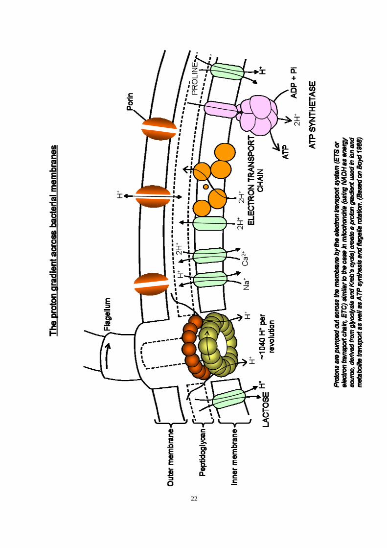
1.6.1 The Proton Gradient Powers Bacterial Flagella
Flagella rotation in bacteria is not driven by consumption of ATP, since mutants in which ATP synthesis is uncoupled and addition of metabolic inhibitors do not effect flagella rotation (Larsen et al. 1974). Experiments have shown that the transmembrane proton-motive force (proton electrochemical gradient) powers flagella rotation. This PMF and its generation are summarized in figure 11.
The proton-motive force (PMF) has two components, the electrical potential across the membrane and the pH gradient across the membrane. Use of valinomycin in K+-free saline (opening K+ channels, causing K+ efflux) to increase the negative membrane potential has shown that the flagellum rotates fastest when the inside of the cell is more negative. Altering pH has also shown that flagella rotate fastest when the inside of the cell is more alkaline than the external medium (Glagolev & Skulachev, 1978). In other words, the faster protons can enter the bacterial cell, the faster the flagella rotates, until it saturates at its maximum rate, at which point the proton carriers must be saturated (Khan and Macnab, 1980). However, these experiments also demonstrate that reversal of PMF, such that protons leave the cell, also supports flagella rotation. Interestingly, in alkalophilic species Na+ influx powers the flagella (Hirota et al. 1981 and Imae and Atsumi, 1989). Not all bacteria have an appreciable transmembrane electrical potential and in this case, pH gradient might be the only component of proton-motive force.
The PMF is also used to generate ATP in bacteria; the way bacterial membranes generate this force is illustrated in fig.7. It is not known for certainty that protons flow through the bacterial motor, but is exchanged for another energy form, like the electrochemical potential of another ionic species. Absence of Ca2+, Mg2+ and Na+ does not inhibit motility (Manson et al. 1977). Collapsing the K+ electrochemical gradient with valinomycin also has no inhibitory effect other than that due to a change in membrane potential. The use of an anion gradient or a high-energy intermediate other than ATP remains a possibility. Block and Berg (1984) estimated that 1040 protons are required for each revolution of the flagella.
1.6.2 Role of the Mot and Switch Complexes
The proteins MotA and MotB are essential for energy transduction during flagella rotation. Mot mutants are unable to rotate their flagella, but otherwise appear normal. Rings of cyto- membrane particles, large enough to accommodate M-rings, have been observed in some bacteria, but are absent in Mot mutants (Khan et al, 1988). Mot proteins probably form part of the basal complex. Various receptors sense environmental signalling and intracellular signals operate a "switch" which controls the direction of flagella rotation. Several features of this control process are well understood (see reviews by Berg (1975), Glagolev (1980) and Jones and Aizawa, 1991).

21
A "switch complex" may be part of the basal complex. These components are common to all bacterial flagella and are believed to be major components of the motor. Freeze-fracture electron micrographs show 16 stud-like particles comprising the M-ring and 17 comprising the S-ring. Block and Berg (1984) used Mot mutants of Escherichia coli. Plasmids bearing the Mot genes under the control of a lac promoter were introduced into the mutants. Addition of a lac inducer caused expression of these Mot genes and flagella rotation was slowly restored. As the flagella "warmed-up" they increased speed in 16 discrete steps. This suggests that each M-ring subunit contributes in the generation of torque (rotary force). The M-ring is, therefore, considered to be the flagella motor. In many models the M-ring generates torque by its interaction with the stationary S-ring (acting as a stator). As the M-ring rotates so it exerts force on the S-ring, which is attached to the cell, and the whole cell spins in the opposite direction to the flagellum. It is assumed that the M-ring is connected to the rod and causes it and the filament to rotate. The filament generates useful thrust (mutants with straight filaments rotate but remain stationary).
MotA-MotB complexes possibly form the proton channels and torque generators of the motor. These are integral membrane proteins. FliG, FliM and FliN are cytosolic proteins required for energisation, switching and assembly of the flagellum (Schuster, 1994). Evidence suggests that these proteins form a structural complex with FliF and FliG in a 1:1 stoichiometry and FliM and FliN also in a 1:1 stoichiometry.
22

23
1.7 Specific Models of Flagella Rotation
1.7.1 Molecular cross-bridges
Berg and Brown (1972) proposed a model in which the periphery of the M-ring was linked to the cell envelope by up to five cross-bridges, similar to those in skeletal muscle. These bridges would detach and reattach at a different site, bringing about rotation. More specifically, Lauger (1988) proposed a similar mechanism in which protons bring about confirmational changes/movements in bridges between the M and S rings (fig. 12).
Fig. 12: Lauger’s model of M-ring rotation (1988). In this model, CW rotation is associated with movement of protons out of the cell via the channels. CCW rotation is associated with the inward movement of protons. 1. When no proton is bound to the channel protein, it is detached from the M-ring. 2. When a proton binds the channel it attaches to a binding site on the M-ring. 3. The channel undergoes a conformational change, rotating the M-ring by theta (q) degrees and loses its proton to the other side. The channel protein now detaches again. C, proton channel acting as a cross-bridge; M, motor ring; S, stator ring (not the M and S rings as we now know them).
1.7.2 Cytomembrane streaming
Adam (1977) imagined that the M-ring subunits resembled the blades of a paddle wheel. These blades are angled such that lipids flowing towards the M-ring slide past one side of the ring while becoming caught by paddles on the other (fig. 13). Lipid streaming can be caused by addition of lipids to membrane on one side and removal of lipids from the other side. A change in the

24
direction of rotation could occur by confirmational changes in the subunit blades or by a change in direction of membrane streaming. Membrane streaming has not been directly demonstrated in bacteria, but may be involved in gliding motility.
Fig. 13: Cytomembrane streaming model (Adam, 1977). A schematical drawing of the M-ring according to the rotational symmetry analysis of De Pamphilis & Adler (1971). Types of hydrodynamic boundary conditions for uniform linear cytomembrane flow of velocity U past the M-ring are indicated

25
1.7.3 Osmoelectric/electrokinetic motors
This type of model has been described by Glagolev and Skulachev (1978) and Lauger (1988), but the one detailed here was put forward by Mitchel (1984). Fig. 14 shows a theoretical turnstile molecular motor. Molecules Sp and Sn represent the stator on either side of the rotor, R. Protons (from outside the cell) bind a negatively charged site on Sp, making it positively charged. This attracts a vacant negatively charged proton-binding site on R, which rotates to pick-up the proton from Sp. The R site becomes positively charged and, as another proton occupies the Sp site, is attracted to the vacant negatively charged proton binding site on Sn. R rotates again, giving its proton to Sn which passes it to the cytosol and the whole cycle repeats. However, in such a system, the motor is equally likely to rotate either way. Introducing asymmetry into the system (fig. 15) biases the direction of rotation. In the case of bacteria, the flagella An array of such stators could occur in a ring (S-ring) above a rotary ring with multiple proton binding sites (M-ring) (fig. 16). The M-ring would be connected to the rod, causing it to rotate (note friction is negligible due to the small size of the components). Signals could bring about a change in rotation by reversing proton flow through the S-ring subunits, so that Sp becomes Sn and Sn becomes Sp. .

26
Fig. 14: A hypothetical “symmetrical well and turnstile molecular motor” in a bilipid membrane. The central cylindrical molecule (R) represents the rotor, and the molecules (Sp and Sn) on either side of it represent the stator. The rotor is shown spinning counter-clockwise in C (looking down from above). Protons are

27
conducted from the aqueous domain P (outside the cell membrane) to a proton-donating site Cps+ in the
molecule Sp; and protons are conducted to the aqueous domain N (inside the cell) from a proton-accepting site Cn
z– in the molecule Sn. A proton-binding site Bm– in the rotor molecule R accepts protons from Cp
s+ at the surface of Sp, and is thereby converted to BHn+, which (after rotation of R) donates protons to Cn
z–, and is thereby converted back to Bm–.
H+
Cps+
Cnz–
Bm–
Sp
Sn
R
H+
BHn+
P
N
A
H+
Cps+
Cnz–
BHn+
Sp
Sn
R
H+
Bm–
P
N
B
H+
Cps+
Cnz–
Bm–
Sp
Sn
R
H+
BHn+
P
N
A
H+
Cps+
Cnz–
BHn+
Sp
Sn
R
H+
Bm–
P
N
B
Fig. 15: “Asymmetric well and turnstile molecular motor” that may exhibit a bias towards (A) counter-clockwise rotation when charge n is greater than charge m and the pathway from Cp to Cn is shorter or (B) clockwise rotation when charge m is greater than charge n and the pathway from Cn to Cp is shorter.

28
Fig. 16: The osmoelectric model of bacterial flagellar rotation (Mitchell, 1984). Several asymmetric well and turnstile molecular motors arranged in a circle, comprising the M and S rings in the plane of the membrane (viewed down the axis of the rotor (R, the rod)). Protons enter the 8 stator components Sp from the aqueous domain (continuous arrows) outside the membrane and leave the 8 stator components Sn for the aqueous domain (dotted arrows) inside the membrane.
1.7.4 Dielectric Motors (Fuhr & Hagedorn, 1989)
Small spherical or cylindrical particles rotate when immersed in liquids and subjected to electromagnetic fields. When a potential difference occurs across such a particle the surfaces become charged (fig. 17a top). Mutual repulsion/attraction causes rotation (fig. 17a middle), then current flows, resetting the charges (fig. 17a bottom). Thus, the particle continues to rotate. A potential difference would be needed within the membrane, either side of the basal complex, not across it and so is not simply the result of a membrane proton potential. Any asymmetry in membrane structure could create a charge difference across the motor, such as protons flowing through a proton channel on one side of the rod (fig. 17b). Such channels could be spaced at intervals in a ring around the rod, such as the M-ring (fig. 17c). In this case, the M-ring might not connect to the rod and could remain stationary. Any of the rings could contribute in generating torque.
As yet, there is no evidence as to which model is most likely, though the osmoelectric-type seems most popular. Mathematical treatment enables most of these models to fit observed kinetics of the bacterial motor.

29
Fig. 17a: The dielectric motor theory (Fuhr & Hagedorn, 1989). Top: a spherical particle in an electric charge field acquires charge and is mutually repulsed and caused to rotate. Middle: the flow of current resets the charge on the particle. Bottom: the particle is recharged and again repulsed and continues to rotate.

30
Fig. 17b: Protons flowing through the membrane, either adjacent to or through the S-ring, through protein channels, setting up a potential difference across the motor.
Fig. 17c: The dielectric motor model of bacterial flagella rotation (Fuhr and Hagedorn, 1989). Proton channels arranged in a ring at intervals around the rod/M-ring causing the latter to rotate as long as the channels remain open.

31
2. Chemosensing 2.1 Chemosensing mode in enteric bacteria (e.g. Escherichia coli) When unstimulated, free-swimming E. coli swarmer cells make long straight runs with infrequent tumbles (fig. 18). During the runs, the flagella rotate CCW and the flagella come together and work as a single bundle, possibly driven together by hydrodynamic forces (note that the cell body rotates in the opposite sense, CW, to the flagella). During the tumbles, the flagella reverse rotation to the CW sense, the wavelength of their filaments also changes and the pitch increases and the flagella mechanically uncouple and fly apart and the cell tumbles (rotates randomly). Chances are that after a tumble when the flagella resume CCW rotation and reform a bundle that the bacterium will start off in a new direction – it has turned by a random number of degrees.
Fig. 18: Left - a computer-simulated ‘random walk’ of a bacterial cell starting at the centre of the arena and tumbling at random intervals. Right - two such simulated cells tracked over 15000 seconds each. Despite starting at the centre they soon reach the edge of the arena (by random chance); they have been programmed to bounce of the walls at the arena’s edge. When E. coli nears the source of an attractant chemical, moving up a concentration gradient, it’s tumbling frequency decreases and it makes longer runs. If tumbling frequency was simply inversely proportional to attractant concentration, then computer simulations reveal that this will actually lead to a repellent effect – the bacterium will tend to move away from the attractant source. However, when the computerised virtual cell is given a memory, allowing it to compare the concentration at different points in time, it is able to measure the gradient rather than simply the concentration. By reducing tumbling as it moves up the gradient toward the attractant source, then it will indeed become ‘attracted’ to the source (meaning that it spends longer near the source) (fig. 19). This is indeed found to be the case – bacteria have a memory of a few minutes that allows them to make temporal comparisons of chemical concentrations in their environment. Repellents work in a converse manner – the bacteria tumble more frequently as they approach the repellent (as the gradient increases) and so tend to avoid it (fig. 20).

32
Fig. 19: Computer-simulated bacterial cells programmed to respond to the (exponential) concentration gradient of a chemo-attractant placed in the centre of the arena. Each cell has been given a one second memory with which to measure changes in attractant concentration over time. The cells tumble less if they move up the concentration gradient and tumble more if they travel down the gradient (within preset minimum and maximum tumbling frequencies). Each picture shows 4 cells traced for 5000 seconds each. Left - each cell started at the edge of the arena at the bottom right corner at about 5 o’clock. Right -: the bacteria start nearer to the arena centre. Note how the bacteria remain near the attractant even though they are in constant motion.
Fig. 20: Computer-simulated bacteria programmed to respond to a chemo-repellent at the centre of the arena, by tumbling more frequently if they travel up the gradient and tumbling less frequently if they travel down the gradient (within preset minimum and maximum tumbling frequencies). Each cell had a one second memory to enable temporal comparisons in repellent concentration. Left – one cell starting at the centre and was tracked for 15000 seconds. Right – 3 cells began in the centre of the arena and were tracked for 15000 seconds each. They can be seen to clearly avoid the repellent and stay near the edge of the arena. What do we mean by frequency of tumbling? The bacterial flagella have a switch mechanism, alternating between two states: CW rotation and CCW rotation, with the rotation rate remaining more or less constant. The Gibbs free energy for the CCW (swimming) → CW (tumbling) transition is about +40 kJ mol-1, and this energy is provided for by random thermal fluctuations. When the cell is triggered to tumble more, it simply increases the probability of tumbling by reducing the free energy required, making it more likely that thermal noise will provide the necessary energy. 2.2 Chemosensing modes in other flagellated bacteria

33
Some alpha-subgroup bacteria also have a CW/CCW flagella switch, for example in Caulobacter crescentus CW rotation leads to swimming, CCW rotation to tumbling (the opposite sense as in E. coli). Others, however, have flagella that rotate in one direction only. In other members of this group very different strategies are employed. Rhodobacter sphaeroides has a single right-handed flagellum in a lateral position which can push the cell at up to ~35 mm/s. This flagellum rotates only CW (or CCW in some variants). If an anaerobically grown cell detects a decreasing gradient of chemoattractant (if it is moving from the source of the attractant) then its probability of turning increases (note that this response is opposite in sense to that of E. coli, but equivalent). During turning, the flagellum stops and assumes the form of a planar spiral. This whole flagellum spiral slowly rotates, turning the cell by a random amount. Sinorhizobium meliloti has 2-6 short, peritrichously or lophotrichously arranged right-handed flagella with fixed CW rotation, during which they form a propulsive bundle. The flagella rotate continuously, however, asynchronous rates of rotation cause the bundle to fly apart and the cell to tumble. 2.3 Mechanism of chemosensing The best-studied chemosensing mechanism of E. coli is the Che phosphorelay system summarised in figures 21 & 22. In E. coli the chemosensors are methyl-accepting chemotaxis proteins (MCPs) that span the inner membrane as dimmers. These are connected to a phosphorelay system. In such phosphorelay systems, the signal is relayed by protein phosphorylation, which involves enzymes called phosphatises (which remove phosphate) phosphorylases (which attach phosphate) and kinases (which transfer phosphate from one molecule to another). These receptors tend to be concentrated at the cell poles. Together with several of the chemotaxis (Che) proteins they form a MCP:CheW:CheA:CheY receptor complex. CheW functions as a relay linker, connecting the MCP to CheA. When an MCP is stimulated by a chemorepellent binding to it, the receptors cluster in groups of about 7 MCP dimmers together with about 7 CheA dimmers and are in their activated state. This state activates CheA which autophosphorylates to CheA-P. CheA-P is an active kinase and phosphorylates CheY to the active CheY-P form. CheY-P then dissociates from the receptor complex and diffduses to the flagella motor where it binds FliM in the C ring (and it also binds the regulator phosphatise CheZ which undergoes delayed activation as it oligomerises to its active form). Binding to CheZ increases the probability of tumbling (CW rotation) by reducing the activation energy of the switch. After a delay CheZ becomes active and this acts as a phosphatise, removing phosphate from CheY-P to yield CheY which reassociates with the receptor complex. Thus, CheZ acts as a time-delay which resets the system, a form of adaptation which helps confer the memory needed for the system to respond to concentration gradients. A second adaptation system also exists, which is methyl-dependent. CheA-P not only phosphorylates and activates CheY, but it also pghosphorylates and activates CheB, at a slower rate, producing CheB-P after a time-delay. CheB-P is a specific methyl esterase which cleaves a methyl group from MCP (MCP-Me → MCP). The methylated form is the active form which stimulates CW flagella rotation and tumbling. The demethylated form becomes inactive and CCW rotation and straight runs become favoured again – returning the cell to its pre-stimulus state. Chemorepellents thus stimulate MCP demethylation. This periodic resetting of the receptor occurs every few minutes, giving the cell a memory of an equivalent duration. CheR also helps to reset the system by antagonising CheB-P as CheR is a methyl transferase which methylates MCPs. Attractants work in a converse manner. When an attractant binds to the MCP, the receptors deactivate and de-cluster into separate dimmers (with associated CheA dimers). This inhibits the

34
autophosphorylation of CheA, deactivating it and reducing the concentration of CheY-P, resulting in an increased probability of CW flagella rotation and a reduced probability of tumbling. Sugars are chemoattractants that do not bind MCPs directly. Instead they either bind their sugar-specific periplasmic binding protein (PBP) which is involved in sugar transport into the cell as well as chemotaxis. Galactose, maltose and ribose work this way. At least the PBP for maltose is known to be localised at the cell poles along with the MCPs. Alternatively, some sugars, e.g. glucose, mannose and mannitol, bind to a specific Enzyme II of the PEP-dependent sugar phosphotransferase system (PTS) (PEP is phosphoenolpyruvate). The PTS Enzyme I modulates the kinase activity of the MCP-CheW-CheA receptor complex by phosphorylating CheA and simultaneously phosphorylates the sugar as it is imported through the EII/EIII proteins. MCPs also respond to changes in pH and temperature. Oxygen acts as an attractant for E. coli and its receptor is Aer (an MCP homologue) which is cytoplasmic and lacks methylation sites for adaptation. Aer also binds FAD (flavine adenine dinucleotide, an electron/hydrogen carrier used in respiration) which acts as an internal signal for the redox state of the electron transport chain (ETC).

35
Chemosensing in E.coli and S. typhimurium
Non-stimulated state: long runs with occasional tumbles, CCW default flagellarrotation → swimming bundles; [CheY-P] low, probability of tumble low.
Approaching attractant
Tumbling frequency ↓
Probability CW rotation ↓
Longer runs, fewer turns
Tumbling frequency ↑
Probability CW rotation ↑
Shorter runs, more turns
Approaching repellent
e.g. oxygen, sugars
MCP dimer / CheA dimer = 1:1
Inactive receptor
MCP dimer / CheA dimer ~ 7:1
Active receptor
autophosphorylation CheA ↓ autophosphorylation CheA ↑
phosphorylation CheY ↑: CheY-P↑
CheY-P dissociates from receptor
CheY-P binds FliM, CheZ
CheB-P ↑ (slower)
FliM-CheY-P
↑ Prob(CW, tumble)
CheZ dephosphorylates CheY-P to CheY
e.g.
CheB-P demethylates MCP, ↓ response
–
phosphorylation CheY ↓: CheY-P↓
CheY associates with receptor
CheR active
FliM-CheY-P
↑ Prob(CW, tumble)
CheZ dephosphorylates CheY-P to CheY
CheR methylates MCP, ↓ response
–
CheW CheW
Fig. 21: A flowchart summary of the main events in chemosensing in enteric bacteria.

36
Y
A A
W W
P
P Y
Z
R
B
MCP
Adaptation
C-ring
Motor
Hook
Filament
Inner membrane
Peptidoglycan
Outer membrane
Y
A A
W W
P
P Y
Z
R
B
MCP
Adaptation
C-ring
Motor
Hook
Filament
Inner membrane
Peptidoglycan
Outer membrane
Fig. 22: A schematic of the Che phosphorelay signalling system in Escherichia coli.

37
3. Motility Mode 2 - Gliding Motility 2.1 What is gliding motility? Gliding motility is a form of locomotion that occurs across a solid surface or substrate. There is no obvious mechanism as the cells simply glide! This form of motility is much slower than flagella-powered locomotion. 2.2 Slime extrusion (slime jets) Gliding motility occurs in some Gram negative bacteria and mycoplasmas. Cyanobacteria can glide at up to 10 mms-1. The envelope of gliding cyanobacteria consists of the following layers arranged from outside to inside: oscillin layer, S-layer, outer membrane, periplasm and the inner membrane. The oscillin layer is a layer of glycoprotein subunits, 8-12 nm in diameter, arranged in helical arrays and is only found in gliding forms. This possibly functions as a passive screw thread, causing the filament to rotate as it glides. The periplasm of gliding forms contains helical fibres, 25-30 nm in diameter, with a 30o pitch. One model of their gliding motility relies on surface waves in a proteinaceous layer, such as in the periplasmic fibres. A second model proposes that slime extruded from junctional pore complexes (JPCs) known to exist on either side of the cell-cell junction be guided by the oscillin to produce rotation (fig. 23). Certainly, slime is extruded during gliding. Acetobacter xylinum extrudes cellulose from pores and glides at 0.05 mms-1. Spirulina also glides and its JPCs are situated inside the spiral and not in contact with the substratum. Hormogonia are motile cyanobacterial filaments that infect plants and establish cyanobacterial-plant symbioses. These filaments do not rotate. These possess pili and possibly glide by twitching motility. Synechocystis is a motile unicellular cyanobacteria and this also has 6-8 nm thick pili. Synechococcus swims, but has no flagella.

38
PC
O S SS
Outer membrane
Periplasm
Peptidoglycan
Cytoplasm
Fig. 23: Junctional pores in Oscillatorium. These pores are about 14-16 nm in diameter and their centre-centre spacing is about twice this distance. These pores form a single row either side of the circumferential junctional furrow. O, helical oscillin fibrils (8-12 nm diameter); PC, pore-complex; S, s-layer; SS, secreted slime. Together, the s-layer and oscillin layer form the ‘external layer’. The inner membrane is hard to see in fully turgid cryofixed cells as it is tightly pressed against the inner surface of the peptidoglycan layer. In Oscillatorium princeps large pores (diameter up to 350 nm at base, narrowing to about 50 nm at the surface, spaced about 400 nm apart centre-centre) also occur further from the septum on either side. These large pores are lined by cytoplasm and inner membrane and represent regular perforations in the peptidoglycan layer.

39
2.3 Twitching Motility – Type IV Pili This form of motility occurs, for example, in Pseudomonas aeruginosa, Neisseria gonorrhoeae and some Escherichia coli strains. Motility occurs in short, intermittent jerks of a few micrometers. A moist surface is required for twitching motility and cells must be within several microns of each other. The pili are polar, 6nm in diameter and up to 4 mm long and occur at one or both poles. These pili actively extend and retract. These pili are also involved in conjugation, bacteriophage infection, biofilm formation and transformation. At least 35 genes are involved in twitching motility and speeds of up to 1 mms-1 are attained. Pili are also involved in attachment and invasion of host cells. 2.4 Motility in Myxobacteria Myxobacteria travel in swarms that co-operatively digest macromolecular food, including prey cells and other bacteria. Myxobacteria aggregate when nutrients become scarce. These aggregations produce multicellular fruiting bodies that release dormant myxospores. These bacteria have large genomes, that of Myxococcus xanthus, for example, is some 9.5 mbp. There are two myxococcal gliding systems, the S system or ‘social’ gliding system and the A system or ‘adventurous’ gliding system. The A system operates in single, isolated cells. Gliding speeds are about 0.4 mms-1. The S system relies upon type IV pili tethering and retraction to pull the cells along. The gliding is smooth and possibly some pili extend as others retract. These pili only attach specifically to extracellular polysaccharide (EPS) slime secreted by other myxobacteria, hence it is specific to social gliding. The A system operates over drier surfaces and does not require pili. Chain-like strands, grouped into bands, wrapped helically around the cell, occur in the periplasm. In Myxococcus fulvus, three or more of these chains are combined into ribbons. The subunit rings have an outer diameter of about 12-16 nm (12.6 to 15.6 nm) joined by elongated linkers 2.8 nm wide and 9.9-11.9 nm long. The linkers are arranged in two parallel rows, so that there is one pair of linkers between each pair of rings. It has been suggested that these periplasmic structures could be involved in A-gliding, however, there is no evidence that they have any role at all in gliding motility and their function remains unknown. There are over 100 nozzles in the outer membrane at each cell pole. The trailing pole secretes jets of slime from these nozzles which push the cell along, whilst nozzles at the leading pole remain inactive. Within one minute the poles may reverse role, with the trailing pole becoming the leading pole as the cell reverses direction.
Myxobacteria apparently glide over moist surfaces by the use of flagella consisting only of the basal complex and hook but no filament. Mutants lacking these structures are immotile (Pate and Shang, 1979). However, for reasons that I have not yet uncovered in my literature searches, this mode of gliding seems to have been ignored by more recent literature. 2.5 The Cytophaga-Flavobacterium Group Bacteria of the Cytophaga-Flavobacterium group glide at about 2-4 mms-1. The cells may rotate as they glide and latex spheres bound to the cell surface move along the cell. The proton-motive force is the likely energy source. Ring-like structures cover the surface of Flavobacterium johnsoniae; are these rotary motors? Fibrils occur within the cell walls of some species. Goblet-

40
shaped structures occur in the envelopes of Flavobacterium polymorphus. Most of these bacteria lack pili and polysaccharide extrusion is considered to be an unlikely mechanism in this group. Gliding motility in this group seems to require sulphonolipids in the outer membrane. These are present in Flavobacterium johnsoniae and other Cytophaga-Flavobacterium group gliders. Upon contact with a surface, more polar sulphonolipids are produced and an OMP becomes cross-linked to the peptidoglycan. One model proposes that adhesive OMPs are moved along tracks fixed to peptidoglycan. Other models propose conveyer belts of polysaccharide or protein fibrils that are exported and imported at different locations. Another model proposes expansion or contraction of elements in the cytoplasm or periplasm. Another proposes rotary motors, and yet another waves in the outer membrane. 2.6 Mycoplasma Gliding The Mycoplasmas are wall-less bacteria related to Gram-positive bacteria. They have very small genomes, that of Mycoplasma genitalium, for example, is 580 kbp. Gliding mycoplasmas have a flask or club-shaped tip (‘head’) that functions as an attachment organelle. This tip leads the gliding cell at 0.1–7 mms-1 depending on species. These bacteria possess a cytoskeleton of over 25 different proteins. 4. Movement of Materials Within Bacteria and Bacterial Cell Size 4.1 Introduction
Bacteria vary in diameter from 0.2-mm nanobacteria to Thiomargarita namibiensis, a colourless sulphur-bacterium 750-mm diameter and their mass varies by 10 orders of magnitude. In large bacteria, inclusions reduce the volume of active cytoplasm. Only the fastest and largest swimming bacteria known, Thiovulum majus, can increase convective nutrient supply by its own motility and these bacteria actively ventilate their population. Other bacteria are diffusion limited. The largest heterotrophic bacterium is the 80 by 600-mm large Epulopiscium spp. That lives in the gut of tropical fish. This organism possibly exists in a nutrient rich medium. Colourless sulphur bacteria oxidise H2S to SO4
2- with oxygen or nitrate. These bacteria form multicellular filaments several cm long, allowing them to penetrate the 500-mm thick diffusive boundary layer and reach water containing the oxygen or nitrate electron acceptor. These bacteria may also store several months reserve of nitrate and sulphur. Thermodiscus has disc-shaped cells, 0.1-0.2 mm thick and 0.2 mm wide and has a cell volume < 1 x 10⁻² mm3 to 2 x 108 mm3. 4.2 Diffusion and the Size limit of Prokaryotes The mixing time of small molecules in a 1mm bacterium is ~ 1ms, that of the larger molecules ~ 10ms and enzyme turnover rates ~ 100/s. Thus, molecules can move through the entire volume of the cell many times during one round of catalysis. The traffic time is the theoretical time taken for any two molecules within a cell to meet. In 1s it is probable that any substrate molecule will have met any enzyme molecule. In large prokaryotes, about 100 mm long, the traffic time (which

41
is proportional to L3) is ~ 10 hours. This may result in regional differences within the cell, or nonbounded compartmentalisation. The viscosity of water dampens fluctuations smaller than the Kolmogorov scale or viscous length Lv ~ 1-6 mm. (This length has its smallest value in the most vigorous turbulence). Cells less than 100 mm in diameter are always surrounded by a diffusion sphere that is not affected by the surrounding turbulence and hence turbulence is not locally important for substrate flux to the cell. Substrates must cross this barrier by diffusion. Smaller cell size efficiently relieves the resultant diffusion limitation. Since diffusion supply is a R, whereas metabolic rate a R-2, cells of 5-30 mm diameter can be diffusion limited. Enzyme release can increase the effective range of substrate utilisation to ~ 10mm. The Péclet number is the ratio of transport by convection to transport by diffusion. If Pe >> 1, then fluid flow or swimming strongly enhances the substrate availability. For bacteria, Pe << 1 and so swimming does not increase substrate transport. The minimum size of a cell to achieve an increase in substrate transport by swimming is ~ 10mm. Thiovulum majus is an exception. It is a large chemotactic sulphur-bacteria (R Y 8 mm). These bacteria increase their substrate uptake 4-fold by swimming at up to 600 mms-1. Their large size and high swimming speed (> 100 mms-1) enables them to overcome Brownian displacement and undergo directional swimming. These bacteria are microaerophilic and seek the oxic-anoxic interface (4% air saturation) ideal for H2S oxidation. These bacteria maintain their position by U-shaped swimming patterns, they swim and rotate in a helical path (3-10 rps, r = 5-40 mm, pitch = 40-250 mm). Smaller bacteria must locate optimum conditions by chemokinesis and they do so by tumbling. 4.3 The Sediment-Water Interface The sediment-water interface has a boundary layer typically ~ 0.5mm. Microbial respiration in the surface sediment establishes a steep oxygen gradient across the diffusive boundary layer (DBL). The mean oxygen diffusion time through the DBL is ~ minutes and there is diffusion limited transport. Filaments reach past the boundary layer. Ciliates may be stalked (e.g. Vorticella) for the same reason. Zoothamnium niveum is a ciliate that grows on highly sulphidic mangrove peat. It becomes overgrown by chemoautotrophic, symbiotic sulphur-oxidising bacteria. Periodic contractions (every 5-30 s) of its 15 mm long with a Re = 2500, changes the water adhering to the surface of the symbiotic bacteria. 4.4 Big Bacteria (Megabacteria, Gigantobacteria) Staphylothermus marinus is about 0.5 – 1 mm wide, but increases to 15 mm in diameter in rich nutrients. In this enlarged condition, only 2% of the cytoplasm is active, the rest is occupied by a large vacuole. Many large bacteria are cyanobacteria or sulphide oxidisers. Thiomargarita namibiensis is about 150 to 200 mm wide, but can be up to 750 mm. Epulopiscium spp. inhabit the guts of herbivorous surgeonfish in the Red Sea and the Great Barrier Reef and are about 10-20 mm wide and 70-200 mm long (5 x 10³ to 5 x 104 mm3). The colourless sulphur bacterium Achromatium oxaliferum ranges from < 1 x 10³ to 8 x 104 mm3 and stores calcium carbonate and sulphur. Big bacteria are found on the sea floor, where there is a large H2S production and store sulphur for periods of less active venting and can also utilise nitrate as an alternative electron acceptor.

42
Some grow on mobile animals, such as polychaetes, ostracods and mayfly larvae, in sulphide rich environments. 4.5 Multicellular Bacteria Bacterial filaments can grow up to 7 cm in length. In magnetotactic forms, there are intercellular connections between 10-30 cells for coordination of locomotion. Beggiatoa spp. form motile filaments on O2-H2S opposed gradients and form dense white mats on marine coasts. These bacteria oxidise H2S with oxygen. They require both molecules, but can only tolerate low oxygen concentrations, since they are unable to breakdown hydrogen peroxide that forms at higher oxygen concentrations. These bacteria inhabit the a zone < 1 mm thick at the sediment-water interface. They consume up to 70% of the sediment’s oxygen and all of its sulphide. Uptake of these molecules is diffusion limited. The filaments coil-up and are several mm to 1 cm long. The zone of coexisting oxygen and sulphide is less than 100 mm thick. These bacteria exhibit a phobic response to oxygen concentrations above about 5% air saturation. After a 20-30 s delay they reverse their direction of gliding. S the filament tips encounter oxygen concentrations above the threshold they retreat, whilst the rest of the filament still glides upwards and as a result the middle part of the filament is forced out to one side at the oxic-anoxic interface, forming loops. These bacteria also exhibit a phobic response to light. In light the benthic diatoms and cyanobacteria produce oxygen. However, at night respiration causes the oxygen – sulphide interface to move above the sediment. In response to this the filaments move upwards. In the daytime they move downwards. With passing clouds and changing light levels, the filaments move up and down to track their optimum environment. Thiovulum majus is a highly motile, spherical bacterium whose biofilm forms a veil over the sediment surface. The veil may be attached or partly floating. These bacteria are chemoautotrophs that oxidise hydrogen sulphide to sulphur and sulphate. Each cell is 9-18 mm across. The cells are motile, with a maximum speed of 615 mms-1. They swarm to the oxygen – sulphide transition-zone and secrete a slime thread. The threads stick together and spread-out to form a 2D mesh containing 105-106 cells cm-2. This mesh separates the flowing oxygenated seawater above from a stagnant boundary layer of hydrogen sulphide enriched water, hence the veil creates its own diffusive boundary layer suspended in the water column. Concerted swimming actively moves the veil up or down. The veil may become tethered by slime thread up to 100 mm long. The flagella create a downward water flow of 200 mms-1 across the veil, which brings in oxygenated water (and possibly sulphide) the outflows are channeled through numerous small openings in the veil, which has a fine lace appearance. The Péclet number for oxygen is about 40, in this system, and therefore convection dominates oxygen transport. I have reviewed biofilms and quorum-sensing in detail elsewhere, and we have just provided a selected introduction here. 4.6 Breaking through the Diffusive Boundary Layer Filament gliding movements ensures that the bacteria can break through the diffusive barrier, even if this boundary thickens. When oxygenated water flow decreases filaments move upwards and when oxygenated water flow decreases the filaments move downwards. 4.7 Surviving Anoxia with a Storage tank of Nitrate Beggiatoa cells are 5-23 mm in diameter and contain sulphur inclusions. This sulphur is oxidised when sulphide supplies diminish. In eutrophic coastal waters, for example fjords, an anoxic

43
bottom layer of water forms during summer. This layer may last for days or months. Nitrate can be utilised as an alternative electron acceptor for sulphide or sulphur oxidation. Nitrate can also penetrate up to 4 cm into the substrate (compared with ~ mm for oxygen). Hydrothermal-vents, hydrocarbon seeps and methane hydrates have high sulphide concentrations and here form mats of unusually large Beggiotoa cells (each cell 40-200 mm in diameter). These enlarged cells contain nitrate accumulated in a central vacuole at up to 160mM. The mats are > 1 cm thick. Pulsating hydrothermal emissions produces pore water flow through the mats. This water is cold and oxygen and nitrate rich. The cells stored reserves carry the cells over the periods in-between the intermittent pulses. 4.8 Commuting Thioplaca is a marine bacterium with a central vacuole that can store up to 500mM nitrate. This organism occurs on shelf sediments and is sometimes the dominant benthic organism. Thioplaca only uses nitrate as an electron acceptor. When oxygen concentrations exceed 10% air saturation, these organisms retreat into their sheaths. The winter El Niňo increases oxygen concentrations in the bottom waters and can reduce the Thioplaca population dramatically. Thioplaca secretes slime whilst gliding. This slime forms tubes within which the filaments move along their tracks. Unused tubes disintegrate rapidly. When up in the water column these bacteria fill their nitrate reservoirs and when down in the sediment they oxidise sulphide and store sulphur. Thus Thioplaca has a double storage capacity. In high sulphide concentrations, the filaments leave their sheaths and live as freely gliding filaments on the sediment surface. Thiomargarita namibiensis is non-motile and the largest known sulphur bacterium. It inhabits semi-fluid diatom ooze where sulphide concentrations are greater than 10mM. These cells only contact nitrate and oxygen when the loose sediment is suspended in the water column following storms, wave pumping or methane eruptions. They have a vacuole occupying 98% of the cell volume. They can respire for 40-50 days or more without taking up new nitrate (for over two years in a cold room). They can survive in air-saturated water and can use oxygen as an electron-acceptor for sulphide oxidation. 5. Magnetotactic Bacteria Magnetotactic bacteria are Gram-negative, flagellated and motile bacteria. They swim at about 100 mms-1. They are microaerophilic, aquatic and contain magnetosomes. These bacteria come from many taxonomic groups. The magnetosomes are intracellular magnetic grains that give each cell a permanent magnetic dipole moment. The local geomagnetic field (about 0.5G) places a magnetic torque upon the bacteria, aligning them with the magnetic field. Aquaspirillum magnetotacticum has Fe3O4 magnetite (lodestone) magnetosomes. Each cell contains about 20 cuboidal-octahedral crystals. Each crystal is about 420 Å in diameter and the crystals are arranged in a chain. Single magnetic domains in magnetite range from 400-1000 Å. Full alignment requires the ratio of the interactive magnetic energy with the applied field to the thermal energy to be greater than about 10. A ratio of 16 is obtained for 22 500 A particles. As we shall see, there are other mechanisms that stabilize the structure and keep the crystals aligned. Thus, the magnetosomes function as a ferromagnetic biocompass. At 0.5G and 30oC, 80-90% of the cells are fully aligned. The cells clump together if they contain too many magnetosomes. Recorded levels of magnetotactic bacteria yield 103-104 cells / ml of slurry in aquatic

44
environments (in New England). These bacteria are absent from heavily polluted waters and absent in limestone caverns, thermal springs, thawed Antarctic sediments and iron-rich seeps. They are abundant in water purification plant settling basins, ponds with organic sediments, and sewage-treatment oxidation ponds. In the Northern Hemisphere, magnetotactic bacteria swim predominantly northward, whilst in the Southern Hemisphere they swim predominantly southward. The vertical component of the geomagnetic field, due to Earth’s curvature, is directed upwards in the Southern Hemisphere, is zero at the geomagnetic equator, and is directed downwards in the Northern Hemisphere. Therefore, the bacteria are directed downwards in both hemispheres and so are abundant in sediments, but absent in surface water. At the geomagnetic equator there are approximately equal proportions of each polarity and the bacteria swim horizontally. Magnetospirillum magnetotacticum (formerly Aquaspirillum magnetotacticum) (fig. 24) is a microaerophilic chemoheterotroph that metabolises organic acids, like fumaric, tartaric and succinic acids as its sole carbon (and energy?) source. Nitrate is the principle electron-acceptor for microaerobic growth on tartrate. Traces of oxygen are required. (As a substrate for oxygenases? For haem production?). Nitrate is reduced to nitrous oxide and ammonia in the process. Thus, this bacterium is a denitrifier. In low oxygen tensions (< 0.2 kPa) these bacteria also reduce ethyne. In high oxygen tension (above about 6 kPa) they do not produce magnetosomes and are not magnetotactic. This bacterium lacks catalase activity and so is sensitive to high oxygen concentrations. They have polar or bipolar flagellation. Pili on coccoid forms, extracellular polysaccharide on spirilla, allow them to adhere to sediment particles.

45
Fig. 24: Magnetospirillum (Aquaspirillum)
Magnetosomes are typically (though not always) arranged in one or more linear chains. Each magnetosome particle is a single crystal of magnetite or greigite enclosed in a phospholipid bilayer membrane. This membrane is derived by invagination of the inner membrane but contains specialized proteins. This causes some confusion in nomenclature – does ‘magnetosome’ refer to the whole chain or just to one of the particles? Each magnetosome particle/vesicle is a tiny magnet and the particles within each chain are aligned –(N-S)-(N-S)-(N-S)- which helps to stabilize the chain as a magnetic north pole is attracted to a magnetic south pole. However, as anyone who has ever played with magnets will know, this arrangement is metastable, meaning that a slight displacement of one of the magnets can cause the whole chain to collapse into an aggregated cluster of magnets. Additional structures are needed to stabilize this arrangement (fig. 25). In Magnetospirillum the actin homologue protein MamK forms a protein filament track along which the magnetosome particles are aligned (see below). There is evidence to suggest that the protein MamJ forms linkers that connect each vesicle to the track. It is also possible that additional structures link neighbouring magnetosome vesicles together. (See review by Thanbichler and Shapiro, 2008).

46
MamK filament
Magnetite crystal in a lipid bilayer membrane
Hypothetical MamJ linker
Hypothetical inter-particle linker
Model of the structure of a magnetosome-chain in Magnetotospirillum
MamK filament
Magnetite crystal in a lipid bilayer membrane
Hypothetical MamJ linker
Hypothetical inter-particle linker
Model of the structure of a magnetosome-chain in Magnetotospirillum
Fig. 25: The magnetosome and associated stabilizing structures in Magnetospirillum.
Some bacteria have two chains of magnetosomes at opposite sides of the cell and it is possible that these are positioned by their mutual magnetic repulsion. Large cells require a lerger torque to turn them and the large rod-shaped cells of Magnetotacticum bavaricum, which are 8-10 mm long, have 200-1000 crystals arranged in 5 chains. However, it is possible that this produces far more torque than needed and magnetosomes may have additional functions. The individual crystals of magnetite are 35-120 nm in diameter. A magnetite crystal contains one or more domains – regions in which the molecules all have their magnetic moments aligned parallel, reinforcing one another. If the crystals were much larger than domain boundaries would occur, where the alignment of the molecules switches between crystal domains with magnetic poles pointing in different directions (typically opposite one another). A crystal with several domains whose magnetic moments tend to oppose and cancel one another will have a reduced magnetic moment. If the crystals were much smaller, then they would lose their permanent magnetism, due to the thermal motion of the atoms. Thus, the size chosen is optimal for maximizing the intensity of the crystal’s magnetic field. Some bacteria use greigite (Fe3S4) instead of magnetite. In particular these bacteria occur in environments where oxygen availability is low and sulphur-availability high. Greigite is less effective, however, as it makes a weaker magnet (one third as strong). At least one known species of bacterium has crystals of both greigite and magnetite (a given magnetosome particle must be one or the other and mixed crystals do not occur). Such bacteria encounter environments of variable oxygen content – if oxygen is scarce then they incorporate sulphur instead.

47
References – for Sections 1 & 2
Acheson, DJ. 1990. Elementary Fluid Mechanics. Oxford University Press, p.235.
Adam, G. 1976. Rotation of Bacterial Flagella as Driven by Cytomembrane streaming. J. Theor. Biol. 1977. 65: 713-726.
Berg, HC. 1974. Dynamic Properties of Bacterial Flagellar Motors. Nature 249: 77-79.
Berg, HC. 1975. Bacterial Behaviour. Nature 254: 389-391.
Berg, H.C. & Brown, D.A. 1972. Nature, London 249, p. 500.
Block, S.M. & Berg, H.C. 1984. Successive Incorporation of Force-Generating Units in the Bacterial Rotary Motor. Nature 309: 470-472.
Boyd, RF. 1988. General Microbiology, 2nd edition, Times Mirror/Mosby College pub. pp. 95-99, 111, 112, 120-124, 277.
Childress, S. 1981. Mechanics of Swimming and Flying. Cambridge University Press.
Coulton, JW. & Murray, RGE. 1977. Biochem. Biophys. Acta 465: 290. Cited in Jones & Aizawa, 1991.
DeRosier, D.J. 1998. The turn of the screw: the bacterial flagellar motor. Cell: 93, 17-20.
Driks, A & Derosier, DJ. 1990. Additional Structures Associated with Bacterial Flagellar Basal Body. J. Mol. Biol. 211: 669-672.
Fuhr, G & Hagedorn, R. 1989. Dielectric Motors -A New Hypothesis for the Bacterial Flagella. J. Theor. Biol. 139: 39-59.
Glagolev, AN. 1980. Reception of the Energy Level in Bacterial Taxis. J. Theor. Biol. 82: 171-185.
Glagolev, AN & Skulachev, VP. 1978. The Proton Pump is the Molecular Engine of Motile Bacteria. Nature 272: 280-282.
Greenbury, CL & Moore, DH. 1966. Immunology 11: 6l7-625.
Hirota, N; Kitada, M & Imae, Y. 1981. FEBS letters 132: 278. Cited in Jones Aizawa 1991.
Holt, SC. 1978. Anatomy and Chemistry of Spirochetes. Microbiol. Rev. 42: 114-160. Cited in Stanier et al., 1989.
Ikeda, T; Homma, M; Iino, T; Asakura, S & Kamiya, R. 1987. J. Bact. 169: 1168.
Imae, Y & Atsumi, T. 1989. J. Bioenergetics and Biomembranes 21: 705. Cited in Jones & Aizawa 1991.
Jones, CJ; Macnab, R.M., Okino, H. & Aizawa, S-I. 1990. J. Mol. Biol. 212, p. 377. Cited in Jones & Aizawa, 1991.

48
Jones, C.J. & Aizawa, S-I. 1991. The Bacterial Flagellum and Flagellar Motor -Structure, Assembly and Function. Adv. Microb. Physiol. 32: 109-172.
Khan, S & Macnab, RM. 1980. Proton Chemical Potential, Proton Electrical Potential and Bacterial Motility. J. Mol. Biol. 138: 599-614.
Khan, S; Dapice, M & Reece, TS. 1988. J. Mol. Biol. 202: 575. Cited in jones & Aizawa 1991.
Kimsey, RB & Spielman, A. 1990. Motility of Lyme-Disease Spirochetes in Fluids as viscous as the Extracellular-Matrix. J. Infectious Diseases 162: 1205-1208.
Kudo, S; Magariyama, Y & Aizawa, S-I. 1990. Nature 346: 677.
Larsen, SH; Reader, RW; Kort, EN; Tso, W-W & Adler, J. 1974. Change in direction of flagella rotation is the basis of the chemotactic response in Escherichia coli. Nature 249: 74.
Lauger, P. 1988. Torque and Rotation Rate of the Bacterial Flagellar Motor. Biophys. J. 53: 53-63.
Listgarten, MA & Socransky, SS. 1964. J. Bacteriol. 88: 1087. Cited in Stanier et al., 1989.
Lowe, G; Meister, M and Berg, HC. 1987. Rapid rotation of flagellar bundles in swimming bacteria. Nature 325: 637-640.
Macnab, RM. 1979. How do Flagella Propel Bacteria? TIBS Jan. N10-N15.
Manson, MD; Berg, HC; Tedesco, PM; Harold, FM & van der Drift, C. 1977. PNAS 74: 3060. Cited in Jones & Aizawa 1991.
Meynell, EW. 1961. J. Gen. Microbiol. 25: 253-290.
Mitchell, P. 1984. Bacterial Flagellar Motors and Osmoelectric Molecular Rotation by an Axially Transmembrane Well and Turnstile Mechanism. FEBS 176: 287-293.
Moat, A.G. 1988. Microbial Physiology, 2nd edition, Wiley-interscience pub. pp. 93-105.
Namba, K; Yamashita, I & Vonderviszt, F. 1989. Structure of the Core and Central Channel of Bacterial Flagella. Nature 342: 648-653.
Neidhardt, FC. 1987. In "Escherichia coli and Salmonella typhimurium: Cellular and Molecular Biology"-(Neidhardt, FC; Ingraham, JL; Low, KB; Magasanik, B; Schaechter, M; & Umbarger, HE, eds), pp. 3-6. American Society for Microbiology, Washington, DC. Cited in Jones & Aizawa, 1991.
Pate, JL & Chang, LYE. 1979. Evidence that gliding motility in prokaryotic cells is driven by rotary assemblies in the cell envelopes. Curr. Microbiol. 2, pp. 59-64. Cited in Cell Movement and Cell Behaviour Lackie, J.M. 1986. pub. Allen & Unwin.
Schuster, SC & Khan, S. 1994. The Bacterial Flagellar Motor. Annu. Rev. Biophys. Biomol. Struct. 23:509-39.
Silverman, MR & Simon, MI. 1972. J. Bact. 112: 986.

49
Silverman, M & Simon, M. 1974. J. Bact. 118: 750 and 120: 1196 and Nature 249: 73.
Stanier, RY; Ingraham, JL; Wheelis, ML & Painter, PR. 1989. General Microbiology, 5th ed., Macmillan, p. 465. References – for Section 5
Thanbichler, M and L. Shapiro, 2008. Getting organized – how bacterial cells move proteins and DNA. Nature Reviews Microbiology 6, 28-40.
Faber, C. 2001. http://www.strangehorizons.com/2001/20010702/living_lodestones.shtml