biology 340 comparative embryology lecture 5 dr. stuart sumida introduction to embryology of...
TRANSCRIPT

Biology 340Comparative EmbryologyLecture 5Dr. Stuart Sumida
Introduction to Embryology of Deuterostomes
Echinodermata - Sea Urchins

PHYLOGENETIC CONTEXT:
Recall that Bilateralia includes two great groups of organisms – Protostomia and Deuterostomia, each of which has a bilaterally symmetrical stage at some point in the lifecycle.

The ciliated larvae of hemichordates are so similar to those of some echinoderms that they were mistaken for echinoderm larvae when first discovered. The dipleurula type of larva is found only in the echinoderms and hemichordates. It has a band of cilia encircling the mouth, whereas the trochophore type of larva found in many protostomes (including some flatworms, molluscs, and annelids) has a band of cilia encircling the body anterior to the mouth. The similarity of the larvae of hemichordates and echinoderms, as well as the similarities in their early embryology, indicate that these two groups probably stem from a common ancestor.

Deuterostomes share a similar larval stage –often referred to as the DIPLEURULA larval stage.
Note the complete gut tube and ciliated circumorbital (no equatorially oriented) band of cilia in the echinoderm.
The similarity between embryos of hemichordates and chordates then shows the similarity of echinoderms to chordates.

CELL ASSOCIATION PATTERNS
Before we go on the patterns of cleavage, etc., we need to define more rigorously some specific cell-association patterns - epithelial versus mesenchymal cell association patterns:
Epithelial tissue - is tightly packed cells that describe a sheet or surface. Epithelial cell association patterns allow virtually no intercellular spaces.
Mesenchymal tissue - is much more loosely arranged cells with lots of intercellular space. Note! Mesenchymal does not necessarily = mesodermal!!!

CELL MOVEMENT PATTERNS
Cell movement patterns are described in association with gastrulation in your book. However, a number of these can take place at a variety of stages in development.
Invagination - infolding of a sheet of cells into an embryo.
Involution - (slightly different from invagination) the inturning of an entire sheet of cells over/onto the basal surface of an outer layer.
Ingression - migration of individual cells into the embryo.
Delamination - splitting of one sheet into two. (Book uses the definition ‘splitting or migration’, but ‘migration’ is a poor term to use.)
Epiboly - expansion of once cell sheet over other cells. (This is sort of an extension of involution in some workers’ opinions.)

Overview of Embryological Cell Movement Patterns

EARLY CLEAVAGE IN DEUTEROSTOMES(vs. Protostomes)
RADIAL VERSUS SPIRAL CLEAVAGE.
Recall that one of the most fundamental differences between Protostomes and Deuterstomes is that their early embryos have a fundamentally different pattern of early cleavage.
Deuterostomes go through an early pattern of cleavage called RADIAL CLEAVAGE. This pattern of cleavage is one in which the organism viewed from above (dorsal, animal pole) is essentially radial in symmetry – where a dorso-ventral slice (i.e. a slice from animal to vegetal pole) in any plane will yield a set of mirror images

EARLY CLEAVAGE IN DEUTEROSTOMES (vs. Protostomes)
RADIAL VERSUS SPIRAL CLEAVAGE.One of the most fundamental differences between Protostomes and
Deuterstomes is that their early embryos have a fundamentally different pattern of early cleavage.

Recall that protostomes don’t have radial cleavage. Rather, they have SPIRAL CLEAVAGE. Spiral cleavage is an early cleavage pattern in which cleavage planes are not parallel or perpendicular to the animal-vegetal pole axis of the egg. Cleavage takes place at oblique angles, forming a “spiral” pattern of daughter blasomeres.

If you’re taking notes with the PowerPoint slides, practice drawing radial versus spiral cleavage here:

Recall that microlecithal eggs are found in many protostomes (annelids, mollusks and nematodes). Echinoderms also have microlecithal eggs - as do primitive Chordates. Thus, the microlecithal condition is probably the primitive condition for Bilateralia in general, as well as Deuterostomia more specifically.
Because there is little or no yolk, there is no impediment to early cleavage. Thus, cleavage is HOLOBLASTIC.

Review of Cleavage Patterns
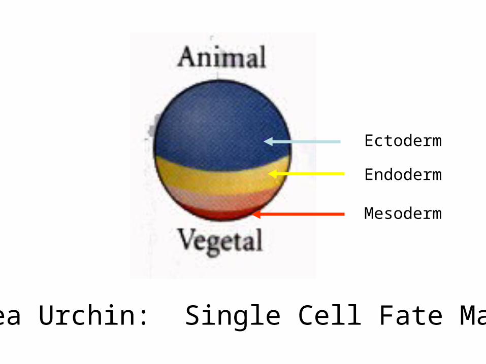
Sea Urchin: Single Cell Fate Map
Ectoderm
Endoderm
Mesoderm

EARLY PATTERNS OF CLEAVAGE IN SEA URCHINS
The earliest patterns of cleavage in sea urchins are holoblastic and very similar to one another.

OVERVIEW OF SEA URCHING EARLY DEVELOPMENT
Zygote Early Blastula Hatched Blastula Gastrula
Prism Larva Pluteus Larva

Early Cleavage in Sea Urchins

The first and second divisions are meridional (animal to vegetal pole) and perpendicular to one another.

Third division is equatorial, separating animal and vegetal hemispheres.


Fourth cleavage is - however - different from the first three:•Four cells of animal hemisphere divide meridonially.•Four cells of vegetal hemisphere divide equatorially and very unequally, resulting in four MACROMERES and four tiny MICROMERES at the vegetal pole.

Fifth cleavage: cells of animal hemisphere divide meridionally, as do macromeres. Micromeres (somewhat more slowly) also divide meridionally.
Beyond this stage, cell divisions are somewhat less regular relative to one another.

Vegetal Pole Animal Pole


BLASTULA FORMATION
Recall that surface:volume constraints demand that embryos eventually take on the morphology of a hollow sphere - the “blastula”. This takes place at approximately the 128 cell stage in sea urchins.
The blastula is a hollow sphere with an internal, central cavity known as the BLASTOCOEL. Tight junctions between cells now help to constitute a seamless epithelial sheet.

Early Blastula Stages in Sea Urchins
Later in the blastula stage, cells at the vegetal pole end thicken, forming the VEGETAL PLATE.
At this point, the cells at the animal pole secrete a digestive enzyme that disintegrates the fertilization envelope, and a free-swimming hatched blastula emerges. Swimming is accomplished by beating cilia.


Sea Urchin: Single Cell Fate Map

SEA URCHIN FATE MAP
Realize of course that the fate map could have been laid on to the single-celled zygote.
In an effort to correspond to the book, we will show it here however.
In normal development, the animal hemisphere consistently gives rise to ectoderm (the larval skin and nervous system’s neurons).


The upper veg1 layer can give rise to ectodermal or endodermal components.

The veg2 layer gives rise to:•endoderm•coelomic mesoderm (internal surface lining)•secondary mesenchyme ( muscle cells, pigment cells, immunocytes)

First tier of micromeres gives rise to:•primary mesenchyme cells (skeletal structures)

Second tier of micromeres gives rise to:•coelomic mesoderm (outer surface lining)

GASTRULATION IN ECHINODERMS
PART I: INGRESSION OF PRIMARY MESEHCHYME
Gastrulation is, essentially, the building and development of the gut and eventually complete gut tube. This demands the movement of cells to the internal volume of the blastula - changing fundamentally the morphology of the organism.
Recall that the blastula is a hollow sphere defined by a single epithelial layer of cells.

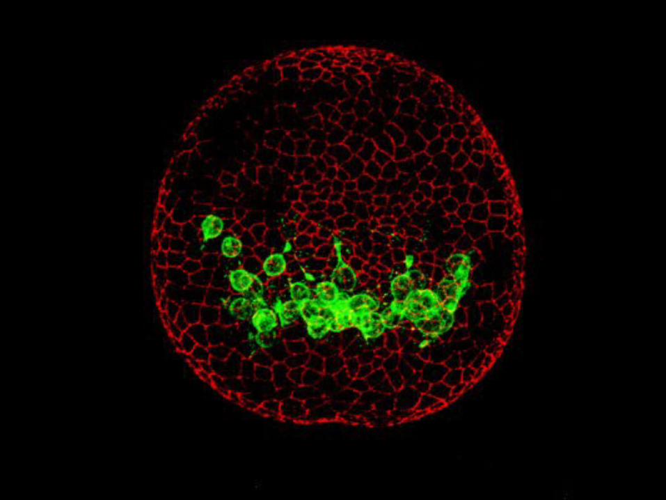
Shortly after hatching, cells derived from the micromeres loose some of their epithelial adhesion and change shape to a bottle-shape.
They then break away from the surface epithelium and enter into the blastocoel. The cells are called the PRIMARY MESENCHYME (sometimes called skeletogenic mesenchyme). They move in independently from other cells, and are thus an example of ingression. They are loosely packed - thus the designation as mesenchyme. They develop spicules and will contribute to skeletal structures.


GASTRULATION IN ECHINODERMS
PART II: INVAGINATION TO FORM FIRST STAGE OF ARCHENTERON.
Note - whereas the primary mesenchyme forms via ingression, the primitive/early gut tube - termed the ARCHENTERON, forms via a movement of an epithelial sheet that retains its integrity, and is thus an invagination.


Cells that remain at the vegetal pole thicken and flatten out to form a VEGETAL PLATE.
The cells move to mend the spaces made by the ingression of the primary mesenchyme cells.
Vegetal plate bends inward, invaginating about 25% the distance into the blastula. The development of this in-turned space is the beginning of the primitive gut - the ARCHENTERON. And, the opening into it is the BLASTOPORE.

Early development of primary archenteron.

Overview of Gastrulation in Sea Urchin

Invagination appears to be caused by shape changes in the cells of the vegetal plate. Vegetal plate cells surrounding cells of vegetal pole become bottle-shaped, constricting their apical ends. This causes the cells to pucker inward.
The secondary mesenchyme is the first group of cells in via ingression. (The book calls it this correctly in the figures, but incorrectly calls it invagination in the text.) They form the tip of the archenteron. This in turn forms pigment cells, muscular around gut, and ceolomic pouches.
Endodermal cells adjacent to micromere-derived mesoderm enter next, becoming foregut.
Next layer of endoderm becomes midgut.The last circumferential row to enter becomes the hindgut and anus linings respectively. (Remember this is a deuterostome!)

SECOND STAGE OF ARCHENTERON FORMATION
During the second stage of archenteron formation, the archenteron extends in length quickly and dramatically – up to tripling its length.
To do this, cells flatten, elongate, and rearrange themselves by migrating over one another

THIRD STAGE OF ARCHENTERON FORMATION
Cells of the secondary mesenchyme contact the inner surface of the blastocoele wall.
They then shorten, pulling the archenteron into contact with distant blastula wall.
Once archenteron makes contact with opposite wall, secondary mesenchyme cells disperse to eventually give rise to other mesodermal organs.
Note dual role of secondary mesenchyme:•Aid in formation of complete gut tube.•Give rise to mesodermal organs.

Prism Larval Stage:
The prism larval stage is achieved when the archenteron reaches distant blastular wall.
This stage is more properly a gastrula now.


Pluteus Larval Stage
In the pluteus larval stage:•Larva elongates.•Coelomic cavities form from secondary mesenchyme.•Right coelom degenerates, but left proliferates into three separate sacs.


FURTHER COELOMIC DIFFERENTIATION
An invagination of ectoderm fuses with the middle of the sacs derived from the elaborated left coelom. This is called the IMAGINAL RUDIMENT.
Imaginal rudiment develops five-fold symmetry.
Secondary mesenchyme cells enter imaginal rudiment to form first skeletal elements.