biological sciences undergraduate research symposium
DESCRIPTION
Loyola's pre-eminent student research experience will be Friday, April 13, from 12:45 to 5 p.m. in Nunemaker Auditorium in Monroe Hall.TRANSCRIPT

LO
YO
LA
U
NIV
ER
SIT
Y
Department of Biological Sciences
22nd
Annual Undergraduate
Research Symposium
BIO
LO
GIC
AL
S
CIE
NC
ES
D
EPA
RT
ME
NT
“Loyola’s Pre-eminent Student Research Experience”
Friday, April 13, 2012
12:45 pm – 5:00 pm
Monroe Hall, Nunemaker Auditorium
Tell me and I forget…
SHOW me and I remember…
INVOLVE me and I understand!

The Mullahy Biology Endowed Fund
for Undergraduate Research
Much of the financial support for the research presented in
the Undergraduate Research Symposium comes from the
Mullahy Biology Endowed Fund for Undergraduate Research.
Established in 1978, the fund is named after Rev. John
Mullahy S.J., who chaired Loyola’s Department of Biological
Sciences for more than 20 years. Fr. Mullahy began involving
undergraduate biology students in original research in the
1950’s long before the current national trend of undergraduate
research. We gratefully acknowledge Fr. Mullahy’s foresight in
establishing this emphasis and tradition in research that has
lasted for more than half a century, as well as the longstanding
and continuous financial commitment of the benefactors of the
Mullahy Biology Endowed Fund.
The Department of Biological Sciences is committed to
strengthening this research experience by increasing the
number of students who can participate in scholarly research
and by developing new research programs. If you are
committed to undergraduate research, we invite you to join our
Campaign. To receive more information or to make a
contribution, please contact Karen E. Anklam, Major Gifts
Officer, Development/Capital Campaigns Department at (504)
861-5423 or [email protected].
LO
YO
LA
U
NIV
ER
SIT
Y
BIO
LO
GIC
AL
S
CIE
NC
ES
D
EPA
RT
ME
NT
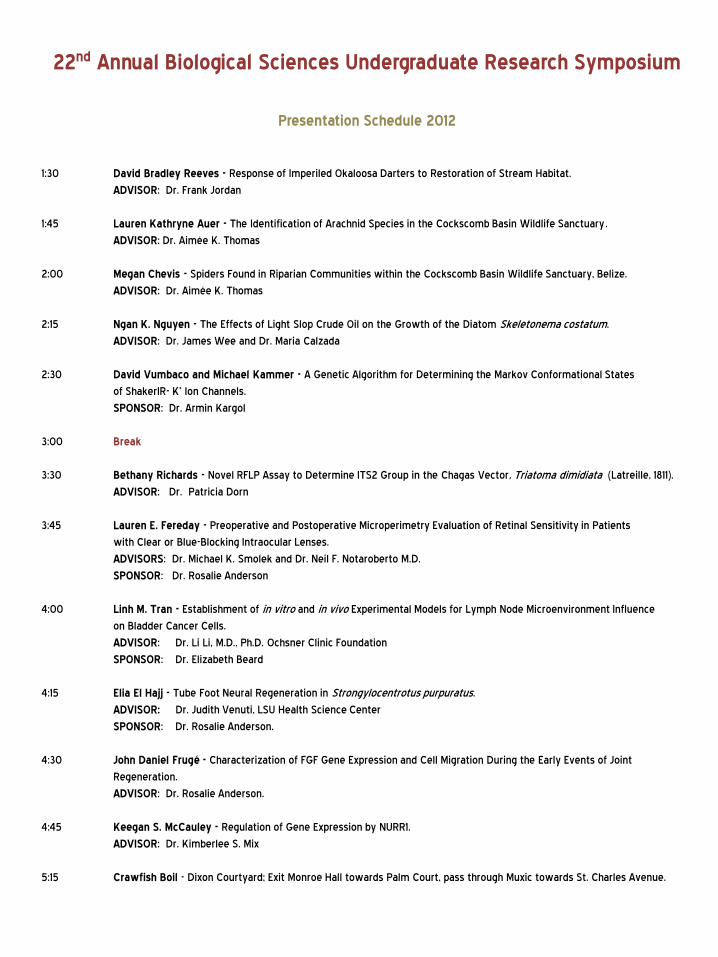
22nd Annual Biological Sciences Undergraduate Research Symposium
Presentation Schedule 2012
1:30 David Bradley Reeves - Response of Imperiled Okaloosa Darters to Restoration of Stream Habitat.
ADVISOR: Dr. Frank Jordan
1:45 Lauren Kathryne Auer - The Identification of Arachnid Species in the Cockscomb Basin Wildlife Sanctuary.
ADVISOR: Dr. Aimée K. Thomas
2:00 Megan Chevis - Spiders Found in Riparian Communities within the Cockscomb Basin Wildlife Sanctuary, Belize.
ADVISOR: Dr. Aimée K. Thomas
2:15 Ngan K. Nguyen - The Effects of Light Slop Crude Oil on the Growth of the Diatom Skeletonema costatum.
ADVISOR: Dr. James Wee and Dr. Maria Calzada
2:30 David Vumbaco and Michael Kammer - A Genetic Algorithm for Determining the Markov Conformational States
of ShakerIR- K+ Ion Channels.
SPONSOR: Dr. Armin Kargol
3:00 Break
3:30 Bethany Richards - Novel RFLP Assay to Determine ITS2 Group in the Chagas Vector, Triatoma dimidiata (Latreille, 1811).
ADVISOR: Dr. Patricia Dorn
3:45 Lauren E. Fereday - Preoperative and Postoperative Microperimetry Evaluation of Retinal Sensitivity in Patients
with Clear or Blue-Blocking Intraocular Lenses.
ADVISORS: Dr. Michael K. Smolek and Dr. Neil F. Notaroberto M.D.
SPONSOR: Dr. Rosalie Anderson
4:00 Linh M. Tran - Establishment of in vitro and in vivo Experimental Models for Lymph Node Microenvironment Influence
on Bladder Cancer Cells.
ADVISOR: Dr. Li Li, M.D., Ph.D. Ochsner Clinic Foundation
SPONSOR: Dr. Elizabeth Beard
4:15 Elia El Hajj - Tube Foot Neural Regeneration in Strongylocentrotus purpuratus.
ADVISOR: Dr. Judith Venuti, LSU Health Science Center
SPONSOR: Dr. Rosalie Anderson.
4:30 John Daniel Frugé - Characterization of FGF Gene Expression and Cell Migration During the Early Events of Joint
Regeneration.
ADVISOR: Dr. Rosalie Anderson.
4:45 Keegan S. McCauley - Regulation of Gene Expression by NURR1.
ADVISOR: Dr. Kimberlee S. Mix
5:15 Crawfish Boil - Dixon Courtyard; Exit Monroe Hall towards Palm Court, pass through Muxic towards St. Charles Avenue.
AB
ST
RA
CT
S
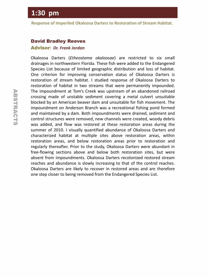
David Bradley Reeves
Advisor: Dr. Frank Jordan
Okaloosa Darters (Etheostoma okaloosae) are restricted to six small drainages in northwestern Florida. These fish were added to the Endangered Species List because of limited geographic distribution and loss of habitat. One criterion for improving conservation status of Okaloosa Darters is restoration of stream habitat. I studied response of Okaloosa Darters to restoration of habitat in two streams that were permanently impounded. The impoundment at Tom’s Creek was upstream of an abandoned railroad crossing made of unstable sediment covering a metal culvert unsuitable blocked by an American beaver dam and unsuitable for fish movement. The impoundment on Anderson Branch was a recreational fishing pond formed and maintained by a dam. Both impoundments were drained, sediment and control structures were removed, new channels were created, woody debris was added, and flow was restored at these restoration areas during the summer of 2010. I visually quantified abundance of Okaloosa Darters and characterized habitat at multiple sites above restoration areas, within restoration areas, and below restoration areas prior to restoration and regularly thereafter. Prior to the study, Okaloosa Darters were abundant in free-flowing sections above and below both restoration sites, but were absent from impoundments. Okaloosa Darters recolonized restored stream reaches and abundance is slowly increasing to that of the control reaches. Okaloosa Darters are likely to recover in restored areas and are therefore one step closer to being removed from the Endangered Species List.
1:30 pm Response of Imperiled Okaloosa Darters to Restoration of Stream Habitat.
AB
ST
RA
CT
S
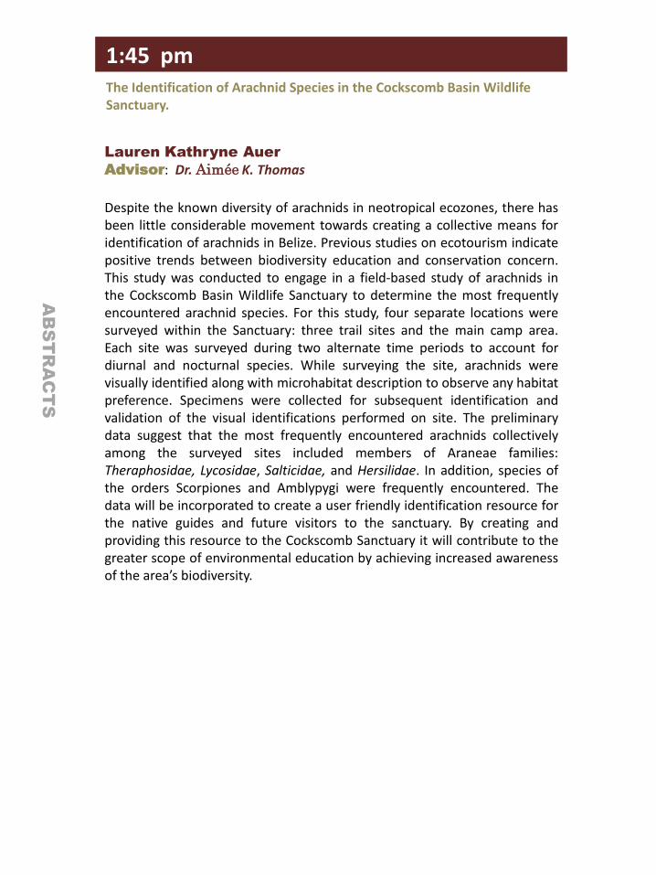
Lauren Kathryne Auer
Advisor: Dr. Aimée K. Thomas
Despite the known diversity of arachnids in neotropical ecozones, there has been little considerable movement towards creating a collective means for identification of arachnids in Belize. Previous studies on ecotourism indicate positive trends between biodiversity education and conservation concern. This study was conducted to engage in a field-based study of arachnids in the Cockscomb Basin Wildlife Sanctuary to determine the most frequently encountered arachnid species. For this study, four separate locations were surveyed within the Sanctuary: three trail sites and the main camp area. Each site was surveyed during two alternate time periods to account for diurnal and nocturnal species. While surveying the site, arachnids were visually identified along with microhabitat description to observe any habitat preference. Specimens were collected for subsequent identification and validation of the visual identifications performed on site. The preliminary data suggest that the most frequently encountered arachnids collectively among the surveyed sites included members of Araneae families: Theraphosidae, Lycosidae, Salticidae, and Hersilidae. In addition, species of the orders Scorpiones and Amblypygi were frequently encountered. The data will be incorporated to create a user friendly identification resource for the native guides and future visitors to the sanctuary. By creating and providing this resource to the Cockscomb Sanctuary it will contribute to the greater scope of environmental education by achieving increased awareness of the area’s biodiversity.
1:45 pm The Identification of Arachnid Species in the Cockscomb Basin Wildlife Sanctuary.
AB
ST
RA
CT
S
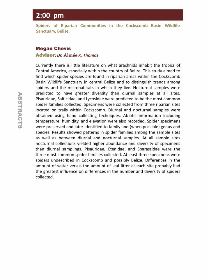
Megan Chevis
Advisor: Dr. Aimée K. Thomas
Currently there is little literature on what arachnids inhabit the tropics of Central America, especially within the country of Belize. This study aimed to find which spider species are found in riparian areas within the Cockscomb Basin Wildlife Sanctuary in central Belize and to distinguish trends among spiders and the microhabitats in which they live. Nocturnal samples were predicted to have greater diversity than diurnal samples at all sites. Pisauridae, Salticidae, and Lycosidae were predicted to be the most common spider families collected. Specimens were collected from three riparian sites located on trails within Cockscomb. Diurnal and nocturnal samples were obtained using hand collecting techniques. Abiotic information including temperature, humidity, and elevation were also recorded. Spider specimens were preserved and later identified to family and (when possible) genus and species. Results showed patterns in spider families among the sample sites as well as between diurnal and nocturnal samples. At all sample sites nocturnal collections yielded higher abundance and diversity of specimens than diurnal samplings. Pisauridae, Ctenidae, and Sparassidae were the three most common spider families collected. At least three specimens were spiders undescribed in Cockscomb and possibly Belize. Differences in the amount of water versus the amount of leaf litter at each site probably had the greatest influence on differences in the number and diversity of spiders collected.
2:00 pm Spiders of Riparian Communities in the Cockscomb Basin Wildlife Sanctuary, Belize.
AB
ST
RA
CT
S
Ngan K. Nguyen
Advisor: Dr. James Wee & Dr. Maria Calzada
With the increase in maritime transport of chemicals, marine environments have become increasingly threatened by toxins. Crude oil spills in particular can cause widespread ecosystem deterioration as demonstrated by the Deepwater Horizon Oil Spill. This study focuses on the effects of Light Slop crude oil on the growth of the diatom, Skeletonema costatum, using a strain isolated from Lake Pontchartrain. Aged seawater from Lake Pontchartrain (salinity = 7.7 ppt) was enriched with nutrients and filtered to make f/2 culture media. Oil contamination was introduced to the culture media via emulsification (1:9, 24 hours, 200 rpm). Experimental treatments included uncontaminated f/2 culture media, oil-emulsified f/2 and oil-emulsified f/2 diluted with uncontaminated f/2 to produce culture media containing 25%, 50% and 75% of oil-emulsified f/2. Preliminary studies indicate that culture tube optical density at = 730 nm correlate to cell counts, and hydrocarbon chain lengths decrease during the course of the experiment. To observe growth of cultures, OD730 readings were recorded on alternate days for ten days. The slopes of the growth curves were compared among the different experimental treatments from day 4 to day 10. Uncontaminated f/2, 25% and 50% treatments showed similar growth curves; the 75% treatment exhibited inhibited growth before recovering at day 4, while no growth occurred in the undiluted, oil-emulsified f/2 treatment. The slopes for the uncontaminated f/2 culture media and 25% treatment were the same (p <0.001), suggesting that oil contamination in the 50% treatment can inhibit growth.
2:15 pm The Effects of Light Slop Crude Oil on the Growth of the Diatom Skeletonema costatum.
AB
ST
RA
CT
S
David Vumbaco and Michael Kammer
Sponsor: Dr. Armin Kargol
Ion channels are trans-membrane proteins that exhibit domains organized to create selectively permeable gated pores through the cell membrane. Ion channels are ubiquitous in the cellular domain. While ionic currents through channels can be measured using a patch clamping technique, it is impossible with current technology to directly measure the conformational states of ion channels. Dr. Kargol’s laboratory has developed an algorithm to determine the Markov conformational states of voltage gated ion channels from experimental data. This is accomplished by taking electrophysiological recordings from mammalian fetal kidney cells type TSA-201 expressing the ShakerIR-K+ ion channel via a voltage clamp apparatus. This data is then processed through a genetic algorithm that determines what the channels’ conformational states were during the initial exposure to the controlled potential. Accurate identification of the bulk conformational state of a cell’s ion channels is an important step in understanding their dynamics at large. It also represents a major step in our larger goal, the realization of non-equilibrium kinetic focusing of ion channels into one conformational state through the use of dichotomous noise and wavelet based protocols.
2:30 pm A Genetic Algorithm for Determining the Markov Conformational States of ShakerIR-K+ Ion Channels.

AB
ST
RA
CT
S
3:30 pm Novel RFLP Assay to Determine ITS2 Group in the Chagas Vector, Triatoma dimidiata (Latreille, 1811).
Bethany Richards
Advisor: Dr. Patricia Dorn
Approximately 10 million people are infected with Trypanosoma cruzi, the protozoan parasite that causes Chagas disease. T. cruzi is transmitted to humans primarily by triatomine insect vectors; the most important Central American species is Triatoma dimidiata. Effective vector control requires a clear understanding of the taxonomy and subdivisions among T. dimidiata populations. The nuclear sequence of the ribosomal internal transcribed spacer 2 (ITS2) is frequently used to illuminate the taxonomy of triatomines because of its relatively high rate of mutation. Oftentimes amplification and sequencing of ITS2 fail, likely due to the large size of the product which may be compromised in older, degraded DNA, and polymerase slippage due to homopolymeric and/or microsatellite regions present near the 5’ end. To overcome these limitations we have designed new primers that amplify only the 3’-most 200 bps of ITS2. This region is sufficient to distinguish the ITS2 group for all known T. dimidiata haplotypes. Furthermore, we have developed an RFLP approach to determine ITS2 group, eliminating the need to sequence the amplified product. The use of new primers and RFLP to determine ITS2 groups will more efficiently distinguish taxa of Triatoma dimidiata, thus facilitating effective vector control.

AB
ST
RA
CT
S
Lauren E. Fereday
Advisor: Dr. Michael K. Smolek, Fellow of the Association for
Research in Vision and Ophthalmology and Dr. Neil F.
Notaroberto, Louisiana Eye Research Institute, and CLEVER
Eye Institute.
Sponsor: Dr. Rosalie Anderson
The retina is protected by the natural light-absorption properties of the crystalline lens, which attenuates the high-energy, UV and blue end of the spectrum. This protection is lost when the natural lens is extracted due to cataract removal. In 2003, Alcon introduced a yellow chromophore intraocular lens (IOL) to partially attenuate blue-light. This lens still generates controversy. Some clinicians suggest that the tint is actually detrimental to vision. We examined records of macular microperimetry to evaluate whether retinal sensitivity is reduced in IOL patients implanted with yellow IOLs. This was a retrospective study based on existing clinical records. Inclusion criteria were IOL patients who had pre- and postoperative Centervue MAIA microperimetry (Ellex USA, Minneapolis, MN) in the 'Expert' mode, and who were implanted with either a clear (CLR-IOL) or yellow-tinted (BB-IOL) IOL. Preoperatively, 16 CLR-IOL and 95 BB-IOL examinations were used, and postoperatively, 10 CLR-IOL and 66 BB-IOL examinations were used. The Expert mode determines absolute sensitivity in decibels (dB) at 36 perifoveal points in the macula and at a central stimulus point presumed to be located at the rod-free fovea. The preoperative data was significantly different between groups. Postoperatively, the perifoveal CLR-IOL and BB-IOL groups had statistically identical levels of mean sensitivity, which suggests that yellow-tinted IOLs have the same effect on visual sensitivity as clear IOLs when measured by microperimetry. Likewise, the central stimulus point had the same outcome. Postoperative sensitivities were on average within the lower end of the normal range (approximately 25 dB perifoveally and 23 dB centrally).
3:45 pm Preoperative and Postoperative Microperimetry Evaluation of Retinal Sensitivity in Patients with Clear or Blue-Blocking Intraocular Lenses.

AB
ST
RA
CT
S
Linh M. Tran
Advisor: Dr. Li Li, MD., Laboratory of Translational Research in
Cellular Immunology at Ochsner Clinic Foundation
Sponsor: Dr. Elizabeth Beard
Human urinary bladder cancer is one of the most expensive diseases to treat. It is the fifth leading carcinoma and second most frequent urologic malignancy in the U.S. We believe its resilience is due to its cancer stem cells (CSCs) that may survive most treatments, cause tumor recurrence, and metastasize elsewhere. Lymph node (LN) metastasis usually gives a negative prognosis because follicular dendritic cells (FDC) of the LN cortex seem to promote the proliferation and drug resistance of these CSCs, many of which can be identified by their biomarkers. This study investigates the relationship between FDC-mediated microenvironments and bladder CSCs. Human bladder cancer cell lines SW780 and UM-UC-3, and FDC stromal cell line HK were used to test in vitro colony assays as well as in vivo NOD/SCID mice xenograft models. Assessment of CSC properties was first performed using cell lines and then confirmed by patient specimens. We found that SW780 expresses biomarkers CD24, CD44, and CD47. UM-UC-3, which proliferates at a higher rate than SW780, expresses CD44, CD47, and CD166. Both cell lines displayed higher proliferation and tumor formation rates in the presence of HK cells in both in vitro and in vivo experimental models. Under slide examinations, xenoplants visually resembled clinic patient specimens. We also identified a CSC-like fraction in both cell lines that interacts with FDC and correlates with the expression of biomarker CD133. Further study of the pro-tumor factors provided by LN stromal cells may lead to a novel treatment protocol to prevent bladder cancer relapse.
4:00 pm Establishment of in vitro and in vivo Experimental Models for Lymph Node Microenvironment Influence on Bladder Cancer Cells.
AB
ST
RA
CT
S
Elia El Hajj
Advisor: Dr. Judith Venuti – LSU Health Science Center, Associate Professor of Anatomy
Sponsor: Dr. Rosalie Anderson
Sea urchins, the most basal deuterostomes, share a common ancestry with humans. With the help of genomics and modern techniques in molecular biology, photo sensory genes have been identified in the sea urchin tube foot. We decided to investigate. We have defined the tube foot nervous system and shown that photo sensory genes, namely vertebrate retinal homologues, are expressed in the tube foot discus. We have confirmed the spatial expression of opsin and pax6 genetic profiles in the discus by means of immunochemistry. To investigate the formation of these photosensory structures on the periphery of the cap, we have determined the time course for neural and dermal regeneration of the tube foot in S. purpuratus urchins. This will be used in future studies to understand the hierarchy of genes involved in the formation of the photosensory structures on the periphery of the cap.
4:15 pm Tube Foot Neural Regeneration in Strongylocentrotus purpuratus.

4:30 pm Characterization of FGF Gene Expression and Cell Migration During the Early Events of Joint Regeneration.
John Daniel Frugé
Advisor: Dr. Rosalie Anderson
Synovial joints, which are crucial to mobility, are subject to degenerative diseases, trauma, and loss of function, yet there is a lack of understanding of their developmental pathways and regenerative capabilities. Using the chicken embryo as a model, this research investigates some of the initial events that occur during joint regeneration. Utilizing the fluorescent, lipophilic tracer DiI, populations of cells were fate mapped during normal development, during regeneration following window excision of the joint, and during wound healing in slice excision limbs. It was found that cells during normal development migrate along the proximal-distal axis. However, during regeneration, cells from the posterior margin of the window excision migrate anteriorly and participate in forming the regenerating joint. Cell migration is not observed following slice excision, suggesting that posterior cell migration is important for joint regeneration. Members of the fibroblast growth factor (FGF) family are candidate molecules that may play a role in this migration and/or in other events that enable regeneration to occur. FGFs have been identified as major contributors to limb development and to regeneration in various regenerative species. In order to determine their role(s) in the development and regeneration of the embryonic chicken joint, their expression must be characterized. Riboprobes were successfully made for FGFs 3, 6, 9, 10, and 18 and their whole-mount gene expression patterns were determined using in-situ hybridization. Subsequently, in-situ hybridization was performed on sections of regenerating and normal limbs. Histological analysis was used to determine expression patterns of the FGFs throughout these limbs.
AB
ST
RA
CT
S
4:45 pm Regulation of Gene Expression by NURR1.
Keegan S. McCauley
Advisor: Dr. Kimberly Mix
Arthritis is one of the leading causes of disability in the US, affecting over 46 million adults. The degradation of cartilage and bone in arthritis leads to severe joint pain and loss of joint function. There is a need for new therapies that antagonize molecules involved in the onset and development of arthritis. We have identified the orphan nuclear receptor related-1 (NURR1) gene as a potential drug target and are currently studying the mechanism of this receptor with respect to arthritis. Elevated levels of NURR1 in arthritic tissues have been documented, and we have established that this receptor has transcriptional activity in joint cells. To identify potential NURR1 target genes, we performed a qPCR array experiment to investigate the differential expression levels of 94 different genes associated with arthritis. In this experiment, 24 genes were induced and 48 genes were repressed by NURR1 over-expression. The most highly induced gene was prolactin (68-fold), and interestingly, this gene codes for a peptide hormone that regulates inflammatory pathways in arthritis. In a subsequent experiment, NURR1 over-expression induced prolactin mRNA levels by 300-fold. Prolactin transcription is regulated by two promoters, and our RT-PCR data suggests that NURR1 activates the extrapituitary promoter in joint cells. This promoter does not appear to contain a consensus binding site for NURR1, meaning that NURR1 may interact with a novel sequence to regulate transcription. In summary, NURR1 may promote inflammation in joint tissues by inducing the expression of prolactin and is therefore a promising target for arthritis therapy.
AB
ST
RA
CT
S

SPECIAL THANKS
We would like to offer our special thanks to the following for
their continued support of this event and our wonderful
students. Without you it would not be possible.
Mrs. Suzanne B. Smith
Mrs. Judith P. Seaman
Dr.. Elizabeth Abboud
Dr. Nicholas A. Danna III
Mrs. Renee F. DiGiovanni
Dr. Patricia Dorn
Dr. Donald Hauber
Mrs. Irene T. Hofstetter
Mr. Randy Englert
Ms. Laura Lee Wilkinson
Mr. Jonathan Kurtz