biocompatibility of six elastomers in vitro
TRANSCRIPT
Biocompatibility of six elastomers in vitro ~ ~
D. Bakker, C. A. Van Blitterswijk, W. Th. Daems*, and J . J . Grote Ear Nose G. Throat Department, Unizlerstty Hospital, Leiden, The Netherlands and *Laboratory for Electron Microscopy, University of Leiden, The Netherlands
The biocompatibility of two silicone rub- bers, Silastic and Dow Corning Elastomer, and of a polyether and a polyester ure- thane, a polyether polyester copolymer, and polypropylene oxide was assessed in uifro. These elastomers were selected for assessment as a possible a!loplastic tym- panic membrane. For these studies use was made of rat middle ear mucosa explants and serially cultured epithelium. The quan- titative results were based on epithelial growth curves, the morphological picture was based on the findings in epithelium, and the aging of a biomaterial was simu- lated. Epithelium morphology was in- vestigated by scanning and transmission electron microscopy and x-ray micro- analysis. Quantitative results showed that
on Dow Corning Elastomer and polypro- pylene oxide, cell proliferation was signifi- cantly lower compared to normal growth curves. The morphological findings were negative for polypropylene oxide, and did not discriminate between the other bio- materials under study. The simulation re- sults indicated better biocompatibility for the polyurethanes and the polyether poly- ester copolymer compared with that of polypropylene oxide and both silicone rubbers. Under the simulation conditions, cells exposed to Silastic showed silicon- containing inclusions. These in vitro results suggest that the biocompatibility of the polyurethanes and the polyether polyester copolymer is better than that of both sili- cone rubbers and polypropylene oxide.
INTRODUCTION
The development of a total alloplastic middle ear prosthesis (TAM)' in our laboratory has encountered problems associated with the need for an arti- ficial tympanic membrane for attachment of the artificial ossicular chain to the hydroxyapatite canal wall of the prosthesis. The bulk material used for the TAM is porous hydroxyapatite.'s2 This ceramic was found to have good biocompatibility in in vitrn studies with rat middle ear epithelium3 and in implantation studies in noninfected4s5 and infected6*' rat middle ears. In long-term clinical studies hydroxyapatite has proven to be suitable for use in ~tology.',~
In the studies reported here, we assessed in vifro the biocompatibility of six biomaterials, for some of which the vibratory properties had been in- vestigated."," The studies were done in cultured tissue; this method offered rapid results and a basis for quantitation of biocompatibility.12 The in vitro investigations comprised quantitation of proliferative activity and obser- vation of morphological changes in rat middle ear epitheliumI3 cultured on the selected biomaterials. The third component of the study was artificial
Journal of Biomedical Materials Research, Vol. 22, 423-439(1988) Q 1988 John Wiley & Sons, Inc. CCC 0021 -9304!88!050423-17$04.00
424 BAKKER ET AL.
aging of a given biomaterial, which was performed according to the method of Homsy.l*
MATERIALS AND METHODS
Materials
The following biomaterials were used; the Dow Corning silicone rubbers Silastic and MDX-4-4210 Clean Grade Elastomer,’i a polyether urethaneI6 (Estane 5714 F1, B. F. Goodrich), a polyester urethane (Pellethane-80A, Upjohn), a poly(ethy1ene oxide-hydantoin)/poly(tetramethylene terephthal- ate) segmented polyether polyester copolymer” (Akzona Inc.), and poly- propylene oxide. For Silastic, commercially available dense films (125 pm) were used, and for the other biomaterials dense films (100-125 pm) were cast from polymer solutions. The films were air dried and kept under vacuum conditions for at least 48 h. All films were cleaned, rinsed extensively first in running tap water, and then in distilled water, and air-dried. Unless stated otherwise, all biomaterials were steam sterilized except polypropylene oxide, which was sterilized by UV radiation.
Explants and epithelium
For the studies on morphology rat middle ear mucosa explants and epi- thelium were cultured as follows. Pieces of middle ear mucosa measuring about 1 mm7 were obtained from the middle ear of male Wistar rats. The pieces were allowed to attach to the substrate in a small drop of Dulbecco’s modification of Eagles medium and F12 (3 : 1) to which hydrocortisone (0.4 pg/mL), isoproterenol M), penicilin (100 U/mL), and streptomycin (100 pg/mL) had been added. After that, normal amounts of medium were used, and epidermal growth factor (10 ng/mL) was added to the medium after three days of culture. All material was cultured in 10% C 0 2 at 37°C and the medium was renewed twice a week.I3
Epithelial cells were harvested by trypsinisation from the outgrowth of an explant, which was cultured for 2 weeks in Costar culture dishes (tissue culture polystyrene TCPS). Serially cultured cells were plated at a density of 2 X 1U4 cells/cm2 in combination with 1.2 x lo4 lethally irradiated 3T3 feeder cells per cm2. For the quantitation of biocompatibility and for the simulation studies, use was made of serially cultured rat middle ear epithelium.
Quantitation
Growth curves of serial cultures of rat middle ear epithelium cultured on the various biomaterials were based on cell counts performed in 35 mm Costar culture dishes after one, three, six, 10, and 14 days. Dishes with and
BIOCOMPATIBILITY OF ELASTOMERS 1N VITRO 425
without (control) a biofilm were UV-sterilized prior to use. Three dishes were used for each of the culture intervals.
Morphology
The morphology of rat middle ear tissue cultured on the various polymer materials and TCPS (control) was established electron microscopically. The observations were performed six and 10 days after the start of the culture of (1) middle ear mucosa explants, (2) epithelium growing out of explants, and (3) serially cultured epithelium.
Artificial aging
For artificial aging of an implant equal amounts of the various biomaterials were heat-exposed to extracellular fluid (PECF) at 115°C for 48 h.14 The PECF was a salt solution comprising most of the salts included in the nutrient medium used for tissue culture and most of the ions in the same proportions as occur in body fluid14 (Table I). Further, aliquots of PECF from a given experiment were used to prepare the nutrient media. In addition to these simulation media, two positive control media and one negative control me- dium were also prepared. The positive controls consisted of PECF that was free of biomaterial during heating and PECF in which Melinex (a transparent thin film made of tissue culture polystyrene) was exposed to heat. PECF containing polyvinyl chloride (PVC) served as negative control. On this basis nine different nutrient media were obtained for serial cultivation of rat middle ear epithelium on TCPS (Table 11).
After three days of culture in routine medium, the various simulation and control media were added to the culture dishes. Three and 10 days later, the cell morphology was compared with the picture after culture in rou- tine medium.
TABLE I Ion Concentrations (in meq./L) in Body Fluid” and PECF
Ion Body fluid PECF
Na + 145 154.5 K + 5 5.4 c1 - 113 11 8.5 co3 30 44 Po:- 2 0
“As defined in ref. 14.
426 BAKKER ET AL.
TABLE I1 Media Used in the Artificial Aging Experiment
Medium Polymer Type -
Melinex PVC Silastic Elastomer Estane Pellethane Copolymer Polypropylene oxide
Positive control 1 Positive control 2 Negative control Biomaterial Biomaterial Biomaterial Biomaterial Biomaterial Biomaterial
Electron microscopy
The specimens destined for transmission electron microscopy were fixed in 1.5% glutaraldehyde in sodium cacodylate buffer (0.14 M, pH 7.4,TC) for 15 min. After being rinsed in phosphate-buffered saline (pH 7.4), the cells were postfixed in a 1% Os04 solution at room temperature for 30 min. Next, the specimens were rinsed in phosphate-buffered saline, dehydrated in graded alcohols, and embedded in Epon. Ultrathin sections were cut on a Reichert ultramicrotome, stained with uranyl acetate and lead citrate, and examined in a Philips 201 electron microscope. The specimens destined for scanning electron microscopy were dehydrated immediately after the glu- taraldehyde and then critical point dried and gold coated. A Cambridge S 180 scanning electron microscope was used.I3 Some of the material prepared for transmission electron microscopy was studied with a Philips EM 400 con- nected to a Tracor Northern (TN) 2000 x-ray micro-analyzer. Both single-spot analysis and x-ray mapping were performed.”
RESULTS
Quantitative analysis
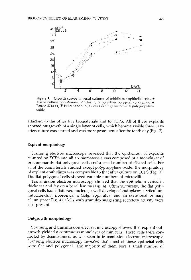
Only a few middle ear epithdial cells adhered to polypropylene oxide, but these cells did not proliferate. The shape of the growth curves of the epi- thelial cells serially cultured on the other five biomaterials and TCPS was sigmoid (Fig. 1). Application of Wilcoxon’s two-sample test (a = 0.05) to the growth curves showed that only the epithelial cells on Dow Corning Elas- tomer and polypropylene oxide proliferated at a significantly lower rate compared with those on TCPS.
Tissue morphology
About half of the middle ear mucosa explants attached to polypropylene oxide but outgrowth of cells did not occur. More than 80% of the explants
BIOCOMPATIBILITY OF ELASTOMERS IN VITRO 427
Figure 1. Growth curves of serial cultures of middle ear epithelial cells. Tissue culture polystyrene, V Silastic, A polyether polyester copolymer, A Estane 5714 F1, V Pellethane 80A, Dow Corning Elastomer, 0 polypropylene oxide.
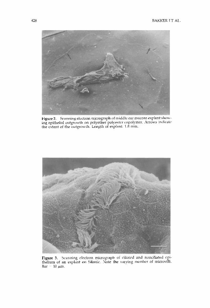
attached to the other five biomaterials and to TCPS. All of these explants showed outgrowth of a single layer of cells, which became visible three days after culture was started and was more prominent after the tenth day (Fig. 2).
Explant morphology
Scanning electron microscopy revealed that the epithelium of explants cultured on TCPS and all six biomaterials was composed of a monolayer of predominantly flat polygonal cells and a small number of ciliated cells. For all of the biomaterials studied except polypropylene oxide, the morphology of explant epithelium was comparable to that after culture on TCPS (Fig. 3). The flat polygonal cells showed variable numbers of microvilli.
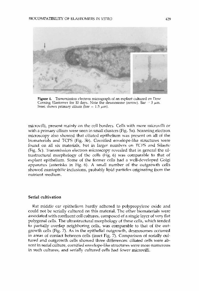
Transmission electron microscopy showed that the epithelium varied in thickness and lay on a basal lamina (Fig. 4). Ultrastructurally, the flat poly- gonal cells had a flattened nucleus, a well-developed endoplasmic reticulum, mitochondria, ribosomes, a Golgi apparatus, and an occasional primary cilium (inset Fig. 4). Cells with granules suggesting secretory activity were also present.
Outgrowth morphology
Scanning and transmission electron microscopy showed that explant out- growth yielded a continuous monolayer of thin cells. 'These cells were con- nected by desmosomes, as was seen in transmission electron microscopy. Scanning electron microscopy revealed that most of these epithelial cells were flat and polygonal. The majority of them bore a small number of
428 BAKKER ET AI,.
Figure 2. Scanning electron tnicrograph of middle ear mucosa explant show- ing epithelial outgrowth on polyether polyester copolymer. Arrows indicate the extent of the outgrowth. Length of explant: 1.8 mm.
Figure 3. Scanning electron micrograph of ciliated and nonciliated epi- thelium of an explant on Silastic. Note the varying number of inicrovilli. Bar = 10 pm.
BIOCOMPATIBILITY OF ELASTOMERS IN VITRO 429
Figure 4. Transmission clcctron micrograph of an explant cultured on Dow Corning Elastomer for 10 days. Note the desmosome (arrow). Bar = 3 pm. Inset shows primary cilium (bar = 1.5 pm).
microvilli, present mainly on the cell borders. Cells with more microvilli or with a primary cilium were seen in small clusters (Fig. 5a). Scanning electron microscopy also showed that ciliated epithelium was present on all of the biomaterials and TCPS (Fig. 5b). Cornified envelope-like structures were found on all six materials, but in larger numbers on TCPS and Silastic (Fig. 5c). Transmission electron microscopy revealed that in general the ul- trastructural morphology of the cells (Fig. 6) was comparable to that of explant epithelium. Some of the former cells had a well-developed Golgi apparatus (asterisks in Fig. 6). A small number of the outgrowth cells showed osmiophilic inclusions, probably lipid particles originating from the nutrient medium.
Serial cultivation
Rat middle ear epithelium hardly adhered to polypropylene oxide and could not be serially cultured on this material. The other biomaterials were associated with confluent cell cultures, composed of a single layer of very flat polygonal cells. The ultrastructural morphology of these cells, which tended to partially overlap neighboring cells, was comparable to that of the out- growth cells (Fig. 7). As in the epithelial outgrowth, desmosomes occurred in areas of contact between cells (inset Fig. 7) . Comparison of serially cul- tured and outgrowth cells showed three differences: ciliated cells were ab- sent in serial culture, cornified envelope-like structures were more numerous in such cultures, and serially cultured cells had fewer microvilli.
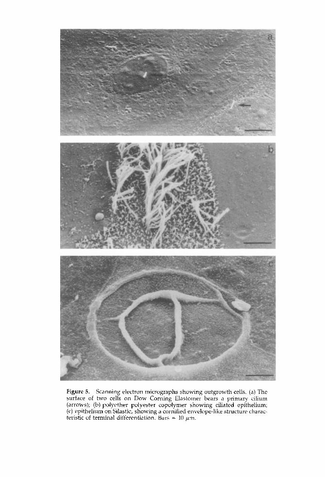
Figure 5. Scanning electron micrographs showing outgrowth cells. (a) The surface of two cells on Dow Corning Elastomer bears a primary cilium (arrows); (b) polyether polyester copolymer showing ciliated epithelium; (c) epithelium on Silastic, showing a cornified envelope-like structure charac- teristic of terminal differentiation. Bars = 10 pm.
BIOCOMPATIBILITY OF ELASTOMERS IN WTXO 431
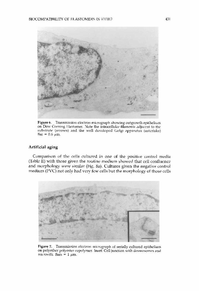
Figure 6. Transmission electron micrograph showing outgrowth epithelium on Dow Corning Elastomer. Note the intracellular filaments adjacent to the substrate (arrows) and the well developed Golgi apparatus (asterisks) Bar = 0.6 pm.
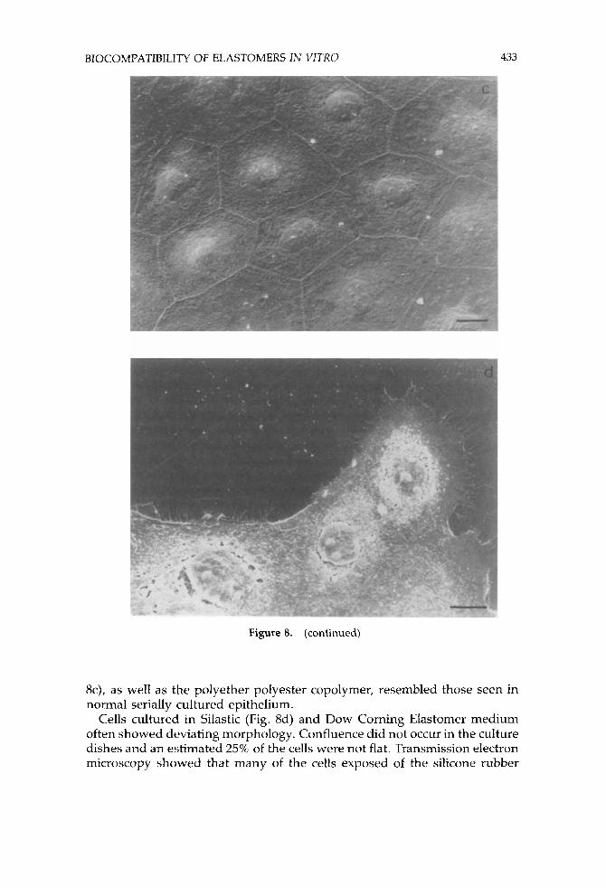
Artificial aging
Comparison of the cells cultured in one of the positive control media (Table 11) with those given the routine medium showed that cell confluence and morphology were similar (Fig. 8a). Cultures given the negative control medium (PVC) not only had very few cells but the morphology of those cells
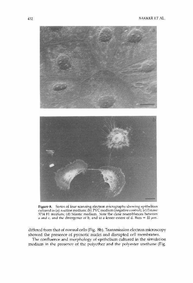
Figure 7. Transmission electron micrograph of serially cultured epithelium on polyether polyester copolymer. Inset: Cell junction with desmosomes and microvilli. Bars = 1 pm.
432 BAKKER ET AL.
Figure 8. Series of four scanning electron micrographs showing epithelium cultured in (a) routine medium; (b) PVC medium (negative control); (c) Estane 5714 F1 medium; (d) Silastic medium. Note the close resemblances between a and c, and the divergence of b, and to a lesser extent of d . Bars = 10 pm.
differed from that of normal cells (Fig. 8b). Transmission electron microscopy showed the presence of pycnotic nuclei and disrupted cell membranes.
The confluence and morphology of epithelium cultured in the simulation medium in the presence of the polyether and the polyester urethane (Fig.
BIOCOMPATIBILITY OF ELASTOMERS IN VlTXO 433
Figure 8. (continued)
8c), as well as the polyether polyester copolymer, resembled those seen in normal serially cultured epithelium.
Cells cultured in Silastic (Fig. 8d) and Dow Corning Elastomer medium often showed deviating morphology. Confluence did not occur in the culture dishes and an estimated 25% of the cells were not flat. Transmission electron microscopy showed that many of the cells exposed of the silicone rubber
434 BAKKER ET AL.
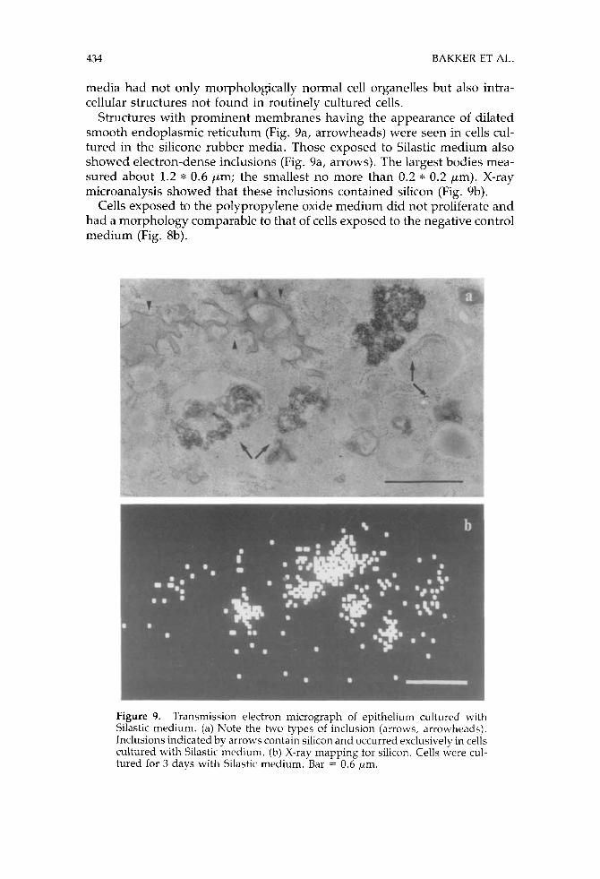
media had not only morphologically normal cell organelles but also intra- cellular structures not found in routinely cultured cells.
Structures with prominent membranes having the appearance of dilated smooth endoplasmic reticulum (Fig. 9a, arrowheads) were seen in cells cul- tured in the silicone rubber media. Those exposed to Silastic medium also showed electron-dense inclusions (Fig. 9a, arrows). The largest bodies mea- sured about 1.2 * 0.6 pm; the smallest no more than 0.2 * 0.2 pm). X-ray microanalysis showed that these inclusions contained silicon (Fig. 9b).
Cells exposed to the polypropylene oxide medium did not proliferate and had a morphology comparable to that of cells exposed to the negative control medium (Fig. 8b).
Figure 9. Transmission electron micrograph of epithelium cultured with Silastic medium. (a) Note the two types of inclusion (arrows, arrowhcads). Inclusions indicated by arrows contain silicon and occurred exclusively in cells cultured with Silastic medium. (b) X-ray mapping for silicon. Cells were cul- tured for 3 days with Silastic medium. Bar = 0.6 pm.
BIOCOMPATIBILITY OF ELASTOMERS IN VITRO 435
DISCUSSION
We assessed the biocompatibility of several candidate tympanic membrane materials in vitro to obtain a basis for selection for our animal implantation studies. Besides quantitation of biocompatibility and sensitivity to toxic materials, in vitro tests permit rapid evaluation of the biocompatibility of a large group of biomaterials. l2
Furthermore, during the early screening phase the assessment of the biocompatibility with human material is by preference performed in vitro. Rat middle ear mucosa explants and middle ear epithelium were used for these studies. This choice was determined by four factors. First, in middle ear surgery the epithelial covering of an implant is considered to influence the success of an implant, because the epithelium is thought to play an important role in middle ear defence against the frequent infections with microorganisms in this region. Second, biocompatibility can be studied in vitro in the same tissue.13
Third, because the explants and cultured cells derive from the rat middle ear, tissue-culture results can be compared with the findings made in im- plantation studies in the rat middle ear. Lastly, these results can be compared with those concerning the in vitro biocompatibility of hydroxyapatite, which has proved to be a highly biocompatible rnateriaL3
With respect to the proliferative activity of the cells under study, there were some interesting findings. TCPS and all of the biomaterials tested except polypropylene oxide - to which cells hardly adhered- showed sig- moid growth curves. The best substrate for cell attachment was TCPS, as indicated by the number of cells adhering to this material after 1 day of cultivation. Adhesion of epithelium, as indicated by cell numbers on day one, was more or less the same for all of the other biomaterials except polypropylene oxide. This seems to be in conflict with results published by Ratner et al.," who reported greater adhesion of chick embryo muscle cells to Pellethane 80A than to Silastic. This divergence might be due to the use of different techniques or other types of tissue.'l Evaluation of the complete growth curves showed significantly lower levels of proliferative activity on Dow Corning Elastomer and polypropylene oxide than on TCPS. The di- vergent cell behavior on Dow Corning Elastomer suggested that it was not the cellisubstrate adhesion that was responsible for the overall lower level of proliferative activity but rather the behavior of the cells cultured on this biomaterial. Both cell behaviorz2 and cellisubstrate adhesionz1 are known to be influenced by the nature of the synthetic substrate.
In our opinion, the alterations in the morphology of explant epithelium reflected exclusively the effects of the presence of toxic substances in the nutrient medium associated with a given biomaterial, because, unlike the situation for serially cultured cells or explant outgrowth, there was no direct contact between explant epithelium and the biomaterial. With respect to toxicity it must be kept in mind that relatively few explants attached to polypropylene oxide. This and the failure of both outgrowth of explants and serial cultures in the presence of polypropylene oxide suggest that toxic
436 BAKKER ET AL.
substances occurred in the nutrient medium in question. For example, the polypropylene oxide we used might have undergone hydrolytic de- gradation, resulting in the release of aldehydes. Explants cultured on TCIPS and on the other five biomaterials showed cell populations resembling nor- mal rat middle ear epithelium as to both morphologically and diversity.
When TCPS or any of the biomaterials except polypropylene oxide was used as synthetic substrate, flat polygonal cells predominated in explant outgrowths, which also showed small numbers of ciliated epithelial cells and terminally differentiated cells. The presence of terminally differentiated cells and ciliated epithelium on these biomaterials seems to be a positive phe- nomenon with respect to biocompatibility, since it shows that these bio- materials allow growth of cells more complex than flat polygonal epithelium. Under serial cultivation, the five biomaterials allowing tissue culture showed only flat polygonal cells with a morphology and a morphological diversity comparable to those seen for TCPS. The absence of ciliated epithelium and the morphological diversity associated with serial cultivation resembled the corresponding characteristics reported by van Blitterswijk et al.I3 Finally, the morphology of middle ear tissue cultured on all of the biomaterials studied except polypropylene oxide, was comparable to that of tissue cul- tured on hydroxyapatite, a biomaterial which showed good biocompatibility both in animals in experimental studies3-'," and in patients when applied clinically. 8,9
The artificial aging of biomaterials was introduced by Homsy, whose ex- perimental results14 indicated that a relatively short exposure of a biomaterial in a medium comparable to body fluid (PECF) in combination with a high temperature mimics long-term implantation conditions with respect to bio- material breakdown. The PECF used in our artificial aging experiments was based on the nutrient medium we used for tissue culture'3 and resembled body fluid as to ion concentration (Table I). According to Flomsy,'4 these ions play the most important role during in uizw degradation of an alloplast.
PVC served as negative control. This polymer has been reported to be cytotoxic in experiments comparable to ours'" as well as in extraction experi- m e n t ~ . ~ ~ Rat middle ear epithelium serially cultured on TCPS with both control media (Table 11) had an ultrastructural morphology comparable to that of epithelium cultured with routine medium, which differed from the morphology of cells cultured with the presence of PVC and polypropylene oxide medium. The latter cultures showed only a very small number of less-flat cells with deviant morphology.
Cells cultured on TCPS with Estane, Pellethane, and polyether polyester copolymer medium showcd a morphology comparable to that of the positive controls. When cultures were given medium based on the PECF in which Silastic or Dow Corning Elastomer had been exposed to heat, the cells showed silicon-containing inclusions and/or structures resembling dilated endoplasmic reticulum. Furthermore, confluence of epithelium was not seen in the culture dishes. It was remarkable that inclusions containing silicon were only found after exposure to Silastic medium and not after Dow Corning Elastomer medium, because both of these biomaterials are silicone
BIOCOMPATIBILITY OF ELASTOMERS IN VITRO 437
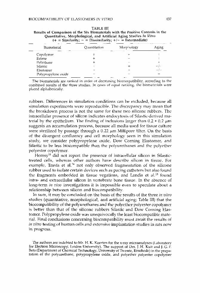
TABLE I11 Results of Comparison of the Six Biomaterials with the Positive Controls in the
Quantitative, Morphological, and Artificial Aging Studies In Wtvo (+ = Similarity; - = Dissimilarity; + I - = Intermediate)
Biomaterial Quantitation Morphology Aging
Copolymer + 7 4 + Estane +
Pellethane + - + Silastic + + +/-
- - r/- Elastomer
-
Polypropylene oxide - - -
The biomaterials are ranked in order of decreasing biocompatibility, according to the combined results of the three studies. In cases of equal ranking, the biomaterials were placed alphabetically.
rubbers. Differences in simulation conditions can be excluded, because all simulation experiments were reproducible. The discrepancy may mean that the breakdown process is not the same for these two silicone rubbers. The intracellular presence of silicon indicates endocytosis of Silastic-derived ma- terial by the epithelium. The finding of inclusions larger than 0.2 * 0.2 pm suggests an accumulation process, because all media used for tissue culture were sterilized by passage through a 0.22 pm Millipore filter. On the basis of the divergent confluency and cell morphology seen in this simulation study, we consider polypropylene oxide, Dow Corning Elastomer, and Silastic to be less biocompatible than the polyurethanes and the polyether polyester copolymer.
HomsyI4 did not report the presence of intracellular silicon in Silastic- treated cells, whereas other authors have describe silicon in tissue. For example, Travis et a1.24 not only observed fragmentation of the silicone rubber used to isolate certain devices such as pacing catheters but also found the fragments embedded in tissue vegations, and Landis et al.25 found intra- and extracellular silicon in vertebrate bone tissue. In the absence of long-term in viuo investigations it is impossible even to speculate about a relationship between silicon and biocompatibility.
In sum, it may be concluded on the basis of the results of the three in vitro studies (quantitative, morphological, and artificial aging; Table 111) that the biocompatibility of the polyurethanes and the polyether polyester copolymer is better than that of the silicone rubbers Silastic and Dow Corning Elas- tomer. Polypropylene oxide was unequivocally the least biocompatible mate- rial. Final conclusions concerning biocompatibility must await the results of in vitro testing of human cells and extensive implantation studies in rats now in progress.
The authors are indebted to Mr. H. K. Koerten for the x-ray microanalyses (Laboratory for Electron Microscopy, Leiden University). The support of Drs. J. H. Kuit and J. G. F. Bots (Department of Chemical Technology, University of Twente, Enschede) in the prepa- ration of the polyurethane, polypropylene oxide, and polyether polyester copolymer
438 BAKKER Er AL.
biofilms is gratefully acknowledged, as is the technical assistance of Ms. P. v. d . Hoek, Mr. B. v. d . Lans, Mr. J . J. Beentjes, Mr. L. D. C. Verschragen, and Mr. S. B. Neijman.
These investigations were supported by the Netherlands Technology Founda- tion (STW).
References
1.
2.
3.
4.
5.
6.
7.
8.
9.
10.
11.
12.
13.
J. J. Grote, ”Tympanoplasty with calciumphosphate,” in Biomaterials in Ofology, Proc. 1st Int. Symp. Biomafs. Otol., J. J . Grote (Ed.), Leiden, The Netherlands, April 21-23, Martinus Nijhoff Publishers, Boston, 1984, pp. 274-280. H. W. Denissen, H . J .A . van Dijk, K. de Groot, P. J. Hopper, J.P. W. Vermeiden, and A. P. Gehring, ”Biological and mechanical evaluation of dense calcium hydroxyapatite made by continuous hot pressing,” in Mechanical Properties of Biomaterials, G. W. Hastings, and D. F. Williams (Eds.), John Wiley & Sons, New York, 1980, pp. 489-495. C. A. van Blitterswijk, W. Kuijpers, W.Th. Daems, and J. J . Grote, “Epithelial reactions to hydroxyapatite: An in vivo and in vitro study,“
C. A. van Blitterswijk, W. Kuijpers, C. J. C. Blok-van Hoek, W. Th. Daems, and J. J . Grote, ”Bioreactions at the hydroxyapatiteitissue inter- face,” Biomaterials, 6, 243-251 (1985). C. A. van Blitterswijk, W. Kuijpers, W. ‘Th. Daems, K. de Groot, and J. J . Grote, ”Macropore tissue ingrowth: A quantitative and qualitative study on hydroxyapatite ceramic,” Biomaterials, 7, 137-143 (1986). C. A. van Blitterswijk, J. J . Grote, K. de Groot, W. Th. Daems, and W. Kuijpers, “The biological performance of calcium phosphate ceramics in an infected implantation site: I. Biological performance of hydroxy- apatite during Staphylococcus aureus infection,” 1. Hiorned. Mater. X e s . , 20, (7), 999-1002 (1986). C. A. van Blitterswijk, D. Bakker J . J. Grote, and W. Th. Daems, “The biological performance of calcium phosphate ceramics in an infected implantation site: 11. Biological evaluation of hydroxyapatite during short-term infection,” 1. Bicimrd. Mnter. Res., 20, (7), 1003-1015 (1986). J . J. Grote and C. A. van Blitterswijk, ”Reconstruction of the posterior auditory canal wall with a hydroxyapatite prosthesis,” Aiiri. Otol. Niinol. Laryrzgd., 95, (2,2) Suppl. 123, 6-9 (1986). J. J. Grote, ”Reconstruction of the ossicular chain with hydroxyapatite implants,” A m . Otol. Rhinol. Latyngol., 95, (2,2) Suppl. 123, 10-12 (1986). W. L. C. Rutten, C. A. van Blitterswijk, C. J . Brenkman, and J. J. Grote, ”Vibrations of natural and artificial middle ear membranes measured by a SQUID-magnetometer,” in Aiomagnetisin, Throry a i d Appiications, H. Weinberg, G. Stroink, and T. Katila (Eds.), Pergamon Press, New York, 1985, pp. 461-465. D. Bakker, C. A. van Blitterswijk, W. I,. C. Rutten, J . G. F. Bots, J. H. Kuit, W. Th. Daems, and J. J. Grote, ”Vibration spectra and biocompatibility of 4 artifical drum membrane materials,” Proc. 10th Europ. Soc. Biorrznter, Bologna, Italy, 1986. T. Rae, “A review of tissue culture techniques suitable for testing the biocompatibility of implant materials,” in Evaluation of Biornaterials, C. D. Winter, 1. L. Leray, and K. de Croot, (Eds.), John Wiley & Sons, New York, 1Y80, pp. 289-293. C. A. van Rlitterswijk, M. Ponec, G. N. Pvan Muijen, M. C. Wijsman, H. K. Kocrten, and J. J . Grote, ”Culture and characterization of rat middle ear epithelium,” Acfn Ufolnryi7gol. (Stockk. i , 101, 453-466 (1986).
Acta Ofolaryngd. (Stockh.1, 101, 231-241 (1986).
BIOCOMPATIBILITY OF ELASTOMERS IN VITRO 439
14,
15.
16. 17.
18.
19.
20.
21.
22.
23.
24.
25.
C. A. Homsy, ”Biocompatibility in selection of materials for im- plantation,” J. Biomed. Muter. Res., 4, 341-356 (1970). Dow Corning MDX-4-4210 Clean Grade Elastomer Bulletin: 51-2-2-01, 1973. 6. F. Goodrich, Technical Service Report 79/11. I? C. Mody, G. L. Wilkes, and K. B. Wagener “Structure-property re- lationships of a new series of segmented polyether-polyester copoly- mers,” J. Appl. Pol. Sci., 26, 2853-2878 (1981). J . G. F. Bots, L. van der Does, and A. Bantjes, ”Small diameter blood vessel prostheses from blends of polyethylene oxide and polypropylene oxide,” Biomutevials, 7, 393-399 (1986). C. A. van Blitterswijk, J . J . Grote, H. K. Koerten, and W. Kuijpers, ”The biological performance of calcium phosphate ceramics in an infected implantation site: 111. Biological performance of p-whitlockite in the non- infected and infected rat middle ear,” J. Biomed. Matev. lies., 20(8) 1197-1218 (1986). B.D. Ratner, T. Horbett, A. S. Hoffman, and S.D. Hauschka, “Cell adhesion to polymeric materials: Implications with respect to bio- compatibility,” J. Riomed. Mater. Res., 9, 407-423 (1975). J. M. Schakenraad, H. J. Busscher C. R. H. Wildevuur, and J. Arends, “The influence of substratum surface free energy on growth and spread- ing of human fibroblasts in the presence and absence of serum proteins,” J. Biomed. Muter. Res., 20(6), 773-784 (1986). M. J . Lydon and C. S. Clay, ”Substratum topography and cell traction on sulphuric acid treated bacteriological-trade plastic,” Cell H i d . Int. Rep., 9(10), 911-921 (1985). H. J. Johnson, S. J . Northup, I? A. Seagraves, M. Atallah, I? J. Garvin, L. Lin, and T. D. Darby ”Biocompatibility test procedures for materials evaluation in vitro. 11. Objective methods of toxicity assessment,” I . Hiomed. Mutev. Res., 19, 489-508 (1985). W.D. Travis, K. Balogh, B.C. Wolf, W.G. Doos, and J .L . Abraham, “Silicone-induced endocarditis,” Arch. Pafhol. Lab. Med., 110, 51-54 (1986). W. J. Landis, D. D. Lee, J . T. Brenna, S. Chandra, and G. H. Morrison, ”Detection and localization of silicon and associated elements in verte- brate bone tissue by immagin ion microscopy,” Calcif. Tissue int . , 52-59 (1986).
Received May 26, 1987 Accepted November 23, 1987