biocompatibility and degradation of poly(ether–ester) microspheres: in vitro and in vivo...
TRANSCRIPT

Biomaterials 23 (2002) 4719–4729
Biocompatibility and degradation of poly(ether–ester) microspheres:in vitro and in vivo evaluation
R. van Dijkhuizen-Radersma*, S.C. Hesseling, P.E. Kaim, K. de Groot, J.M. Bezemer
IsoTis N.V., P.O. Box 98, 3720 AB, Bilthoven, The Netherlands
Received 20 November 2001; accepted 30 May 2002
Abstract
Microspheres of a hydrophobic and a hydrophilic poly(ether–ester) copolymer were evaluated for their in vitro and in vivo
biocompatibility and degradation. The microspheres prior to and after sterilization were tested for in vitro cytotoxicity. The in vivo
biocompatibility of the poly(ethylene glycol) terephthalate and poly(butylene terephthalate) (PEGT/PBT) microspheres was
evaluated subcutaneously and intramuscularly for 24 weeks in rabbits. The in vivo degradation of the microspheres was studied
microscopically and compared to the in vitro degradation. The in vitro and in vivo studies showed the biocompatibility of the
microspheres of both the hydrophobic and the hydrophilic PEGT/PBT copolymer. Extracts of these microspheres showed no
cytotoxic reactivity in the in vitro cytotoxicity test. Sterilization of the microspheres by gamma irradiation did not affect the
cytotoxicity. PEGT/PBT microspheres injected subcutaneously and intramuscularly in rabbits showed a mild tissue response in vivo,
in terms of the inflammatory response, the foreign body reaction and the granulation tissue response. Although an in vitro
degradation experiment showed a decrease in molecular weight due to hydrolysis, the in vivo degradation of the microspheres was
slower than previously published. r 2002 Elsevier Science Ltd. All rights reserved.
Keywords: Poly(ether–ester); Microspheres; Biocompatibility; Degradation; Cytotoxicity; Gamma irradiation
1. Introduction
Polymeric microspheres have been widely investigatedas controlled release system for proteins and peptides[1]. A critical aspect in the application of these micro-spheres is their biocompatibility [2]. Recently, a series ofpoly(ether–ester) multiblock copolymers composed ofpoly(ethylene glycol) terephthalate and poly(butyleneterephthalate) (PEGT/PBT) was introduced as matrixfor controlled release systems [3,4]. This copolymersystem is currently applied for a wide range ofbiomedical applications [5–7], including a FDA ap-proved cement restrictor. Although many in vivo and invitro studies have shown that PEGT/PBT copolymersare biocompatible and can be made biodegradable[6–13], all of these studies dealt with larger implants.Biocompatibility of biomaterials, however, depends to alarge extent on the size and the shape of the implant [2].For controlled release applications, easily injectable
microspheres are preferred. Injection of microspheresresults in the implantation of a high surface area at alow volume biomaterial, which may show a more intensetissue response [2].In this publication, we report for the first time results
of an in vivo biocompatibility study on PEGT/PBTmicrospheres. Two different types of poly(ether–ester)copolymers were selected; a hydrophobic copolymer as amodel matrix for the release of peptides [3] and a morehydrophilic copolymer as a model matrix for sustainedprotein delivery [4].Prior to injection, the microspheres were tested for
cytotoxicity, which has been used as in vitro biocompat-ibility test for several biodegradable polymers [8,14,15].The in vivo biocompatibility of the PEGT/PBT micro-spheres was evaluated for 24 weeks in rabbits. Bothsubcutaneous and intramuscular injection sites wereinvestigated. The in vivo degradation of the micro-spheres was studied microscopically and compared tothe in vitro degradation.In addition, the effects of sterilization of the PEGT/
PBT microspheres by gamma irradiation on themolecular weight and cytotoxicity were evaluated.
*Corresponding author. Fax: +31-30-2280255.
E-mail address: [email protected] (R. van Dijk-
huizen-Radersma).
0142-9612/02/$ - see front matter r 2002 Elsevier Science Ltd. All rights reserved.
PII: S 0 1 4 2 - 9 6 1 2 ( 0 2 ) 0 0 2 2 0 - X

2. Materials and methods
2.1. Materials
The PEGT/PBT copolymers were obtained fromIsoTis (Bilthoven, The Netherlands) and were indicatedas aPEGTbPBTc, in which a is the PEG molecularweight, b the wt% PEG-terephthalate and c (=100�b)the wt% PBT. The hydrophobic 300PEGT53PBT47copolymer and the hydrophilic 1000PEGT71PBT29copolymer were selected for this study (compositiondetermined by NMR). The average molecular weight(Mw) determined by GPC (relative to PMMA stan-dards) was 78.4 kg/mol for 300PEGT53PBT47 and85.8 kg/mol for 1000PEGT71PBT29. The PEGT/PBTcopolymers contained 0.2wt% a-tocopherol as antiox-idant (determined by HPLC). Phosphate buffered saline(PBS), (pH 7.4) and minimum essential medium (MEM)were purchased from Life Technologies Ltd. (Paisley,Scotland). Polyvinylalcohol (PVA, 13.000–23.000 g/mol,87–89% hydrolyzed) was obtained from Aldrich Che-mical Company, Inc. (Milwaukee, USA). Chloroformpurchased from Fluka Chemie GmbH (Buchs, Switzer-land) was of analytical grade. NPBI (Emmer-Compas-cuum, The Netherlands) was the supplier of water forinjection and saline buffer (0.9% sodium chloride).Dimethylthiazol dephenyltetrazolium bromide (MTT)was obtained from Sigma Chemical Co. (St Louis,USA). Para rubber (Hilversum Rubber Factory, Hil-versum, The Netherlands) and ultra high molecularweight (UHMW) polyethylene (Goodfellow, Cam-bridge, England) were used as controls in cytotoxicitytests.
2.2. Preparation of PEGT/PBT microspheres
Microspheres of the PEGT/PBT copolymers wereprepared by an oil-in-water (o/w) emulsion method. Theoil phase consisted of PEGT/PBT copolymer (25 g)dissolved in 175ml chloroform. The polymer solutionwas poured into 400ml PBS containing 4% (w/v) ofPVA. After 5min stirring at 1100 rpm, 1500ml water forinjection was added. The oil-in-water emulsion wasstirred at constant speed for 16 h at room temperature.The microspheres were collected by using sieves withmesh widths of 25 and 75 mm. The fraction between 25and 75 mm was washed with water for injection andfreeze-dried for 48 h.
2.3. Sterilization
Microspheres, packed under vacuum in two alumi-num pouches, were sterilized by gamma irradiation. Aminimum irradiation dose of 25 kGy was applied in aJS6500 Tote Box Irratiator at Gammaster B.V. (Ede,The Netherlands).
2.4. Microspheres characterization
The size (number and volume weight mean diameter)and the size distribution of microspheres suspended inwater were determined with an optical particle sizer(Accusizer, model 770, Santa Barbara, California,USA). Water was used as eluent. Mean particlediameters (number weight mean (Dn) and volume weightmean (Dv) according to Edmunson [16]) were calculatedwith Nicomp Particle Sizing Systems, Accusizer C770software version 2.54.A Philips XL 30 Environmental Scanning Electron
Microscope (ESEM) was used to evaluate the surfacecharacteristics of the microspheres. Samples weresputter-coated with a thin gold layer.To study the effect of the sterilization process, the
molecular weight of the microspheres before and aftergamma irradiation was determined using gel permeationchromatography (GPC). Samples were eluted in 0.02msodiumtrifluoroacetate (NaF3Ac) in hexafluoroisopropa-nol (HFIP) through a Polymer Labs HFIP gel guardcolumn (50� 7.5mm) and two PL HFIP gel analyticalcolumns (300� 7.5mm). Flow rate was 1ml/min and arefraction index (RI) detector was used. Columntemperature was 401C and sample concentration was20mg/ml. The molecular weights (Mn and Mw) weredetermined relative to polymethylmethacrylate (PMMA)standards.
2.5. Cytotoxicity
Microspheres, before and after sterilization, weretested for cytotoxicity towards the growth, mor-phology and metabolism of fibroblasts. The cytotoxicitytest (MEM-extract test, MTT test system, 72 hincubation) was conducted according to the ISO10993/EN 30993 standard. In this study, 100mg ofthe dry microspheres (equals at least 120 cm2) wereextracted at 371C for 24 h in 20ml medium. The mediumconsisted of MEM supplemented with 10% fetalcalf serum. 60 cm2 of Para rubber was extractedidentically for a positive control. As a negative controlmaterial, UHMW polyethylene (60 cm2) was used. Themaximum negative control was cells cultured instandard medium.A monolayer of cells (mouse lung fibroblasts, L929)
were grown to 70–80% confluency, which was examinedand scored microscopically. Then the cells were chal-lenged with an extract of the microspheres (n ¼ 6), thenegative control material (n ¼ 6), the positive controlmaterial (n ¼ 6), and with medium only (n ¼ 6). Afterexposure to the extract for 72 h at 371C, the medium wasremoved, leaving a film of medium in each well, and thecells were examined and scored microscopically forcytotoxic effects: confluency of the monolayer andchange of cellular morphology. These qualitative scores
R. van Dijkhuizen-Radersma et al. / Biomaterials 23 (2002) 4719–47294720

are corrected for the negative control. The grades ofcytotoxicity are given in Table 1.Quantitative scores were obtained by addition of
MTT, a water soluble tetrazolium salt yielding ayellowish solution, to each culture being assayed.MTT is converted into an insoluble purple formazandye by mitochondrial dehydrogenase enzymes. Onlyactive mitochondrial dehydrogenases of living cells willconvert MTT into the insoluble purple formazan dye.The cultures were incubated for 3 h at 371C. Afterintroduction of absolute isopropanol, cells are lysed andthe precipitated formazan is dissolved. Formazanconcentrations are quantitatively determined by mea-suring the optical density (OD) at 570 nm with back-ground correction of the OD at 690 nm. The meanOD570 value obtained for the negative control isstandardized as 0% inhibition. The mean OD570 valueof the positive control is standardized as 100%inhibition. The mean OD570 value of a test sample isexpressed as % inhibition, resulting in a cytotoxicitygrade (Table 1). Finally, the cytotoxic response is gradedby addition of the mean score for microscopic changeand the mean score for growth inhibition.
2.6. Implantation
The rabbit model was chosen to evaluate thebiocompatibility of the microspheres. New ZealandWhite rabbits (3–3.5 kg) were anesthetized and injectedsubcutaneously and intramuscularly under sterile con-ditions. Prior to subcutaneous injection, per dose 150mgmicrospheres were suspended in 1.0ml of a sterile 0.9%saline solution. Per rabbit, 3 doses of 300PEG-T53PBT47 microspheres were injected on the right flankand 3 doses of 1000PEGT71PBT29 microspheres wereinjected on the left flank. For the intramuscularinjection, per dose 50mg of microspheres were sus-pended in 0.5ml of sterile saline. Three doses of300PEGT53PBT47 microspheres (right musculus para-vertebralis) and 3 doses of 1000PEGT71PBT29 micro-spheres (left musculus paravertebralis) were injected perrabbit. At 1, 4, 12 and 24 weeks after injection, onerabbit was sacrificed by using an overdose euthasate.
The microspheres were removed with excess surround-ing tissue for evaluation. The animal experiment in thisstudy was performed according to the legal guidelinesconcerning animal welfare ISO 10993 part 2, 1997,European Directive 86/609/EEC and to the DutchLaboratory Animal Act.
2.7. Characterization in vivo samples
After explantation, the samples remained for 1–2 daysin the fixative (4% formaldehyde, 2% glutaraldehyde)after which they were washed in PBS and dehydrated ingraded series of ethanol. For histological analyses,pieces of the samples were embedded in glycol metha-crylate. Subsequently, the samples were sawed by usinga microtome and stained with methylene blue and basicfuchsin. The slides were examined under a lightmicroscope (Nikon Eclipse E400) and scored accordingto the following system: 0=no infiltration up to5=severe infiltration of leukocytes, lymphocytes,macrophages and foreign body giant cells. The slideswere also evaluated for the presence of fibroblasts,neovascularization and the formation of a fibrouscapsule around the microspheres.Scanning electron microscopy (SEM) was used to
evaluate the morphology of the explanted microspheresand surrounding tissue. Following dehydration by seriesof ethanol, pieces of the samples were critical point driedfrom carbon dioxide in a Balzers model CPD 030Critical Point Dryer. The dried samples were sputter-coated with a thin gold layer and studied on a PhilipsXL 30 ESEM.
2.8. In vitro degradation
To determine the in vitro degradation, microspheres(approximately 0.25 g) were immersed in 10ml PBS (pH7.4) at 371C in a shaking bath for 1, 4, 8, 15 and 24weeks. Each week, the buffer was refreshed. After 1, 4,8, 15 and 24 weeks, respectively, the microspheres werecollected by centrifugation and freeze-dried. Subse-quently, the molecular weight (Mw) was determined byGPC analyses as described above.
Table 1
Criteria for scores in cytotoxicity test
Grade Confluency of monolayera Change of cell morphologyb,a Inhibition of cell metabolismb,c
0 100% No differences from negative control o10%
1 90–100% Slight changes, few cell different 10–30%
2 60–90% Mild changes, some cells different/rounded 30–50%
3 30–60% Moderate changes, many cells rounded 50–70%
4 o30% Severe changes, about all cells show morphologic change 70–100%
aQualitatively determined.bCompared to negative control.cQuantitatively determined.
R. van Dijkhuizen-Radersma et al. / Biomaterials 23 (2002) 4719–4729 4721
3. Results and discussion
3.1. Microspheres characteristics
To prepare PEGT/PBT microspheres, a polymersolution was dispersed in an aqueous poly(vinyl alcohol)solution, resulting in an oil-in-water (o/w) emulsion.Hardening of the microspheres was accomplished byevaporation of the organic solvent. For this in vivostudy, two types of poly(ether–ester) copolymers wereselected; a hydrophobic copolymer with PEG segmentsof 300 g/mol and 47wt% PBT (300PEGT53PBT47) anda more hydrophilic opolymer: 1000PEGT71PBT29 (Mw
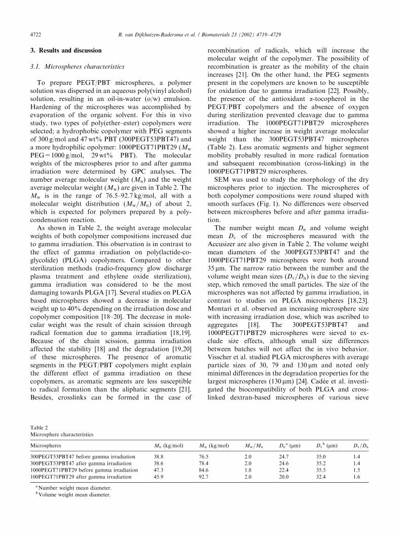
PEG=1000 g/mol, 29wt% PBT). The molecularweights of the microspheres prior to and after gammairradiation were determined by GPC analyses. Thenumber average molecular weight (Mn) and the weightaverage molecular weight (Mw) are given in Table 2. TheMw is in the range of 76.5–92.7 kg/mol, all with amolecular weight distribution (Mw=Mn) of about 2,which is expected for polymers prepared by a poly-condensation reaction.As shown in Table 2, the weight average molecular
weights of both copolymer compositions increased dueto gamma irradiation. This observation is in contrast tothe effect of gamma irradiation on poly(lactide-co-glycolide) (PLGA) copolymers. Compared to othersterilization methods (radio-frequency glow dischargeplasma treatment and ethylene oxide sterilization),gamma irradiation was considered to be the mostdamaging towards PLGA [17]. Several studies on PLGAbased microspheres showed a decrease in molecularweight up to 40% depending on the irradiation dose andcopolymer composition [18–20]. The decrease in mole-cular weight was the result of chain scission throughradical formation due to gamma irradiation [18,19].Because of the chain scission, gamma irradiationaffected the stability [18] and the degradation [19,20]of these microspheres. The presence of aromaticsegments in the PEGT/PBT copolymers might explainthe different effect of gamma irradiation on thesecopolymers, as aromatic segments are less susceptibleto radical formation than the aliphatic segments [21].Besides, crosslinks can be formed in the case of
recombination of radicals, which will increase themolecular weight of the copolymer. The possibility ofrecombination is greater as the mobility of the chainincreases [21]. On the other hand, the PEG segmentspresent in the copolymers are known to be susceptiblefor oxidation due to gamma irradiation [22]. Possibly,the presence of the antioxidant a-tocopherol in thePEGT/PBT copolymers and the absence of oxygenduring sterilization prevented cleavage due to gammairradiation. The 1000PEGT71PBT29 microspheresshowed a higher increase in weight average molecularweight than the 300PEGT53PBT47 microspheres(Table 2). Less aromatic segments and higher segmentmobility probably resulted in more radical formationand subsequent recombination (cross-linking) in the1000PEGT71PBT29 microspheres.SEM was used to study the morphology of the dry
microspheres prior to injection. The microspheres ofboth copolymer compositions were round shaped withsmooth surfaces (Fig. 1). No differences were observedbetween microspheres before and after gamma irradia-tion.The number weight mean Dn and volume weight
mean Dv of the microspheres measured with theAccusizer are also given in Table 2. The volume weightmean diameters of the 300PEGT53PBT47 and the1000PEGT71PBT29 microspheres were both around35 mm. The narrow ratio between the number and thevolume weight mean sizes (Dv=Dn) is due to the sievingstep, which removed the small particles. The size of themicrospheres was not affected by gamma irradiation, incontrast to studies on PLGA microspheres [18,23].Montari et al. observed an increasing microsphere sizewith increasing irradiation dose, which was ascribed toaggregates [18]. The 300PEGT53PBT47 and1000PEGT71PBT29 microspheres were sieved to ex-clude size effects, although small size differencesbetween batches will not affect the in vivo behavior.Visscher et al. studied PLGA microspheres with averageparticle sizes of 30, 79 and 130 mm and noted onlyminimal differences in the degradation properties for thelargest microspheres (130 mm) [24]. Cad!ee et al. investi-gated the biocompatibility of both PLGA and cross-linked dextran-based microspheres of various sieve
Table 2
Microsphere characteristics
Microspheres Mn (kg/mol) Mw (kg/mol) Mw=Mn Dna (mm) Dv
b (mm) Dv=Dn
300PEGT53PBT47 before gamma irradiation 38.8 76.5 2.0 24.7 35.0 1.4
300PEGT53PBT47 after gamma irradiation 38.6 78.4 2.0 24.6 35.2 1.4
1000PEGT71PBT29 before gamma irradiation 47.3 84.6 1.8 22.4 35.5 1.5
100PEGT71PBT29 after gamma irradiation 45.9 92.7 2.0 20.0 32.4 1.6
aNumber weight mean diameter.bVolume weight mean diameter.
R. van Dijkhuizen-Radersma et al. / Biomaterials 23 (2002) 4719–47294722

fractions [25]. Only, PLGA particles smaller than 10 mminduced an extensive tissue reaction. Macrophages andforeign body giant cells are known to phagocytozeparticles of this size [2].
3.2. Cytotoxicity
To evaluate the cytotoxicity of the PEGT/PBTmicrospheres, before and after sterilization, extracts ofthese materials were tested towards the growth, mor-phology and metabolism of fibroblasts. In the presenceof an extract of UHMW polyethylene, which served asthe negative control, the cell growth was comparable tothe medium control cultures (maximum negative con-trol). After exposure to the extract for 72 h, an almostconfluent layer of cells was formed and no morpholo-gical changes were observed (grade 0 in Table 1). Incontrast, in the presence of Para rubber, which was usedas a positive control, no confluent layer could beobserved. Lots of cells died or rounded off as a resultof the exposure to the Para rubber extract (grade 4 inTable 1). The confluency of the monolayer and thechange of cellular morphology after exposure to extractsof PEGT/PBT microspheres, before and after gammairradiation, were also scored microscopically. Thesequalitative scores are given in Table 3. For the micro-spheres of both copolymer compositions, before and
after gamma irradiation, no differences with respect tothe monolayer and the cell morphology could beobserved compared to the negative control. Qualitativescores on the cell metabolism were obtained from theMTT test, which indicates the mitochondrial dehydro-genase activity. The fibroblasts exposed to the extract ofthe Para rubber (positive control) revealed a strongMTT reduction (Table 3). The extracts of the micro-spheres, however, showed no inhibition of the cellmetabolism compared to the negative control (UHMWpolyethylene). From both the qualitative and quantita-tive scores, it was concluded that the extracts of themicrospheres demonstrated no cytotoxic reactivity inthis test. The sterilization process by gamma irradiationhad no effect on the cytotoxicity of the microspheres.This in vitro biocompatibility experiment focused on
the initial cytotoxicity, while the effect of degradationproducts on the cytotoxicity will be more pronounced inlong-term evaluations. Van Loon et al. investigatedthe cytotoxicity of PEGT/PBT copolymers by realtime testing [8]. Samples of these copolymers weredegraded in pseudo-extracellular fluid [26] at 371C up to52 weeks. None of the extracts of the tested copolymersshowed any cytotoxicity in cell proliferation tests andMTT tests. If these results can be extrapolated for long-term implantation has been tested in the in vivobiocompatibility study.
Fig. 1. Scanning electron micrographs of sterilized 1000PEGT71PBT29 (A) and 300PEGT53PBT47 (B) microspheres prior to injection.
Table 3
Scores (grades) in cytotoxicity test for microspheres before and after gamma irradiation
Sample Confluency of
monolayer
Change of cell
morphology
Inhibition of cell
metabolism
Mean score
UHMW polyethylene (negative control) 0 0 0 0
300PEGT53PBT47 before gamma irradiation 0 0 0 0
300PEGT53PBT47 after gamma irradiation 0 0 0 0
1000PEGT71PBT29 before gamma irradiation 0 0 0 0
100PEGT71PBT29 after gamma irradiation 0 0 0 0
Para rubber (positive control) 4 4 4 4
R. van Dijkhuizen-Radersma et al. / Biomaterials 23 (2002) 4719–4729 4723
3.3. In vivo biocompatibility
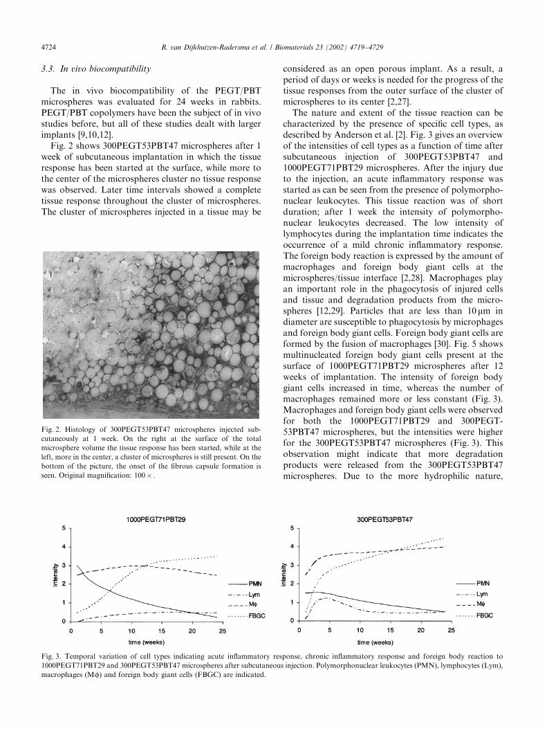
The in vivo biocompatibility of the PEGT/PBTmicrospheres was evaluated for 24 weeks in rabbits.PEGT/PBT copolymers have been the subject of in vivostudies before, but all of these studies dealt with largerimplants [9,10,12].Fig. 2 shows 300PEGT53PBT47 microspheres after 1
week of subcutaneous implantation in which the tissueresponse has been started at the surface, while more tothe center of the microspheres cluster no tissue responsewas observed. Later time intervals showed a completetissue response throughout the cluster of microspheres.The cluster of microspheres injected in a tissue may be
considered as an open porous implant. As a result, aperiod of days or weeks is needed for the progress of thetissue responses from the outer surface of the cluster ofmicrospheres to its center [2,27].The nature and extent of the tissue reaction can be
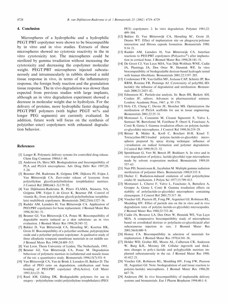
characterized by the presence of specific cell types, asdescribed by Anderson et al. [2]. Fig. 3 gives an overviewof the intensities of cell types as a function of time aftersubcutaneous injection of 300PEGT53PBT47 and1000PEGT71PBT29 microspheres. After the injury dueto the injection, an acute inflammatory response wasstarted as can be seen from the presence of polymorpho-nuclear leukocytes. This tissue reaction was of shortduration; after 1 week the intensity of polymorpho-nuclear leukocytes decreased. The low intensity oflymphocytes during the implantation time indicates theoccurrence of a mild chronic inflammatory response.The foreign body reaction is expressed by the amount ofmacrophages and foreign body giant cells at themicrospheres/tissue interface [2,28]. Macrophages playan important role in the phagocytosis of injured cellsand tissue and degradation products from the micro-spheres [12,29]. Particles that are less than 10 mm indiameter are susceptible to phagocytosis by microphagesand foreign body giant cells. Foreign body giant cells areformed by the fusion of macrophages [30]. Fig. 5 showsmultinucleated foreign body giant cells present at thesurface of 1000PEGT71PBT29 microspheres after 12weeks of implantation. The intensity of foreign bodygiant cells increased in time, whereas the number ofmacrophages remained more or less constant (Fig. 3).Macrophages and foreign body giant cells were observedfor both the 1000PEGT71PBT29 and 300PEGT-53PBT47 microspheres, but the intensities were higherfor the 300PEGT53PBT47 microspheres (Fig. 3). Thisobservation might indicate that more degradationproducts were released from the 300PEGT53PBT47microspheres. Due to the more hydrophilic nature,
Fig. 2. Histology of 300PEGT53PBT47 microspheres injected sub-
cutaneously at 1 week. On the right at the surface of the total
microsphere volume the tissue response has been started, while at the
left, more in the center, a cluster of microspheres is still present. On the
bottom of the picture, the onset of the fibrous capsule formation is
seen. Original magnification: 100� .
Fig. 3. Temporal variation of cell types indicating acute inflammatory response, chronic inflammatory response and foreign body reaction to
1000PEGT71PBT29 and 300PEGT53PBT47 microspheres after subcutaneous injection. Polymorphonuclear leukocytes (PMN), lymphocytes (Lym),
macrophages (Mf) and foreign body giant cells (FBGC) are indicated.
R. van Dijkhuizen-Radersma et al. / Biomaterials 23 (2002) 4719–47294724

however, faster degradation is expected for the1000PEGT71PBT29 microspheres (next paragraph).More likely, the difference in foreign body giant cellintensity is caused by surface properties [31]. The surfaceof the 1000PEGT71PBT29 microspheres containshydrophilic PEG chains, which prevents protein adsorp-tion [32]. This can also affect the formation of foreignbody giant cell, as this process is surface dependent [2].Besides phagocytosis, macrophages initiate the for-
mation of granulation tissue, which is the scaffold fortissue repair. The granulation tissue response is char-acterized by fibroblast infiltration and the developmentof blood capillaries [30]. If an implant is not phagocy-tosed, the body tends to completely isolate the foreignimplant by forming a fibrous capsule around theimplant [29,32,33]. Fig. 4 shows the granulation tissueresponse as observed after subcutaneous injection of1000PEGT71PBT29 and 300PEGT53PBT47 micro-spheres. The fibroblast infiltration and the neovasular-ization increased in time for both types of microspheres,but the intensities for the 1000PEGT71PBT29 micro-spheres were higher. Fibrous tissue ingrowth and newlyformed blood vessels around 1000PEGT71PBT29 mi-crospheres after 12 weeks are shown in Fig. 5. Fig. 2shows the onset of fibrous capsule formation for the300PEGT53PBT47 microspheres after 1 week of sub-cutaneous implantation. An onset of a fibrous capsulewas observed for both 1000PEGT71PBT29 and300PEGT53PBT47 microspheres. The maximum cap-sule thickness was 5–10 cell layers after 12 weeks, but,surprisingly, decreased to almost no fibrous capsuleafter 24 weeks. Especially for the application of themicrospheres as matrix for drug delivery, the amount offibrous capsule might be of importance. Studies ofAnderson showed that the fibrous capsule influenced therelease behavior of the drug [34]. The in vivo drugrelease could be delayed, or prevented by the fibrouscapsule. Wood et al. isolated fibrous capsules fromsubcutaneously implanted silastic discs for drug perme-
ability experiments [35]. Three model compounds withvarious molecular weights were tested in an in vitrodiffusion cell model. These experiments showed that thefibrous capsule is even permeable for dextran with amolecular weight of 70000 g/mol (permeability:5.6� 10�6 cm/s). Thus, the fibrous capsule around themicrospheres will not necessarily prevent the release ofproteins and peptides, but a delayed release might beobserved.Besides the inflammatory response, the foreign body
reaction and the granulation tissue response, the slidesfor histological analyses were checked for tissue necrosisand changes in tissue morphology (like fatty degenera-tion). These signs of bioincompatibility were notobserved with either the 1000PEGT71PBT29 or the300PEGT53PBT47 microspheres. From these results, it
Fig. 5. Histology of 1000PEGT71PBT29 microspheres (MS) injected
subcutaneously at 12 weeks. Macrophages (M, dark spots) and foreign
body giant (F, dark areas) cells surround the microspheres. Fibroblasts
produced fibrous connective tissue around clusters of microspheres.
Blood capillaries (B) within the fibrous tissue (FT) are seen at the top
of the figure. Original magnification: 200� .
Fig. 4. Granulation tissue response to 1000PEGT71PBT29 and 300PEGT53PBT47 microspheres after subcutaneous injection as function of time.
The presence of fibroblasts (Fib), newly formed blood capillaries (Vas) and the intensity of fibrous capsule (Cap) are indicated.
R. van Dijkhuizen-Radersma et al. / Biomaterials 23 (2002) 4719–4729 4725

was concluded that the PEGT/PBT microspheres werewell tolerated by the surrounding subcutaneous tissue.The results reported above were obtained from
the subcutaneously injected microspheres. To study theeffect of the injection site on the biocompatibility, themicrospheres were also injected intramuscularly. How-ever, evaluation of the intramuscular injection sites wasdifficult as it was hard to retrieve the microspheres fromthe muscular tissue. This might be related to the loweramount injected intramuscularly compared to thesubcutaneously injected amount. Besides, maybe moredegradation or transportation of the microspheres hadoccurred in the muscular tissue [9]. The few samples ofintramuscularly injected microspheres that could beretrieved showed no clear differences in inflammatoryand foreign body response compared to the subcuta-neously injected microspheres. The granulation tissueresponse was less obvious for the intramuscularlyinjected microspheres, as fibrous tissue ingrowth andfibrous capsule were observed to a lesser extent (Fig. 6).Previous studies with PEGT/PBT films showed a similartissue response for intramuscular and subcutaneousimplantation in terms of foreign body reaction, in-growth of fibrous tissue and neovascularization [9].Usually, a more intense tissue reaction is expected formicrospheres, as the surface area is much largercompared to an implant with an identical volume [2].In some cases, however, the opposite effect wasobserved. For example, Cad!ee et al. studied the in vivobiocompatibility of both degradable and nondegradabledextran-based microspheres and discs [25,33]. Forthe discs more exudate and fibrin was observed in thesurrounding tissue compared to the microspheres.
Concerning the tissue reaction observed for themicrospheres in this study, PEGT/PBT copolymershave good potential as matrices for controlled drugdelivery. Repeated injections, however, may enhance theforeign body reaction as was shown for severalbiomaterials by van Luyn [36]. This phenomenon hasto be evaluated for PEGT/PBT matrices. Although thetwo PEGT/PBT copolymer compositions used in thisstudy varied in hydrophilicity and degradation rate, thein vivo biocompatibility study on the two types ofmicrospheres showed only marginal differences in thetissue responses.
3.4. In vitro and in vivo degradation
Histology showed intact microspheres up to 12 weeksof implantation (Figs. 2, 5 and 6). Even after 24 weeks,no fragmented particles have been observed (Fig. 7),although the number of microspheres seemed to bereduced and the amount of fibrous tissue had increased.Histology, however, is hard to use as quantitativemethod for the determination of mass loss. No signsof degradation were observed at the surfaces of the300PEGT53PBT47 microspheres. Closer examination,however, of the 1000PEGT71PBT29 microspheres,surrounded by macrophages and foreign body giantcells, showed that biomaterial–tissue interfaces becameless defined, which may be a sign of resorption (Fig. 8).In previous studies with implants of a similar PEGT/PBT copolymer composition, transmission electronmicroscopy (TEM) showed phagocytes with intracellu-lar polymeric particles [8–10]. In addition, extensivefragmentation of the implants was visible within 4weeks, enlarging the total implant surface [9,10].With increasing PEGT content, the fragmentation ofthe copolymer was more pronounced [10]. Higher
Fig. 7. Scanning electron micrograph of 1000PEGT71PBT29 micro-
spheres injected subcutaneously at 24 weeks.
Fig. 6. Histology of 1000PEGT71PBT29 microspheres injected intra-
muscularly at 12 weeks. Macrophages and foreign body giant cells
surround the microspheres. Muscle cells are seen at the bottom of the
picture. A tiny fibrous capsule is present shown between the muscle
cells and the microspheres. Less fibrous tissue was observed for the
intramuscularly injected microspheres compared to the subcutaneously
injected microspheres (Fig. 5). Original magnification: 200� .
R. van Dijkhuizen-Radersma et al. / Biomaterials 23 (2002) 4719–47294726

fragmentation rate and smaller average particle sizewere observed at the intramuscular implantation site ascompared to the subcutaneous site [9]. This wasattributed to the higher stresses and higher extent oftissue vascularization in muscular tissue. The polymericfragments, however, varied from 300 up to 1200 mm,which is at least 10 times larger than the microspheresinjected in this study. It is, therefore, unlikely that in ourstudy the microspheres will show fragmentation due tomechanical stresses.As described for PLGA based microspheres, degrada-
tion may have started without being visible as fragmen-tation or mass loss [2,24,28,37]. As the polymerdegrades, no loss of integrity of the microspheres maybe observed and the foreign body reaction remainsvisually the same. In a later stage, the decrease inmolecular weight may progress to the point where theintegrity of the microspheres can no longer be main-tained and the microspheres break down into particles(o10 mm), which undergo macrophage phagocytosisand complete degradation [2]. To check this hypothesisfor the PEGT/PBT microspheres, the in vitro degrada-tion in PBS was investigated. Fig. 9 shows the weightaverage molecular weight of the microspheres deter-mined by GPC analyses, as function of time. Themolecular weight of the 1000PEGT71PBT29 micro-spheres decreased from 92.7–37.4 kg/mol in 24 weeks.However, similar to the in vivo situation, SEM showedno signs of fragmentation or mass loss of the micro-spheres. Apparently, the point where the integrity of themicrospheres can no longer be maintained had not beenreached after 24 weeks of degradation. In addition, themild tissue reaction already indicated that a low amountof degradation products had been released. For the
300PEGT53PBT47 microspheres, no significant changein molecular weight could be observed.The degradation mechanisms of poly(ether–ester)s
have been subject of many studies [11,38–40]. Thedegradation pathways of PEGT/PBT that are supposedto occur in vivo are hydrolysis and oxidation [38]. Theester bonds can be hydrolyzed and the ether bonds can beoxidized due to the presence of ions or radicals producedby cells [38]. In our in vitro degradation study on PEGT/PBT microspheres, only the hydrolysis was taken intoaccount. Deschamps et al. studied the effect of radicalson the degradation of PEGT/PBT films in vitro [39].Incubation in a H2O2/CoCl2 solution had a dramaticeffect on the molecular weight and the mechanicalproperties of the samples. Based on these results, the invivo degradation of the microspheres was expected to befaster than the in vitro degradation in PBS.The in vivo degradation of the microspheres was
slower than reported in previous studies with macro-scopic PEGT/PBT implants [9,10]. This might limitthe suitability for repeated injections, in particular forthe release of peptides from hydrophobic matrices likethe 300PEGT53PBT47 matrices. For the sustainedrelease of proteins, however, more hydrophilic PEGT/PBT copolymer compositions are available, which showa faster degradation [10]. The in vivo behavior of thesefaster degrading copolymers is the subject of furtherexperiments. In addition, unloaded microspheres wereevaluated in this study, while for controlled releaseapplications the microspheres will be loaded with adrug. The biological activity of the incorporated drugmay alter the tissue response [2]. In addition, a differentinternal structure due to the presence of the drug mayaffect the degradation behavior as well.
Fig. 9. The weight average molecular weights as determined by GPC
of 1000PEGT71PBT29 (solid line) and 300PEGT53PBT47 (dotted
line) microspheres degraded in PBS versus time.
Fig. 8. Histology of 1000PEGT71PBT29 microspheres injected sub-
cutaneously at 12 weeks. The biomaterial–tissue interface of the
microsphere on the right, surrounded by macrophages and foreign
body giant cells, became less defined, which may be a sign of
resorption. Original magnification: 1000� .
R. van Dijkhuizen-Radersma et al. / Biomaterials 23 (2002) 4719–4729 4727
4. Conclusion
Microspheres of a hydrophobic and a hydrophilicPEGT/PBT copolymer were shown to be biocompatibleby in vitro and in vivo studies. Extracts of thesemicrospheres showed no cytotoxic reactivity in the invitro cytotoxicity test. The microspheres could besterilized by gamma irradiation without increasing thecytotoxicity and decreasing the copolymer molecularweight. PEGT/PBT microspheres injected subcuta-neously and intramuscularly in rabbits showed a mildtissue response in vivo, in terms of the inflammatoryresponse, the foreign body reaction and the granulationtissue response. The in vivo degradation was slower thanexpected from previous studies with large implants,although an in vitro degradation experiment showed adecrease in molecular weight due to hydrolysis. For thedelivery of proteins, more hydrophilic faster degradingPEGT/PBT polymers (with higher PEGT wt% andlonger PEG segments) are currently evaluated. Inaddition, future work will focus on the synthesis ofpoly(ether–ester) copolymers with enhanced degrada-tion behavior.
References
[1] Langer R. Polymeric delivery systems for controlled drug release.
Chem Eng Commun 1980;6:1–48.
[2] Anderson JA, Shive MS. Biodegradation and biocompatibility of
PLA and PLGA microspheres. Adv Drug Deliv Rev 1997;28:
5–24.
[3] Bezemer JM, Radersma R, Grijpma DW, Dijkstra PJ, Feijen J,
Van Blitterswijk CA. Zero-order release of lysozyme from
poly(ethylene glycol)/poly(buthylene terephthalate) matrices.
J Control Rel 2000;64(1–3):179–92.
[4] Van Dijkhuizen-Radersma R, P!eters FLAMA, Stienstra NA,
Grijpma DW, Feijen J, de Groot K, Bezemer JM. Control of
vitamin B12 from poly(ethylene glycol)/poly(buthylene terephtha-
late) multiblock copolymers. Biomaterials 2002;23(6):1527–36.
[5] Radder AM, Leenders H, Van Blitterswijk CA. Application of
PEO/PBT copolymers for bone replacement. J Biomed Mater Res
1996;30:341–51.
[6] Beumer GJ, Van Blitterswijk CA, Ponec M. Biocompatibility of
degradable matrix induced as a skin substitute: an in vivo
evaluation. J Biomed Mater Res 1994;28:545–52.
[7] Bakker D, Van Blitterswijk CA, Hesseling SC, Koerten HK,
Grote JJ. Biocompatibility of a polyether urethane, polypropylene
oxide and a polyether polyester copolymer. A qualitative study of
three alloplastic tympanic membrane materials in rat middle ear.
J Biomed Mater Res 1990;24:489–515.
[8] Van Loon. Thesis University of Leiden, The Netherlands, 1995.
[9] Beumer GJ, Van Blitterswijk CA, Ponec M. Degradation
behaviour of polymeric matrices in (sub)dermal and muscle tissue
of the rat: a quantitative study. Biomaterials 1994;15(7):551–9.
[10] Van Blitterswijk CA, Van de Brink J, Leenders H, Bakker D. The
effect of PEO ratio on degradation, calcification and bone
bonding of PEO/PBT copolymer (PolyActive). Cell Mater
1993;3(1):23–36.
[11] Reed AM, Gilding DK. Biodegradable polymers for use in
surgery—poly(ethylene oxide) poly(ethylene terephthalate) (PEO/
PET) copolymers: 2. In vitro degradation. Polymer 1981;22:
499–504.
[12] Bakker D, Van Blitterswijk CA, Hesseling SC, Grote JJ,
Deams WT. Effect of implantation site on phagocyte/polymer
interaction and fibrous capsule formation. Biomaterials 1988;
9:14–21.
[13] Radder AM, Leenders H, Van Blitterswijk CA. Interface
reactions to PEO/PBT copolymers (Polyactives) after implanta-
tion in cortical bone. J Biomed Mater Res 1994;28:141–51.
[14] De Groot CJ, Van Luyn MJA, Van Dijk-Wolthuis WNE, Cad!ee
JA, Plantinga JA, Den Otter W, Hennink WE. In vitro
biocompatibility of biodegradable dextran-based hydrogels tested
with human fibroblasts. Biomaterials 2001;22:1197–203.
[15] Cordewener FW, Van Geffen MF, Joziasse CAP, Schmitz JP, Bos
RRM, Rozema FR, Pennings AJ. Cytotoxicity of poly(96L/4D-
lactide): the influence of degradation and sterilization. Biomate-
rials 2000;21:2433–42.
[16] Edmunson IC. Particle-size analysis. In: Bean HS, Beckett AH,
Couless JF, editors. Advances in pharmaceutical sciences.
London: Academic Press, 1967. p. 95–179.
[17] Holy CE, Cheng C, Davies JE, Shoichet MS. Optimization the
sterilization of PLGA scaffolds for use in tissue engineering.
Biomaterials 2001;22:25–31.
[18] Montanari L, Costantini M, Ciranni Signoretti E, Valvo L,
Santucci M, Bartolomei M, Fattibene P, Onori S, Faucitano A,
Conti B, Genta I. Gamma irradiation effects on poly(dl-lactide-
co-glycolide) microspheres. J Control Rel 1998;56:219–29.
[19] Bittner B, M.ader K, Kroll C, Borchert H-H, Kissel T.
Tetracycline-HCl-loaded poly(dl-lactide-co-glycolide) micro-
spheres prepared by spray drying technique: influence of
g-irradiation on radical formation and polymer degradation.
J Control Rel 1999;59:23–32.
[20] Spenlehauer G, Vert M, Benoit JP, Boddaert A. In vitro and in
vivo degradation of poly(d,l lactide/glycolide) type microspheres
made by solvent evaporation method. Biomaterials 1989;10:
557–63.
[21] Nair PD, Sreenivasan K, JayabalanM. Multiple gamma radiation
sterilization of polyester fibers. Biomaterials 1988;9:335–8.
[22] Decker C. Radiation-induced oxidation of solid poly(ethylene
oxide) II: mechanism. J Polym Sci 1977;15:799–813.
[23] Montanari L, Cilurzo F, Valvo L, Faucitano A, Buttafave A,
Groppo A, Genta I, Conti B. Gamma irradiation effects on
stability of poly(lactide-co-glycolide) microspheres containing
clonazepam. J Control Rel 2001;75:317–30.
[24] Visscher GE, Pearson JE, Fong JW, Argentieri GJ, Robinson RL,
Maulding HV. Effect of particle size on the in vitro and in vivo
degradation rates of poly(dl-lactide-co-glycolide) microcapsules.
J Biomed Mater Res 1988;22:733–46.
[25] Cad!ee JA, Brouwer LA, Den Otter W, Hennink WE, Van Luyn
MJA. A comparative biocompatibility study of microspheres
based on crosslinked dextran or poly(lactic-co-glycolic)acid after
subcutaneous injection in rats. J Biomed Mater Res
2001;56(4):600–9.
[26] Homsy CA. Biocompatibility in selection of materials for
implantation. J Biomed Mater Res 1970;4:341–56.
[27] Holder WD, Gruber HE, Moore AL, Culberson CR, Anderson
W, Burg KJL, Mooney DJ. Cellular ingrowth and thick-
ness changes in poly-l-lactide and polyglycolide matrices im-
planted subcutaneously in the rat. J Biomed Mater Res 1998;
41:412–21.
[28] Visscher GE, Robinson RL, Maulding HV, Fong FW, Pearson
JE, Argentieri GJ. Note: biodegradation of and tissue reaction to
poly(dl-lactide) microspheres. J Biomed Mater Res 1986;20:
667–76.
[29] Anderson JM. In vivo biocompatibility of implantable delivery
systems and biomaterials. Eur J Pharm Biopharm 1994;40:1–8.
R. van Dijkhuizen-Radersma et al. / Biomaterials 23 (2002) 4719–47294728

[30] Anderson JM, Langone JJ. Issues and perspectives on the
biocompatibility and immunotoxicity evaluation of implanted
controlled release systems. J Control Rel 1999;57:107–13.
[31] Tabata Y, Ikada Y. Phagocytosis of polymer microspheres by
macrophages. Adv Polym Sci 1990;94:107–41.
[32] Park H, Park K. Biocompatibility issues of implantable drug
delivery systems. Pharm Res 1996;13(12):1770–6.
[33] Cad!ee JA, Van Luyn MJA, Brouwer LA, Plantinga JA, Van
Wachem PB, De Groot CJ, Den Otter W, Hennink WE. In vivo
biocompatibility of dextran-based hydrogels. J Biomed Mater Res
200;50:397–404.
[34] Anderson JM, Niven H, Pelagalli J, Olanoff LS, Jones RD. The role
of the fibrous capsule in the function of implanted drug-polymer
sustained release systems. J Biomed Mater Res 1981;15:889–902.
[35] Wood RC, LeCluyse EL, Fix JA. Assessment of a model for
measuring drug diffusion through implant-generated fibrous
capsule membranes. Biomaterials 1995;16(12):957–9.
[36] Van Luyn MJA, Plantinga JA, Brouwer LA, Khouw IMSL, De
Leij LFMH, Van Wachem PB. Repetitive subcutaneous implan-
tation of different types of (biodegradable) biomaterials alter the
foreign body reaction. Biomaterials 2001;22:1385–91.
[37] Visscher GE, Robison RL, Maulding HV, Fong JW, Pearson JE,
Argentieri GJ. Biodegradation of and tissue reaction to 50:50
poly(dl-lactide-co-glycolide) microcapsules. J Biomed Mater Res
1985;19:349–65.
[38] Goedemoed JH, Hennink WH, Bezemer JM, Feijen J, Van
Blitterswijk CA, De Bruijn JD. Polyetherester copolymers as
drug delivery matrices. European Patent Application 97202533.2,
1998.
[39] Deschamps AA, Claase MB, Sleijster MWJ, De Bruijn JD,
Grijpma DW, Feijen J. Design of segmented poly(ether ester)
materials and structures for tissue engineering of bone. J Control
Rel 2002;78(1–3):175–86.
[40] Bezemer JM, Grijpma DW, Dijkstra PJ, Van Blitterswijk CA,
Feijen J. A controlled release system for proteins based on
poly(ether–ester) block-copolymers: polymer network character-
ization. J Control Rel 1999;62(3):393–405.
R. van Dijkhuizen-Radersma et al. / Biomaterials 23 (2002) 4719–4729 4729