biochar and microbial signaling: production conditions determine effects on microbial communication
TRANSCRIPT

Biochar and Microbial Signaling: Production Conditions DetermineEffects on Microbial CommunicationCaroline A. Masiello,*,† Ye Chen,‡ Xiaodong Gao,† Shirley Liu,‡ Hsiao-Ying Cheng,§
Matthew R. Bennett,‡ Jennifer A. Rudgers,⊥ Daniel S. Wagner,‡ Kyriacos Zygourakis,¶
and Jonathan J. Silberg*,‡,§
†Department of Earth Science, ‡Department of Biochemistry and Cell Biology, §Department of Bioengineering, and ¶Department ofChemical and Biomolecular Engineering, Rice University, 6100 Main Street, Houston, Texas 77005, United States⊥Department of Biology, University of New Mexico, 167 Castetter Hall, Albuquerque, New Mexico 87131, United States
*S Supporting Information
ABSTRACT: Charcoal has a long soil residence time, which has resulted in itsproduction and use as a carbon sequestration technique (biochar). A range ofbiological effects can be triggered by soil biochar that can positively andnegatively influence carbon storage, such as changing the decomposition rate oforganic matter and altering plant biomass production. Sorption of cellular signalshas been hypothesized to underlie some of these effects, but it remains unknownwhether the binding of biochemical signals occurs, and if so, on time scalesrelevant to microbial growth and communication. We examined biochar sorptionof N-3-oxo-dodecanoyl-L-homoserine lactone, an acyl-homoserine lactone(AHL) intercellular signaling molecule used by many gram-negative soilmicrobes to regulate gene expression. We show that wood biochars disruptcommunication within a growing multicellular system that is made up of sendercells that synthesize AHL and receiver cells that express green fluorescent proteinin response to an AHL signal. However, biochar inhibition of AHL-mediated cell−cell communication varied, with the biocharprepared at 700 °C (surface area of 301 m2/g) inhibiting cellular communication 10-fold more than an equivalent mass ofbiochar prepared at 300 °C (surface area of 3 m2/g). These findings provide the first direct evidence that biochars elicit a range ofeffects on gene expression dependent on intercellular signaling, implicating the method of biochar preparation as a parameterthat could be tuned to regulate microbial-dependent soil processes, like nitrogen fixation and pest attack of root crops.
■ INTRODUCTION
Charcoal is a ubiquitous component of the Earth system, presentnot only in the soils of fire-affected ecosystems, but also inmarinesediments and terrestrial and marine dissolved organic carbonpools.1 It also forms the basis for a form of carbon sequestrationcalled soil biochar amendment,2 where charcoal is intentionallyadded to soil with the aim of increasing the size of the stableorganic carbon pool while potentially delivering other agronomicbenefits such as increased crop productivity,1,3−5 decreased soiltensile strength,2,6 altered soil water properties,7−10 improvedplant pest resistance,11 and decreased nitrogen loss from soils.12
The carbon sequestration potential of biochar is based on theassumption that a large fraction of this material is an inertcomponent of the soil organic matter pool with a very long soilresidence time.13 Large-scale data on the biogeochemical cyclingof charcoal back up the idea that it decomposes slowly in theEarth system,14,15 and laboratory and ecosystem studies arenarrowing down the factors that control charcoal soil residencetime.16−20
As commercial applications of biochar have begun, results haveappeared which challenge the inert nature of this material andsuggest more complex carbon cycle roles. For example, biochar
addition to soil has been shown to promote the loss ofnoncharcoal organic matter (priming) in some studies,21 but notin others.22 This priming of soil decomposition causes anenhanced flux of CO2 into the atmosphere from soils, potentiallydecreasing the carbon sequestration benefits of biochar, as well asdecreasing ecosystem services provided by soil organic matter.Other evidence has appeared suggesting that biochar canenhance retention of nitrogen molecules that contribute to soilfertility,23,24 stimulate colonization of roots by mycorrhizalfungi,25,26 change soil microbial composition,27 and confer plantresistance to microbial pathogens.28 These biological effects havebeen proposed to arise in part because biochars sorb diffusiblesmall molecules that soil organisms use for intercellularcommunication and coordinated decision-making.25,29 Biocharsare known to bind diverse organic molecules,30 many of whichare nonpolar like the molecules used for intercellularcommunication. Numerous studies have analyzed the kinetics
Received: April 3, 2013Revised: September 5, 2013Accepted: September 11, 2013
Article
pubs.acs.org/est
© XXXX American Chemical Society A dx.doi.org/10.1021/es401458s | Environ. Sci. Technol. XXXX, XXX, XXX−XXX
of biochar sorption to organics on time scales (days to months)relevant to the mobilization of pollutants.29,31−33 However, itremains unclear if communication can be altered by the presenceof biochars on the time scales of microbial signaling and geneexpression (minutes to hours).It is challenging to directly demonstrate biochar-driven
mechanisms for observed biological effects within the environ-ment because of the complexity and diversity of conversationsoccurring among microbes and plants. Bacteria communicatewith one another using a variety of biochemicals,34 which aredistinct from the molecules used by fungi for intraspeciescommunication.35 Plants also synthesize flavinoids that regulatemicrobial behaviors, such as the establishment of root nodules,36
and microbes synthesize nodulation signals37 and planthormones38 that influence plant development and nutrientuptake. The diversity of signals present in the environmentcreates challenges in attributing causality to any particularintercellular conservation. The high light absorptivity of biocharand soils adds to this challenge. While there exist microbialbiosensors capable of synthesizing reporters that can be imagedwhen they encounter biological signals within the rhizo-sphere,39,40 fluorescent reporters tend to absorb and emit lightin a visible range that is unsuitable for imaging cells withinbiochar-amended materials, which have high light absorption.To better understand the effects of biochars on cellular
communication, we investigated how biochar materials createdunder different pyrolysis temperatures (300, 350, 400, 450, 550,600, and 700 °C) influence signal detection within a syntheticmicrobial system.41 Because the relevant question is thedetectability of molecular signals by microbes (as opposed tothe chemically extractable fraction of signal present in the soil),we used a microbial sensor to determine when signalingmolecules fell below a level detectable to bacteria in the presenceof different biochars. We focused our attention on N-3-oxo-dodecanoyl-L-homoserine lactone, a member of the acyl-homoserine lactone (AHL) signaling molecule family that areused for intraspecies communication and quorum sensing bymany gram-negative bacteria, including nitrogen fixing plantsymbionts and pathogens that cause soft rot in plants.42,43
■ MATERIALS AND METHODSMaterials. Escherichia coli XL1-Blue were from Stratagene,
and E. coli BLIM cells were kindly provided by K. S. Matthews.44
The acylhomoserine lactone (AHL) used, N-3-oxo-dodecanoyl-L-homoserine lactone, was from Cayman Chemical, bacterialgrowth media components were from BD Biosciences, and allother reagents were from Sigma-Aldrich and VWR.Biochar Synthesis. Slow pyrolysis of Prosopis glandulosa
(mesquite) wood to generate biochar was performed using afixed bed reactor as described previously.7 In brief, mesquitefeedstocks ground to 20 mesh (<0.853 mm) were placed in astainless steel crucible, which was then plugged with ceramicwool, capped with a ceramic bowl, and buried in fine-grainedquartz sand inside a larger, open-top stainless steel crucible. Thisreactor systemwas heated in amuffle furnace at 5 °Cmin−1 to thedesired reaction temperatures (300, 350, 400, 450, 550, 600, and700 °C) and held at each temperature for 4 h. The products weremanually mixed after cooling to increase homogeneity.Surface Area Measurements. We measured the biochar
surface area using a Quantachrome Autosorb-3b SurfaceAnalyzer. Prior to analysis, samples were placed in ashed (550°C for 4 h) glass cells and vacuum-dried overnight at 200 °C.Nitrogen adsorption/desorption isotherms were obtained at 77
K by a 26-point analysis for relative pressures P/P0 from 1.21 ×10⁻⁴ to 0.99, where P is the adsorption equilibrium pressure andP0 is the vapor pressure of bulk liquid N2 at the experimentaltemperature. Specific surface area was calculated usingBrunauer−Emmett−Teller (BET) theory.45
Receiver Plasmids. Receiver plasmids contained thesynthetic PlasR promoter fused to a strong RBS (BBa_B0034)and GFP gene (BBa_E0040) from the IGEM registry. Theseplasmids additionally contained a pMB1 origin from pET28a andeither a chloramphenicol (CmR) or a kanamycin (KanR)selectable marker.
Sender Plasmid. The sender plasmid contained the PA1lacO‑1promoter46 fused to a strong RBS and a gene fusion that wasmade up of the LasI gene (BBa_C0078) fused to the mCherrygene through a (GGGGS)3 peptide linker. This plasmidadditionally contained a pMB1 origin, a CmR marker, and theLacI gene from pET28a. In this sender plasmid, the N-3-oxo-dodecanoyl-L-homoserine lactone synthase gene, LasI, isexpressed as a fusion to mCherry through a (GGGGS)3 linkerusing a PA1lacO‑1 promoter, which is constitutively active in cellsthat lack the repressor LacI, such as E. coli BLIM cells.44 Thefunction of this fusion protein is indistinguishable from LasI.
Effect of AHL on GFP Expression in Liquid Culture. E.coli XL1 Blue harboring the receiver plasmid (CmR) were grownto stationary phase in LB medium containing 34 μg/mLchloramphenicol, diluted to an A600 = 0.05 in 50% LB mediumthat contained varying concentrations of AHL (0, 5, 25, 50, 125,250, 500, 1000, and 2000 nM), and grown to stationary phase at30 °C. GFP levels were determined by measuring whole cellfluorescence emission (509 nm) upon excitation of 488 nm usinga Tecan M1000 plate reader. Fluorescence was normalized toculture absorbance at 600 nm to account for variability in growth.
Effect of Biochar on AHL Availability in Water. Varyingconcentrations of each biochar (1, 5, 10, 25, and 50 mg/mL)prepared at different temperatures (300, 350, 400, 450, 550, 600,and 700 °C) were incubated with 1 μM AHL within water for 6,60, or 1440 min. After incubation, biochar was removed throughcentrifugation (14 000g for 1 min), and 100 μL of the solublefraction was mixed with an equal volume of LB mediumcontaining E. coli transformed with the CmR receiver plasmid atan OD600 = 0.05 and 50 μg/mL chloramphenicol. This mixturewas grown for 18 h within 96-well microtiter plates incubated at30 °C and shaken at 250 rpm. Green cellular fluorescence (λex =488; λem = 509) and absorbance (600 nm) were measured using200 μL of each culture after growing cells in a shaking incubator(30 °C; 250 rpm) for 18 h. To account for variation in celldensity, fluorescence to absorbance ratio was calculated for eachwell, and the calculated values were normalized to those observedwith cells that were grown in the absence of AHL and in thepresence of AHL that was not incubated with a biochar.
Effect of Colony Separation on Intercellular Signaling.E. coli XL1 Blue harboring the KanR receiver plasmid and E. coliBLIM transformed with the CmR sender plasmid were grown tostationary phase in LB containing 50 μg/mL kanamycin and 50μg/mL chloramphenicol, respectively. Bacteria (1 mL) wereharvested by centrifugation, washed with 1 mL of 25% glycerol,resuspended in 1 mL of 25% glycerol, and diluted 10-fold in 25%glycerol. Resuspended receiver (8 μL) and sender cells (3 μL)were spotted at different distances (1, 10, 20, and 30 mm) fromone another on 1.5% agar plates containing M9minimal mediumand 20 amino acids. A smaller volume of the sender strain wasspotted to avoid growth of these cells over the biochar agar slabsduring the overnight incubation. Spotting was performed in
Environmental Science & Technology Article
dx.doi.org/10.1021/es401458s | Environ. Sci. Technol. XXXX, XXX, XXX−XXXB

triplicate at each separation distance. After spotting, plates wereincubated overnight at 37 °C and then photographed with aCanonDigital Rebel mounted on a LeicaMZFLIII microscope at0.4× magnification with white light (5 ms exposure), a Chroma41012 GFP long pass filter set (200 and 500ms exposures), and aChroma 11002 Green filter (200 and 500 ms exposures). Theimage analysis software ImageJ was used to quantify thefluorescence from receiver colonies on each agar plate.47
Effect of Biochar on Cell−Cell Communication on SolidMedium. Agar plates for assessing cellular communication wereprepared by placing a mold (sterilized through immersion inethanol) within sterile polystyrene plates (60 × 15 mm) andpouring 8 mL of 1.5% agar containing M9medium and 20 aminoacids. Upon cooling the mold was removed to leave a pair ofempty adjacent wells. The left well (control) on each plate wasfilled with 1 mL of liquid agar (1.5%) dissolved in water (65 °C).The right well (experimental) was then filled with 1 mL of liquidagar containing or lacking 10 mg of 300 or 700 °C biochar. E. coliXL1 Blue harboring the KanR receiver plasmid and E. coli BLIMtransformed with the CmR sender plasmid were grown tostationary phase in LB containing 50 μg/mL kanamycin and 50μg/mL chloramphenicol. Bacteria were harvested by centrifuga-tion, washed with 25% glycerol, resuspended in 25% glycerol,and diluted 10-fold into 25% glycerol. The 10-fold dilution ofsender cells (3 μL) was spotted between the two agar slabs onplates, whereas the 10-fold dilution of receiver cells (8 μL) wasspotted on the outside of each agar slab. After spotting, plateswere incubated overnight at 37 °C and then photographed with aCanon Digital Rebel on a Leica MZFLIII microscope at 0.4×magnification with white light (5 ms exposure), a Chroma 41012GFP long pass filter set (200 and 500 ms exposures), and aChroma 11002 Green filter (200 and 500 ms exposures). ImageJwas used to quantify the fluorescence from the pair of receivercolonies on each agar plate. The ratio of the signals from the pairof receiver colonies on each plate was calculated (receiver
adjacent to biochar/receiver adjacent to empty agar) todetermine the level of signal inhibition elicited by each biochar.All measurements were performed in triplicate, and valuesreported represent the average of three independent experiments±1σ.
Calculating the Effect of Biochar on AHL Diffusion. Toestimate the effect of biochar on AHL diffusion within agar plates,the effective diffusion coefficient De of AHL was calculated as (1− ε)D0/[1 − 0.5 ln(1 − ε)], where D0 is the actual diffusioncoefficient in biochar-free agar medium and ε is the volumefraction occupied by biochar particles in the agar-biochar cube.48
This analysis revealed that the low volume fraction used in ourexperiments (less than 2% v/v biochar) decreases the AHLdiffusion constant by less than 3%. This implicates sorption as themajor mechanism responsible for biochar effects on sender−receiver cell−cell signaling.
■ RESULTS AND DISCUSSIONAutoinducer Sorption to Biochars. To assay whether
biochars influence the availability of AHLs, we constructed abacterial biosensor39 that expresses a green fluorescent protein(GFP) reporter when bacterial cells encounter the AHL N-3-oxo-dodecanoyl-L-homoserine lactone in their local environ-ment. This was accomplished by transforming E. coli with aplasmid that (i) constitutively expresses the protein LasR, aPseudomonas aeruginosa AHL-dependent protein that activatestranscription from the promoter PlasR, and (ii) expresses GFPunder control of the LasR-dependent promoter PlasR. Cellscontaining this receiver plasmid displayed increased GFPproduction when they were grown in medium containing AHL(Supporting Information, Figure S1), because LasR requiresbound AHL to activate transcription of genes whose expression isregulated by the LasR promoter.49 We assayed AHL signaling inE. coli because this organism does not use AHL to communicate,lacks the complex responses of soil bacteria that are sensitive to
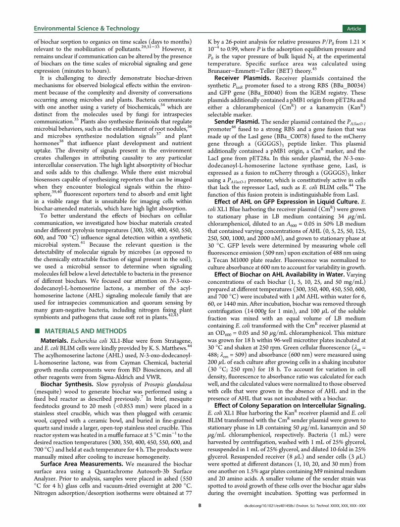
Figure 1. Sorption of AHL by biochars pyrolyzed over a range of temperatures. (A) E. coli transformed with the receiver plasmid synthesize GFP whengrown in an environment containing the AHL N-3-oxo-dodecanoyl-L-homoserine lactone. The transcriptional activator LasR, which is constitutivelyexpressed from this plasmid, turns on transcription of the GFP gene upon binding AHL. (B) Effect of biochar-treated AHL on GFP expression within E.coli harboring the receiver plasmid. Varying concentrations of biochar were incubated with AHL for 1 h prior to adding the soluble fraction to cellsharboring the receiver plasmid. Cells were grown 18 h to allow for AHL-induced GFP expression, which was normalized to the value observed when cellswere grown in the presence of untreated AHL. All measurements were performed in triplicate and are reported as the mean ±1 standard deviation.Biochar is designated BC in the figure.
Environmental Science & Technology Article
dx.doi.org/10.1021/es401458s | Environ. Sci. Technol. XXXX, XXX, XXX−XXXC
one or more AHL, and does not produce lactonase enzymes thathydrolyze AHL. With this bacterial biosensor, only twoparameters determine AHL levels in the medium: the amountof AHL added to the medium and fraction of the AHL that sorbsto biochar.We produced a suite of biochars by pyrolyzing Prosopis
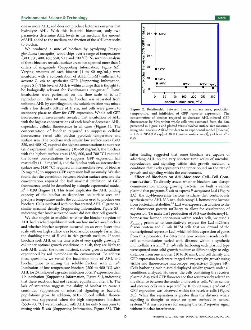
glandulosa (mesquite) wood chips over a range of temperatures(300, 350, 400, 450, 550, 600, and 700 °C). N2 sorption analysisof these biochars revealed surface areas that spanned more than 2orders of magnitude (Supporting Information, Figure S2).Varying amounts of each biochar (1 to 50 mg/mL) wereincubated with a concentration of AHL (1 μM) sufficient toactivate E. coli to synthesize GFP (Supporting Information,Figure S1). This level of AHL is within a range that is thought tobe biologically relevant for Pseudomonas aeruginosa.50 Initialincubations were performed on the time scale of E. colireproduction. After 60 min, the biochar was separated fromunbound AHL by centrifugation, the soluble fraction was mixedwith a low density culture of E. coli, and cells were grown tostationary phase to allow for GFP expression. Whole cell GFPfluorescence measurements revealed that incubation of AHLwith the highest concentrations of each biochar decreased AHL-dependent cellular fluorescence in all cases (Figure 1). Theconcentration of biochar required to suppress cellularfluorescence varied with biochar pyrolysis temperature andsurface area. The biochars with similar low surface areas (300,350, and 400 °C) required the highest concentrations to suppressGFP expression half maximally (10−50 mg/mL), the biocharswith the highest surface areas (550, 600, and 700 °C) requiredthe lowest concentrations to suppress GFP expression halfmaximally (1−2 mg/mL), and the biochar with an intermediatesurface area (450 °C) required an intermediate level of biochar(5 mg/mL) to suppress GFP expression half maximally. We alsofound that the correlation between biochar surface area and theconcentration required for half maximal inhibition of GFPfluorescence could be described by a simple exponential model,R2 = 0.99 (Figure 2). This trend implicates the AHL bindingcapacity of the biochar as dependent on surface area andpyrolysis temperature under the conditions used to produce ourbiochars. Cells incubated with biochar-treated AHL all grew to asimilar maximal density (Supporting Information, Figure S3),indicating that biochar-treated water did not alter cell growth.We also sought to establish whether the biochar sorption of
AHL had reached equilibrium with our low surface area biocharsand whether biochar sorption occurred on an even faster timescale with our high surface area biochars, for example, faster thanthe doubling time of E. coli in rich growth medium. If somebiochars sorb AHL on the time scale of very rapidly growing E.coli under optimal growth conditions in a lab, they are likely tosorb AHL under the lower nutrient, slower growth conditionsexperienced by soil microbes in the environment. To addressthese questions, we varied the incubation time of AHL andbiochar prior to mixing the soluble fraction with E. coli.Incubation of low temperature biochars (300 to 400 °C) withAHL for 24 h showed a greater inhibition of GFP expression than1 h incubation (Supporting Information, Figure S4), indicatingthat these reactions had not reached equilibrium after 1 h. Thelack of saturation suggests the ability of biochar to cause acontinued suppression of microbial signaling as bacterialpopulations grow. In addition, AHL-induced cellular fluores-cence was suppressed when the high temperature biochars(550−700 °C) were incubated with AHL for only 6 min prior tomixing with E. coli (Supporting Information, Figure S5). This
latter finding suggested that some biochars are capable ofadsorbing AHL on the very shortest time scales of microbialreproduction and signaling within rich growth medium, acondition that likely represents the upper bound on the rate ofgrowth and signaling within the environment.
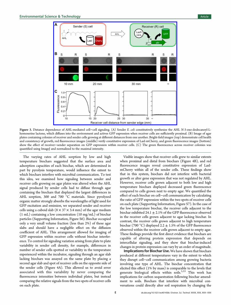
Effect of Biochars on AHL-Mediated Cell−Cell Com-munication. To directly assess whether biochars can disruptcommunication among growing bacteria, we built a senderplasmid that programs E. coli to express P. aeruginosa LasI (Figure3A), the acyl-homoserine lactone synthase that continuouslysynthesizes the AHLN-3-oxo-dodecanoyl-L-homoserine lactonefrom bacterial metabolites.51 LasI was expressed as a fusion to thered fluorescent protein mCherry to allow for visualization ofexpression. To make LasI production of N-3-oxo-dodecanoyl-L-homoserine lactone continuous within sender cells, we used aPA1lacO‑1 promoter to constitutively express the LasI-mCherryfusion protein and E. coli BLIM cells that are devoid of thetranscriptional repressor LacI, which inhibits expression of genesfrom this promoter. To determine how receiver−sender cell−cell communication varied with distance within a syntheticmulticellular system,41 E. coli cells harboring each plasmid typewere spotted onto solid growthmedium at different edge-to-edgedistances from one another (10 to 30 mm), and cell density andGFP expression levels were imaged after overnight growth usingvisible and fluorescence microscopy, respectively (Figure 3B).Cells harboring each plasmid displayed similar growth under allconditions analyzed. However, the cells containing the receiverplasmid displayed GFP fluorescence that was inversely related tothe distance between the sender and receiver cells. When senderand receiver cells were separated by 10 to 20 mm, a gradient ofGFP expression was observed within the receiver cells (Figure3C). While this separation is greater than the distance wheresignaling is thought to occur on plant surfaces in naturalsystems,52 it was necessary for imaging the GFP reporter signalwithout biochar interference.
Figure 2. Relationship between biochar surface area, productiontemperature, and inhibition of GFP reporter expression. Theconcentration of biochar required to decrease AHL-induced GFPfluorescence by 50% within whole cells was estimated from the datapresented in Figure 1 and plotted versus biochar surface area measuredusing BET analysis. A fit of this data to an exponential model, [biochar]= 1.99 + 2061.9 × exp[−1.38 × (biochar surface area)], yields an R2 =0.99.
Environmental Science & Technology Article
dx.doi.org/10.1021/es401458s | Environ. Sci. Technol. XXXX, XXX, XXX−XXXD
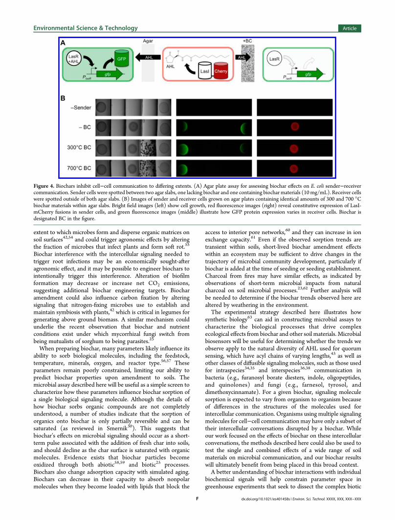
The varying rates of AHL sorption by low and hightemperature biochars suggested that the surface area andadsorption capacities of each biochar, which are determined inpart by pyrolysis temperature, would influence the extent towhich biochars interfere with microbial communication. To testthis idea, we examined how signaling between sender andreceiver cells growing on agar plates was altered when the AHLsignal produced by sender cells had to diffuse through agarcontaining the biochars that displayed the largest differences inAHL sorption, 300 and 700 °C materials. Since pyrolyzedorganic matter strongly absorbs the wavelengths of light used forGFP excitation and emission, we separated sender and receivercells using a cuboid slab (8 × 37 × 3.4 mm) of the agar medium(1 mL) containing a low concentration (10 mg/mL) of biocharparticles (Supporting Information, Figure S6). Biochar occupiedonly a very small volume fraction (less than 2%) of these agarslabs and should have a negligible effect on the diffusioncoefficient of AHL. This arrangement allowed for imaging ofGFP expression within receiver cells without biochar interfer-ence. To control for signaling variation arising from plate to platevariability in sender cell density, for example, differences innumber of sender cells spotted or variability in the temperatureexperienced within the incubator, signaling through an agar slablacking biochars was assayed on the same plate by placing asecond agar slab and spot of receiver cells on the opposite side ofthe sender cells (Figure 4A). This allowed us to avoid errorassociated with this variability by never comparing thefluorescence intensities between individual plates, but insteadcomparing the relative signals from the two spots of receiver cellson each plate.
Visible images show that receiver cells grew to similar extentswhen proximal and distal from biochars (Figure 4B), and redfluorescence images reveal constitutive expression of LasI-mCherry within all of the sender cells. These findings showthat in this system, biochars did not interfere with bacterialgrowth or alter gene expression that was not regulated by AHL.However, receiver cells grown adjacent to both low and hightemperature biochars displayed decreased green fluorescencecompared to cells grown next to empty agar. We quantified theeffect of each biochar on cell−cell communication by calculatingthe ratio of GFP expression within the two spots of receiver cellson each plate (Supporting Information, Figure S7). In the case ofthe low temperature biochar, the receiver cells adjacent to thebiochar exhibited 24.1 ± 2.1% of the GFP fluorescence observedin the receiver cells grown adjacent to agar lacking biochar. Incontrast, the receiver cells grown adjacent to high temperaturebiochar (700 °C) displayed 2.2 ± 1.5% of the GFP fluorescenceobserved within the receiver cells grown adjacent to empty agar.These findings provide the first direct evidence that biochars arecapable of altering protein expression that depends onintercellular signaling, and they show that biochar-inducedchanges in protein expression can vary by an order of magnitude.
Implications for Biochar Use.Wehave shown that biocharsproduced at different temperatures vary in the extent to whichthey disrupt cell−cell communication among growing bacteriainvolving one type of AHL. The biochar concentration thatelicited this effect (1% by mass) is comparable to the levels thatgenerate biological effects within soils.5,53 This work hasimplications for carbon sequestration following biochar amend-ment to soils. Biochars that interfere with microbial con-versations could directly alter soil respiration by changing the
Figure 3. Distance dependence of AHL-mediated cell−cell signaling. (A) Sender E. coli constitutively synthesize the AHL N-3-oxo-dodecanoyl-L-homoserine lactone, which diffuses into the environment and actives GFP expression when receiver cells are sufficiently proximal. (B) Image of agarplates containing colonies of receiver and sender cells growing at different distances from one another. Bright-field images (top) demonstrate cell healthand consistency of growth, red fluorescence images (middle) verify constitutive expression of LasI-mCherry, and green fluorescence images (bottom)show the effect of receiver−sender separation on GFP expression within receiver cells. (C) The green fluorescence across receiver colonies wasquantified using ImageJ and normalized to the maximal intensity.
Environmental Science & Technology Article
dx.doi.org/10.1021/es401458s | Environ. Sci. Technol. XXXX, XXX, XXX−XXXE
extent to which microbes form and disperse organic matrices onsoil surfaces42,54 and could trigger agronomic effects by alteringthe fraction of microbes that infect plants and form soft rot.55
Biochar interference with the intercellular signaling needed totrigger root infections may be an economically sought-afteragronomic effect, and it may be possible to engineer biochars tointentionally trigger this interference. Alteration of biofilmformation may decrease or increase net CO2 emissions,suggesting additional biochar engineering targets. Biocharamendment could also influence carbon fixation by alteringsignaling that nitrogen-fixing microbes use to establish andmaintain symbiosis with plants,42 which is critical in legumes forgenerating above ground biomass. A similar mechanism couldunderlie the recent observation that biochar and nutrientconditions exist under which mycorrhizal fungi switch frombeing mutualists of sorghum to being parasites.25
When preparing biochar, many parameters likely influence itsability to sorb biological molecules, including the feedstock,temperature, minerals, oxygen, and reactor type.56,57 Theseparameters remain poorly constrained, limiting our ability topredict biochar properties upon amendment to soils. Themicrobial assay described here will be useful as a simple screen tocharacterize how these parameters influence biochar sorption ofa single biological signaling molecule. Although the details ofhow biochar sorbs organic compounds are not completelyunderstood, a number of studies indicate that the sorption oforganics onto biochar is only partially reversible and can besaturated (as reviewed in Smernik30). This suggests thatbiochar’s effects on microbial signaling should occur as a short-term pulse associated with the addition of fresh char into soils,and should decline as the char surface is saturated with organicmolecules. Evidence exists that biochar particles becomeoxidized through both abiotic58,59 and biotic25 processes.Biochars also change adsorption capacity with simulated aging.Biochars can decrease in their capacity to absorb nonpolarmolecules when they become loaded with lipids that block the
access to interior pore networks,60 and they can increase in ionexchange capacity.61 Even if the observed sorption trends aretransient within soils, short-lived biochar amendment effectswithin an ecosystem may be sufficient to drive changes in thetrajectory of microbial community development, particularly ifbiochar is added at the time of seeding or seeding establishment.Charcoal from fires may have similar effects, as indicated byobservations of short-term microbial impacts from naturalcharcoal on soil microbial processes.23,62 Further analysis willbe needed to determine if the biochar trends observed here arealtered by weathering in the environment.The experimental strategy described here illustrates how
synthetic biology63 can aid in constructing microbial assays tocharacterize the biological processes that drive complexecological effects from biochar and other soil materials. Microbialbiosensors will be useful for determining whether the trends weobserve apply to the natural diversity of AHL used for quorumsensing, which have acyl chains of varying lengths,43 as well asother classes of diffusible signaling molecules, such as those usedfor intraspecies34,35 and interspecies36,38 communication inbacteria (e.g., furanosyl borate diesters, indole, oligopeptides,and quinolones) and fungi (e.g., farnesol, tyrosol, anddimethoxycinnamate). For a given biochar, signaling moleculesorption is expected to vary from organism to organism becauseof differences in the structures of the molecules used forintercellular communication. Organisms using multiple signalingmolecules for cell−cell communication may have only a subset oftheir intercellular conversations disrupted by a biochar. Whileour work focused on the effects of biochar on these intercellularconversations, the methods described here could also be used totest the single and combined effects of a wide range of soilmaterials on microbial communication, and our biochar resultswill ultimately benefit from being placed in this broad context.A better understanding of biochar interactions with individual
biochemical signals will help constrain parameter space ingreenhouse experiments that seek to dissect the complex biotic
Figure 4. Biochars inhibit cell−cell communication to differing extents. (A) Agar plate assay for assessing biochar effects on E. coli sender−receivercommunication. Sender cells were spotted between two agar slabs, one lacking biochar and one containing biochar materials (10mg/mL). Receiver cellswere spotted outside of both agar slabs. (B) Images of sender and receiver cells grown on agar plates containing identical amounts of 300 and 700 °Cbiochar materials within agar slabs. Bright field images (left) show cell growth, red fluorescence images (right) reveal constitutive expression of LasI-mCherry fusions in sender cells, and green fluorescence images (middle) illustrate how GFP protein expression varies in receiver cells. Biochar isdesignated BC in the figure.
Environmental Science & Technology Article
dx.doi.org/10.1021/es401458s | Environ. Sci. Technol. XXXX, XXX, XXX−XXXF

effects caused by biochar and require significant investments intime and space to achieve replication. Such information will becritical for anticipating the biological effects of soil biocharamendment and making land use decisions that maximizeagricultural productivity and carbon sequestration whileminimizing greenhouse gas emissions.
■ ASSOCIATED CONTENT*S Supporting InformationSeven figures related to the concentration dependence of AHL-induced gene expression, biochar surface areas, cellular growth inthe presence of AHL treated medium, charcoal sorption of AHLafter different incubation times, methodology for creating agarplates, and biochar effects on cellular communication. Thismaterial is available free of charge via the Internet at http://pubs.acs.org.
■ AUTHOR INFORMATIONCorresponding Author*(J.J.S.) Tel.: 713-348-3849. E-mail: [email protected]. (C.A.M)Tel.: 713-348-5234. E-mail: [email protected].
NotesThe authors declare no competing financial interest.
■ ACKNOWLEDGMENTSWe are grateful for financial support from the Hamill Foundationgrant (to C.A.M., J.A.R., J.J.S., and K.Z.), Robert A. WelchFoundation Grants C-1614 (to J.J.S.) and C-1729 (to M.R.B.),National Science Foundation 0911685 (to C.A.M.), NationalInstitute of Health R01GM104974 (to M.R.B.), as part of thejoint NSF/NIGMS Mathematical Biology Program, and TaiwanMinistry of Education Scholarship (to H.Y.C.).
■ REFERENCES(1) Masiello, C. A. New directions in black carbon organicgeochemistry. Mar. Chem. 2004, 92, 201−213.(2) Lehmann, J.; Gaunt, J.; Rondon, M. Bio-char sequestration interrestrial ecosystems−A review.Mitig. Adapt. Strat. Glob. Change 2006,11, 395−419.(3) Major, J.; Rondon, M.; Molina, D.; Riha, S. J.; Lehmann, J. Maizeyield and nutrition during 4 years after biochar application to aColombian savanna oxisol. Plant Soil 2010, 333, 117−128.(4) Zhang, A.; Liu, Y.; Pan, G.; Hussain, Q.; Li, L.; Zheng, J.; Zhang, X.Effect of biochar amendment on maize yield and greenhouse gasemissions from a soil organic carbon poor calcareous loamy soil fromCentral China Plain. Plant Soil 2011, 351, 263−275.(5) Zhang, A.; Bian, R.; Pan, G.; Cui, L.; Hussain, Q.; Li, L. Effects ofbiochar amendment on soil quality, crop yield and greenhouse gasemission in a Chinese rice paddy: A field study of 2 consecutive ricegrowing cycles. Field Crops Res. 2012, 127, 153−160.(6) Chan, K. Y.; Van Zwieten, L.; Meszaros, I.; Downie, A.; Joseph, S.Agronomic values of greenwaste biochar as a soil amendment. Aust. J.Soil Res. 2007, 45, 629.(7) Kinney, T. J.; Masiello, C. A.; Dugan, B.; Hockaday, W. C.; Dean,M. R.; Zygourakis, K.; Barnes, R. T. Hydrologic properties of biocharsproduced at different temperatures. Biomass Bioenergy 2012, 41, 34−43.(8) Briggs, C.; Breiner, J. M.; Graham, R. C. Physical and chemicalproperties of Pinus ponderosa charcoal. Soil Science 2012, 177, 263−268.(9) Novak, J. M.; Busscher, W. J.; Watts, D. W.; Amonette, J. E.;Ippolito, J. A.; Lima, I. M.; Gaskin, J.; Das, K. C.; Stephen; Ahmedna, M.;Rehrah, D.; Schomberg, H. Biochars impact on soil-moisture storage inan ultisol and two aridisols. Soil Science 2012, 177, 310−320.(10) Liu, J.; Schulz, H.; Brandl, S.; Miehtke, H.; Huwe, B.; Glaser, B.Short-term effect of biochar and compost on soil fertility and water
status of a Dystric Cambisol in NE Germany under field conditions. J.Plant Nutr. Soil Sci. 2012, 175, 698−707.(11) Elad, Y.; David, D. R.; Harel, Y. M.; Borenshtein, M.; Kalifa, H. B.;Silber, A.; Graber, E. R. Induction of systemic resistance in plants bybiochar, a soil-applied carbon sequestering agent. Phytopathology 2010,100, 913−921.(12) Ventura, M.; Sorrenti, G.; Panzacchi, P.; George, E.; Tonon, G.Biochar reduces short-term nitrate leaching from a horizon in an appleorchard. J. Environ. Qual. 2013, 42, 76.(13) Skjemstad, J. O.; Spouncer, L. R.; Cowie, B.; Swift, R. S.Calibration of the Rothamsted organic carbon turnover model (RothCver. 26.3), using measurable soil organic carbon pools. Aust. J. Soil Res.2004, 42, 79−88.(14) Masiello, C. A. Black carbon in deep-sea sediments. Science 1998,280, 1911−1913.(15) Ziolkowski, L. A.; Druffel, E. Aged black carbon identified inmarine dissolved organic carbon. Geophys. Res. Lett. 2010, 37, L16601.(16) Kuzyakov, Y.; Subbotina, I.; Chen, H.; Bogomolova, I. Blackcarbon decomposition and incorporation into soil microbial biomassestimated by 14C labeling. Soil Biol. Biochem. 2009, 41, 210−219.(17) Santos, F.; Torn, M. S.; Bird, J. A. Biological degradation ofpyrogenic organic matter in temperate forest soils. Soil Biol. Biochem.2012, 51, 115−124.(18) Singh, N.; Abiven, S.; Torn, M. S.; Schmidt, M. W. I. Fire-derivedorganic carbon in soil turns over on a centennial scale. Biogeosciences2012, 9, 2847−2857.(19) Zimmerman, A. R. Abiotic and microbial oxidation of laboratory-produced black carbon (biochar). Environ. Sci. Technol. 2010, 44, 1295−1301.(20) Schmidt, M. W. I.; Torn, M. S.; Abiven, S.; Dittmar, T.;Guggenberger, G.; Janssens, I. A.; Kleber, M.; Kogel-Knabner, I.;Lehmann, J.; Manning, D. A. C.; Nannipieri, P.; Rasse, D. P.; Weiner, S.;Trumbore, S. E. Persistence of soil organic matter as an ecosystemproperty. Nature 2012, 478, 49−56.(21) Wardle, D. A.; Nilsson, M.-C.; Zackrisson, O. Fire-derivedcharcoal causes loss of forest humus. Science 2008, 320, 629.(22) Zimmerman, A. R.; Gao, B.; Ahn, M.-Y. Positive and negativecarbon mineralization priming effects among a variety of biochar-amended soils. Soil Biol. Biochem. 2011, 43, 1169−1179.(23)Mackenzie, M. D.; DeLuca, T. H. Charcoal and shrubs modify soilprocesses in ponderosa pine forests of western Montana. Plant Soil2006, 287, 257−266.(24) Dempster, D. N.; Gleeson, D. B.; Solaiman, Z. M.; Jones, D. L.;Murphy, D. V. Decreased soil microbial biomass and nitrogenmineralisation with Eucalyptus biochar addition to a coarse texturedsoil. Plant Soil 2011, 354, 311−324.(25) LeCroy, C.; Masiello, C. A.; Rudgers, J. A.; Hockaday, W. C.;Silberg, J. J. Nitrogen, biochar, and mycorrhizae: Alteration of thesymbiosis and oxidation of the char surface. Soil Biol. Biochem. 2013, 58,248−254.(26) Warnock, D. D.; Lehmann, J.; Kuyper, T. W.; Rillig, M. C.Mycorrhizal responses to biochar in soilConcepts and mechanisms.Plant Soil 2007, 300, 9−20.(27) Khodadad, C. L. M.; Zimmerman, A. R.; Green, S. J.; Uthandi, S.;Foster, J. S. Taxa-specific changes in soil microbial communitycomposition induced by pyrogenic carbon amendments. Soil Biol.Biochem. 2011, 43, 385−392.(28) Elad, Y.; Cytryn, E.; Harel, Y. M.; Lew, B.; Graber, E. R. Thebiochar effect: plant resistance to biotic stresses. Phytopathol. Mediter.2012, 50, 335−349.(29) Lehmann, J.; Rillig, M. C.; Thies, J.; Masiello, C. A.; Hockaday, W.C.; Crowley, D. Biochar effects on soil biota − A review. Soil Biol.Biochem. 2011, 43, 1812−1836.(30) Smernik, R. J. Biochar and sorption of organic compounds. InBiochar for Environmental Management: Science and Technology;Lehmann, J., Joseph, S., Eds.; Science: London, 2009; pp 289−300.(31) Spokas, K. A.; Koskinen, W. C.; Baker, J. M.; Reicosky, D. C.Impacts of woodchip biochar additions on greenhouse gas production
Environmental Science & Technology Article
dx.doi.org/10.1021/es401458s | Environ. Sci. Technol. XXXX, XXX, XXX−XXXG
and sorption/degradation of two herbicides in a Minnesota soil.Chemosphere 2009, 77, 574−581.(32) Beesley, L.; Moreno-Jimenez, E.; Gomez-Eyles, J. L. Effects ofbiochar and greenwaste compost amendments on mobility, bioavail-ability and toxicity of inorganic and organic contaminants in a multi-element polluted soil. Environ. Pollut. 2010, 158, 2282−2287.(33) Zheng, W.; Guo, M.; Chow, T.; Bennett, D. N.; Rajagopalan, N.Sorption properties of greenwaste biochar for two triazine pesticides. J.Hazard. Mater. 2010, 181, 121−126.(34) Ng, W.-L.; Bassler, B. L. Bacterial quorum-sensing networkarchitectures. Annu. Rev. Genet. 2009, 43, 197−222.(35) Hogan, D. A. Talking to themselves: Autoregulation and quorumsensing in fungi. Eukaryotic Cell 2006, 5, 613−619.(36) Shaw, L. J.; Morris, P.; Hooker, J. E. Perception and modificationof plant flavonoid signals by rhizosphere microorganisms. Environ.Microbiol. 2006, 8, 1867−1880.(37) Oldroyd, G. E.; Downie, J. A. Coordinating nodule morpho-genesis with rhizobial infection in legumes. Annu. Rev. Plant Biol. 2008,59, 519−546.(38) Lambrecht, M.; Okon, Y.; Vande Broek, A.; Vanderleyden, J.Indole-3-acetic acid: A reciprocal signalling molecule in bacteria-plantinteractions. Trends Microbiol. 2000, 8, 298−300.(39) DeAngelis, K. M.; Firestone, M. K.; Lindow, S. E. Sensitive whole-cell biosensor suitable for detecting a variety of N-acyl homoserinelactones in intact rhizosphere microbial communities. Appl. Environ.Microbiol. 2007, 73, 3724−3727.(40) DeAngelis, K. M.; Lindow, S. E.; Firestone, M. K. Bacterialquorum sensing and nitrogen cycling in rhizosphere soil. FEMSMicrobiol. Ecol. 2008, 66, 197−207.(41) Basu, S.; Gerchman, Y.; Collins, C. H.; Arnold, F. H.; Weiss, R. Asynthetic multicellular system for programmed pattern formation.Nature 2005, 434, 1130−1134.(42) Gonzalez, J. E.; Marketon, M. M. Quorum sensing in nitrogen-fixing rhizobia. Microbiol. Mol. Biol. Rev. 2003, 67, 574−592.(43) Churchill, M. E. A.; Chen, L. Structural basis of acyl-homoserinelactone-dependent signaling. Chem. Rev. 2011, 111, 68−85.(44) Wycuff, D. R.; Matthews, K. S. Generation of an AraC-araBADpromoter-regulated T7 expression system. Anal. Biochem. 2000, 277,67−73.(45) Gregg, S. J.; Sing, K. Adsorption, Surface Area, and Porosity; 2nd ed.Academic Press: London, 1983.(46) Lutz, R.; Bujard, H. Independent and tight regulation oftranscriptional units in Escherichia coli via the LacR/O, the TetR/Oand AraC/I1-I2 regulatory elements. Nucleic Acids Res. 1997, 25, 1203−1210.(47) Schneider, C. A.; Rasband, W. S.; Eliceiri, K. W. NIH Image toImageJ: 25 years of image analysis. Nat. Methods 2012, 9, 671−675.(48) Weissberg, H. L. Effective diffusion coefficient in porous media. J.Appl. Phys. 1963, 34, 2636.(49) Pearson, J. P.; Gray, K. M.; Passador, L.; Tucker, K. D.; Eberhard,A.; Iglewski, B. H.; Greenberg, E. P. Structure of the autoinducerrequired for expression of Pseudomonas aeruginosa virulence genes. Proc.Natl. Acad. Sci. U.S.A. 1994, 91, 197−201.(50) Charlton, T. S.; de Nys, R.; Netting, A.; Kumar, N.; Hentzer, M.;Givskov, M.; Kjelleberg, S. A novel and sensitive method for thequantification of N-3-oxoacyl homoserine lactones using gas chroma-tography−mass spectrometry: application to a model bacterial biofilm.Environ. Microbiol. 2000, 2, 530−541.(51) Passador, L.; Cook, J. M.; Gambello, M. J.; Rust, L.; Iglewski, B. H.Expression of Pseudomonas aeruginosa virulence genes requires cell-to-cell communication. Science 1993, 260, 1127−1130.(52) Decho, A. W.; Frey, R. L.; Ferry, J. L. Chemical challenges tobacterial AHL signaling in the environment. Chem. Rev. 2011, 111, 86−99.(53) Rondon, M. A.; Lehmann, J.; Ramírez, J.; Hurtado, M. Biologicalnitrogen fixation by common beans (Phaseolus vulgaris L.) increases withbio-char additions. Biol. Fertil. Soils 2006, 43, 699−708.
(54) Karatan, E.; Watnick, P. Signals, regulatory networks, andmaterials that build and break bacterial biofilms.Microbiol. Mol. Biol. Rev.2009, 73, 310−347.(55) Dong, Y.-H.; Xu, J.-L.; Li, X.-Z.; Zhang, L.-H. AiiA, an enzyme thatinactivates the acylhomoserine lactone quorum-sensing signal andattenuates the virulence of Erwinia carotovora. Proc. Natl. Acad. Sci.U.S.A. 2000, 97, 3526−3531.(56) Masek, O.; Brownsort, P.; Cross, A.; Sohi, S. Influence ofproduction conditions on the yield and environmental stability ofbiochar. Fuel 2013, 103, 151−155.(57) Lin, Y.; Munroe, P.; Joseph, S.; Ziolkowski, A.; Van Zwieten, L.;Kimber, S.; Rust, J. Chemical and structural analysis of enhancedbiochars: Thermally treated mixtures of biochar, chicken litter, clay andminerals. Chemosphere 2013, 91, 35−40.(58) Nguyen, B. T.; Lehmann, J.; Kinyangi, J.; Smernik, R.; Riha, S. J.;Engelhard, M. H. Long-term black carbon dynamics in cultivated soil.Biogeochemistry 2008, 92, 163−176.(59) Yao, F. X.; Arbestain, M. C.; Virgel, S.; Blanco, F.; Arostegui, J.;Macia-Agullo, J. A.; Macías, F. Simulated geochemical weathering of amineral ash-rich biochar in a modified Soxhlet reactor. Chemosphere2010, 80, 724−732.(60) Kwon, S.; Pignatello, J. J. Effect of natural organic substances onthe surface and adsorptive properties of environmental black carbon(char): Pseudo pore blockage by model lipid components and itsimplications for N2-probed surface properties of natural sorbents.Environ. Sci. Technol. 2005, 39, 7932−7939.(61) Cheng, C.-H.; Lehmann, J.; Engelhard, M. H. Natural oxidation ofblack carbon in soils: Changes in molecular form and surface chargealong a climosequence. Geochim. Cosmochim. Acta 2008, 72, 1598−1610.(62) DeLuca, T. H.; Mackenzie, M. D.; Gundale, M. J.; Holben, W. E.Wildfire-produced charcoal directly influences nitrogen cycling inponderosa pine forests. Soil Sci. Soc. Am. J. 2006, 70, 448−453.(63) Slusarczyk, A. L.; Lin, A.;Weiss, R. Foundations for the design andimplementation of synthetic genetic circuits. Nat. Rev. Genet. 2012, 13,406−420.
Environmental Science & Technology Article
dx.doi.org/10.1021/es401458s | Environ. Sci. Technol. XXXX, XXX, XXX−XXXH