bioactive polysaccharides from marine algae · bioactive polysaccharides from marine algae tamara...
TRANSCRIPT
Available online at www.sciencedirect.com
www.elsevier.com/locate/bcdf
B i o a c t i v e C a r b o h y d r a t e s a n d D i e t a r y F i b r e 4 ( 2 0 1 4 ) 1 2 5 – 1 3 8
http://dx.doi.org/102212-6198/& 2014 El
nCorresponding autE-mail address: b
Bioactive polysaccharides from marine algae
Tamara Barahonaa, Marıa V. Encinasa, Monica Imaraib, Andres Mansillac,Betty Matsuhiroa,n, Rodrigo Torresa, Beatriz Valenzuelab
aDepartamento de Ciencias del Ambiente, Santiago, ChilebDepartamento de Biología, Facultad de Química y Biología, Universidad de Santiago de Chile, Av. L. B. O’Higgins 3363,Santiago, ChilecDepartamento de Ciencias y Recursos Naturales, Facultad de Ciencias, Universidad de Magallanes, Av. Bulnes 1465,Punta Arenas, Chile
a r t i c l e i n f o
Article history:
Received 13 June 2014
Received in revised form
5 September 2014
Accepted 10 September 2014
Keywords:
Fucoidans
Laminaran
Sulfated galactans
Oversulfation
Antioxidant
Anticoagulant
.1016/j.bcdf.2014.09.002sevier Ltd. All rights rese
hor. Tel.: þ56 2 [email protected]
a b s t r a c t
Laminaran, fucoidan and sulfated galactan were extracted from the seaweeds Desmarestia
distans, Lessonia vadosa, and Gigartina skottsbergii, respectively. Modified polysaccharides
were prepared by desulfation, depolymerization, and sulfation. Antioxidant, anticoagulant
and immunostimulating activities of native and modified polysaccharides were assayed
in vitro. The oversulfated derivatives showed high antioxidant capacity towards oxygen
radical assay; however, no direct relation between sulfate content and antioxidant capacity
was found. Oversulfated polysaccharides presented higher antioxidant capacity towards
hydroxyl radicals than the native polysaccharide. Regarding the ABTS�þradical cation
assay moderate inhibition values (35.1–3.4%) were observed. The anticoagulant activity of
native and modified polysaccharides was measured using the activated partial thrombo-
plastin time assay; the native sulfated galactan from G. skottsbergii presented the highest
value, close to that shown by heparin at similar concentrations. The immunostimulating
activity of polysaccharides was measured through their effects on bone marrow-derived
mice dendritic cell maturation. The native sulfated galactan from G. skottsbergii presented
good dose-dependent activity inducing increased levels of MHC class II in dendritic cells.
& 2014 Elsevier Ltd. All rights reserved.
rved.
(B. Matsuhiro).
1. Introduction
In the last decades the study of polysaccharides from marine
algae has gained renewed interest for their many valuable
biological properties (Pomin & Mourão, 2010; Wijesekara,
Pangestuti, & Kim, 2011). Brown seaweeds (Phaeophyceae)
mainly produce alginic acid, and to lesser extent fucans and
laminarans. Fucans, the polymers of L-fucose, may also
contain galactose, mannose, xylose and uronic acids and
sulfate groups; the term fucoidan is restricted to sulfated
homo-L-fucans (Patankar, Oehninger, Barnett, Williams, &
Clark, 1993; Chevolot, Mulloy, Ratiskol, Foucault, & Colliec-
Jouault, 2001; Bilan et al., 2002). Fucoidans have received
much attention due to their many biological activities includ-
ing, anticoagulant, antithrombotic, antitumor, antioxidant,
antiviral and anti-inflammatory properties (Berteau &
Mulloy, 2003; Li, Lu, Wei, & Zhao, 2008). Recently, commercial
fucoidan had shown immunomodulatory properties on
human and mice dendritic cells (Kim & Joo, 2008; Yang
et al., 2008b). Laminarans are neutral 1-3-beta-D-glucans

B i o a c t i v e C a r b o h y d r a t e s a n d D i e t a r y F i b r e 4 ( 2 0 1 4 ) 1 2 5 – 1 3 8126
with beta-1,6 branching (Painter, 1983; Rioux, Turgeon, &Beaulieu, 2010). Lai et al. (2010) studied the immunomodula-tory and adjuvant activities of a high molecular weight 1-3-beta-D-glucan fraction isolated from Ganoderma lucidum. Theyfound that in mice this glucan showed an increase in thenumber of dendritic cells and displayed potent adjuvantactivity while in vitro it induced the maturation of dendriticcells and stimulated the production of cytokines and chemo-kines. Agarans and carrageenans are the most commonsulfated galactans from red seaweeds; they differ in theconfiguration of alfa-galactopyranosyl residues and in thepattern of sulfation (Lahaye, 2001; Van de Velde, Knutsen,Usov, Rollema, & Cerezo, 2002; Campo, Kawano, Braz da Silva,& Carvalho, 2009). Various sulfated galactans have beenstudied in search of a relation between sulfate presence andanticoagulant activity (Farías, Valente, Pereira, & Mourão,2000; Pereira, Melo, & Mourão, 2002; Pereira et al., 2005;Fonseca et al., 2008). It seems that there is no correlationbetween coagulation with sulfate content. However, Opoku,Qiu, and Doctor (2006) found that oversulfation of kappa-carrageenan gave a derivative with 30 times higher antic-oagulant activity than the native carrageenan. Moreover, ithas been reported that sulfated polysaccharides from sea-weeds showed in vitro antioxidant capacity (Ruperez,Ahrazem, & Leal, 2002; Kim et al., 2007; Rocha de Souzaet al., 2007; Barahona, Chandía, Encinas, Matsuhiro, & Zúñiga,2011). Recently, Gómez-Ordóñez, Jiménez-Escrig, and Rupérez(2014) reported the antioxidant and anticoagulant activities ofkappa-iota hybrid carrageenans from Mastocarpus stellatus(Rhodophyta). The brown seaweeds Desmarestia distans andLessonia vadosa, and the green variant of tetrasporic Gigartinaskottsbergii (Rhodophyta) grow abundantly in southern Chile,but so far they are not exploited for industrial purposes. Theaim of this work is the study of antioxidant, anticoagulant,and immunostimulating properties of polysaccharidesextracted from these polysaccharides. In order to study therelationship among sulfate content, molecular weight andbiological activity, native polysaccharides were modified byoversulfation, desulfation and partial hydrolysis.
2. Materials and methods
2.1. Materials and general methods
D. distans (C. Agardh) J. Agardh, L. vadosa Searles, and thegreen variant of tetrasporic G. skottsbergii Setchellet Gardnerwere collected in Fuerte Bulnes (53137055 6″S, 70155017 9″W),Magellan region. Specimens were deposited in Sala de Colec-ciones, Departamento de Ciencias y Recursos Naturales,Universidad de Magallanes, Punta Arenas, Chile. Preparationof fucoidan from L. vadosa was performed according toChandía and Matsuhiro (2008). Isolation of sulfated galactansfrom the green variant of tetrasporic G. skottsbergii and theirchemical modifications were previously described (Barahona,Encinas, Mansilla, Matsuhiro, & Zúñiga, 2012). FT-IR spectrain KBr pellets were registered in the 4000–400 cm�1 regionusing a Bruker IFS 66v instrument (Bruker, Coventry,UK) according to Leal, Matsuhiro, Rossi, and Caruso (2008).Absorbance was measured in a Genesys 5 Thermospectronic
spectrophotometer (Thermo Fisher Scientific, Waltham, MA,USA). 1H NMR (400.13 MHz) and 13C (100.62 MHz) spectra ofthe polysaccharides were recorded in D2O, after isotopicexchange (3�0.75 mL) at 70 1C on a Bruker Avance DRX 400spectrometer (Bruker, Coventry, UK) using the sodium salt of3-(trimethylsilyl)-1-propane-d4-sulfonic acid) as internal refer-ence. Gas–liquid chromatography (GC) was carried out on aShimadzu GC-14B chromatograph (Shimadzu, Tokyo, Japan)equipped with a flame ionization detector using a SP 2330column (0.25mm�30m) and performed with an initial 5 minhold at 150 1C and then at 5 1C/min to 210 1C for 10 min. Thehelium flow was 20mL/min. Molecular weight of polysacchar-ides was determined by the reducing end method (Park &Johnson, 1949; Cáceres, Carlucci, Damonte, Matsuhiro, &Zúñiga, 2000). Reagent grade solvents were purchased fromMerck (Darmstadt, Germany); DEAE-Sephadex, Sephadex 100and reagent grade chemicals were purchased from Sigma(St. Louis, MO, USA).
2.2. Extraction of Desmarestia distans
Blades of D. distans were oven dried at 45 1C for 48 h. The dryseaweed (120 g) was milled and stirred for 30 min with 900mLof n-hexane. The supernatant was concentrated in vacuo andthe treatment was repeated four times. The treated seaweedwas dried at room temperature for 48 h and soaked into2000mL of a solution containing 96% ethanol and 36% offormaldehyde in a 4:1 v/v ratio. After 72 h the seaweed wasdecanted and air dried. One hundred grams of the dried algawere stirred with 3% CaCl2 solution at 85 1C for 4 h, and themixture was cooled and centrifuged at 3000� g for 20 min. Thesolid was recovered and the extraction process was repeatedthree more times. The supernatants were dialyzed usingSpectra/Por membrane (MWCO 3500) (Spectrum Laboratories,Rancho Domínguez, CA, USA) against tap water, followed bydistilled water, concentrated in vacuo and freeze-dried (ChristAlpha 1–2 Freeze Dryer, Osterode am Harz, Germany). Theresulting solid was dissolved in 75mL of distilled water, stirredfor 2 h with 1 M HCl (50 mL) and centrifuged. The supernatantwas neutralized with 1 M NaOH solution, dialyzed againstdistilled water, concentrated and freeze-dried.
2.3. Ion-exchange chromatography
The CaCl2 extract from D. distans was dissolved in water(0.2 g/mL solution) and was deposited on a DEAE-SephadexA-50 column (30 cm�3 cm). Elution was carried out with agradient increasing concentrations of NaCl solutions (0.2, 0.4,0.6, 0.8, 1.0, 1.2, 1.4 1.6, 1.8, 2.0, and 2.5 M) and 3 mL fractionswere collected. Elution was monitored by the Dubois method(Chaplin, 1986). Fraction 1 was submitted to total acid hydro-lysis as previously described and the constituent monosac-charides were analyzed by GLC as alditol acetates (Chandíaand Matsuhiro, 2008).
2.4. Partial hydrolysis of fucoidan from Lessonia vadosa
2.4.1. Partial acid hydrolysisNative polysaccharide from L. vadosa (0.500 g) was stirred with50mL of a 0.5 M HCl solution at 60 1C. Aliquots were taken at
B i o a c t i v e C a r b o h y d r a t e s a n d D i e t a r y F i b r e 4 ( 2 0 1 4 ) 1 2 5 – 1 3 8 127
half an hour and then every hour until six hours. The reactionwas stopped by addition of 2 M NaOH until neutrality. Each ofthe aliquots was dialyzed against distilled water for 9 h,exchanging water every hour, concentrated and freeze-dried.Each fraction (3 mg in 1mL of distilled water) was chromato-graphed on a Sephadex G-100 column (30�3 cm) using 0.4 MNaCl solution as eluant. The column was calibrated with BlueDextran 2000, dextran sulfates (500 and 8 kDA, Sigma) andglucose, and 3mL fractions were collected. Elution was mon-itored with the phenol-H2SO4 acid reagent (Chaplin, 1986). Thehydrolysis process was repeated with one gram of fucoidanduring 4 h.
2.4.2. Free radical depolymerizationPartial free radical depolymerization of native polysaccharidefrom Li et al. (2008). After 6 h, the reaction was stopped byaddition of EDTA (500 mg), and the solution was dialyzedusing Spectra/Por membranes (MWCO 1000) against distilledwater for 12 h, exchanging water every one hour. The result-ing solution was concentrated in vacuo and freeze-dried(44.2% yield). The resulting solid was chromatographed on aSephadex G 100 column as in Section 2.4.1.
2.5. Desulfation of the sulfated polysaccharide fromGigartina skottsbergii
Desulfation of the polysaccharide was performed as pre-viously described (Barahona et al., 2012). In brief, the pyridi-nium salt of the polysaccharide was prepared by successivedialysis against pyridinium hydrochloride and then freeze-dried. The product obtained (0.1 g) was dissolved in 40 mL of amixture of anhydrous DMSO–MeOH–pyridine (89:10:1 v/v/v)and maintained at 100 1C for 4 h. After the solution wascooled, distilled water (5 mL) was added and the mixturewas dialyzed against distilled water and then freeze-dried(yield 50.3%).
2.6. Sulfation of polysaccharides
Sulfation was conducted according to Mähner, Lechner, andNordmeier (2001). Briefly, a solution of the polysaccharide(0.200 g) in 20 mL of formamide was added to a solution ofchlorosulfonic acid (0.8 mL) in dry pyridine (4 mL) andrefluxed at 65 1C for 4 h, cooled and poured over ice-waterwith stirring. The resulting solution was added to 150 mL ofcold methanol and the precipitate was separated by centri-fugation, dissolved in distilled water and neutralized with 1 MNaOH, dialyzed against distilled water and freeze-dried.
2.7. Antioxidant capacity assays
2.7.1. Oxygen radical absorbance capacity (ORAC) assayThe consumption of fluorescein associated with its incubationwith AAPH [2,20-azo-bis(2-amidinopropane) dihydrochloride] wasestimated from fluorescence measurements (Alarcón, Campos,Edwards, Lissi, & López-Alarcón, 2008). A reaction mixturecontaining 10mM AAPH with and without the polysaccharidewith different concentrations (0.1–1.0 mg/mL) in distilled waterwas incubated in a phosphate buffer (10mM, pH 7.0) at 37 1C.Fluorescein (1.5 μM) consumption was evaluated from the
decrease in the sample fluorescence intensity (excitation491 nm, emission 512 nm). Fluorescence measurements wereconducted using a Fluorolog-Spex 1681/0.22m spectrofluori-meter (Spex, Metuchen, NJ, USA). Bandwidths of 1.25 nm wereused for excitation and emission slits. Values of the fluorescenceintensity IF in relation to initial value IF
o (IF/IF1) were plotted astime function. Integration of the area under the curve (AUC) wasperformed up to a time such that (IF/IF1) reached a value close tozero. ORAC values were obtained from the ratio of the slope ofthe plots of (AUC–AUCo) versus concentrations of polysaccharideand vitamin C.
2.7.2. Ferrous ion chelating abilityThe method reported by Decker and Welch (1990) was used toinvestigate the ferrous ion chelating ability. The polysacchar-ides in the concentration range of 0.1–2.0 mg/mL were mixedwith 0.1 mL of 2 mM FeCl2 and 0.2 mL of 5 mM ferrozinesolutions. The absorbance as time function was measured at562 nm where the complex of ferrozine- Fe2þ showed strongabsorbance. All absorbance measurements were done usingan HP8453 diode array spectrophotometer (Hewlett Packard,Waldbronn, Germany).
2.7.3. Hydroxyl radical scavenging activity assay (HRS)Hydroxyl radicals were generated from FeSO4 and H2O2
(Fenton reaction) at 20 1C and their antioxidant capacitywas studied using salicylate as probe (Smirnoff & Cumbes,1989). The absorbance at 520 nm of aqueous solutions of20 mM sodium salicylate, 1.5 mM FeSO4, and varying concen-trations of polysaccharide (0–1.5 mg/mL) were measured astime function immediately after the addition of H2O2.
2.7.4. ABTS assayThe ABTS radical cation (ABTS�þ) was produced by the reactionof ABTS (2,20-azinobis(3-ethylbenzothiazoline-6-sulfonic aciddiammonium salt) with 2.45 mM potassium persulfate at roomtemperature for 16 h. The ABTS radical cation solution wasdiluted with PBS (pH 7.0) to an absorbance of 0.70 at 734 nm. Topolysaccharide solutions (0.3 to 20mg/mL), 500 μL of ABTS�þ
was added. Absorbance spectra and the absorbance at 734 nmwere registered in order to measure the kinetics of the reaction.The inhibition of ABTS�þ was calculated as follows: Inhibitionrate (%)¼100� (Abs0�Abs45)/Abs0 where Abs0 was absorbanceof ABTS�þ and Abs45 was the absorbance of the test group at45 min.
2.8. Anticoagulant activity by APTT assay
Human plasma (0.4 mL) from healthy donors was mixed with0.1 mL of polysaccharide samples (1–7�10�3 mg/mL), andheparin (0.2–6.0�10�3 mg/mL) (Claris Lifescience, Ahmeda-bad, India). All samples were incubated with 0.005 mL of STA-Cephascreen solution (DiagnosticaStago Inc., Parsippany, NJ,USA) at 37 1C for 4 min. Then, 0.005 mL of 0.025 M CaCl2solution was added and the clotting time was measured intriplicate in a STA Compact (DiagnosticaStago, Inc., Parsip-pany, NJ, USA) equipment and repeated on three differentdays. Distilled water was used as negative control. Theanticoagulant activity was expressed as sample APTT/nega-tive control APTT (APTTs/APTTNC).
B i o a c t i v e C a r b o h y d r a t e s a n d D i e t a r y F i b r e 4 ( 2 0 1 4 ) 1 2 5 – 1 3 8128
2.9. Immunostimulating assay
2.9.1. Dendritic cellsBone marrow-derived dendritic cells were obtained as pre-viously described by Inaba et al. (1992). Briefly, female miceBALB/C of 8 weeks old were sacrificed by cervical dislocationand femurs and tibias were removed. Bones were placed in70% alcohol, washed with phosphate buffered saline (PBS)and transferred into RPMI-1640 without supplement forbone marrow extraction. Cells were collected by centrifuga-tion and erythrocytes were lyzed with ACK buffer pH 7.2(0.15 M NH4Cl, 10 mM KHCO3, 0.1 mM EDTA). Bone marrowcells (1�106) were placed in RPMI-1640 medium supplemen-ted with 10% SFB, 4 mM de L-Glutamine and 10 mg/mL ofGM-CSF (granulocyte/macrophage colony-stimulating fac-tor). The immature dendritic cells were obtained after 6days of culture at 37 1C with 5% CO2. The medium waschanged on day 2 (with cytokine) and day 4 (withoutcytokine) aspirating 75% of the volume.
2.9.2. Cytotoxicity assayDendritic cells were incubated with the native and modifiedpolysaccharides samples (10–1000 μg/mL) for 6 h at 37 1Cand 5% CO2. After incubation, treated cells were collectedusing a cell scraper and washed and recovered by centri-fugation. Cells were suspended in 0.4 mL of IF (PBS pH7.2and 2% fetal calf serum) and 2 μL of 1 mg/mL propidiumiodide (IP) were added to each sample. Viability was thenexamined using a BD FACSCanto™ II flow cytometer (Bec-ton Dickinson, Franklin Lakes, NJ, USA) and data wereanalyzed using FacsDiva. Analysis was done in triplicateand a sample of dendritic cells incubated with 30% ethanolwas used as positive control.
2.9.3. Immunostimulating activity assayThe assay was conducted after Spisek et al. (2004). Briefly,the native polysaccharides samples and the modified poly-saccharides (10–1000 mg/mL) were incubated with dendriticcells for 6 h at 37 1C and 5% CO2. After incubation, cells wereremoved and centrifuged at 1800�g during 8 min. Thecells were suspended in 0.3 mL of IF containing PE conju-gated anti-mouse CD11c (clone HL3, Pharmingen, 1:300) andFITC conjugated anti-mouse MHC II antibodies (IAd cloneAMS-32.1, Pharmingen, 1:300). The samples were incub-ated at 4 1C for 45 min and then 0.5 mL of IF was added.The resulting mixtures were centrifuged at 1800� g during8 min at room temperature and the pellets were suspen-ded in 0.4 mL of IF and 1–2 mL of IP was added. Sampleswere analyzed by flow cytometry and data were expressedas Mean Fluorescence Intensity (MFI). Analyses were per-formed in triplicate.
2.10. Statistical analysis
The data obtained were means7S.D. of three determinations,and followed by the Student’s t-test. Differences were con-sidered to be statistically significant if Po0.05.
3. Results and discussion
3.1. Extraction, characterization and chemicalmodifications of polysaccharides
Blades of D. distans were pretreated with n-hexane, followed byformaldehyde-ethanol to eliminate lipids and polymerize phe-nolic compounds, respectively. Extraction of D. distans withcalcium chloride afforded in 5.5% yield, a white powder whichwas separated into two fractions by ion-exchange chromato-graphy. Fraction I (32.3% yield) that eluted with distilled watershowed to contain glucose as a sole monosaccharide by totalhydrolysis and GLC analysis. Its IR spectrum presented acharacteristic band at 890 cm�1 assigned to beta-linked gluco-pyranosyl residues; this result was confirmed by NMR spectro-scopy. The 1H NMR spectrum showed a doublet centered at4.80 ppm (J¼8.0 Hz), while the 13C NMR spectrum showed sixsignals at 105.37 (C1), 76.1 (C2), 87.05 (C3), 70.95 (C4), 78.45 (C5),and 63.53 ppm (C6) (figures not shown) which are in goodagreement with data published for 1-3 beta-glucan (Yang &Zhang, 2009). These results confirmed that the structure offraction I corresponds to a laminaran. This neutral polysacchar-ide is not often found in brown seaweeds. Fraction II (7.2%yield) was eluted with a 0.2 M NaCl aqueous solution showed tocontain 19.8% of sulfate group and 7.5% of uronic acids. Totalacid hydrolysis of fraction II and GLC analysis of alditol acetatesindicated that it contained 96.4% of glucose and 3.6% of fucose.These results indicate that this fraction is a mixture of neutralglucan and a fucan. Due to the low yield obtained, fraction IIwas not further studied. Fraction I of CaCl2 extract from D.distans herein after named laminaran, was sulfated withchlorosulfonic acid affording in 92.3% yield, a sulfated poly-saccharide with a glucose:sulfate molar ratio of 1.0:2.1. Its IRspectrum showed the characteristic bands of sulfate groups at1232.9 cm�1, 854 cm�1, 821.7 cm�1, and 582.5 cm�1. Moreover,the 13C NMR spectrum of the sulfated polysaccharide showednew peaks (figure not shown), in relation to the native poly-saccharide, at 72.65 and 72.34 ppm, which could be assigned tosubstitution by sulfate group at O-6 position, and a signal at76.15 ppm which might be due to sulfation at O-4 position.
Extraction of the brown seaweed L. vadosa with CaCl2solution gave a polysaccharide in 15.4% yield. Total acidhydrolysis of the purified extract and GLC analysis of thereduced and acetylated hydrolyzate showed the sole pre-sence of per-O-acetyl-fucitol. The 1H NMR spectrum of theextracted polysaccharide was almost superimposable withthe previously published spectrum of the extract fromL. vadosa, indicating the presence of a fucoidan; spectroscopicanalyses by IR and 2D NMR confirmed that its structurecorresponded to alfa 1-3 fucoidan, mainly sulfated at posi-tion O-4 and partially sulfated at position O-2, as previouslyreported by Chandía and Matsuhiro (2008). This polysacchar-ide was also sulfated affording in 95.2% yield a derivativewhich contained 36.0% of sulfate groups.
The native polysaccharide from L. vadosa was partiallydepolymerized by two methods, an acidic depolymerizationwith 0.5 M HCl solution and a free radical depolymerizationusing Cu2þ as promoter of hydroxyl radicals. ProtonNMR spectra of the polysaccharide and both depolymerized
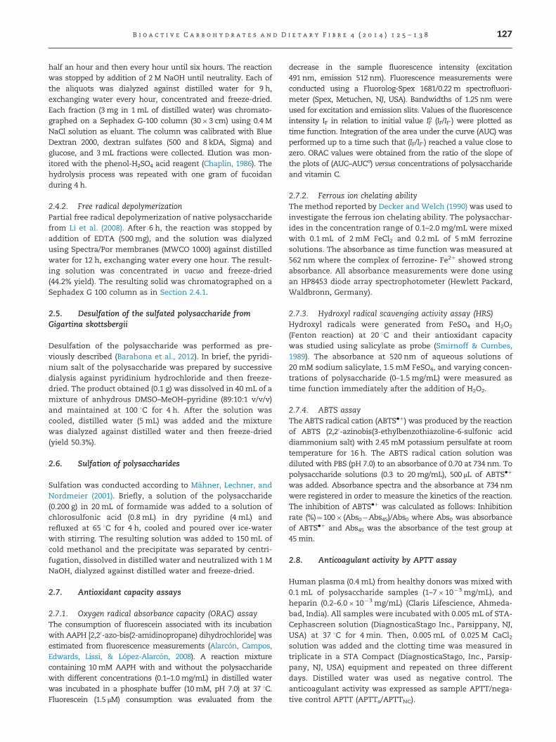
Fig. 1 – 1H NMR (100.62 MHz) spectra in D2O of fucoidan ofLessonia vadosa and its depolymerized fractions. NPL: nativepolysaccharide from Lessonia vadosa, ADPL: aciddepolymerized polysaccharide from L. vadosa, FRPL: freeradical depolymerized polysaccharide from L. vadosa.
B i o a c t i v e C a r b o h y d r a t e s a n d D i e t a r y F i b r e 4 ( 2 0 1 4 ) 1 2 5 – 1 3 8 129
fractions were very similar which indicated that the mainstructural features of native fucoidan was maintained duringthe depolymerization (Fig. 1). The acid partial hydrolysisafforded a homogeneous fraction (MW 6000) after 4 h ofreaction (39.5%, yield), which contained fucose and sulfategroup in the molar ratio 1:0.9. On the other hand, free radicaldepolymerization gave a homogeneous fraction (MW 8000) in44.2% yield; it contained fucose and sulfate group in themolar ratio 1.0:1.2. The higher sulfate content of this fractionin relation to the one obtained by acid hydrolysis may beexplained considering that in the free radical depolymeriza-tion reaction a complex is formed by interaction of Cu2þ withthe anionic sulfate groups of the fucoidan which may preventtheir hydrolysis; moreover, a selective depolymerization of 4-O-sulfate fucopyranosyl residues may take place (Torres,Ferraudi, Chandía, & Matsuhiro, 2011).
We recently reported the preparation and chemical prop-erties of the sulfated galactan from the green variant oftetrasporic G. skottsbergii and its partial acid depolymeriza-tion (Barahona et al., 2012). It was shown that this sulfatedgalactan, although it had a lambda-carrageenan type back-bone, contained lower amount of sulfate groups (25.9%) anda different sulfation pattern compared with commerciallambda-carrageenan. Sulfation with chlorosulfonic acidincreased the sulfate content to 40.1%; however, the IRspectrum is very similar to that of the native polysaccharide,indicating that sulfate groups were not incorporated at newpositions, but rather completed the sulfation at positionsO-2 and O-6 of the galactopyranosyl residues. Besides, thenative sulfated galactan from the green variant of tetraspo-ric G. skottsbergii was desulfated by solvolysis, giving a 50.3%yield of a polysaccharide which by elemental analysis wasshown to contain no sulfur.
3.2. Antioxidant capacity assays
Several methods for evaluating the in vitro antioxidant capa-city are known; generally, they are based on scavenging
capacity assays against specific reactive oxygen and nitrogenspecies or scavenging capacity assays against non-biologicalradicals. According to Niki (2011, 2012) peroxyl radicals (ROO�)are the most suitable radicals for the study of antioxidantssince they are involved in lipid peroxidation as chain-carrying radicals. The oxygen radical absorbance capacity(ORAC) was applied to assess the antioxidant capacity ofhydrophilic samples, such as beverages, tea, and biologicalsamples (Cao, Sofic, & Prior, 1996; López-Alarcón, & Lissi,2006; Omata, Saito, Yoshida, & Niki, 2008; Alarcón et al., 2008;Atala, Vásquez, Speisky, Lissi, & López-Alarcón, 2009; Poblete,López-Alarcón, Lissi, & Campos, 2009; Zulueta, Esteve, &Frígola, 2009). Recently, the ORAC method has been used inour group for evaluating the antioxidant capacity of seaweedpolysaccharides; and in order to establish a comparison,hydroxyl radical scavenging capacity assay (HRS) was alsoevaluated (Barahona et al., 2011, 2012). The hydroxyl radicalsproduced in HRS assay was assessed by the competitivereaction with Brillant Green dye, kinetics of the reaction withseaweed polysaccharides showed complex behavior, indicat-ing a fast consumption of Brillant Green followed by forma-tion of new products. It was suggested that two mechanismsmay be involved in HRS capacity, the suppression againsthydroxyl radical generation by chelation of Feþ2 with sulfategroups of the polysaccharide, and the chemical reaction ofradicals with the polysaccharide. Salicylate was also used asa probe (Smirnoff & Cumbes, 1989). Salicylate in the presenceof Fe2þ forms a complex that absorbs with a maximum at520 nm; absorption decreases in the presence of hydroxylradicals due to the salicylate hydroxylation. In the lastdecades many studies on the antioxidant capacity in vitro ofpolysaccharides using free radical scavenging assays, such asDPPH free radical, have been published (Ruperez et al., 2002;Chen, Xie, Nie, Li, & Wang, 2008; Yang, Zhao, Shi, Yang, &Jiang,2008a; Kong et al., 2010; Zhang, Li, Wu, & Kuang, 2012;Chen et al., 2013). However, DPPH free radical is soluble inorganic solvents and the DPPH assay is not suitable forhydrophilic compounds (Niki, 2011). The long-lived 2,20-azi-nonobis(3-ethylbenzthiazoline-6-sulfonic acid (ABTS) radicalcation, which has to be generated from ABTS is soluble inwater and organic solvents and can be used for the determi-nation of antioxidant capacity of hydrophilic and lipophiliccompounds and biological samples (Niki, 2011).
3.2.1. Oxygen radical absorbance capacity (ORAC) assayThe ORACmethod is based in hydrogen atom abstraction. It hasthe advantage to produce oxygen centered radicals through thedecomposition of an azo-compound (Alarcón et al., 2008;Pereira et al., 2005). The peroxyl radicals scavenging wasmeasured using fluorescein as fluorescent probe that presentsan intensive emission band in the region 480–580 nm. In thepresence of peroxyl radicals the fluorescence decreased andshowed a total consumption at 15 min reaction with a negli-gible inhibition time. The addition of polysaccharides, mea-sured by the integrated area under the fluorescence decay,decreased the reaction rate and then increased the consump-tion time. In all cases the area under the kinetic plots waslinearly related to the amount of the polysaccharide addition.The protective effects of the different polysaccharides, reportedrespect to vitamin C are depicted in Table 1. The ORAC values
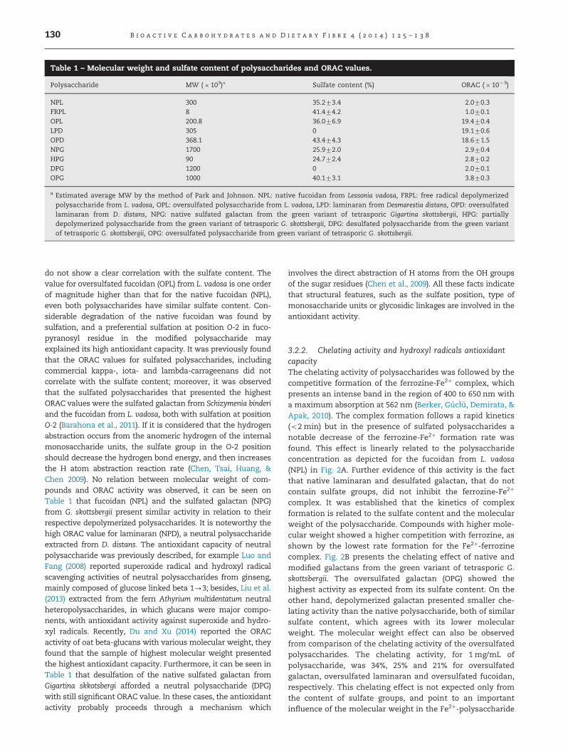
Table 1 – Molecular weight and sulfate content of polysaccharides and ORAC values.
Polysaccharide MW (�103)a Sulfate content (%) ORAC (� 10�3)
NPL 300 35.273.4 2.070.3FRPL 8 41.474.2 1.070.1OPL 200.8 36.076.9 19.470.4LPD 305 0 19.170.6OPD 368.1 43.474.3 18.671.5NPG 1700 25.972.0 2.970.4HPG 90 24.772.4 2.870.2DPG 1200 0 2.070.1OPG 1000 40.173.1 3.870.3
a Estimated average MW by the method of Park and Johnson. NPL: native fucoidan from Lessonia vadosa, FRPL: free radical depolymerizedpolysaccharide from L. vadosa, OPL: oversulfated polysaccharide from L. vadosa, LPD: laminaran from Desmarestia distans, OPD: oversulfatedlaminaran from D. distans, NPG: native sulfated galactan from the green variant of tetrasporic Gigartina skottsbergii, HPG: partiallydepolymerized polysaccharide from the green variant of tetrasporic G. skottsbergii, DPG: desulfated polysaccharide from the green variantof tetrasporic G. skottsbergii, OPG: oversulfated polysaccharide from green variant of tetrasporic G. skottsbergii.
B i o a c t i v e C a r b o h y d r a t e s a n d D i e t a r y F i b r e 4 ( 2 0 1 4 ) 1 2 5 – 1 3 8130
do not show a clear correlation with the sulfate content. Thevalue for oversulfated fucoidan (OPL) from L. vadosa is one orderof magnitude higher than that for the native fucoidan (NPL),even both polysaccharides have similar sulfate content. Con-siderable degradation of the native fucoidan was found bysulfation, and a preferential sulfation at position O-2 in fuco-pyranosyl residue in the modified polysaccharide mayexplained its high antioxidant capacity. It was previously foundthat the ORAC values for sulfated polysaccharides, includingcommercial kappa-, iota- and lambda-carrageenans did notcorrelate with the sulfate content; moreover, it was observedthat the sulfated polysaccharides that presented the highestORAC values were the sulfated galactan from Schizymenia binderiand the fucoidan from L. vadosa, both with sulfation at positionO-2 (Barahona et al., 2011). If it is considered that the hydrogenabstraction occurs from the anomeric hydrogen of the internalmonosaccharide units, the sulfate group in the O-2 positionshould decrease the hydrogen bond energy, and then increasesthe H atom abstraction reaction rate (Chen, Tsai, Huang, &Chen 2009). No relation between molecular weight of com-pounds and ORAC activity was observed, it can be seen onTable 1 that fucoidan (NPL) and the sulfated galactan (NPG)from G. skottsbergii present similar activity in relation to theirrespective depolymerized polysaccharides. It is noteworthy thehigh ORAC value for laminaran (NPD), a neutral polysaccharideextracted from D. distans. The antioxidant capacity of neutralpolysaccharide was previously described, for example Luo andFang (2008) reported superoxide radical and hydroxyl radicalscavenging activities of neutral polysaccharides from ginseng,mainly composed of glucose linked beta 1-3; besides, Liu et al.(2013) extracted from the fern Athyrium multidentatum neutralheteropolysaccharides, in which glucans were major compo-nents, with antioxidant activity against superoxide and hydro-xyl radicals. Recently, Du and Xu (2014) reported the ORACactivity of oat beta-glucans with various molecular weight, theyfound that the sample of highest molecular weight presentedthe highest antioxidant capacity. Furthermore, it can be seen inTable 1 that desulfation of the native sulfated galactan fromGigartina skkotsbergi afforded a neutral polysaccharide (DPG)with still significant ORAC value. In these cases, the antioxidantactivity probably proceeds through a mechanism which
involves the direct abstraction of H atoms from the OH groupsof the sugar residues (Chen et al., 2009). All these facts indicatethat structural features, such as the sulfate position, type ofmonosaccharide units or glycosidic linkages are involved in theantioxidant activity.
3.2.2. Chelating activity and hydroxyl radicals antioxidantcapacityThe chelating activity of polysaccharides was followed by thecompetitive formation of the ferrozine-Fe2þ complex, whichpresents an intense band in the region of 400 to 650 nm witha maximum absorption at 562 nm (Berker, Gúclü, Demirata, &Apak, 2010). The complex formation follows a rapid kinetics(o2 min) but in the presence of sulfated polysaccharides anotable decrease of the ferrozine-Fe2þ formation rate wasfound. This effect is linearly related to the polysaccharideconcentration as depicted for the fucoidan from L. vadosa(NPL) in Fig. 2A. Further evidence of this activity is the factthat native laminaran and desulfated galactan, that do notcontain sulfate groups, did not inhibit the ferrozine-Fe2þ
complex. It was established that the kinetics of complexformation is related to the sulfate content and the molecularweight of the polysaccharide. Compounds with higher mole-cular weight showed a higher competition with ferrozine, asshown by the lowest rate formation for the Fe2þ-ferrozinecomplex. Fig. 2B presents the chelating effect of native andmodified galactans from the green variant of tetrasporic G.skottsbergii. The oversulfated galactan (OPG) showed thehighest activity as expected from its sulfate content. On theother hand, depolymerized galactan presented smaller che-lating activity than the native polysaccharide, both of similarsulfate content, which agrees with its lower molecularweight. The molecular weight effect can also be observedfrom comparison of the chelating activity of the oversulfatedpolysaccharides. The chelating activity, for 1 mg/mL ofpolysaccharide, was 34%, 25% and 21% for oversulfatedgalactan, oversulfated laminaran and oversulfated fucoidan,respectively. This chelating effect is not expected only fromthe content of sulfate groups, and point to an importantinfluence of the molecular weight in the Fe2þ-polysaccharide
0.0
0.2
0.4
0.6
0.8
0 15 30 45 600 15 30 45 600.0
0.2
0.4
0.6
0.8
e
d
ca, b
Abs
orba
nce
(562
nm
)
Time (min)
mg/mL 0 0.25 0.50 1.0 1.5 2.0
Abs
orba
nce
(562
nm
)
Time (min)
Fig. 2 – Effect of polysaccharide addition on the kinetic formation of ferrozine-Fe2þ complex. (A) Effect of fucoidan from Lessoniavadosa concentration. (B) Effect of polysaccharides from the green variant of tetrasporic Gigartina skottsbergii at 0.5 mg/mL:(a) in the absence of polysaccharide, and in the presence of (b) desulfated (DPG), (c) partially acid depolymerized (HPG),(d) native (NPG), (e) oversulfated (OPG). (For interpretation of the references to color in this figure legend, the reader is referredto the web version of this article.)
0 10 20 30 400.0
0.2
0.4
0.6
0.8
ed
cba
Abs
orba
nce
(520
nm
)
Time (min)
4510
20
Fig. 3 – Decrease of the salicylate-Fe2þ complex absorbance in(a) absence of polysaccharide and in the presence ofoversulfated laminaran from D. distans (OPD): (b) 0.125mg/mL,(c) 0.25mg/mL, (d) 0.5 mg/mL, and (e) 1.0 mg/mL. Inset: spectraat different reaction times in the presence of 1mg/mL of OPD.
B i o a c t i v e C a r b o h y d r a t e s a n d D i e t a r y F i b r e 4 ( 2 0 1 4 ) 1 2 5 – 1 3 8 131
chelating activity being more favorable at higher molecularweight.
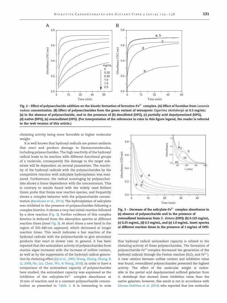
It is well known that hydroxyl radicals are potent oxidantsthat react and produce damage to biomacromolecules,including polysaccharides. The high reactivity of the hydroxylradical leads to its reaction with different functional groupsof a molecule, consequently the damage to the target sub-strate will be dependent on several parameters. The reactiv-ity of the hydroxyl radicals with the polysaccharides by thecompetitive reaction with salicylate hydroxylation was mea-sured. Furthermore, the radical scavenging by polysacchar-ides shows a linear dependence with the concentration. Thisis contrary to results found with the widely used BrillantGreen probe that forms new reactive species, and frequentlyshows a complex behavior with the polysaccharide concen-tration (Barahona et al., 2011). The hydroxylation of salicylatewas inhibited in the presence of polysaccharides following acomplex kinetics. It shows a very fast initial reaction followedby a slow reaction (Fig. 3). Further evidence of this complexkinetics is deduced from the absorption spectra at differentreaction times (inset Fig. 3). At short times a new band in theregion of 325–440 nm appeared, which decreased at longerreaction times. This result indicates a fast reaction of thehydroxyl radicals with the polysaccharide to give secondaryproducts that react at slower rate. In general, it has beenreported that the antioxidant activity of polysaccharides frommarine algae increases with the increase of sulfate content,as well as by the suppression of the hydroxyl radical genera-tion by chelating effect (Qi et al., 2005; Wang, Zhang, Zhang, &Li, 2008; Hu, Liu, Chen, Wu, & Wang, 2010). In order to have acomparison of the antioxidant capacity of polysaccharideshere studied, the antioxidant capacity was expressed as theinhibition of the salicylate-Fe2þ complex absorbance at10 min of reaction and at a constant polysaccharide concen-tration as presented in Table 2. It is interesting to note
that hydroxyl radical antioxidant capacity is related to thechelating activity of these polysaccharides. The formation ofpolysaccharide-Fe2þcomplex decreased the generation of thehydroxyl radicals through the Fenton reaction (H2O2 and Fe2þ).A clear relation between sulfate content and inhibition valuewas found, oversulfated polysaccharides presented the highestactivity. The effect of the molecular weight is notice-able in the partial acid depolymerized sulfated galactan fromG. skottsbergii that showed lower inhibition value than thenative galactan; however, this result is not in accordance withGómez-Ordóñez et al. (2014) who reported that low molecular
400 600 800 1000
0.0
0.5
1.0
min0 0.5545
Abs
orba
nce
λ (nm)
Fig. 4 – Decrease of ABTS�þ radical cation absorbance in thepresence of free radical depolymerized polysaccharide fromLessonia vadosa (FRPL) (2 mg/mL) at different reaction times.Absorption spectra of ABTS�þ: (–) in the absence, and (—) inthe presence of polysaccharide.
Table 2 – Antioxidant capacity of polysaccharidestowards hydroxyl radicals at 10 min reaction. Polysac-charide concentration 1 mg/mL.
Sample Inhibition (%)
NPG 58.676.3OPG 86.074.5HPG 38.071.8OPD 63.073.4NPL 62.872.2OPL 79.172.9FRPL 60.973.2
NPG: native sulfated galactan from the green variant of tetrasporicGigartina skottsbergii, OPG: oversulfated polysaccharide from thegreen variant of tetrasporic G. skottsbergii, HPG: partially aciddepolymerized polysaccharide from the green variant of tetraspo-ric G. skottsbergii, OPD: oversulfated laminaran from Desmarestiadistans, NPL: native fucoidan from Lessonia vadosa, OPL: oversul-fated polysaccharide from L. vadosa, FRPL: free radical depolymer-ized polysaccharide from L. vadosa.
B i o a c t i v e C a r b o h y d r a t e s a n d D i e t a r y F i b r e 4 ( 2 0 1 4 ) 1 2 5 – 1 3 8132
weight polysaccharides, due to their conformation in solutionpresent more potentially available hydroxyl groups reactingwith free radicals. On the contrary, fraction obtained by freeradical depolymerization of fucoidan did not present anincrease in the antioxidant capacity towards hydroxyl radicals;similar results for the native polysaccharide from Lesonia vadosaand its free radical depolymerized fraction against hydroxylradical using Brillant Green as a probe was found (Barahonaet al., 2011). These results indicated that other structuralfeatures such as position of sulfate groups in the fucopyranosylresidues may have an effect on the antioxidant capacity. Inaddition, Yuan, Zhang, Li, Li, and Gao (2005) reported no effectof molecular weight on the antioxidant capacity towardshydroxyl radical of sulfated and acetylated fractions obtainedby mild acid hydrolysis of κ-carrageenan. Comparison of theactivity of the different polysaccharides indicates that thechemical structure of the sugar units also plays a role in theantioxidant capacity. It was previously found that the antiox-idant capacity of commercial λ-carrageenan towards hydroxylradicals also showed a complex kinetics, as measured withBrillant Green as probe. Its activity was higher than thatobserved for the sulfated galactan from the green variant oftetrasporic G. skottsbergii with lower sulfate content (Barahonaet al., 2012). However, to understand the complex reaction ofthe highly reactive hydroxyl radicals it is not enough to carryout the measurements at a fixed point, it is necessary to knowits time dependence. Also, more direct measurements of thehydroxyl radical reaction would be helpful.
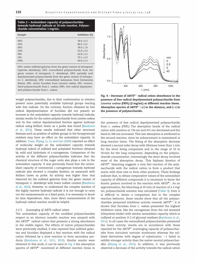
3.2.3. Scavenging of ABTS radical cationThe antioxidant capacity of the modified polysaccharidesrespect to an electron transfer reaction was assayed withthe ABTS�þ radical cation that presents a strong absorbancein the visible region. The effects of native polysaccharideswere previously studied, it was reported that sulfated galac-tan and fucoidan displayed a fast reaction with the radicalcation followed by a slow reaction to form secondary pro-ducts (Barahona et al., 2011, 2012). Similar results wereobtained in this work, it can be seen in Fig. 4 the absorptionspectra of ABTS�þ monitored at different reaction times in
the presence of free radical depolymerized polysaccharidefrom L. vadosa (FRPL) The absorption bands of the radicalcation with maxima at 734 nm and 415 nm decreased and theband at 340 nm increased. This last absorption is attributed tothe second reaction, since its enhancement is maintained atlong reaction times. The fitting of the absorption decreaseshowed a second order decay with lifetimes lower than 1 minfor the short living component and in the range of 10 to50 min for the long component, depending on the polysac-charide concentration. Interestingly the short decay involvedmost of the absorption decay. This biphasic kinetics ofABTS�þ bleaching suggests a very fast reaction of the poly-saccharide with the radical cation to form a product thatreacts with slow rate to form other products. These findingsindicate that, to obtain comparative values of the antioxidantcapacity of different compounds it is necessary to know thekinetic pattern involved in the reaction with ABTS�þ. As anapproximation, the bleaching at 45 min of reaction of a 2 mg/mL polysaccharide solution was calculated (Table 3). Even itis difficult to obtain a comparison due to the complexreaction behavior, these results show that all the polysac-charides presented inhibition activity towards ABTS�þ. It isshown that fucoidan from L. vadosa presented the highestinhibition value, like the carrageenan from the red seaweedSchizymenia binderi with similar antioxidant capacity which issulfated at position O-2 of glycosyl residues (Barahona et al.,2011). In all cases the oversulfated polysaccharides presentedthe lower activity; results are in accordance with thosereported for the ABTS�þ scavenging capacity of polysacchar-ides from Auricularic auricular mushroom whereas the sul-fated derivatives with degree of substitution �1 failed toexhibit stronger activity than the native neutral polysacchar-ides (Zhang et al., 2011). In addition, it was previouslyobserved a decrease of the activity towards the radical cation

Table 3 – Inhibition (%) at 45 min reaction time of ABTS�þby polysaccharides (2 mg/mL).
Samples Inhibition at 45 min (%)
NPG 20.772.3DPG 13.871.4OPG 3.470.6HPG 22.172.2OPD 16.772.2NPL 35.173.9FRPL 24.070.8OPL 19.571.5
NPG: native sulfated galactan from the green variant of tetrasporicGigartina skottsbergii, DPG: desulfated polysaccharide from thegreen variant of tetrasporic G. skottsbergii, OPG: oversulfated poly-saccharide from green variant of tetrasporic G. skottsbergii, HPG:partially acid hydrolyzed polysaccharide from the green variant oftetrasporic G. skottsbergii, OPD: oversulfated laminaran fromDesmarestia distans, NPL: native fucoidan from Lessonia vadosa,FRPL: free radical depolymerized polysaccharide from L. vadosa,OPL: oversulfated polysaccharide from L. vadosa.
B i o a c t i v e C a r b o h y d r a t e s a n d D i e t a r y F i b r e 4 ( 2 0 1 4 ) 1 2 5 – 1 3 8 133
with the increase in the sulfate content for commercial κ-, ι-,and λ-carrageenans (Barahona et al., 2011). Desulfated galac-tan from tetrasporic Gigartina skottsbergi presented significantinhibition of ABTS�þ, with similar values to some of theoversulfated derivatives. It has been reported that neutralpolysaccharides from Dendrobium plants showed high anti-oxidant capacity towards ABTS�þ. Luo, He, Zhou, Fan, andChun (2010) isolated from D. nobile four neutral mannogluco-galactans of MW between 136 and 11.4 kDa, although all thepolysaccharides presented antioxidant capacity, the polysac-charide with MW 11.4 kDa presented high antioxidant capa-city close to vitamin C. It is noteworthy that the authors alsoassayed the scavenging capacity of the polysaccharidestowards DPPH radical, and found that only the polysacchar-ide with the lowest MW showed significant capacity. Luo andFan (2011) reported the antioxidant capacity of D. fimhriatumpolysaccharide (MW 209 kDa) and also found ABTS�þ scaven-ging capacity close to vitamin C, and insufficient antioxidantcapacity towards DPPH radical. On the other hand, Shao,Chen, and Sun (2014) reported the antioxidant capacity ofthree polysaccharides with sulfate content between (19.4–11.4%) isolated from the brown seaweed Sargassum horneri,they found that the fraction with lowest MW (11.2 kDa) andwith lowest content of sulfate groups presented the highestantioxidant capacity against ABTS�þ. However, Gómez-Ordóñez et al. (2014), reported that fractions with the highestMW (1248–1425 kDa) and lowest MW (8–10 kDa) of galactanswith low content of sulfate groups (3.9–1.9%) showed signifi-cant scavenging activity towards ABTS�þ. Altogether, resultsfound in this work and literature data indicate that thereaction of polysaccharides with ABTS�þ is complex, nocorrelation between sulfate content and antioxidant capacitywas found, structural features such as monosaccharide com-position, molecular weight, position of sulfate groups, andglycosidic linkage are involved.
3.3.3. Anticoagulant activityThe anticoagulant activity of native and modified polysacchar-ides was measured using the activated partial thromboplastin
time (APTT) assay. Results are shown in Table 4. It was foundthat the native polysaccharide from the green variant oftetrasporic G. skottsbergii presented the highest value, amongall the samples, close to those shown by heparin at similarconcentrations. This polysaccharide presented sulfation atposition O-2 of 4-linked alfa-D-galactopyranosyl residue and atposition O-2 of 3-linked beta-D-galactopyranosyl residue, and itshowed a molecular weight of �1�106 (Barahona et al., 2012).Partial acid depolymerization of the native polysaccharidedecreased the anticoagulant activity probably due to thedecrease in molecular weight. On the other hand, oversulfationof the native polysaccharide decreased the anticoagulant activ-ity which may indicate that sulfation at other positions in thegalactopyranosyl residues did not play a rol in the interactionwith coagulation cofactors, and produced a deleterious effect.Recently, De Araujo et al. (2013) reported that regioselectiveoversulfation of kappa-, iota- and theta-carrageenan increasedtheir anticoagulant activity determined by the APPT assay; theyfound that sulfation at position O-6 in beta-D-galactopyranosylresidues increased the activity. Pereira et al. (2002) studied theanticoagulant properties of sulfated galactans and fucans frominvertebrates, they found that sulfation at position O-2 in alfa-L-galactans was responsible for the anticoagulant propertieswhereas sulfation at position O-4 in alfa-L-fucans was necessaryfor anticoagulant activity. In relation to anticoagulant proper-ties of fucoidans, it was previously found that the nativefucoidan from L. vadosa showed good anticoagulant activitymeasured by thrombin time (TT) while the free radical depoly-merized fraction (MW 32,000) presented Li et al. (2008). How-ever, it is shown in Table 4 that the free radical depolymerizedfucoidan (FRPL) presented good anticoagulant activity whereasthe partial acid depolymerized fucoidan (ADPL) had loweranticoagulant activity; the latter contained lower amount ofsulfate groups. It is noteworthy that FRPL despite low molecularweight presented anticoagulant activity; its FT-IR spectrum (notshown) was very similar to the native polysaccharide andsuggested mainly the presence of sulfate groups at position O-4 and partially at position O-2 in 3-linked alfa-L-fucopyranosylresidues. Wang, Zhang, Zhang, Song, and Li (2010) reportedgood anticoagulant activity for low molecular weight sulfatedpolysaccharides (MW 4800–7.800) isolated from Laminaria japo-nica; according to chemicals analysis, the polysaccharides weregalactofucans which contained sulfate groups in high amount(30–42%), unfortunately the authors did not report the sulfatepositions in the glycosyl residues. On the other hand, thedesulfated polysaccharide from G. skottsbergii did not showanticoagulant activity in the concentration range of 0.01 to1.44 mg/mL; moreover, oversulfation of sulfated polysacchar-ides from G. skottsbergii and L. vadosa did not increased theiranticoagulant activity. These results agree with those publishedby Wang et al. (2010), and De Araujo et al. (2013) and indicatethat the anticoagulant effect of sulfated polysaccharides wasstereo-specific and independent of the sulfate content.
3.4. Immunostimulating activity
The immunostimulating activity of the native and modifiedpolysaccharides was measured in vitro through their effectson dendritic cell (DC) maturation (Costa et al., 2010). DCs areantigen presenting cells which play a critical role initiating
Table 4 – Anticoagulant activity of native and modified polysaccharidesa.
NPG Concentration(μg/mL)
24 15.2 7.6 4.6 3.0
APTTa,b Máx.c 4.5570.19 3.2370.40 2.4170.24 1.6770.11
NPD Concentration(μg/mL)
640 64
APTT 0.9770.03 1.0470.06
HPG Concentration(μg/mL)
92.4 74 55.4 37 28 4.6
APTT Max. 3.8570.36 2.7570.09 2.2170.04 1.9470.05 1.2770.03
FRPL Concentration(μg/mL)
35 20 10 5 2.5 1 0.5
APTT 3.9370.37 2.3170.50 1.6070.16 1.2770.13 1.1370.04 1.0970.06 1.0470.05
ADPL Concentration(μg/mL)
1430 720 520 260 100 50 10
APTT 2.6870.33 1.8570.09 1.7570.14 1.4670.13 1.2470.01 1.1070.06 1.0370.02
OPG Concentration(μg/mL)
240 102 77 51.2 25.6 12.8
APTT Máx. 3.9770.41 3.3070.46 2.4770.17 1.8970.05 1.5870.02
OPL Concentration(μg/mL)
584 464 290 174 58 46 29 17
APTT 1.5270.24 1.4170.18 1.2670.14 1.1770.07 1.0870.07 1.0370.07 1.0270.04 1.0170.03
OPD Concentration(μg/mL)
680 544 340 204 68 54 34 20
APTT 1.0870.06 1.0770.04 1.0270.05 1.0070.04 1.0170.10 1.0270.04 1.0070.03 1.0070.05
DPG Concentration(μg/mL)
1440
APTT 1.0670.07
Hep Concentration(mg/mL)
6.0 4.9 2.1 0.7 0.4 0.2
APTT 4.2570.66 4.0071.09 1.8770.22 1.3170.10 1.1270.11 1.0270.02
NPG: native polysaccharide from the green variant of tetrasporic G. skottsbergii, NPD: native polysaccharide from D. distans, HPG: partially acidhydrolyzed polysaccharide from the green variant of tetrasporic G. skottsbergii, FRPL: partially free radical depolymerized polysaccharide fromL. vadosa, ADPL: partially acid depolymerized polysaccharide from L. vadosa, OPG:oversulfated polysaccharide from the green variant oftetrasporic G. skottsbergii, OPD: oversulfated polysaccharide from D. distans, OPL: oversulfated polysaccharide from L. vadosa, DPG: desulfatedpolysaccharide from the green variant of tetrasporic G. skottsbergii, Hep:heparin.a APTT is expressed as the quotient between sample APTT and negative control APTT.b Error value is expressed using a P¼0.05.c Clotting time exceeded the equipment maximum value.
B i o a c t i v e C a r b o h y d r a t e s a n d D i e t a r y F i b r e 4 ( 2 0 1 4 ) 1 2 5 – 1 3 8134
primary immune responses. These cells capture and processprotein antigens derived from pathogens in the peripheraltissues and derived peptides are loaded in the major histo-compatibility complex (MHC) class I or class II molecules onthe DC cell surface. DCs thus undergo maturation andmigrate to lymphoid organs where they activate resting Tlymphocytes to initiate antigen-specific immune responses(Bauchereau et al., 2000). Mature dendritic cells have highlevels of major histocompatibility complex (MHC) class mole-cules therefore analysis of expression level of the MHC classII is a good indicator of the induction of DC maturation.
Before determining the immunostimulating activity, thecytotoxicity effects of the polysaccharide samples were mea-sured. All polysaccharide samples presented no cytotoxiceffects in the concentration range of 10–100 μg/mL; moreover,it was remarkable that the native fucoidan from L. vadosa didnot induce cytotoxicity in the 10–1000 μg/mL range. Fig. 5shows the MHC class II levels on the surface of control andtreated dendritic cells examined by flow cytometry. Values
are expressed as mean fluorescence intensity (MFI). Thesulfated galactan from tetrasporic G. skottsbergii presentedgood dose-dependent activity inducing increased levels ofMHC class II in dendritic cells (NPG in Fig. 5). Activity was lostafter increasing sulfate content but also after desulfation(DPG and OPG in Fig. 5) which indicate that it is not thecontent of sulfate groups but the native pattern of sulfationthe structural feature needed for the immunostimulatingactivity. Sulfation recognition patterns may function asmolecular recognition elements for dendritic cells as it hasbeen demonstrated with glycosaminoglycans (Tully et al.,2006). For sulfated galactans, the complex and heterogeneousstructures have precluded a better understanding of struc-tural motifs which are required to disclose the mechanismsof recognition and biological activities. Native fucoidan fromL. vadosa also induced dendritic cell maturation but differencedid not reach statistical significance (NPL in Fig. 5). Theimmunostimulating effect of fucoidan in mouse spleen lym-phocytes was previously studied by Choi, Kim, Kim, and

Fig. 5 – Mean Fluorescence Intensity (MFI) of mono- and polysaccharides samples. NPG: native sulfated galactan from thegreen variant of tetrasporic Gigartina skottsbergii, DPG: desulfated polysaccharide from the green variant of tetrasporicG. skottsbergii, OPG: oversulfated polysaccharide from the green variant of tetrasporic G. skottsbergii, HPG: partially aciddepolymerized polysaccharide from the green variant of tetrasporic G. skottsbergii, LPD: laminaran from Desmarestia distans,NPL: native fucoidan from Lessonia vadosa, FRPL: free radical depolymerized polysaccharide from L. vadosa, ADPL: aciddepolymerized polysaccharide from L. vadosa. (For interpretation of the references to color in this figure legend, the reader isreferred to the web version of this article.)
B i o a c t i v e C a r b o h y d r a t e s a n d D i e t a r y F i b r e 4 ( 2 0 1 4 ) 1 2 5 – 1 3 8 135
Hwang (2005) and in human dendritic cells by Yang et al.(2008b); however, both investigations employed commercialfucoidan, with unknown sulfate content and molecularweight. Depolymerization of fucoidan (FRPL and ADPL inFig. 5) from L. vadosa and sulfated galactan from tetrasporicG. skottsbergii (HPG in Fig. 5) achieved lower molecular weightpolysaccharides devoid of activity; furthermore, neither theconstituent monosaccharides of the sulfated polysacchar-ides, L-fucose and D-galactose, showed activity. Results arein agreement with the model where the complex structure ofpolysaccharides, the structural motifs of sulfation and highmolecular weights are important for immunostimulant activ-ity tested on dendritic cells. Mechanisms of stimulation mayinvolve C-type lectins carbohydrate recognition, a signal that
potentiate cell endocytosis and microbicidal activity andcontribute to innate and adaptive immunity (Sancho & deSousa, 2012).
4. Conclusions
A simple correlation between sulfate content of polysacchar-ides and ORAC antioxidant capacity could not be found;structural features, such as the sulfate position, type ofmonosaccharide units or glycosidic linkages, could beinvolved. The chelating effect of sulfated polysaccharidesagrees not only with the content of sulfate groups, and pointto an important influence of the molecular weight in the

B i o a c t i v e C a r b o h y d r a t e s a n d D i e t a r y F i b r e 4 ( 2 0 1 4 ) 1 2 5 – 1 3 8136
Fe2þ-polysaccharide complex formation being more favorableat higher molecular weight. The antioxidant activity of poly-saccharides towards HO � radicals is related to the chelatingactivity of these compounds.
No relation was found between sulfate content of poly-saccharides and the anticoagulant activity; results indicatedthat it depends on the position of sulfate groups, thestructure of monosaccharide residues and the glycosidiclinkages in the polysaccharides.
Results found in the immunomodulating effect of nativeand modified polysaccharides towards dendritic cells indicatea molecular weight effect over the sulfate content.
Based on the results found in this work, it can beconcluded that native sulfated galactan from the greenvariant of tetrasporic G. skottsbergii is the most suitablepolysaccharide among the native and modified polysacchar-ides here studied, for potential applications in food andpharmaceutical industries.
Acknowledgements
This work was supported by DICYT of Universidad de San-tiago de Chile, and Innova-Chile (Grant 09-MCSS6698). A. M.appreciates the support of Millennium Scientific Initiative(Grant P05-002 ICM) Chile and the Basal Financing Program ofCONICYT (Grant PFB-23), Chile.
r e f e r e n c e s
Alarcon, E., Campos, A. M., Edwards, A. M., Lissi, E., & Lopez-Alarcon, C. (2008). Antioxidant capacity of herbal infusionsand tea extracts: A comparison of ORAC-fluorescein andORAC-pyrogallol red methodologies. Food Chemistry, 107,1114–1119.
Atala, E., Vasquez, L., Speisky, H., Lissi, E., & Lopez-Alarcon, C.(2009). Ascorbic acid contribution to ORAC values in berryextracts. An evaluation by the ORAC-pyrogallol redmethodology. Food Chemistry, 113, 331–335.
Barahona, T., Chandıa, N. P., Encinas, M. V., Matsuhiro, B., &Zuniga, E. A. (2011). Antioxidant capacity of sulfatedpolysaccharides from seaweeds. A kinetic approach. FoodHydrocolloids, 25, 529–535.
Barahona, T., Encinas, M. V., Mansilla, A., Matsuhiro, B., & Zuniga,E. A. (2012). A sulfated galactan with antioxidant capacityfrom the green variant of tetrasporic Gigartina skottsbergii(Gigartinales, Rhodophyta). Carbohydrate Research, 347,114–120.
Bauchereau, J., Briere, F., Caux, C., Davoust, J., Lebecque, S., Liu, Y.-J.,et al. (2000). Immunobiology of dendritic cells. Annual Review ofImmunology, 18, 767–811.
Berker, K. I., Guclu, K., Demirata, B., & Apak, R. (2010). A novelantioxidant assay of ferric reducing capacity measurementusing ferrozine as the colour forming complexation reagent.Analytical Methods, 2, 1770–1778.
Berteau, O., & Mulloy, B. (2003). Sulfated fucans, freshperspectives: Structures, functions, and biological propertiesof sulfated fucans and an overview of enzymes active towardthis class of polysaccharide. Glycobiology, 13, 29R–40R.
Bilan, M. I., Grachev, A. A., Ustuzhanina, N. E., Shaskov, A. S.,Nifantiev, N. E., & Usov, A. I. (2002). Structure of a fucoidanfrom the brown seaweed Fucus evanescens C.Ag. CarbohydrateResearch, 337, 719–730.
Caceres, P. J., Carlucci, M. C., Damonte, E. B., Matsuhiro, B., &Zuniga, E. A. (2000). Carrageenans from Chilean samples ofStenogramme interrupta (Phyllophoraceae): Structural analysisand biological activity. Phytochemistry, 53, 81–86.
Campo, V. L., Kawano, D. F., Braz da Silva, D., & Carvalho, I. (2009).Carrageenans: Biological properties, chemical modificationsand structural analysis—a review. Carbohydrate Polymers, 77,167–180.
Cao, G., Sofic, E., & Prior, R. (1996). Antioxidant capacity of tea andcommon vegetables. Journal of Agricultural and Food Chemistry,44, 3426–3431.
Chandıa, N. P., & Matsuhiro, B. (2008). Characterization of afucoidan from Lessonia vadosa (Phaeophyta) and itsanticoagulant and elicitor properties. International Journal ofBiological Macromolecules, 42, 235–240.
Chaplin, M. F. (1986). Monosaccharides. In M. F. Chaplin, & J. F.Kennedy (Eds.), Carbohydrate analysis: a practical approach (pp. 1–36).Oxford: IRL Press.
Chen, P., Zhang, Y., Zhu, L., Jin, H., Zhang, G., Su, D., et al. (2013).Chemical analysis and antioxidant activity in vitro ofpolysaccharides extracted from lower grade green tea.Advance Journal of Food Science and Technology, 5, 1355–1360.
Chen, S. K., Tsai, M. L., Huang, J. R., & Chen, R. H. (2009). In vitroantioxidant activities of low-molecular-weightpolysaccharides with various functional groups. Journal ofAgriculture and Food Chemistry, 57, 2699–2704.
Chen, Y., Xie, M.-Y., Nie, S.-P., Li, C., & Wang, Y.-X. (2008).Purification, composition analysis and antioxidant activity ofa polysaccharide from the fruiting bodies of Ganoderma atrum.Food Chemistry, 107, 231–241.
Chevolot, L., Mulloy, B., Ratiskol, J., Foucault, A., & Colliec-Jouault,S. (2001). A disaccharide repeat unit is the major structure infucoidans from two species of brown algae. CarbohydrateResearch, 330, 529–535.
Choi, E.-M., Kim, A.–J., Kim, Y.-O., & Hwang, J.-K. (2005).Immunomodulating activity of arabinogalactan and fucoidanin vitro. Journal of Medicinal Food, 8, 446–453.
Costa, L. S., Fidelis, G. P., Cordeiro, S. L., Oliveira, R. M., Sabry,D. A., Camara, R. B. G., et al. (2010). Biological activities ofsulfated polysaccharides from tropical seaweeds. Biomedicine& Pharmacotherapy, 64, 21–28.
De Araujo, C. A., Noseda, M. D., Cipriani, T. R., Goncalves, A. G.,Duarte, M. E. R., & Ducatti, D. R.B (2013). Selective sulfation ofcarrageenans and the influence of sulfate regiochemistry onanticoagulant properties. Carbohydrate Polymers, 91, 483–491.
Decker, E., & Welch, B. (1990). Role of ferritin as a lipid oxidationcatalyst in muscle food. Journal of Agriculture and FoodChemistry, 38, 674–677.
Du, B., & Xu, B. (2014). Oxygen radical absorbance capacity (ORAC)and ferric reducing antioxidant power (FRAP) of β-glucansfrom different sources with various molecular weight.Bioactive Carbohydrates and Dietary Fibre, 3(11-16), 2014.
Farıas, W. R., Valente, A.-P., Pereira, M. S., & Mourao, P. A. S. (2000).Structure and anticoagulant activity of sulfated galactans.Journal of Biological Chemistry, 275, 29299–29307.
Fonseca, R. J. C., Oliveira, S.-N. M. C. G., Melo, F. R., Pereira, M. G.,Benevides, N. M. B., & Mourao, P. A. S. (2008). Slight differencesin sulfation of algal galactans account for differences in theiranticoagulant and venous antithrombotic activities.Thrombosis and Haemostasis, 99, 539–545.
Gomez-Ordonez, E., Jimenez-Escrig, A., & Ruperez, P. (2014).Bioactivity of sulfated polysaccharides from the edible redseaweed Mastocarpus stellatus. Bioactive Carbohydrates andDietary Fibre, 3, 29–40.
Hu, T., Liu, D., Chen, Y., Wu, J., & Wang, S. (2010). Antioxidantactivity of sulfated polysaccharide fractions extracted fromUndaria pinnitafida in vitro. International Journal of BiologicalMacromolecules, 46, 193–198.
B i o a c t i v e C a r b o h y d r a t e s a n d D i e t a r y F i b r e 4 ( 2 0 1 4 ) 1 2 5 – 1 3 8 137
Inaba, K., Inaba, M., Romani, N., Haya, H., Deguchi, M., Ikehara, S.,et al. (1992). Generation of large numbers of dendritic cellsfrom mouse bone marrow cultures supplemented withgranulocyte/macrophage colony-stimulating factor. Journal ofExperimental Medicine, 176, 1693–1702.
Kim, M.-H., & Joo, H.-G. (2008). Immunostimulatory effects offucoidan on bone marrow-derived dendritic cells. ImmunologyLetters, 115, 138–143.
Kim, S.-H., Choi, D.-S., Athukorala, Y., Jeon, Y.-J., Senevirathne, M.,& Rha, C. K. (2007). Antioxidant activity of sulphatedpolysaccharides isolated from Sargassum fulvellum. Journal ofFood Science and Nutrition, 12, 65–73.
Kong, F., Zhang, M., Liao, S., Yu, S., Chi, J., & Wei, Z. (2010).Antioxidant activity of polysaccharide-enriched fractionsextracted from pulp tissue of Litchi Chinensis Sonn. Molecules,15, 2152–2165.
Lahaye, M. (2001). Developments on gelling algal galactans, theirstructure and physico-chemistry. Journal of Applied Phycology,13, 173–184.
Lai, C.-Y., Hung, J.-T., Lin, H.-H., Yu, A. L., Chen, S.-H., Tsai, S.-H.,et al. (2010). Immunomodulatory and adjuvant activities of apolysaccharide extract of Ganoderma lucidum in vivo andin vitro. Vaccine, 28, 4945–4954.
Leal, D., Matsuhiro, B., Rossi, M., & Caruso, F. (2008). FT-IR spectraof alginic acid block fractions in three species of brownseaweeds. Carbohydrate Research, 343, 308–316.
Li, B., Lu, F., Wei, X., & Zhao, R. (2008). Fucoidan: Structure andbioactivity. Molecules, 13, 1671–1695.
Liu, D., Sheng, J., Li, Z., Qi, H., Sun, Y., Duan, Y., et al. (2013).Antioxidant activity of polysaccharide fractions extractedfrom Athyrium multidentatum (Doll.) Ching. International Journalof Biological Macromolecules, 56, 1–5.
Lopez-Alarcon, C., & Lissi, E. (2006). A novel and simple ORACmethod methodology based on the interaction of pyrogallolred with peroxyl radicals. Free Radical Research, 40, 979–985.
Luo, D., & Fang, B. (2008). Structural identification of ginsengpolysaccharides and testing of their antioxidant activities.Carbohydrate Polymers, 72, 376–381.
Luo, A., & Fan, Y. (2011). In vitro antioxidant of a water-solublepolysaccharide from Dendrobium fimhriatum Hook.var.oculatum Hook. International Journal of Molecular Sciences, 12,4068–4079.
Luo, A., He, X., Zhou, S. D., Fan, A. S., & Chun, Z. (2010).Purification, composition analysis and antioxidant activity ofthe polysaccharides from Dendrobium nobile Lindl. CarbohydratePolymers, 79, 1014–1019.
Mahner, C., Lechner, M., & Nordmeier, E. (2001). Synthesis andcharacterisation of dextran and pullulan sulphate.Carbohydrate Research, 331, 203–208.
Niki, E. (2011). Antioxidant capacity: Which capacity and how toassess it?. Journal of Berry Research, 1, 169–176.
Niki, E. (2012). Do antioxidants impair signaling by reactiveoxygen species and lipid oxidation products?. FEBS Letters, 586,3767–3770.
Omata, Y., Saito, Y., Yoshida, Y., & Niki, E. (2008). Simpleassessment of radical scavenging capacity of beverages.Journal of Agricultural and Food Chemistry, 56, 3386–3390.
Opoku, G., Qiu, X., & Doctor, V. (2006). Effect of oversulfation onthe chemical and biological properties of kappa carrageenan.Carbohydrate Polymers, 65, 134–138.
Painter, T. J. (1983). Algal Polysaccharides. In G. O. Aspinall (Ed.),The Polysaccharides (pp. 196–285). New York: Academic Press.
Park, J. T., & Johnson, M. J. (1949). A submicrodetermination ofglucose. Journal of Biological Chemistry, 181, 149–151.
Patankar, M. S., Oehninger, S., Barnett, T., Williams, R. L., & Clark, G. F.(1993). A revised structure for fucoidan may explain some of itsbiological activities. Journal of Biological Chemistry, 268,21770–21776.
Pereira, M. G., Benevides, N. M. B., Melo, M. R. S., Valente, A. P.,
Melo, F. R., & Mourao, P. A. S. (2005). Structure and
anticoagulant activity of a sulfated galactan from the red alga,
Gelidium crinale. Is there a specific structural requirement for
the anticoagulant action?. Carbohydrate Research, 340,2015–2023.
Pereira, M. S., Melo, F. R., & Mourao, P. A. (2002). Is there a
correlation between structure and anticoagulant action of
sulfated galactans and sulfated fucans?. Glycobiology, 12,573–580.
Poblete, A., Lopez-Alarcon, C., Lissi, E., & Campos, A. M. (2009).
Oxygen radical antioxidant capacity (ORAC) valnues of herbal
teas obtained by employing different methodologies can
provide complementary data. Journal of the Chilean ChemicalSociety, 54, 154–157.
Pomin, V., & Mourao, P. A. S. (2010). Structure, biology, evolution,
and medical importance of sulfated fucans and galactans.
Glycobiology, 18, 1016–1027.Qi, H., Zhao, T., Zhang, Q., Li, Z., Zhao, Z., & Xing, R. (2005).
Antioxidant activity of different molecular weight sulfated
polysaccharides from Ulva pertusa Kjellm (Chlorophyta).
Journal of Applied Phycology, 17, 527–534.Rioux, L.-E., Turgeon, S. L., & Beaulieu, M. (2010). Structural
characterization of laminaran and galactofucan extracted
from the brown seaweed Saccharina longicruris. Phytochemistry,71, 1586–1595.
Rocha de Souza, M. C., Teixeira Marques, C., Guerra Dore, C. M.,
Ferreira da Silva, F. R., Oliveira Rocha, H. A., & Lisboa Leite, E.
(2007). Antioxidant activities of sulfated polysaccharides from
brown and red seaweeds. Journal of Applied Phycology, 19,153–160.
Ruperez, P., Ahrazem, O., & Leal, J. A. (2002). Potential antioxidant
capacity of sulfated polysaccharides from the edible marine
brown seaweed Fucus vesiculosus. Journal of Agriculture and FoodChemistry, 50, 840–845.
Sancho, D., & de Sousa, C. R. (2012). Signaling by myeloid C-type
lectin receptors in immunity and homeostasis. Annual Reviewof Immunology, 30, 491–529.
Shao, P., Chen, X., & Sun, P. (2014). Chemical characterization,
antioxidant and antitumor activity of sulfated polysaccharide
from Sargassum horneri. Carbohydrate Polymers, 105, 260–269.Smirnoff, N., & Cumbes, Q. J. (1989). Hydroxyl radical scavenging
activity of compatible solutes. Phytochemistry, 28, 1057–1060.Spisek, R., Brazova, J., Rozkova, D., Zapletalova, K., Sediva, A., &
Bartunkova, J. (2004). Maturation of dendritic cells by bacterial
immunomodulators. Vaccine, 22, 2761–2768.Torres, S., Ferraudi, G., Chandıa, N. P., & Matsuhiro, B. (2011).
Observations on the mechanisms of the termal and
photoinduced oxidation of D-mannitol and fucoidan by
transition metal complexes and inorganic radicals. Journal ofCoordination Chemistry, 64, 377–389.
Tully, S. E., Sotogaku, N., Clark, P. M., Rawat, M., Vaidehi, N.,
Goddard, W. A., III, et al. (2006). Sulfation patterns of
glycosaminoglycans encode molecular recognition and
activity. Nature Chemical Biology, 2, 467–473.Van de Velde, F., Knutsen, S. H., Usov, A. L., Rollema, H. S., &
Cerezo, A. S. (2002). 1H and 13C high resolution NMR
spectroscopy of carrageenans: Application in research and
industry. Trends in Food Science & Technology, 13, 73–92.Wang, J., Zhang, Q., Zhang, Z., & Li, Z. (2008). Antioxidant activity
of sulfated polysaccharide fractions extracted from Laminariajaponica. International Journal of Biological Macromolecules, 42,127–132.
Wang, J., Zhang, Q., Zhang, Z., Song, H., & Li, P. (2010). Potential
antioxidant and anticoagulant capacity of low molecular
weight fucoidan fractions extracted from Laminaria japonica.International Journal of Biological Macromolecules, 46, 6–12.
B i o a c t i v e C a r b o h y d r a t e s a n d D i e t a r y F i b r e 4 ( 2 0 1 4 ) 1 2 5 – 1 3 8138
Wijesekara, I., Pangestuti, R., & Kim, S.-K. (2011). Biologicalactivities and potential health benefits of sulfatedpolysaccharides derived from marine algae. CarbohydrateResearch, 84, 14–21.
Yang, B., Zhao, M., Shi, J., Yang, N., & Jiang, Y. (2008a). Effect ofultrasonic treatment on the recovering and DPPH radicalscavenging activity of polysaccharides from longan fruitpericarp. Food Chemistry, 106, 685–690.
Yang, M., Ma, C., Sun, J., Shao, Q., Gao, W., Zhang, Y., et al. (2008b).Fucoidan stimulation induces a functional maturation ofhuman monocyte-derived dendritic cells. InternationalImmunopharmacology, 8, 1754–1760.
Yang, L., & Zhang, L.-M. (2009). Chemical structural and chainconformational characterization of some bioactivepolysaccharides isolated from natural sources. CarbohydratePolymers, 76, 349–361.
Yuan, H., Zhang, W., Li, X., Li, N., Gao, X., et al. (2005). Preparationand in vitro antioxidant activity of κ-carrageenanoligosaccharides and their oversulfated, acetylated, andphosphorylated derivatives. Carbohydrate Research, 340,685–692.
Zhang, S., Li, X.-Z., Wu, Z.-P., & Kuang, C.-T. (2012). Antioxidantactivity of polysaccharide from camellia cake against ABTSand DPPH free radicals. Advanced Materials Research, 550-553,1545–1549.
Zhang, H., Wang, Z.-Y., Yang, L., Yang, X., Wang, X., & Zhang, Z.(2011). In vitro antioxidant activities of sulfated derivatives ofpolysaccharides extracted from Auricularic auricular.International Journal of Molecular Sciences, 12, 3288–3302.
Zulueta, A., Esteve, M. J., & Frıgola, A. (2009). ORAC and TEACassays comparison to measure the antioxidant capacity offood products. Food Chemistry, 114, 310–316.