bio 241 histology review human blood dr. tim ballard department of biology and marine biology
TRANSCRIPT
BIO 241 HISTOLOGY REVIEWHuman Blood
Dr. Tim BallardDepartment of Biology and Marine Biology
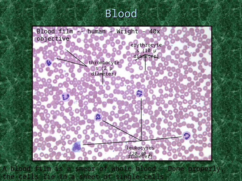
A blood film is a smear of whole blood. Done properly, the cells lie in a sheet of single cells.
BloodBlood
Blood film -- human – Wright – 40x objective
thrombocytes (2 μ
diameter)
erythrocytes (10 μ
diameter)
leukocytes(20-30 μ diameter)
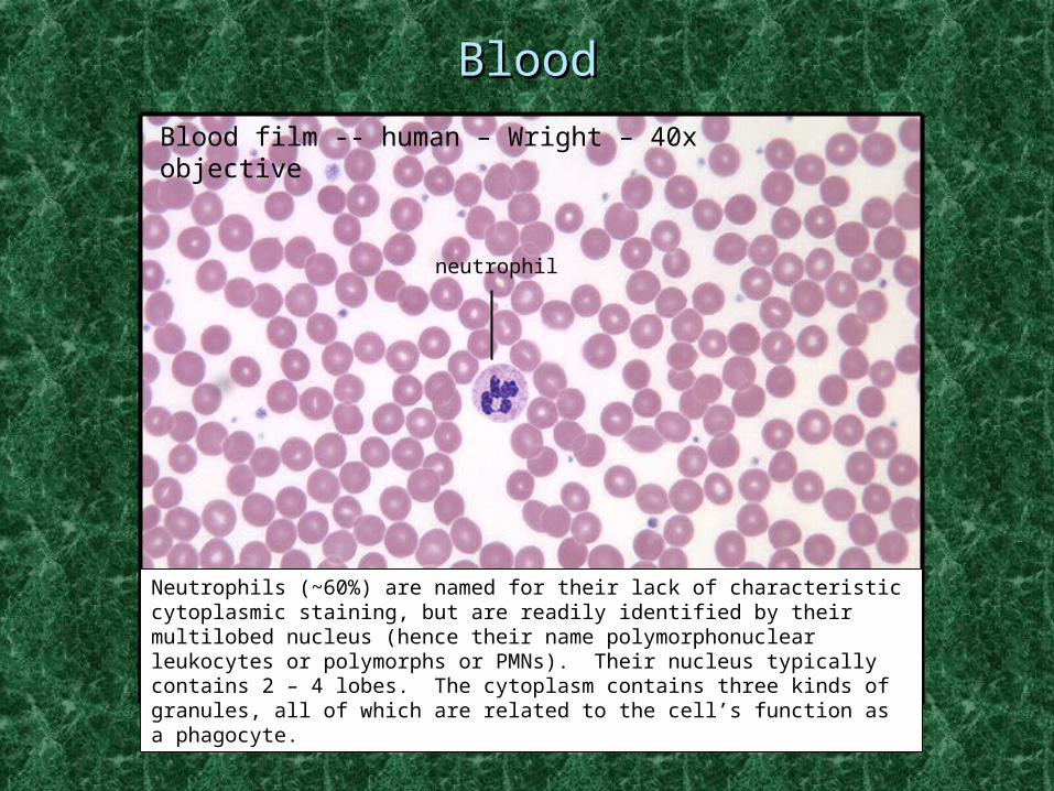
BloodBlood
Blood film -- human – Wright – 40x objective
neutrophil
Neutrophils (~60%) are named for their lack of characteristic cytoplasmic staining, but are readily identified by their multilobed nucleus (hence their name polymorphonuclear leukocytes or polymorphs or PMNs). Their nucleus typically contains 2 – 4 lobes. The cytoplasm contains three kinds of granules, all of which are related to the cell’s function as a phagocyte.
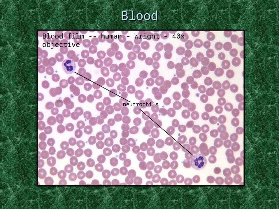
BloodBlood
Blood film -- human – Wright – 40x objective
neutrophils
BloodBlood
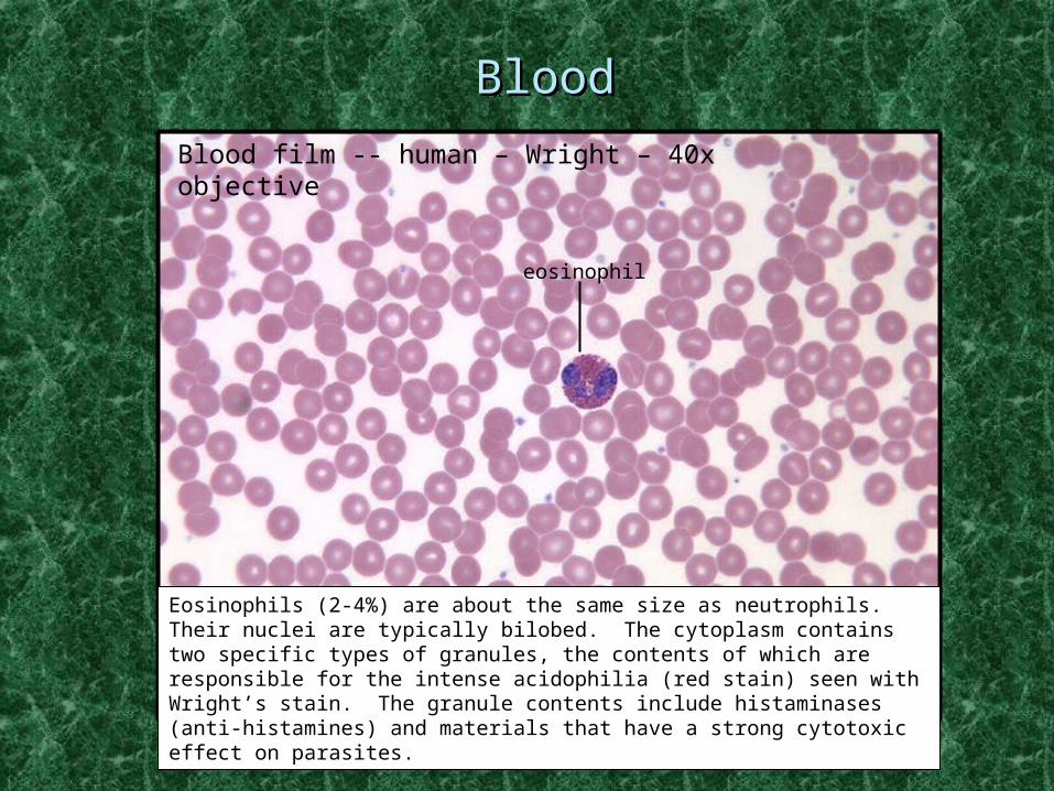
Blood film -- human – Wright – 40x objective
eosinophil
Eosinophils (2-4%) are about the same size as neutrophils. Their nuclei are typically bilobed. The cytoplasm contains two specific types of granules, the contents of which are responsible for the intense acidophilia (red stain) seen with Wright’s stain. The granule contents include histaminases (anti-histamines) and materials that have a strong cytotoxic effect on parasites.

BloodBlood
Blood film -- human – Wright – 40x objective
neutrophil
eosinophil
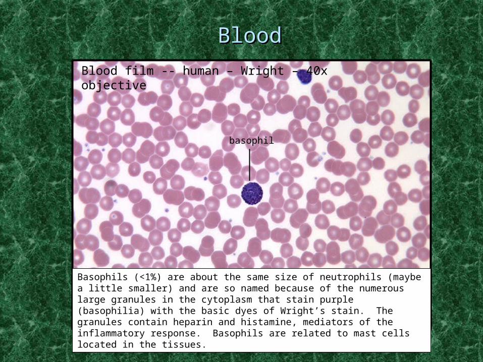
BloodBlood
Blood film -- human – Wright – 40x objective
basophil
Basophils (<1%) are about the same size of neutrophils (maybe a little smaller) and are so named because of the numerous large granules in the cytoplasm that stain purple (basophilia) with the basic dyes of Wright’s stain. The granules contain heparin and histamine, mediators of the inflammatory response. Basophils are related to mast cells located in the tissues.

BloodBlood
Blood film -- human – Wright – 40x objective
eosinophil
basophil

BloodBlood
Blood film -- human – Wright – 40x objective
monocyte
Monocytes (3-8%) are the largest of the WBCs. They leave the general circulation in 1.5 – 2 days to become wandering macrophages in the tissues and osteoclasts in the bone. The nucleus of the monocyte is typically flattened or indented. The cells are classified as agranulocytes because their granules are small and hard to visualize. The granules are lysosomes in keeping with the function of the cells as macrophages.

BloodBlood
Blood film -- human – Wright – 40x objective
monocyte
neutrophils
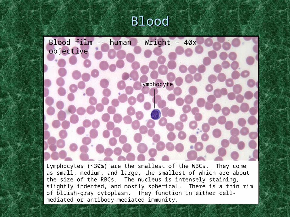
BloodBlood
Blood film -- human – Wright – 40x objective
lymphocyte
Lymphocytes (~30%) are the smallest of the WBCs. They come as small, medium, and large, the smallest of which are about the size of the RBCs. The nucleus is intensely staining, slightly indented, and mostly spherical. There is a thin rim of bluish-gray cytoplasm. They function in either cell-mediated or antibody-mediated immunity.
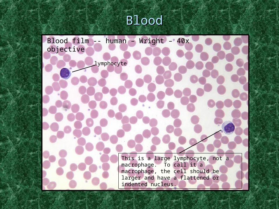
BloodBlood
Blood film -- human – Wright – 40x objective
lymphocyte
This is a large lymphocyte, not a macrophage. To call it a macrophage, the cell should be larger and have a flattened or indented nucleus.

BloodBlood
Blood film -- human – Wright – 40x objective
lymphocyte
neutrophil
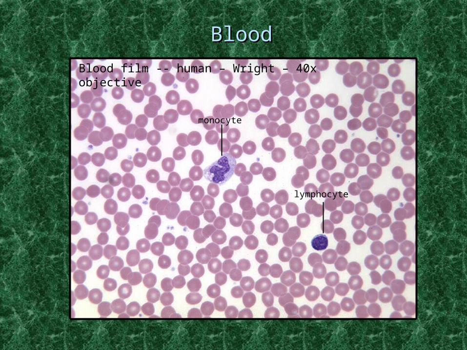
BloodBlood
Blood film -- human – Wright – 40x objective
monocyte
lymphocyte