bell, han, sawtell (2008)
TRANSCRIPT
ANRV346-NE31-01 ARI 14 May 2008 6:50
Cerebellum-Like Structuresand Their Implications forCerebellar FunctionCurtis C. Bell,1 Victor Han,2
and Nathaniel B. Sawtell11Neurological Sciences Institute, Oregon Health and Science University,Beaverton, Oregon 97006; email: [email protected], [email protected] Regional Primate Center, Oregon Health and Science University,Beaverton, Oregon 97006; email: [email protected]
Annu. Rev. Neurosci. 2008. 31:1–24
First published online as a Review in Advance onFebruary 14, 2008
The Annual Review of Neuroscience is online atneuro.annualreviews.org
This article’s doi:10.1146/annurev.neuro.30.051606.094225
Copyright c© 2008 by Annual Reviews.All rights reserved
0147-006X/08/0721-0001$20.00
Key Words
forward model, synaptic plasticity, electric fish, cerebellum
AbstractThe nervous systems of most vertebrates include both the cerebellumand structures that are architecturally similar to the cerebellum.The cerebellum-like structures are sensory structures that receiveinput from the periphery in their deep layers and parallel fiber inputin their molecular layers. This review describes these cerebellum-like structures and compares them with the cerebellum itself. Thecerebellum-like structures in three groups of fish act as adaptivesensory processors in which the signals conveyed by parallel fibers inthe molecular layer predict the patterns of sensory input to the deeplayers through a process of associative synaptic plasticity. Similaritiesbetween the cerebellum-like structures and the cerebellum suggestthat the cerebellum may also generate predictions about expectedsensory inputs or states of the system, as suggested also by clinical,experimental, and theoretical studies of the cerebellum. Understandingthe process of predicting sensory patterns in cerebellum-like structuresmay therefore be a source of insight into cerebellar function.
1
Click here for quick links to
Annual Reviews content online,
including:
• Other articles in this volume
• Top cited articles
• Top downloaded articles
• Our comprehensive search
FurtherANNUALREVIEWS
Ann
u. R
ev. N
euro
sci.
2008
.31:
1-24
. Dow
nloa
ded
from
arj
ourn
als.
annu
alre
view
s.or
gby
CO
RN
EL
L U
NIV
ER
SIT
Y o
n 10
/26/
09. F
or p
erso
nal u
se o
nly.
ANRV346-NE31-01 ARI 14 May 2008 6:50
Contents
LOCAL CIRCUITRY, GENEEXPRESSION, ANDEVOLUTION OFCEREBELLUM-LIKESTRUCTURES . . . . . . . . . . . . . . . . . . . 2General Features. . . . . . . . . . . . . . . . . . . 2Local Circuitry of Different
Cerebellum-Like Structures . . . . . 3Comparison of the Local Circuitries
of Cerebellum-Like Structuresand the Cerebellum . . . . . . . . . . . . . 9
Patterns of Gene Expression inCerebellum-Like Structuresand the Cerebellum . . . . . . . . . . . . . 10
Evolution of Cerebellum-LikeStructures and the Cerebellum . . 11
PREDICTIONS AND PLASTICITYIN CEREBELLUM-LIKESTRUCTURES AND THECEREBELLUM . . . . . . . . . . . . . . . . . . 11Predictions and Plasticity in
Cerebellum-Like Structures . . . . . 11Predictions in the Cerebellum . . . . . . 15
DIRECTIONS FOR FUTURERESEARCH . . . . . . . . . . . . . . . . . . . . . . 16Activity Patterns in Granule Cells . . . 16Adaptive Filtering in Electrosensory
Systems . . . . . . . . . . . . . . . . . . . . . . . . 17Adaptive Filtering in the DCN
and Less-StudiedCerebellum-Like Structures . . . . . 17
Purkinje-Like Cells . . . . . . . . . . . . . . . . 17Primitive Cerebellums . . . . . . . . . . . . . 17
LOCAL CIRCUITRY, GENEEXPRESSION, AND EVOLUTIONOF CEREBELLUM-LIKESTRUCTURES
General Features
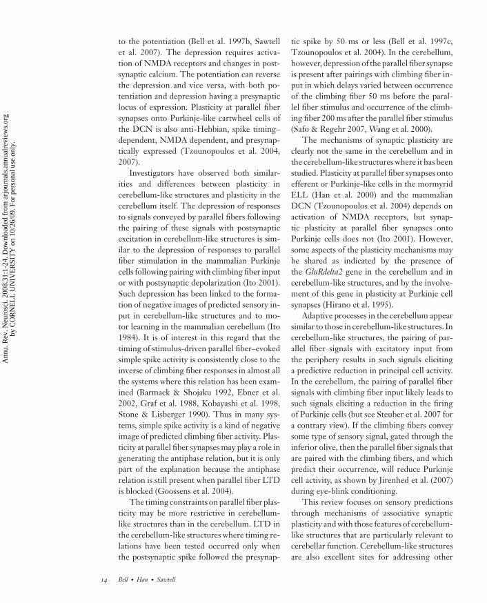
A distinctive molecular layer is a key identi-fying feature of all cerebellum-like structures(Figure 1). The molecular layer is composed of
parallel fibers together with the dendrites andcell bodies on which the fibers terminate. Theparallel fibers are numerous and closely packed.The granule cells that give rise to the paral-lel fibers in cerebellum-like structures are mor-phologically similar to cerebellar granular cells(Mugnaini et al. 1980a,b) but are usually locatedin an external granule cell mass rather than ina granule cell layer beneath the molecular layeras in the cerebellum. Unipolar brush cells andGolgi cells similar to those present in the gran-ular layer of the cerebellum are also presentin some cerebellum-like structures (Campbellet al. 2007, Mugnaini et al. 1997).
Functionally, the parallel fibers convey a richvariety of information from other central struc-tures, which includes corollary discharge in-formation associated with motor commands,information from higher levels of the same sen-sory modality represented in the deep layers,and information from other sensory modalities.In general, the types of signals conveyed by par-allel fibers are signals that are likely to be asso-ciated with changes in the sensory input to thedeep layers and that can therefore serve to pre-dict such sensory input (“predictive inputs” inFigure 1).
The parallel fibers terminate on the den-dritic spines of principal cells and on the smoothdendrites of inhibitory stellate cells in a man-ner very similar to the termination of paral-lel fibers on Purkinje cells and molecular layerinterneurons of the cerebellum. We use theterm principal cells to refer to large cells withspine-covered dendrites that extend through-out the molecular layer. Some of these prin-cipal cells are excitatory efferent cells thatproject to higher levels of the sensory system,whereas others are inhibitory neurons that ter-minate locally on each other and on the effer-ent cells. The latter are sometimes referred toas “Purkinje-like.” The cell bodies of principalcells are usually located in a separate layer be-low the molecular layer, like the Purkinje celllayer of the cerebellum.
Afferent input from the periphery termi-nates in the deep layers of cerebellum-likestructures, on basilar dendrites of principal
2 Bell · Han · Sawtell
Ann
u. R
ev. N
euro
sci.
2008
.31:
1-24
. Dow
nloa
ded
from
arj
ourn
als.
annu
alre
view
s.or
gby
CO
RN
EL
L U
NIV
ER
SIT
Y o
n 10
/26/
09. F
or p
erso
nal u
se o
nly.

ANRV346-NE31-01 ARI 14 May 2008 6:50
Sensory input layer
Principal cell layer
Molecular layer
Granule layer
Input from a sensory surface
Predictive inputs
Corollary discharge signalsHigher levels of the same modalityOther sensory modalities (e.g. proprioception)???
Figure 1Schematic drawing showing major features of cerebellum-like sensory structures. Inhibitory stellate cells ofthe molecular layer are shown in black. Blue upward arrows indicate afferent input from the peripheryterminating in the sensory input layer. In some cerebellum-like structures the afferent input also terminateson the smooth proximal portion of the apical dendrites as indicated by the small blue arrowheads.
cells, on proximal apical dendrites of principalcells, or on interneurons that relay the informa-tion from the periphery to the principal cells.Some of the interneurons of the deep layersare inhibitory, allowing for a change of sign,whereby excitation in the periphery is con-verted into inhibition of some principal cells.The peripheral input to the deep layers forms amap of a sensory surface, such as the skin sur-face, the retina, or the cochlea.
Local Circuitry of DifferentCerebellum-Like Structures
The brains of all major groups of craniatesexcept reptiles and birds have cerebellum-likestructures (Figures 2 and 3). The similari-ties among the different cerebellum-like struc-tures are clear, but so are the differences. Dif-ferent structures may have different types ofcells in addition to the principal cells, stel-late cells, and granule cells that are present inall cerebellum-like structures. Moreover, somestructures have additional inputs besides the in-puts from the periphery and the parallel fibers.
MON: medialoctavolateral nucleus
This review describes major features of the dif-ferent cerebellum-like structures of craniatesbut is not exhaustive. Recent reviews (Bell 2002,Bell & Maler 2005, Montgomery et al. 1995)and the original papers on individual structures,as provided below, should be consulted for morecomplete descriptions. Some of the structuresare also much better known than others, whichis reflected in the level of detail in the followingdescriptions.
Medial octavolateral nucleus. The medialoctavolateral nucleus (MON) processes pri-mary afferent input from the mechanical lateralline system and, in some fish, from eighth nerveend organs (Bell 1981b, McCormick 1999). Itis present in all basal aquatic craniates with me-chanical lateral line sensory systems (Figures 2,3a–d, 4a). Myxinoids (atlantic hagfish; C.B.Braun, personal communication) and aquaticamniotes (reptiles, birds, and mammals; Mont-gomery et al. 1995) do not have lateral line sys-tems and do not have an MON.
The efferent cells of the MON extend theirspiny apical dendrites up into a molecular
www.annualreviews.org • Cerebellum-Like Structures and Their Implications for Cerebellar Function 3
Ann
u. R
ev. N
euro
sci.
2008
.31:
1-24
. Dow
nloa
ded
from
arj
ourn
als.
annu
alre
view
s.or
gby
CO
RN
EL
L U
NIV
ER
SIT
Y o
n 10
/26/
09. F
or p
erso
nal u
se o
nly.
ANRV346-NE31-01 ARI 14 May 2008 6:50
Cra
niat
a
Ver
tebr
ata
Gna
thos
tom
ata
Myxinoidea
PetromyzontiformesEptatretidae
Myxinoids
CBM MON DON OTML ELL RLN DCN
Lampetra
ChondrichthyesHolocephalii
Elasmobranchii
Sar
copt
eryg
ii
Dipneustii
Tetrapoda AmphibiaAnura
Urodela
Apoda
Aves
Mammalia
Act
inop
tery
gii
Chondrosteii
Neopterygii
Holosteii
Osteoglossomorpha
Elopomorpha
Clupeomorpha
Euteleostei
?
?
?
?
?
Crossopterygii
Reptilia
Teleosteii
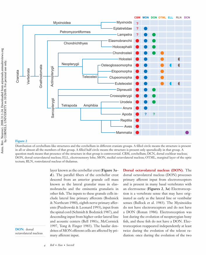
Figure 2Distribution of cerebellum-like structures and the cerebellum in different craniate groups. A filled circle means the structure is presentin all or almost all the members of that group. A filled half circle means the structure is present only sporadically in that group. Aquestion mark means that presence of the structure in that group is controversial. CBM, cerebellum; DCN, dorsal cochlear nucleus;DON, dorsal octavolateral nucleus; ELL, electrosensory lobe; MON, medial octavolateral nucleus; OTML, marginal layer of the optictectum; RLN, rostrolateral nucleus of thalamus.
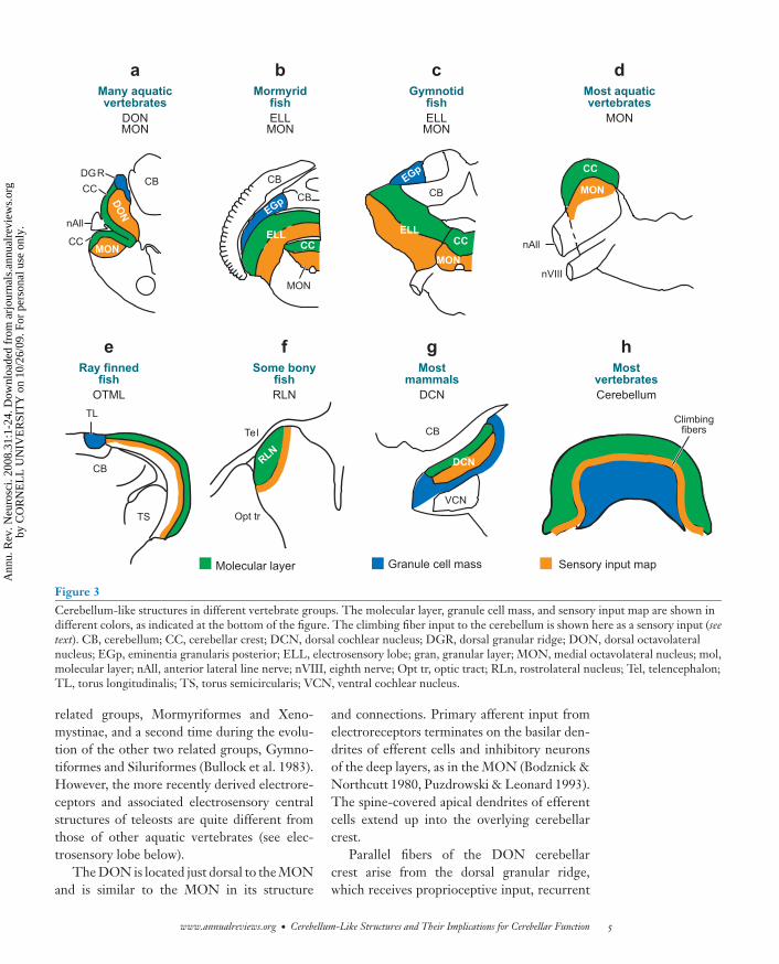
DON: dorsaloctavolateral nucleus
layer known as the cerebellar crest (Figure 3a–d ). The parallel fibers of the cerebellar crestdescend from an anterior granule cell massknown as the lateral granular mass in elas-mobranchs and the eminentia granularis inother fish. The inputs to these granule cells in-clude lateral line primary afferents (Bodznick& Northcutt 1980), eighth nerve primary affer-ents (Puzdrowski & Leonard 1993), input fromthe spinal cord (Schmidt & Bodznick 1987), anddescending input from higher-order lateral lineand acoustic centers (Bell 1981c, McCormick1997, Tong & Finger 1983). The basilar den-drites of MON efferent cells are affected by pri-mary afferent input.
Dorsal octavolateral nucleus (DON). Thedorsal octavolateral nucleus (DON) processesprimary afferent input from electroreceptorsand is present in many basal vertebrates withan electrosense (Figures 2, 3a) Electrorecep-tion is a vertebrate sense that may have orig-inated as early as the lateral line or vestibularsenses (Bullock et al. 1983). The Myxinoideado not have electroreceptors and do not havea DON (Ronan 1986). Electroreception waslost during the evolution of neopterygian bonyfish, and these fish do not have a DON. Elec-troreception reappeared independently at leasttwice during the evolution of the teleost ra-diation: once during the evolution of the two
4 Bell · Han · Sawtell
Ann
u. R
ev. N
euro
sci.
2008
.31:
1-24
. Dow
nloa
ded
from
arj
ourn
als.
annu
alre
view
s.or
gby
CO
RN
EL
L U
NIV
ER
SIT
Y o
n 10
/26/
09. F
or p
erso
nal u
se o
nly.
ANRV346-NE31-01 ARI 14 May 2008 6:50
MONDON
CB
CBEGp
EGp
ELLCC
MON
CC
MON
nVIII
nAll
nAll
CB
DO
N
MONCC
CC
DGR
ELL
CB
TL
TS Opt tr
RLN
Tel
CB
CC
MON
Many aquaticvertebrates
a b c d
e f g h
MON
Most aquaticvertebrates
MONELL
Mormyridfish
OTML
Ray finnedfish
RLN
Some bonyfish
DCN
Mostmammals
Cerebellum
Mostvertebrates
MONELL
Gymnotidfish
CB
VCN
DCN
Sensory input mapMolecular layer Granule cell mass
Climbingfibers
Figure 3Cerebellum-like structures in different vertebrate groups. The molecular layer, granule cell mass, and sensory input map are shown indifferent colors, as indicated at the bottom of the figure. The climbing fiber input to the cerebellum is shown here as a sensory input (seetext). CB, cerebellum; CC, cerebellar crest; DCN, dorsal cochlear nucleus; DGR, dorsal granular ridge; DON, dorsal octavolateralnucleus; EGp, eminentia granularis posterior; ELL, electrosensory lobe; gran, granular layer; MON, medial octavolateral nucleus; mol,molecular layer; nAll, anterior lateral line nerve; nVIII, eighth nerve; Opt tr, optic tract; RLn, rostrolateral nucleus; Tel, telencephalon;TL, torus longitudinalis; TS, torus semicircularis; VCN, ventral cochlear nucleus.
related groups, Mormyriformes and Xeno-mystinae, and a second time during the evolu-tion of the other two related groups, Gymno-tiformes and Siluriformes (Bullock et al. 1983).However, the more recently derived electrore-ceptors and associated electrosensory centralstructures of teleosts are quite different fromthose of other aquatic vertebrates (see elec-trosensory lobe below).
The DON is located just dorsal to the MONand is similar to the MON in its structure
and connections. Primary afferent input fromelectroreceptors terminates on the basilar den-drites of efferent cells and inhibitory neuronsof the deep layers, as in the MON (Bodznick &Northcutt 1980, Puzdrowski & Leonard 1993).The spine-covered apical dendrites of efferentcells extend up into the overlying cerebellarcrest.
Parallel fibers of the DON cerebellarcrest arise from the dorsal granular ridge,which receives proprioceptive input, recurrent
www.annualreviews.org • Cerebellum-Like Structures and Their Implications for Cerebellar Function 5
Ann
u. R
ev. N
euro
sci.
2008
.31:
1-24
. Dow
nloa
ded
from
arj
ourn
als.
annu
alre
view
s.or
gby
CO
RN
EL
L U
NIV
ER
SIT
Y o
n 10
/26/
09. F
or p
erso
nal u
se o
nly.
ANRV346-NE31-01 ARI 14 May 2008 6:50
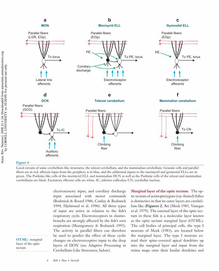
Mormyrid ELL Gymnotid ELLMON
Teleost cerebellum Mammalian cerebellumDCN
Parallel fibers(LGR, EGp)
Lateral lineafferents
To torus To PE, torusPE
Parallel fibers(EGp)
Electroreceptorafferents
To PE, torus
Corollarydischarge
PE
Parallel fibers(EGp)
Electroreceptorafferents
Auditoryafferents
To IC
Parallel fibers(GCD)
Climbingfiber
Parallel fibers
Climbingfiber
To CN
Parallel fibers
a b c
d e f
Figure 4Local circuits of some cerebellum-like structures, the teleost cerebellum, and the mammalian cerebellum. Granule cells and parallelfibers are in red, afferent input from the periphery is in blue, and the additional inputs to the mormyrid and gymnotid ELLs are ingreen. The Purkinje-like cells of the mormyrid ELL and mammalian DCN as well as the Purkinje cells of the teleost and mammaliancerebellums are black. Excitatory efferent cells are white. IC, inferior colliculus; CN, cerebellar nucleus.
OTML: marginallayer of the optictectum
electrosensory input, and corollary dischargeinput associated with motor commands(Bodznick & Boord 1986, Conley & Bodznick1994, Hjelmstad et al. 1996). All three typesof input are active in relation to the fish’srespiratory cycle. Electroreceptors in elasmo-branchs are strongly affected by the fish’s ownrespiration (Montgomery & Bodznick 1993).The activity in parallel fibers can thereforebe used to predict the effect of these cyclicchanges on electroreceptive input to the deeplayers of DON (see Adaptive Processing inCerebellum-Like Structures, below).
Marginal layer of the optic tectum. The op-tic tectum of actinopterygian (ray-finned) fishesis distinctive in that its outer layers are cerebel-lum like (Figures 2, 3e) (Meek 1983, Vanegaset al. 1979). The external layer of the optic tec-tum in these fish is a molecular layer knownas the optic tectum marginal layer (OTML).The cell bodies of principal cells, the type Ineurons of Meek (1983), are located belowthe marginal layer. The type I neurons ex-tend their spine-covered apical dendrites upinto the marginal layer and input from theretina maps onto their basilar dendrites and
6 Bell · Han · Sawtell
Ann
u. R
ev. N
euro
sci.
2008
.31:
1-24
. Dow
nloa
ded
from
arj
ourn
als.
annu
alre
view
s.or
gby
CO
RN
EL
L U
NIV
ER
SIT
Y o
n 10
/26/
09. F
or p
erso
nal u
se o
nly.
ANRV346-NE31-01 ARI 14 May 2008 6:50
the smooth proximal portions of their apicaldendrites.
The parallel fibers of the marginal layerarise from a medially located granule cell massknown as the torus longitudinalis. Granule cellsof the torus longitudinalis respond to corol-lary discharge signals associated with the motorcommands that evoke eye movements and re-spond to visual stimuli as well (Northmore et al.1983). Parallel fiber activity driven by corol-lary discharge signals associated with eye move-ments could predict changes in retinal input tothe deep layers, a possible interaction betweenthe two types of input similar to that describedabove for the DON.
Electrosensory lobe (ELL). Electrorecep-tion is present in four groups of teleosts:Mormyriformes, an order of electric fish fromAfrica; Gymnotiformes, a superorder of elec-tric fish from South America; Siluriformes, theorder of catfish; and Xenomystinae, an Africansubfamily of the family Notopteridae (Bullock& Heiligenberg 1986). All these fish have acerebellum-like electrosensory lobe (ELL) thatreceives primary afferent input from electrore-ceptors (Figures 2, 3b,c) (Bell & Russell 1978,Braford 1982, Finger & Tong 1984, Maler et al.1981).
The Mormyriformes and Gymnotiformesare electric fish with electric organs as wellas electroreceptors. The order Mormyri-formes includes the family Mormyridae, all ofwhich have electric organ discharges (EODs)that are brief and pulse like, and the single-species family Gymnarchidae, which has a con-tinuous wave-like EOD. The order Gymno-tiformes includes some families with wave-likeEODs and other families with pulsatile EODs.The ELLs of pulsatile mormyrids and wavegymnotids have been studied most extensively,although some work has been done on the ELLsof wave mormyriforms (Kawasaki & Guo 1998,Matsushita & Kawasaki 2005) and pulse gym-notiforms (Caputi et al. 2002, Schlegel 1973).
The spine-covered apical dendrites of ELLprincipal cells extend up into the overlyingmolecular layer. Primary afferent fibers from
ELL: electrosensorylobe
EOD: electric organdischarge
EOCD: electric organcorollary discharge
electroreceptors in the skin map onto the deeplayers, terminating on the basilar dendrites ofprincipal cells or on interneurons (Bell & Maler2005). The ELL efferent cells of mormyrid(Bell et al. 1997b), gymnotid (Saunders & Bas-tian 1984), and silurid fish (McCreery 1977) areof two main types: E-cells, which are excited byan increase in peripheral stimulus strength inthe center of their receptive fields, and I-cells,which are inhibited by such an increase. Thesetwo functionally distinct cell types are also mor-phologically distinct; the E-cells have more ex-tensive basilar dendrites.
Parallel fibers of ELLs arise from gran-ule cells of the eminentia granularis posterior(EGp), which in mormyrids, at least, also con-tains Golgi cells and unipolar brush cells similarto the same cell types in the mammalian cere-bellum (Campbell et al. 2007). The inputs toEGp in mormyrid and gymnotid fish includeproprioceptive signals associated with bendingof the body or the fins, recurrent electrosen-sory input from a higher levels of the system,and in mormyrids only, a corollary dischargesignal associated with the motor command thatelicits the electric organ (corollary) discharge(EOCD) (Bastian & Bratton 1990; Bell et al.1992; Carr & Maler 1986; Szabo et al. 1979,1990). These different inputs to EGp are re-layed to ELL as parallel fiber inputs, where theycan predict changes in electroreceptor input tothe deep layers associated with tail movements,some other electrosensory input, or the EOD(see Adaptive Processing in Cerebellum-LikeStructures).
The mormyrid (Bell et al. 1981), gym-notid (Carr & Maler 1986), and silurid (Tong1982) ELLs receive additional input asidefrom the peripheral and parallel fiber inputs.They receive direct recurrent input from ahigher-order electrosensory nucleus just ros-tral to ELL, the nucleus preeminentialis dor-salis (PE) (Figures 4b,c). The deep layers of themormyrid ELL also receive EOCD input di-rectly from an EOD motor command–relatednucleus (Bell & von der Emde 1995). This in-put is in addition to the EOCD input conveyedvia parallel fibers.
www.annualreviews.org • Cerebellum-Like Structures and Their Implications for Cerebellar Function 7
Ann
u. R
ev. N
euro
sci.
2008
.31:
1-24
. Dow
nloa
ded
from
arj
ourn
als.
annu
alre
view
s.or
gby
CO
RN
EL
L U
NIV
ER
SIT
Y o
n 10
/26/
09. F
or p
erso
nal u
se o
nly.
ANRV346-NE31-01 ARI 14 May 2008 6:50
RLN: rostrolateralnucleus of thethalamus
DCN: dorsal cochlearnucleus
The ELLs of mormyrid and gymnotid fishhave differences as well as similarities. Most im-portant, the mormyrid ELL includes a prin-cipal cell that is not present in the gymnotidELL (Figures 4b,c), the medium ganglion cell(Meek et al. 1996). These cells are referred to asPurkinje-like because they are GABAergic withextensive spine-covered dendrites in the overly-ing molecular layer. However, they differ fromPurkinje cells because they have basilar den-drites and do not receive climbing fiber input.The medium ganglion cells are interneuronsthat inhibit both nearby efferent cells and eachother (Figure 4b). They are more numerousthan the efferent cells and have many more den-drites and spines in the molecular layer (Meeket al. 1996). They must therefore have a centralrole in the integration of peripheral and paral-lel fiber inputs in the mormyrid ELL. Theseand other differences between the mormyridand gymnotid ELLs are consistent with theirindependent evolutionary origins.
Rostrolateral nucleus of the thalamus. Therostrolateral nucleus (RLN) (Figures 2, 3f ) ofthe thalamus is a small, cerebellum-like struc-ture found in the thalamus of a few widelyscattered neopterygian fish (Figure 2) (Butler& Saidel 1992). The principal cells of RLNreceive topographically organized direct in-put from the retina on the smooth proximalparts of their apical dendrites. The more dis-tal apical dendrites are covered with spinesand receive parallel fiber input from the toruslongitudinalis.
Dorsal cochlear nucleus. All mammalspossess a dorsal cochlear nucleus (DCN)(Figures 2, 3g, 4d ). The DCN is laminatedand cerebellum-like in marsupials and euthe-rian mammals but not in monotremes (Cant1992, Nieuwenhuys et al. 1997). Fusiformcells are the major efferent cell type of theDCN. Their basilar dendrites are contactedby primary afferent fibers from the cochlea,which form a topographic map of the cochleain the deeper layers below the molecular layer.The fusiform cells extend their spine-covered
apical dendrites up into the molecular layerwhere they are contacted by parallel fibers.The parallel fibers arise from granule cellslocated around the margins of the nucleus.The parallel fibers course at right angles tothe isofrequency bands in the deeper layers.Thus, parallel fibers cross through differentfrequency-specific regions of DCN.
The cartwheel cell is a second type of princi-pal cell in the DCN (Cant 1992, Nieuwenhuyset al. 1997). These cells are Purkinje-likebecause they are GABAergic, have extensivespine-covered dendrites in the molecular layer,and inhibit the efferent fusiform cells. The cellbodies of cartwheel cells are in the molecularlayer, and their dendrites are restricted to themolecular layer.
The local circuits of the DCN and themormyrid ELL are very similar to the localcircuit of the cerebellar cortex in actinopteryr-ian fish where most Purkinje cells are in-terneurons that terminate locally on efferentcells (Figure 4e) (Finger 1978, Meek 1998).The parallel fibers of the DCN, the mormyridELL, and the actinopterygian cerebellum passthrough and excite the dendrites of bothefferent cells and Purkinje or Purkinje-likecells. In all three cases, the Purkinje cells orPurkinje-like cells inhibit nearby efferent cells(Figures 4b,d,e). The efferent neurons of theactinopterygian cerebellum are equivalent tothe cerebellar nucleus neurons of mammals(Figure 4f ).
The granule cells of the DCN receive var-ious types of input: recurrent auditory inputfrom the inferior colliculus (Caicedo & Herbert1993) and auditory cortex (Weedman & Ryugo1996); primary vestibular afferent input (Burian& Gstoettner 1988); input from the pontine nu-clei (Ohlrogge et al. 2001); somatosensory in-put from the dorsal column nuclei (Weinberg& Rustioni 1987), the trigeminal nuclei (Zhou& Shore 2004), and the somatosensory cortex(Wolff & Kunzle 1997); and direct input fromthe cochlea via fine unmyelinated Type II affer-ents (Brown et al. 1988). DCN granule cellsalso receive input from brainstem nuclei as-sociated with vocalization and respiration that
8 Bell · Han · Sawtell
Ann
u. R
ev. N
euro
sci.
2008
.31:
1-24
. Dow
nloa
ded
from
arj
ourn
als.
annu
alre
view
s.or
gby
CO
RN
EL
L U
NIV
ER
SIT
Y o
n 10
/26/
09. F
or p
erso
nal u
se o
nly.
ANRV346-NE31-01 ARI 14 May 2008 6:50
may convey corollary discharge signals (Shore& Zhou 2006). Proprioceptive input from thepinna has particularly strong effects on DCNgranule cells in the cat (Kanold & Young 2001).Movements of the animal’s pinna, head, or bodyhave predictable effects on how the cochlearesponds to an external sound source, and ananimal’s own vocalization and respiration willhave predictable consequences on auditory in-put. Thus the signals conveyed by the parallelfibers in the DCN molecular layer could gen-erate predictions about changes in afferent ac-tivity from the cochlea that arrive at the deeplayers, as in other cerebellum-like structures.
Comparison of the Local Circuitriesof Cerebellum-Like Structuresand the Cerebellum
Many similarities in cell types and local cir-cuitry between the cerebellum and cerebellum-like structures have been described in the pre-ceding section. The similar cellular elementsinclude the granule cells, the Golgi cells, theunipolar brush cells, the parallel fibers, the stel-late cells, and the spine-covered molecular layerdendrites of principal cells.
The most crucial similarity is that betweenthe two inputs to cerebellum-like structuresand the two inputs to cerebellar Purkinjecells. Cerebellum-like structures receive paral-lel fiber and peripheral input, whereas Purkinjecells of the cerebellum receive parallel fiber in-put and climbing fiber input. In both cases, oneinput, the parallel fibers, conveys a rich varietyof information to an entire set of principal cellsor Purkinje cells. In both cases, a second input—peripheral input for cerebellum-like structuresand climbing fiber input for the cerebellum—conveys specific information that subdivides theset of Purkinje cells that share the same parallelfiber input.
Olivary input to Purkinje cells is more spe-cific than the peripheral input to the deep lay-ers of cerebellum-like structures insofar as it isconveyed by just a single climbing fiber. Effer-ent cells and Purkinje-like cells in cerebellum-like structures do not have such single fiber in-
puts. The cerebellums of different vertebratescan vary markedly, but all the cerebellums thathave been closely examined have a specific in-put from the inferior olive that terminates asclimbing fibers. We suggest that the presenceof a climbing fiber is the defining characteris-tic of the cerebellum that distinguishes it fromcerebellum-like structures.
Climbing fibers and the peripheral sensoryinput to cerebellum-like structures are similarin many respects. Climbing fibers signal ratherspecific sensory events in most of the caseswhere the information they convey has beenidentified. Such sensory signals include retinalslip in a particular direction (Maekawa & Simp-son 1972), somatosensory stimulation within asmall region of skin (Ekerot & Jorntell 2001,Robertson 1985), and vestibular stimulationwith tilt in a particular direction (Barmack &Shojaku 1992). Moreover, the climbing fibers ofvertebrates other than mammals do not termi-nate throughout the molecular layer as in mam-mals. They terminate instead on smooth, prox-imal dendrites at the base of the molecular layer(Nieuwenhuys et al. 1997) in a manner similarto that of retinal input onto the smooth, prox-imal dendrites of principal cells in the OTMLand RLN. This is not to say that the inferiorolive is a simple sensory relay. It is not. Butclearly sensory stimuli have a strong influenceon the inferior olive and on climbing fibers, aresult consistent with the origin of the inferiorolive from the embryo’s alar or sensory plate.Devor (2002) has in fact suggested that the in-ferior olive has been interposed between pe-ripheral sensory structures and the cerebellumto gate sensory signals by motor commands andby the inferior olive’s own intrinsic rhythmicity.
As noted in the previous section, the parallelfibers of cerebellum-like structures convey in-formation that is associated with sensory inputchanges to the deep layers and that can there-fore predict such changes. The parallel fibersof the cerebellum similarly convey informationthat can predict the occurrence of climbingfiber input. Climbing fibers in the floccu-lonodular lobe of the mammalian cerebel-lum, for example, signal retinal slip (Maekawa
www.annualreviews.org • Cerebellum-Like Structures and Their Implications for Cerebellar Function 9
Ann
u. R
ev. N
euro
sci.
2008
.31:
1-24
. Dow
nloa
ded
from
arj
ourn
als.
annu
alre
view
s.or
gby
CO
RN
EL
L U
NIV
ER
SIT
Y o
n 10
/26/
09. F
or p
erso
nal u
se o
nly.
ANRV346-NE31-01 ARI 14 May 2008 6:50
LTD: long-termdepression
& Simpson 1972), and the parallel fibers inthis region convey vestibular information abouthead movement (Lisberger & Fuchs 1974),corollary discharge information about eyemovement (Noda & Warabi 1982), and propri-oceptive information from the neck (Matsushita& Tanami 1987), all of which could be used topredict movement of an image on the retina.
The presence of a climbing fiber is perhapsthe critical difference between the cerebellumand cerebellum-like structures. Other differ-ences include the presence of basilar dendriteson most principal cells of cerebellum-like struc-tures but not on Purkinje cells; the presence ofplanar dendritic trees in most Purkinje cells butnot in most principal cells; the presence of celltypes in cerebellum-like structures not presentin the cerebellum; and the presence of other in-puts besides parallel fibers and climbing fibersin cerebellum-like structures not present in thecerebellum, such as the preeminential input inelectroreceptive teleosts (Figures 4b,c).
Patterns of Gene Expressionin Cerebellum-Like Structuresand the Cerebellum
Similarities and differences between the differ-ent cerebellum-like structures and the cerebel-lum itself are also revealed in gene expressionpatterns. Some genes are expressed in manydifferent cerebellar and cerebellum-like struc-tures, whereas others are expressed in only afew of these structures (Bell 2002). Commonpatterns of gene expression between cerebellarPurkinje cells and cartwheel cells of the DCNare particularly prominent, and many muta-tions affect both cell types (Berrebi et al. 1990).
One gene, the GluRdelta2 gene, may be ex-pressed in most if not all cerebellum-like struc-tures and also in the cerebellum, but not in otherstructures. This gene is structurally related tothe ionotropic glutamate receptors but doesnot form ion channels (Yuzaki 2003). The geneis necessary for long-term depression (LTD)at the parallel fiber to Purkinje cell synapse(Yawata et al. 2006). In mammals, the GluR-delta2 gene is expressed in Purkinje cells (Yuzaki
2003) and in the principal cells of the DCN(Petralia et al. 1996). In zebrafish, the GluR-delta2 gene is expressed in the molecular layersof the cerebellum, the MON, and the OTML,but not elsewhere in the brain as shown forboth the gene and the protein (Mikami et al.2004). Similarly, in the mormyrid brain, theGluRdelta2 protein is present in the molecularlayers of the cerebellum, the ELL, the MON,and the OTML, but not elsewhere in the brain( J. Zhang & C. Bell, unpublished observa-tions). Expression of the GluRdelta2 gene in stillother cerebellum-like structures remains to beestablished.
Some genes are expressed in some of thecerebellum-like structures or the cerebellum inthe adult but are expressed only in other suchstructures during development. The zebrin IIgene, for example, is expressed only in Purkinjecells in adult mammals, birds, and fish (Hawkes& Herrup 1995, Lannoo et al. 1991) but is ex-pressed transiently during development in theMON and in part of the ELL of gymnotidfish (Lannoo et al. 1992). Similarly, functionalN-methyl-D-aspartate (NMDA) receptors arepresent on principal cells of the adult mormyridand gymnotid ELLs (Grant et al. 1998, Bermanet al. 2001), as well as principal cells of theadult DCN (Manis & Molitor 1996), but arepresent on cerebellar Purkinje only during de-velopment (Dupont et al. 1987).
The common features in the local circuitryand in the gene expression patterns suggestthe presence of a shared genetic-developmentalprogram in all craniates, a program thatonce activated can generate a cerebellum orcerebellum-like structure. Some findings fromexperimental embryology support this idea.Thus, ectopic cerebellum-like structures de-velop in the forebrain or midbrain of a chickembryo if beads are coated with fibroblastgrowth factor 8 and placed at those sites inthe embryo (Martinez et al. 1999). Similarly,cerebellar tissue will develop ectopically in themidbrain and forebrain of a mouse embryowith a genome that is Otx1+/- and Otx2+/-(Drosophila orthodenticle protein, a transcrip-tion factor) (Acampora et al. 1997).
10 Bell · Han · Sawtell
Ann
u. R
ev. N
euro
sci.
2008
.31:
1-24
. Dow
nloa
ded
from
arj
ourn
als.
annu
alre
view
s.or
gby
CO
RN
EL
L U
NIV
ER
SIT
Y o
n 10
/26/
09. F
or p
erso
nal u
se o
nly.
ANRV346-NE31-01 ARI 14 May 2008 6:50
Evolution of Cerebellum-LikeStructures and the Cerebellum
The similarities between all the differentcerebellum-like and cerebellar structures can-not be explained solely by homology in thesense of historical or phylogenetic homology(Butler & Saidel 2000). In this usage of the term,a feature is considered homologous across dif-ferent taxa if the taxa have inherited the fea-ture from a common ancestor that also had thefeature. However, some of the individual struc-tures described here are homologous. Thus themost parsimonious explanation for the pres-ence of a cerebellum in all vertebrates is thatit was present in a common ancestor. A com-mon ancestor is also the most parsimoniousexplanation for the presence of an MON, aDON, or a DCN in some groups of craniates.However, we find no evidence for an ancestralcerebellum-like structure from which the cere-bellum, MON, DON, marginal layer of the tec-tum, ELL, RLN, and DCN all evolved. (SeeBell 2002 for a more complete analysis of theevolution of cerebellum-like structures.)
How then can we explain the clear simi-larities among the different cerebellums andcerebellum-like structures? The best explana-tion may be the presence of a developmental-genetic program that can generate a cerebellumor cerebellum-like structure, as described pre-viously, together with evolutionary pressure forthe type of information processing that thesestructures can perform.
Cerebellum-like structures may haveevolved before the cerebellum itself. An MONis clearly present in some myxinoids, and bothan MON and a DON are clearly present inlampreys, but the presence of a cerebellum isnot well established in either of these groups.Some comparative anatomists affirm thepresence of a cerebellum in myxinoids (Larsell1967), whereas others deny it (Nieuwenhuyset al. 1997), and arguments have also beenmade both for (Larsell 1967, Nieuwenhuyset al. 1997) and against (Crosby 1969) thepresence of a cerebellum in lampreys. Assuggested previously, the identification of
climbing fibers on putative Purkinje cellscould indicate the presence of a cerebellum,but no efforts to identify climbing fibershave been made in myxinoids and lampreys.Purkinje cell–specific markers that do not staincerebellum-like structures could also helpdetermine the presence of a cerebellum. Thusthe finding that the Zebrin II antibody doesnot stain cells in what some consider to be thelamprey cerebellum is of interest (Lannoo &Hawkes 1997) but is not conclusive because theZebrin II antibody does not stain all Purkinjecells.
PREDICTIONS AND PLASTICITYIN CEREBELLUM-LIKESTRUCTURES ANDTHE CEREBELLUM
Predictions and Plasticity inCerebellum-Like Structures
Cerebellum-like structures process informa-tion from peripheral sensory receptors in com-bination with an array of central signals con-veyed by parallel fibers. If a common functionexists among all cerebellum-like structures, itmust involve the interaction between these twotypes of inputs. Progress toward understandingthese interactions has been made in cerebellum-like structures concerned with the processingof electrosensory information in three distinctgroups of fish: elasmobranchs, gymnoti-form teleosts, and mormyrid teleosts. Thecerebellum-like structures of these fish act asadaptive filters, removing predictable featuresof the sensory input (for reviews, see Bastian& Zakon 2005, Bell 2001, Bell et al. 1997a).
In these systems, the animals’ own behav-ior strongly affects electroreceptors and couldinterfere with sensing weak electrosensory sig-nals from the environment. In the passive elec-trosensory system of elasmobranch fish, for ex-ample, ventilatory movements modulate thefish’s standing bioelectric field and can driveelectroreceptor afferents through their en-tire dynamic range (Montgomery & Bodznick
www.annualreviews.org • Cerebellum-Like Structures and Their Implications for Cerebellar Function 11
Ann
u. R
ev. N
euro
sci.
2008
.31:
1-24
. Dow
nloa
ded
from
arj
ourn
als.
annu
alre
view
s.or
gby
CO
RN
EL
L U
NIV
ER
SIT
Y o
n 10
/26/
09. F
or p
erso
nal u
se o
nly.
ANRV346-NE31-01 ARI 14 May 2008 6:50
1999). In the active electrosensory systems ofmormyrid and gymnotid fish, movements of theelectric organ (located in the tail) relative tosensory surface cause large changes in EOD-evoked electroreceptor input that could over-whelm the small changes resulting from nearbyobjects.
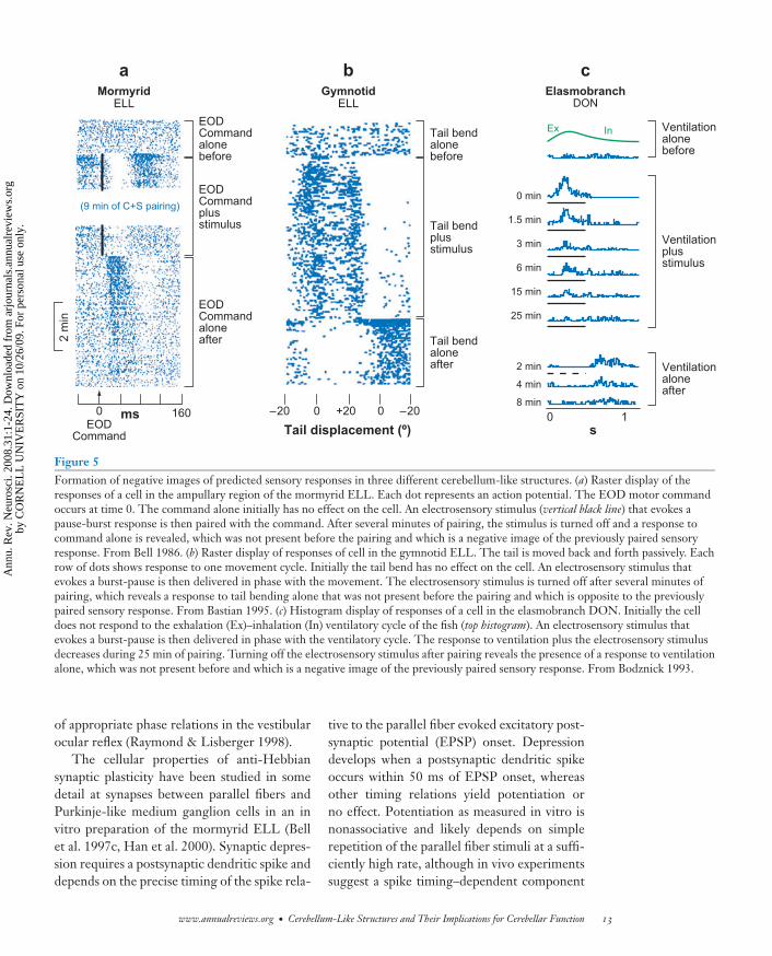
Parallel fiber inputs to cerebellum-likestructures involved in electrolocation conveyproprioceptive, corollary discharge, and elec-trosensory signals that could be used to pre-dict the electrosensory consequences of the an-imals’ own behavior. Direct evidence for thegeneration of such predictions has been ob-tained from in vivo recordings from princi-pal cells in the mormyrid and gymnotid ELLand elasmobranch DON (Bastian 1996a, Bell1981a, Bell et al. 1997b, Bodznick et al. 1999).In each case, pairing artificial electrosensorystimuli with central predictive signals—a corol-lary discharge signal at a particular delay afterthe EOD motor command in the case of themormyrid ELL (Figure 5a), a proprioceptivesignal at a particular tail angle in the case of thegymnotid ELL (Figure 5b), and a propriocep-tive or corollary discharge signal at a particularphase of the ventilatory cycle in the case of theelasmobranch DON (Figure 5c)—results in achange in the response to the predictive sig-nals alone that resembles a negative image ofthe response to the previously paired (and nowpredicted) stimulus. The negative images de-velop rapidly over the course of a few minutesof pairing and are specific to the sign as wellas to the spatial and temporal patterns of activ-ity evoked by the stimulus. On the basis of theseresults investigators suggested that cerebellum-like circuitry could operate as an adaptive filterby continually generating and updating sensorypredictions on the basis of associations betweencentral signals and current sensory inputs andsubtracting these predictions from the neuralresponse. Adaptive filtering could thus allowexternal electrosensory signals to be detectedmore easily.
Several lines of evidence confirm that for-mation of negative images is due, at least inlarge part, to plastic changes occurring within
the cerebellum-like structures themselves (Bell2001). Pairing predictive signals with intracel-lular current injections in vivo results in theformation of negative images in principal cellsin all three groups of fish, indicating that theinputs to the recorded cell are plastic (Bastian1996b, Bell et al. 1993, Bodznick et al. 1999).Given the types of predictive signals involvedin negative image formation, synapses betweenparallel fibers and principal cells are the mostnatural candidates for the site of plastic changes.Negative image formation requires that theplasticity be anti-Hebbian in character, i.e., cor-relations between pre- and postsynaptic ac-tivity should decrease synaptic strength, andresearchers have obtained evidence for anti-Hebbian plasticity at parallel fiber synapses withprincipal cells in all three classes of fish. Anti-Hebbian plasticity at parallel fiber synapses hasalso been shown recently in the DCN of mam-mals (Fujino & Oertel 2003, Tzounopouloset al. 2004) but has not yet been connected tosystems-level adaptive filtering.
Modeling studies have helped to link theproperties of negative image formation withmechanisms of synaptic plasticity (Nelson &Paulin 1995, Roberts 1999, Roberts & Bell2000). Temporal specificity is a key feature ofnegative image formation. In the mormyridELL, parallel fibers convey corollary dischargesignals related to the motor command thatdrives the EOD. Pairing with electrosensorystimuli at various delays relative to the motorcommand results in negative images that arespecific to the paired delay (Bell 1982). Re-sults of modeling studies suggest that tempo-rally specific negative images could be gener-ated using an anti-Hebbian learning rule similarto that observed experimentally (see below) to-gether with an array of parallel fiber inputsactive at different delays following the mo-tor command (Roberts 1999, Roberts & Bell2000). The mechanisms for generating tem-porally specific negative images in this modelare quite similar to those proposed for someforms of cerebellar learning, such as the learn-ing of adaptively timed responses in classicaleye-blink conditioning (Medina et al. 2000) or
12 Bell · Han · Sawtell
Ann
u. R
ev. N
euro
sci.
2008
.31:
1-24
. Dow
nloa
ded
from
arj
ourn
als.
annu
alre
view
s.or
gby
CO
RN
EL
L U
NIV
ER
SIT
Y o
n 10
/26/
09. F
or p
erso
nal u
se o
nly.
ANRV346-NE31-01 ARI 14 May 2008 6:50
0 min
1.5 min
3 min
6 min
15 min
25 min
2 min
4 min
8 min
0 1
Ex InEODCommandalonebefore
EODCommandplusstimulus
(9 min of C+S pairing)
2 m
in
160mssTail displacement (º)
0
EODCommandaloneafter
EODCommand
0 0 –20–20 +20
Tail bendalonebefore
Tail bendplusstimulus
Tail bendaloneafter
Ventilationalonebefore
Ventilationplus stimulus
Ventilationaloneafter
a
ELLMormyrid
b
ELLGymnotid
c
DONElasmobranch
Figure 5Formation of negative images of predicted sensory responses in three different cerebellum-like structures. (a) Raster display of theresponses of a cell in the ampullary region of the mormyrid ELL. Each dot represents an action potential. The EOD motor commandoccurs at time 0. The command alone initially has no effect on the cell. An electrosensory stimulus (vertical black line) that evokes apause-burst response is then paired with the command. After several minutes of pairing, the stimulus is turned off and a response tocommand alone is revealed, which was not present before the pairing and which is a negative image of the previously paired sensoryresponse. From Bell 1986. (b) Raster display of responses of cell in the gymnotid ELL. The tail is moved back and forth passively. Eachrow of dots shows response to one movement cycle. Initially the tail bend has no effect on the cell. An electrosensory stimulus thatevokes a burst-pause is then delivered in phase with the movement. The electrosensory stimulus is turned off after several minutes ofpairing, which reveals a response to tail bending alone that was not present before the pairing and which is opposite to the previouslypaired sensory response. From Bastian 1995. (c) Histogram display of responses of a cell in the elasmobranch DON. Initially the celldoes not respond to the exhalation (Ex)–inhalation (In) ventilatory cycle of the fish (top histogram). An electrosensory stimulus thatevokes a burst-pause is then delivered in phase with the ventilatory cycle. The response to ventilation plus the electrosensory stimulusdecreases during 25 min of pairing. Turning off the electrosensory stimulus after pairing reveals the presence of a response to ventilationalone, which was not present before and which is a negative image of the previously paired sensory response. From Bodznick 1993.
of appropriate phase relations in the vestibularocular reflex (Raymond & Lisberger 1998).
The cellular properties of anti-Hebbiansynaptic plasticity have been studied in somedetail at synapses between parallel fibers andPurkinje-like medium ganglion cells in an invitro preparation of the mormyrid ELL (Bellet al. 1997c, Han et al. 2000). Synaptic depres-sion requires a postsynaptic dendritic spike anddepends on the precise timing of the spike rela-
tive to the parallel fiber evoked excitatory post-synaptic potential (EPSP) onset. Depressiondevelops when a postsynaptic dendritic spikeoccurs within 50 ms of EPSP onset, whereasother timing relations yield potentiation orno effect. Potentiation as measured in vitro isnonassociative and likely depends on simplerepetition of the parallel fiber stimuli at a suffi-ciently high rate, although in vivo experimentssuggest a spike timing–dependent component
www.annualreviews.org • Cerebellum-Like Structures and Their Implications for Cerebellar Function 13
Ann
u. R
ev. N
euro
sci.
2008
.31:
1-24
. Dow
nloa
ded
from
arj
ourn
als.
annu
alre
view
s.or
gby
CO
RN
EL
L U
NIV
ER
SIT
Y o
n 10
/26/
09. F
or p
erso
nal u
se o
nly.
ANRV346-NE31-01 ARI 14 May 2008 6:50
to the potentiation (Bell et al. 1997b, Sawtellet al. 2007). The depression requires activa-tion of NMDA receptors and changes in post-synaptic calcium. The potentiation can reversethe depression and vice versa, with both po-tentiation and depression having a presynapticlocus of expression. Plasticity at parallel fibersynapses onto Purkinje-like cartwheel cells ofthe DCN is also anti-Hebbian, spike timing–dependent, NMDA dependent, and presynap-tically expressed (Tzounopoulos et al. 2004,2007).
Investigators have observed both similar-ities and differences between plasticity incerebellum-like structures and plasticity in thecerebellum itself. The depression of responsesto signals conveyed by parallel fibers followingthe pairing of these signals with postsynapticexcitation in cerebellum-like structures is sim-ilar to the depression of responses to parallelfiber stimulation in the mammalian Purkinjecells following pairing with climbing fiber inputor with postsynaptic depolarization (Ito 2001).Such depression has been linked to the forma-tion of negative images of predicted sensory in-put in cerebellum-like structures and to mo-tor learning in the mammalian cerebellum (Ito1984). It is of interest in this regard that thetiming of stimulus-driven parallel fiber–evokedsimple spike activity is consistently close to theinverse of climbing fiber responses in almost allthe systems where this relation has been exam-ined (Barmack & Shojaku 1992, Ebner et al.2002, Graf et al. 1988, Kobayashi et al. 1998,Stone & Lisberger 1990). Thus in many sys-tems, simple spike activity is a kind of negativeimage of predicted climbing fiber activity. Plas-ticity at parallel fiber synapses may play a role ingenerating the antiphase relation, but it is onlypart of the explanation because the antiphaserelation is still present when parallel fiber LTDis blocked (Goossens et al. 2004).
The timing constraints on parallel fiber plas-ticity may be more restrictive in cerebellum-like structures than in the cerebellum. LTD inthe cerebellum-like structures where timing re-lations have been tested occurred only whenthe postsynaptic spike followed the presynap-
tic spike by 50 ms or less (Bell et al. 1997c,Tzounopoulos et al. 2004). In the cerebellum,however, depression of the parallel fiber synapseis present after pairings with climbing fiber in-put in which delays varied between occurrenceof the climbing fiber 50 ms before the paral-lel fiber stimulus and occurrence of the climb-ing fiber 200 ms after the parallel fiber stimulus(Safo & Regehr 2007, Wang et al. 2000).
The mechanisms of synaptic plasticity areclearly not the same in the cerebellum and inthe cerebellum-like structures where it has beenstudied. Plasticity at parallel fiber synapses ontoefferent or Purkinje-like cells in the mormyridELL (Han et al. 2000) and the mammalianDCN (Tzounopoulos et al. 2004) depends onactivation of NMDA receptors, but synap-tic plasticity at parallel fiber synapses ontoPurkinje cells does not (Ito 2001). However,some aspects of the plasticity mechanisms maybe shared as indicated by the presence ofthe GluRdelta2 gene in the cerebellum and incerebellum-like structures, and by the involve-ment of this gene in plasticity at Purkinje cellsynapses (Hirano et al. 1995).
Adaptive processes in the cerebellum appearsimilar to those in cerebellum-like structures. Incerebellum-like structures, the pairing of par-allel fiber signals with excitatory input fromthe periphery results in such signals elicitinga predictive reduction in principal cell activity.In the cerebellum, the pairing of parallel fibersignals with climbing fiber input likely leads tosuch signals eliciting a reduction in the firingof Purkinje cells (but see Steuber et al. 2007 fora contrary view). If the climbing fibers conveysome type of sensory signal, gated through theinferior olive, then the parallel fiber signals thatare paired with the climbing fibers, and whichpredict their occurrence, will reduce Purkinjecell activity, as shown by Jirenhed et al. (2007)during eye-blink conditioning.
This review focuses on sensory predictionsthrough mechanisms of associative synapticplasticity and with those features of cerebellum-like structures that are particularly relevant tocerebellar function. Cerebellum-like structuresare also excellent sites for addressing other
14 Bell · Han · Sawtell
Ann
u. R
ev. N
euro
sci.
2008
.31:
1-24
. Dow
nloa
ded
from
arj
ourn
als.
annu
alre
view
s.or
gby
CO
RN
EL
L U
NIV
ER
SIT
Y o
n 10
/26/
09. F
or p
erso
nal u
se o
nly.
ANRV346-NE31-01 ARI 14 May 2008 6:50
important issues in neuroscience, which cannotbe discussed here because of space constraints.These include the roles of recurrent feedbackfrom higher to lower levels of the same sen-sory system (Chacron et al. 2003, 2005; Do-iron et al. 2003), the effects of motor commandson sensory processing (Bell & Grant 1992), thepreservation and analysis of temporal informa-tion (Kawasaki 2005), and the neural process-ing of spectral cues for sound localization in theDCN (Young & Davis 2002).
Predictions in the Cerebellum
The many similarities between cerebellum-likestructures and the cerebellum suggest that thecerebellum too may be involved in generatingpredictions concerning expected sensory inputor states of the system (Bell et al. 1997a, Devor2000), and a variety of experimental, clinical,and theoretical studies of the cerebellum sup-port this hypothesis (Diedrichsen et al. 2007,Nixon 2001, Paulin 2005, Wolpert et al. 1998).
The probable involvement of the cere-bellum in predictive or feedforward controlthrough learning is well recognized (Bastian2006, Ito 1984, Miall et al. 1993, Ohyama et al.2003, Wolpert et al. 1998). Predictive controlallows for prior knowledge to shape an action,as in knowing if a cup is full or empty beforepicking it up. Several studies indicate that pre-dictive feedforward control is deficient in cere-bellar patients (Morton & Bastian 2006, Smith& Shadmehr 2005). Such patients do not adapttheir responses to predictable perturbations, al-though they respond quite well to sudden un-predictable perturbations of a movement, indi-cating that feedback control from the peripheryis functional.
Theoreticians have proposed that the cere-bellum may act in an adaptive and predictivemanner through the generation of two typesof models: forward models and inverse mod-els (Wolpert et al. 1998). In a forward model,copies of a motor command are conveyed to thecerebellum together with information aboutthe current state of the system such as posi-tions and velocities of the limbs. The cerebel-
Forward model:predicts the futurestate of the system onthe basis of the currentstate and the motorcommand
Inverse model:generates anappropriate motorcommand that willcause a desired changein the state of thesystem
lum then generates a prediction about the sen-sory consequences of the commanded motor actin the current context. In an inverse model, thedesired goal of an action together with infor-mation about the current state are conveyed tothe cerebellum, which then generates the pre-cise motor commands that will yield the desiredgoal. Both types of models must be capable ofplastic change or learning to adapt to changesin the task or in the system, such as changes inload or initial limb position.
Forward models are particularly importantin generating fast, coordinated movement se-quences. Feedback from peripheral sensory re-ceptors is slow. An appropriate command forone phase of a movement must often be issuedbefore peripheral feedback can arrive about theconsequences of a motor command that evokeda previous phase of the movement. A forwardmodel that predicts the sensory consequencesof a motor command, accounting for all that isknown about the current state of the system, al-lows the next motor command in a sequence tobe issued appropriately and in accord with theexpected consequences of previous commands.Such a process allows for the chunking of sepa-rate components of a motor sequence and theirautomatization, as described by Nixon (2001).Moreover, classic symptoms of cerebellar dam-age such as decomposition of movement, slow-ness, and tremor can all be understood as dueto the absence of predictive forward models andreliance on peripheral feedback (Bastian 2006,Nixon 2001).
What is required in such automatization ofa sequence of movements is the predicted ef-fect of the motor command: the sensory con-sequences or state that results from the action,not simply the motor command itself. Recentexperiments by Pasalar et al. (2006) suggestthat the Purkinje cell output from large regionsof the cerebellar hemispheres is indeed moretightly coupled with predictions about conse-quences of the movement than with the mo-tor commands themselves (but see Yamamotoet al. 2007). Pasalar et al. (2006) recorded fromPurkinje cells over a wide area of the hemi-sphere in monkeys that had been trained to
www.annualreviews.org • Cerebellum-Like Structures and Their Implications for Cerebellar Function 15
Ann
u. R
ev. N
euro
sci.
2008
.31:
1-24
. Dow
nloa
ded
from
arj
ourn
als.
annu
alre
view
s.or
gby
CO
RN
EL
L U
NIV
ER
SIT
Y o
n 10
/26/
09. F
or p
erso
nal u
se o
nly.
ANRV346-NE31-01 ARI 14 May 2008 6:50
control a cursor on a screen with a manipulan-dum and to make the cursor track a circularlymoving stimulus. They then altered the forcesrequired to move the manipulandum. The elec-tromyograms in the arm muscles varied sys-tematically with the changes in required forces,but Purkinje cell simple spike activity was un-affected by the changes in force. Purkinje cellsimple spikes depended only on the position, di-rection, and velocity of the movement. Purkinjecell activity was phase advanced, that is, predic-tive of the movement parameters or state of thearm (T. Ebner, personal communication).
Pasalar et al. (2006) took their results as anargument against an inverse model in the cere-bellum because Purkinje cell activity had littlerelation to the motor commands to the mus-cles. Although one could argue that the ac-tivity reflects a high-level motor command, inmovement rather than muscle coordinates, thesimpler explanation is that the Purkinje cell ac-tivity reflects a forward model of expected con-sequences, as required for the automatization ofmovement sequences. Their experiments sug-gest that not all sensory consequences are pre-dicted; only those critical for accomplishing thetask are predicted. Thus presumed changes intouch or muscle receptors associated with forcechanges were not predicted by Purkinje cell ac-tivity; only velocity and position of the limbwere predicted.
Examples of what are, in effect, forwardmodels in the cerebellum-like structures ofmormyrid and elasmobranch fish are describedin the previous section, showing that forwardmodels can indeed be generated within struc-tures such as the cerebellum. In these sys-tems, corollary discharge signals come to elicita prediction about the sensory input patternthat is expected to follow the motor com-mand. The possibility of such corollary dis-charge effects in the OTML and DCN was alsomentioned.
Cerebellum-like structures can generatepredictions on the basis of other sensory in-puts (Bastian 1996a, Bodznick et al. 1999), notjust on the basis of motor commands, and thecerebellum may do so also. For example, in
eye-blink conditioning, which is thought to in-volve the cerebellum, the timing of one sen-sory signal, an air puff to the cornea (signaledby the climbing fiber), is predicted from an-other sensory signal, a tone (signaled by mossyfibers) (Kim & Thompson 1997). Similarly,cerebellar modulation of the vestibular ocu-lar reflex involves the prediction of one sen-sory stimulus, retinal slip (signaled by climbingfibers), by the occurrence of another sensorystimulus, vestibular input (signaled by mossyfibers). More broadly, Paulin (1993, 2005) hassuggested that the cerebellum estimates futurestates of the organism or environment usinga combination of sensory, motor, and possiblyother types of information.
In simpler systems, such as the vestibularocular reflex, in which Purkinje cell output iscoupled quite directly with motor pathways, theadaptive alteration in Purkinje cell activity afterpairing with the climbing fiber can be viewed aseither a prediction about a sensory input or asa motor command. In more complex systems,where Purkinje cell output is less tightly cou-pled with motor pathways, as in the trackingtask studied by Pasalar et al. (2006), the hy-pothesis of Purkinje cell activity as a predic-tor of consequences may provide a more usefulperspective.
DIRECTIONS FORFUTURE RESEARCH
Our understanding of adaptive processing incerebellum-like structures is far from complete,and future work will be useful both for under-standing the neural mechanisms of sensory pro-cessing and for understanding the cerebellum.Promising lines of research are outlined brieflybelow.
Activity Patterns in Granule Cells
How the different types of predictive inputs arecombined and represented in the granule cellsthat are associated with cerebellum-like struc-tures remains unclear, as is also the case for cere-bellar granule cells.
16 Bell · Han · Sawtell
Ann
u. R
ev. N
euro
sci.
2008
.31:
1-24
. Dow
nloa
ded
from
arj
ourn
als.
annu
alre
view
s.or
gby
CO
RN
EL
L U
NIV
ER
SIT
Y o
n 10
/26/
09. F
or p
erso
nal u
se o
nly.
ANRV346-NE31-01 ARI 14 May 2008 6:50
Adaptive Filtering inElectrosensory Systems
Several aspects of adaptive filtering incerebellum-like structures require furtherinvestigation, including (a) the behavioralconsequences of adaptive filtering; (b) theeffects of adaptive filtering on encoding natu-ralistic stimuli in the presence of self-generatedinterference; (c) the mechanisms of plasticityand the presence of plasticity at other sites,such as inhibitory synapses; and (d ) the possiblegeneration of more complex expectations suchas those based on memories of entire scenesor sequences. The possibility of more complexexpectations is suggested by the massive de-scending inputs that cerebellum-like structuresreceive from higher levels of the same sensorysystems.
Adaptive Filtering in the DCNand Less-StudiedCerebellum-Like Structures
Recent studies have found synaptic plasticityat parallel fiber synapses onto Purkinje-likecartwheel cells and fusiform cells in the DCNin vitro. Yet very little is known at the systemslevel regarding the role of such plastic parallel
fiber inputs in auditory processing. Similarly,very little is known about adaptive filtering inthe MON or OTML.
Purkinje-Like Cells
The functional roles of Purkinje-like cellsremain unclear. Recent work has shownthat dendritic spikes that drive anti-Hebbianplasticity in Purkinje-like MG cells of themormyrid ELL are strongly regulated bycentral signals, suggesting a parallel to su-pervised learning mediated by climbing fiberinputs to the cerebellum (Sawtell et al. 2007).In addition, the mormyrid ELL, the DCN, andthe teleost cerebellum all provide excellent op-portunities for examining interactions betweenPurkinje or Purkinje-like cells and neighboringefferent cells (analogous to deep cerebellar nu-clear cells in the mammalian cerebellum).
Primitive Cerebellums
As discussed previously, the earliest craniatespossess cerebellum-like structures, but it is notclear if they possess a cerebellum. Identifica-tion of a structure similar to the inferior olivein hagfish or lampreys would help to resolve thisissue.
DISCLOSURE STATEMENT
The authors are not aware of any biases that might be perceived as affecting the objectivity of thisreview.
ACKNOWLEDGMENTS
We thank Drs. Neal Barmack, Timothy Ebner, and Johannes Meek for their critical reviews ofthe manuscript. The work was supported by grants from the National Institutes of Health (MH49792 to C.C.B. and NS44961 to V.H.) and the National Science Foundation (IOB 0618212 toN.B.S.) and by a National Research Service Award (NS049728 to N.B.S.).
LITERATURE CITED
Acampora D, Avantaggiato V, Tuorto F, Simeone A. 1997. Genetic control of brain morphogenesisthrough Otx gene dosage requirement. Development 124(18):3639–50
Barmack NH, Shojaku H. 1992. Vestibularly induced slow oscillations in climbing fiber responsesof Purkinje cells in the cerebellar nodulus of the rabbit. Neuroscience 50:1–5
www.annualreviews.org • Cerebellum-Like Structures and Their Implications for Cerebellar Function 17
Ann
u. R
ev. N
euro
sci.
2008
.31:
1-24
. Dow
nloa
ded
from
arj
ourn
als.
annu
alre
view
s.or
gby
CO
RN
EL
L U
NIV
ER
SIT
Y o
n 10
/26/
09. F
or p
erso
nal u
se o
nly.
ANRV346-NE31-01 ARI 14 May 2008 6:50
Bastian AJ. 2006. Learning to predict the future: the cerebellum adapts feedforward movementcontrol. Curr. Opin. Neurobiol. 16(6):645–49
Bastian J. 1995. Pyramidal-cell plasticity in weakly electric fish: a mechanism for attenuatingresponses to reafferent electrosensory inputs. J. Comp. Physiol. 176:63–78
Bastian J. 1996a. Plasticity in an electrosensory system. I. General features of dynamic sensoryfilter. J. Neurophysiol. 76:2483–96
Bastian J. 1996b. Plasticity in an electrosensory system. II. Postsynaptic events associated with adynamic sensory filter. J. Neurophysiol. 76:2497–507
Bastian J, Bratton B. 1990. Descending control of electroreception. I. Properties of nu-cleus praeeminentialis neurons projecting indirectly to the electrosensory lateral line lobe.J. Neurosci. 10:1226–40
Bastian J, Zakon H. 2005. Plasticity of sense organs and brain. See Bullock et al. 2005,pp. 195–228
Bell CC. 1981a. An efference copy modified by reafferent input. Science 214:450–53Bell CC. 1981b. Central distribution of octavolateral afferents and efferents in a teleost (Mormyri-
dae). J. Comp. Neurol. 195:391–414Bell CC. 1981c. Some central connections of medullary octavolateral centers in a mormyrid fish.
In Hearing and Sound Communication in Fishes, ed. RR Fay, AN Popper, WN Tavolga, pp.383–92. Berlin: Heidelberg, Springer-Verlag
Bell CC. 1982. Properties of a modifiable efference copy in electric fish. J. Neurophysiol. 47:1043–56Bell CC. 1986. Duration of plastic change in a modifiable efference copy. Brain Res. 369:29–
36Bell CC. 2001. Memory-based expectations in electrosensory systems. Curr. Opin. Neurobiol.
11:481–87Bell CC. 2002. Evolution of cerebellum-like structures. Brain Behav. Evol. 59:312–
26Bell CC, Bodznick D, Montgomery J, Bastian J. 1997a. The generation and subtraction
of sensory expectations within cerebellum-like structures. Brain Behav. Evol. 50:17–31
Bell CC, Caputi A, Grant K. 1997b. Physiology and plasticity of morphologically identified cellsin the mormyrid electrosensory lobe. J. Neurosci. 17:6409–22
Bell CC, Caputi A, Grant K, Serrier J. 1993. Storage of a sensory pattern by anti-Hebbian synapticplasticity in an electric fish. Proc. Natl. Acad. Sci. USA 90:4650–54
Bell CC, Finger TE, Russell CJ. 1981. Central connections of the posterior lateral line lobe inmormyrid fish. Exp. Brain Res. 42:9–22
Bell CC, Grant K. 1992. Corollary discharge effects and sensory processing in the mormyromastregions of the mormyrid electrosensory lobe: II. Cell types and corollary discharge plasticity.J. Neurophysiol. 68:859–75
Bell CC, Grant K, Serrier J. 1992. Corollary discharge effects and sensory processing in themormyrid electrosensory lobe: I. Field potentials and cellular activity in associated structures.J. Neurophysiol. 68:843–58
Bell CC, Han VZ, Sugawara S, Grant K. 1997c. Synaptic plasticity in a cerebellum-like structuredepends on temporal order. Nature 387:278–81
Bell CC, Maler L. 2005. Central neuroanatomy of electrosensory systems in fish. See Bullocket al. 2005, pp. 68–111
Bell CC, Russell CJ. 1978. Termination of electroreceptor and mechanical lateral line afferents inthe mormyrid acousticolateral area. J. Comp. Neurol. 182:367–82
Bell CC, von der Emde G. 1995. Electric organ corollary discharge pathways in mormyrid fish:II. The medial juxtalobar nucleus. J. Comp. Physiol. A. 177:463–79
18 Bell · Han · Sawtell
Ann
u. R
ev. N
euro
sci.
2008
.31:
1-24
. Dow
nloa
ded
from
arj
ourn
als.
annu
alre
view
s.or
gby
CO
RN
EL
L U
NIV
ER
SIT
Y o
n 10
/26/
09. F
or p
erso
nal u
se o
nly.
ANRV346-NE31-01 ARI 14 May 2008 6:50
Berman N, Dunn RJ, Maler L. 2001. Function of NMDA receptors in a feedback pathway of theelectrosensory system. J. Neurophysiol. 86:1612–21
Berrebi AS, Morgan JI, Mugnaini E. 1990. The Purkinje cell class may extend beyond the cere-bellum. J. Neurocytol. 19(5):643–54
Bodznick D. 1993. The specificity of an adaptive filter that suppresses unwanted reafference inelectrosensory neurons of the skate medulla. Biol. Bull. 185:312–14
Bodznick D, Boord RL. 1986. Electroreception in Chondrichthyes: central anatomy and physi-ology. See Bullock & Heiligenberg 1986, pp. 225–56
Bodznick D, Montgomery JC, Carey M. 1999. Adaptive mechanisms in the elasmobranch hind-brain. J. Exp. Biol. 202:1357–64
Bodznick D, Northcutt RG. 1980. Segregation of electro- and mechanoreceptive inputs to theelasmobranch medulla. Brain Res. 195:313–21
Braford MR. 1982. African, but not Asian, notopterid fishes are electroreceptive: evidence frombrain characters. Neurosci. Lett. 32:35–39
Brown MC, Berglund AM, Kiang NY, Ryugo DK. 1988. Central trajectories of type II spiralganglion neurons. J. Comp. Neurol. 278(4):581–90
Bullock TH, Bodznick DA, Northcutt RG. 1983. The phylogenetic distribution of electrorecep-tion: evidence for convergent evolution of a primitive vertebrate sense modality. Brain Res.Rev. 6:25–46
Bullock TH, Heiligenberg W. 1986. Electroreception. New York: WileyBullock TH, Hopkins CD, Popper AN, Fay RR, eds. 2005. Electroreception. New York: SpringerBurian M, Gstoettner W. 1988. Projection of primary vestibular afferent fibres to the cochlear
nucleus in the guinea pig. Neurosci. Lett. 84(1):13–17Butler AB, Saidel WM. 1992. Tectal projection to an unusual nucleus in the diencephalon of a
teleost fish, Pantodon buchholzi. Neurosci. Lett. 145:193–96Butler AB, Saidel WM. 2000. Defining sameness: historical, biological, and generative homology.
BioEssays 22(9):846–53Caicedo A, Herbert H. 1993. Topography of descending projections from the inferior colliculus
to auditory brainstem nuclei in the rat. J. Comp. Neurol. 328(3):377–92Campbell HR, Meek J, Zhang J, Bell CC. 2007. Anatomy of the posterior caudal lobe of the cere-
bellum and the eminentia granularis posterior in a mormyrid fish. J. Comp. Neurol. 502(5):714–35
Cant NB. 1992. The cochlear nucleus: neuronal types and their synaptic organization. InThe Mammalian Auditory Pathway: Neuroanatomy, ed. DB Webster, AN Popper, RR Fay,pp. 66–116. New York: Springer
Caputi AA, Castello ME, Aguilera P, Trujillo-Cenoz O. 2002. Electrolocation and electrocommu-nication in pulse gymnotids: signal carriers, prereceptor mechanisms and the electrosensorymosaic. J. Physiol. Paris 96(5–6):493–505
Carr CE, Maler L. 1986. Electroreception in gymnatiform fish: central anatomy and physiology.See Bullock & Heiligenberg 1986, pp. 319–74
Chacron MJ, Doiron B, Maler L, Longtin A, Bastian J. 2003. Non-classical receptive field mediatesswitch in a sensory neuron’s frequency tuning. Nature 423(6935):77–81
Chacron MJ, Maler L, Bastian J. 2005. Feedback and feedforward control of frequency tuning tonaturalistic stimuli. J. Neurosci. 25(23):5521–32
Conley RA, Bodznick D. 1994. The cerebellar dorsal granular ridge in an elasmobranch has pro-prioceptive and electroreceptive representations and projects homotopically to the medullaryelectrosensory nucleus. J. Comp. Physiol. A 174:707–21
Crosby EC. 1969. Comparative aspects of cerebellar morphology. In Neurobiology of CerebellarEvolution and Development, ed. R Llinas, pp. 19–41. Chicago: Am. Med. Assoc.
www.annualreviews.org • Cerebellum-Like Structures and Their Implications for Cerebellar Function 19
Ann
u. R
ev. N
euro
sci.
2008
.31:
1-24
. Dow
nloa
ded
from
arj
ourn
als.
annu
alre
view
s.or
gby
CO
RN
EL
L U
NIV
ER
SIT
Y o
n 10
/26/
09. F
or p
erso
nal u
se o
nly.

ANRV346-NE31-01 ARI 14 May 2008 6:50
Devor A. 2000. Is the cerebellum like cerebellar-like structures? Brain Res. Rev. 34(3):149–56Devor A. 2002. The great gate: control of sensory information flow to the cerebellum.
Cerebellum 1(1):27–34Diedrichsen J, Criscimagna-Hemminger SE, Shadmehr R. 2007. Dissociating timing and coor-
dination as functions of the cerebellum. J. Neurosci. 27(23):6291–301Doiron B, Chacron MJ, Maler L, Longtin A, Bastian J. 2003. Inhibitory feedback required for
network oscillatory responses to communication but not prey stimuli. Nature 421(6922):539–43
Dupont JL, Gardette R, Crepel F. 1987. Postnatal development of the chemosensitivity of ratcerebellar Purkinje cells to excitatory amino acids. An in vitro study. Brain Res. 431(1):59–68
Ebner TJ, Johnson MT, Roitman A, Fu Q. 2002. What do complex spikes signal about limbmovements? Ann. N.Y. Acad. Sci. 978:205–18
Ekerot CF, Jorntell H. 2001. Parallel fibre receptive fields of Purkinje cells and interneurons areclimbing fibre-specific. Eur. J. Neurosci. 13:1303–10
Finger TE. 1978. Efferent neurons of the teleost cerebellum. Brain Res. 153:608–14Finger TE, Tong SL. 1984. Central organization of eighth nerve and mechanosensory lateral line
systems in the brainstem of ictalurid catfish. J. Comp. Neurol. 229:129–51Fujino K, Oertel D. 2003. Bidirectional synaptic plasticity in the cerebellum-like mammalian
dorsal cochlear nucleus. Proc. Natl. Acad. Sci. USA 100(1):265–70Goossens HH, Hoebeek FE, van Alphen AM, Van Der SJ, Stahl JS, et al. 2004. Simple spike and
complex spike activity of floccular Purkinje cells during the optokinetic reflex in mice lackingcerebellar long-term depression. Eur. J. Neurosci. 19(3):687–97
Graf W, Simpson JI, Leonard CS. 1988. Spatial organization of visual messages of the rabbit’scerebellar flocculus. II. Complex and simple spike responses of Purkinje cells. J. Neurophysiol.60(6):2091–121
Grant K, Sugawara S, Gomez L, Han VZ, Bell CC. 1998. The Mormyrid electrosensory lobe invitro: physiology and pharmacology of cells and circuits. J. Neurosci. 18:6009–25
Han VZ, Grant G, Bell CC. 2000. Reversible associative depression and nonassociative potenti-ation at a parallel fiber synapse. Neuron 27:611–22
Hawkes R, Herrup K. 1995. Aldolase C/zebrin II and the regionalization of the cerebellum.J. Mol. Neurosci. 6(3):147–58
Hirano T, Kasono K, Araki K, Mishina M. 1995. Suppression of LTD in cultured Purkinje cellsdeficient in the glutamate receptor d2 subunit. NeuroReport 6:524–26
Hjelmstad GO, Parks G, Bodznick D. 1996. Motor corollary discharge activity and sensory re-sponses related to ventilation in the skate vestibulolateral cerebellum: implications for elec-trosensory processing. J. Exp. Biol. 199:673–81
Ito M. 1984. The Cerebellum and Neural Control. New York: RavenIto M. 2001. Cerebellar long-term depression: characterization, signal transduction, and functional
roles. Physiol. Rev. 81(3):1143–95Jirenhed DA, Bengtsson F, Hesslow G. 2007. Acquisition, extinction, and reacquisition of a cere-
bellar cortical memory trace. J. Neurosci. 27(10):2493–502Kanold PO, Young ED. 2001. Proprioceptive information from the pinna provides somatosensory
input to cat dorsal cochlear nucleus. J. Neurosci. 21(19):7848–58Kawasaki M. 2005. Physiology of tuberous electrosensory systems. See Bullock et al. 2005,
pp. 154–194Kawasaki M, Guo YX. 1998. Parallel projection of amplitude and phase information from the hind-
brain to the midbrain of the African electric fish Gymnarchus niloticus. J. Neurosci. 18(18):7599–611
20 Bell · Han · Sawtell
Ann
u. R
ev. N
euro
sci.
2008
.31:
1-24
. Dow
nloa
ded
from
arj
ourn
als.
annu
alre
view
s.or
gby
CO
RN
EL
L U
NIV
ER
SIT
Y o
n 10
/26/
09. F
or p
erso
nal u
se o
nly.
ANRV346-NE31-01 ARI 14 May 2008 6:50
Kim JJ, Thompson RF. 1997. Cerebellar circuits and synaptic mechanisms involved in classicaleyeblink conditioning. Trends Neurosci. 20:177–81
Kobayashi Y, Kawano K, Takemura A, Inoue Y, Kitama T, et al. 1998. Temporal firing patternsof Purkinje cells in the cerebellar ventral paraflocculus during ocular following responses inmonkeys II. Complex spikes. J. Neurophysiol. 80(2):832–48
Lannoo M, Hawkes R. 1997. A search for primitive Purkinje cells: zebrin II expression in sealampreys (Petromyzon marinus). Neurosci. Lett. 237:53–55
Lannoo MJ, Maler L, Hawkes R. 1992. Zebrin II distinguishes the ampullary organ receptive mapfrom the tuberous organ receptive maps during development in the teleost electrosensorylateral line lobe. Brain Res. 586:176–80
Lannoo MJ, Ross L, Maler L, Hawkes R. 1991. Development of the cerebellum and its extracellularPurkinje cell projection in teleost fishes as determined by zebrin II immunocytochemistry.Prog. Neurobiol. 37:329–63
Larsell O. 1967. The Comparative Anatomy and Histology of the Cerebellum from Myxinoids throughBirds. Minneapolis: Univ. Minn. Press
Lisberger SG, Fuchs AF. 1974. Response of flocculus Purkinje cells to adequate vestibular stim-ulation in the alert monkey: fixation vs compensatory eye movements. Brain Res. 69:347–53
Maekawa K, Simpson JI. 1972. Climbing fiber activation of Purkinje cells in the flocculus byimpulses transferred through the visual pathway. Brain Res. 39:245–51
Maler L, Sas EKB, Rogers J. 1981. The cytology of the posterior lateral line lobe of high frequencyweakly electric fish (Gymnotidae): dendritic differentiation and synaptic specificity in a simplecortex. J. Comp. Neurol. 195:87–140
Manis PB, Molitor SC. 1996. N-methyl-d-aspartate receptors at parallel fiber synapses in thedorsal cochlear nucleus. J. Neurophysiol. 76:1639–55
Martinez S, Crossley PH, Cobos I, Rubenstein JL, Martin GR. 1999. FGF8 induces formationof an ectopic isthmic organizer and isthmocerebellar development via a repressive effect onOtx2 expression. Development 126(6):1189–200
Matsushita A, Kawasaki M. 2005. Neuronal sensitivity to microsecond time disparities in theelectrosensory system of Gymnarchus niloticus. J. Neurosci. 25(49):11424–32
Matsushita M, Tanami T. 1987. Spinocerebellar projections from the central cervical nucleus inthe cat, as studied by anterograde transport of wheat germ agglutinin-horseradish peroxidase.J. Comp. Neurol. 266(3):376–97
McCormick CA. 1997. Organization and connections of octaval and lateral line centers in themedulla of a clupeid, Dorosoma cepedianum. Hear Res. 110(1–2):39–60
McCormick CA. 1999. Anatomy of the central auditory pathways of fish and amphibians. InComparative Hearing: Fish and Amphibians, ed. RR Fay, AN Popper, pp. 155–217. New York:Springer Verlag
McCreery DB. 1977. Two types of electroreceptive lateral lemniscal neurons of the lateral linelobe of the catfish Ictalurus nebulosus; connections from the lateral line nerve and steady-statefrequency response characteristics. J. Comp. Physiol. 113:317–39
Medina JF, Garcia KS, Nores WL, Taylor NM, Mauk MD. 2000. Timing mechanisms in thecerebellum: testing predictions of a large-scale computer simulation. J. Neurosci. 20(14):5516–25
Meek J. 1983. Functional anatomy of the tectum mesencephali of the goldfish. An explorativeanalysis of the functional implication of the laminar structural organization of the tectum.Brain Res. Rev. 6:247–97
Meek J. 1998. Holosteans and teleosts. In The Central Nervous System of Vertebrates, ed. R Nieuwen-huys, HJ Ten Donkelaar, C Nicholson, Vol. 15, pp. 759–937. Berlin: Springer
www.annualreviews.org • Cerebellum-Like Structures and Their Implications for Cerebellar Function 21
Ann
u. R
ev. N
euro
sci.
2008
.31:
1-24
. Dow
nloa
ded
from
arj
ourn
als.
annu
alre
view
s.or
gby
CO
RN
EL
L U
NIV
ER
SIT
Y o
n 10
/26/
09. F
or p
erso
nal u
se o
nly.
ANRV346-NE31-01 ARI 14 May 2008 6:50
Meek J, Grant K, Sugawara S, Hafmans TGM, Veron M, Denizot JP. 1996. Interneurons of theganglionic layer in the mormyrid electrosensory lateral line lobe: morphology, immunocyto-chemistry, and synaptology. J. Comp. Neurol. 375:43–65
Miall RC, Weir DJ, Wolpert DM, Stein JF. 1993. Is the Cerebellum a smith predictor. J. MotorBehav. 25(3):203–16
Mikami Y, Yoshida T, Matsuda N, Mishina M. 2004. Expression of zebrafish glutamate receptordelta2 in neurons with cerebellum-like wiring. Biochem. Biophys. Res. Commun. 322(1):168–76
Montgomery JC, Bodznick D. 1993. Hindbrain circuitry mediating common mode suppres-sion of ventilatory reafference in the electrosensory system of the little skate Raja erinacea.J. Exp. Biol. 183:203–15
Montgomery JC, Bodznick D. 1999. Signals and noise in the elasmobranch electrosensory system.J. Exp. Biol. 202(Pt. 10):1349–55
Montgomery JC, Coombs S, Conley RA, Bodznick D. 1995. Hindbrain sensory processing inlateral line, electrosensory, and auditory systems: a comparative overview of anatomical andfunctional similarities. Auditory Neurosc. 1:207–31
Morton SM, Bastian AJ. 2006. Cerebellar contributions to locomotor adaptations during splitbelttreadmill walking. J. Neurosci. 26(36):9107–16
Mugnaini E, Dino MR, Jaarsma D. 1997. The unipolar brush cells of the mammalian cerebellumand cochlear nucleus: cytology and microcircuitry. Prog. Brain Res. 114:131–50
Mugnaini E, Osen KK, Dahl AL, Friedrich VL Jr, Korte G. 1980a. Fine structure of granule cellsand related interneurons (termed Golgi cells) in the cochlear nuclear complex of cat, rat andmouse. J. Neurocytol. 9(4):537–70
Mugnaini E, Warr WB, Osen KK. 1980b. Distribution and light microscopic features of granulecells in the cochlear nuclei of cat, rat, and mouse. J. Comp. Neurol. 191(4):581–606
Nelson ME, Paulin MG. 1995. Neural simulations of adaptive reafference suppression in theelasmobranch electrosensory system. J. Comp. Physiol. A 177:723–36
Nieuwenhuys R, Ten Donkelaar HJ, Nicholson C. 1997. The Central Nervous System of Vertebrates.Heidelberg: Springer
Nixon PD, Passingham RE. 2001. Predicting sensory events: the role of the cerebellum in motorlearning. Exp. Brain. Res. 138:251–57
Noda H, Warabi T. 1982. Eye position signals in the flocculus of the monkey during smooth-pursuit eye movements. J. Physiol. 324:187–202
Northmore DPM, Williams B, Vanegas H. 1983. The teleostean torus longitudinalis: responsesrelated to eye movements, visuotopic mapping, and functional relations with the optic tectum.J. Comp. Physiol. A 150:39–50
Ohlrogge M, Doucet JR, Ryugo DK. 2001. Projections of the pontine nuclei to the cochlearnucleus in rats. J. Comp. Neurol. 436(3):290–303
Ohyama T, Nores WL, Murphy M, Mauk MD. 2003. What the cerebellum computes. TrendsNeurosci. 26(4):222–27
Pasalar S, Roitman AV, Durfee WK, Ebner TJ. 2006. Force field effects on cerbellar Purkinje celldischarge with implications for internal models. Nat. Neurosci. 9(11):1404–11
Paulin MG. 1993. The role of the cerebellum in motor control and perception. Brain Behav. Evol.41:39–50
Paulin MG. 2005. Evolution of the cerebellum as a neuronal machine for Bayesian state estimation.J. Neural. Eng. 2(3):S219–34
Petralia RS, Wang YX, Zhao HM, Wenthold RJ. 1996. Ionotropic and metabotropic glutamatereceptors show unique postsynaptic, presynaptic, and glial localizations in the dorsal cochlearnucleus. J. Comp. Neurol. 372(3):356–83
22 Bell · Han · Sawtell
Ann
u. R
ev. N
euro
sci.
2008
.31:
1-24
. Dow
nloa
ded
from
arj
ourn
als.
annu
alre
view
s.or
gby
CO
RN
EL
L U
NIV
ER
SIT
Y o
n 10
/26/
09. F
or p
erso
nal u
se o
nly.
ANRV346-NE31-01 ARI 14 May 2008 6:50
Puzdrowski RL, Leonard RB. 1993. The octavolateral systems in the stingray, Dasyatis sabina. I.Primary projections of the octaval and lateral line nerves. J. Comp. Neurol. 332(1):21–37
Raymond JL, Lisberger SG. 1998. Neural learning rules for the vestibulo-ocular reflex.J. Neurosci. 18(21):9112–29 (Abstr.)
Roberts PD. 1999. Computational consequences of temporally asymmetric learning rules: I. dif-ferential Hebbian learning. J. Comp. Neurosci. 7:235–46
Roberts PD, Bell CC. 2000. Computational consequences of temporally asymmetric learningrules: II. sensory image cancellation. J. Comput. Neurosci. 9:67–83
Robertson LT. 1985. Somatosensory representation of the climbing fiber system in the rostralintermediate cerebellum. Exp. Brain Res. 61(1):73–86
Ronan M 1986. Electroreception in cyclostomes. See Bullock & Heiligenberg 1986, pp. 209–24Safo P, Regehr WG. 2008. Timing dependence of the induction of cerebellar LTD. Neurophar-
macology. In pressSaunders J, Bastian J. 1984. The physiology and morphology of two types of electrosensory neurons
in the weakly electric fish Apteronotus leptorhynchus. J. Comp. Physiol. A 154:199–209Sawtell NB, Williams A, Bell CC. 2007. Central control of dendritic spikes shapes the responses of
Purkinje-like cells through spike timing-dependent synaptic plasticity. J. Neurosci. 27(7):1552–65
Schlegel P. 1973. Perception of objects in weakly electric fish Gymnotus carapo as studied in record-ings from rhombencephalic neurons. Exp. Brain Res. 18:340–54
Schmidt AW, Bodznick D. 1987. Afferent and efferent connections of the vestibulolateral cere-bellum of the little skate, Raja erinacea. Brain Behav. Evol. 30:282–302
Shore SE, Zhou J. 2006. Somatosensory influence on the cochlear nucleus and beyond. Hear. Res.216–17:90–99
Smith MA, Shadmehr R. 2005. Intact ability to learn internal models of arm dynamics in Hunt-ington’s disease but not cerebellar degeneration. J. Neurophysiol. 93(5):2809–21
Steuber V, Mittman W, Hoebeek FE, Silver RA, De Zeeuw CI, et al. 2007. Cerebellar LTD andpattern recognition by Purkinje cells. Neuron 54(1):121–36
Stone LS, Lisberger SG. 1990. Visual responses of Purkinje cells in the cerebellar flocculus duringsmooth-pursuit eye movements in monkeys. II. Complex spikes. J. Neurophysiol. 63(5):1262–75
Szabo T, Libouban S, Haugede-Carre F. 1979. Convergence of common and specific sensoryafferents to the cerebellar auricle (auricula cerebelli) in the teleost fish Gnathonemus demon-strated by HRP method. Brain Res. 168:619–22
Szabo T, Libouban S, Denizot JP. 1990. A well defined spinocerebellar system in the weaklyelectric teleost fish Gnathonemus petersii. Arch. Ital. Biol. 128:229–47
Tong S. 1982. The nucleus praeeminentialis: an electro- and mechanoreceptive center in thebrainstem of the catfish. J. Comp. Physiol. A 145:299–309
Tong S, Finger TE. 1983. Central organization of the electrosensory lateral line system in bullheadcarfish Ictalurus nebulosus. J. Comp. Neurol. 217:1–16
Tzounopoulos T, Kim Y, Oertel D, Trussell LO. 2004. Cell-specific, spike timing-dependentplasticities in the dorsal cochlear nucleus. Nat. Neurosci. 7(7):719–25
Tzounopoulos T, Rubio ME, Keen JE, Trussell LO. 2007. Coactivation of pre- and postsynap-tic signaling mechanisms determines cell-specific spike-timing-dependent plasticity. Neuron54(2):291–301
Vanegas H, Williams B, Freeman JA. 1979. Responses to stimulation of marginal fibers in theteleostean optic tectum. Exp. Brain Res. 34(2):335–49
Wang SS, Denk W, Hausser M. 2000. Coincidence detection in single dendritic spines mediatedby calcium release. Nat. Neurosci. 3:1266–73
www.annualreviews.org • Cerebellum-Like Structures and Their Implications for Cerebellar Function 23
Ann
u. R
ev. N
euro
sci.
2008
.31:
1-24
. Dow
nloa
ded
from
arj
ourn
als.
annu
alre
view
s.or
gby
CO
RN
EL
L U
NIV
ER
SIT
Y o
n 10
/26/
09. F
or p
erso
nal u
se o
nly.
ANRV346-NE31-01 ARI 14 May 2008 6:50
Weedman DL, Ryugo DK. 1996. Projections from auditory cortex to the cochlear nucleus in rats:synapses on granule cell dendrites. J. Comp. Neurol. 371:311–24
Weinberg RJ, Rustioni A. 1987. A cuneocochlear pathway in the rat. Neuroscience 20(1):209–19
Wolff A, Kunzle H. 1997. Cortical and medullary somatosensory projections to the cochlearnuclear complex in the hedgehog tenrec. Neurosci. Lett. 221(2–3):125–28
Wolpert DM, Miall C, Kawato M. 1998. Internal models in the cerebellum. Trends Cogn. Sci.2:338–47
Yamamoto K, Kawato M, Kotosaka S, Kitazawa S. 2007. Encoding coding of movements dynamicsby Purkinje cell simple spike activity during fast arm movements under resistive and assistiveforce fields. J. Neurophysiol. 97:1588–99
Yawata S, Tsuchida H, Kengaku M, Hirano T. 2006. Membrane-proximal region of gluta-mate receptor delta2 subunit is critical for long-term depression and interaction with pro-tein interacting with C kinase 1 in a cerebellar Purkinje neuron. J. Neurosci. 26(14):3626–33
Young ED, Davis KA. 2002. Circuitry and function of the dorsal cochlear nucleus. In IntegrativeFunctions in the Mammalian Auditory Pathway, ed. D Oertel, AN Popper, RR Fay, pp. 160–206.New York: Springer-Verlag
Yuzaki M. 2003. The delta2 glutamate receptor: 10 years later. Neurosci. Res. 46(1):11–22Zhou J, Shore S. 2004. Projections from the trigeminal nuclear complex to the cochlear nuclei: a
retrograde and anterograde tracing study in the guinea pig. J. Neurosci. Res. 78(6):901–7
24 Bell · Han · Sawtell
Ann
u. R
ev. N
euro
sci.
2008
.31:
1-24
. Dow
nloa
ded
from
arj
ourn
als.
annu
alre
view
s.or
gby
CO
RN
EL
L U
NIV
ER
SIT
Y o
n 10
/26/
09. F
or p
erso
nal u
se o
nly.
AR346-FM ARI 20 May 2008 15:1
Annual Review ofNeuroscience
Volume 31, 2008Contents
Cerebellum-Like Structures and Their Implications for CerebellarFunctionCurtis C. Bell, Victor Han, and Nathaniel B. Sawtell � � � � � � � � � � � � � � � � � � � � � � � � � � � � � � � � � � � � � � � 1
Spike Timing–Dependent Plasticity: A Hebbian Learning RuleNatalia Caporale and Yang Dan � � � � � � � � � � � � � � � � � � � � � � � � � � � � � � � � � � � � � � � � � � � � � � � � � � � � � � � � � � � � � �25
Balancing Structure and Function at Hippocampal Dendritic SpinesJennifer N. Bourne and Kristen M. Harris � � � � � � � � � � � � � � � � � � � � � � � � � � � � � � � � � � � � � � � � � � � � � � � � � �47
Place Cells, Grid Cells, and the Brain’s Spatial Representation SystemEdvard I. Moser, Emilio Kropff, and May-Britt Moser � � � � � � � � � � � � � � � � � � � � � � � � � � � � � � � � � � � � �69
Mitochondrial Disorders in the Nervous SystemSalvatore DiMauro and Eric A. Schon � � � � � � � � � � � � � � � � � � � � � � � � � � � � � � � � � � � � � � � � � � � � � � � � � � � � � � �91
Vestibular System: The Many Facets of a Multimodal SenseDora E. Angelaki and Kathleen E. Cullen � � � � � � � � � � � � � � � � � � � � � � � � � � � � � � � � � � � � � � � � � � � � � � � � � 125
Role of Axonal Transport in Neurodegenerative DiseasesKurt J. De Vos, Andrew J. Grierson, Steven Ackerley, and Christopher C.J. Miller � � � 151
Active and Passive Immunotherapy for Neurodegenerative DisordersDavid L. Brody and David M. Holtzman � � � � � � � � � � � � � � � � � � � � � � � � � � � � � � � � � � � � � � � � � � � � � � � � � 175
Descending Pathways in Motor ControlRoger N. Lemon � � � � � � � � � � � � � � � � � � � � � � � � � � � � � � � � � � � � � � � � � � � � � � � � � � � � � � � � � � � � � � � � � � � � � � � � � � � � � 195
Task Set and Prefrontal CortexKatsuyuki Sakai � � � � � � � � � � � � � � � � � � � � � � � � � � � � � � � � � � � � � � � � � � � � � � � � � � � � � � � � � � � � � � � � � � � � � � � � � � � � � 219
Multiple Sclerosis: An Immune or Neurodegenerative Disorder?Bruce D. Trapp and Klaus-Armin Nave � � � � � � � � � � � � � � � � � � � � � � � � � � � � � � � � � � � � � � � � � � � � � � � � � � � 247
Multifunctional Pattern-Generating CircuitsK.L. Briggman and W.B. Kristan, Jr. � � � � � � � � � � � � � � � � � � � � � � � � � � � � � � � � � � � � � � � � � � � � � � � � � � � � � � 271
Retinal Axon Growth at the Optic Chiasm: To Cross or Not to CrossTimothy J. Petros, Alexandra Rebsam, and Carol A. Mason � � � � � � � � � � � � � � � � � � � � � � � � � � � � � 295
v
Ann
u. R
ev. N
euro
sci.
2008
.31:
1-24
. Dow
nloa
ded
from
arj
ourn
als.
annu
alre
view
s.or
gby
CO
RN
EL
L U
NIV
ER
SIT
Y o
n 10
/26/
09. F
or p
erso
nal u
se o
nly.
AR346-FM ARI 20 May 2008 15:1
Brain Circuits for the Internal Monitoring of MovementsMarc A. Sommer and Robert H. Wurtz � � � � � � � � � � � � � � � � � � � � � � � � � � � � � � � � � � � � � � � � � � � � � � � � � � � 317
Wnt Signaling in Neural Circuit AssemblyPatricia C. Salinas and Yimin Zou � � � � � � � � � � � � � � � � � � � � � � � � � � � � � � � � � � � � � � � � � � � � � � � � � � � � � � � � � 339
Habits, Rituals, and the Evaluative BrainAnn M. Graybiel � � � � � � � � � � � � � � � � � � � � � � � � � � � � � � � � � � � � � � � � � � � � � � � � � � � � � � � � � � � � � � � � � � � � � � � � � � � � 359
Mechanisms of Self-Motion PerceptionKenneth H. Britten � � � � � � � � � � � � � � � � � � � � � � � � � � � � � � � � � � � � � � � � � � � � � � � � � � � � � � � � � � � � � � � � � � � � � � � � � � 389
Mechanisms of Face PerceptionDoris Y. Tsao and Margaret S. Livingstone � � � � � � � � � � � � � � � � � � � � � � � � � � � � � � � � � � � � � � � � � � � � � � � � 411
The Prion’s Elusive Reason for BeingAdriano Aguzzi, Frank Baumann, and Juliane Bremer � � � � � � � � � � � � � � � � � � � � � � � � � � � � � � � � � � 439
Mechanisms Underlying Development of Visual Maps andReceptive FieldsAndrew D. Huberman, Marla B. Feller, and Barbara Chapman � � � � � � � � � � � � � � � � � � � � � � � � 479
Neural Substrates of Language AcquisitionPatricia K. Kuhl and Maritza Rivera-Gaxiola � � � � � � � � � � � � � � � � � � � � � � � � � � � � � � � � � � � � � � � � � � � � 511
Axon-Glial Signaling and the Glial Support of Axon FunctionKlaus-Armin Nave and Bruce D. Trapp � � � � � � � � � � � � � � � � � � � � � � � � � � � � � � � � � � � � � � � � � � � � � � � � � � � 535
Signaling Mechanisms Linking Neuronal Activity to Gene Expressionand Plasticity of the Nervous SystemSteven W. Flavell and Michael E. Greenberg � � � � � � � � � � � � � � � � � � � � � � � � � � � � � � � � � � � � � � � � � � � � � � 563
Indexes
Cumulative Index of Contributing Authors, Volumes 22–31 � � � � � � � � � � � � � � � � � � � � � � � � � � � 591
Cumulative Index of Chapter Titles, Volumes 22–31 � � � � � � � � � � � � � � � � � � � � � � � � � � � � � � � � � � � � 595
Errata
An online log of corrections to Annual Review of Neuroscience articles may be found athttp://neuro.annualreviews.org/
vi Contents
Ann
u. R
ev. N
euro
sci.
2008
.31:
1-24
. Dow
nloa
ded
from
arj
ourn
als.
annu
alre
view
s.or
gby
CO
RN
EL
L U
NIV
ER
SIT
Y o
n 10
/26/
09. F
or p
erso
nal u
se o
nly.