basket assisted stone extraction - boston scientific · improper removal of stones with basket....
TRANSCRIPT

Presented by:
Adam Elfant, MDAssociate Head of Gastroenterology and Liver Disease
Director of Therapeutic Endoscopy
Associate Professor of Medicine
Cooper University Hospital
Robert Wood Johnson Medical School at Camden
BASKET ASSISTEDSTONE EXTRACTIONOVERVIEW, PROCEDURAL TECHNIQUE AND CASE REPORT
Boston Scientific CorporationOne Boston Scientific PlaceNatick, MA 01760-1537www.bostonscientific.com/endoscopy
Ordering Information1.800.225.3226
© 2009 Boston Scientific Corporationor its affiliates. All rights reserved.
DVG2660 2.5M November 2009
CASE REPORT
Patient History and AssessmentA 37-year-old female was admitted acutely to the hospital with nausea, vomiting and progressive abdominal pain over the
previous 48 hours. Past medical history was pertinent for a laparoscopic cholecystectomy six years earlier. There was no other
significant medical history. Admission laboratories revealed a total bilirubin of 8 g/dLwith a normal white count. Transabdominal
imaging revealed a markedly dilated intra and extrahepatic ductal system with the presence of calculi noted.
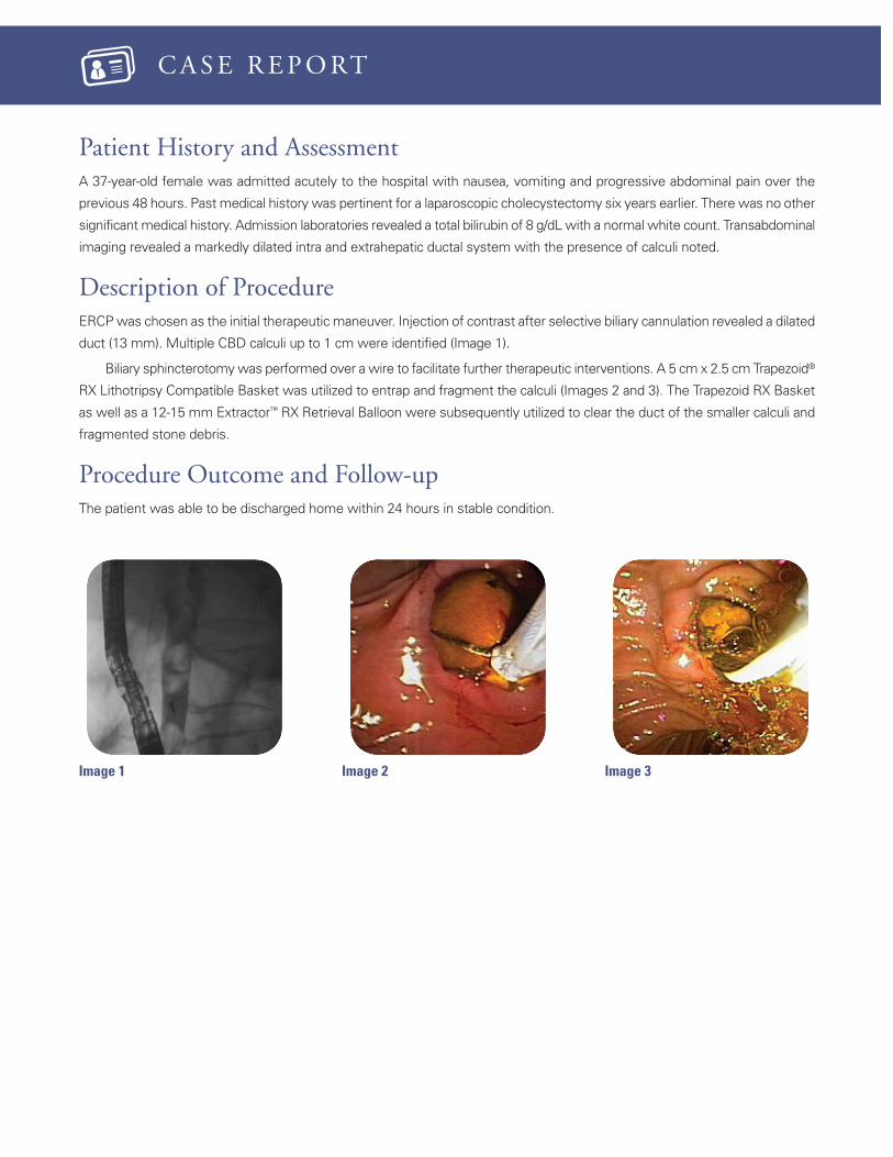
Description of ProcedureERCPwas chosen as the initial therapeutic maneuver. Injection of contrast after selective biliary cannulation revealed a dilated
duct (13 mm). Multiple CBD calculi up to 1 cm were identified (Image 1).
Biliary sphincterotomywas performed over a wire to facilitate further therapeutic interventions. A 5 cm x 2.5 cm Trapezoid®
RX Lithotripsy Compatible Basket was utilized to entrap and fragment the calculi (Images 2 and 3). The Trapezoid RX Basket
as well as a 12-15 mm Extractor™ RX Retrieval Balloon were subsequently utilized to clear the duct of the smaller calculi and
fragmented stone debris.
Procedure Outcome and Follow-upThe patient was able to be discharged home within 24 hours in stable condition.
Image 1 Image 2 Image 3
Lithotripter baskets add to the endoscopists’ ability to deal with a variety of stone cases. While all lithotripter baskets are effective inthe management of medium and large calculi, additional safety issues may warrant special considerations. Infrequently, stones maynot respond to the forces generated by mechanical lithotripsy. In these situations, the patient may require surgical exploration if thestones cannot be removed or released. Fortunately, some lithotripter baskets have a built-in safety feature allowing for wire/tipfracture and removal of the basket if specific tensile forces are exceeded. While the residual ductal calculi still need to be dealt with,the need for urgent surgery is avoided.
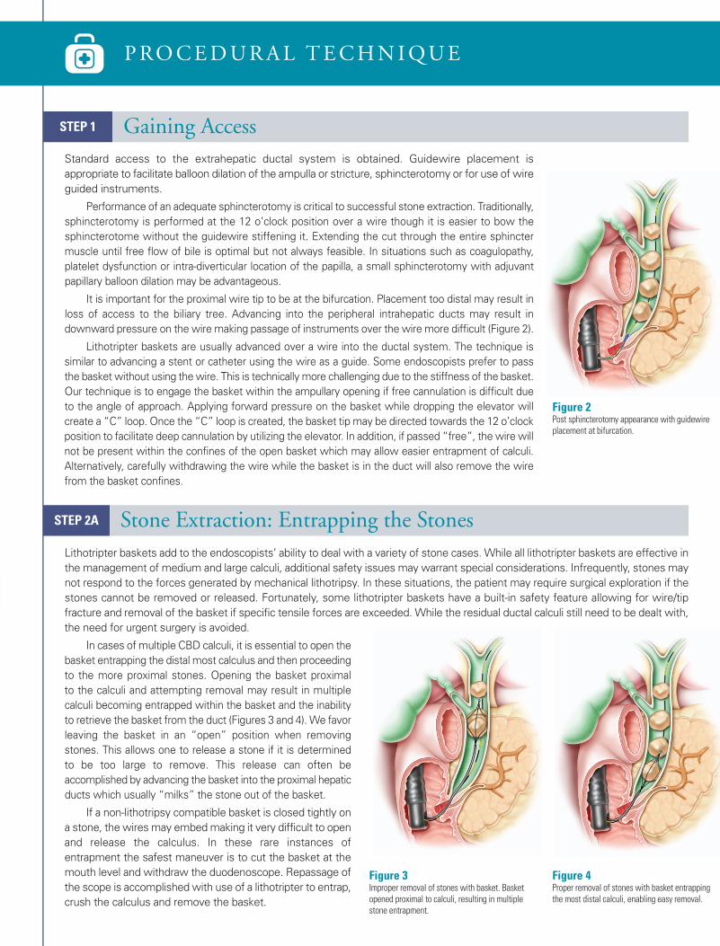
In cases of multiple CBD calculi, it is essential to open thebasket entrapping the distal most calculus and then proceedingto the more proximal stones. Opening the basket proximalto the calculi and attempting removal may result in multiplecalculi becoming entrapped within the basket and the inabilityto retrieve the basket from the duct (Figures 3 and 4). We favorleaving the basket in an “open” position when removingstones. This allows one to release a stone if it is determinedto be too large to remove. This release can often beaccomplished by advancing the basket into the proximal hepaticducts which usually “milks” the stone out of the basket.
If a non-lithotripsy compatible basket is closed tightly ona stone, the wiresmay embedmaking it very difficult to openand release the calculus. In these rare instances ofentrapment the safest maneuver is to cut the basket at themouth level and withdraw the duodenoscope. Repassage ofthe scope is accomplishedwith use of a lithotripter to entrap,crush the calculus and remove the basket.
Standard access to the extrahepatic ductal system is obtained. Guidewire placement isappropriate to facilitate balloon dilation of the ampulla or stricture, sphincterotomy or for use of wireguided instruments.
Performance of an adequate sphincterotomy is critical to successful stone extraction. Traditionally,sphincterotomy is performed at the 12 o’clock position over a wire though it is easier to bow thesphincterotome without the guidewire stiffening it. Extending the cut through the entire sphinctermuscle until free flow of bile is optimal but not always feasible. In situations such as coagulopathy,platelet dysfunction or intra-diverticular location of the papilla, a small sphincterotomy with adjuvantpapillary balloon dilation may be advantageous.
It is important for the proximal wire tip to be at the bifurcation. Placement too distal may result inloss of access to the biliary tree. Advancing into the peripheral intrahepatic ducts may result indownward pressure on the wire making passage of instruments over the wire more difficult (Figure 2).
Lithotripter baskets are usually advanced over a wire into the ductal system. The technique issimilar to advancing a stent or catheter using the wire as a guide. Some endoscopists prefer to passthe basket without using thewire. This is technically more challenging due to the stiffness of the basket.Our technique is to engage the basket within the ampullary opening if free cannulation is difficult dueto the angle of approach. Applying forward pressure on the basket while dropping the elevator willcreate a “C” loop. Once the “C” loop is created, the basket tip may be directed towards the 12 o’clockposition to facilitate deep cannulation by utilizing the elevator. In addition, if passed “free”, the wire willnot be present within the confines of the open basket which may allow easier entrapment of calculi.Alternatively, carefully withdrawing the wire while the basket is in the duct will also remove the wirefrom the basket confines.
STEP 1 Gaining Access
Figure 2Post sphincterotomy appearance with guidewireplacement at bifurcation.
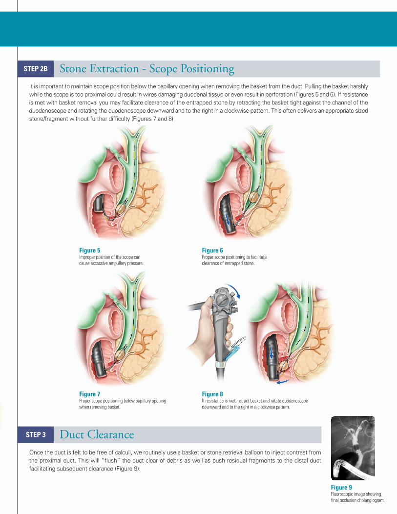
Figure 6Proper scope positioning to facilitateclearance of entrapped stone.
Figure 7Proper scope positioning below papillary openingwhen removing basket.
Figure 8If resistance is met, retract basket and rotate duodenoscopedownward and to the right in a clockwise pattern.
PROCEDURAL TECHNIQUE
Introduction
Many techniques are utilized to facilitate removal of biliary
ductal calculi, including sphincterotomy, hydrostatic balloon
dilatation of the ampulla, stone retrieval balloons, baskets, and
electrohydraulic, mechanical and laser lithotripsy. Basket assisted
techniques have generally been less favored due to concern of
intraductal entrapment. This concern is prudent in cases where
calculi are larger than the distal duct diameter or under conditions
where only a small sphincterotomy may be performed (i.e.
coagulopathy, periampullary diverticula). However, proper
selection and use of baskets will allow successful extraction of most calculi without difficulty. This
paper is designed to allay unwarranted concerns and provide advice on proper use of this technique.
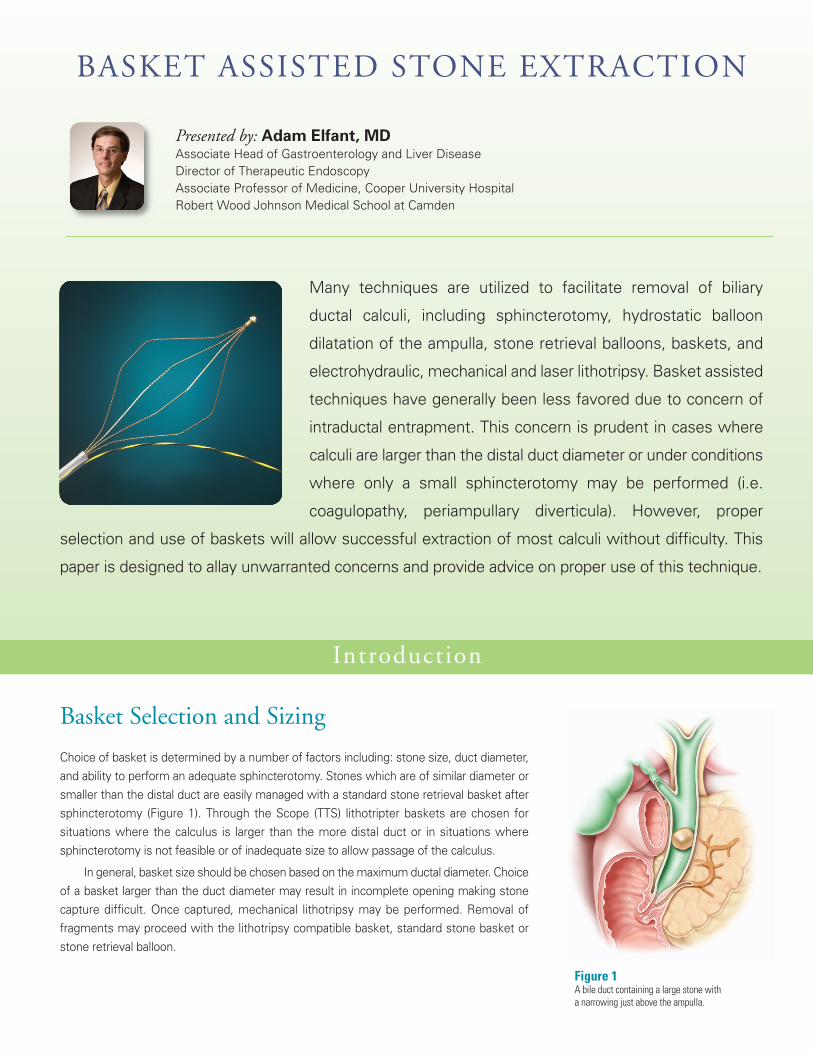
Figure 1A bile duct containing a large stone witha narrowing just above the ampulla.
Basket Selection and Sizing
Choice of basket is determined by a number of factors including: stone size, duct diameter,and ability to perform an adequate sphincterotomy. Stones which are of similar diameter orsmaller than the distal duct are easily managed with a standard stone retrieval basket aftersphincterotomy (Figure 1). Through the Scope (TTS) lithotripter baskets are chosen forsituations where the calculus is larger than the more distal duct or in situations wheresphincterotomy is not feasible or of inadequate size to allow passage of the calculus.
In general, basket size should be chosen based on themaximum ductal diameter. Choiceof a basket larger than the duct diameter may result in incomplete opening making stonecapture difficult. Once captured, mechanical lithotripsy may be performed. Removal offragments may proceed with the lithotripsy compatible basket, standard stone basket orstone retrieval balloon.
STEP 2A Stone Extraction: Entrapping the Stones
STEP 2B Stone Extraction - Scope PositioningIt is important tomaintain scope position below the papillary opening when removing the basket from the duct. Pulling the basket harshlywhile the scope is too proximal could result in wires damaging duodenal tissue or even result in perforation (Figures 5 and 6). If resistanceis met with basket removal you may facilitate clearance of the entrapped stone by retracting the basket tight against the channel of theduodenoscope and rotating the duodenoscope downward and to the right in a clockwise pattern. This often delivers an appropriate sizedstone/fragment without further difficulty (Figures 7 and 8).
STEP 3 Duct ClearanceOnce the duct is felt to be free of calculi, we routinely use a basket or stone retrieval balloon to inject contrast fromthe proximal duct. This will “flush” the duct clear of debris as well as push residual fragments to the distal ductfacilitating subsequent clearance (Figure 9).
Figure 3Improper removal of stones with basket. Basketopened proximal to calculi, resulting in multiplestone entrapment.
Figure 4Proper removal of stones with basket entrappingthe most distal calculi, enabling easy removal.
BASKET ASSISTED STONE EXTRACTION
Figure 5Improper position of the scope cancause excessive ampullary pressure.
Presented by: Adam Elfant, MDAssociate Head of Gastroenterology and Liver DiseaseDirector of Therapeutic EndoscopyAssociate Professor of Medicine, Cooper University HospitalRobert Wood Johnson Medical School at Camden
Figure 9Fluoroscopic image showingfinal occlusion cholangiogram.
Lithotripter baskets add to the endoscopists’ ability to deal with a variety of stone cases. While all lithotripter baskets are effective inthe management of medium and large calculi, additional safety issues may warrant special considerations. Infrequently, stones maynot respond to the forces generated by mechanical lithotripsy. In these situations, the patient may require surgical exploration if thestones cannot be removed or released. Fortunately, some lithotripter baskets have a built-in safety feature allowing for wire/tipfracture and removal of the basket if specific tensile forces are exceeded. While the residual ductal calculi still need to be dealt with,the need for urgent surgery is avoided.
In cases of multiple CBD calculi, it is essential to open thebasket entrapping the distal most calculus and then proceedingto the more proximal stones. Opening the basket proximalto the calculi and attempting removal may result in multiplecalculi becoming entrapped within the basket and the inabilityto retrieve the basket from the duct (Figures 3 and 4). We favorleaving the basket in an “open” position when removingstones. This allows one to release a stone if it is determinedto be too large to remove. This release can often beaccomplished by advancing the basket into the proximal hepaticducts which usually “milks” the stone out of the basket.
If a non-lithotripsy compatible basket is closed tightly ona stone, the wiresmay embedmaking it very difficult to openand release the calculus. In these rare instances ofentrapment the safest maneuver is to cut the basket at themouth level and withdraw the duodenoscope. Repassage ofthe scope is accomplishedwith use of a lithotripter to entrap,crush the calculus and remove the basket.
Standard access to the extrahepatic ductal system is obtained. Guidewire placement isappropriate to facilitate balloon dilation of the ampulla or stricture, sphincterotomy or for use of wireguided instruments.
Performance of an adequate sphincterotomy is critical to successful stone extraction. Traditionally,sphincterotomy is performed at the 12 o’clock position over a wire though it is easier to bow thesphincterotome without the guidewire stiffening it. Extending the cut through the entire sphinctermuscle until free flow of bile is optimal but not always feasible. In situations such as coagulopathy,platelet dysfunction or intra-diverticular location of the papilla, a small sphincterotomy with adjuvantpapillary balloon dilation may be advantageous.
It is important for the proximal wire tip to be at the bifurcation. Placement too distal may result inloss of access to the biliary tree. Advancing into the peripheral intrahepatic ducts may result indownward pressure on the wire making passage of instruments over the wire more difficult (Figure 2).
Lithotripter baskets are usually advanced over a wire into the ductal system. The technique issimilar to advancing a stent or catheter using the wire as a guide. Some endoscopists prefer to passthe basket without using thewire. This is technically more challenging due to the stiffness of the basket.Our technique is to engage the basket within the ampullary opening if free cannulation is difficult dueto the angle of approach. Applying forward pressure on the basket while dropping the elevator willcreate a “C” loop. Once the “C” loop is created, the basket tip may be directed towards the 12 o’clockposition to facilitate deep cannulation by utilizing the elevator. In addition, if passed “free”, the wire willnot be present within the confines of the open basket which may allow easier entrapment of calculi.Alternatively, carefully withdrawing the wire while the basket is in the duct will also remove the wirefrom the basket confines.
STEP 1 Gaining Access
Figure 2Post sphincterotomy appearance with guidewireplacement at bifurcation.
Figure 6Proper scope positioning to facilitateclearance of entrapped stone.
Figure 7Proper scope positioning below papillary openingwhen removing basket.
Figure 8If resistance is met, retract basket and rotate duodenoscopedownward and to the right in a clockwise pattern.
PROCEDURAL TECHNIQUE
Introduction
Many techniques are utilized to facilitate removal of biliary
ductal calculi, including sphincterotomy, hydrostatic balloon
dilatation of the ampulla, stone retrieval balloons, baskets, and
electrohydraulic, mechanical and laser lithotripsy. Basket assisted
techniques have generally been less favored due to concern of
intraductal entrapment. This concern is prudent in cases where
calculi are larger than the distal duct diameter or under conditions
where only a small sphincterotomy may be performed (i.e.
coagulopathy, periampullary diverticula). However, proper
selection and use of baskets will allow successful extraction of most calculi without difficulty. This
paper is designed to allay unwarranted concerns and provide advice on proper use of this technique.
Figure 1A bile duct containing a large stone witha narrowing just above the ampulla.
Basket Selection and Sizing
Choice of basket is determined by a number of factors including: stone size, duct diameter,and ability to perform an adequate sphincterotomy. Stones which are of similar diameter orsmaller than the distal duct are easily managed with a standard stone retrieval basket aftersphincterotomy (Figure 1). Through the Scope (TTS) lithotripter baskets are chosen forsituations where the calculus is larger than the more distal duct or in situations wheresphincterotomy is not feasible or of inadequate size to allow passage of the calculus.
In general, basket size should be chosen based on themaximum ductal diameter. Choiceof a basket larger than the duct diameter may result in incomplete opening making stonecapture difficult. Once captured, mechanical lithotripsy may be performed. Removal offragments may proceed with the lithotripsy compatible basket, standard stone basket orstone retrieval balloon.
STEP 2A Stone Extraction: Entrapping the Stones
STEP 2B Stone Extraction - Scope PositioningIt is important tomaintain scope position below the papillary opening when removing the basket from the duct. Pulling the basket harshlywhile the scope is too proximal could result in wires damaging duodenal tissue or even result in perforation (Figures 5 and 6). If resistanceis met with basket removal you may facilitate clearance of the entrapped stone by retracting the basket tight against the channel of theduodenoscope and rotating the duodenoscope downward and to the right in a clockwise pattern. This often delivers an appropriate sizedstone/fragment without further difficulty (Figures 7 and 8).
STEP 3 Duct ClearanceOnce the duct is felt to be free of calculi, we routinely use a basket or stone retrieval balloon to inject contrast fromthe proximal duct. This will “flush” the duct clear of debris as well as push residual fragments to the distal ductfacilitating subsequent clearance (Figure 9).
Figure 3Improper removal of stones with basket. Basketopened proximal to calculi, resulting in multiplestone entrapment.
Figure 4Proper removal of stones with basket entrappingthe most distal calculi, enabling easy removal.
BASKET ASSISTED STONE EXTRACTION
Figure 5Improper position of the scope cancause excessive ampullary pressure.
Presented by: Adam Elfant, MDAssociate Head of Gastroenterology and Liver DiseaseDirector of Therapeutic EndoscopyAssociate Professor of Medicine, Cooper University HospitalRobert Wood Johnson Medical School at Camden
Figure 9Fluoroscopic image showingfinal occlusion cholangiogram.
Presented by:
Adam Elfant, MDAssociate Head of Gastroenterology and Liver Disease
Director of Therapeutic Endoscopy
Associate Professor of Medicine
Cooper University Hospital
Robert Wood Johnson Medical School at Camden
BASKET ASSISTEDSTONE EXTRACTIONOVERVIEW, PROCEDURAL TECHNIQUE AND CASE REPORT
Boston Scientific CorporationOne Boston Scientific PlaceNatick, MA 01760-1537www.bostonscientific.com/endoscopy
Ordering Information1.800.225.3226
© 2009 Boston Scientific Corporationor its affiliates. All rights reserved.
DVG2660 2.5M November 2009
CASE REPORT
Patient History and AssessmentA 37-year-old female was admitted acutely to the hospital with nausea, vomiting and progressive abdominal pain over the
previous 48 hours. Past medical history was pertinent for a laparoscopic cholecystectomy six years earlier. There was no other
significant medical history. Admission laboratories revealed a total bilirubin of 8 g/dLwith a normal white count. Transabdominal
imaging revealed a markedly dilated intra and extrahepatic ductal system with the presence of calculi noted.
Description of ProcedureERCPwas chosen as the initial therapeutic maneuver. Injection of contrast after selective biliary cannulation revealed a dilated
duct (13 mm). Multiple CBD calculi up to 1 cm were identified (Image 1).
Biliary sphincterotomywas performed over a wire to facilitate further therapeutic interventions. A 5 cm x 2.5 cm Trapezoid®
RX Lithotripsy Compatible Basket was utilized to entrap and fragment the calculi (Images 2 and 3). The Trapezoid RX Basket
as well as a 12-15 mm Extractor™ RX Retrieval Balloon were subsequently utilized to clear the duct of the smaller calculi and
fragmented stone debris.
Procedure Outcome and Follow-upThe patient was able to be discharged home within 24 hours in stable condition.
Image 1 Image 2 Image 3
ENDO-150707-AA June 2013