basic introduction to cvs diseases by dr. mohamed abd almoneim attia
TRANSCRIPT
Basic introduction to CVS diseases
ByDr. MOHAMED ABD ALMONEIM ATTIA
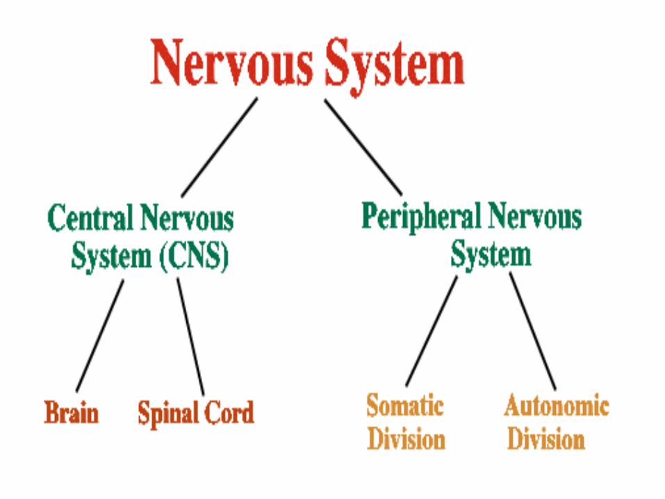
Peripheral Nervous System
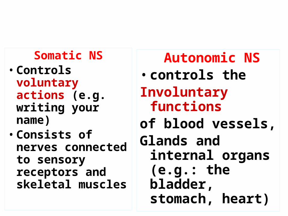
Somatic NS• Controls voluntary
actions (e.g. writing your name)
• Consists of nerves connected to sensory receptors and skeletal muscles
Autonomic NS• controls theInvoluntary functionsof blood vessels,Glands and internal
organs (e.g.: the bladder, stomach, heart)
Somatic NS Vs. Autonomic NS
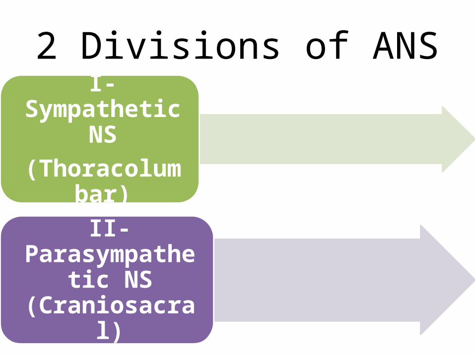
2 Divisions of ANSI- Sympathetic
NS(Thoracolumbar)
II- Parasympathetic NS (Craniosacral)
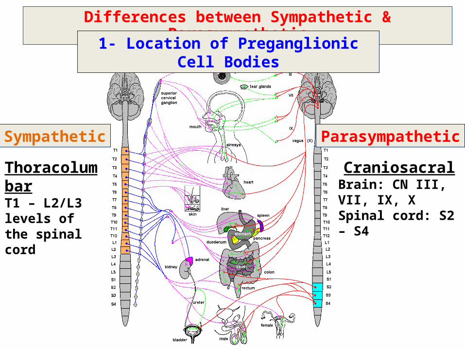
Differences between Sympathetic & Parasympathetic
1- Location of Preganglionic Cell Bodies
ThoracolumbarT1 – L2/L3 levels of the spinal cord
CraniosacralBrain: CN III, VII, IX, XSpinal cord: S2 – S4
Sympathetic Parasympathetic
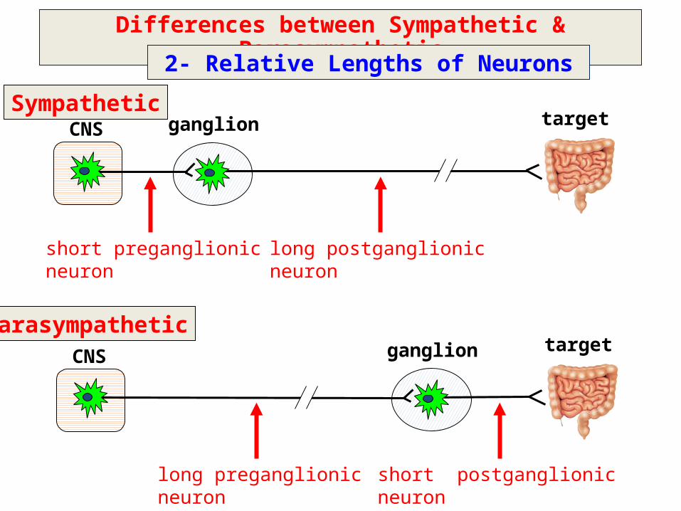
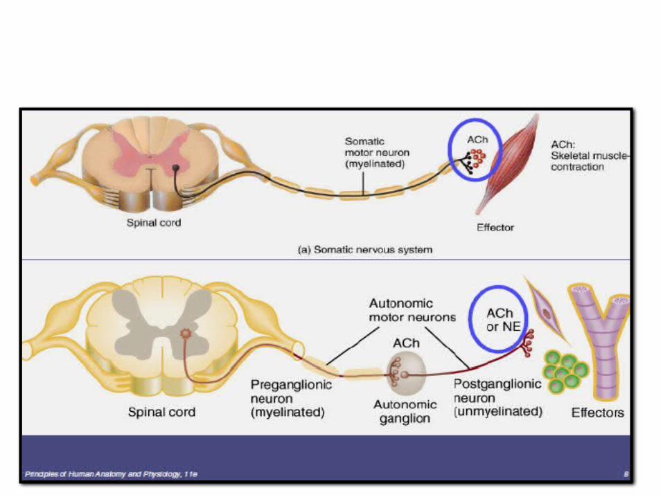
SympatheticCNS ganglion
short preganglionicneuron
long postganglionicneuron
target
ParasympatheticCNS ganglion
long preganglionicneuron
target
short postganglionicneuron
Differences between Sympathetic & Parasympathetic
2- Relative Lengths of Neurons
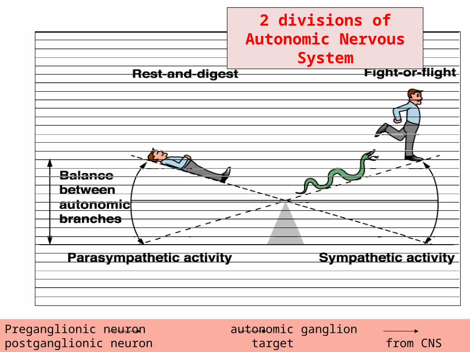
2 divisions of Autonomic Nervous System
Preganglionic neuron autonomic ganglion postganglionic neuron target from CNS outside CNS
2 divisions of ANS
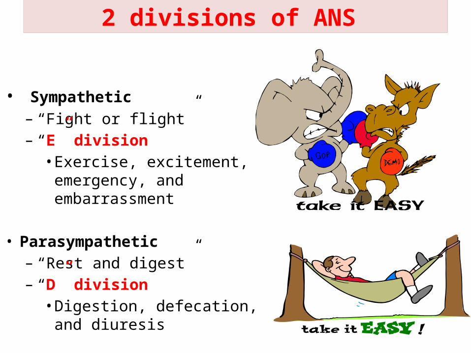
• Sympathetic – “Fight or flight”– “E” division• Exercise, excitement,
emergency, and embarrassment
• Parasympathetic – “Rest and digest”– “D” division• Digestion, defecation, and
diuresis
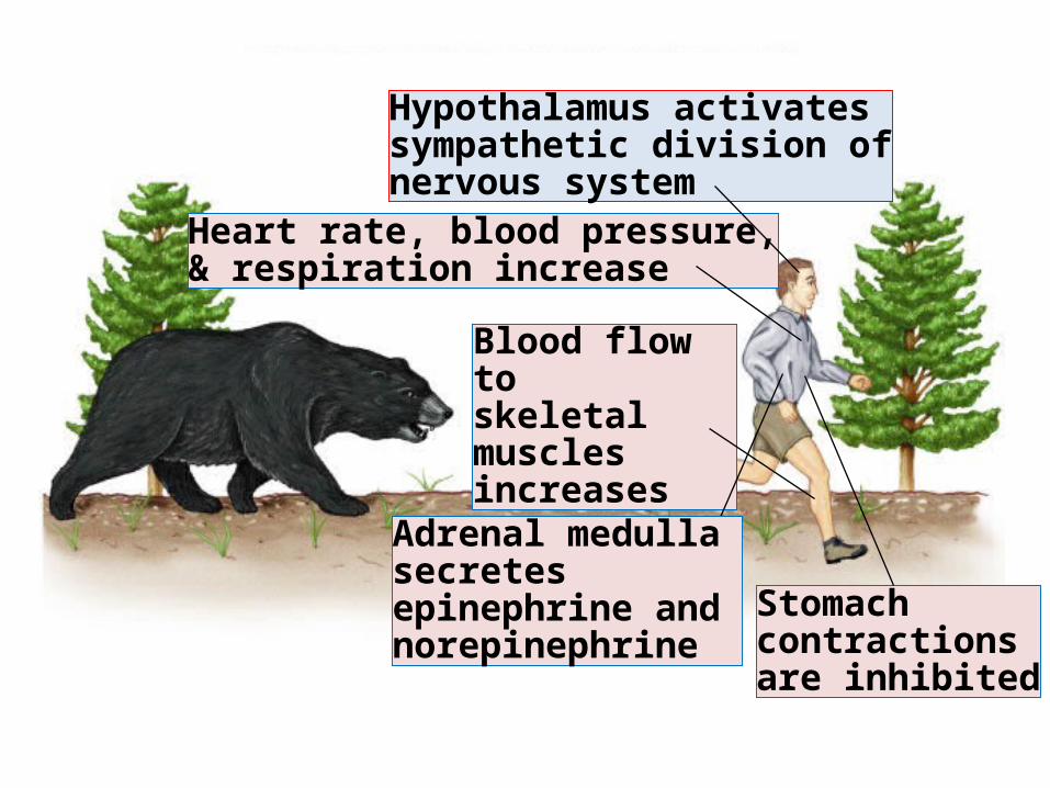
Fig. 45.34(TE Art)Hypothalamus activatessympathetic division ofnervous system
Heart rate, blood pressure,& respiration increase
Blood flow toskeletal musclesincreases
Stomachcontractions are inhibited
Adrenal medulla secretes epinephrine and norepinephrine
Autonomic supply to different tissues and organsMost organs of the body receive dual innervation
consisting of sympathetic and parasympathetic In general the parasympathetic and sympathetic
neurones mediate opposing responses in the effector organ, although some exceptions to this generalization exist.
Some organs in the body receive only sympathetic innervation, e.g., most blood vessels.
Some organs in the body receive only parasympathetic innervation, e.g., ciliary body.
How Autonomic physiological responses are mediated?
• Responses mediated by ANS are produced through chemical neurotransmitters carrying the message (orders) and acting on autonomic receptors in effectors organs
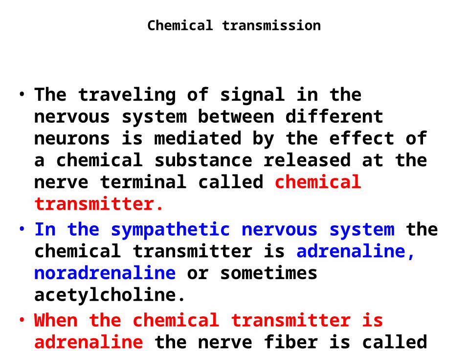
Chemical transmission
• The traveling of signal in the nervous system between different neurons is mediated by the effect of a chemical substance released at the nerve terminal called chemical transmitter.
• In the sympathetic nervous system the chemical transmitter is adrenaline, noradrenaline or sometimes acetylcholine.
• When the chemical transmitter is adrenaline the nerve fiber is called adrenergic,
• when the chemical transmitter is acetylcholine, the nerve fiber is called cholinergic.
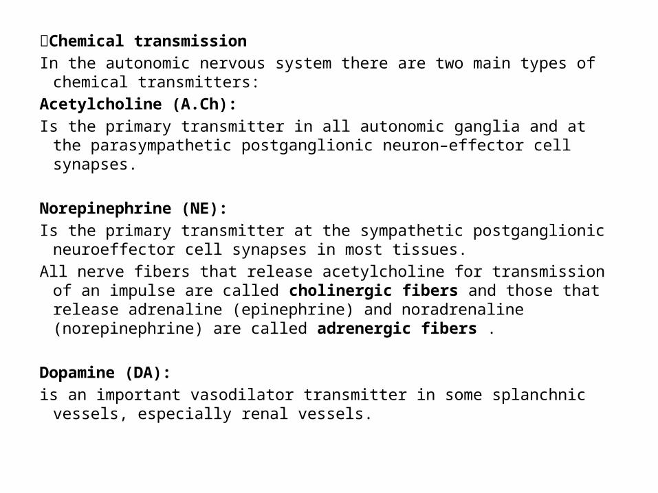
Chemical transmissionIn the autonomic nervous system there are two main types of chemical
transmitters:Acetylcholine (A.Ch): Is the primary transmitter in all autonomic ganglia and at the
parasympathetic postganglionic neuron–effector cell synapses. Norepinephrine (NE): Is the primary transmitter at the sympathetic postganglionic neuroeffector
cell synapses in most tissues. All nerve fibers that release acetylcholine for transmission of an impulse are
called cholinergic fibers and those that release adrenaline (epinephrine) and noradrenaline (norepinephrine) are called adrenergic fibers .
Dopamine (DA): is an important vasodilator transmitter in some splanchnic vessels,
especially renal vessels.
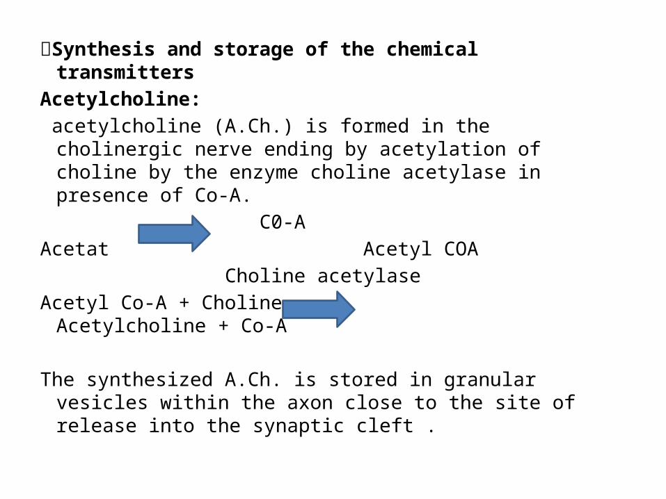
Synthesis and storage of the chemical transmittersAcetylcholine: acetylcholine (A.Ch.) is formed in the cholinergic nerve
ending by acetylation of choline by the enzyme choline acetylase in presence of Co-A.
C0-A Acetat Acetyl COA Choline acetylaseAcetyl Co-A + Choline Acetylcholine + Co-A The synthesized A.Ch. is stored in granular vesicles within
the axon close to the site of release into the synaptic cleft .
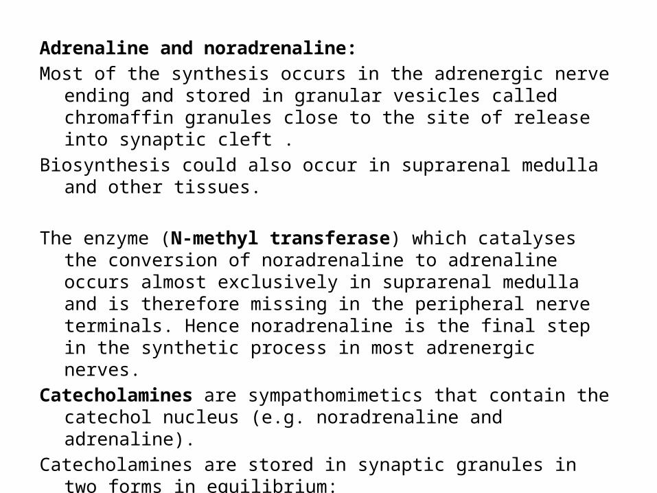
Adrenaline and noradrenaline: Most of the synthesis occurs in the adrenergic nerve ending and stored in
granular vesicles called chromaffin granules close to the site of release into synaptic cleft .
Biosynthesis could also occur in suprarenal medulla and other tissues. The enzyme (N-methyl transferase) which catalyses the conversion of
noradrenaline to adrenaline occurs almost exclusively in suprarenal medulla and is therefore missing in the peripheral nerve terminals. Hence noradrenaline is the final step in the synthetic process in most adrenergic nerves.
Catecholamines are sympathomimetics that contain the catechol nucleus (e.g. noradrenaline and adrenaline).
Catecholamines are stored in synaptic granules in two forms in equilibrium:Bound noradrenaline with ATP and protein is the (inactive part). Free noradrenaline is released by nerve stimulation. Another portion of it is stored in the cytoplasm in free form (cytoplasmic free
noradrenaline).
Release of the chemical transmittersOn the arrival of an action potential to the nerve terminal, vesicles
containing acetylcholine or granules containing norepinephrine accumulate at the nerve ending facing the membrane.
Once they got closer to the membrane they open releasing their contents of the chemical transmitter through an exocytotic process.
Calcium ion is essential for this step (through Ca++ channels). The vesicle membranes are retained to return back to the nerve
cytoplasm to be used for storage again. The released chemical transmitter will stimulate the specific
autonomic receptors. The interaction of chemical transmitter with the receptors will
produce the post receptor events that will elicit the change in the function of the organ that contains that receptor.
Fate of the chemical transmitters ►Fate of noradrenaline: 1. Re-uptake: (80% of the released amount): It is the major mechanism by which the
released NE is removed from the vicinity of the sympathetic ends. Three uptake processes participate in removing NE:Neuronal uptake (uptake 1): active transport of NE into the neuronal
cytoplasmGranular uptake (uptake III): active transport of NE from the cytoplasm of the
nerve ends into the storage granules.Non-neuronal uptake (uptake II): uptake to the tissueN.B. 2. Metabolism by specific enzymes: NE and other catecholamines are metabolized into biologically inactive
products by oxidation (monoamine oxidase; MAO enzyme) and methylation (cathecol-Omethyl transferase; COMT enzyme)
N.B.Vanillyl Mandelic Acid (VMA) is the main
catecholamine metabolite in the urine. Normal Values is 4-8 mg/day. High Levels suggest the presence of a tumour in the suprarenal medulla (pheochromocytoma) that secretes excess catecholamines leading to hypertension. 2% of NE is excreted unmetabolized in the urine.
Autonomic receptorsThe released chemical transmitter will produce its physiological or
pharmacological action via stimulating the corresponding receptor on the effector cell.
Receptors that respond to acetylcholine are called cholinoceptors and those that respond to epinephrine and NE are called adrenoceptors
A. Adrenoceptors: Can be subdivided into alpha and beta adrenoceptor types. Alpha adrenoceptors Alpha 1 adrenoceptorsSite: postsynaptic in the effector tissues .Alpha 2 adrenoceptorsSite: PresynapticPostsynaptic
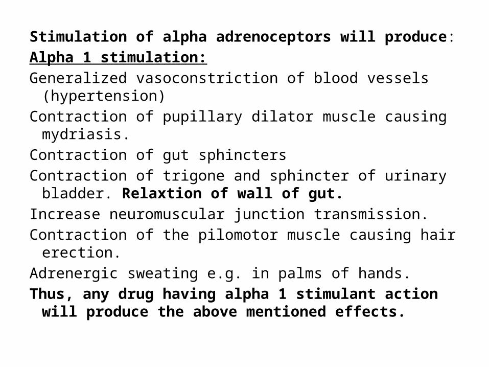
Stimulation of alpha adrenoceptors will produce:Alpha 1 stimulation:Generalized vasoconstriction of blood vessels
(hypertension)Contraction of pupillary dilator muscle causing mydriasis.Contraction of gut sphinctersContraction of trigone and sphincter of urinary bladder.
Relaxtion of wall of gut.Increase neuromuscular junction transmission.Contraction of the pilomotor muscle causing hair erection.Adrenergic sweating e.g. in palms of hands. Thus, any drug having alpha 1 stimulant action will
produce the above mentioned effects.
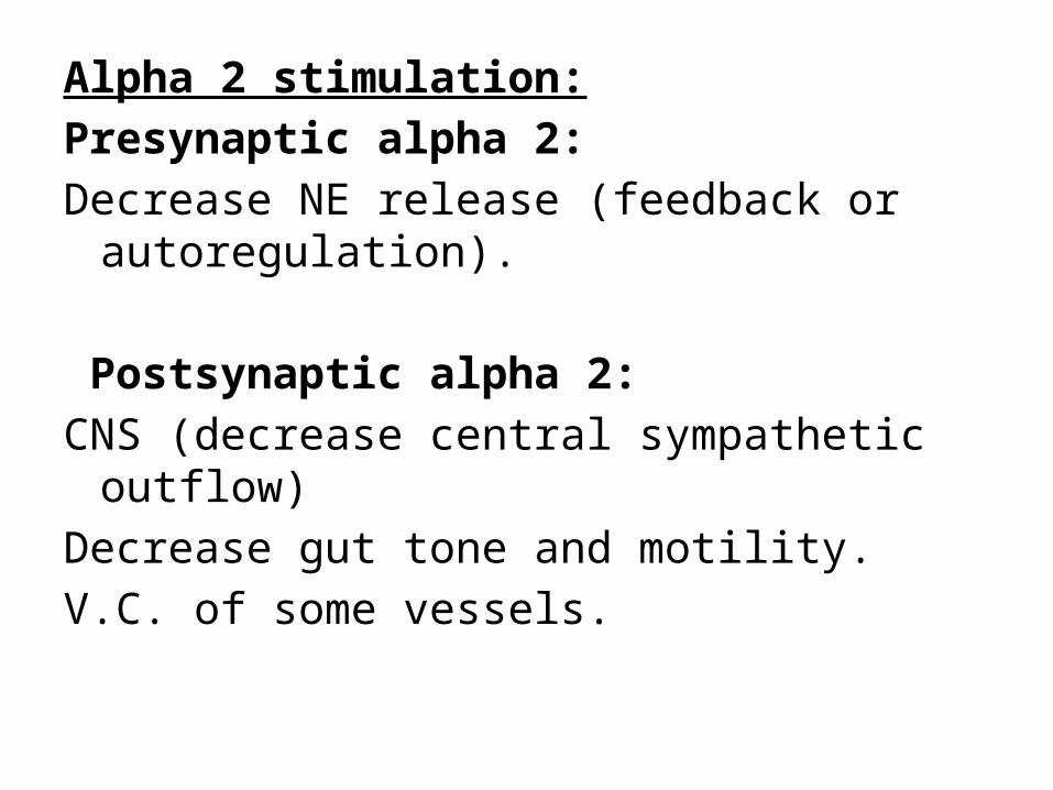
Alpha 2 stimulation:Presynaptic alpha 2:Decrease NE release (feedback or
autoregulation). Postsynaptic alpha 2:CNS (decrease central sympathetic outflow)Decrease gut tone and motility.V.C. of some vessels.
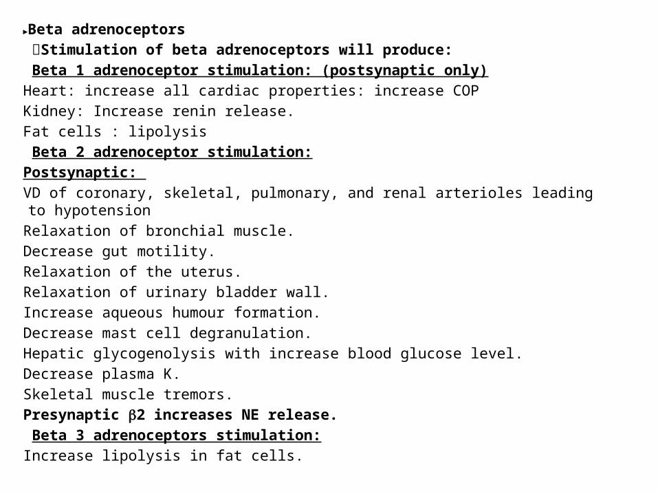
►Beta adrenoceptors Stimulation of beta adrenoceptors will produce: Beta 1 adrenoceptor stimulation: (postsynaptic only)Heart: increase all cardiac properties: increase COPKidney: Increase renin release.Fat cells : lipolysis Beta 2 adrenoceptor stimulation:Postsynaptic: VD of coronary, skeletal, pulmonary, and renal arterioles leading to hypotensionRelaxation of bronchial muscle.Decrease gut motility.Relaxation of the uterus.Relaxation of urinary bladder wall.Increase aqueous humour formation.Decrease mast cell degranulation.Hepatic glycogenolysis with increase blood glucose level.Decrease plasma K.Skeletal muscle tremors.Presynaptic 2 increases NE release. Beta 3 adrenoceptors stimulation:Increase lipolysis in fat cells.
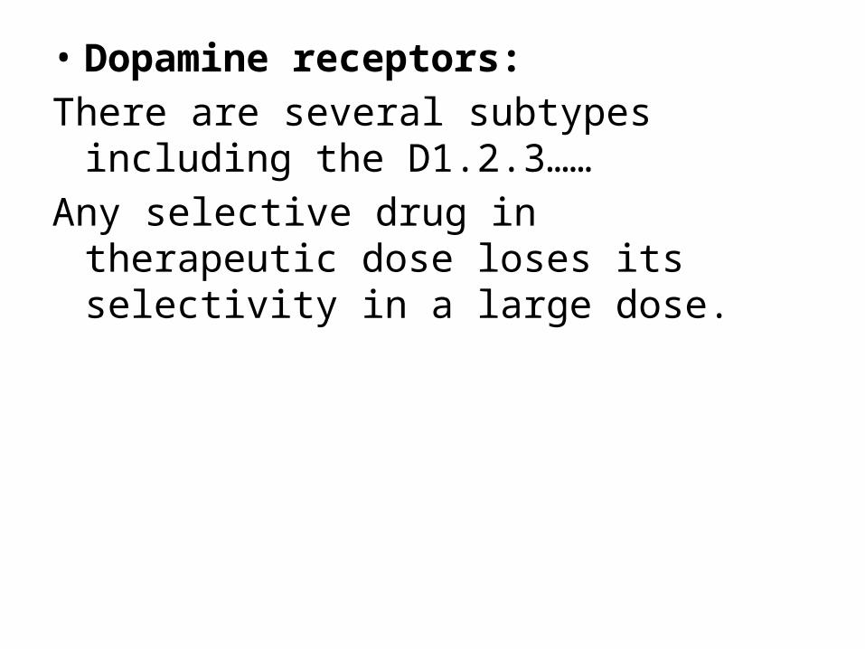
• Dopamine receptors:There are several subtypes including the
D1.2.3…… Any selective drug in therapeutic dose loses its
selectivity in a large dose.