basic hematology - aacc
TRANSCRIPT

Presented by AACC and NACB
Basic Hematology
Neil Harris MD Dept. of Pathology, Immunology and
Laboratory Medicine University of Florida College of
Medicine Gainesville, FL
LEARNING OBJECTIVES
1. Define “CBC” 2. Define and explain red cell indices including
derived values such as MCV, MCHC and MCH 3. Define and explain erythrocyte sedimentation rate 4. Describe the main cell types observed in peripheral
blood. 5. Describe what is meant by the term “differential
count”. 6. Describe the distribution & morphology of cells in
certain common “Blood Pictures”
Clinical Diagnostic Laboratories offer:
1. “CBC” 2. “CBC plus Diff.” [CBC + Differential
count] 3. Above ± “Reticulocyte count” What do these terms mean, and how are
they used?
CBC 1. White Cell count (leukocyte
count)
2. Platelet count
3. Red cell count, hemoglobin concentration and red cell indices
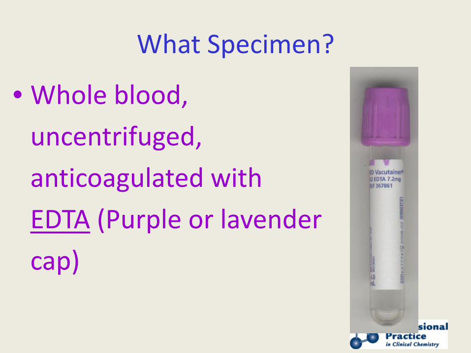
What Specimen?
• Whole blood, uncentrifuged, anticoagulated with EDTA (Purple or lavender cap)
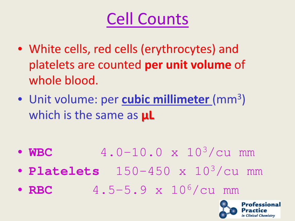
Cell Counts
• White cells, red cells (erythrocytes) and platelets are counted per unit volume of whole blood.
• Unit volume: per cubic millimeter (mm3) which is the same as µL
• WBC 4.0-10.0 x 10³/cu mm • Platelets 150-450 x 103/cu mm • RBC 4.5-5.9 x 10⁶/cu mm
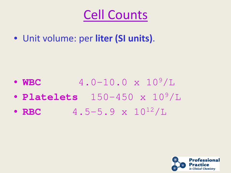
Cell Counts
• Unit volume: per liter (SI units).
• WBC 4.0-10.0 x 10⁹/L • Platelets 150-450 x 109/L • RBC 4.5-5.9 x 10¹²/L
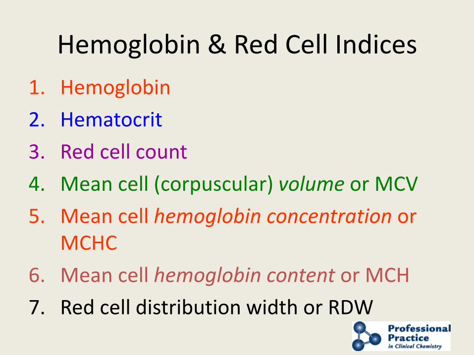
Hemoglobin & Red Cell Indices 1. Hemoglobin 2. Hematocrit 3. Red cell count 4. Mean cell (corpuscular) volume or MCV 5. Mean cell hemoglobin concentration or
MCHC 6. Mean cell hemoglobin content or MCH 7. Red cell distribution width or RDW
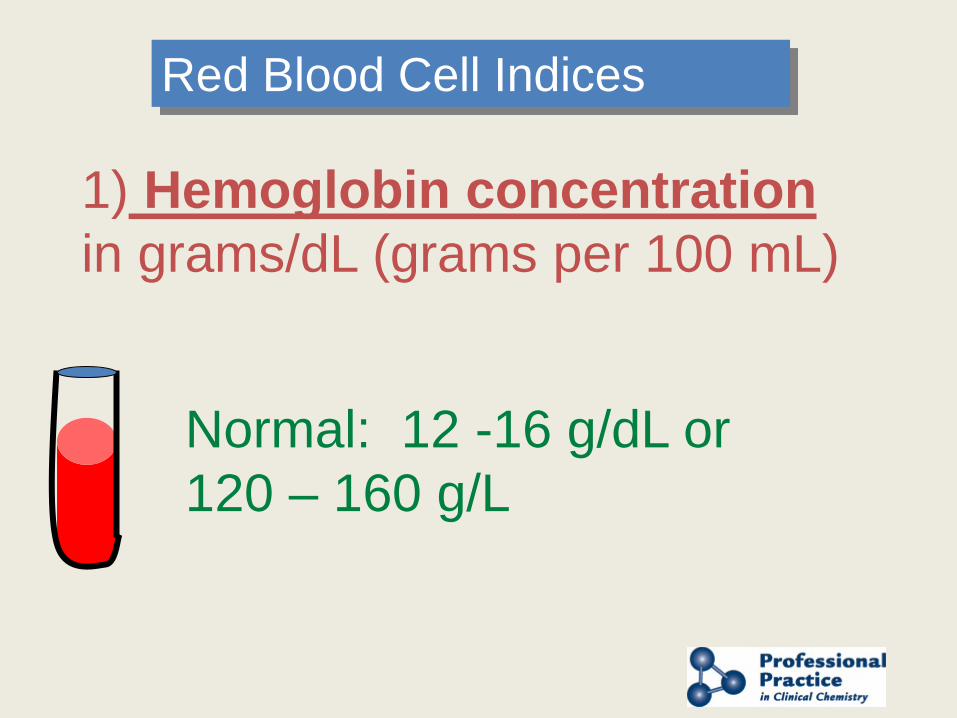
1) Hemoglobin concentration in grams/dL (grams per 100 mL)
Normal: 12 -16 g/dL or 120 – 160 g/L
Red Blood Cell Indices
Hemoglobin concentration
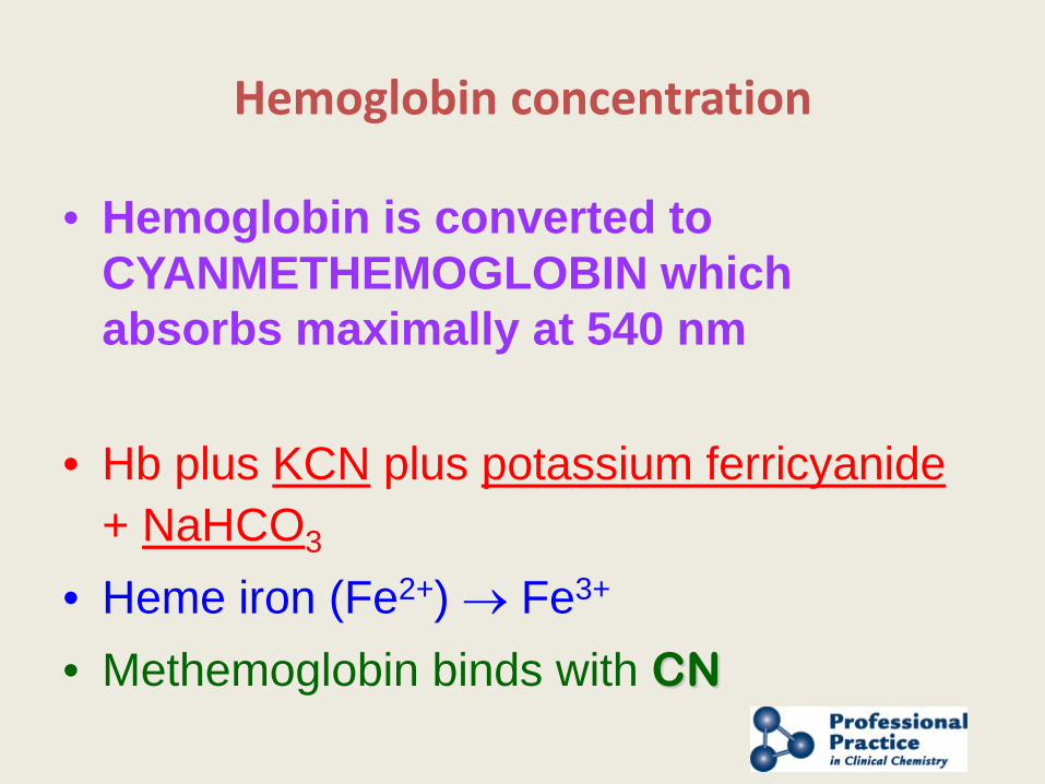
• Hemoglobin is converted to CYANMETHEMOGLOBIN which absorbs maximally at 540 nm
• Hb plus KCN plus potassium ferricyanide + NaHCO3
• Heme iron (Fe2+) → Fe3+ • Methemoglobin binds with CN
Hemoglobin concentration
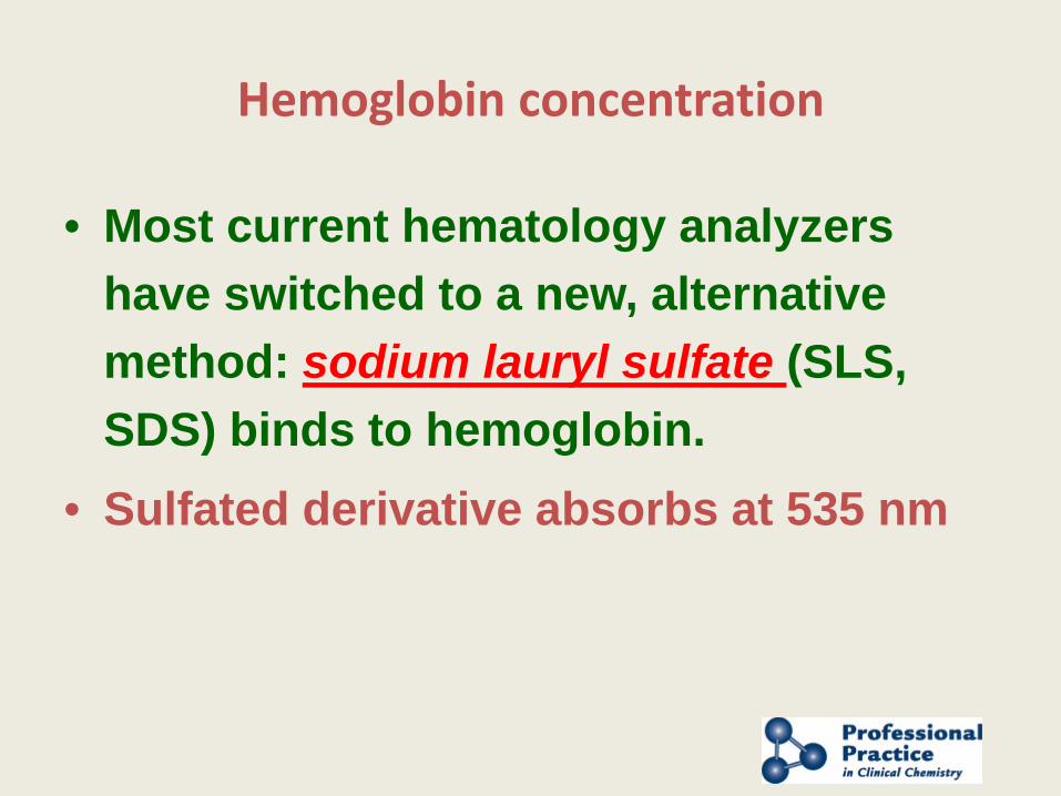
• Most current hematology analyzers have switched to a new, alternative method: sodium lauryl sulfate (SLS, SDS) binds to hemoglobin.
• Sulfated derivative absorbs at 535 nm
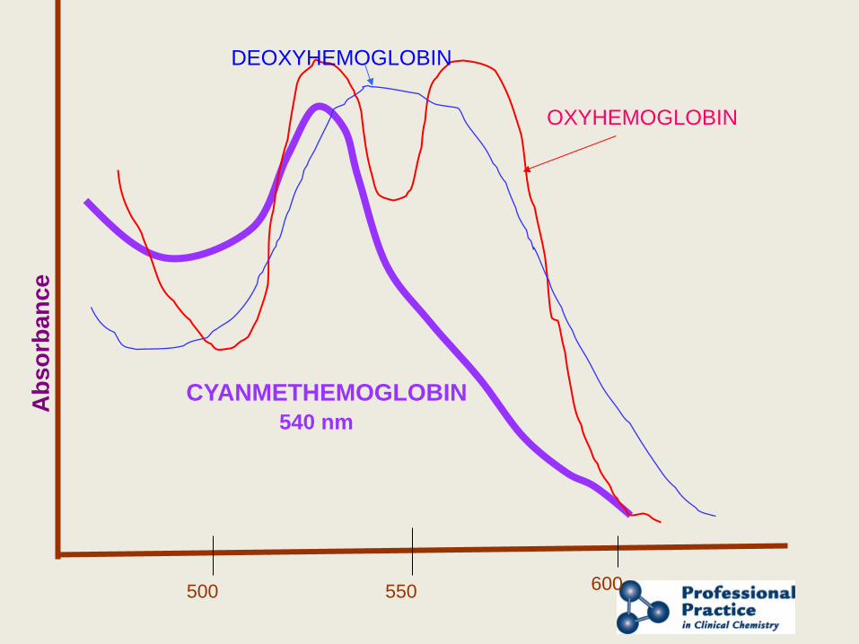
500 550 600
CYANMETHEMOGLOBIN 540 nm
OXYHEMOGLOBIN
DEOXYHEMOGLOBIN
Abs
orba
nce
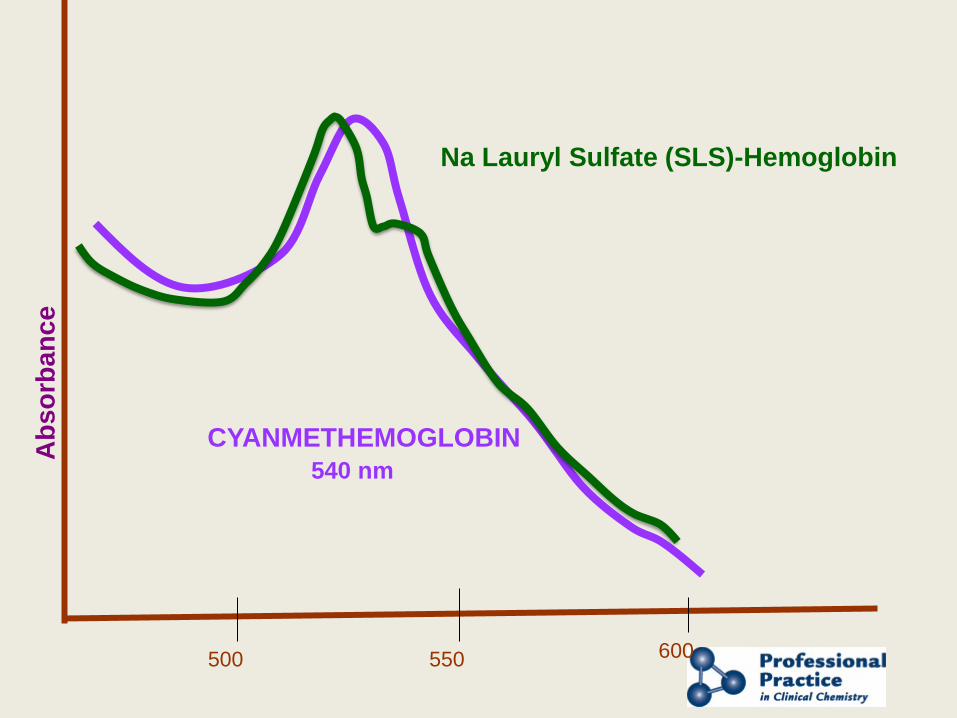
500 550 600
CYANMETHEMOGLOBIN 540 nm
Abs
orba
nce
Na Lauryl Sulfate (SLS)-Hemoglobin
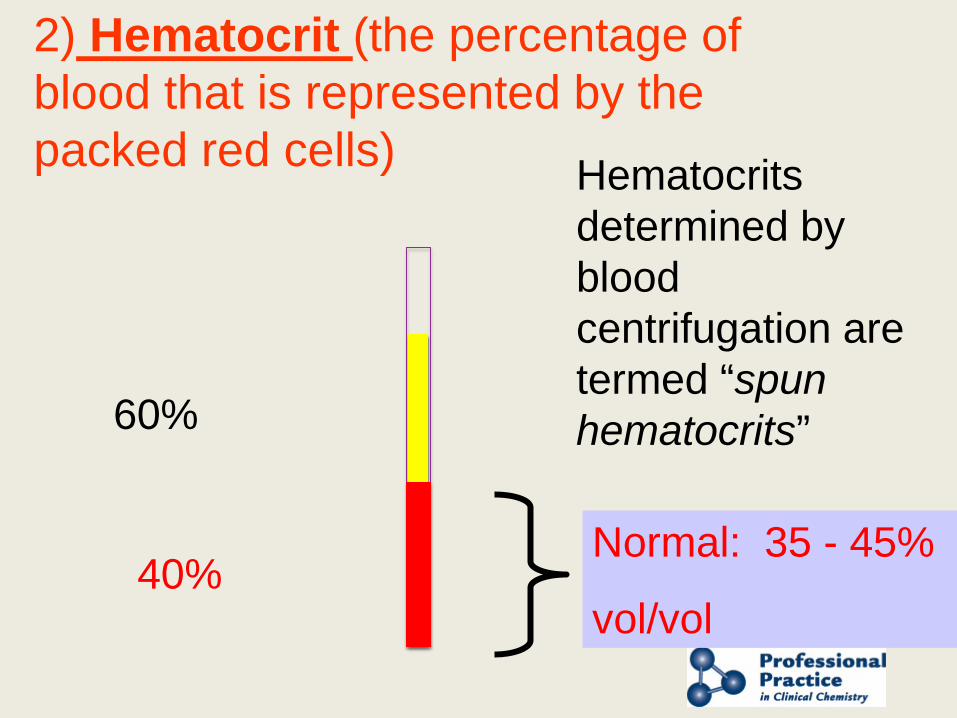
2) Hematocrit (the percentage of blood that is represented by the packed red cells)
40%
60%
Normal: 35 - 45%
vol/vol
Hematocrits determined by blood centrifugation are termed “spun hematocrits”
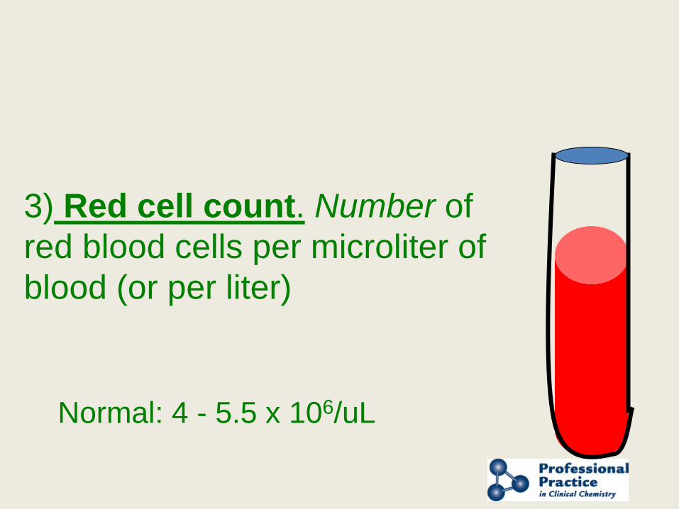
3) Red cell count. Number of red blood cells per microliter of blood (or per liter)
Normal: 4 - 5.5 x 10⁶/uL
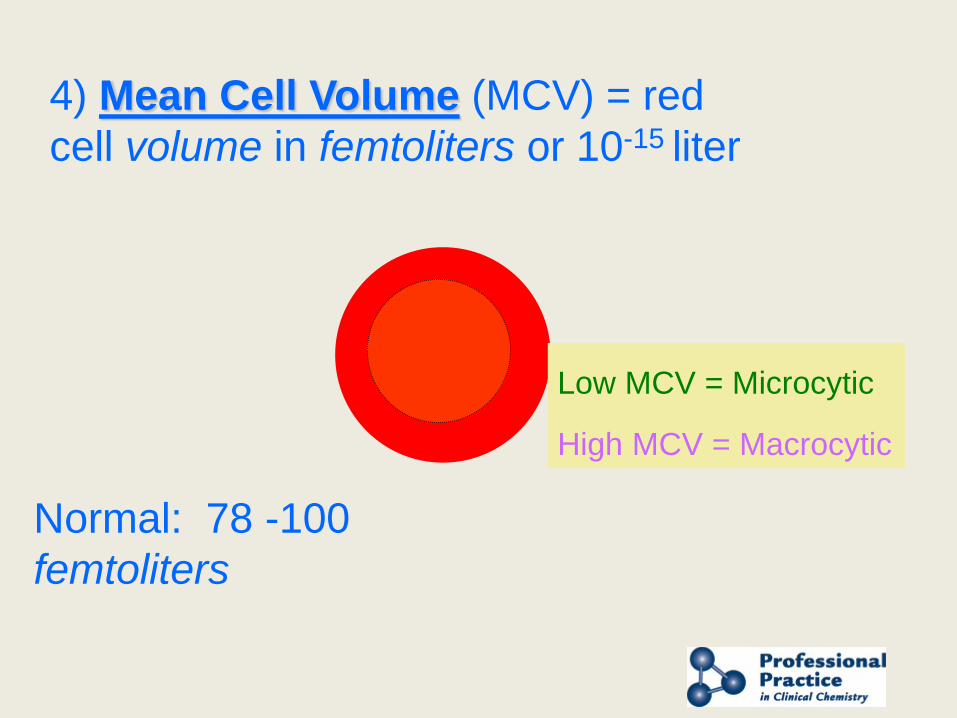
4) Mean Cell Volume (MCV) = red cell volume in femtoliters or 10-15 liter
Normal: 78 -100 femtoliters
Low MCV = Microcytic
High MCV = Macrocytic
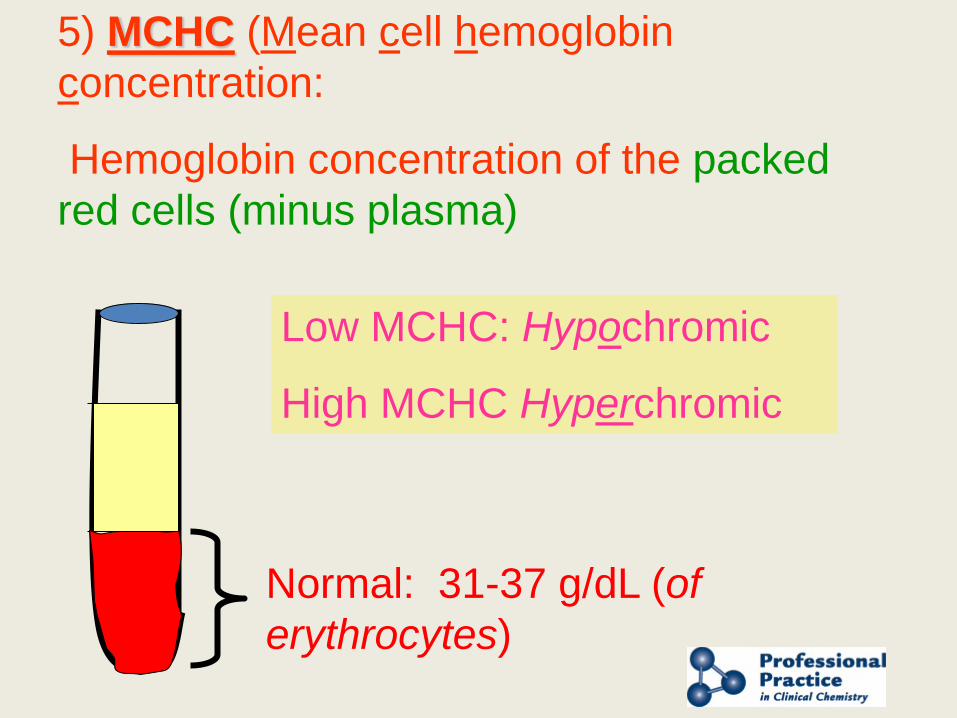
5) MCHC (Mean cell hemoglobin concentration:
Hemoglobin concentration of the packed red cells (minus plasma)
Normal: 31-37 g/dL (of erythrocytes)
Low MCHC: Hypochromic
High MCHC Hyperchromic
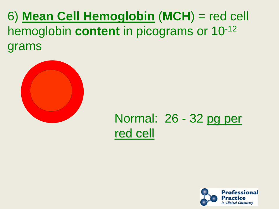
6) Mean Cell Hemoglobin (MCH) = red cell hemoglobin content in picograms or 10-12
grams
Normal: 26 - 32 pg per red cell
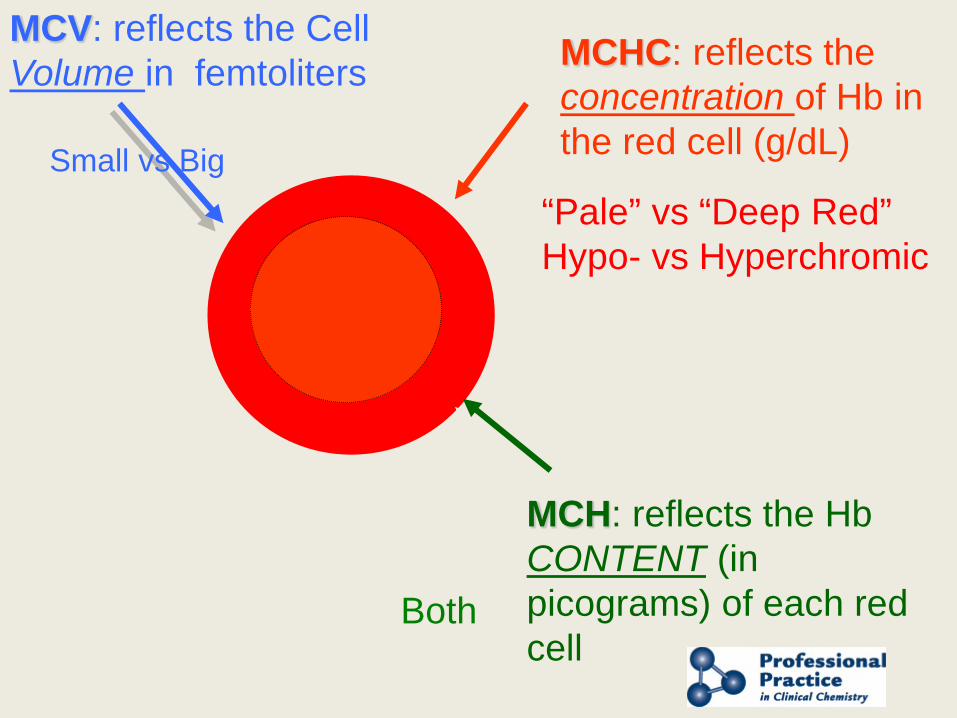
MCHC: reflects the concentration of Hb in the red cell (g/dL)
MCH: reflects the Hb CONTENT (in picograms) of each red cell
MCV: reflects the Cell Volume in femtoliters
Small vs Big “Pale” vs “Deep Red” Hypo- vs Hyperchromic
Both
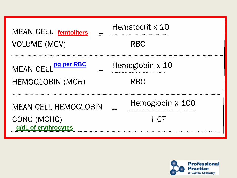
pg per RBC
femtoliters
g/dL of erythrocytes
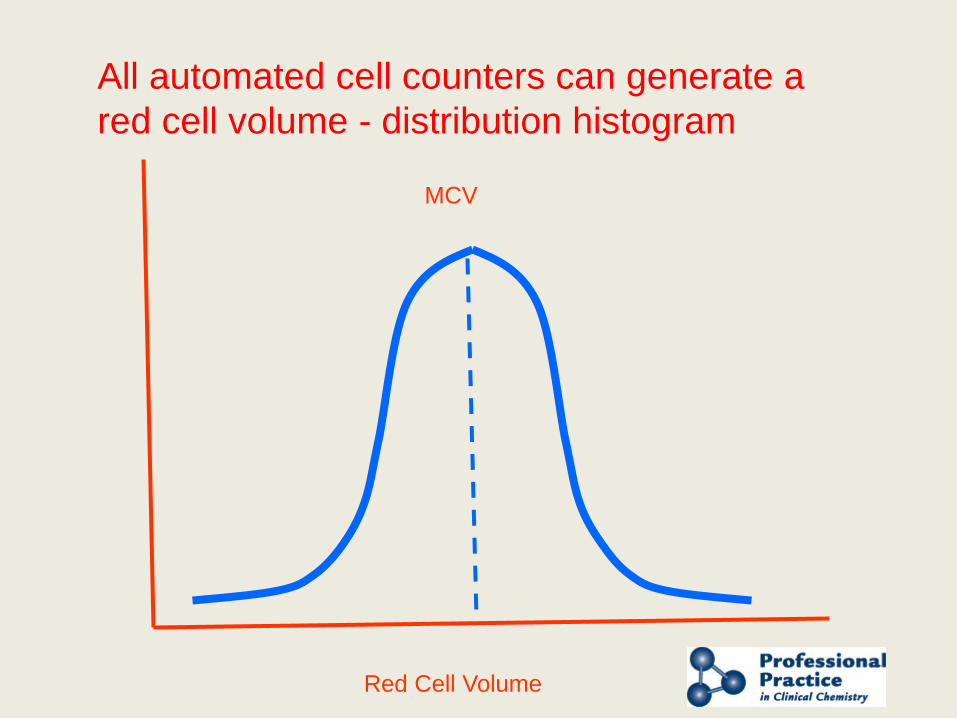
Red Cell Volume
MCV
All automated cell counters can generate a red cell volume - distribution histogram
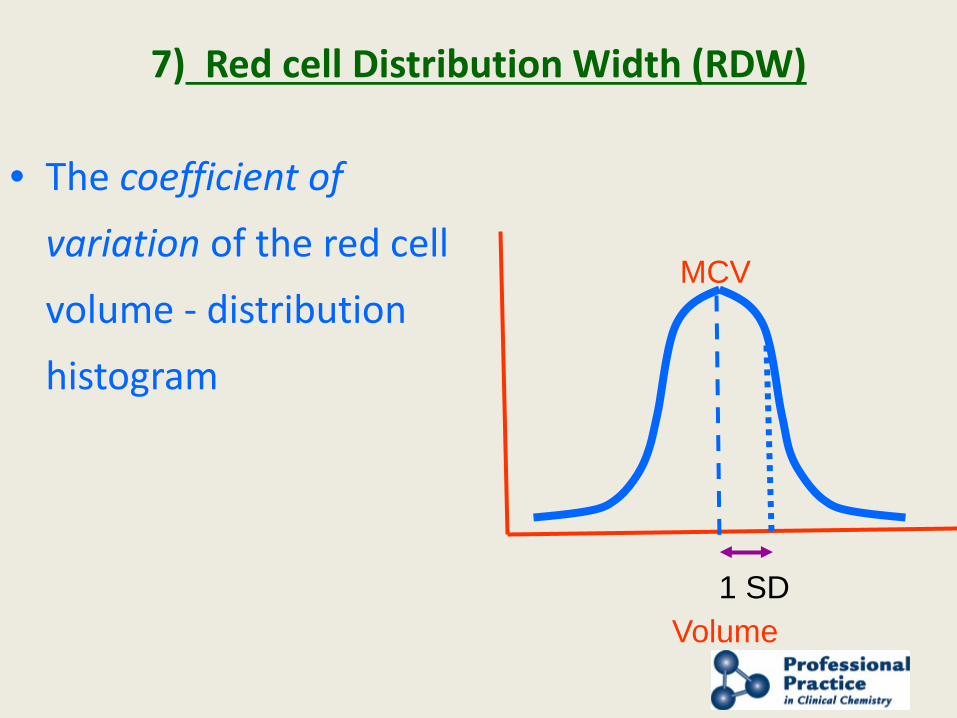
7) Red cell Distribution Width (RDW)
• The coefficient of variation of the red cell volume - distribution histogram
Volume
MCV
1 SD
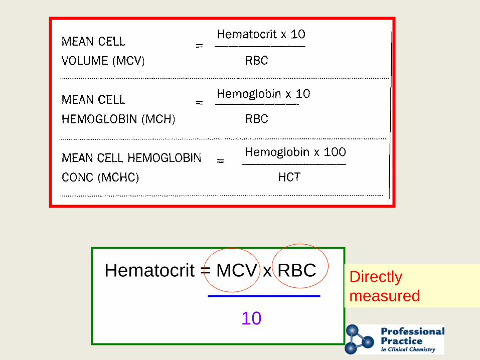
Hematocrit = MCV x RBC
10
Directly measured
Presented by AACC and NACB
Reticulocytes
1. Immature red cells containing residual RNA
2. Indicate rate of new RBC production
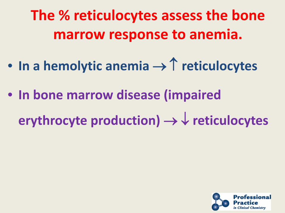
The % reticulocytes assess the bone
marrow response to anemia. • In a hemolytic anemia → ↑ reticulocytes
• In bone marrow disease (impaired
erythrocyte production) → ↓ reticulocytes
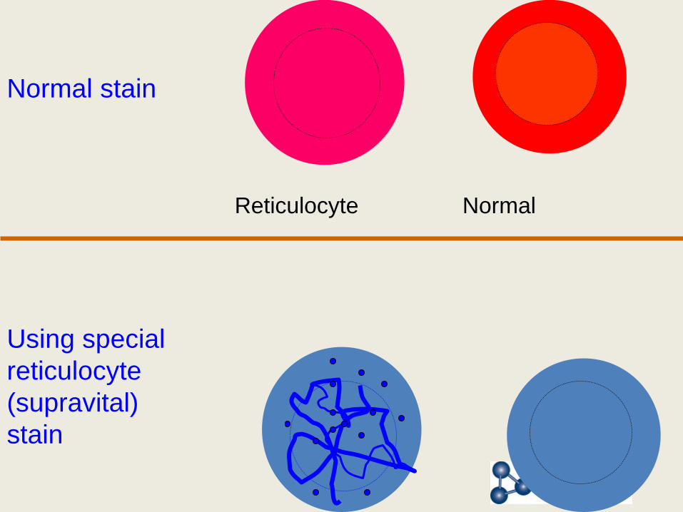
Normal Reticulocyte
Using special reticulocyte (supravital) stain
Normal stain
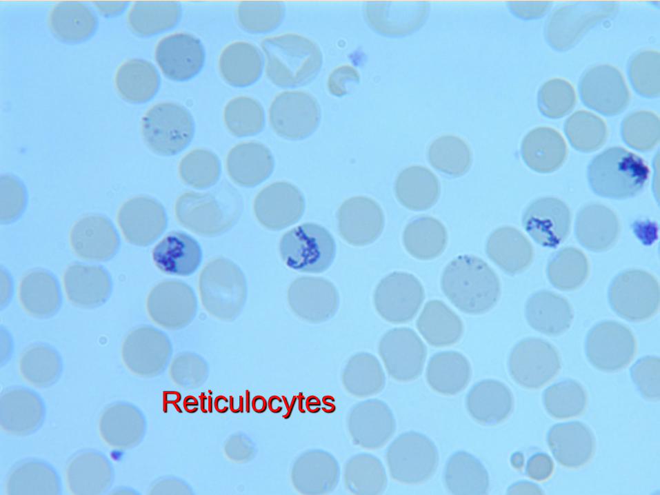
Reticulocytes
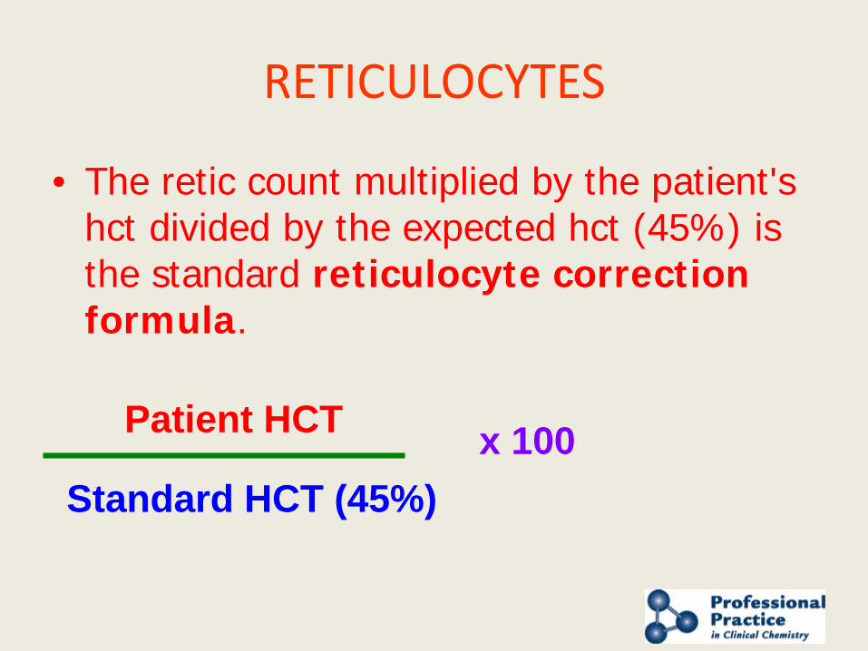
RETICULOCYTES
• The retic count multiplied by the patient's hct divided by the expected hct (45%) is the standard reticulocyte correction formula.
Patient HCT
Standard HCT (45%) x 100
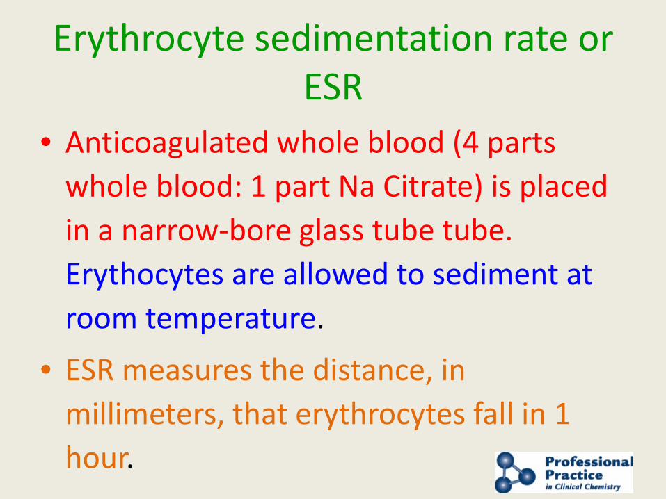
Erythrocyte sedimentation rate or ESR
• Anticoagulated whole blood (4 parts whole blood: 1 part Na Citrate) is placed in a narrow-bore glass tube tube. Erythocytes are allowed to sediment at room temperature.
• ESR measures the distance, in millimeters, that erythrocytes fall in 1 hour.
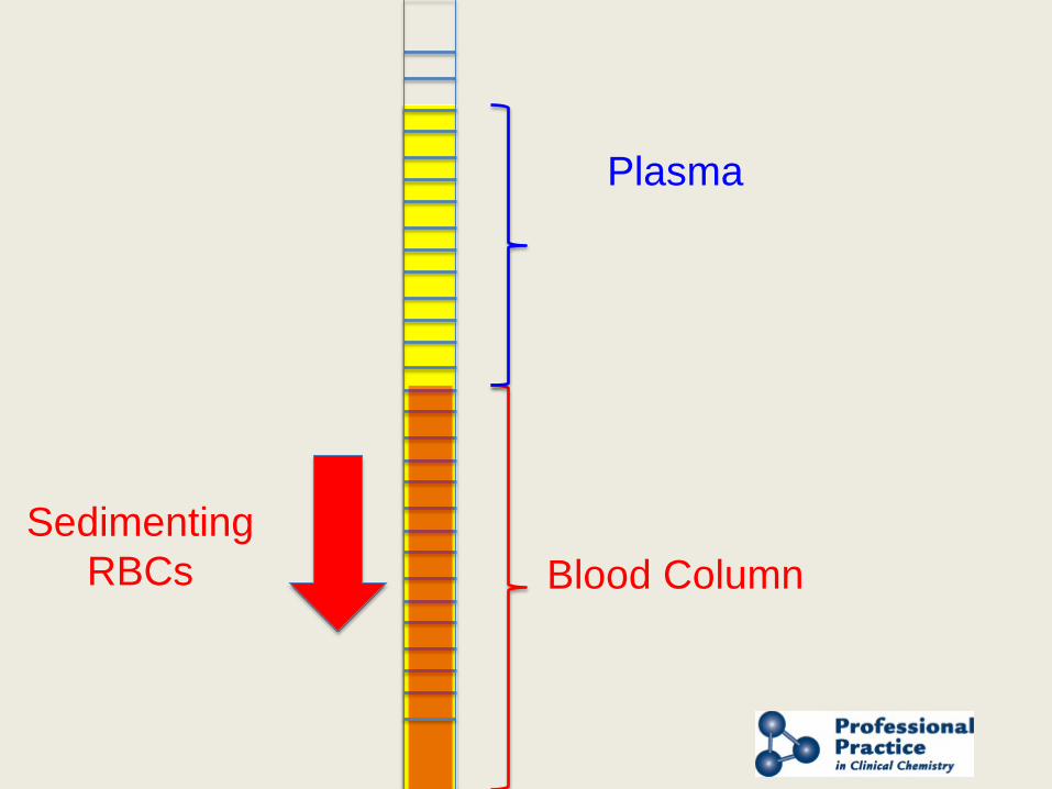
Blood Column
Plasma
Sedimenting RBCs
Erythrocyte sedimentation rate or ESR
• Rate of sedimentation is determined by plasma proteins. ESR increases with acute phase response
• This is an indirect determination of inflammation
• Used to follow rheumatoid arthritis, SLE, vasculitis and many inflammatory conditions
• VERY LOW SPECIFICITY
Erythrocyte sedimentation rate or ESR
• Westergren Method: 200 mm tube • Wintrobe Method: 100 mm tube • Modern Methods: Semi-automated
systems that measure sedimentation by infra-red light. Measured over a shorter period than 1 hour & extrapolated to give Westergren ESR.
• Can interface with LIS

Presented by AACC and NACB
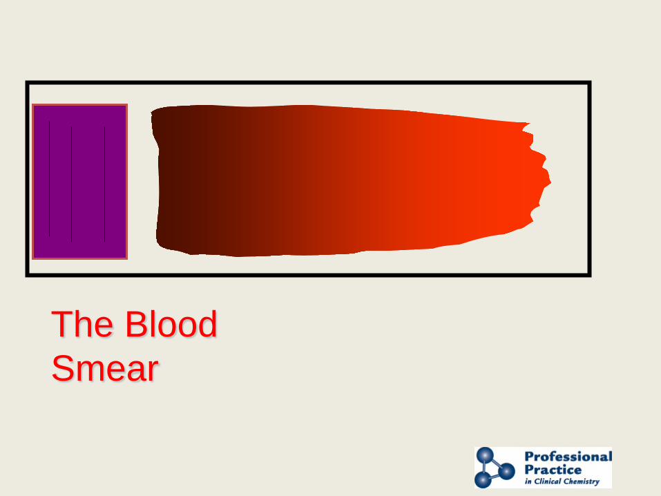
The Blood Smear

Stained Blood Smear
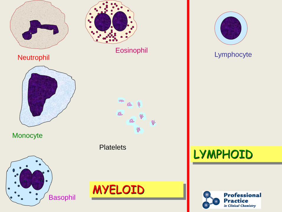
Lymphocyte
MYELOID
LYMPHOID
Neutrophil
Monocyte
Eosinophil
Basophil
Platelets
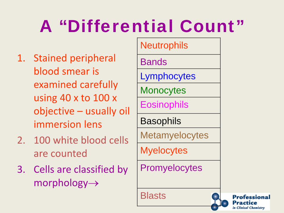
A “Differential Count” 1. Stained peripheral
blood smear is examined carefully using 40 x to 100 x objective – usually oil immersion lens
2. 100 white blood cells are counted
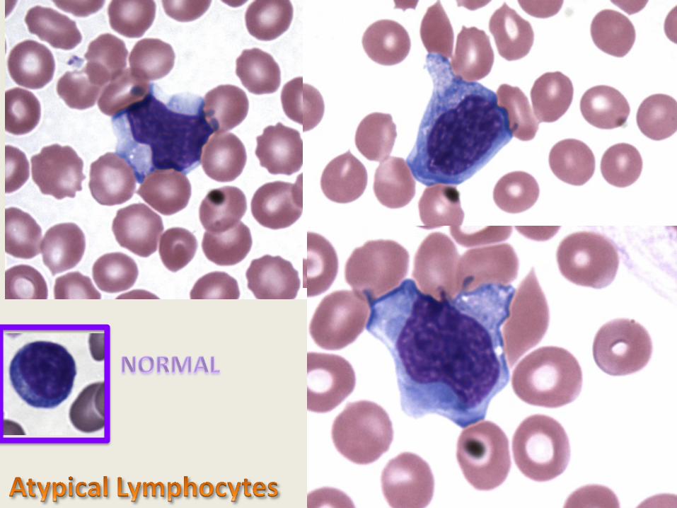
3. Cells are classified by morphology→
Neutrophils
Bands Lymphocytes Monocytes Eosinophils
Basophils Metamyelocytes Myelocytes
Promyelocytes Blasts
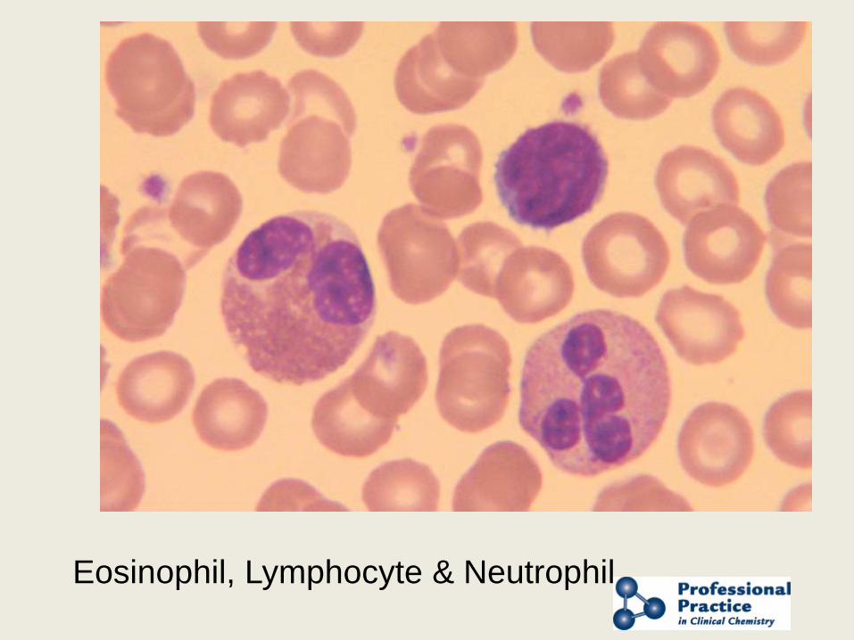
Eosinophil, Lymphocyte & Neutrophil

Neutrophil and Lymphocyte
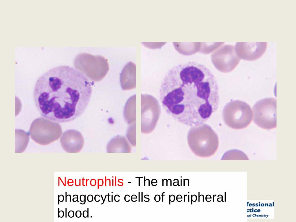
Neutrophils - The main phagocytic cells of peripheral blood.
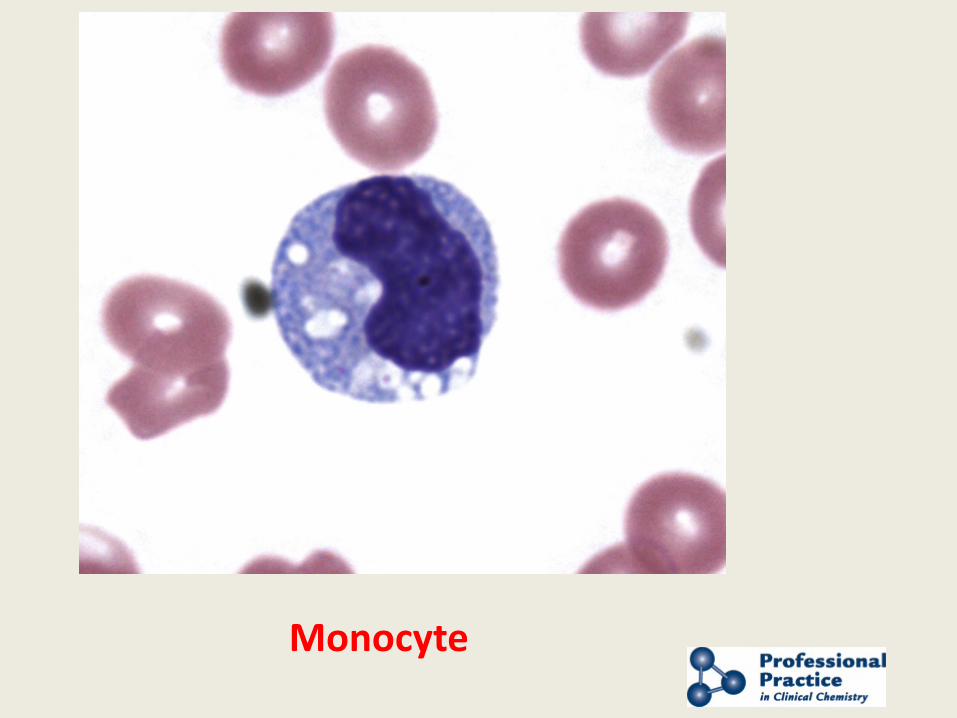
Monocyte
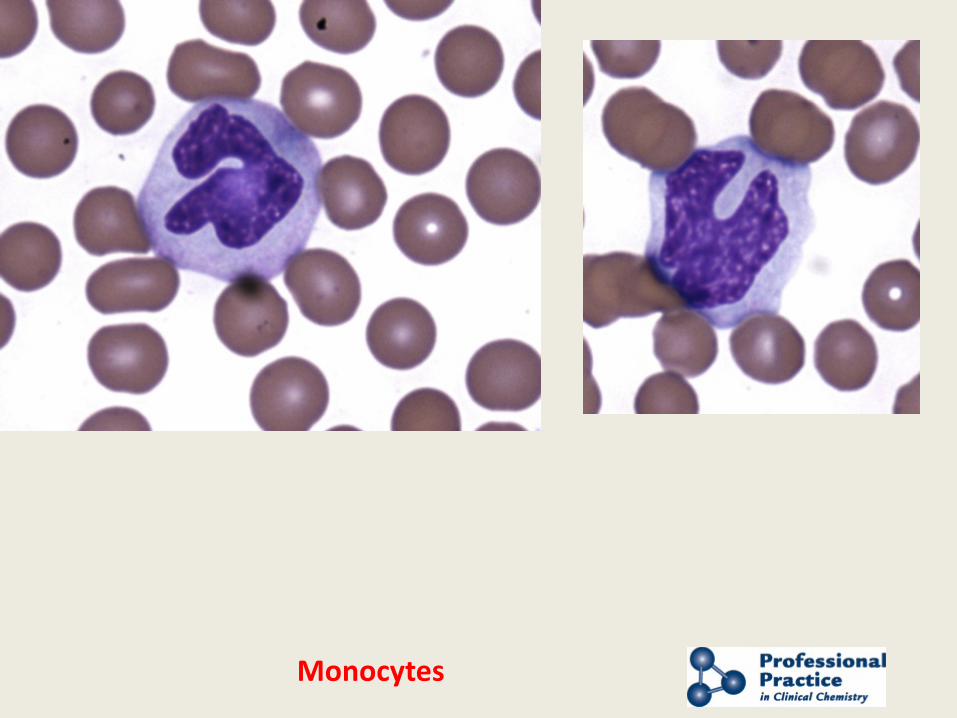
Monocytes
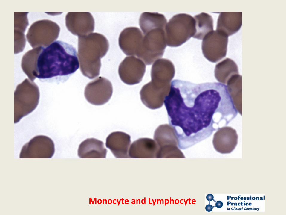
Monocyte and Lymphocyte
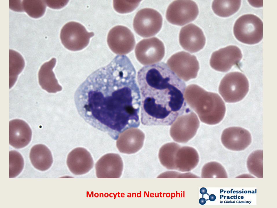
Monocyte and Neutrophil
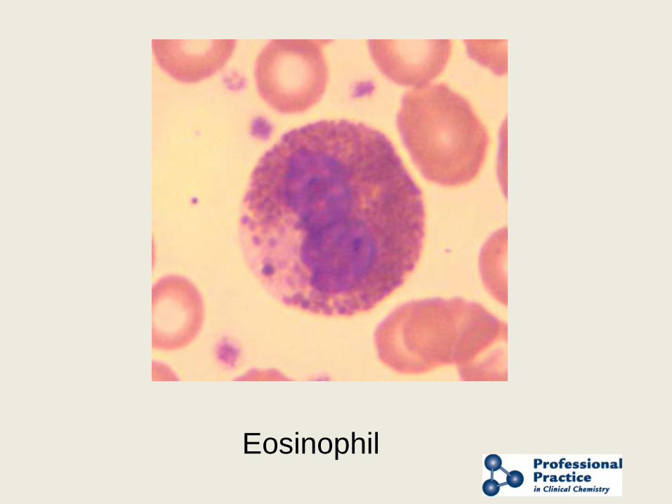
Eosinophil
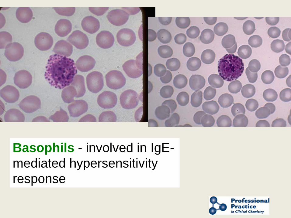
Basophils - involved in IgE-mediated hypersensitivity response
Presented by AACC and NACB
Neutrophil maturation
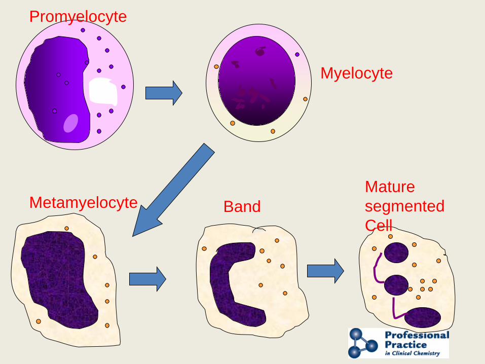
Promyelocyte
Band Mature segmented Cell
Metamyelocyte
Myelocyte
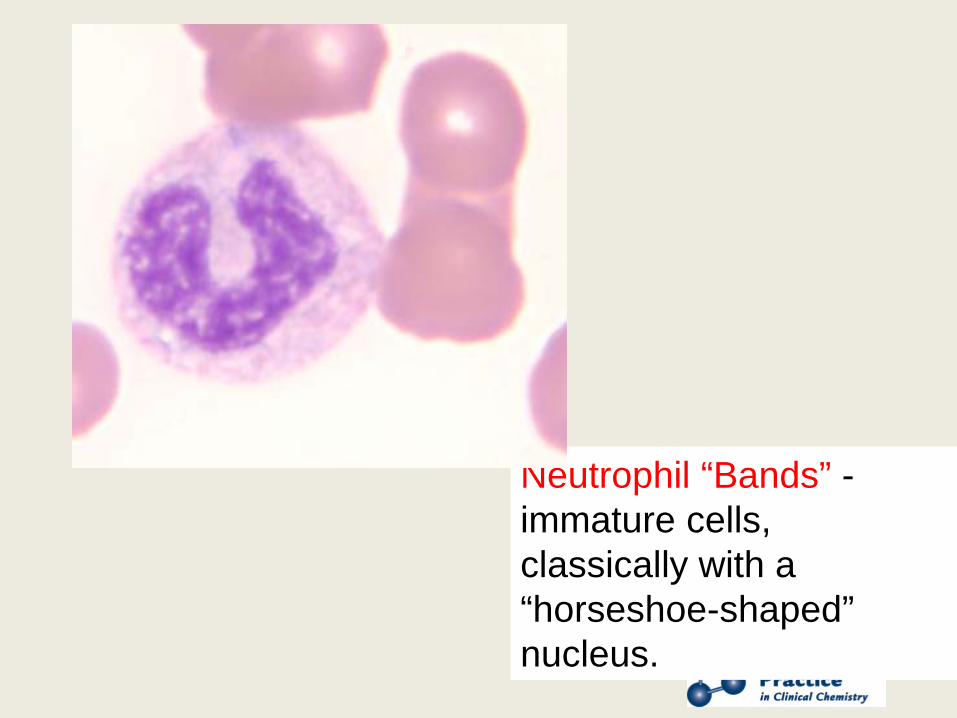
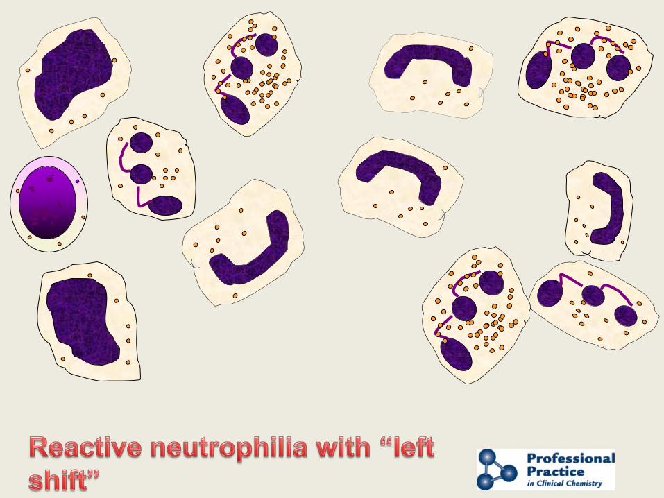
Neutrophil “Bands” -immature cells, classically with a “horseshoe-shaped” nucleus.
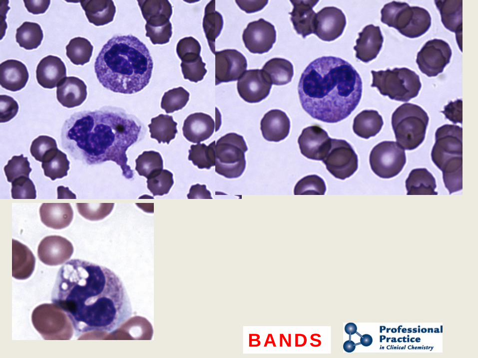
BANDS
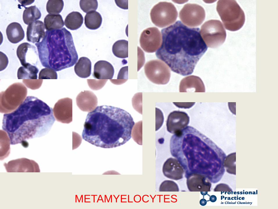
METAMYELOCYTES
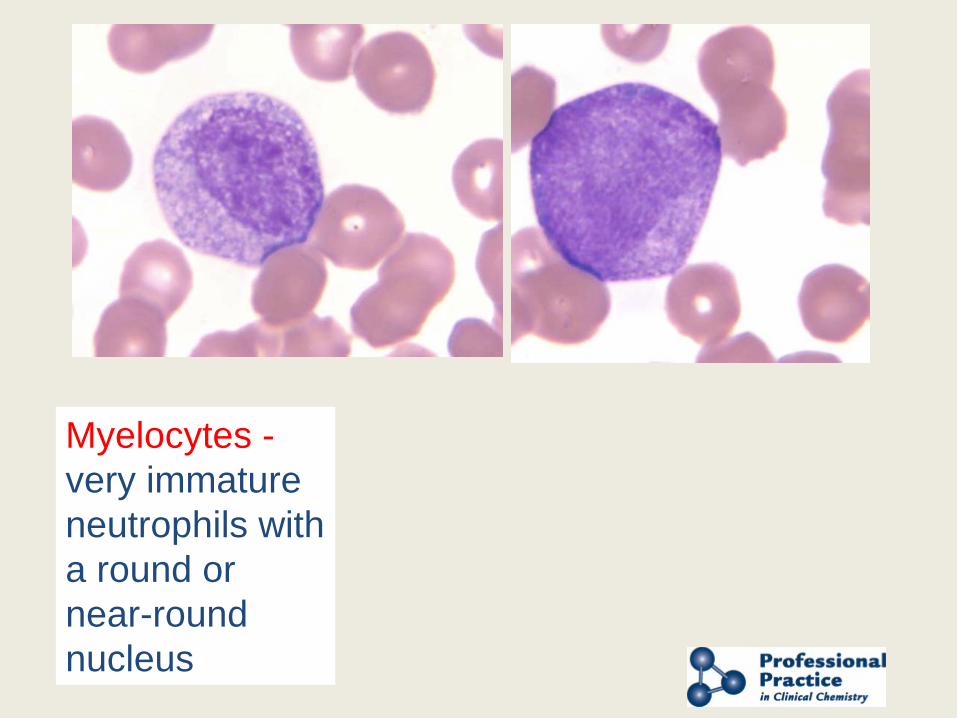
Myelocytes - very immature neutrophils with a round or near-round nucleus
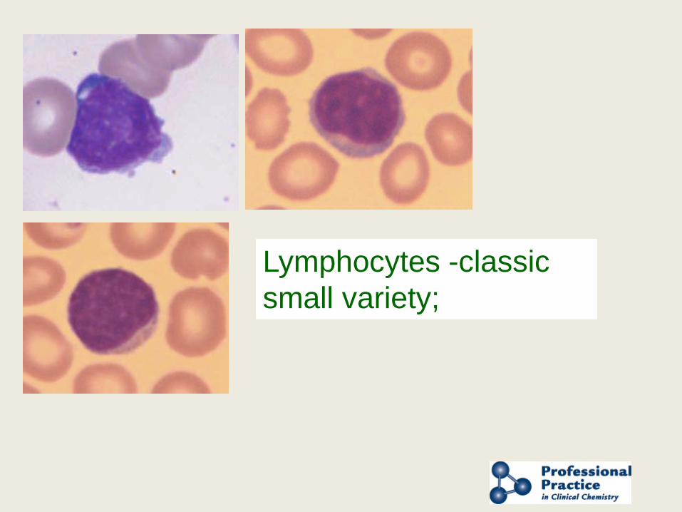
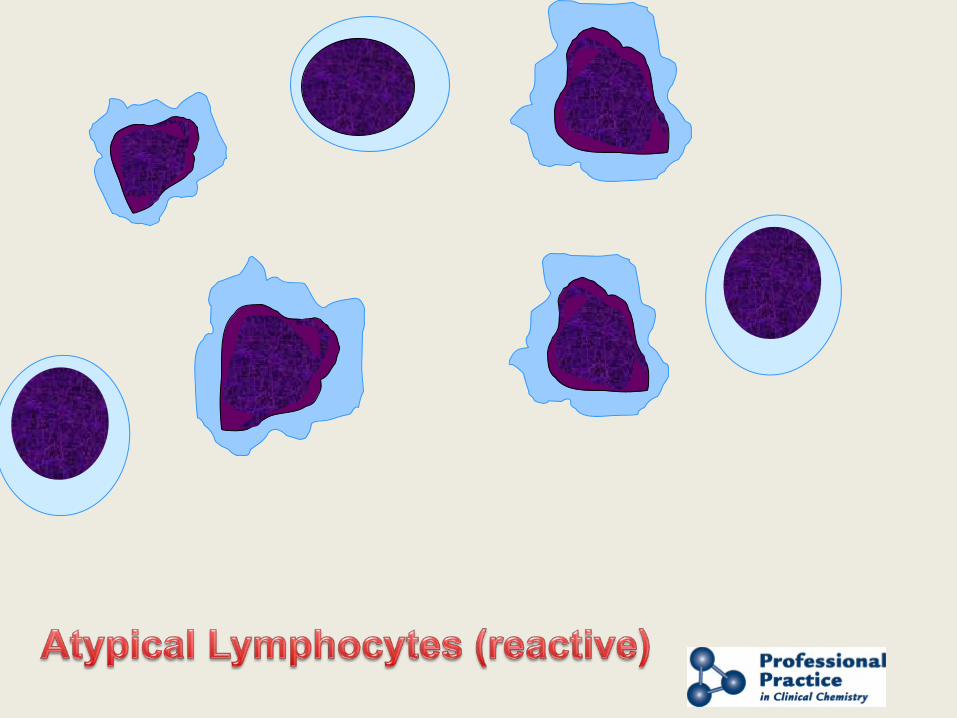
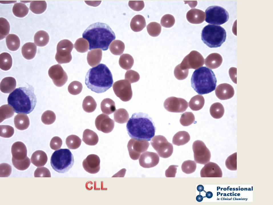
Lymphocytes -classic small variety;
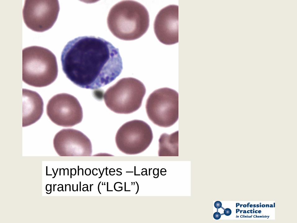
Lymphocytes –Large granular (“LGL”)
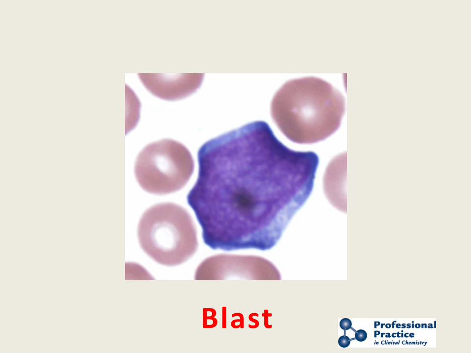
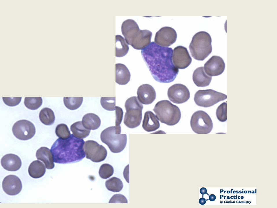
Blast
Presented by AACC and NACB
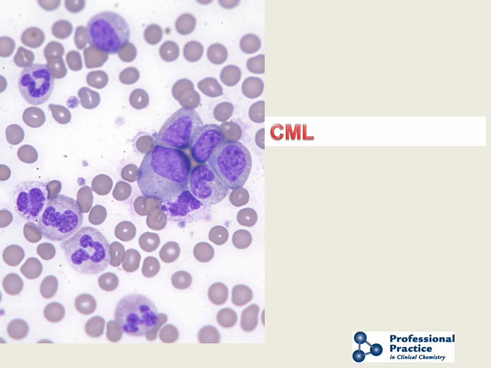
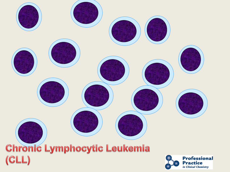
Common Blood “Pictures”
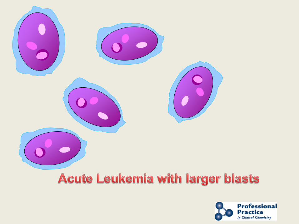
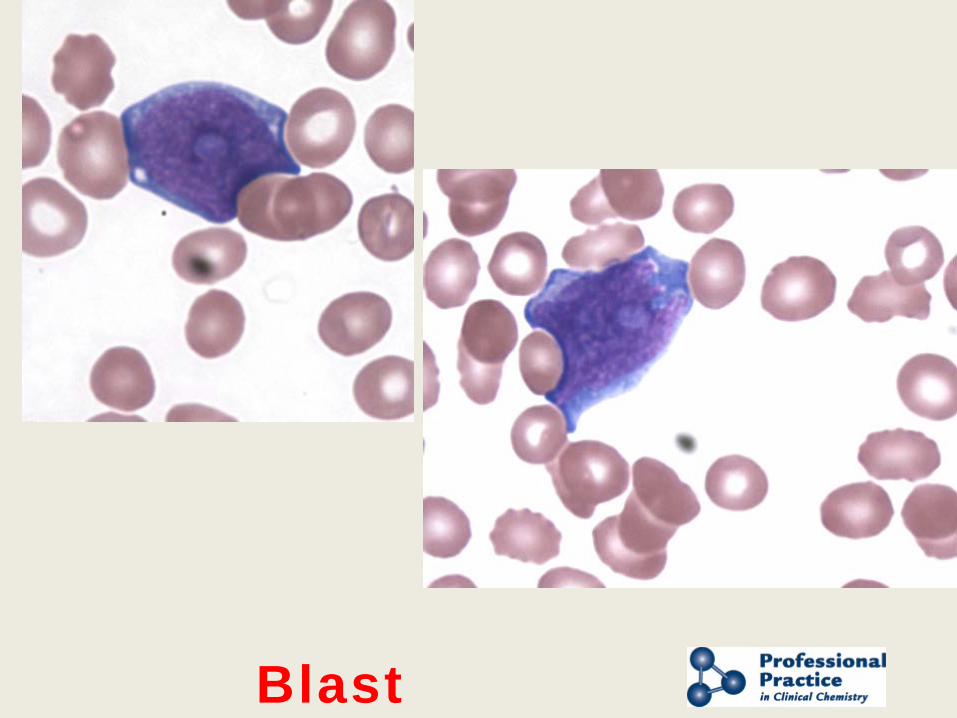
Blast
Presented by AACC and NACB
END
Self-Assessment Question #1 Regarding the mean cell volume or MCV: a) This is now determined directly by electrical or flow cytometric methods. b) it is always calculated from the spun hematocrit. c) it is determined by light microscopy. d) it is determined by electron microscopy. e) it is derived from the white cell count. Explanation: The red cell MCV is measured directly on cell counters; this is true whatever method is employed to enumerate cells. The counter is able to plot a red cell volume histogram, and the mean is determined. MCV can be calculated from the spun hematocrit as in option b. This was the original method for determining MCV but it is not commonly used anymore.
Self-Assessment Question #2 The Erythrocyte Sedimentation Rate (ESR) is:
a) the rate of sedimentation of fibrinogen in a 5 ml glass tube. b) the rate of sedimentation of white cells in a standard narrow-bore glass tube. c) the rate of clotting in the presence of erythrocytes d) a test of platelet function e) the rate of sedimentation of erythrocytes in a standard narrow-bore glass tube Explanation: The ESR is the rate of sedimentation of red cells in a whole blood specimen enclosed in a narrow-bore glass tube. ESR is increased in acute inflammation.
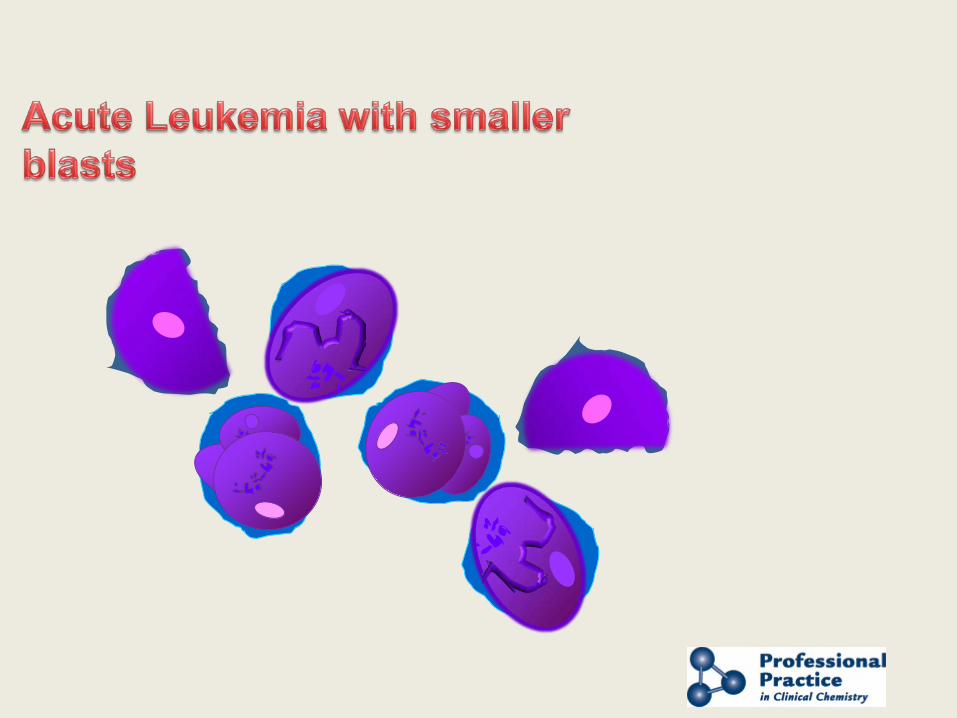
Self-Assessment Question #3 Acute leukemia is characterized by presence of ____ in the stained peripheral blood smear: a) blasts b) neutrophils c) lymphocytes d) platelet clumps e) basophils Explanation: The hallmark of acute leukemia is the increased presence of blasts in the bone marrow and in peripheral blood. Blasts are immature progenitor cells with characteristic morphology and cell surface markers.
Presented by AACC and NACB
APPENDIX
Presented by AACC and NACB
Derivation of RBC Indices
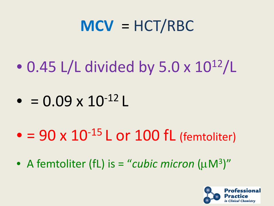
MCV = HCT/RBC
• 0.45 L/L divided by 5.0 x 10¹²/L
• = 0.09 x 10⁻¹² L
• = 90 x 10-15 L or 100 fL (femtoliter)
• A femtoliter (fL) is = “cubic micron (µM3)”
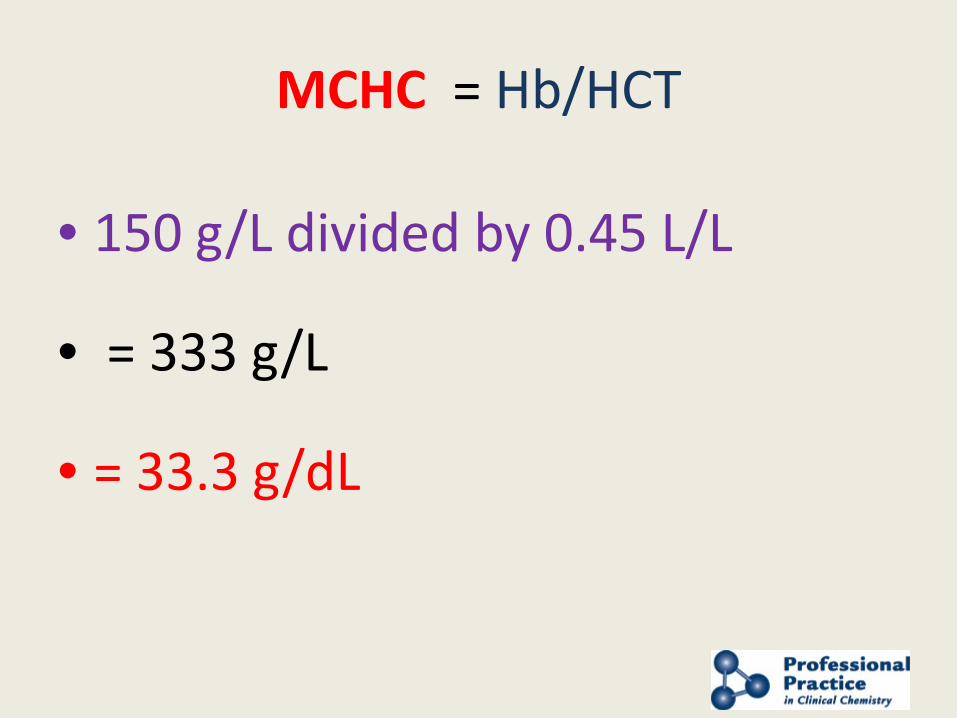
MCHC = Hb/HCT
• 150 g/L divided by 0.45 L/L
• = 333 g/L
• = 33.3 g/dL
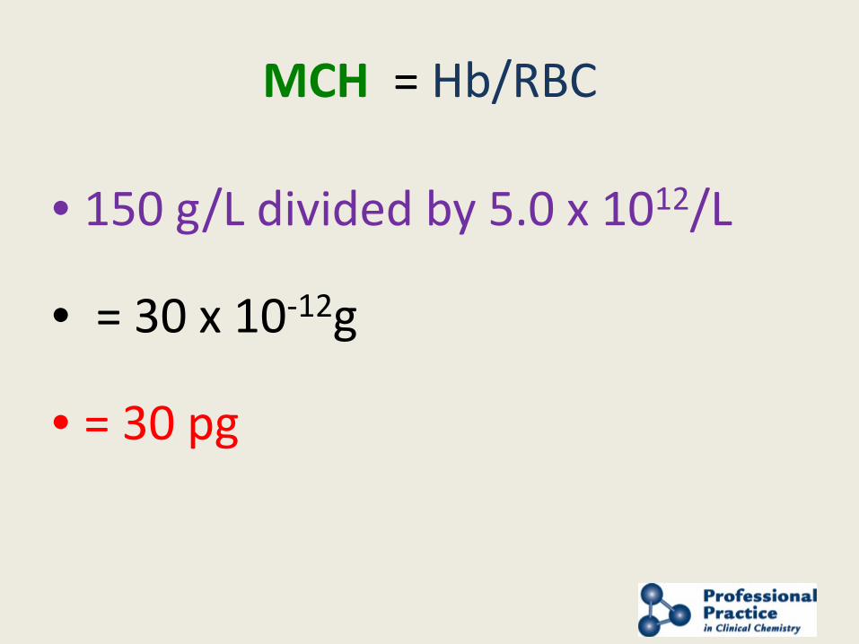
MCH = Hb/RBC
• 150 g/L divided by 5.0 x 10¹²/L
• = 30 x 10-12g
• = 30 pg
Presented by AACC and NACB
AUTOMATED CELL Analysis
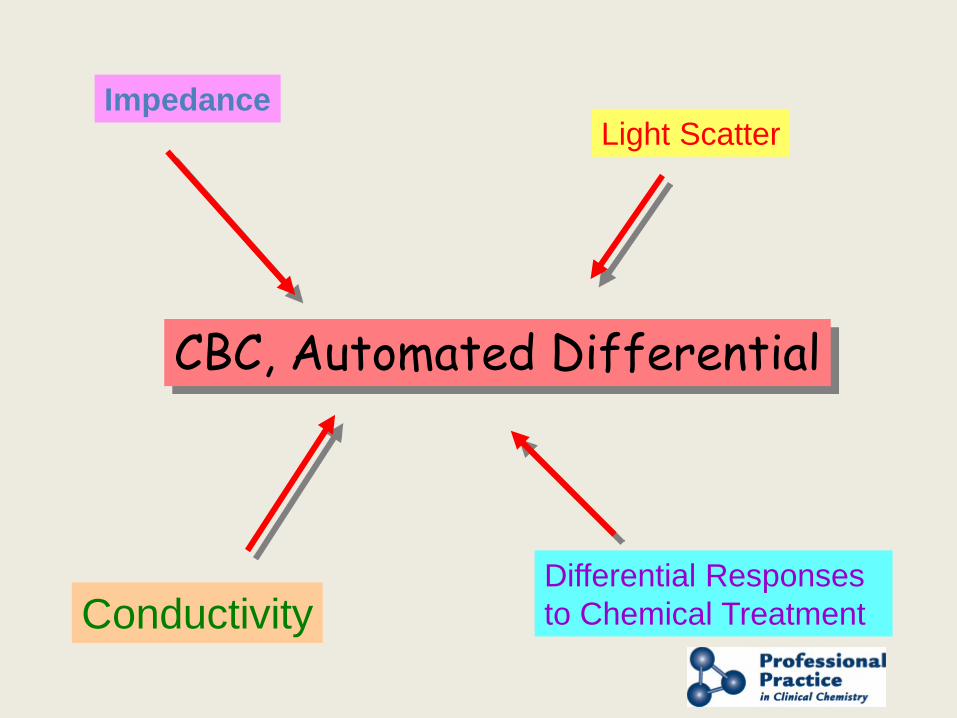
Impedance
Differential Responses to Chemical Treatment
Light Scatter
Conductivity
CBC, Automated Differential
Presented by AACC and NACB
1) IMPEDANCE
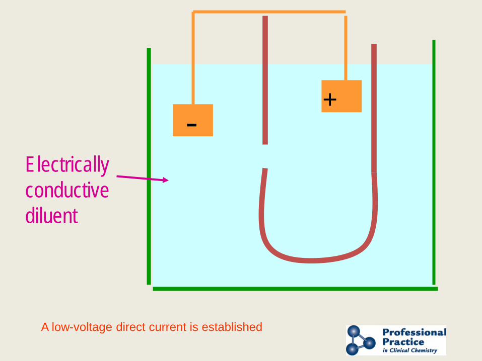
- +
Electrically conductive diluent
A low-voltage direct current is established
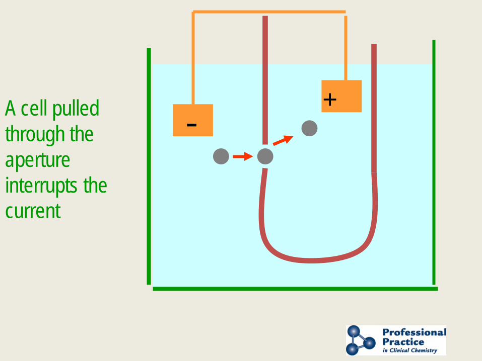
- + A cell pulled
through the aperture interrupts the current
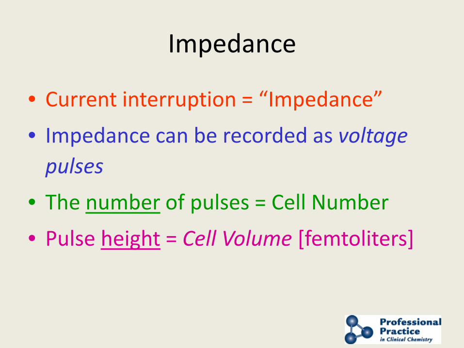
Impedance
• Current interruption = “Impedance” • Impedance can be recorded as voltage
pulses • The number of pulses = Cell Number • Pulse height = Cell Volume [femtoliters]

Presented by AACC and NACB
2) CONDUCTIVITY
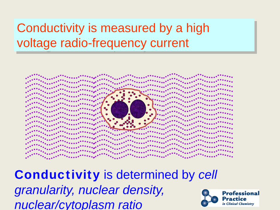
Conductivity is measured by a high voltage radio-frequency current
Conductivity is determined by cell granularity, nuclear density, nuclear/cytoplasm ratio
Presented by AACC and NACB
3) LIGHT SCATTER (Flow Cytometry)
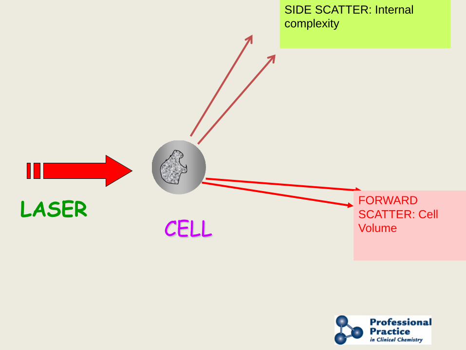
LASER
SIDE SCATTER: Internal complexity
FORWARD SCATTER: Cell Volume CELL
Presented by AACC and NACB
4) Differential Responses to Chemical treatment
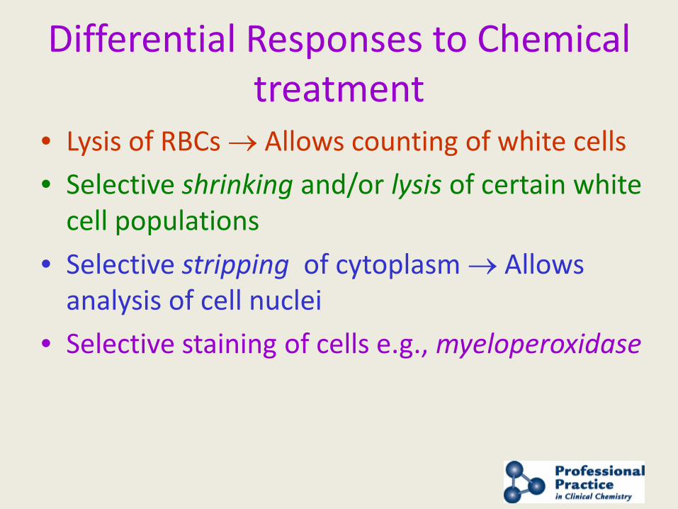
Differential Responses to Chemical treatment
• Lysis of RBCs → Allows counting of white cells • Selective shrinking and/or lysis of certain white
cell populations • Selective stripping of cytoplasm → Allows
analysis of cell nuclei • Selective staining of cells e.g., myeloperoxidase
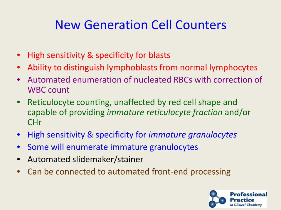
New Generation Cell Counters
• High sensitivity & specificity for blasts • Ability to distinguish lymphoblasts from normal lymphocytes • Automated enumeration of nucleated RBCs with correction of
WBC count • Reticulocyte counting, unaffected by red cell shape and
capable of providing immature reticulocyte fraction and/or CHr
• High sensitivity & specificity for immature granulocytes • Some will enumerate immature granulocytes • Automated slidemaker/stainer • Can be connected to automated front-end processing