b major thalassemia - sbmu.ac.irpchd.sbmu.ac.ir/uploads/iron_defficiency_and_thalassemia.pdfb...
TRANSCRIPT


B Major Thalassemia

Anemia
It is a reductionof the red cell volume or hemoglobin
concentration below -2SD for age, sex.

Normal Range,Hb
Birth:16/6 gr/dl2 Mo:11/53-6 Mo 11/5 6-24 M :122-6 Y:12/56-12 Y:13/512-18 Y: F =12-14 M=14-16

Normal Range,MCV
Birth:108FL2 Mo:96 3-6 Mo:91 6-24 M :78 2-6 Y:816-12 Y:8612-18 Y: F =90 M=88

Iron deficiency
Infectious/inflammatory disorders
Malaria
Hemoglobinopathies
HIV infection
Helminth infection
Vitamin B12deficiency
Folate deficiency
Anemia

Iron is vital for allliving organisms;oxygen Transport

Red blood cells specialisationsno nucleus extra space inside
contain haemoglobin the oxygen
carrying molecule
biconcave shape
increases thesurface area somore oxygen can becarried

Iron deficiency is a major healthproblem worldwide and especially indeveloping countries.
Iron-deficiency is the most prevalentnutritional deficiency worldwide
Iron deficiency is the most commonsingle cause of anemia worldwide

Review Of Articles 1Prevalence of iron deficiency anemia in 6mo-5 years
old children in Fars , southern IRAN Kadivar MR & Collegues. Med Sci Monit,2003;9(2);CR 100-104
541 patients: 110 p(%19.7): Serum Ferritin level < 12ng/ml 101 P(%18.7): low serum Hb Developing Countries: IDA%25-%35Industerialized Country: IDA %5-%8 Iron supplements by Health care centers , Free of
charge

The concentration of Iron in:Infant: 75-80 mg/kg(BW)
50mg/kg: Hb Mass
25mg/kg: Storage Iron
5mg/kg: Myoglobin & tissueIron
Iron status

The concentration of Iron in:Adult: 40-50 mg/kg(BW)
30mg/kg: Hb Mass6-7mg/kg: Myoglobin, Heme
enzymes& non heme enzymes* 6-7mg/kg (F) storage Iron* 10-12 mg/kg (M)< 0/5%: Transport Iron
Iron status

Cellular sequestration & Metabolism ofIron is mediated by 3 proteins:
Transferrin
Transferrin receptor
Ferritin
Iron Metabolism

FerritinFerritin is the major storage protein with 24
subunit:* Light chain (L), 19 kD
* Heavy chain (H) 21 kDH gene locus: ch 11: Heart, Iron – Metabolism
L gene locus: ch 19: Liver & spleen- Ironstorage function
Ferritin is found in virtually all cellsespecially:
Erythroied precursorsMacrophagesHepatocytes
F.molecule: 4500 Iron atomsHalf life: 60 hour
Catabolism of, F: Reutilization of Iron coreHemosiderin conversion

Body Iron Distribution and StorageDietary iron
Utilization Utilization
Duodenum(average, 1 - 2 mg
per day)
Muscle(myoglobin)
(300 mg)
Liver(1,000 mg)
Bonemarrow(300 mg)Circulating
erythrocytes(hemoglobin)
(1,800 mg)
Reticuloendothelialmacrophages
(600 mg)
Sloughed mucosal cellsDesquamation/Menstruation
Other blood loss(average, 1 - 2 mg per day)
Storageiron
Plasmatransferrin
(3 mg)
Iron loss
(Ferritin)
(TIBC)


Iron balance is physiologically regulatedby controlling Iron absorption.The availability of dietry Iron forabsorption is dependent to: The amount of Iron Form of Iron Composition of the diet GI factors
Iron balance

CBCHb, HCT -Anemia
WBC-leukocytosis-leukopenia-Abnormal cells
Platelet -Thrombocytosis-Thrombocytopenia

peripheral Blood,CBCoR BC
o Hb (is not specific)
o MCV
o MCH
o Reticulocyte
oPeripheral blood smear,Morphology

Indirect: 1- plasma ferritin:
(the most useful) in the absence of:
* Tissue necrosis
* Inflammation
* Neoplasm
* liver disorder
* turn over of RBC

Prussian Blue Stainof Bone Marrow
Iron Present No Iron Present

Iron deficiency anemiaIron deficiency anemia is themost common cause ofanemia.
Growth & diet are almostalways contributing factors inchildhood

Etiology / IDA
Blood LossGastrointestinal Tract: Milk -induced
Enthropathy Peptic ulcer Inflamatory Bowel Diseaes Meckel Diverticuculm &Polips Drugs: Salicylates Hookworm Infestation
Pulmonary Hemosiderosis
Iatrogenic
Menstural Blood Loss
Urinary Blood Loss(rare)
Red Blood Cells

Etiology/IDA
Increased Physiologic Requirement-Pregnancy-Infancy-Adolescence
Malabsorption- Inflamatory Bowel Diseaes-Tropical Sprue Gastrectomy Pica
Dietary inadequacy: Iron Poor Diet
Combinations of above

Clinical manifestations*Hematologic* Non Hematologic
• Pallor• Weakness, fatigue, Irritability• Anorexia• Pica• Blue sclera•Koilonychias (spoon- shaped nails)• Glossitis• Angular stomatatis• Post cricoid esophageal web (plummer winsonsyndrome)• Impair of intellectual & learning• Impaired of immunity• Slightly enlarged spleen• Cardiopulmonary failure & death.


Laboratory test:1-Serum Ferritin: < 10-12 ug/l
2- Serum Iron( Decrease)
3-Total iron binding capacity TIBC
4- peripheral blood : RBC, Hb- HCT↓MCV, MCH ↓
(RDW(Red blood cell distributaion width )
Reticulocyte , Mild
4- Serum Soluble Transferrin Receptor
5-FEP
6- BMA & BM Biopsy (Prussian Blue Staining)

Reticulocyte countNormal = 0.2-2 %
Corrected reticulocyte = Pt HCT X Reti.Normal HCT

CBCHb, HCT -Anemia
WBC-leukocytosis-leukopenia-Abnormal cells
Platelet -Thrombocytosis-Thrombocytopenia


IDA

Differential Diagnosis ofI.D.Anemia
1- . Thalassemia minor
2- . Thalassemia major
3- Chronic disorders
4- lead poisining
5- . Thalassemia

B Thalassemia trait (Heterozygous)
Expression of one gene is impaired bymutation where as the other gene is normal.
Slight ineffective erythropoiesis & modestlydecrease of RBC survival
Mild erythrocytosisMarked microcytosis Peripheral Blood: microcytosis, hypochromia
& targeting

Differntial DiagnosisB Thalassemia trait / Iron Deficiency Anemia
B. Th. Trait:Increase of RBC- Mild Erythrocytosis,Marked microcytosis IDA : RBC count decreased, MCV is rarely
as low as B. Th .Trait RDW ( Red Cell Disrtribution Width by
Automated cell counter) : Increased in IDAMentzer Index( MCV/RBC ): B .Th .Trait <13 IDA > 13
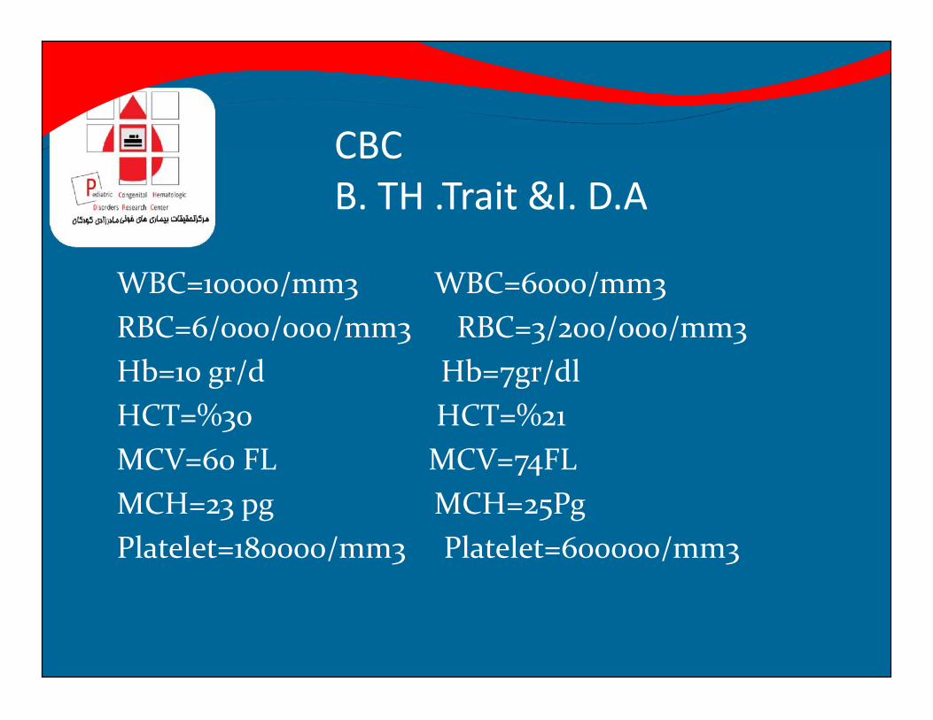
CBCB. TH .Trait &I. D.A
WBC=10000/mm3 WBC=6000/mm3RBC=6/000/000/mm3 RBC=3/200/000/mm3Hb=10 gr/d Hb=7gr/dlHCT=%30 HCT=%21MCV=60 FL MCV=74FLMCH=23 pg MCH=25PgPlatelet=180000/mm3 Platelet=600000/mm3

thalassemiaHb A
Hb A2
Hb F
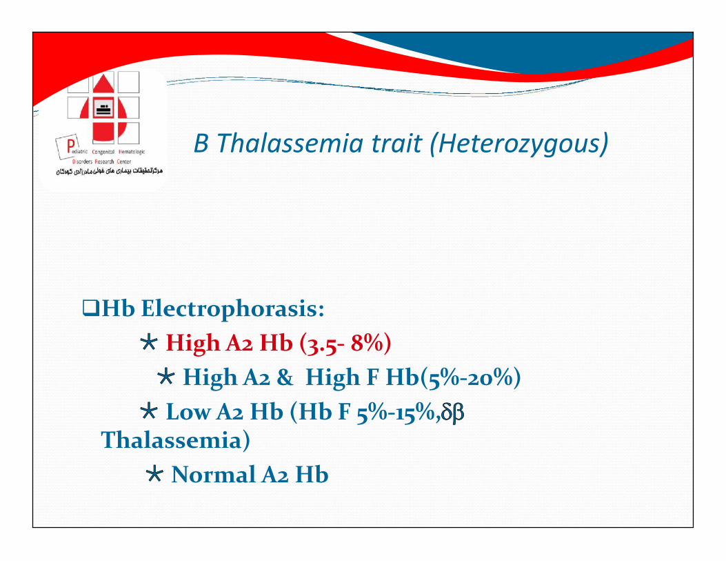
B Thalassemia trait (Heterozygous)
Hb Electrophorasis:High A2 Hb (3.5- 8%)High A2 & High F Hb(5%-20%) Low A2 Hb (Hb F 5%-15%,
Thalassemia) Normal A2 Hb


IRON DEFICIENCY versus ACD
Serum Iron Transferrin Ferritin
Iron Deficiency
ACD

Major Thalassemia /CooleyAnemia
Compound heterozygous state for two different globin gene mutationsHomozygous state for the same mutation.Age of diagnosis: 6-12 months 60%: first year 29%: second year 9%: later

o Palloro Failure to thriveo Irritabilityo Icteruso Hepatosplenomegalyo Skeletal changeso Prone to infection
Clinical manifestations


B MAJOR THALASSEMIA

Hepatosplenmegaly

o Hypochromic & microcytic anemiao NRBC o Serum Iron & Ferritin o BMA: marked E hyperplasiao E/M: 20/1o Hb electrophoresis:o Hb F o Hb A2: variable levelso Hb A: reduced or absento Th. Trait in both parentso Globin biosynthetic ratio: diagnostic
Laboratory Test


Treatment of Thalassemia Major• Gene therapy
• Stem cell transplantation
• Blood transfusion

o Determine the blood type & minorRed Cell Antigen :
o ABO, RH ,Kell, Kidd, Duffy
Blood Transfusion in .Th. Major

Guidelines For Blood Transfusion
oPatients should not receive PRBC more thantwo weeks oldoHb level:9/5-11/5 gr/dl
oVolume of PRC:10-20 Ml/kg of leukocyte-poorand filterd RBCoTransfusion interval:3-5 weeks
opretransfusion laboratory tests:CBCocross match,RBC antibody screen

Replenishment of body IronCorrection of factor responsible for IrondeficiencyIron administration:
*Oral: safe, cheap & effective*Parenteral: IM, IV
Parentrarl Indication :* poor tolerance* GI Iron absorption is compromised* has Iron needs that can not be met oral
therapy because of chronic uncontrollablebleeding
Treatment of I.D.A

Ferrus sulphate is the preferred, salt.The Iron element: 20%Dose: 3-6mg/kg/day divided doseAdministration: between mealsSide effects ,10-20%: Nausea
VomitingDiarrhea, constipationAbdominal pain
Plan for side effects : 1- Administrationimmediately after meal2- dose
Oral Iron therapy

Parenteral Iron therapyDextran :Side effects 1- Anaphylaxia
2- Serum sickness- like reaction3- Skin staining (IM)4- Muscle necrosis5- Phlebitis6- Persistent pain7- Artralgia
Because of anaphylaxia: Test dose 0/5cc1 hour before.
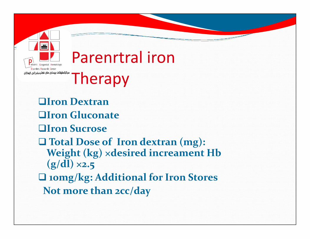
Parenrtral ironTherapy
Iron DextranIron GluconateIron Sucrose Total Dose of Iron dextran (mg):
Weight (kg) ×desired increament Hb(g/dl) ×2.5 10mg/kg: Additional for Iron Stores
Not more than 2cc/day

Timing for Iron replacementin infant
1- Breast milk infant: 1mg/kg/day Ironsupplementation beyond 6 months
2- Infant with Iron supplementedformula: 12mg/lit Iron
3- Cow’s milk should be avoidedduring the first year.
4- premature infants should receiveIron supplements immediately.

THE END