axial & appendicular skeleton - cardinal newman high...
TRANSCRIPT

Axial & Appendicular Skeleton
INCLUDING JOINTS

Are these anatomical positions correct?
A B

The Axial Skeleton

• Sutures: join immovable Joints
• Processes: Extending portion of the bone
• Septum: Dividing structurre

The Skull BonesThe Skull consists of
• 8 Cranial Bones – covers & protects the brain
connected by sutures
• 13 Facial Bones – encases eyes & attached to facial muscles
The Ears consists of 6 Bones
Floating in the throat is the Hyoid Bone: the only bone NOT touching another
Inner Ear

THE SKULL• Frontal: forehead under eyebrows upper
eye orbit
• Parietal: Superior & lateral
• Sphenoid:forms the base of the cranium,
behind the eye and below the front part of
the brain
• Temporal: inferior to the parietal
• Nasal Bone:Nose Bridge
• Zygomatic:Cheekbone
• Vomer:Forms the septum in the nose
• Maxilla:Upper Jaw
• Mandible:Lower jaw

• 12 pairs of RIBS
– True ribs(1-7): attach directly to
the sternum
– False ribs(8-12):Indirect or no
attachement to the sternum
– 2 floating ribs(11&12) no
attachment to the sternum
• 1 STERNUM
(breastbone)
Thoracic Cage(Rib Cage)

Vertebral ColumnThe Vertebral Column (Spinal
Column or Backbone)
– 7 Cervical (Neck) Vertebrae,
– 12 Thoracic (Chest Cavity)
– 5 Lumbar(Lower Back): support
most of the weight
– Sacrum: 5 Fused Vertebrae
(posterior wall of the pelvis)
– Coccyx: 4 Small Fused Vertebrae
Into 1 (Tail Bone)

APPENDICULAR SKELETON
Consists Of Bones Of The:
– ARMS (upper limbs)
– LEGS (lower limbs)
– SHOULDER GIRDLE (pectoral girdle)
– HIP GIRDLE (pelvic girdle)

Shoulder Girdles and Arms• Shoulder girdle is also
called the pectoral girdle
• Consists of 4 bones
• Upper limbs consist of 60
bones
• Hands and wrist
contain 54
separate
bones

Hip Girdles and Legs• Hip girdle is also called the pelvic girdle
• Consists of 2 bones
• Lower limbs consist of
60 bones
• Ankles and feet contain
52 separate bones
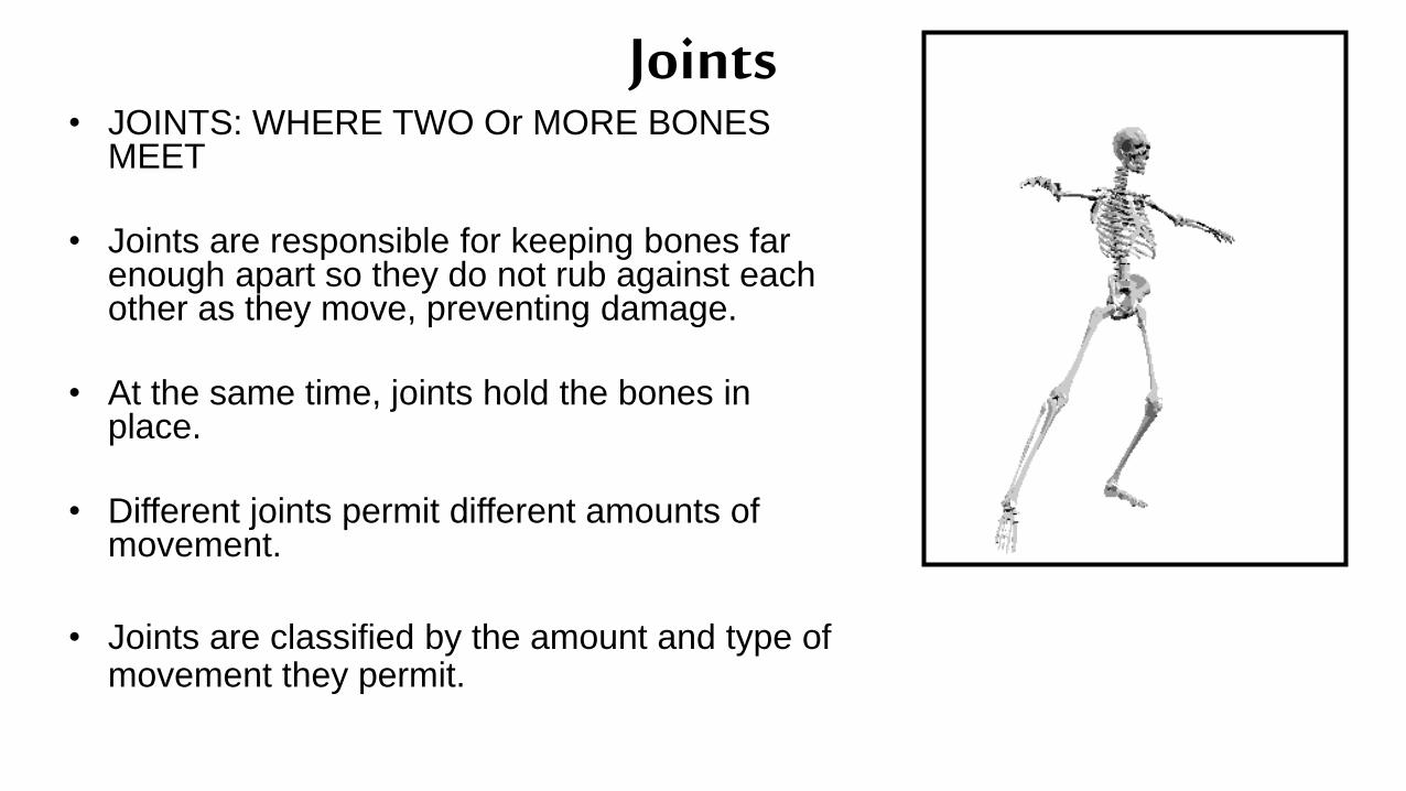
Joints• JOINTS: WHERE TWO Or MORE BONES
MEET
• Joints are responsible for keeping bones far enough apart so they do not rub against each other as they move, preventing damage.
• At the same time, joints hold the bones in place.
• Different joints permit different amounts of movement.
• Joints are classified by the amount and type of movement they permit.

Classification of Joints
Three Main Functional Types
• Immovable(Synarthroses) A fixed joint, one that allows no movement
• Slightly movable (Amphiarthoses)joint that permits a small amount of restricted movement
• Freely movable(Diarthoses) Permit movement in one or more directions

Classification of Joints
• Immovable
– bones of skull,
– pelvis, and sacrum
• Slightly movable
– between vertebrae,
– two bones of lower leg
Tibia and FibulaskullPelvis
Vertebra
Ribs

Freely Movable JointsTYPES OF FREELY MOVABLE JOINTS
A. BALL AND SOCKET JOINT –
Permits circular movement – the widest range of
movement.
– SHOULDER Joint- which enables you to move
your arm up, down, forward and backward, as
well as to rotate it in a complete circle.
– HIP Joint- same range of motion.

Freely Movable Joints (cont’d)
B. HINGED JOINT - Permits a
back-and-forth motion.
– The Knee- enables your leg to
flex and extend.
– The Elbow -allows you to move
your forearm forward and
backward.
– The Phalanges

Freely Movable Joints (cont’d)• C. PIVOT JOINT -
Permits rotation of one bone around another.
– The elbow enables your hand to turn over. (radiusrotates around ulna)
– It also allows you to turn yourhead from side to side.
(rotates around axis)

Freely Movable Joints (cont’d)
D. GLIDING JOINT (Plane )
Permits a sliding motion of one bone
over another.
– Found at ends of the collarbones,
– between wrist bones,
– and between anklebones.

Freely Movable Joints (cont’d)
E. SADDLE JOINT-Permits movement in two planes.
– This type of joint is found at the base of the thumb

Anatomy of a JointStructures of a freely movable joint
• 2 or more bones
• Cartilage
• Joint capsule
• Synovial membrane
• Synovial fluid
• Fat
• Bursa
• Meniscus
• Ligaments
• Tendons

• Cartilage - at the joint, the
bones are covered with
cartilage, which is wear-
resistant and helps reduce
the friction of movement.
• Joint capsule- is a thick,
tough layer that envelops
the joint cavity forming a
membrane or sac that
adheres firmly to the
periosteum of the
articulating bones
Anatomy of a Joint

Anatomy of a Joint• Synovial membrane -
a tissue that lines the
joint and seals it into a
joint capsule. The
synovial membrane
secretes synovial fluid.
• Synovial fluid - a
clear, sticky fluid
secreted by the
synovial membrane to
lubricate the joint.
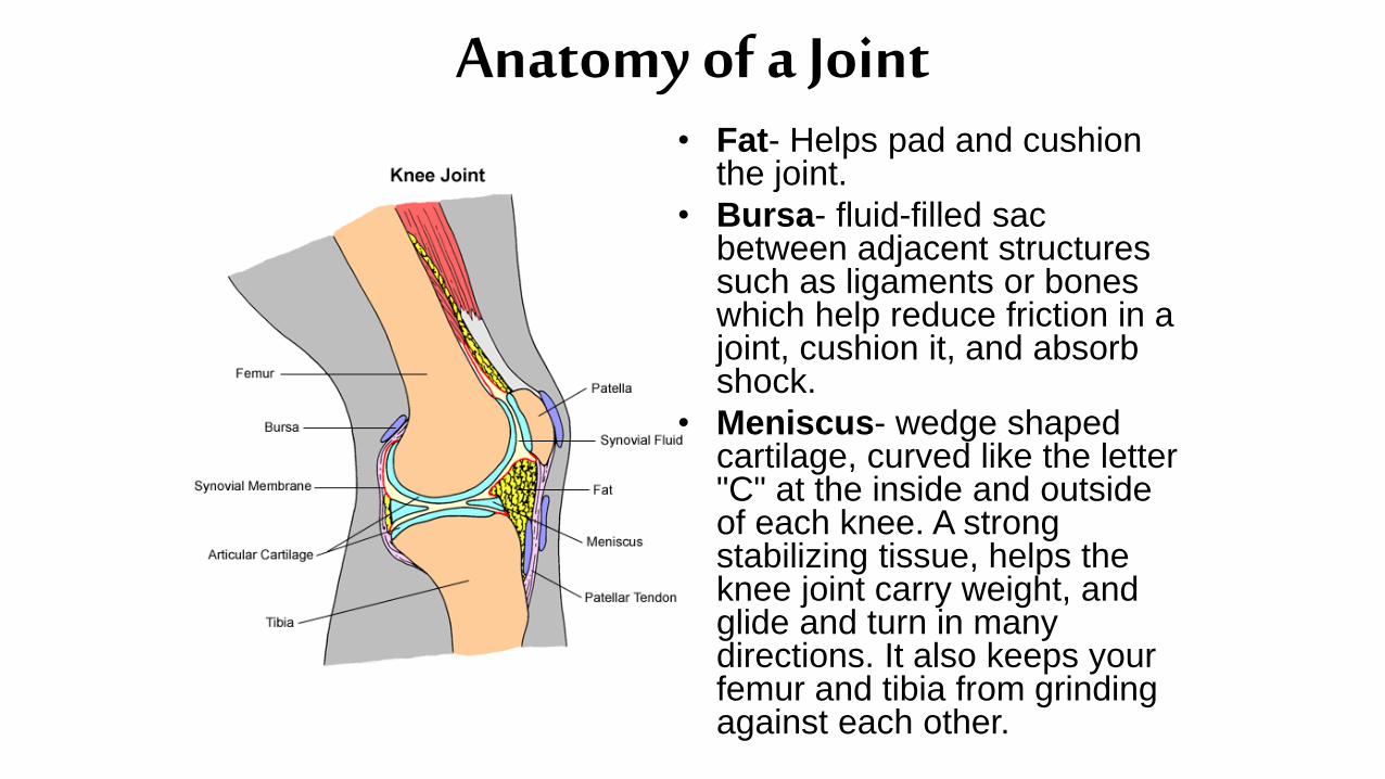
Anatomy of a Joint• Fat- Helps pad and cushion
the joint.
• Bursa- fluid-filled sac between adjacent structures such as ligaments or bones which help reduce friction in a joint, cushion it, and absorb shock.
• Meniscus- wedge shaped cartilage, curved like the letter "C" at the inside and outside of each knee. A strong stabilizing tissue, helps the knee joint carry weight, and glide and turn in many directions. It also keeps your femur and tibia from grinding against each other.

Anatomy of a Joint• ligaments - tough, elastic
bands of connective tissue
– surround the joint to give
support and limit the
joint's movement.
– Attach bone to bone
• tendons – another type of
tough connective tissue
– on each side of a joint
attached to muscles that
control movement of the
joint.
– Attach muscle to bone
Knee
Joint