avoiding pitfalls in mammographic interpretation
TRANSCRIPT

Canadian Association of Radiologists Journal 62 (2011) 50e59www.carjonline.org
Thoracic and Cardiac Imaging / Imagerie cardiaque et imagerie thoracique
Avoiding Pitfalls in Mammographic Interpretation
Shayna Roberts-Klein, MD, Elaine Iuanow, MD, Priscilla J. Slanetz, MD, MPH, FACR*
Division of Breast Imaging, Department of Radiology, Beth Israel Deaconess Medical Center, Boston, MA
Abstract
There is a public misconception that screening mammography detects all breast malignancies. The objective of this pictorial essay is toreview classic mammographic features of malignancy that, if missed, could potentially result in malpractice litigation. By identifyingradiologic themes, we attempt to improve awareness about the imaging characteristics of a variety of subtle malignancies.
Resume
Il existe une idee fausse repandue dans la population voulant que la mammographie de depistage permette de deceler tous les types detumeurs malignes aux seins. Cet article descriptif a pour but de passer en revue les caracteristiques classiques des tumeurs malignes a lamammographie qui, si elles ne sont pas detectees, peuvent mener a un litige pour faute professionnelle. En definissant des themes lies a laradiologie, nous tentons d’ameliorer la connaissance des caracteristiques d’un eventail de tumeurs malignes subtiles visibles par imagerie.� 2011 Canadian Association of Radiologists. All rights reserved.
Key Words: Breast; Breast cancer; Mammography; Malpractice
Introduction
Mammography is the standard screening tool for detec-tion of breast cancer, yet 10%e30% of cancers may bemissed with mammography [1]. Education of the generalpublic regarding the limitations of mammography is lacking,and many malpractice cases arise because of the miscon-ception that mammography detects all malignancies and thatany delay in diagnosis leads to a worse outcome [2]. Delay indiagnosis of breast cancer remains a leading cause ofmalpractice, particularly for radiologists who practice breastimaging [3].
From the imaging perspective, breast cancers may bemissed because of failure to perceive an abnormality,incorrect interpretation of an abnormality, lesions obscuredby dense parenchyma, or lesions missed because of poorpositioning or technique [1,4]. Many of the imaging findingsin these types of cases are subtle and likely do not relate toactual negligence on the part of the interpreting radiologist.
* Address for correspondence: Priscilla J. Slanetz, MD, MPH, FACR,
Department of Radiology, TCC4, Beth Israel Deaconess Medical Center, 330
Brookline Avenue, Boston, MA 02215.
E-mail address: [email protected] (P. J. Slanetz).
0846-5371/$ - see front matter � 2011 Canadian Association of Radiologists. A
doi:10.1016/j.carj.2010.07.004
However, these types of cases are difficult to defend ina court of law, because juries often side with the plaintiff.
In this article, we review some common pitfalls in breastimaging, which, if overlooked or misinterpreted, might leadto an unnecessary delay in diagnosis. The topics discussedbelow include: (1) edge of film findings; (2) suspicious, butstable morphology; (3) slowly developing asymmetry;(4) architectural distortion; (5) benign-appearing nodule;(6) presumed intramammary lymph node; (7) shrinkingbreast; and (8) scar carcinoma. By identifying radiologicthemes, we aim to increase awareness of these subtleimaging presentations in hopes of earlier detection andimproved outcomes.
Edge of Film Findings
Edge of film findings refer to any finding visualized atthe margin of an image. As seen in Figure 1, there isa subtle mammographic asymmetry only seen on the edgeof the craniocaudal (CC) view (Figure 1). Another subtlemalignancy, only seen on 1 of the conventional images,which could be easily overlooked, is demonstrated in Figure2. In reality, even if a finding is only seen on the edge ofa single view, additional imaging is necessary to confirm
ll rights reserved.
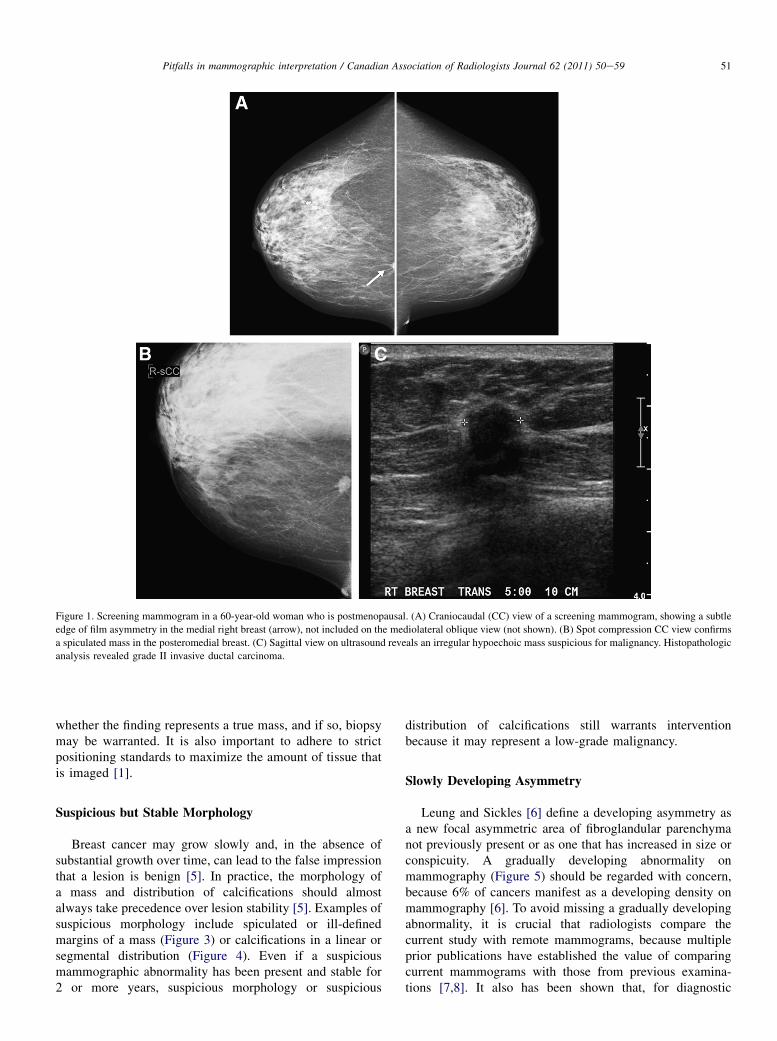
Figure 1. Screening mammogram in a 60-year-old woman who is postmenopausal. (A) Craniocaudal (CC) view of a screening mammogram, showing a subtle
edge of film asymmetry in the medial right breast (arrow), not included on the mediolateral oblique view (not shown). (B) Spot compression CC view confirms
a spiculated mass in the posteromedial breast. (C) Sagittal view on ultrasound reveals an irregular hypoechoic mass suspicious for malignancy. Histopathologic
analysis revealed grade II invasive ductal carcinoma.
51Pitfalls in mammographic interpretation / Canadian Association of Radiologists Journal 62 (2011) 50e59
whether the finding represents a true mass, and if so, biopsymay be warranted. It is also important to adhere to strictpositioning standards to maximize the amount of tissue thatis imaged [1].
Suspicious but Stable Morphology
Breast cancer may grow slowly and, in the absence ofsubstantial growth over time, can lead to the false impressionthat a lesion is benign [5]. In practice, the morphology ofa mass and distribution of calcifications should almostalways take precedence over lesion stability [5]. Examples ofsuspicious morphology include spiculated or ill-definedmargins of a mass (Figure 3) or calcifications in a linear orsegmental distribution (Figure 4). Even if a suspiciousmammographic abnormality has been present and stable for2 or more years, suspicious morphology or suspicious
distribution of calcifications still warrants interventionbecause it may represent a low-grade malignancy.
Slowly Developing Asymmetry
Leung and Sickles [6] define a developing asymmetry asa new focal asymmetric area of fibroglandular parenchymanot previously present or as one that has increased in size orconspicuity. A gradually developing abnormality onmammography (Figure 5) should be regarded with concern,because 6% of cancers manifest as a developing density onmammography [6]. To avoid missing a gradually developingabnormality, it is crucial that radiologists compare thecurrent study with remote mammograms, because multipleprior publications have established the value of comparingcurrent mammograms with those from previous examina-tions [7,8]. It also has been shown that, for diagnostic

Figure 2. Screening mammogram in a 72-year-old woman. (A) Craniocaudal (CC) view from screening mammogram, showing an asymmetry in the deep
lateral right breast (arrow), not included in the mediolateral oblique (MLO) view (not shown) and not visible on prior screening studies. (B) CC and MLO spot
compression and mediolateral views demonstrate the mass. (C) Sagittal view on ultrasound, revealing an irregular hypoechoic solid mass, which was path-
ologically proven invasive ductal carcinoma, grade II, with metastatic adenocarcinoma, involving 4 of 12 lymph nodes.
52 S. Roberts-Klein et al. / Canadian Association of Radiologists Journal 62 (2011) 50e59
mammography, comparison with prior mammograms isassociated with an increase in the cancer detection rate [9].In cases in which the mammographic evaluation is equivocal,targeted ultrasound or breast magnetic resonance imaging(MRI) can be useful to help exclude an underlyingmalignancy.
Architectural Distortion
Architectural distortion refers to a focal area of breasttissue that appears distorted because of radiating spicula-tions (Figure 6) and focal areas of retraction and tethering,without a discrete associated mass (Figure 7) [10]. Becausearchitectural distortion may mimic the normal appearanceof overlapping breast tissue, this finding can be subtle andmay be difficult to detect [11]. Distortion can be associatedwith benign lesions, such as surgical scars, radial scars, orfat necrosis; however, the malignancy rates for architec-tural distortion range from almost one-half to two-thirds ofthe cases [10,11]. One study that evaluated the efficacy ofcommercially available computer-aided detection systems
(CAD) showed that CAD had a low sensitivity for thedetection of architectural distortion, identifying fewer thanone-half of the cases, and hence, breast imagers shouldremain vigilant in the assessment of distortion [11].Although there is considerable interobserver variabilitywith regard to architectural distortion, double reading ofmammograms has been shown to increase the detectionrate for breast cancer up to 15% and may help improvedetection of this subtle finding [1]. Therefore, having a lowthreshold to recall patients for additional imaging evalua-tion of distortion is critical. Even on the diagnosticworkup, it is important to carefully evaluate this type offinding. If the tissue is at all fibroglandular, even if itappears to dissipate on spot mammographic views, thenultrasound or MRI can aid in excluding an underlyingmalignancy.
Benign-appearing Nodule
It is important to remember that not all cancers havespiculated margins and an irregular shape. Some cancers

Figure 3. An 80-year-old woman for screening. (A) Right craniocaudal and mediolateral oblique views, showing a focal asymmetry in the deep upper outer
breast (arrow), which was stable for 10 years and classified as glandular asymmetry by the interpreting radiologist. (B) Spot compression images demonstrating
the mass to better advantage. (C) Sagittal view on ultrasound confirms a hypoechoic irregular mass at 10-o’clock position, 9 cm from the nipple, which was
pathologically proven invasive carcinoma, predominantly mucinous type.
Figure 4. A 56-year-old woman who presented for a 6-month follow-up of calcifications in the left breast. (A) Craniocaudal (CC) and mediolateral oblique
(MLO) views from left diagnostic mammogram and (B) left spot magnification CC and MLO views, demonstrating calcifications in a segmental distribution in
the lower medial left breast. Although these calcifications appear coarse and many have lucent centres that suggest a benign etiology, the distribution was
suspicious, and biopsy revealed ductal carcinoma in situ, intermediate-to-high nuclear grade.
53Pitfalls in mammographic interpretation / Canadian Association of Radiologists Journal 62 (2011) 50e59

Figure 5. A 67-year-old woman for screening. (A) Craniocaudal and (B) mediolateral oblique views from left consecutive annual mammograms over 3 years,
showing a slowly developing asymmetry in the upper outer quadrant (arrow), which was pathologically proven grade I invasive ductal carcinoma.
54 S. Roberts-Klein et al. / Canadian Association of Radiologists Journal 62 (2011) 50e59
present as relatively well-circumscribed masses on screeningmammography, particularly medullary, mucinous, andpapillary subtypes [4]. However, on spot compression, these‘‘circumscribed’’ cancers usually have ill-defined margins(Figure 8) [12]. Therefore, it is important to be wary of a newmass that appears to be circumscribed at first glance.Although many of these will be benign lesions, such as cystsor fibroadenomas, it is necessary to image the patient withadditional mammographic views and ultrasound to bettercharacterize the finding. Ultrasound is particularly helpful inpredicting the likelihood of a malignancy of a circumscribed
mass, and it is important to correlate the sonographic find-ings with those on mammography [4].
Presumed Intramammary Lymph Node
Benign-appearing intramammary lymph nodes aregenerally found in the upper outer quadrant and typicallyare less than 1 cm in diameter, well-circumscribed, ovalor lobulated masses with radiolucent fatty hilum visibleon mammography [13,14]. On ultrasound, a benign-appearing intramammary lymph node should have

Figure 6. A 61-year-old woman for screening. (A) Craniocaudal and (B) mediolateral oblique (MLO) views from screening mammogram, showing an area of
architectural distortion in the right upper outer quadrant (arrow). (C) Right diagnostic spot MLO view, showing persistent distortion (arrow), but this was not
perceived by the interpreting radiologist and was thought to represent superimposed breast tissue. No ultrasound was performed at that time. The patient
returned 5 months later with a palpable lump in the region of distortion. Histopathologic analysis revealed invasive ductal carcinoma with metastatic carcinoma
that involved 1 of 7 lymph nodes.
55Pitfalls in mammographic interpretation / Canadian Association of Radiologists Journal 62 (2011) 50e59
smooth or lobulated margins, with a thin hypoechoiccortex and an echogenic central hilum. Any irregularity ofthe margins of mass should raise concern for malignancyand warrant intervention (Figure 9). Rarely, benign-appearing intramammary lymph nodes may be seen inlocations other than the upper outer quadrant; however,because of the infrequency of this occurrence, additionalimaging with mammography and ultrasound should beperformed to confirm that the mass meets the morpho-logic criteria for a normal-appearing lymph node ratherthan a malignancy [14].
Shrinking Breast
A decrease in breast size has been described withadvanced cases of infiltrating lobular carcinoma (ILC) [15].This subtype of breast cancer is known to be difficult todiagnose on both mammography and on physical examina-tion because it infiltrates the breast as a single layer of cells,often referred to as ‘‘Indian-filing,’’ with little disruption ofthe normal parenchymal architecture until it has diffuselyspread through a substantial amount of breast tissue [15].This infiltration can eventually lead to the appearance of

Figure 7. A 66-year-old woman who presented with a palpable lump in the right breast 10 months after a ‘‘negative’’ screening examination. (A) Re-review of
the craniocaudal (CC) and (B) mediolateral oblique (MLO) views of the right breast on consecutive annual mammograms over 3 years, showing developing
architectural distortion (arrow) in the central upper breast (area of distortion was palpable in 2009). (C) Magnified spot compression CC and MLO views,
demonstrating distortion and pleomorphic calcifications that correspond to the area of concern to the patient. (D) Sagittal view on ultrasound, showing an
irregular hypoechoic mass, which was pathologically proven invasive ductal carcinoma arising in extensive high-grade ductal carcinoma in situ.
56 S. Roberts-Klein et al. / Canadian Association of Radiologists Journal 62 (2011) 50e59
distortion and retraction of the glandular tissue and trabec-ular thickening, eventually accompanied by loss of breastvolume [16]. On imaging, this process manifests as a poorlycompressible breast that becomes smaller over time becauseof the diffusely infiltrating tumour (Figure 10) [15,16].
Because the mammographic findings in these cases aresubtle, often without a discrete mass, comparison with priorstudies will aid the radiologist in recognizing cases ofa diffusely infiltrative process. Focusing on the posterior andsuperficial interfaces of fat and glandular tissue will also helpin detecting those subtle findings, because as the breast‘‘shrinks,’’ more fat becomes visible behind the glandulartissue and underneath the skin surface [16].
Scar Carcinoma
Breast cancer that develops from a surgical scar related toprior benign breast biopsy is rare, with only 13 cases of this
type of malignancy reported in the literature to date [17].Carcinoma that forms in the scar from a benign breast biopsycan be subtle and may present with vague developingasymmetry, suspicious microcalcifications, or fullness of thebiopsy bed (Figure 11). Correlation with clinical history toexclude a superimposed acute infection or recent trauma andcomparison with old studies can be helpful to identify thesesubtle changes. When the mammogram is indeterminate,MRI can also be used to further evaluate because suspiciousenhancement in a biopsy bed would be concerning formalignancy.
Discussion
As many as 30% of breast cancers may be missed withmammography [1]. For a specific case to meet the criteriaof malpractice, the interpreting radiologist must be negli-gent in the mammographic interpretation and the resulting
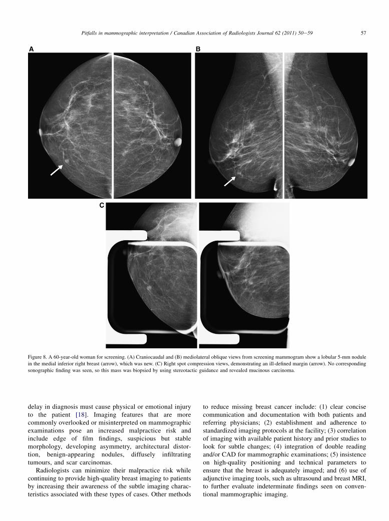
Figure 8. A 60-year-old woman for screening. (A) Craniocaudal and (B) mediolateral oblique views from screening mammogram show a lobular 5-mm nodule
in the medial inferior right breast (arrow), which was new. (C) Right spot compression views, demonstrating an ill-defined margin (arrow). No corresponding
sonographic finding was seen, so this mass was biopsied by using stereotactic guidance and revealed mucinous carcinoma.
57Pitfalls in mammographic interpretation / Canadian Association of Radiologists Journal 62 (2011) 50e59
delay in diagnosis must cause physical or emotional injuryto the patient [18]. Imaging features that are morecommonly overlooked or misinterpreted on mammographicexaminations pose an increased malpractice risk andinclude edge of film findings, suspicious but stablemorphology, developing asymmetry, architectural distor-tion, benign-appearing nodules, diffusely infiltratingtumours, and scar carcinomas.
Radiologists can minimize their malpractice risk whilecontinuing to provide high-quality breast imaging to patientsby increasing their awareness of the subtle imaging charac-teristics associated with these types of cases. Other methods
to reduce missing breast cancer include: (1) clear concisecommunication and documentation with both patients andreferring physicians; (2) establishment and adherence tostandardized imaging protocols at the facility; (3) correlationof imaging with available patient history and prior studies tolook for subtle changes; (4) integration of double readingand/or CAD for mammographic examinations; (5) insistenceon high-quality positioning and technical parameters toensure that the breast is adequately imaged; and (6) use ofadjunctive imaging tools, such as ultrasound and breast MRI,to further evaluate indeterminate findings seen on conven-tional mammographic imaging.
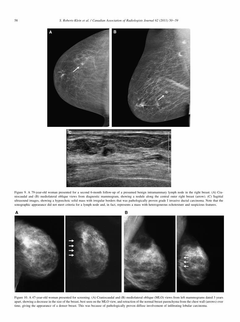
Figure 9. A 79-year-old woman presented for a second 6-month follow-up of a presumed benign intramammary lymph node in the right breast. (A) Cra-
niocaudal and (B) mediolateral oblique views from diagnostic mammogram, showing a nodule along the central outer right breast (arrow). (C) Sagittal
ultrasound images, showing a hypoechoic solid mass with irregular borders that was pathologically proven grade I invasive ductal carcinoma. Note that the
sonographic appearance did not meet criteria for a lymph node and, in fact, represents a mass with heterogeneous echotexture and suspicious features.
Figure 10. A 47-year-old woman presented for screening. (A) Craniocaudal and (B) mediolateral oblique (MLO) views from left mammograms dated 3 years
apart, showing a decrease in the size of the breast, best seen on the MLO view, and retraction of the normal breast parenchyma from the chest wall (arrows) over
time, giving the appearance of a denser breast. This was because of pathologically proven diffuse involvement of infiltrating lobular carcinoma.
58 S. Roberts-Klein et al. / Canadian Association of Radiologists Journal 62 (2011) 50e59

Figure 11. A 63-year-old woman with a history of 2 benign right breast excisional biopsies 8 years before presentation. (A) Craniocaudal (CC) and mediolateral
oblique (MLO) right mammographic views of the right breast, showing a dense mass along the deep medial upper breast adjacent to the post-surgical distortion.
(B) Spot compression CC and MLO views, better demonstrating the spiculated mass. (C) Ultrasound in the radial plane, showing a hypoechoic irregular mass
with posterior shadowing (arrow) adjacent to the scar. Biopsy of this finding confirmed a grade 1 invasive ductal carcinoma.
59Pitfalls in mammographic interpretation / Canadian Association of Radiologists Journal 62 (2011) 50e59
References
[1] Majid AS, Shaw de Paredes E, Doherty RD, et al. Missed breast
carcinoma: pitfalls and pearls. Radiographics 2003;23:881e95.
[2] Berlin L. Malpractice and breast cancer: perceptions versus reality.
AJR Am J Roentgenol 2009;92:334e6.
[3] Kopans DB. Mammography screening is saving thousands of lives, but
will it survive medical malpractice. Radiology 2004;230:20e4.
[4] Ikeda DM, Birdwell RL, O’Shaughnessy KF, et al. Analysis of 172
subtle findings on prior normal mammograms in women with breast
cancer detected at follow-up screening. Radiology 2003;226:
494e503.
[5] Harvey JA, Nicholson BT, Cohen MA. Finding early invasive breast
cancers: a practical approach. Radiology 2008;248:61e76.
[6] Leung JW, Sickles EA. Developing asymmetry identified on
mammography: correlation with imaging outcome and pathologic
findings. AJR Am J Roentgenol 2007;188:667e75.[7] Frankel SD, Sickles EA, Curpen BN, et al. Initial versus subsequent
screening mammography: comparison of findings and their prognostic
significance. AJR Am J Roentgenol 1995;164:1107e9.[8] Callaway MP, Boggis CRM, Astley SA, et al. The influences of
previous films on screening mammographic interpretation and detec-
tion of breast carcinoma. Clin Radiol 1997;52:527e9.
[9] Burnside ES, Sickles EA, Sohlich RE, et al. Differential value
of comparison with previous examinations in diagnostic versus
screening mammography. AJR Am J Roentgenol 2002;179:
1173e7.
[10] Samardar P, Shaw de Paredes E, Grimes MM, et al. Focal asymmetric
densities seen at mammography: US and pathologic correlation.
Radiographics 2002;22:19e33.[11] Baker JA, Rosen EL, Lo JY, et al. Computer-aided detection (CAD) in
screening mammography: sensitivity of commercial CAD systems for
detecting architectural distortion. AJR Am J Roentgenol 2003;181:
1083e8.[12] Harvey JA. Unusual breast cancers: useful clues to expanding the
differential diagnosis. Radiology 2007;242:683e94.
[13] Cyrlak D, Carpenter PM. Breast imaging case of the day. Radio-
graphics 1999;19:S73e9.
[14] Meyer JE, Ferraro FA, Frenna TH, et al. Mammographic appearance of
normal intramammary lymph nodes in an atypical location. AJR Am
J Roentgenol 1993;161:779e80.[15] Harvey JA, Fechner RE, Moore MM. Apparent ipsilateral decrease in
breast size at mammography: a sign of infiltrating lobular carcinoma.
Radiology 2000;214:883e9.
[16] Jafri NF, Slanetz PJ. The shrinking breast: an unusual mammographic
finding of invasive lobular carcinoma. Radiology Case Reports 2007;2:
94.
[17] Kim MJ, Kim EK, Lee JY, et al. Breast cancer from the excisional scar
of a benign mass. Korean J Radiol 2007;8:254e7.[18] Berlin L. Fear of cancer. AJR Am J Roentgenol 2004;183:267e72.