attia general surgery review manual
TRANSCRIPT
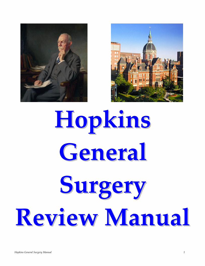
Hopkins General Surgery Manual 1
HHooppkkiinnss GGeenneerraall SSuurrggeerryy
RReevviieeww MMaannuuaall
Hopkins General Surgery Manual 2
Introduction This “manual” is a compilation of study notes I have made over the past 5 years based on a number of sources, including those listed here:
Text books (see reference list) Review books (see reference list) Didactic lectures and conferences at both Johns Hopkins and the NCI Surgery Branch Presentations I gave during weekly conferences at NCI Primary and review articles Points made by attendings and other residents on rounds or in the OR Intern Sunday morning lecture with Dr. Cameron (2001 – 2002) Halsted quizzes SESAP questions UpToDate®
Disclaimer: Individual illustrations and material may belong to a third party. Unless otherwise stated all figures and tables by Peter Attia When I began putting my notes together on random pieces of paper and my Palm Pilot, I did not intend to do much else with them. However, in time, they became so numerous that I needed to organize them in a better way. A resident from the Brigham whom I worked with in the lab at NIH encouraged me to put them together in what he jokingly referred to as an “Attia Bible” of surgical wisdom, something he had done with his own notes. The intent of these notes was not as much to be a review for a specific test per se, as it was an “all‐purpose” compilation of salient points to consider as I go through residency. Of course, these notes come with the standard disclaimer that they are not meant to replace reading from primary sources, rather to supplement it. In addition, while I have tried to be as accurate as possible, during my readings I encountered several “facts” that were either contradictory to “facts” I had been taught as a resident or read in other sources. For this reason I can make no guarantees about the validity of each statement made here. I have tried my best to amalgamate each set of facts into a somewhat concise, yet accurate document. Hopefully, these notes will provide you with some benefit as well. I welcome all criticism and correction and look forward to supplementing and augmenting this first edition many times over. Peter Attia, MD Surgical Resident The Johns Hopkins Hospital [email protected] Copyright, Peter Attia, 2005. All rights reserved.
Hopkins General Surgery Manual 3
Reference List
1. Vascular Surgery 3rd Ed. House Officer Series. Faust GR, Cohen JR., 1998. 2. ABSITE Killer. Lipkin AP, 2000 3. Rush University Review of Surgery 3rd Ed. Deziel, Witt, Bines, et al., 2000. 4. Current Surgical Therapy 6th Ed. Cameron JL, 1998. 5. Current Therapy of Trauma 4th Ed. Trunkey DD and Lewis FR, 1999. 6. Surgery: Scientific Principles and Practice 3rd Ed. Greenfield LJ, et al., 2001. 7. Shackelford’s Surgery of the Alimentary Tract 5th Ed. Yeo CJ and Zuidema GD. Volumes I – V, 2001. 8. Atlas of Human Anatomy 8th Ed. Netter FH, 1995. 9. Atlas of Surgical Operations 7th Ed. Zollinger & Zollinger, 1993. 10. General Surgery Board Review 3rd Ed. Gold MS, Scher LA, and Weinberg G, 1999. 11. General Surgery Review. Makary MA, 2004. 12. Advances in Surgery® Vol 33. Ed. Cameron JL, et al., 1999. 13. Pitfalls of Data Analysis. Clay Helberg, 1995. 14. Principles of Biostatistics. 2nd Ed. Pagano M and Gauvreau K. 2000.
Editors
The following individuals have been generous with their time and thoughts, and have made several changes and additions to my original “manual”.
H. Richard Alexander Surgery Branch, National Cancer Institute William A. Baumgartner The Johns Hopkins Hospital John L. Cameron The Johns Hopkins Hospital Michael A. Choti The Johns Hopkins Hospital Peter L. Choyke Department of Radiology, National Institutes of Health Paul M. Colombani The Johns Hopkins Hospital Matthew Cooper The University of Maryland Edward E. Cornwell, III The Johns Hopkins Hospital Todd Dorman The Johns Hopkins Hospital Frederic E. Eckhauser The Johns Hopkins Hospital David T. Efron The Johns Hopkins Hospital Anne C. Fischer The Johns Hopkins Hospital Julie A. Freischlag The Johns Hopkins Hospital Susan L. Gearhart The Johns Hopkins Hospital Vincent L. Gott The Johns Hopkins Hospital McDonald Horne Department of Hematology, National Institutes of Health Udai S. Kammula Surgery Branch, National Cancer Institute Herbert Kotz Department of Gynecology, National Cancer Institute Steven K. Libutti Surgery Branch, National Cancer Institute Pamela A. Lipsett The Johns Hopkins Hospital Martin A. Makary The Johns Hopkins Hospital Bruce J. Perler The Johns Hopkins Hospital Peter A. Pinto Urology Branch, National Cancer Institute Jorge D. Salazar University of Texas, San Antonio, TX Anthony P. Tufaro The Johns Hopkins Hospital Peter S. Walinsky Presbyterian Heart Group, Albuquerque, NM Stephen S. Yang The Johns Hopkins Hospital Charles J. Yeo Thomas Jefferson University Martha A. Zeiger The Johns Hopkins Hospital
Hopkins General Surgery Manual 4
Table of Contents Breast Disease ................................................................................................................................................................................... 5 Head & Neck Disease ...................................................................................................................................................................... 9 Thyroid Gland and Disease ......................................................................................................................................................... 12 Parathyroid Gland and Disease................................................................................................................................................... 15 Multiple Endocrine Neoplasia (MEN) ....................................................................................................................................... 18 Gastrinoma ...................................................................................................................................................................................... 21 Glucagonoma .................................................................................................................................................................................. 22 Insulinoma ...................................................................................................................................................................................... 23 Adrenal Gland ................................................................................................................................................................................ 24 Pheochromocytoma........................................................................................................................................................................ 27 Pituitary Gland ............................................................................................................................................................................... 28 Thoracic Surgery ............................................................................................................................................................................ 29 Mediastinal Disease ...................................................................................................................................................................... 32 Cardiac Surgery: Congenital Defects ......................................................................................................................................... 34 Cardiac Surgery: Acquired Defects............................................................................................................................................. 36 Vascular Surgery ............................................................................................................................................................................ 42 Urology............................................................................................................................................................................................. 54 Orthopedic Surgery ....................................................................................................................................................................... 55 Gynecologic Pathology ................................................................................................................................................................. 56 Neurosurgery .................................................................................................................................................................................. 57 Cancer Epidemiology .................................................................................................................................................................... 58 Esophageal Disease........................................................................................................................................................................ 59 Stomach & Gut Physiology and Disease ................................................................................................................................... 63 Small Bowel Physiology and Disease ........................................................................................................................................ 67 Colorectal Disease.......................................................................................................................................................................... 71 Pediatric Surgery ............................................................................................................................................................................ 79 Spleen and Splenectomy .............................................................................................................................................................. 84 Hepatobiliary Anatomy, Physiology, and Disease .................................................................................................................. 86 Pancreas............................................................................................................................................................................................ 97 Sarcoma .......................................................................................................................................................................................... 102 Melanoma ...................................................................................................................................................................................... 103 Hernia & Abdominal Wall ......................................................................................................................................................... 105 Trauma Principles ........................................................................................................................................................................ 106 Critical Care................................................................................................................................................................................... 117 Hemostasis & Transfusion ......................................................................................................................................................... 126 Metabolism.................................................................................................................................................................................... 130 Transplant Surgery ...................................................................................................................................................................... 131 Nutrition ........................................................................................................................................................................................ 134 Fluids & Electrolytes ................................................................................................................................................................... 136 Renal Physiology.......................................................................................................................................................................... 137 Immunology/Infections .............................................................................................................................................................. 139 Burns............................................................................................................................................................................................... 140 Skin & Wound Healing............................................................................................................................................................... 141 Pharmacology................................................................................................................................................................................ 142 Radiology....................................................................................................................................................................................... 143 Statistics in Medicine .................................................................................................................................................................. 149 Notes............................................................................................................................................................................................... 154
Hopkins General Surgery Manual 5
Breast Disease Surgical Anatomy:
Intercostobrachial nerve (off 2nd intercostal nerve) sensation to medial arm can sacrifice Long thoracic nerve: to serratus anterior winged scapula Thoracodorsal nerve: to latissimus dorsi weak arm adduction Medial pectoral nerve to pec minor and major; lateral pectoral nerve to pec minor only
Batson’s plexus: valveless vertebral veins allow direct metastases to spine Poland syndrome: amastia, hypoplastic shoulder, no pecs Mastodynia: Rx with danazol, OCP Mondor’s disease: thrombophlebitis of superficial vein of breast Rx with NSAID DCIS
• Highly curable with survival of 94 – 100% • 50% of recurrences are invasive • Excision and radiotherapy OR mastectomy; axillary lymph node dissection (ALND) not required
(only 1% have positive nodes). NSABP‐17 showed that lumpectomy alone had 13.4% recurrent DCIS and 13.4% recurrent invasive cancer vs. 8.2% and 3.9%, respectively for lumpectomy + radiation.
• Tamoxifen decreases rate of ipsilateral and contralateral breast cancer in ER positive women, role in ER negative women, if any, unknown; but must be balanced against risk factors (1 – 2% DVT, PE; Endometrial cancer). Tamoxifen has NOT been shown to increase survival, only to decrease rate of recurrence (DCIS and ipsilateral/contralateral invasive breast cancer). Several large studies have been done (NSABP‐24, 1800 patients, [Fisher B, et al. Lancet 1999;353:1993]) and failed to identify a survival advantage, despite adequate power.
• Ongoing research to identify subset of patients who could be treated without radiation • Role of Sentinel Lymph is undefined. NO evidence to support use as of 2004. • Van Nuys classification MAY identify patients who can benefit from lumpectomy alone (low‐grade,
without necrosis; margin > 1 cm; lesion < 1.5 cm) LCIS
1. Aka Lobular Neoplasia, encompasses LCIS (> 50% lobular involvement) and Atypical Lobular Hyperplasia (ALH, < 50% lobular involvement)
2. Not clinically, radiographically, grossly detectable 3. 7 – 10 x increased risk of invasive cancer in either breast (especially in young women with a family
history) 4. 17% risk at 15 years, 5.6% at 5 years; 20% lifetime risk (70% of which will be ductal invasive, 30%
will be lobular invasive); ≈ 1% per year 5. Margins are irrelevant, disease is diffuse (unlike DCIS) 6. LCIS is not itself pre‐cancerous, it is simply a marker of a susceptible field
Phyllodes tumor: 10% malignant; large; rare nodes (spread, if any, hematogenous): Rx WLE, mastectomy not necessary; NO ALND Intraductal papilloma: No risk of cancer; #1 cause of bloody nipple discharge
Hopkins General Surgery Manual 6
Comedo breast cancer: Likely multicentric; do mastectomy; poor prognosis Paget’s disease of the breast: Eczematous lesion on nipple underlying DCIS or ductal CA Most recent screening recommendations: First at 40; q 1 – 2 years until 50; yearly thereafter Radial Scar: associated with carcinoma anywhere in the scar; do not stereotactically biopsy (↑ chance of sampling error), instead excisional biopsy Staging: T1: < 2 cm T2: 2.1 – 5 cm T3: > 5 cm T4: skin involvement (inflammatory dermal lymphatic invasion)
N1: + ax nodes N2: matted/fixed N3: internal mammary nodes
Stage I: T1 Stage II: up to T2N1 or T3N0 Stage III: T4 or N3 Stage IV: any M
Survival by stage (5 years): I: 90 – 95% II: 50 – 80% III: 30 – 50% IV: 15 – 20%
Note: FNA cannot distinguish between DCIS and invasive Who gets chemotherapy? 1. Pre‐menopausal:
ER/PR‐ T > 1 cm Any N, including micro (SN+)
2. Post‐menopausal (up to 90% are ER/PR+ get tamoxifen): ER/PR‐ & T > 2 cm ≥ 4 nodes OR matted nodes (regardless of ER/PR)
(Hence, ER/PR+, ≤ 3 unmatted nodes no chemo)
Who gets axillary radiation? (In general, want to avoid axillary radiation following dissection) + supraclavicular node matted nodes (extracapsular extension) ≥ 4 nodes
Who gets breast irradiation? any segmental resection for invasive or DCIS inflammatory disease (T4/skin involvement); some T3
Major studies evaluating role of adjuvant radiation* therapy:
1. The addition of post‐op irradiation to chemotherapy (CMF) for women with stage II or III breast cancer following mastectomy increased overall survival and reduced locoregional recurrence. [Postoperative radiotherapy in high‐risk premenopausal women with breast cancer who receive adjuvant chemotherapy. Danish Breast Cancer Cooperative Group 82b Trial. Overgaard M, et al. NEJM 1997;337:949].
2. Radiotherapy combined with chemotherapy (CMF) after modified radical mastectomy decreases rates of locoregional and systemic relapse and reduces mortality from breast cancer. [Adjuvant radiotherapy and chemotherapy in node‐positive premenopausal women with breast cancer. Ragaz J, et al. NEJM 1997;337:956].
*Trental is very effective in treating radiation mastitis
Hopkins General Surgery Manual 7
Locally Advanced Breast Cancer • Locally Advanced Breast Cancer (LABC) & Inflammatory Breast Cancer (IBC) sometimes
(incorrectly) used interchangeably • Strictly speaking, LABC includes: T3+N1 – 3 or T4+N0 – 3 or any T+N2 – 3 (i.e. Stage III A/B disease) • Term IBC first used in 1924 by Lee and Tannenbaum at Memorial Hospital to describe clinical
presentation of 28 patients with: “…breast of affected side usually increased in size…skin becomes deep red or reddish purple…to the touch brawny and infiltrated…after the fashion of erysipelas…”
• Accounts for 1 – 6% of all breast carcinomas (IBC) • 50 – 75% axillary involvement at diagnosis
Overall prognosis median survival: 2 years
• Diagnosis based on histology of invasive carcinoma PLUS 1. Erythema 2. Edema, or peau d’orange 3. Wheals, or ridging of the skin secondary to dermal lymphatic invasion (although tumor
invasion only seen in 30%) • Neoadjuvant treatment and early diagnosis crucial for successful treatment • Approximately 75% undergo CR or PR to induction therapy response predicts outcome
Effectiveness of mastectomy by response to induction chemotherapy for control of Inflammatory Breast Cancer [Fleming R, et al. Ann Surg Onc 1997 4:452]
Initial Response to Induction therapy: • CR median survival: 120 months (12%) • PR median survival: 48 months (62%) • NR median survival: < 24 months (26%)
Further Breakdown: • If > 1 cm3 residual tumor median survival: 36 months • If < 1 cm3 residual tumor 70% alive at 5 years
Role of Mastectomy: • If CR or PR Chemo + RT + Mastectomy increased median survival from 48 to 120 months (vs.
Chemo + RT) • If NR Chemo + RT + Mastectomy did not influence median survival (< 24 months), or disease‐
free interval
Summary for Treatment for Inflammatory breast cancer: 1. Neoadjuvant chemo (cytoxan/adriamycin); response to this predicts survival (10% CR, 80%PR) 2. MRM (if PR or CR) 3. Adjuvant chemo (taxane based) 4. Radiation to chest wall
Hopkins General Surgery Manual 8
Chemotherapy/Hormonal* Treatment:
Premenopausal Postmenopausal chemo for almost any tumor > 1 cm (regardless of
nodal status) cytoxan & adriamycin add taxane if node positive tamoxifen if ER/PR positive arimidex and aromatase inhibitors not effective in
premenopausal since can’t compete with estrogen produced
tamoxifen or arimidex if node negative and ER/PR+ Chemo if poorly differentiated and > 1 cm (even if node
negative) cytoxan & adriamycin ± taxane if node positive tamoxifen or adriamycin if elderly, node positive, and
ER/PR+
*Responses to hormonal therapy by marker: ER/PR+ 80% ER‐/PR+ 45% ER+/PR‐ 35% ER/PR‐ 10% Inherited Breast Cancer Syndromes: 4 appear to be important
1. Li‐Fraumeni Syndrome mutation of p53 2. Mutation of bcl‐2 (18q21) ↑ expression of bcl‐2, which is anti‐apoptotic 3. BRCA‐1 on long arm of 17 4. BRCA‐2 on short region of 13q12‐13
BRCA 1 Ch 17q21; reported 1990, positionally cloned 1994 ↑ Risk of breast cancer (85%) and ovarian cancer (40 – 50%)
BRCA 2 Ch 13q12‐13; reported 1994; positionally cloned 1995 ↑ Risk of breast cancer (85%) and ovarian cancer (10%) ↑ Risk of male breast cancer (6%)
Risks of Tamoxifen use ↑Uterine adenocarcinoma, sarcoma ↑Cataracts ↑DVT, PE ↓osteoporosis No change in incidence of heart disease
Hopkins General Surgery Manual 9
Head & Neck Disease Parotiditis: Usually caused staph spp; seen in elderly, dehydrated; Rx: antibiotics drainage of abscess if not improving
“Ludwig’s angina”: Sublingual space infection (severe deep soft tissue infection of neck involving the floor of the mouth); if airway compromise perform awake tracheostomy under local anesthetic operative debridement
Leukoplakia can be premalignant; erythroplakia is premalignant (and of much more concern)
Head & Neck SCC: Stage I, II (up to 4 cm, no nodes) single modality treatment (surgery or RT) Stage III, IV combined modality Perform FNA, not excisional biopsy for suspicious masses
Nasopharyngeal SCC: associated with EBV; 50% present late as neck mass; drainage to posterior neck nodes; most common nasopharyngeal cancer in adults (lymphoma is most common in kids). Often see in Asian population
Glottic Cancer: if cords not fixed RT; if fixed surgery + RT. Chemo + RT used more often for organ preservation
Lip Cancer (99% epidermoid [i.e. squamous] carcinoma): Lower > upper lip (because of sun exposure) resect with primary closure if < ½ lip; otherwise flap
Tongue Cancer: usually surgery + RT; seen ↑ in Plummer Vinson (dysphagia, spoon fingers, anemia). More commonly seen in smokers/drinkers
As salivary gland size ↑ [sublingual (60%), submandibular (50%), parotid (20%)] incidence of malignant disease ↓
Pharyngeal cancers have worse prognosis than oral cancers
Mucoepidermoid carcinoma: #1 malignant salivary tumor overall
Adenoid cystic carcinoma: #1 malignant salivary tumor of submandibular/minor glands. Overall: poor prognosis
Pleomorphic adenoma ≡ mixed parotid tumor = #1 benign tumor (40 – 70% of all salivary gland tumors) Do NOT enucleate (or will recur) needs superficial parotidectomy (spare CN VII). If malignant take whole gland + CN VII; If high grade (anaplastic) need neck dissection
Warthin’s tumor (adenolymphoma) #2 benign salivary tumor; male predominance; 10% bilateral; 70% of bilateral parotid tumors are Warthin’s tumor; Rx superficial parotidectomy
Frey’s Syndrome: late complication of parotidectomy (occurs ≈ 50% when facial nerve is preserved); perfuse perspiration over cheek following salivary stimulation. Intracutaneous injection of Botox A ≈ 100% effective in treatment, but responses may be short‐lived (can be repeated). Usually self‐limiting.
Ipsilateral drooling following submandibular gland resection: likely injury to marginal mandibular nerve
Radical neck dissection: takes CN XI, SCM, IJ, submandibular gland; most morbid is CN XI
Hopkins General Surgery Manual 10
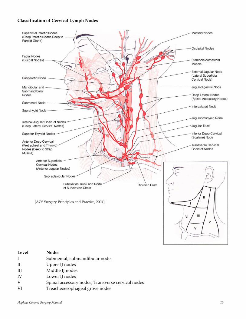
Classification of Cervical Lymph Nodes
Level Nodes I Submental, submandibular nodes II Upper IJ nodes III Middle IJ nodes IV Lower IJ nodes V Spinal accessory nodes, Transverse cervical nodes VI Treacheoesophageal grove nodes
[ACS Surgery Principles and Practice, 2004]
Hopkins General Surgery Manual 11
Cancers of the oral cavity usually metastasize to the nodes in levels I – III. Laryngeal cancers typically metastasize to the nodes in levels II – IV. Presence of Horner Syndrome (paralysis of the vagus nerve, phrenic nerve, invasion of brachial plexus, and/or paravertebral musculature) generally indicates tumor unresectability Tracheo‐innominate fistula
Massive bleeding from trachea is innominate artery until proven otherwise; avoid by making tracheostomy no lower than 3rd ring
Usually occurs 2 – 3 weeks post tracheostomy; poor nutrition and steroids use may contribute Mortality ≈ 80% Sentinel bleed to OR for bronchoscopy Temporary control (on route to OR) via cuff hyperinflation or finger compression of innominate
artery (anterior pressure) Treatment is ligation of innominate artery
Most common locations for mandibular fractures: angle (25%) and subcondyl (30%); the most common long‐term complication of mandibular fracture is malocclusion Carotid body: chemoreceptor within the adventitia of the CCA (posteromedial side); responds to ↓O2 tension, ↑CO2 tension, ↑blood acidity, and ↑blood temperature by ↑HR, ↑BP, and ↑rate & depth of respiration in an attempt to overcome the above stimuli
Carotid sinus: pressure sensor within wall of proximal ICA; responds to ↑BP by ↓HR and ↓BP
Hopkins General Surgery Manual 12
Thyroid Gland and Disease
from the Greek work “Theros” (shield) and “eidos” (form) secretes hormones (T4, T3, calcitonin) from basal membrane side (into bloodstream) antithyroid agents impair (i) iodination and (ii) coupling of DIT/MIT T4 T3 peripherally (kidney, liver) (T3; 10 x more active than T4). Propothiouracil (PTU) blocks
peripheral conversion of T4 T3 Note: Suppression of iodine uptake in patients with increased T3 and T4 levels is pathognomonic for subacute thyroiditis
Usual Causes of Hyperthyroidism: 1. Toxic nodule 2. Toxic multinodular goiter 3. Graves’ disease 4. Early subacute thyroiditis
Ways to Treat Hyperthyroidism: 1. Medical (PTU, methimazole): interfere with iodine conversion; up to 60% recur 2. Radioiodine Ablation (I131): weeks to months; 1st choice by many except in pregnancy 3. Surgery: risks of surgery
Thyroid Storm: untreated hyperthyroidism + stress (trauma, infection, pregnancy, DKA, etc) Rx: fluids, O2, glucose, anti‐thyroid drugs, but first treat underlying cause; NB: do not use ASA, as it displaces T4 from thyroglobulin
(Differentiated) Thyroid Cancer 15,000 – 20,000 cases/yr US 15,000,000 nodules/yr (5 – 10% harbor cancer) mortality < 1%
Risks age < 14, > 65 previous thyroid cancer family history enlarging nodule on thyroid hormone suppression exposure to low‐dose radiation Graves’ disease or thyroiditis syndromes (MEN II, Carney’s)
Cancer Histology Papillary (60%) Follicular variant of papillary (20%) Follicular (< 5%)* Hürthle cell carcinoma (< 5%) Medullary (5%) Anaplastic (1%) Other (1%)
*difficult on FNA to differentiate follicular adenoma from carcinoma
Hopkins General Surgery Manual 13
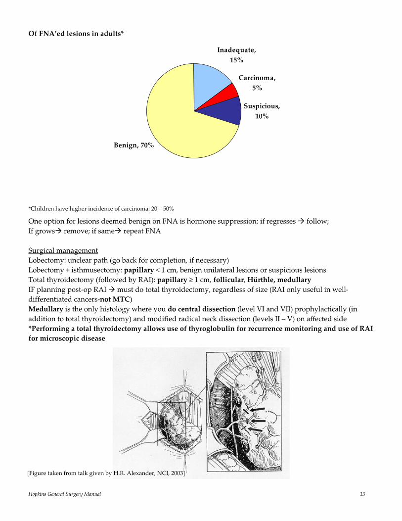
Of FNA’ed lesions in adults*
Inadequate, 15%
Carcinoma, 5%
Suspicious, 10%
Benign, 70%
*Children have higher incidence of carcinoma: 20 – 50%
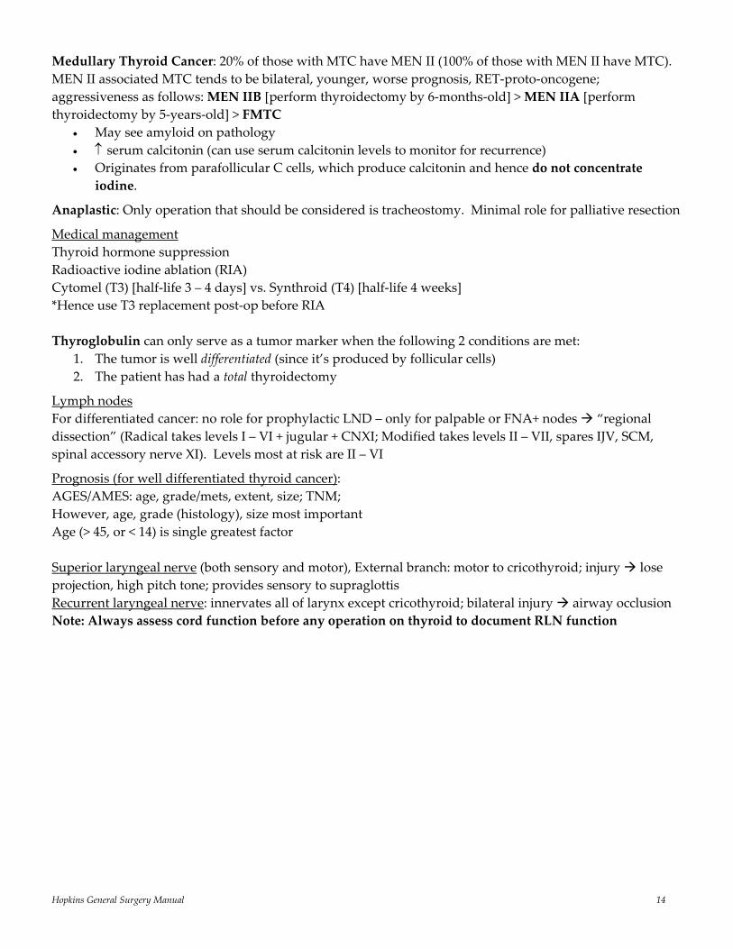
One option for lesions deemed benign on FNA is hormone suppression: if regresses follow; If grows remove; if same repeat FNA Surgical management Lobectomy: unclear path (go back for completion, if necessary) Lobectomy + isthmusectomy: papillary < 1 cm, benign unilateral lesions or suspicious lesions Total thyroidectomy (followed by RAI): papillary ≥ 1 cm, follicular, Hürthle, medullary IF planning post‐op RAI must do total thyroidectomy, regardless of size (RAI only useful in well‐differentiated cancers‐not MTC) Medullary is the only histology where you do central dissection (level VI and VII) prophylactically (in addition to total thyroidectomy) and modified radical neck dissection (levels II – V) on affected side *Performing a total thyroidectomy allows use of thyroglobulin for recurrence monitoring and use of RAI for microscopic disease
[Figure taken from talk given by H.R. Alexander, NCI, 2003]
Hopkins General Surgery Manual 14
Medullary Thyroid Cancer: 20% of those with MTC have MEN II (100% of those with MEN II have MTC). MEN II associated MTC tends to be bilateral, younger, worse prognosis, RET‐proto‐oncogene; aggressiveness as follows: MEN IIB [perform thyroidectomy by 6‐months‐old] > MEN IIA [perform thyroidectomy by 5‐years‐old] > FMTC
• May see amyloid on pathology • ↑ serum calcitonin (can use serum calcitonin levels to monitor for recurrence) • Originates from parafollicular C cells, which produce calcitonin and hence do not concentrate
iodine.
Anaplastic: Only operation that should be considered is tracheostomy. Minimal role for palliative resection
Medical management Thyroid hormone suppression Radioactive iodine ablation (RIA) Cytomel (T3) [half‐life 3 – 4 days] vs. Synthroid (T4) [half‐life 4 weeks] *Hence use T3 replacement post‐op before RIA Thyroglobulin can only serve as a tumor marker when the following 2 conditions are met:
1. The tumor is well differentiated (since it’s produced by follicular cells) 2. The patient has had a total thyroidectomy
Lymph nodes For differentiated cancer: no role for prophylactic LND – only for palpable or FNA+ nodes “regional dissection” (Radical takes levels I – VI + jugular + CNXI; Modified takes levels II – VII, spares IJV, SCM, spinal accessory nerve XI). Levels most at risk are II – VI
Prognosis (for well differentiated thyroid cancer): AGES/AMES: age, grade/mets, extent, size; TNM; However, age, grade (histology), size most important Age (> 45, or < 14) is single greatest factor Superior laryngeal nerve (both sensory and motor), External branch: motor to cricothyroid; injury lose projection, high pitch tone; provides sensory to supraglottis Recurrent laryngeal nerve: innervates all of larynx except cricothyroid; bilateral injury airway occlusion Note: Always assess cord function before any operation on thyroid to document RLN function
Hopkins General Surgery Manual 15
Parathyroid Gland and Disease Superior parathyroid glands from 4th pharyngeal pouch; Inferior (and thymus) from 3rd pharyngeal pouch more variable position (since longer distance traveled)
All parathyroid glands generally receive blood supply from the inferior thyroid artery
If only 3 glands found at surgery, fourth may be in: • Thymus, anterior mediastinum • Thyroid • Carotid sheath • Tracheoesophageal groove*, posterior mediastinum • Behind esophagus *Most common ectopic site
PTH produced by Chief cells increases Ca++ via bone breakdown, GI absorption, increased kidney re‐absorption, excretion of phosphate by kidney Hyperparathyroidism
1. Primary: ↑ PTH secretion by parathyroid (high Ca++, low PO4; look for Cl‐/PO4 > 33, even with normal Ca++)
2. Secondary: ↑ PTH secretion due to renal failure or decreased GI Ca++ abs (Ca++ low or normal) 3. Tertiary: ↑ PTH after correction of 2° hyperparathyroidism (high Ca++) 4. Familial Hypercalcemia Hypocaluria (FHH): see ↑ serum Ca++, PTH, but ↓ urine Ca++ (defect in set‐
point for “normal” Ca++ levels; patients do not experience the sequelae of elevated Ca++); No surgery Parathyroid Imagining:
• Sestamibi scan • U/S • 201Technetium‐thallium subtraction scan • CT/MRI
Primary Hyperparathyroidism Incidence: 1/4000 Risks: MEN I, IIa, irradiation, family history (autosomal dominant) Adenoma > 85% [1], Hyperplasia ≈ 10% [4], Carcinoma ≈ 1% [1], [# glands typically involved] Typically: [Cl‐]/[PO4] > 33 Initial medical treatment: IV fluids, lasix, NOT thiazides
Treatment • 1° Adenoma: Surgically remove adenoma (± biopsy all enlarged glands) • 1° Hyperplasia: Bilateral neck exploration and intraoperative PTH. Subtotal parathyroidectomy
(leave ½ lower gland in situ) or total parathyroidectomy with autotransplantation • 1° Carcinoma: WLE with ipsilateral thyroidectomy and lymph node dissection
• 2°: Correct Ca++ and PO4, perform renal transplant (no parathyroid surgery)
• 3°: Correct Ca++ and PO4, perform renal transplant, remove parathyroid glands and re‐implant 30 to 40 mg in forearm
Hopkins General Surgery Manual 16
Parathyroid Carcinoma Signs/Sx: HyperCa++, elevated PTH, palpable gland (50%), neck pain, recurrent laryngeal nerve paralysis HCG is a marker Treatment: En bloc resection including ipsilateral thyroid lobe + associated lymph nodes Post op Complications:
• Recurrent laryngeal nerve injury • Neck hematoma (open at bedside if breathing compromised) • HypoCa++
Parathyroid Pearls
• 90% of primary hyperparathyroidism due to a single adenoma unilateral exposure is ok (with intraop PTH)
• MUST exclude familial/MEN disease (a different entity altogether which requires subtotal parathyroidectomy leave ½ of a lower gland in situ)
• Nuclear medicine expertise is crucial: if possible, subtraction of Tc99m pertechnetate (potassium analog specific for thyroid) from Tc99m Sestamibi (taken up by both thyroid and parathyroid)
• For intra‐op PTH to be valid must have >50% drop in baseline PTH within 10 minutes
• Intra‐op PTH must be used if doing single gland exploration (MIP), else must do 4 gland exploration
If disease recurs, MUST distinguish between persistent and recurrent:
• Persistent: Only transient “cure”. Almost always implies missed adenoma. #1 place is TE groove on right side; also consider ectopic glands
• Recurrent (> 6 months normocalcemia): Implies hyperplasia with re‐growth (e.g. familial, possibly cancer)
• 10 x increase in RLN injury during re‐do surgery. Hence, first step in re‐do is confirm diagnosis with 24 hour urinary Ca++ (if normal no disease). Second, check for family history of MEN I manifestations
• Localization with Sestamibi and U/S. Consider CT/MRI (very bright on T2 to differentiate from LNs)
Hopkins General Surgery Manual 17
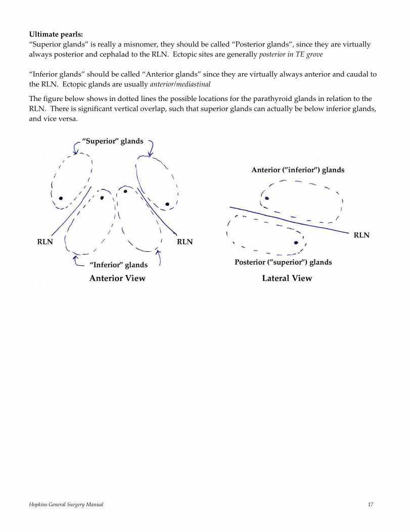
Ultimate pearls: “Superior glands” is really a misnomer, they should be called “Posterior glands”, since they are virtually always posterior and cephalad to the RLN. Ectopic sites are generally posterior in TE grove “Inferior glands” should be called “Anterior glands” since they are virtually always anterior and caudal to the RLN. Ectopic glands are usually anterior/mediastinal
The figure below shows in dotted lines the possible locations for the parathyroid glands in relation to the RLN. There is significant vertical overlap, such that superior glands can actually be below inferior glands, and vice versa.
Hopkins General Surgery Manual 18
Multiple Endocrine Neoplasia (MEN) *inherited autosomal dominant (with variable penetrance) MEN Type I aka Wermer’s Syndrome (“PPP”) •Parathyroid Hyperplasia (≈ 90%) HyperCa++ usually first •Pancreatic islet cell tumors (≈ 67%) Gastrinoma (ZES) (≈ 50%) Insulinoma (≈ 20%) •Pituitary Tumor (≈ 67%) most often PL‐secreting tumor MEN Type IIA aka Sipple’s Syndrome (“MPP”) •Medullary Thyroid Carcinoma (100%) 2nd to 3rd decade calcitonin secreting usually quite indolent •Pheochromocytoma (> 33%) catechol excess usually benign, bilateral, adrenal •Parathyroid Hyperplasia (≈ 50%) hyperCa++ MEN Type IIB ʺMMMPʺ •Mucosal Neuromas (100%) naso, oropharynx, larynx, conjunctiva •Medullary Thyroid Carcinoma (≈ 85%) more aggressive than IIA •Marfanoid body habitus •Pheochromocytoma (≈ 50%) often bilateral (70%) MEN1 Consensus Summary Statements (loss of function)
Diverse array of defects (missense, nonsense, frameshift, mRNA splicing); hence difficult to screen because of so many possible mutations
1997: gene Menin found of Ch 11. Exact function unknown, but it is a tumor suppressor gene
The MEN1 germline mutation test is recommended for MEN1 carrier identification.
All kindred with MEN1 are likely to have a mutation in the MEN1 gene.
However, MEN1 germline mutation tests fail to detect 10 – 20% of mutations. If a family lacks an identifiable MEN1 mutation, 11q13 haplotype testing about the MEN1 locus or genetic linkage analysis can identify MEN1 carriers. Periodic biochemical testing is an alternative when DNA‐based tests are not possible.
Hopkins General Surgery Manual 19
The main candidates for MEN1 mutation analysis include index cases with MEN1, their unaffected relatives, and some cases with features atypical for MEN1.
MEN1 carrier analysis should be used mainly for information. It should rarely determine a major intervention.
MEN1 tumor patterns in families do not have clear variants or specific correlations with an MEN1 germline mutation pattern. Thus, the MEN1 carriers in a family with either typical or atypical expression of MEN1 should be monitored similarly for typical expressions of MEN1 tumors.
MEN1 tumors cause morbidity through hormone excess (PTH, gastrin, PRL, etc.) and through malignancies (gastrinoma/islet cell or foregut carcinoid).
Medications control most features of hormone excess (gastrin, PRL, etc.). Surgery should control features of excess of some other hormones (PTH and insulin). Surgery has not been shown to prevent or cure MEN1‐related cancers.
Hyperparathyroidism develops in over 90% of MEN1 carriers. There is controversy over indications
for parathyroid surgery in MEN1 patients.
The preferred parathyroid operation in the HPT of MEN1 is subtotal parathyroidectomy (without autograft); transcervical near‐total thymectomy is also simultaneously. Parathyroid tissue should be cryopreserved.
Curative surgery for gastrinoma in MEN1 is rare. There is controversy over the indications for surgery for gastrinomas in MEN1.
Surgery in MEN1 is indicated and is usually successful for insulinoma. For most other pancreatic islet tumors, except gastrinomas, surgery is also indicated; however, there is no consensus over tumor criteria for the latter operations.
The management of pituitary tumor in MEN1 should be similar to that in sporadic cases. MEN2 Consensus Summary Statements (gain of function)
1995: RET‐proto‐oncogene (responsible for tyrosine kinase activity) identified on Ch 10
Fewer possible mutations (codons 609, 611, 618, 620, 634; involve replacement of a cystine residue)
The main morbidity from MEN2 is MTC. MEN2 variants differ in aggressiveness of MTC, in decreasing order as follows: MEN2B > MEN2A > FMTC.
MEN2 carrier detection should be the basis for recommending thyroidectomy to prevent or cure MTC. This carrier testing is mandatory in all children at 50% risk.
Compared with RET mutation testing, immunoassay of basal or stimulated CT results in more frequent false positive diagnoses and delays of the true positive diagnosis of the MEN2 carrier state. However, the CT test still should be used to monitor the tumor status of MTC.
RET germline mutation (10q11 – 12) testing has replaced CT testing as the basis for carrier diagnosis in MEN2 families. It reveals a RET mutation in over 95% of MEN2 index cases.
The RET codon mutations can be stratified into three levels of risk from MTC. These three categories predict the MEN2 syndromic variant, the age of onset of MTC, and the aggressiveness of MTC.
Hopkins General Surgery Manual 20
Thyroidectomy should be performed before age 6 months in MEN2B, perhaps much earlier, and before age 5 yr in MEN2A. Policies about central lymph node dissection at initial thyroidectomy are controversial and may differ among the MEN2 variants.
MEN2 has distinctive variants. MEN2A and MEN2B are the MEN2 variants with the greatest syndromic consistency.
FMTC is the mildest variant of MEN2. To avoid missing a diagnosis of MEN2A with its risk of pheochromocytoma, physicians should diagnose FMTC only from rigorous criteria.
Morbidity from pheochromocytoma in MEN2 has been markedly decreased by improved recognition and management. The preferred treatment for unilateral pheochromocytoma in MEN2 is laparoscopic adrenalectomy.
HPT is less intense in MEN2 than in MEN1. Parathyroidectomy should be the same as in other disorders with multiple parathyroid tumors.
Hopkins General Surgery Manual 21
Gastrinoma
• 50% of patients with MEN I have a gastrinoma • 33% of patients with ZES have MEN I (Clinical Triad: 1. PUD 2. gastric acid hypersecretion 3. Islet
cell tumor) • In MEN I associated ZES: Most common site is duodenum (2nd is pancreas) • ⅔ are malignant • 25% of patients with pheochromocytoma have ZES • Risk of metastatic behavior from gastrinoma rises sharply at 3 cm (< 3 cm < 2% chance of mets) The sporadic disease is a different one from the MEN‐associated one • Sporadic ZES occurs outside of the duodenum 30 – 60% of the time; MEN version is virtually always
in the duodenum (with or without pancreatic involvement) • Sporadic ZES is much more likely to be malignant (the extrapancreatic primaries are less
biologically aggressive in both diseases) • Sporadic ZES is more often solitary and larger at the time of detection, usually > 2 cm • MEN ZES is a diffuse disease, rarely amenable to true “cure”
“Gastrinoma Triangle” joins: 1. junction of cystic duct & CBD 2. junction of 2nd and 3rd portion duodenum 3. junction of neck & body of pancreas (90% found in this triangle) [Am J Surg 1984 147:25 Stabile, Morrow, Passaro]
Ddx for increased gastrin: Gastrinoma Retained excluded antrum (a surgical mistake) Gastric outlet obstruction Antral G‐cell hyperplasia/hyperfunction Postvagotomy Pernicious anemia Atrophic gastritis Short gut syndrome Renal failure H2 blocker, proton pump inhibitor* *pH < 2 inhibits gastrin secretion in normal patients
Check gastrin levels in patients with: 1. recurrent ulcers 2. ulcers in unusual places (e.g. jejunum), 3. refractory to medical management 4. prior to any elective operation for an ulcer 5. unexplained or persistent diarrhea 6. peptic ulcer and any endocrinopathy 7. family history of PUD 8. family history of MEN I
Check: 1. fasting gastrin level 2. post‐secretin challenge gastrin level (synthetic secretin – 2 units/kg IV bolus) 3. Ca++ (MEN I screen) 4. Chemistry panel
High acid
Low/minimal acid
Hopkins General Surgery Manual 22
Look for: • ZES fasting: 200 – 1000 pg/mL (normal < 100 pg/mL) • Basal acid secretion: ZES > 15 mEq/hr (normal < 10 mEq/hr)
Note: generally will see failure of feedback: fasting gastrin ≈ 1000 with gastric pH < 2.5
Secretin Stimulation Test:
• IV secretin administered, gastrin measured • ZES: increased gastrin (by > 200 pg/mL) within 10 to 20 minutes (normal response is decrease in
gastrin) Management issues:
• Some believe distal pancreatectomy should be done in any patient with MEN I with either hormonal syndrome or a neuroendocrine tumor regardless of location in pancreas or duodenum. Virtually all patients with MEN I ZES have concomitant neuroendocrine tumors in neck, body, or tail.
• If patient has gastrinoma and hyperparathyroidism remove parathyroid first to normalize Ca++ levels (since hypercalcemia is more dangerous than hypergastrinemia)
• Higher than “normal” doses of PPI are needed for achlorhydria • Minimal role for CT scan/octreoscan • Imamura Test: intra‐arterial secretin into visceral arteries to measure hepatic vein gastrin levels
(look for step up) is good for localization • No role for debulking functional gastrinoma mets, since patients can be managed medically in this
setting with PPIs
Post‐Op Must stay on acid suppression for 3 – 4 months because even after resection acid secretion high for some time Glucagonoma The 4D Syndrome: diabetes, diarrhea, dementia, dermatitis (patients look cachectic) Many patients also have normochromic normocytic anemia, hypoalbuminemia, weight loss, beefy red tongue (glossitis), stomatitis, angular chelosis (i.e. signs of malnutrition)
Usually found in tail of pancreas, but can be anywhere. Usually > 3 cm at time of diagnosis; 70% malignant
Diagnosis simple by measuring serum glucagon level, although most patients with elevated glucagons do not have gastrinoma [see review: Wermers RA, et al. Medicine (Baltimore). 1996;75:53]
Sx: Necrotizing Migratory Erythema‐NME (usually below waist), glossitis, stomatitis, diabetes
• IV Tolbutamide results in elevated glucagon
• Medical treatment for Necrotizing migratory erythema: Somatostatin/octreotide, IV Amino acids (TPN)
• Aggressive surgical resections are indicated, even if metastatic
Hopkins General Surgery Manual 23
Insulinoma
Number 1 islet cell neoplasm; associated with MEN I
• 80 – 90% are benign solitary adenomas cured by surgical resection • 30% < 1 cm • 10% multiple • 10 – 15% malignant • 10% hyperplasia or nesidioblastosis
Presents with sympathetic nervous system symptoms due to hypoglycemia (patients look like “Pillsbury Dough Boy”)
Whipple’s Triad: 1. Hypoglycemia < 50 mg/dl 2. CNS symptoms. 3. Reversal of CNS symptoms. with glucose admin.
Ddx for hyperinsulinemia: • Reactive hypoglycemia (very common), • Functional hypoglycemia with gastrectomy, • Adrenal insufficiency, hypopituitarism, hepatic insufficiency, • Munchausen’s syndrome (self injection) • Tumors secreting insulin‐like molecule (sarcoma, mesothelioma, etc.)
First, check for proinsulin, then: Get 72 hour fasting levels with q6 hour checks until patient becomes symptomatic
Insulin:Glucose ratio = insulin (uU/ml)/glucose (mg/dl) > 0.3 found in almost all patients with insulinoma.
Accuracy increased by “Amended ratio” = insulin (uU/ml)/ [glucose (mg/dl) – 30] > 0.3
Localizing Tests: • CT, A‐gram, endoscopic ultrasound, venous catheterization (sample blood along portal and splenic
veins) • Calcium angiogram: Ca++ causes insulin secretion localize to artery (e.g. splenic for tail) where
tumor nearest • Intraoperative U/S is probably the best test for localization
Medical Treatment:
• Diazoxide to suppress insulin levels (until resection). Diazoxide inhibits the sulfonylurea receptor 1 (SUR1) on the beta cell, which is a component of the K+ATPase responsible for insulin secretion
• Octreotide, IV glucose
Hopkins General Surgery Manual 24
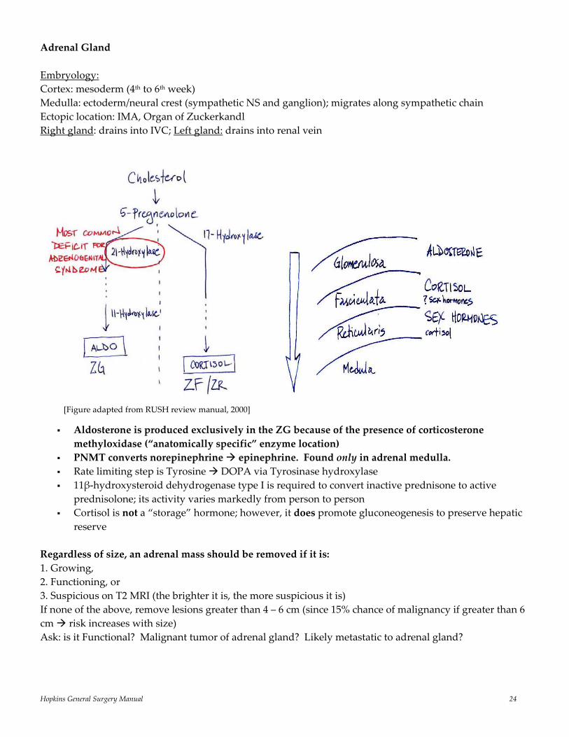
Adrenal Gland Embryology: Cortex: mesoderm (4th to 6th week) Medulla: ectoderm/neural crest (sympathetic NS and ganglion); migrates along sympathetic chain Ectopic location: IMA, Organ of Zuckerkandl Right gland: drains into IVC; Left gland: drains into renal vein
Aldosterone is produced exclusively in the ZG because of the presence of corticosterone methyloxidase (“anatomically specific” enzyme location)
PNMT converts norepinephrine epinephrine. Found only in adrenal medulla. Rate limiting step is Tyrosine DOPA via Tyrosinase hydroxylase 11β‐hydroxysteroid dehydrogenase type I is required to convert inactive prednisone to active
prednisolone; its activity varies markedly from person to person Cortisol is not a “storage” hormone; however, it does promote gluconeogenesis to preserve hepatic
reserve Regardless of size, an adrenal mass should be removed if it is: 1. Growing, 2. Functioning, or 3. Suspicious on T2 MRI (the brighter it is, the more suspicious it is) If none of the above, remove lesions greater than 4 – 6 cm (since 15% chance of malignancy if greater than 6 cm risk increases with size) Ask: is it Functional? Malignant tumor of adrenal gland? Likely metastatic to adrenal gland?
[Figure adapted from RUSH review manual, 2000]
Hopkins General Surgery Manual 25
Syndromes: I. Conn’s (↑ aldosterone): 75% unilateral adenoma Rx: with resection 25% bilateral hyperplasia Rx: spironolactone + C++ channel blocker II. Addison’s (↓ aldosterone and cortisol): low Na+, high K+, hypoglycemia; can present in crisis with hypotension III. Waterhouse Friedrickson: adrenal hemorrhage with meningococcal sepsis IV. Nelson: post adrenalectomy (10%) ↑ ACTH, pigmentation, change in vision from ↑ pituitary response V. Cushing’s Disease (pituitary): 80% of non‐iatrogenic causes; pituitary microadenoma ↑ ACTH
(will also see urine 17‐OH progesterone) Adrenal Cushing’s Syndrome: (aka ACTH‐independent Cushing’s) 15% of non‐iatrogenic causes 10% adrenal adenoma, 5% adrenal carcinoma; bilateral hyperplasia is very rare; will see ↓ ACTH Ectopic Cushing’s Syndrome: 20% of ACTH‐dependent; sources of ectopic ACTH including: Pulmonary (SCLC, bronchial, thymic carcinoids), Neuroendocrine tumors, Pheochromocytoma, MTC
Diagnosis:
1. Start with 24 hour urine free cortisol and plasma ACTH 2. Low dose dexamethasone suppression will suppress causes of hypercortisolism such as obesity
and excess ethanol ingestion, but not others (confirms dx) 3. High dose dexamethasone suppression will suppress pituitary adenoma, but not ectopic sources
(locates cause) 4. MRI, CT, and/or petrosal venous sampling
Treatment:
• “Medical Adrenalectomy” = metyrapone and aminoglutethimide • Surgical removal of all functional adrenal masses is indicated, including bilateral adrenalectomy for
diffuse disease in patients recalcitrant to medical management Stress Dose Steroids [Chernow et al. Ann Surg 1994, 219:416]
• Under normal conditions, body produces 30 mg hydrocortisone equivalent (solucortef)/day • Under extreme stress up to 300 mg/day • Prednisone is 4:1 (to solucortef) • Solu‐Medrol is 5:1 • Decadron is 25:1 Normal adrenal secretion is 25 – 30 mg cortisol/24h Appropriate stress test: 250 mcg cosyntropin 1. ↑ in plasma cortisol by ≥ 7 mcg/dL, or 2. Absolute level > 20 mcg/dL
Hopkins General Surgery Manual 26
For adrenal crisis: 200 mg hydrocortisone immediately 100 mg q8 hours x 48 hours then taper by 50% reduction q2 days until 25 mg reached Above 50 mg hydrocortisone/day you are getting enough mineralocorticoid activity (except dexamethasone), BUT below 50 mg/day must replace aldosterone with Florinef Acetate Proven Adrenal Insufficiency/Chronic Steroids [give following in addition to maintenance doses]: I. Mild illness/non‐febrile •no replacement II. Mod illness (fever, minor trauma or surgery) •15 mg prednisolone qd until 24 h post resolution III. Severe illness or major trauma or surgery • 50 mg hydrocortisone q6h taper to normal by 50%/day IV. Septic Shock • 50 mg hydrocortisone q6h ± 50 mcg florinef qd x 7 days
Hopkins General Surgery Manual 27
Pheochromocytoma Tumor of adrenal medulla and sympathetic ganglion (from chromaffin cell lines) producing catecolamines (NE > Epi)
Incidence: ≈ 0.2% (1/500 hypertensives) “Rule of 10’s”
10% malignant 10% bilateral 10% in kids 10% multiple tumors 10% extraadrenal
Found in MEN II (A and B) always rule out MEN with dx of pheochromocytoma
Classic Triad 1. Palpitations 2. Headache 3. Episodic diaphoresis (also, 50% hypertensive)
Ddx: Renovascular HTN, menopause, migraines, carcinoid syndrome, pre‐eclampsia, neuroblastoma, anxiety disorder, hyperthyroidism, insulinoma
Locations: Adrenal (90%) Organ of Zuckerkandl (embryonic chromaffin cells around the abdominal aorta near IMA; normally
atrophies in childhood) Thorax Bladder Scrotum
(Note: if epi high, must be at or near adrenals, since nonadrenal sites lack ability to methylate NE to epi)
Locators: CT, MRI, 131I‐MIBG (an NE analog that collects in adrenergic vesicles)
Note: Histology can’t determine malignancy; only spread can Preop Treatment:
Increase intravascular volume Must give α‐blockers (phenoxybenzamine or prazosin) for 5 – 7 days prior to surgery to control
HTN. If, after BP controlled, still tachycardic add on β‐blocker for 2 to 4 days. Catastrophic error to begin with β‐blocker because this will lead to unopposed vasoconstriction
which can cause acute heart failure.
Hopkins General Surgery Manual 28
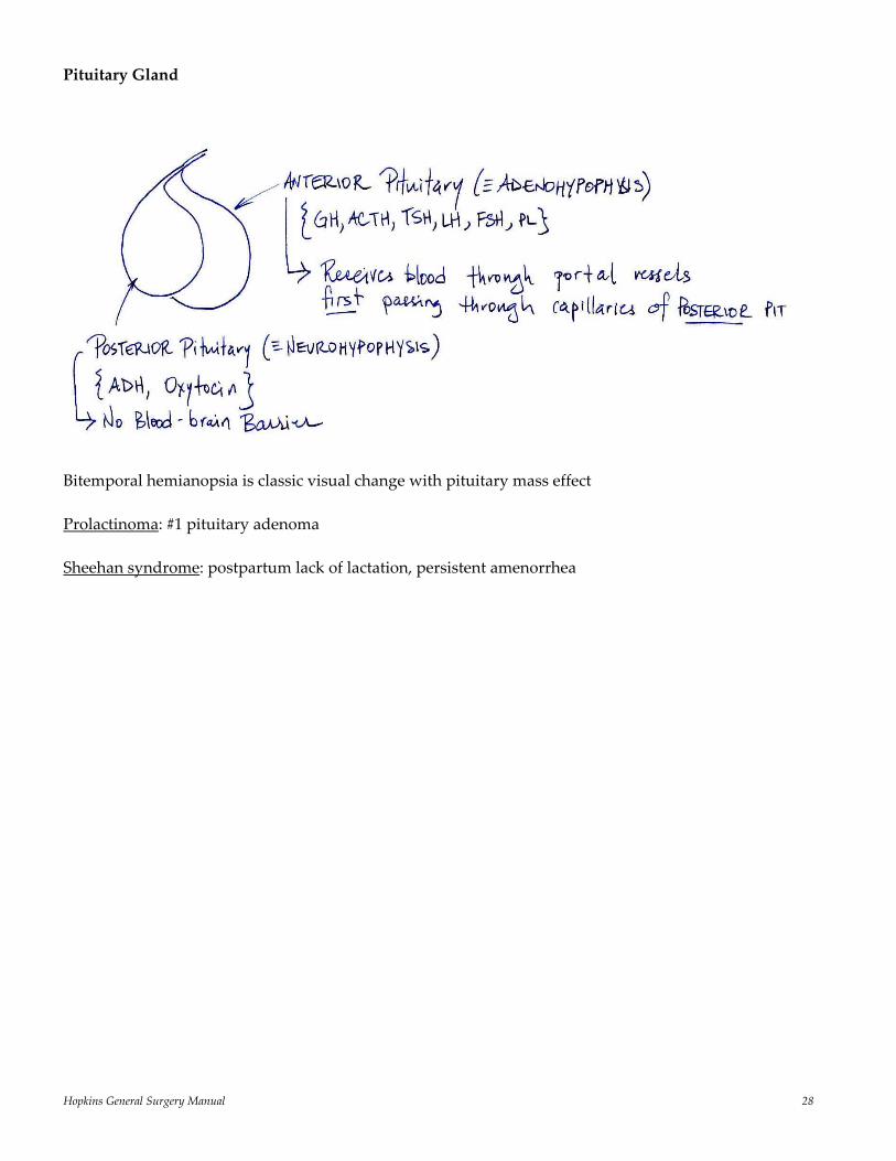
Pituitary Gland
Bitemporal hemianopsia is classic visual change with pituitary mass effect Prolactinoma: #1 pituitary adenoma Sheehan syndrome: postpartum lack of lactation, persistent amenorrhea
Hopkins General Surgery Manual 29
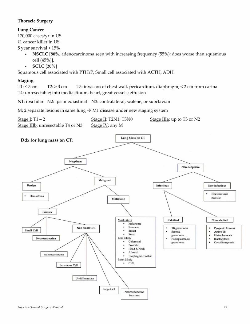
Thoracic Surgery
Lung Cancer 170,000 cases/yr in US #1 cancer killer in US 5 year survival < 15%
NSCLC [80%; adenocarcinoma seen with increasing frequency (55%); does worse than squamous cell (45%)],
SCLC [20%] Squamous cell associated with PTHrP; Small cell associated with ACTH, ADH
Staging: T1: ≤ 3 cm T2: > 3 cm T3: invasion of chest wall, pericardium, diaphragm, < 2 cm from carina T4: unresectable; into mediastinum, heart, great vessels; effusion
N1: ipsi hilar N2: ipsi mediastinal N3: contralateral, scalene, or subclavian
M: 2 separate lesions in same lung M1 disease under new staging system
Stage I: T1 – 2 Stage II: T2N1, T3N0 Stage IIIa: up to T3 or N2 Stage IIIb: unresectable T4 or N3 Stage IV: any M
Ddx for lung mass on CT:
Hopkins General Surgery Manual 30
Association of smoking and lung cancer • 90% of lung cancer occurs in smokers • 14 – 20 fold higher risk in smokers; 2 – 5 fold higher (never zero) in former smokers • Shown to be a multistep process [NEJM 265:253] of “field cancerization”; more smoking more CIS
on autopsy • 3p14 loss of heterozygosity 88% smokers; 45% formers; 0% nonsmokers
Risk of surgery: Pre thoracotomy PFT’s: need FEV1 > 2L, 1L for pneumonectomy/lobectmy Want Post‐op FEV1 > 800 – 1000 mL (40% predicted) DLCO < 60% significant risk; MVO2 < 10 mL/kg/min significant risk
Stage III disease: 1. Confirm N2 status (FNA, mediastinoscopy, VATS) 2. CDDP/Etoposide or Taxol/CDDP + surgery +XRT
Several studies [Rosell NEJM ‘94, Roth JNCI ‘94 + follow‐ups] could end the role of surgery in IIIA disease. Showed that with induction chemo + XRT ± surgery had equal survival with significantly more deaths in surgery arm, but currently stage IIIA patients undergo surgical resection
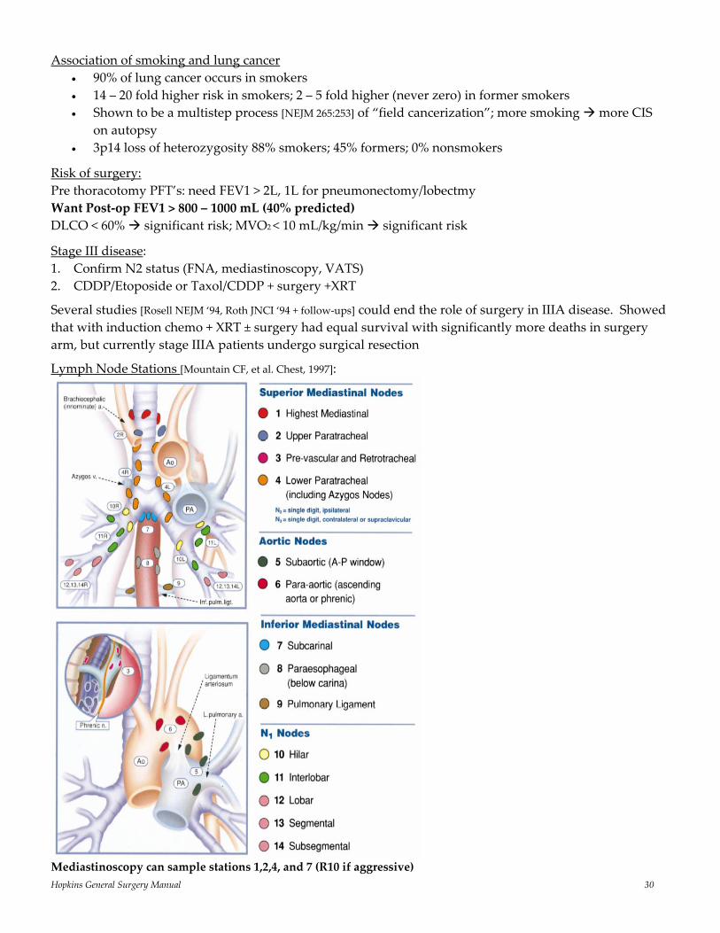
Lymph Node Stations [Mountain CF, et al. Chest, 1997]:
Mediastinoscopy can sample stations 1,2,4, and 7 (R10 if aggressive)
Hopkins General Surgery Manual 31
Pancoast Tumor: involves sympathetic chain (Horner’s syndrome); shoulder/medial scapula pain is most common presentation; Mediastinoscopy induction chemo radical resection XRT Significantly higher incidence of right‐sided node involvement with left‐sided lung tumors because of lymphatic pathways which travel from left right; the reverse in not observed
Metastatic tumors (lung, breast) to pleura >> primary pleural tumor (mesothelioma)
In Summary‡: 1. Surgery is still standard of care for I, II, selected IIIA, and selected IIIB NSCLC (and very rare IV disease,
e.g. isolated brain or adrenal N0 disease) 2. XRT improves local control, but not survival 3. Adjuvant RT for > T2, N1 – 2 disease 4. Induction chemo is proven for stage III disease, but is untested for early disease.
‡See excellent review: Multidisciplinary Management of Lung Cancer [Spira et al. NEJM 350;379, 2004]
Factors that ↑ air leak after resection: 1. Neoadjuvant chemo/radiation 2. Deep dissection 3. Blebs/emphysema
“Massive” hemoptysis: > 600 mL in 24 hours
Spontaneous pneumothorax: usually result of ruptured small bleb; 15 – 20% of recurrence after initial event (much greater after 2nd event)
Chyle Leak: • Thoracic duct enters chest on right (with aorta) crosses to left at T4/5 joins at IJ/subclavian
junction on left • Most often iatrogenic • Normal chyle flow varies between 1.5 and 2.5 L/day according to diet • High lymphocytes (making it resistant to infection) and 10 x TG of serum • Treat with 2 week trial of NPO and drainage (50 – 70% success); if still > 500 mL/day to OR for
duct ligation
Empyema: exudative (thin, free‐flowing fluid) fibrinopurulent (fibrin deposition, beginning to loculate) organizing (ingrowth of fibroblasts, “peel”)
Exudative Stage: may respond to antibiotics ± drainage
Fibrinopurulent Stage: requires chest tube ± surgery
Thin WBC < 1000/mm3 LDH < 500 – 1000 IU (pleural fluid/serum > 0.6) Pleural fluid/serum protein > 0.5 pH > 7.30 Glucose > 60 mg/dL
Turbid, bacterial cellular debris Glucose < 40 mg/dL LDU > 1000 IU WBC >5000/mm3 pH < 7.10
Hopkins General Surgery Manual 32
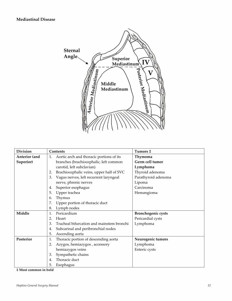
Mediastinal Disease
Division Contents Tumors ‡ Anterior (and Superior)
1. Aortic arch and thoracic portions of its branches (brachiocephalic, left common carotid, left subclavian)
2. Brachiocephalic veins, upper half of SVC 3. Vagus nerves, left recurrent laryngeal
nerve, phrenic nerves 4. Superior esophagus 5. Upper trachea 6. Thymus 7. Upper portion of thoracic duct 8. Lymph nodes
Thymoma Germ cell tumor Lymphoma Thyroid adenoma Parathyroid adenoma Lipoma Carcinoma Hemangioma
Middle 1. Pericardium 2. Heart 3. Tracheal bifurcation and mainstem bronchi 4. Subcarinal and peribronchial nodes 5. Ascending aorta
Bronchogenic cysts Pericardial cysts Lymphoma
Posterior 1. Thoracic portion of descending aorta 2. Azygos, hemiazygos , accessory
hemiazygos veins 3. Sympathetic chains 4. Thoracic duct 5. Esophagus
Neurogenic tumors Lymphoma Enteric cysts
‡ Most common in bold
Hopkins General Surgery Manual 33
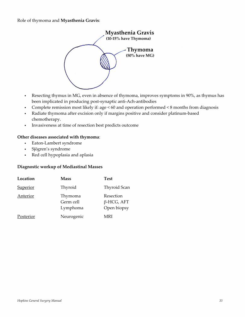
Role of thymoma and Myasthenia Gravis:
Resecting thymus in MG, even in absence of thymoma, improves symptoms in 90%, as thymus has
been implicated in producing post‐synaptic anti‐Ach‐antibodies Complete remission most likely if: age < 60 and operation performed < 8 months from diagnosis Radiate thymoma after excision only if margins positive and consider platinum‐based
chemotherapy. Invasiveness at time of resection best predicts outcome
Other diseases associated with thymoma:
Eaton‐Lambert syndrome Sjögren’s syndrome Red cell hypoplasia and aplasia
Diagnostic workup of Mediastinal Masses Location Mass Test
Superior Thyroid Thyroid Scan
Anterior Thymoma Resection Germ cell β‐HCG, AFT Lymphoma Open biopsy
Posterior Neurogenic MRI
Hopkins General Surgery Manual 34
Cardiac Surgery: Congenital Defects
Ventricular Septal Defect (VSD)
Most common CHD (25%, 1/1000 live births) Multiple types: perimembranous is most common Up to 50% have associated anomaly (PDA, coarctation, AS) PVR ↑ over time because of pulmonary overcirculation, and my be irreversible at 2 years May go on develop Eisenmenger’s syndrome with shunt reversal across the VSD Fix if shunt fraction (Qp/Qs) > 2 (twice blood flow to lungs) Repair unrestrictive VSD prior to 1‐year of age (prevent failure to thrive)
Patent Ductus Ateriosis (PDA) Communication between upper descending aorta and left/main PA Presentation dependant on degree of left right shunt (pulmonary overcirculation and steal from
systemic perfusion) Persistent PDA is more common in premature infants Medical closure: indomethacin (up to three doses) Long term complication of pulmonary overcirculation include: pulmonary HTN, CHF, increased
respiratory infections Early surgical closure indicated for symptomatic patients who are recalcitrant to medical therapy, or
are not suitable candidates for medical therapy Use prostaglandin to keep open in cyanotic infants; can also close in cath lab
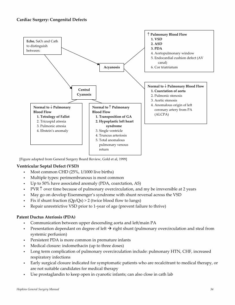
Acyanosis
Echo, SaO2 and Cath to distinguish between:
Central Cyanosis
Normal to ↓ Pulmonary Blood Flow1. Coarctation of aorta 2. Pulmonic stenosis 3. Aortic stenosis 4. Anomalous origin of left coronary artery from PA (ALCPA)
Normal to ↑ Pulmonary Blood Flow 1. Transposition of GA 2. Hypoplastic left heart
syndrome 3. Single ventricle 4. Truncus arteriosis 5. Total anomalous pulmonary venous return
Normal to ↓ Pulmonary Blood Flow 1. Tetralogy of Fallot 2. Tricuspid atresia 3. Pulmonic atresia 4. Ebstein’s anomaly
↑ Pulmonary Blood Flow 1. VSD 2. ASD 3. PDA 4. Aortopulmonary window 5. Endocardial cushion defect (AV
canal) 6. Cor triatriatum
[Figure adapted from General Surgery Board Review, Gold et al, 1999]
Hopkins General Surgery Manual 35
Coarctation of the Aorta Accounts for 6 – 8% of CHD (≈ 2 – 5x more common in males) The usual location of the discrete coarctation is juxtaductal (just distal to the left subclavian artery) Less often, the coarctation is just proximal to the left subclavian artery; can also be diffuse Two theories:
1. Reduced antegrade intrauterine blood flow, which causes underdevelopment of the aortic arch 2. Extension of the ductal tissue into the thoracic aorta which, when it constricts, causes coarctation of the aorta
The most common clinical manifestation is a difference in systolic pressure between the upper and lower extremities (diastolic pressures are usually similar), manifested by:
1. Upper extremity hypertension 2. Absent/delayed femoral pulses 3. Low/unobtainable blood pressure in the lower extremities
Treatment options include angioplasty ± stenting (if > 25 kg) or surgical repair (resection + end‐to‐end spatulated anastomosis, bypass if segment too long for primary repair)
Atrial Septal Defect (ASD) Strictly speaking, a Patent Foramen Ovale (PFO) only shuts: right left Usually an ASD shunts: left right Accounts for 10 – 15% of CHD (most common one in adults) Secundum defect is most common ASD (PFO is more common) Spontaneous closure rare > 2‐years of age Typically asymptomatic with murmur; 50 – 60% have easy fatigability Fix almost all persistent ASDs Can be closed via catheterization
Tetralogy of Fallot Four anatomic features (key is RVOT obstruction):
1. Stenosis of PA 2. RV hypertrophy 3. VSD (usually single, large, and unrestricted; in the perimembranous region of the septum) 4. Aorta overriding the VSD
Accounts for 7 – 10% of all congenital heart disease (3.3 per 10,000 live births) Approximately 15% of children have extracardiac anomalies (e.g. trisomy 21) Physiology and clinical presentation of tetralogy is determined primarily by the extent of RV
outflow obstruction. Most children are cyanotic and symptomatic. • Severe obstruction with poor pulmonary flow: profound cyanosis during newborn period • Moderate obstruction with balanced pulmonary and systemic flow: may be identified during
elective workup for a murmur • Minimal obstruction: pulmonary overcirculation and late heart failure
Options for repair include: Patch repair of the RVOT, possibly rendering the pulmonary valve incompetent, which, if severe,
may have significant long term hemodynamic and electrophysiologic consequences. An alternate procedure is the insertion of a valved conduit from the RV to the distal main
pulmonary artery if there is pulmonary atresia or a coronary anomaly precluding a transanular incision
Hopkins General Surgery Manual 36
Cardiac Surgery: Acquired Defects
4 “buzz words” to describe cardiac physiology • Inotropy: force of contraction (systolic) • Chronotropy: rate of contraction • Lusitropy: rate of relaxation (diastolic) • Dromotropy: conduction
Coronary Artery Bypass is associated with improved survival in patients with • triple vessel disease • left main disease • patients with ↓EF
i.e. the more extensive disease the greater the benefit
Indications: Intractable symptoms, medically refractory > 50% left main disease Triple vessel disease with depressed EF Left dominant circulation with high grade LAD stenosis
Dominance: 85% are right dominant, which means RCA supplies: 1. PDA 2. AV node 3. Posterior crux of heart (near IVC)
Branches of main cardiac vessels 1. Left circumflex: obtuse marginals 2. Left anterior descending: diagonals and septals 3. Right coronary: acute marginal branches; if right dominant: PDA, AV nodal; septals
Saphenous vein patency ≈ 50 – 60% at 10 years IMA patency ≈ 95% at 10 years Rate of recurrence of angina following CAB is ≈ 5 – 7% per year
IABP Positioned just distal to left subclavian artery (aortic knob on CXR) Inflates during diastole (40 msec before T wave; ↓ afterload) and deflates with p wave (↑ coronary perfusion); AI is a contraindication
Acute MI Complications
1. Arrhythmias: PVC, ventricular ectopy, VT/ VF, PEA (pulseless electrical activity) (0 – 48 hours) Reperfusion can cause PVCs
2. Recurrence: (0 – 7 days)
3. Pericardial Disease*: pericarditis; Dressler’s syndrome (pericarditis + effusion) (6 hours – 14 days); likely autoimmune inflammation of pericardium; pleuritic chest pain, low grade fever, malaise; treat with steroids or NSAIDs; *key is to differentiate from mediastinitis
Hopkins General Surgery Manual 37
4. Mediastinitis: follows ≈ 1 – 3% of cardiac surgery; risks include: DM, age > 60, re‐exploration for bleeding, steroids, obesity with bilateral IMA harvest; need re‐operative drainage and flap (advancement pec, or transfer)
5. Structural Catastrophes (3 – 5 days) a) papillary muscle tear; usually follows posterior MI: acute MR‐pulmonary edema b) septal rupture: VSD (SOB) c) free wall rupture: rapid death
6. Aneurysm formation (weeks); ≈ 10% of patients post‐MI; usually after anterolateral infarct caused by proximal LAD occlusion (anterior and apical); can develop progressive LV failure with CHF; nidus for emboli and arrhythmia; surgical intervention if symptomatic
7. CHF + Recurrence: ANYTIME
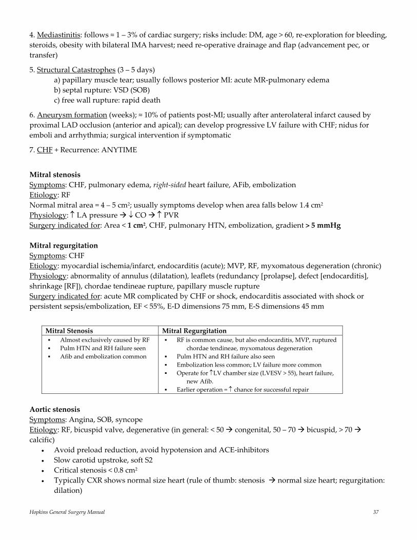
Mitral stenosis Symptoms: CHF, pulmonary edema, right‐sided heart failure, AFib, embolization Etiology: RF Normal mitral area = 4 – 5 cm2; usually symptoms develop when area falls below 1.4 cm2 Physiology: ↑ LA pressure ↓ CO ↑ PVR Surgery indicated for: Area < 1 cm2, CHF, pulmonary HTN, embolization, gradient > 5 mmHg Mitral regurgitation Symptoms: CHF Etiology: myocardial ischemia/infarct, endocarditis (acute); MVP, RF, myxomatous degeneration (chronic) Physiology: abnormality of annulus (dilatation), leaflets (redundancy [prolapse], defect [endocarditis], shrinkage [RF]), chordae tendineae rupture, papillary muscle rupture Surgery indicated for: acute MR complicated by CHF or shock, endocarditis associated with shock or persistent sepsis/embolization, EF < 55%, E‐D dimensions 75 mm, E‐S dimensions 45 mm
Mitral Stenosis Mitral Regurgitation Almost exclusively caused by RF Pulm HTN and RH failure seen Afib and embolization common
RF is common cause, but also endocarditis, MVP, ruptured chordae tendineae, myxomatous degeneration
Pulm HTN and RH failure also seen Embolization less common; LV failure more common Operate for ↑LV chamber size (LVESV > 55), heart failure,
new Afib. Earlier operation = ↑ chance for successful repair
Aortic stenosis Symptoms: Angina, SOB, syncope Etiology: RF, bicuspid valve, degenerative (in general: < 50 congenital, 50 – 70 bicuspid, > 70 calcific)
• Avoid preload reduction, avoid hypotension and ACE‐inhibitors • Slow carotid upstroke, soft S2 • Critical stenosis < 0.8 cm2 • Typically CXR shows normal size heart (rule of thumb: stenosis normal size heart; regurgitation:
dilation)
Hopkins General Surgery Manual 38
Prognosis depends on symptoms: 1. CHF ‐ worst prognosis (1.5 years*); occurs at approximately 0.7 – 0.8 cm2 2. Syncope post exertional (3 years*) 3. Angina (5 years*) *Mean survival from initiation of symptoms
• need antibiotic prophylaxis • critical AS diagnosed by symptoms, not area • symptoms need surgery, good outcome even in 80s • average progression 0.12 cm2/year
Surgery indicated for: Area < 0.8 – 1.0 cm2, gradient > 50 mmHg, symptoms, evidence of rapid cardiac enlargement Aortic regurgitation (AR/AI)
3F’s: Fast (must be kept tachy), Forward (reduce AL), Full (preload dependent)
Symptoms: Fatigue, angina, progressive dyspnea, palpitations, peripheral vasomotor changes; bounding peripheral pulses (wide pulse pressure) ESLV diameter greater than 5 cm has increased death rate (19% vs. 5% per year) Acute Rx: afterload reduction, diuretics for CHF Surgery: symptoms, evidence of ventricular enlargement Perioperative MI
Most likely 2 – 3 days post‐op Differentiate Acute MI from Acute Coronary Syndrome
A. Acute MI: CP, ST elevations, + enzymes Goals are: 1. Reperfusion (fibrinolytic 0 – 6 hours; cath), and 2. Decrease O2 demand
B. Acute Coronary Syndrome is subdivided into Non Q‐wave MI and Unstable Angina (CP, with no EKG changes or enzymes) Non Q‐wave MI: CP, EKG changes without ST elevation, +enzymes; give MONA (MSO4, O2, nitrate, ASA) + β‐blocker then IIb/IIIa + heparin
*Note: Inferior infarct RCA 90% chance of AV nodal involvement 2:1 block more likely to be Mobitz I > II give atropine
Hopkins General Surgery Manual 39
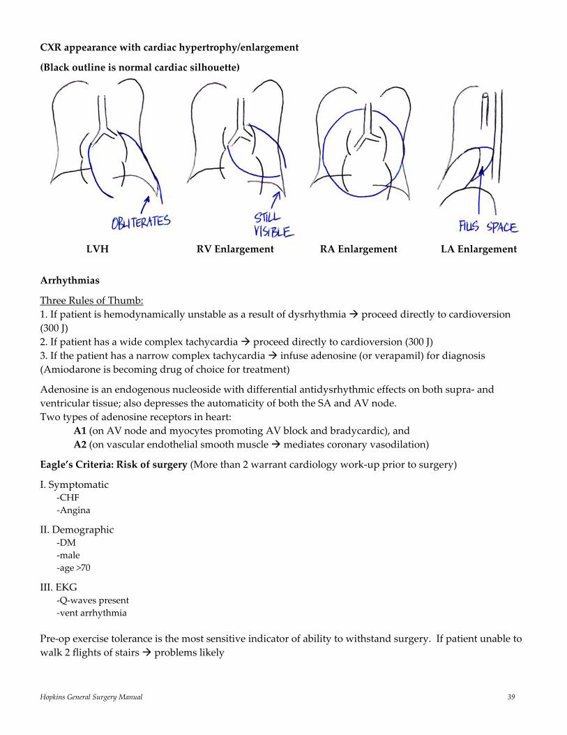
CXR appearance with cardiac hypertrophy/enlargement
(Black outline is normal cardiac silhouette)
LVH RV Enlargement RA Enlargement LA Enlargement
Arrhythmias
Three Rules of Thumb: 1. If patient is hemodynamically unstable as a result of dysrhythmia proceed directly to cardioversion (300 J) 2. If patient has a wide complex tachycardia proceed directly to cardioversion (300 J) 3. If the patient has a narrow complex tachycardia infuse adenosine (or verapamil) for diagnosis (Amiodarone is becoming drug of choice for treatment)
Adenosine is an endogenous nucleoside with differential antidysrhythmic effects on both supra‐ and ventricular tissue; also depresses the automaticity of both the SA and AV node. Two types of adenosine receptors in heart: A1 (on AV node and myocytes promoting AV block and bradycardic), and A2 (on vascular endothelial smooth muscle mediates coronary vasodilation)
Eagle’s Criteria: Risk of surgery (More than 2 warrant cardiology work‐up prior to surgery)
I. Symptomatic ‐CHF ‐Angina
II. Demographic ‐DM ‐male ‐age >70
III. EKG ‐Q‐waves present ‐vent arrhythmia
Pre‐op exercise tolerance is the most sensitive indicator of ability to withstand surgery. If patient unable to walk 2 flights of stairs problems likely
Hopkins General Surgery Manual 40
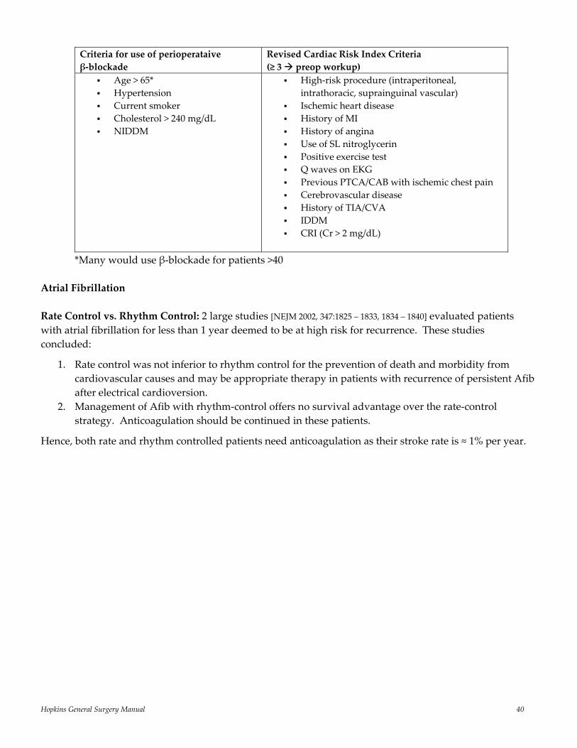
Criteria for use of perioperataive β‐blockade
Revised Cardiac Risk Index Criteria (≥ 3 preop workup)
Age > 65* Hypertension Current smoker Cholesterol > 240 mg/dL NIDDM
High‐risk procedure (intraperitoneal, intrathoracic, suprainguinal vascular)
Ischemic heart disease History of MI History of angina Use of SL nitroglycerin Positive exercise test Q waves on EKG Previous PTCA/CAB with ischemic chest pain Cerebrovascular disease History of TIA/CVA IDDM CRI (Cr > 2 mg/dL)
*Many would use β‐blockade for patients >40 Atrial Fibrillation Rate Control vs. Rhythm Control: 2 large studies [NEJM 2002, 347:1825 – 1833, 1834 – 1840] evaluated patients with atrial fibrillation for less than 1 year deemed to be at high risk for recurrence. These studies concluded:
1. Rate control was not inferior to rhythm control for the prevention of death and morbidity from cardiovascular causes and may be appropriate therapy in patients with recurrence of persistent Afib after electrical cardioversion.
2. Management of Afib with rhythm‐control offers no survival advantage over the rate‐control strategy. Anticoagulation should be continued in these patients.
Hence, both rate and rhythm controlled patients need anticoagulation as their stroke rate is ≈ 1% per year.
Hopkins General Surgery Manual 41
Surgical Approaches:
[Nitta T, et al. Ann Thor Surg 1999 67:27]
• Radiofrequency ablation, cryoablation possible interventions for chronic Afib (RFA is becoming popular because of the complexity of the Maze procedure).
• Electrical cardioversion is rarely successful in converting chronic Afib. • 90% of paroxysmal Afib cured with pulmonary vein isolation alone • Persistent Afib generally need full Cox Maze III; RFA ≈ 60 – 70% cure; Maze ≈ 90% cure
Hopkins General Surgery Manual 42
Vascular Surgery
Nitric Oxide: derived from L‐arginine; reduces free radicals (by scavenging) and prevents atherosclerosis; cGMP acts as the 2nd messenger
Cerebral ischemia most often caused by atheroembolization (50% of strokes due to HTN, 25% due to carotid disease, 25% due to bleed or other cause). Clinical Classification
1. Asymptomatic: bruits (+ bruit 30 – 50% have significant stenosis; + significant stenosis 20 – 50% bruit; Bruit are actually a significant predictor of cardiac disease)
2. TIA: < 24 hour resolution (90% resolve within 2 hours) 3. RIND (Reversible Ischemic Neurologic Deficit): 24 – 48 hour resolution 4. Fixed deficit: stroke
Risks of Stroke:
700,000 per year 160,000 deaths/year 1 year post stroke 2/3 of survivors have disability TIA: 15% stroke in first year, then about 6% per year thereafter (40% chance of stroke in 5 years without ASA) Cortical TIA Vertebral TIA Unilateral Dizzy Arm weakness Bilateral “woozy” Decreased vision “Drop attack” (transient loss of motor tone)
Asymptomatic: Stenosis > 50%: about 4% per year Stenosis > 80%: 35% risk over 2 years 2% per year risk of stroke for patients > 60 Carotid Stenosis
Low resistance arterial systems (such as ICA): total blood flow does not decrease until stenosis > 50%. Hence, no need to repair stenosis < 50%
Up to 50% of patients who suffer stroke have had previous TIA
Asymptomatic Carotid Artery Stenosis Trial (ACAS) demonstrated that patients with an asymptomatic stenosis of 60% or greater had a 53% relative risk reduction of stroke after undergoing CEA + ASA compared to ASA alone [11 5%]. The benefit was much greater in men than women.
North American Symptomatic Carotid Endarterectomy Trial (NASCET I) demonstrated that CEA is highly beneficial in patients with recent hemispheric or retinal TIA or nondisabaling stroke and an ipsilateral high grade stenosis (70 – 99%). The benefit of surgery was seen within 3 months of operation. The incidence of stroke was decreased in all subgroups but was largest in patients who experienced major ipsilateral stroke with an 81% risk reduction. Overall, 26% of patients with high‐grade (70 – 99%) stenosis sustained a stroke within 18 months with medical management vs. 9% with surgery at 2 years [26 9%]
NASCET II: looked at symptomatic patients with 50 – 69% stenosis and found a reduction from 22 16% (p<0.045). More hospitals participated, hence increased morbidity.
Hopkins General Surgery Manual 43
Diagnosis: Ask 3 questions (ICA disease? % stenosis? Characteristics of plaque?) Neck duplex consists of 2 parts: B‐mode U/S image and spectral velocity analysis IC/CC systolic ratio gives % stenosis:
• <2 <50% • 2 – 3.9 50 – 69% • >4 70 – 99%
Following CEA:
• If ipsilateral pre‐orbital headache think hyperperfusion (since stenosis removed); usually ≤ 3 – 5 days post surgery.
• Most common CN injury vagus nerve (clamp) hoarseness • CN XII tongue deviation to side of injury; marginal mandibular lip droop • CAD is main cause of post‐op mortality
Re‐stenosis rate ≈ 5 – 10% (>50% stenosis within 2 years: myointimal hyperplasia, not atherosclerosis; F > M)
Upper limit guidelines for acceptable M&M rates for CEA: Asymptomatic: 3% Symptomatic: 5% Symptomatic/CVA: 7% Recurrent Stenosis: 10% To participate in ACAS and NASCET trials centers had to have < 5% overall mortality for CEA Carotid Anatomy
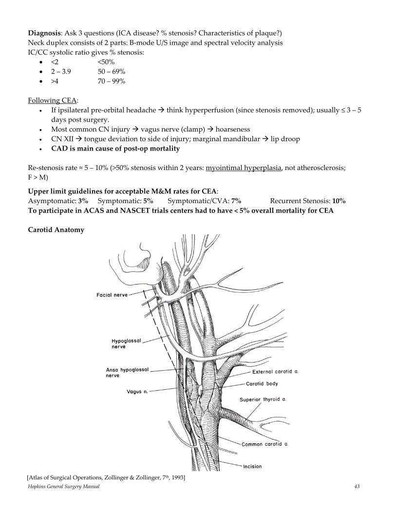
[Atlas of Surgical Operations, Zollinger & Zollinger, 7th, 1993]
Hopkins General Surgery Manual 44
Abdominal Aortic Aneurysm
95% infrarenal approximately 75% asymptomatic and discovered incidentally growth rate ≈ 0.4 cm/year in diameter risk of rupture for small (< 5.5 cm) aneurysms: 0.6 – 1% per year [NEJM 2002, 346: 1437‐1444, 1445‐1452] Pathology: ↑ matrix metalloprotease activity (MMP‐1,2,3; MMP‐9) risk of rupture related to size:
• < 5 cm: 20% 5‐year risk • 5 – 7 cm: 33% • > 7 cm: 95%
10 – 20% involve iliacs when removing distal clamps, remove INTERNAL iliac first, then EXTERNAL iliac to avoid distal
embolization to lower extremities following rupture with operative repair: cardiac complications are most common cause of early
death; renal complications for late death CAD is most common cause of death for patients with “small” AAA (< 6 cm); Rupture (75 – 90% mortality) is most common cause of death for patients with “large” AAA (> 6 cm)
[unless they have metastatic cancer or debilitating CHF]
Indications for Repair:
Good risk patients: 1. AAA > 5.5 cm + life expectancy > 2 years 2. AAA < 5.5 cm with COPD, expansion > 0.5 cm/6 months High risk patients: 1. AAA > 6 cm, or symptomatic
The UK Small Aneurysm Trial: [NEJM 346(19):1445, 2002] Randomly assigned over 1000 patients with aneurysms ranging from 4.0 to 5.5 cm to either undergo early elective repair or observation with U/S surveillance. Mean follow‐up was 8 years. The mean survival was 6.5 years in the surveillance group vs. 6.7 years in the treatment group (p=0.29). Early mortality was greater in the surgery group, but total mortality was greater in the surveillance group at 8 years. Indications for Retroperitoneal Approach‡:
hostile abdomen Inflammatory AAA juxtarenal AAA Right iliac disease is a relative contraindication
‡ Benefits (vs. anterior approach) include: ↓respiratory problems, ↓LOS, minimal ileus
Hopkins General Surgery Manual 45
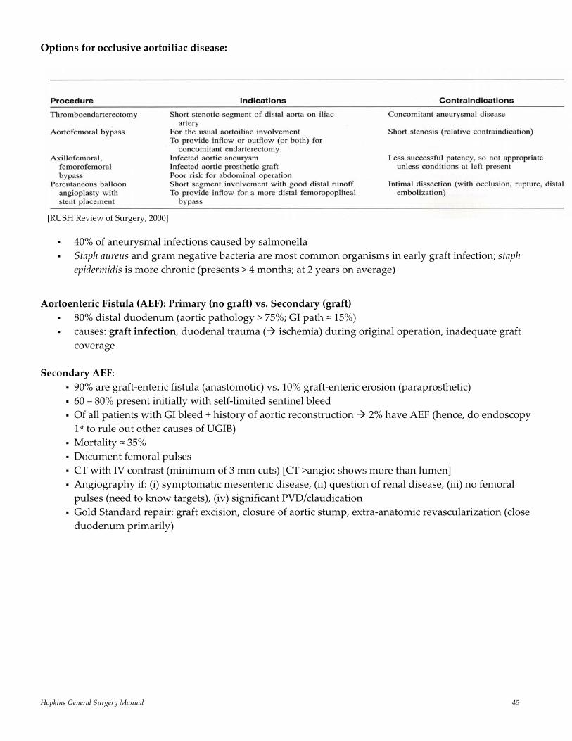
Options for occlusive aortoiliac disease:
40% of aneurysmal infections caused by salmonella Staph aureus and gram negative bacteria are most common organisms in early graft infection; staph epidermidis is more chronic (presents > 4 months; at 2 years on average)
Aortoenteric Fistula (AEF): Primary (no graft) vs. Secondary (graft)
80% distal duodenum (aortic pathology > 75%; GI path ≈ 15%) causes: graft infection, duodenal trauma ( ischemia) during original operation, inadequate graft
coverage Secondary AEF:
90% are graft‐enteric fistula (anastomotic) vs. 10% graft‐enteric erosion (paraprosthetic) 60 – 80% present initially with self‐limited sentinel bleed Of all patients with GI bleed + history of aortic reconstruction 2% have AEF (hence, do endoscopy 1st to rule out other causes of UGIB)
Mortality ≈ 35%
Document femoral pulses CT with IV contrast (minimum of 3 mm cuts) [CT >angio: shows more than lumen] Angiography if: (i) symptomatic mesenteric disease, (ii) question of renal disease, (iii) no femoral pulses (need to know targets), (iv) significant PVD/claudication
Gold Standard repair: graft excision, closure of aortic stump, extra‐anatomic revascularization (close duodenum primarily)
[RUSH Review of Surgery, 2000]
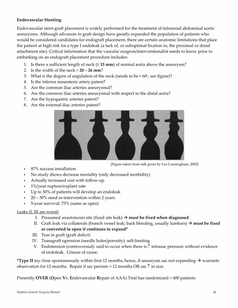
Hopkins General Surgery Manual 46
Endovascular Stenting:
Endovascular stent‐graft placement is widely performed for the treatment of infrarenal abdominal aortic aneurysms. Although advances in graft design have greatly expanded the population of patients who would be considered candidates for endograft placement, there are certain anatomic limitations that place the patient at high risk for a type I endoleak (a lack of, or suboptimal fixation in, the proximal or distal attachment site). Critical information that the vascular surgeon/interventionalist needs to know prior to embarking on an endograft placement procedure includes:
1. Is there a sufficient length of neck (≥ 15 mm) of normal aorta above the aneurysm? 2. Is the width of the neck < 20 – 26 mm? 3. What is the degree of angulation of the neck (needs to be < 60o, see figure)? 4. Is the inferior mesenteric artery patent? 5. Are the common iliac arteries aneurysmal? 6. Are the common iliac arteries aneurysmal with respect to the distal aorta? 7. Are the hypogastric arteries patent? 8. Are the external iliac arteries patent?
97% success installation No study shows decrease mortality (only decreased morbidity) Actually increased cost with follow‐up 1%/year rupture/explant rate Up to 50% of patients will develop an endoleak 20 – 30% need re‐intervention within 2 years 5‐year survival: 75% (same as open)
Leaks (I, III are worst) I. Presumed anastomosis site (fixed site leak) must be fixed when diagnosed II. Graft leak via collaterals (branch vessel leak; back bleeding, usually lumbars) must be fixed
or converted to open if continues to expand* III. Tear in graft (graft defect) IV. Transgraft egression (needle holes/porosity): self‐limiting V. Endotension (controversial): said to occur when there is ↑ intrasac pressure without evidence
of endoleak. Unsure of cause.
*Type II my close spontaneously within first 12 months; hence, if aneurysm sac not expanding warrants observation for 12 months. Repair if sac persists > 12 months OR sac ↑ in size. Presently OVER (Open Vs. Endovascular Repair of AAA) Trial has randomized > 400 patients
[Figure taken from talk given by Les Cunningham, 2005]
Hopkins General Surgery Manual 47
Peripheral Vascular Disease Claudication:
• History is key: Reproducible pain of buttock, thigh and/or calf associated with ambulation and relieved by rest.
• Initial treatment is smoking cessation, exercise; Pletal > Trental (but ↑ cost); not surgery • These patients have the same risk of death from cardiovascular disease as patients with known
cardiac disease (50% die within 5 years) • Progresses to gangrene 2 – 3% annually (only 10% ever lose leg)
Rest pain/ulcers indication for bypass
Clinical manifestations of critical limb ischemia (according to European Consensus Conference): 1. Rest pain requiring analgesia for at least 2 weeks 2. Ankle systolic pressure < 50 mmHg (with or without tissue loss/gangrene)
Exercise Test “positive” if > 20% fall in ankle systolic pressure requiring > 3 min to recover
Arterial Flow is triphasic: 1. Forward, 2. Reverse, 3. Late forward (Note: will be normally monophasic in low resistance system, like ICA)
ABI Values: Calcified (diabetic) > 1.2; normal > 1; Claudication 0.5 – 0.99; Rest pain ≤ 0.3 *Require ABI > 0.5 to heal a lower extremity amputation
1‐year survival after amputation for ischemic disease is 75% 2‐year survival after amputation for ischemic disease is 60% 3‐year survival after amputation for ischemic disease is 50% 4‐year survival after amputation for ischemic disease is 45% 50% of patients lose other leg within 5 years of 1st amputation
*Most common site of lower limb atherosclerosis: SFA in region of the adductor canal (Hunter’s canal) *Cardioatrial emboli most frequently occlude CFA
Reversed saphenous vein patency (gold standard below inguinal ligament): 80 – 90% at 1 year; 75% at 5 years; persistent smoking is #1 reason for late graft failure (valve leaflet remnants is #1 reason during 1st two years)
Fem‐pop bypass: # Vessel run‐off 5‐year patency 3 70% 2 35% 1 15 – 20%
Popliteal Artery Aneurysm Most common peripheral aneurysm (≈ 70% of all) 50% are bilateral and 30% also have AAA 20 – 30% of limb loss with distal emboli elective repair of all, regardless of size Management options: ‐ medial exploration: proximal/distal ligation & bypass
‐ acute thrombosis preop thrombolytics ‐ endovascular repair (not yet accepted)
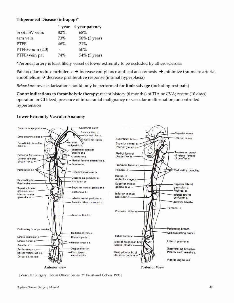
Hopkins General Surgery Manual 48
Tibperoneal Disease (infrapop)*
1‐year 4‐year patency in situ SV vein: 82% 68% arm vein 73% 58% (3 year) PTFE 46% 21% PTFE+coum (2.0) ‐ 50% PTFE+vein pat 74% 54% (5 year)
*Peroneal artery is least likely vessel of lower extremity to be occluded by atherosclerosis
Patch/collar reduce turbulence increase compliance at distal anastomosis minimize trauma to arterial endothelium decrease proliferative response (intimal hyperplasia)
Below knee revascularization should only be performed for limb salvage (including rest pain)
Contraindications to thrombolytic therapy: recent history (6 months) of TIA or CVA; recent (10 days) operation or GI bleed; presence of intracranial malignancy or vascular malformation; uncontrolled hypertension Lower Extremity Vascular Anatomy
Anterior view Posterior View [Vascular Surgery, House Officer Series, 3rd Faust and Cohen, 1998]

Hopkins General Surgery Manual 49
Evaluating Lower Extremity Ulcers
First diagnose the cause (diabetic, venous insufficiency, arterial insufficiency, vasculitis, IBD, etc.)
The most common are diabetic, arterial, and venous insufficiency:
I. Diabetic: typically occur on pressure points on the foot and are painless secondary to the acquired neuropathy
II. Arterial Insufficiency: extremely painful, associated with rest pain in distal foot, have grayish granulation tissue, surrounded by blue and mottled skin, and do not bleed when debrided. Pain is most common over metatarsal heads, not toes (usually occur at pressure points).
III. Venous insufficiency: large, irregular, shallow, have red granulation tissue, occur around medial and lateral maleoli, and are surrounded by brawny edema and stasis pigmentation. Leukocytes are thought to play an important role in the pathophysiology because they have been found to be sequestered in the ankle region of patients with elevated venous pressures, especially in the dependent position. They plug capillaries and become activated and release their enzymes and superoxide radicals, decreasing flow, leading to ischemia and ulceration
Phlegmasia alba dolens: venous clot results in such increase in venous pressure edematous, swollen, pale, cyanotic extremity; blanched appearance result of edema; can impair arterial inflow; a surgical emergency requiring thrombectomy ± creation of AV fistula (can use TPA) Evaluating Limb Ischemia
The presentation of peripheral limb ischemia determines the chronicity Acute limb ischemia is 60% thrombotic and 40% embolic Always check inflow (femoral pulses) Absent popliteal pulses imply SFA occlusion or multiple proximal occlusions
When describing angiographic lesions/stenoses use terms like: none, mild, moderate, severe, and distinguish between diffuse and focal
SVS Clinical Classification of Limb Ischemia
Sensory Motor Doppler Arterial¥
Doppler Venous¥
Viable Normal Normal Normal Normal Threatened Marginal
+/‐ toes Normal Abnormal Normal
Threatened Immediate
Beyond toes Mild‐moderate deficit
Abnormal Normal
Irreversible Profound Paralysis Abnormal Abnormal ¥ If you hear a monophasic doppler signal distinguish between arterial and venous (venous will change with sequential calf compression)
Treatment
Viable: heparin‡ further work‐up Threatened (Marginal): heparin further work‐up Threatened (Immediate): heparin to OR for intra‐op angiogram +/‐ intervention Irreversible: to OR amputation ‡dose: bolus 80 units/kg followed by drip of 18 units/kg
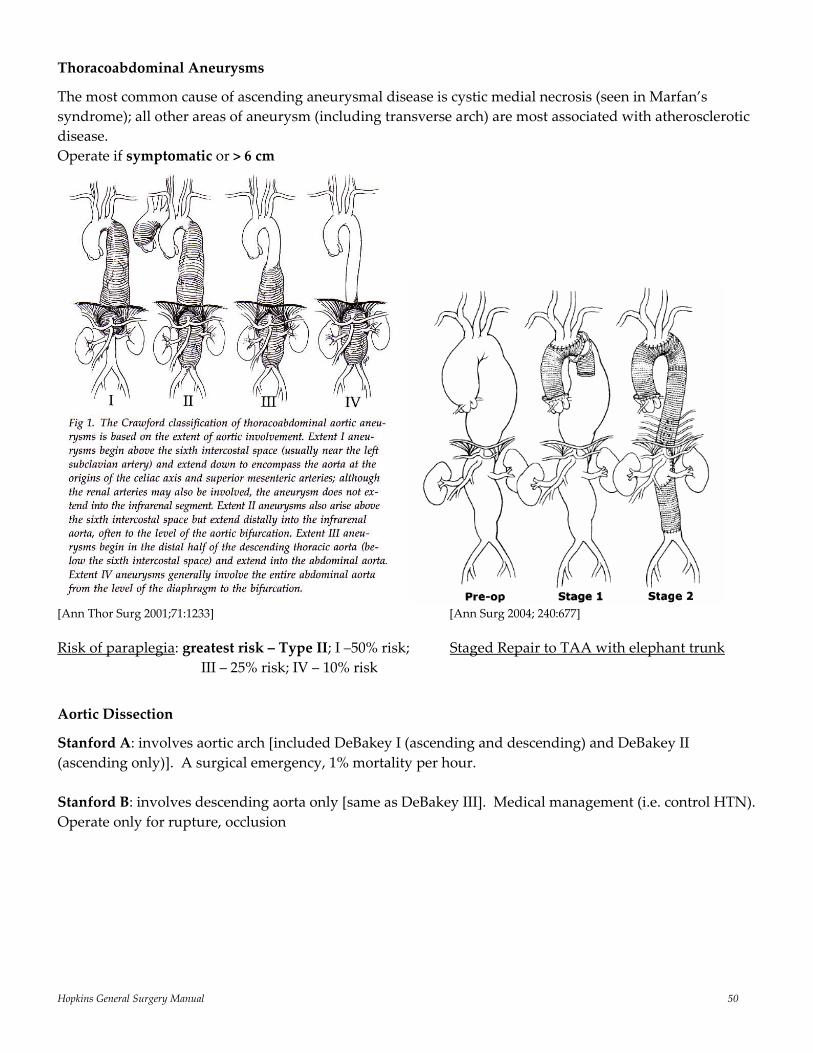
Hopkins General Surgery Manual 50
Thoracoabdominal Aneurysms
The most common cause of ascending aneurysmal disease is cystic medial necrosis (seen in Marfan’s syndrome); all other areas of aneurysm (including transverse arch) are most associated with atherosclerotic disease. Operate if symptomatic or > 6 cm
[Ann Thor Surg 2001;71:1233] [Ann Surg 2004; 240:677] Risk of paraplegia: greatest risk – Type II; I –50% risk; Staged Repair to TAA with elephant trunk III – 25% risk; IV – 10% risk
Aortic Dissection
Stanford A: involves aortic arch [included DeBakey I (ascending and descending) and DeBakey II (ascending only)]. A surgical emergency, 1% mortality per hour. Stanford B: involves descending aorta only [same as DeBakey III]. Medical management (i.e. control HTN). Operate only for rupture, occlusion

Hopkins General Surgery Manual 51
Splancnic Artery Aneurysms
Site Incidence Pathology Clinical Presentation
Diagnosis Natural History Treatment
Splenic Most common; 1/1000
Atherosclerosis F > M 60% occur during pregnancy
Most asymptomatic 20% with variable symptoms
Often incidental Calcs in LUQ CT Arteriography is gold std
Unclear; if symptomatic, enlarging fix Pregnant fix > 2 – 3 cm fix Some say ok to watch if 1. Asympt + > 60 2. calc + < 1.5 cm
If distal splenectomy If proximal ligate at both ends (no need to reconstruct because of short gastrics)
Hepatic 2nd most common splancnic
Infectious most common; Mycotic following sepsis (e.g. bacterial endocarditis); Approx 80% outside of liver
Most symptomatic; Persistent RUQ pain
Rare pre‐op; Selective celiac Arteriography is gold std
Unknown, but rupture devastating, so fix all unless serious contraindication
Proximal to GDA
aneurysmectomy and ligate (retrograde GDA flow) May do same for PHA, LHA, or RHA, but recon with saphenous vein is best
SMA 3rd most common splancnic
Rarely atherosclerotic; > 50% mycotic (2o to SBE)
Almost always cause symptoms; Intermittent or constant epigastric/back pain
Epigastric pain, tender NONFIXED pulsitile mass
Spontaneous rupture occurs in over 50%; Operation always warranted
Aneursymectomy + bypass (vein preferred because of infection); Dacron acceptable
Celiac Rare; 1/8000 Atherosclerosis;Infectious, also
Usually asymptomatic
Usually incidental
Unknown; probably high risk of rupture
Aneurysmectomy with arterial reconstruction is desired; Greatest risk is intestinal ischemia
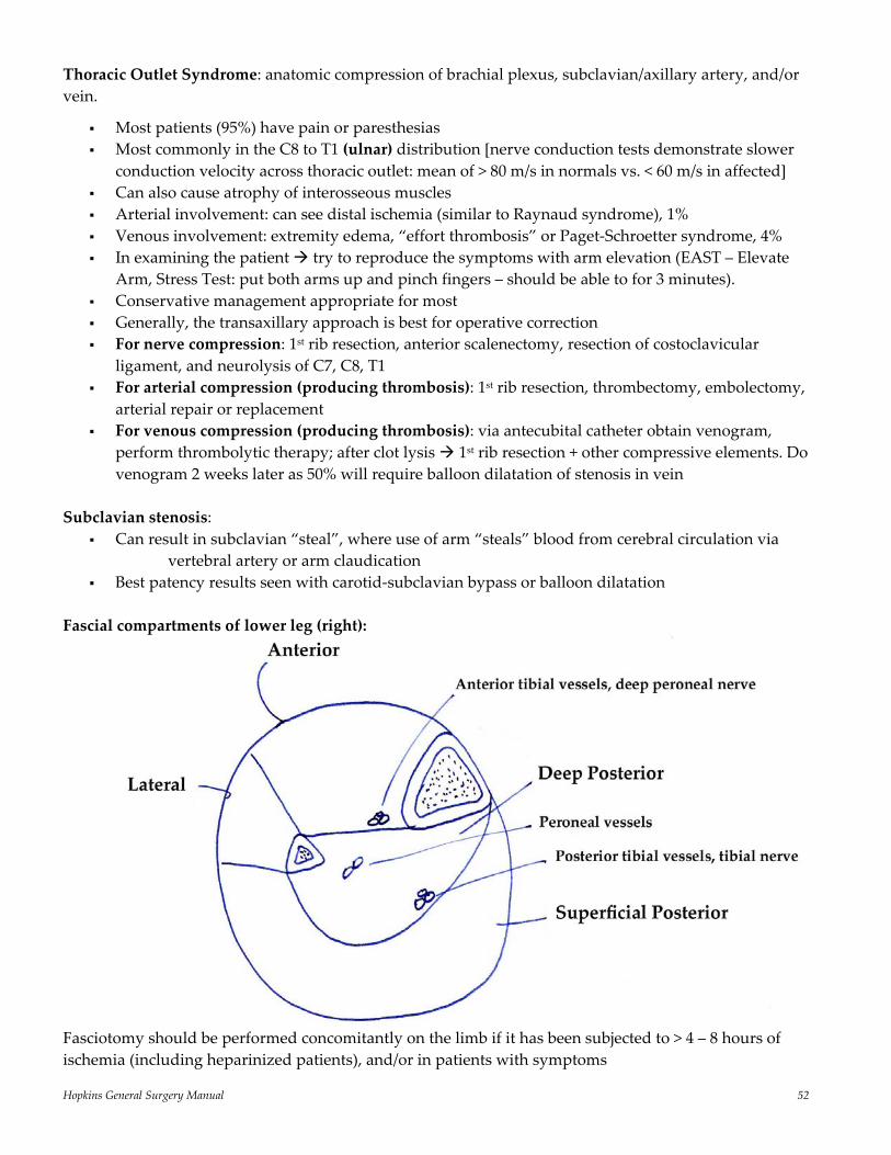
Hopkins General Surgery Manual 52
Thoracic Outlet Syndrome: anatomic compression of brachial plexus, subclavian/axillary artery, and/or vein.
Most patients (95%) have pain or paresthesias Most commonly in the C8 to T1 (ulnar) distribution [nerve conduction tests demonstrate slower
conduction velocity across thoracic outlet: mean of > 80 m/s in normals vs. < 60 m/s in affected] Can also cause atrophy of interosseous muscles Arterial involvement: can see distal ischemia (similar to Raynaud syndrome), 1% Venous involvement: extremity edema, “effort thrombosis” or Paget‐Schroetter syndrome, 4% In examining the patient try to reproduce the symptoms with arm elevation (EAST – Elevate
Arm, Stress Test: put both arms up and pinch fingers – should be able to for 3 minutes). Conservative management appropriate for most Generally, the transaxillary approach is best for operative correction For nerve compression: 1st rib resection, anterior scalenectomy, resection of costoclavicular
ligament, and neurolysis of C7, C8, T1 For arterial compression (producing thrombosis): 1st rib resection, thrombectomy, embolectomy,
arterial repair or replacement For venous compression (producing thrombosis): via antecubital catheter obtain venogram,
perform thrombolytic therapy; after clot lysis 1st rib resection + other compressive elements. Do venogram 2 weeks later as 50% will require balloon dilatation of stenosis in vein
Subclavian stenosis:
Can result in subclavian “steal”, where use of arm “steals” blood from cerebral circulation via vertebral artery or arm claudication
Best patency results seen with carotid‐subclavian bypass or balloon dilatation
Fascial compartments of lower leg (right):
Fasciotomy should be performed concomitantly on the limb if it has been subjected to > 4 – 8 hours of ischemia (including heparinized patients), and/or in patients with symptoms
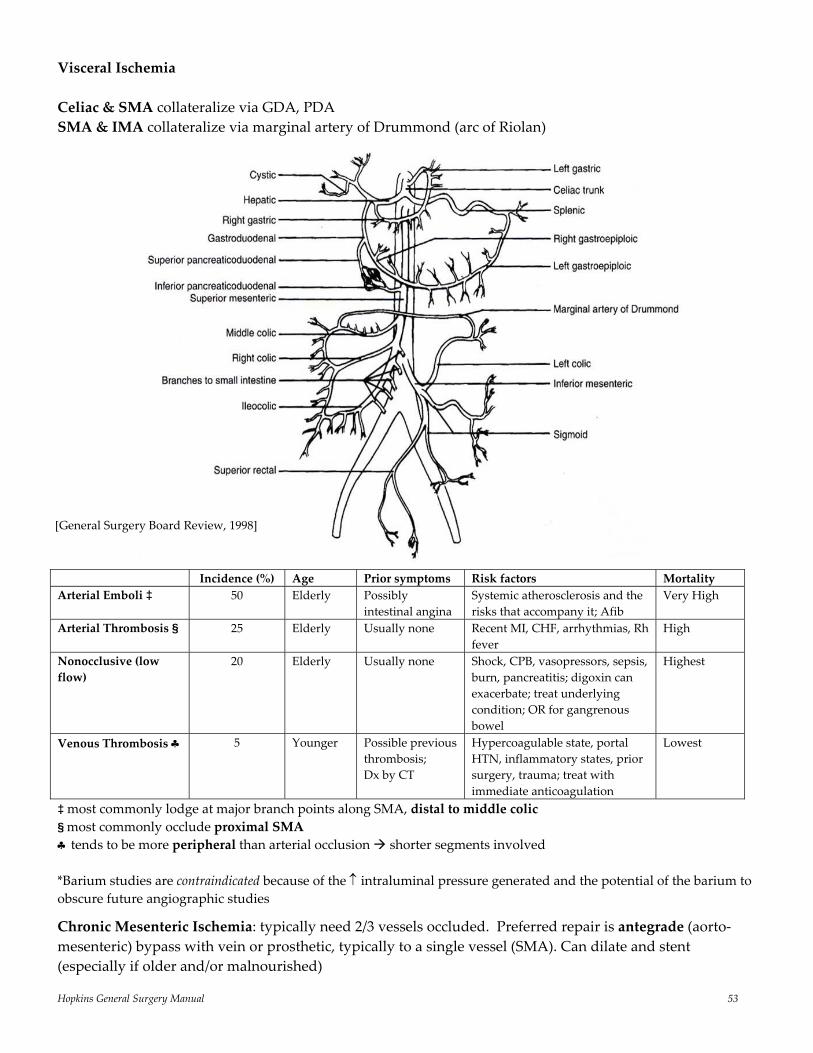
Hopkins General Surgery Manual 53
Visceral Ischemia Celiac & SMA collateralize via GDA, PDA SMA & IMA collateralize via marginal artery of Drummond (arc of Riolan)
Incidence (%) Age Prior symptoms Risk factors Mortality Arterial Emboli ‡ 50 Elderly Possibly
intestinal angina Systemic atherosclerosis and the risks that accompany it; Afib
Very High
Arterial Thrombosis § 25 Elderly Usually none Recent MI, CHF, arrhythmias, Rh fever
High
Nonocclusive (low flow)
20 Elderly Usually none Shock, CPB, vasopressors, sepsis, burn, pancreatitis; digoxin can exacerbate; treat underlying condition; OR for gangrenous bowel
Highest
Venous Thrombosis ♣ 5 Younger Possible previous thrombosis; Dx by CT
Hypercoagulable state, portal HTN, inflammatory states, prior surgery, trauma; treat with immediate anticoagulation
Lowest
‡ most commonly lodge at major branch points along SMA, distal to middle colic § most commonly occlude proximal SMA ♣ tends to be more peripheral than arterial occlusion shorter segments involved *Barium studies are contraindicated because of the ↑ intraluminal pressure generated and the potential of the barium to obscure future angiographic studies
Chronic Mesenteric Ischemia: typically need 2/3 vessels occluded. Preferred repair is antegrade (aorto‐mesenteric) bypass with vein or prosthetic, typically to a single vessel (SMA). Can dilate and stent (especially if older and/or malnourished)
[General Surgery Board Review, 1998]
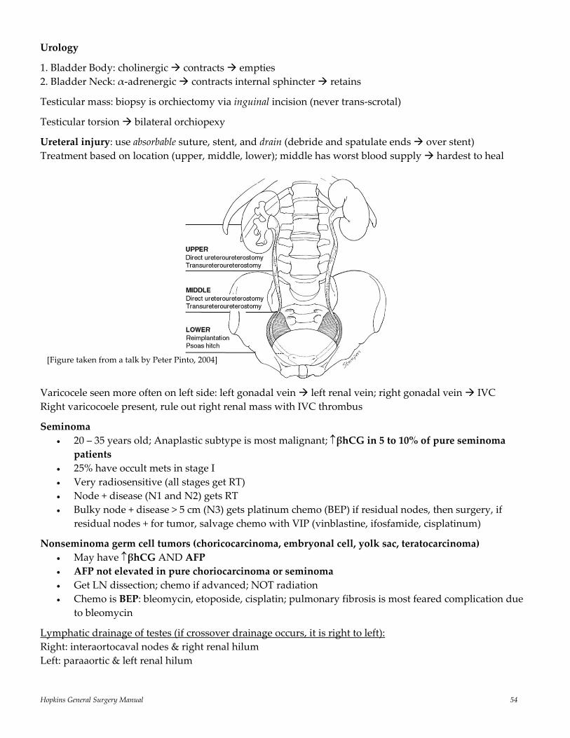
Hopkins General Surgery Manual 54
Urology
1. Bladder Body: cholinergic contracts empties 2. Bladder Neck: α‐adrenergic contracts internal sphincter retains
Testicular mass: biopsy is orchiectomy via inguinal incision (never trans‐scrotal)
Testicular torsion bilateral orchiopexy
Ureteral injury: use absorbable suture, stent, and drain (debride and spatulate ends over stent) Treatment based on location (upper, middle, lower); middle has worst blood supply hardest to heal
Varicocele seen more often on left side: left gonadal vein left renal vein; right gonadal vein IVC Right varicocoele present, rule out right renal mass with IVC thrombus
Seminoma • 20 – 35 years old; Anaplastic subtype is most malignant; ↑βhCG in 5 to 10% of pure seminoma
patients • 25% have occult mets in stage I • Very radiosensitive (all stages get RT) • Node + disease (N1 and N2) gets RT • Bulky node + disease > 5 cm (N3) gets platinum chemo (BEP) if residual nodes, then surgery, if
residual nodes + for tumor, salvage chemo with VIP (vinblastine, ifosfamide, cisplatinum)
Nonseminoma germ cell tumors (choricocarcinoma, embryonal cell, yolk sac, teratocarcinoma) • May have ↑βhCG AND AFP • AFP not elevated in pure choriocarcinoma or seminoma • Get LN dissection; chemo if advanced; NOT radiation • Chemo is BEP: bleomycin, etoposide, cisplatin; pulmonary fibrosis is most feared complication due
to bleomycin
Lymphatic drainage of testes (if crossover drainage occurs, it is right to left): Right: interaortocaval nodes & right renal hilum Left: paraaortic & left renal hilum
[Figure taken from a talk by Peter Pinto, 2004]
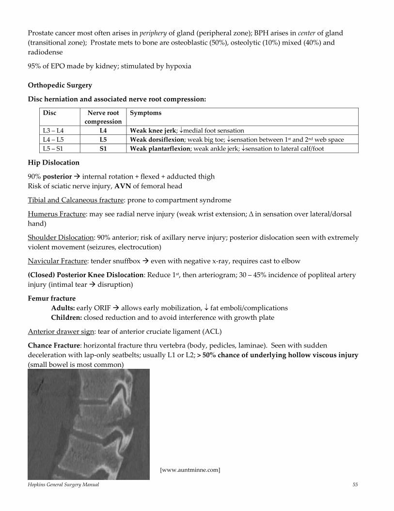
Hopkins General Surgery Manual 55
Prostate cancer most often arises in periphery of gland (peripheral zone); BPH arises in center of gland (transitional zone); Prostate mets to bone are osteoblastic (50%), osteolytic (10%) mixed (40%) and radiodense
95% of EPO made by kidney; stimulated by hypoxia Orthopedic Surgery
Disc herniation and associated nerve root compression:
Disc Nerve root compression
Symptoms
L3 – L4 L4 Weak knee jerk; ↓medial foot sensation L4 – L5 L5 Weak dorsiflexion; weak big toe; ↓sensation between 1st and 2nd web space L5 – S1 S1 Weak plantarflexion; weak ankle jerk; ↓sensation to lateral calf/foot
Hip Dislocation
90% posterior internal rotation + flexed + adducted thigh Risk of sciatic nerve injury, AVN of femoral head
Tibial and Calcaneous fracture: prone to compartment syndrome
Humerus Fracture: may see radial nerve injury (weak wrist extension; ∆ in sensation over lateral/dorsal hand)
Shoulder Dislocation: 90% anterior; risk of axillary nerve injury; posterior dislocation seen with extremely violent movement (seizures, electrocution)
Navicular Fracture: tender snuffbox even with negative x‐ray, requires cast to elbow
(Closed) Posterior Knee Dislocation: Reduce 1st, then arteriogram; 30 – 45% incidence of popliteal artery injury (intimal tear disruption)
Femur fracture Adults: early ORIF allows early mobilization, ↓ fat emboli/complications Children: closed reduction and to avoid interference with growth plate
Anterior drawer sign: tear of anterior cruciate ligament (ACL)
Chance Fracture: horizontal fracture thru vertebra (body, pedicles, laminae). Seen with sudden deceleration with lap‐only seatbelts; usually L1 or L2; > 50% chance of underlying hollow viscous injury (small bowel is most common)
[www.auntminne.com]
Hopkins General Surgery Manual 56
Gynecologic Pathology
Ovarian Cancer 5 major classes (based on histology and embryologic etiology):
1. neoplasms derived from celomic epithelium 2. neoplasms derived from germ cells 3. neoplasms derived from gonadal stroma 4. neoplasms derived from nonspecific mesenchyme 5. metastatic lesions to the ovary (usually GI, breast, or uterine)
Responsible for half of all gynecologic deaths each year; 25,000 cases/year in US (33% 5‐year survival)
All comers: 20% of ovarian neoplasms are malignant (↑ with age) Woman aged 20 – 30: 10% chance of malignancy; ≥ 50: 50% chance of malignancy Usually diagnosed in advanced stage (2/3 present as stage III or IV) CA‐125 of limited use for screening, especially in premenopausal women Cytoreductive surgery is most effective treatment (TAH+BSO; must include omentum, peritoneal washings; no need for lymphadenectomy since tumors spreads by exfoliation of cells)
Uterine Cancer Endometrial cancer is the most common gynecologic malignancy in US (40,000 cases/year) ≈ 75% are diagnosed as stage I, 5‐year survival > 75% Risk factors are associated with ↑estrogen exposure 80% of cases are in postmenopausal women (5% in women < 40) Abnormal bleeding is #1 presentation; histology is adenocarcinoma > 90% Treatment is TAH+BSO, peritoneal washings, LN sampling Post‐operative radiation (5000 rad over 5 weeks) reserved for: + pelvic nodes, poor differentiation,
invasion into myometrium, occult cervical involvement Uterine sarcoma has poor prognosis
Cervical Cancer Most (> 80%) are squamous cell Primarily affects women aged 35 – 45 Painless bleeding is #1 presentation (pain often signals advanced disease) Pap smear allows most to be diagnosed as premalignant lesions Highly associated with HPV infection (serotypes 16 and 18 virtually always involved) Treatment for early stage: radical hysterectomy (no need for oophorectomy unless >45 or has
ovarian pathology) Chemoradiation if poor surgical candidate or advanced disease
Hopkins General Surgery Manual 57
Post‐menopausal HRT is associated with:
Increase in:
Decrease in:
Endometrial cancer Breast cancer Venous thrombosis Stroke Coronary artery
disease Gallbladder disease
Vasomotor symptoms of post‐menopause
Vertebral/hip/pelvic fractures
Osteoperosis Colorectal Cancer
Surgery during pregnancy:
Second trimester is preferred Both laparoscopic and open procedures during first trimester are associated with ↑ risk of
spontaneous abortion and possible risk of teratogenicity During the third trimester surgery is associated with ↑ risk of premature labor and damage to the
uterus Neurosurgery
Diabetes Insipidus – central or renal (ADH inhibited)
SIADH
↑ urine output Low urine Osm, specific gravity High serum Osm, hypernatremic
↓ urine output High urine Osm, specific gravity Low Serum Osm, hyponatremic
Peripheral Nerve Injuries Neuropraxia: focal demyelination improves Axonotmesis: loss of axon continuity (nerve and sheath intact) regenerates at 1 mm/day Neurotmesis: loss of nerve continuity surgery required to repair
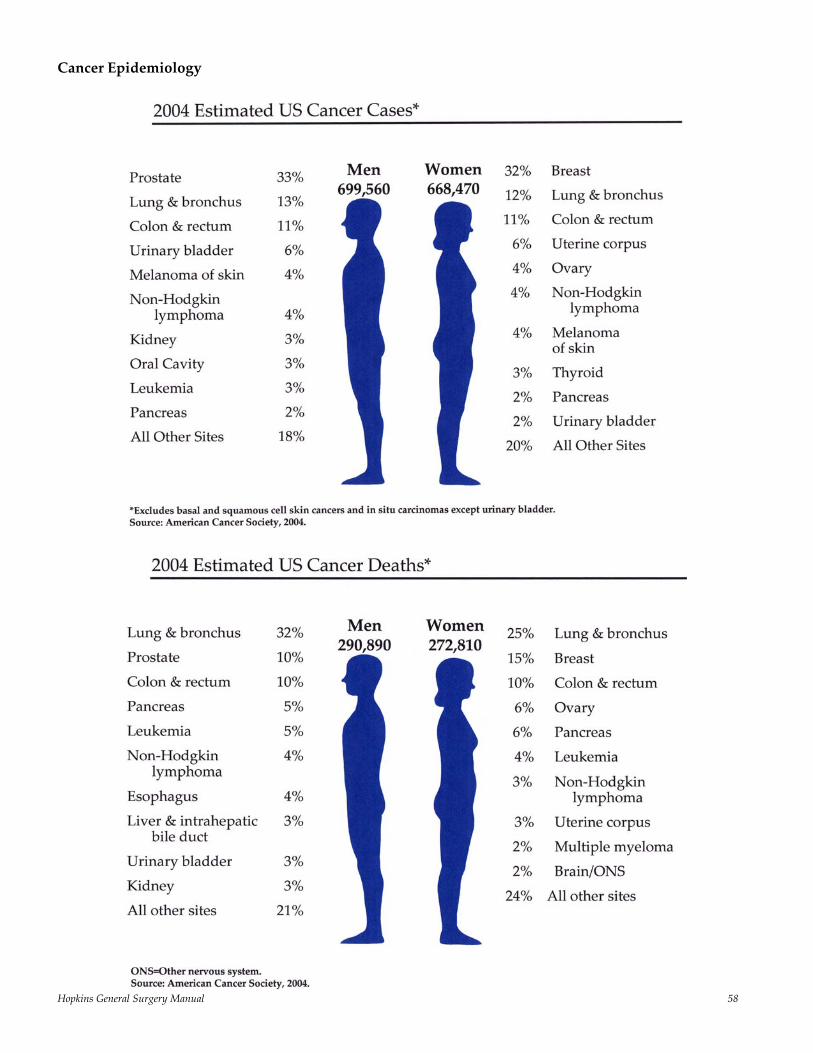
Hopkins General Surgery Manual 58
Cancer Epidemiology
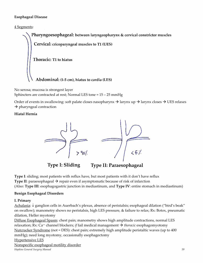
Hopkins General Surgery Manual 59
Esophageal Disease 4 Segments:
No serosa; mucosa is strongest layer Sphincters are contracted at rest; Normal LES tone ≈ 15 – 25 mmHg
Order of events in swallowing: soft palate closes nasopharynx larynx up larynx closes UES relaxes pharyngeal contraction
Hiatal Hernia
Type I: sliding; most patients with reflux have, but most patients with it don’t have reflux Type II: paraesophageal repair even if asymptomatic because of risk of infarction (Also: Type III: esophagogastric junction in mediastinum, and Type IV: entire stomach in mediastinum)
Benign Esophageal Disorders
I. Primary Achalasia: ↓ ganglion cells in Auerbach’s plexus, absence of peristalsis; esophageal dilation (“bird’s beak” on swallow); manometry shows no peristalsis, high LES pressure, & failure to relax; Rx: Botox, pneumatic dilation, Heller myotomy Diffuse Esophageal Spasm: chest pain; manometry shows high amplitude contractions, normal LES relaxation; Rx: Ca++ channel blockers; if fail medical management thoracic esophagomyotomy Nutcracker Syndrome (not = DES): chest pain; extremely high amplitude peristaltic waves (up to 400 mmHg); need long myotomy, occasionally esophagectomy Hypertensive LES Nonspecific esophageal motility disorder

Hopkins General Surgery Manual 60
II. Secondary Collagen vascular (SLE, systemic sclerosis, polymyositis, dermatomyositis) Chronic idiopathic intestinal pseudo‐obstruction Neuromuscular disease Endocrine disorder Zenker’s Diverticulum • Loss of compliance in the pharyngoesophageal segment; manifested by increase bolus pressure • Muscle biopsies have shown histologic evidence of restrictive myopathy correlating with decreased compliance of upper esophagus
• Repetitive stress of bolus through noncompliant muscle diverticulum through Killian’s triangle (between cricopharyngeus and thyropharyngeus muscles)
• Primary reason: dyscoordination of the sphincter relaxation with pharyngeal contraction together with impaired sphincter opening
• Dx: with barium swallow; not EGD • Rx: myotomy and diverticulectomy (or ‐pexy if unfit for resection) via left cervical approach
Zenker’s and epiphrenic are both false diverticuli (mucosa only); epiphrenic requires long myotomy at 1800 Traction diverticuli are True: located mid‐esophagus; associated with TB/inflammatory diseases
Esophageal Claudication: chest pain caused by a burst of uncoordinated esophageal motor activity under ischemic conditions (i.e. esophageal blood supply is interrupted during these bursts in situations where blood flow may already be compromised)
Esophageal foreign body: usually at points of natural narrowing: below cricopharyngeus, near arch of aorta, behind right mainstem; 95% are immediately below cricopharyngeus muscle rigid scope under GA is treatment of choice
Esophageal rupture (Boerhaave’s): full thickness injury (vs. partial thickness injury of Mallory‐Weiss); often left posterior/lateral; 85% die if diagnosis delayed > 36 hours (Rx: early repair, late diversion)
Mallory‐Weiss Tear: repeated emesis; about 10% present with massive hemorrhage gastric bleeding (usually lesser curvature); since arterial bleed, pressure tamponade of little help usually stops spontaneously; Dx with endoscopy gastrotomy & oversew if doesn’t stop Esophageal Perforation
50% instrumentation, 20% trauma, 15% spontaneous 3/10k EGD, 11/10k rigid
Presentation depends on 1. Location, 2. Size, 3. Elapsed time, and 4. Underlying pathology/etiology
• Cervical: neck pain (especially with flexion), crepitus, right pleural effusion • Spontaneous: usually distal left
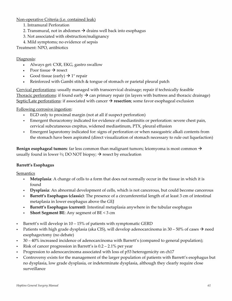
Hopkins General Surgery Manual 61
Non‐operative Criteria (i.e. contained leak) 1. Intramural Perforation 2. Transmural, not in abdomen drains well back into esophagus 3. Not associated with obstruction/malignancy 4. Mild symptoms; no evidence of sepsis
Treatment: NPO, antibiotics Diagnosis:
• Always get: CXR, EKG, gastro swallow • Poor tissue resect • Good tissue (early) 1° repair • Reinforced with Gambi stitch & tongue of stomach or parietal pleural patch
Cervical perforations: usually managed with transcervical drainage; repair if technically feasible Thoracic perforations: if found early can primary repair (in layers with buttress and thoracic drainage) Septic/Late perforations: if associated with cancer resection; some favor esophageal exclusion
Following corrosive ingestion: EGD only to proximal margin (not at all if suspect perforation) Emergent thoracotomy indicated for evidence of mediastinitis or perforation: severe chest pain,
cervical subcutaneous crepitus, widened mediastinum, PTX, pleural effusion Emergent laparotomy indicated for: signs of perforation or when nasogastric alkali contents from
the stomach have been aspirated (direct visualization of stomach necessary to rule out liquefaction) Benign esophageal tumors: far less common than malignant tumors; leiomyoma is most common usually found in lower ⅔; DO NOT biopsy; resect by enucleation
Barrett’s Esophagus
Semantics Metaplasia: A change of cells to a form that does not normally occur in the tissue in which it is
found Dysplasia: An abnormal development of cells, which is not cancerous, but could become cancerous Barrett’s Esophagus (classic): The presence of a circumferential length of at least 3 cm of intestinal
metaplasia in lower esophagus above the GEJ Barrett’s Esophagus (current): Intestinal metaplasia anywhere in the tubular esophagus Short Segment BE: Any segment of BE < 3 cm
Barrett’s will develop in 10 – 15% of patients with symptomatic GERD Patients with high grade dysplasia (aka CIS), will develop adenocarcinoma in 30 – 50% of cases need
esophagectomy (no debate) 30 – 40% increased incidence of adenocarcinoma with Barrett’s (compared to general population); Risk of cancer progression in Barrett’s is 0.2 – 2.1% per year Progression to adenocarcinoma associated with loss of p53 heterogenicity on ch17 Controversy exists for the management of the larger population of patients with Barrett’s esophagus but
no dysplasia, low grade dysplasia, or indeterminate dysplasia, although they clearly require close surveillance

Hopkins General Surgery Manual 62
Esophageal Cancer
Top 10 cancer worldwide Geographic variation 17/100k U.S. (Adenocarcinoma, Barrett’s) 100/100k Asia (SCC) Lymphatics run longitudinally in esophagus straight to thoracic duct; hence small primary can still spread aggressively via lymphatics
T1: to lamina propria (does not breach submucosa) T2: to muscularis propria (does not breach muscularis propria) T3: Adventitia T4: Adjacent structures
No role for adjuvant chemo/XRT (except adjuvant XRT for margin + to decrease local recurrence)
Role of Neoadjuvant Treatment [4 studies]
20 – 25% PR to induction chemo/XRT on final path (50% of those CR) Survival benefit (including LN +) with more aggressive lymphadenectomy and resection
1. [Kelson NEJM 1998]: 5FU+CDDP+surgery vs. surgery (Prospective randomized): no survival difference at 2 years 35% vs. 37%
2. [Herskovic NEJM 1999]: Nonsurgical patients: 5FU+CDDP+XRT (50 Gr) vs. XRT (6400 Gr) [difference is because these chemo agents make tissue more radiosensitive] 12.9 months vs. 8.9 months (significant survival, local, distal disease)
3. [*Walsh NEJM 1996]: 5FU+CDDP+XRT + surgery vs. surgery alone (10 protocol violations vs. 1); 16 vs. 11 months (p < 0.01); 1 & 3 year survivals: 52/32% vs. 44/6% (p < 0.01)
*Controversy: (i) Protocol violations (ii) Poor surgical survival compared to previous studies (iii) U/S and CXR for staging (no CT) (iv) Proportion of stage III (13 vs. 38)
4. [Meluch Cancer J 2003]: Phase II trial of Taxol/Carbo/5FU/XRT/Surg: median survival 22 months
What about radical resection? Stage III 5 year survival US: 10 – 17% (standard resection) Japan: 27 – 34% (radical resection) [Altorki/Skinner Ann Surg 2001 and Altorki Ann Surg 2002]
EUS: valuable tool for staging (better than CT for T‐stage; good for N‐staging)
Surgical Approaches Cervical esophagus best approached via left neck (cervical esophagus is left of midline) Thoracic esophagus best approached via right thoracotomy (Ivor Lewis) Lower esophagus best approached via left thoracotomy ± celiotomy
Options for resection include: “3 hole” (left neck, right thoracotomy, celiotomy) offers complete exposure, but greatest morbidity
(if intrathoracic anastomosis) Transhiatal: no thoracotomy, cervical anastomosis (very low morbidity, but higher leak rate)
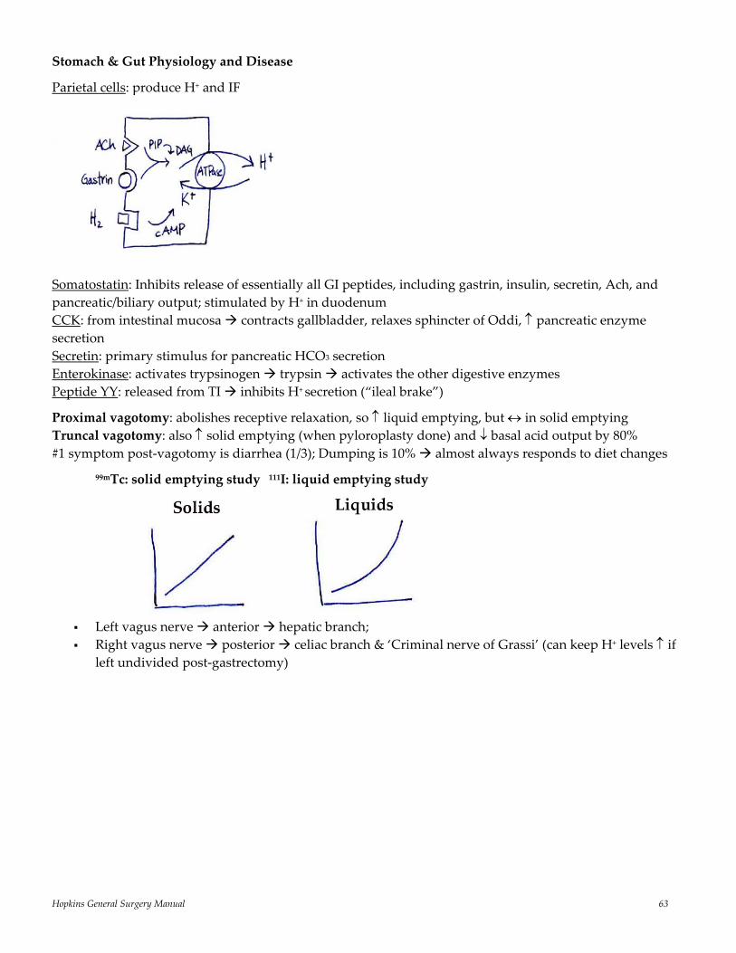
Hopkins General Surgery Manual 63
Stomach & Gut Physiology and Disease
Parietal cells: produce H+ and IF
Somatostatin: Inhibits release of essentially all GI peptides, including gastrin, insulin, secretin, Ach, and pancreatic/biliary output; stimulated by H+ in duodenum CCK: from intestinal mucosa contracts gallbladder, relaxes sphincter of Oddi, ↑ pancreatic enzyme secretion Secretin: primary stimulus for pancreatic HCO3 secretion Enterokinase: activates trypsinogen trypsin activates the other digestive enzymes Peptide YY: released from TI inhibits H+ secretion (“ileal brake”)
Proximal vagotomy: abolishes receptive relaxation, so ↑ liquid emptying, but ↔ in solid emptying Truncal vagotomy: also ↑ solid emptying (when pyloroplasty done) and ↓ basal acid output by 80% #1 symptom post‐vagotomy is diarrhea (1/3); Dumping is 10% almost always responds to diet changes
99mTc: solid emptying study 111I: liquid emptying study
Left vagus nerve anterior hepatic branch; Right vagus nerve posterior celiac branch & ‘Criminal nerve of Grassi’ (can keep H+ levels ↑ if
left undivided post‐gastrectomy)
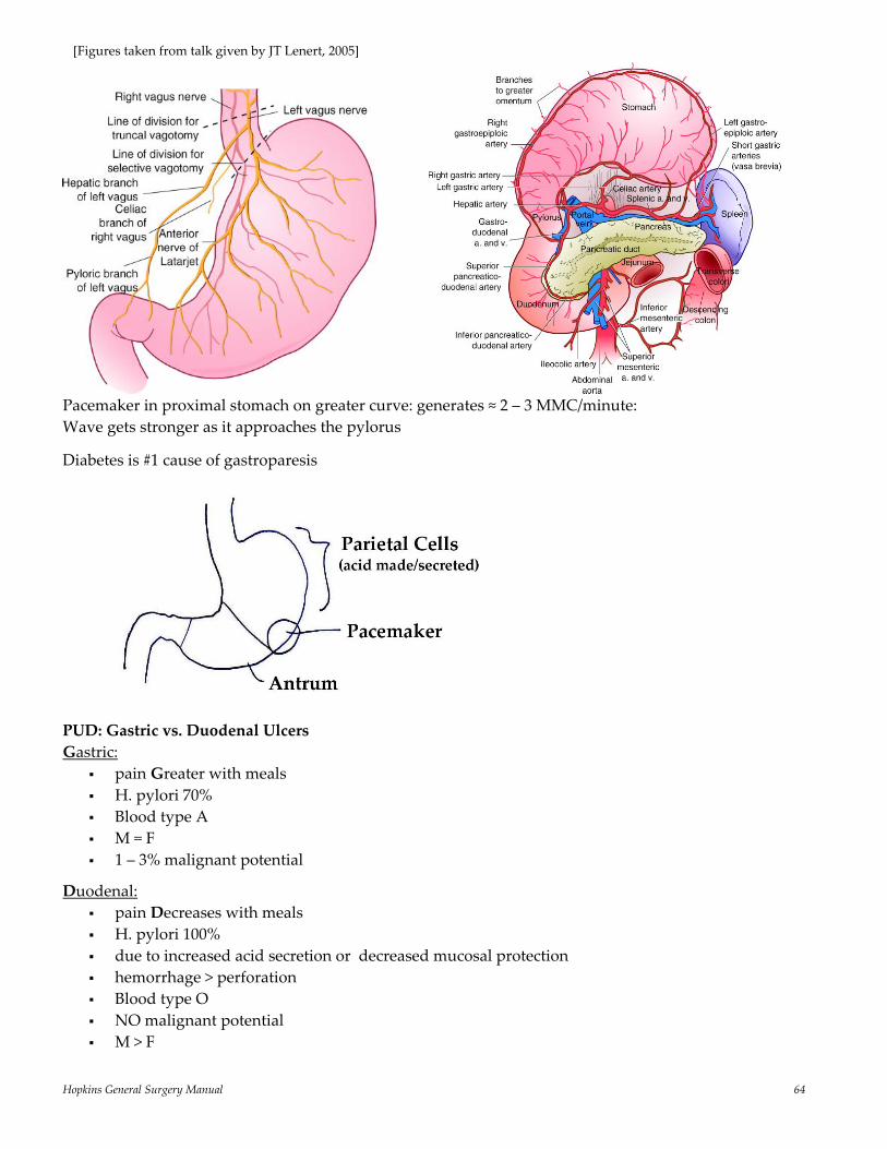
Hopkins General Surgery Manual 64
Pacemaker in proximal stomach on greater curve: generates ≈ 2 – 3 MMC/minute: Wave gets stronger as it approaches the pylorus
Diabetes is #1 cause of gastroparesis
PUD: Gastric vs. Duodenal Ulcers Gastric:
pain Greater with meals H. pylori 70% Blood type A M = F 1 – 3% malignant potential
Duodenal: pain Decreases with meals H. pylori 100% due to increased acid secretion or decreased mucosal protection hemorrhage > perforation Blood type O NO malignant potential M > F
[Figures taken from talk given by JT Lenert, 2005]
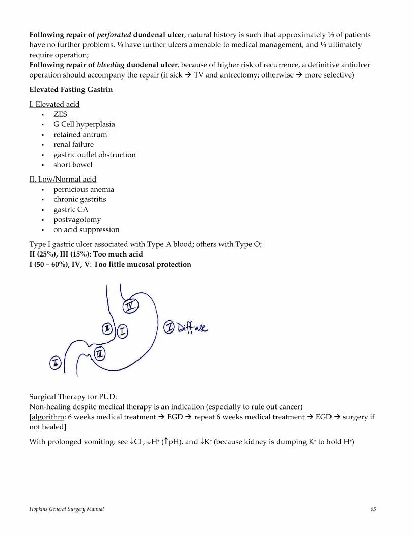
Hopkins General Surgery Manual 65
Following repair of perforated duodenal ulcer, natural history is such that approximately ⅓ of patients have no further problems, ⅓ have further ulcers amenable to medical management, and ⅓ ultimately require operation; Following repair of bleeding duodenal ulcer, because of higher risk of recurrence, a definitive antiulcer operation should accompany the repair (if sick TV and antrectomy; otherwise more selective)
Elevated Fasting Gastrin
I. Elevated acid ZES G Cell hyperplasia retained antrum renal failure gastric outlet obstruction short bowel
II. Low/Normal acid pernicious anemia chronic gastritis gastric CA postvagotomy on acid suppression
Type I gastric ulcer associated with Type A blood; others with Type O; II (25%), III (15%): Too much acid I (50 – 60%), IV, V: Too little mucosal protection
Surgical Therapy for PUD: Non‐healing despite medical therapy is an indication (especially to rule out cancer) [algorithm: 6 weeks medical treatment EGD repeat 6 weeks medical treatment EGD surgery if not healed]
With prolonged vomiting: see ↓Cl‐, ↓H+ (↑pH), and ↓K+ (because kidney is dumping K+ to hold H+)
Hopkins General Surgery Manual 66
Distribution of Upper GI Bleeding
PUD 55% Esophagogastric varices 14% Arteriovenous malformations 6% Mallory‐Weiss tears 5% Tumors/erosions 4% each Other lesions‡ 12% ‡ Includes: Dieulafoyʹs Lesion: dilated aberrant submucosal vessel (usually arterial) usually high in the gastric fundus; can lead to significant bleeding. Gastric antral vascular ectasia (GAVE): Also known as “Watermelon Stomach” is generally idiopathic, but may be associated with autoimmune disease
Gastric Volvulus: 2 types: Organoaxial (more common): rotation around the axis of line connecting cardia to pylorus Mesenterioaxial: axis is orthogonal to above
Gastric Dilation: causes hypotension, bradycardia, abdominal pain
Obesity Class I: BMI > 30 Class II: BMI > 35* surgery if develop complications of obesity; *If BMI > 35 + significant GERD Gastric Bypass is much preferred over an anti‐reflux procedure Class III: BMI > 40 surgery (Note: previous jejuno‐ileal bypasses led to renal failure because of development of CaOxalate stones)
Gastric Cancer
Gastrointestinal Stromal Tumor (GIST) Arises from interstitial cell of Cajal (intestinal pacemaker); C‐kit mutation/CD117+ Gain of function tyrosine kinase Resect if possible; Gleevac for mets; role of Gleevac in adjuvant being currently evaluated
Adenocarcinoma: Resect with 6 cm margins + draining lymph nodes + omentum; no obvious role for extended
lymphadenectomy Chronic atrophic gastritis underlies most gastric cancer; other risks: adenoma > 2 cm, Type A blood, nitrosamines, pernicious anemia
Lymphoma: distinguish between T‐cell, NHL (non‐MALT), and MALT Extranodal marginal Zone B‐Cell lymphoma (low grade B‐cell lymphoma of Mucosa‐Associated Lymphoid Tissue, MALT):
50% of patients with gastric NHL have the indolent MALT type gastric MALT is frequently associated with chronic gastritis and H.pylori infection
*the standard treatment for MALT patients (who are H.pylori +) is antibiotics and follow‐up EGD 3 and 6 months later:
if CR done if PR continue antibiotics before XRT (not surgery)
Surgery reserved for complications Note: the thicker the lesion the less likely it will regress with eradication of H.pylori alone
Hopkins General Surgery Manual 67
Small Bowel Physiology and Disease MMC: interdigestive motility; 90 minutes cycles; starts in stomach goes to TI
Phase I: quiescence Phase II: gallbladder contraction Phase III: peristalsis Phase IV: subsiding electric activity
MMC return to normal 6 – 24 hours after laparotomy; stomach and colon take longer to return to normal tone
SB transit is approximately 1 inch per minute
Jejunum absorbs more Na+ and H2O (paracellular) than ileum
80 – 100 cm (30%) of the small bowel is required for absorption, unless the IC junction is absent, in which case approximately 150 cm is required.
Derangements seen with SB resection leading to malabsorption/short‐gut include: 1. Fat 2. B12 3. Electrolytes 4. H2O
Following bowel resection Ca++/Mg++ soap form ↓cations to complex with oxalate in colon ↑oxalate absorption (worsened by Vit C consumption). Treat deficiencies with Ca++, Mg++, potassium citrate, Vit B6, and avoid Vit C
Small Bowel Neoplasms
Represent only 5% of GI neoplasms (1 – 2% of all neoplasms)
Most common benign: Most common malignant: 1. Adenoma (25 – 35%) 1. Adenocarcinoma (50%) 2. GIST 2. Carcinoid 3. Lipoma 3. Lymphoma 4. GIST
All should be resected, even if asymptomatic (need path to confirm lack of malignant behavior) also perform regional lymphadenectomy with resection
Adenocarcinomas are most common in the duodenum, the remaining malignant tumors are more common distally, with frequency proportional to length of segment (ileum > jejunum > duodenum)
Periampullary Whipple D3/D4 segmental resection + duodenojejunostomy
GIST arise from multiple mesodermal components (muscle, nervous tissue, connective tissue, vascular elements, fat)
Hopkins General Surgery Manual 68
Carcinoids arise from the Kulchitsky cell and are found in the appendix 85% of time Mets 2% if less than 1 cm; 80 – 90% if > 2 cm Most common locations for GI carcinoids: 1. Appendix, 2. Ileum, 3. Rectum 4. Stomach Small bowel carcinoids are multiple ≈ 30%; appendiceal usually solitary Urinary 5‐HIAA only elevated if able to bypass first pass (extensive liver mets, drains directly into
systemic circulation) Metabolizes tryptophan serotonin 5‐HIAA (measured in urine); carcinoids utilize 60% of
body’s tryptophan, hence side effects of tryptophan deficiency (3 D’s: dermatitis, dementia, diarrhea)
Clinical Manifestations Depends on location: i) Foregut: stomach‐pain, bleeding; bronchus‐hemoptysis, pneumonitis, wheezing ii) Midgut: appendix‐obstructive appendicitis; jejunoilium‐obstruction, intusessuption
Localization CXR, Chest CT, Barium enema, colonoscopy, superior mesenteric angiography in advanced tumors Biopsy: + argyrophil stain is suggestive, but EM of neurosecretory granules is gold standard If one found, especially in colon, 36 – 40% incidence of synchronous lesion look everywhere Metastatic disease diagnosed ONLY by mets, not histology Treatment Appendix: If > 1.5 cm, involving base of appendix, or regional lymphadenopathy present right hemicolectomy indicated Gastroduodenal: If < 1 cm endoscopic resection; >1 cm or mets subtotal gastrectomy and omentectomy Rectal: If < 1 cm endoscopic excision; 1 – 2 cm resection with negative margins (2 – 3 cm); > 2 cm low anterior resection (LAR) or abdominal perineal resection (APR) if low Any tumor with mets en bloc resection
Outcome > 2 cm portends a poorer prognosis Noninvasive appendiceal and rectal < 2 cm ≈ 100% 5‐year survival If > 2 cm ≈ 40%; with liver mets: 20 – 40%
Approximately 10% of patients with carcinoid tumor develop the “Carcinoid Syndrome‡” of flushing, sweating, diarrhea, wheezing, abdominal pain, right‐sided cardiac valvular fibrosis, and pellagra dermatosis
‡Tumor needs access to venous drainage that escapes portal circulation, such as when: 1. Hepatic mets are present 2. Venous blood from extensive retroperitoneal mets drains into paravertebral veins 3. Primary tumor is outside the GI tract (bronchial, ovarian, testicular)
Carcinoid Crisis: results from overwhelming release of serotonin (liver unable to break down) Can occur in OR during manipulation of tumor Results in hypo‐ or hypertension Hypertension should be treated with volume expansion, octreotide, and ketanserin (somostatin
analog)
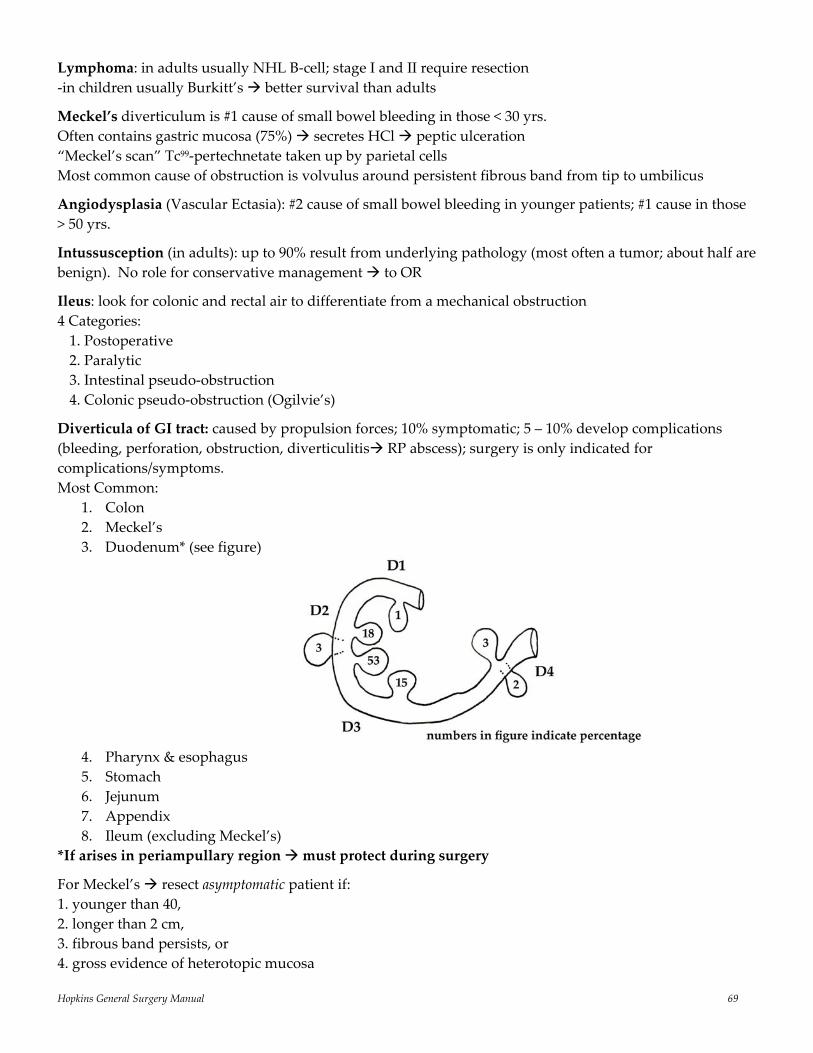
Hopkins General Surgery Manual 69
Lymphoma: in adults usually NHL B‐cell; stage I and II require resection ‐in children usually Burkitt’s better survival than adults
Meckel’s diverticulum is #1 cause of small bowel bleeding in those < 30 yrs. Often contains gastric mucosa (75%) secretes HCl peptic ulceration “Meckel’s scan” Tc99‐pertechnetate taken up by parietal cells Most common cause of obstruction is volvulus around persistent fibrous band from tip to umbilicus
Angiodysplasia (Vascular Ectasia): #2 cause of small bowel bleeding in younger patients; #1 cause in those > 50 yrs.
Intussusception (in adults): up to 90% result from underlying pathology (most often a tumor; about half are benign). No role for conservative management to OR
Ileus: look for colonic and rectal air to differentiate from a mechanical obstruction 4 Categories: 1. Postoperative 2. Paralytic 3. Intestinal pseudo‐obstruction 4. Colonic pseudo‐obstruction (Ogilvie’s)
Diverticula of GI tract: caused by propulsion forces; 10% symptomatic; 5 – 10% develop complications (bleeding, perforation, obstruction, diverticulitis RP abscess); surgery is only indicated for complications/symptoms. Most Common:
1. Colon 2. Meckel’s 3. Duodenum* (see figure)
4. Pharynx & esophagus 5. Stomach 6. Jejunum 7. Appendix 8. Ileum (excluding Meckel’s)
*If arises in periampullary region must protect during surgery
For Meckel’s resect asymptomatic patient if: 1. younger than 40, 2. longer than 2 cm, 3. fibrous band persists, or 4. gross evidence of heterotopic mucosa
Hopkins General Surgery Manual 70
Most common reasons for surgery (i.e. surgery is reserved for select cases) in patients with Crohn’s disease 1. Failure of medical management 2. Obstruction 3. Inflammatory mass or abscess 4. Fistula Note: If appendicitis suspected find Crohn’s do appendectomy provided the base of appendix not involved Small Bowel Fistula: “High output” (≥ 500 mL/24 hours) are 3 x less likely to close than low output Overall mortality: 20% (higher for jejunal; lower for ileal) In patients with Crohn’s disease + high output fistula TPN does promote fistula closure Schilling test for Vitamin B12 deficiency: reveals a type of urinary excretion of B12 similarly to that seen with pernicious anemia, except not corrected with the addition of IF, but is corrected with the use of oral tetracycline (nonabsorbable) Appendix
A component of the secretory immune system Appendicitis is initiated by obstruction of the lumen. In adults fecalith; in children lymphoid
hyperplasia Continued secretion of mucus leads to ↑ pressure (up to 126 cmH2O within 14 hours) gangrene &
perforation The area of the appendix with the poorest blood supply is mid‐portion of antimesenteric side, hence
location of most frequent gangrene and perforation Presentation of Appendicitis:
Classically, abdominal pain begins in periumbilical region (somatic pain from appendiceal distention) then localizes to site of appendix (e.g. RLQ) as visceral pain once the serosa is involved.
Anorexia precedes pain Vomiting occurs in about 75% of patients and typically follows the onset of pain Hence: anorexia pain vomiting is observed ≈ 95% of time
Acute appendicitis is the most common cause of an acute abdomen in women after the first trimester of pregnancy; they may present with RUQ pain, especially during the last trimester; however, pregnancy does not ↑ risk per se. Immediate operation is warranted. A CT scan is safe during pregnancy.
Hopkins General Surgery Manual 71
Colorectal Disease Colon:
actively secretes K+ and HCO3 absorbs Na+ against both concentration and electrical gradients to avoid hyponatremia normally absorbs 1 – 2 L of H2O/day; can absorb up to 5 – 6 L/day
Lower GI bleeding: diverticulosis + angiodysplasia = 90% of causes (for those >50)
Angiography can detect bleeding rates as low as 1 – 5 mL/min (in some series as low as 0.5 mL/min) Tag RBC can detect bleeding rates as low as 0.5 – 1 mL/min (in some series as low as 0.1 mL/min) 1st test to perform on LGIB (i.e. NG aspirate is bilious), after starting resuscitation, is rigid proctoscopy Volvulus
Cecal (rare); also known as cecal bascule
Sigmoid
presents with SBO young (25 – 35) OR (only 25% success with scope) most
do R hemi; but some attempt cecopexy
present with colonic obstruction old, debilitated patients (nursing homes) 70% success with scope tube
decompression bowel prep sigmoid colectomy during that admission
Ulcerative Colitis:
• Limited to mucosa and submucosa • 5 – 6% develop colorectal cancer; ↑ risk with disease duration, pancolitis, PSC • Proctocolectomy does not help sclerosing cholangitis or arthritis, but may help skin manifestations • 20% will require proctocolectomy
Ischemic colitis:
• A disease of small arterioles • Can occur in any segment of colon, but most common in watershed areas, which rely on
“Meandering” arteries, such as splenic flexure (Griffith’s point) and distal sigmoid colon (Sudeck’s point)
• See thumbprinting on bowel wall • Dark discoloration on colonoscopy (black/green) is indication for surgical resection
Diverticulosis
Vascular Ectasia
50% right‐sided ruptured vasa recta at neck of
diverticula arterial bleeding; severe
25 – 50% re‐bleed rate most visualize on angiography
virtually all right‐sided venous bleeding 85% re‐bleed rate only 8 – 10% extravasation on
angiography
Hopkins General Surgery Manual 72
Colorectal Cancer
120,000 – 140,000 new cases/year; 60,000 deaths/year in U.S. 3rd most common cancer in western society 2nd in deaths (to lung) 50% mortality 80% of patients present eligible for resection (i.e. 20% stage IV); 67% of these will recur; 80% of these
recurrences will be intra‐abdominal (liver #1) 5 cm gross margin for resection in colon; 1 cm distal mucosal margin (UNfixed) is adequate because
rectal cancers recur based on radial spread, rather than longitudinal spread; need 3 – 5 cm of distal mesorectal margin, if possible
Staging Tis: mucosa only N0: no nodes T1: into submucosa N1: 1 – 3 regional T2: into muscularis propria N2: > 3 regional T3: into subserosa T4: into adjacent structure (through serosa) Stage I: T1, T2, N0, M0 Survival Stage IIA/B: T3 (A), T4 (B), N0, M0 1‐year (all): 83% Stage IIIA: T1‐2, N1, M0 10‐year (all): 55% Stage IIIB: T3‐4, N1, M0 Stage IIIC: any T, N2, M0 Stage IV: any T, any N, M1 5‐year survival: I, II: 90%; III: 65%; IV: 9% Treatment
Stage I,II colon: Surgery alone (current trials looking into adjuvant chemo for II) Stage III colon: Surgery + Chemo (FL ± what other trials throw in) Stage II, III rectal: Surgery + Chemo/XRT (adjuvant)
With respect to adjuvant vs. neoadjuvant XRT, good evidence of down staging (e.g. to sphincter preserving operation) and decreased local recurrence rates, but no evidence of survival advantage, particularly in era of TME
Stage IV: 5FU/Leukovorin (FL), Oxaliplatin, CPT‐11(Irinotechan), Avastin; all combinations In 2004: IFL (Saltz regimen) + Avastin increased median survival to 20.3 months from 15.6 months (IFL alone); BUT NO INCREASE IN 5‐YEAR SURVIVAL (no regimen has impacted this in nearly 20 years) Post‐Resection Follow‐Up (Debatable)
*CEA q3 months *colonoscopy q1 – 3 years Serial CT q6 months Liver enzymes q2 – 3 months (LDH is most important) CXR q6 months *most agree on
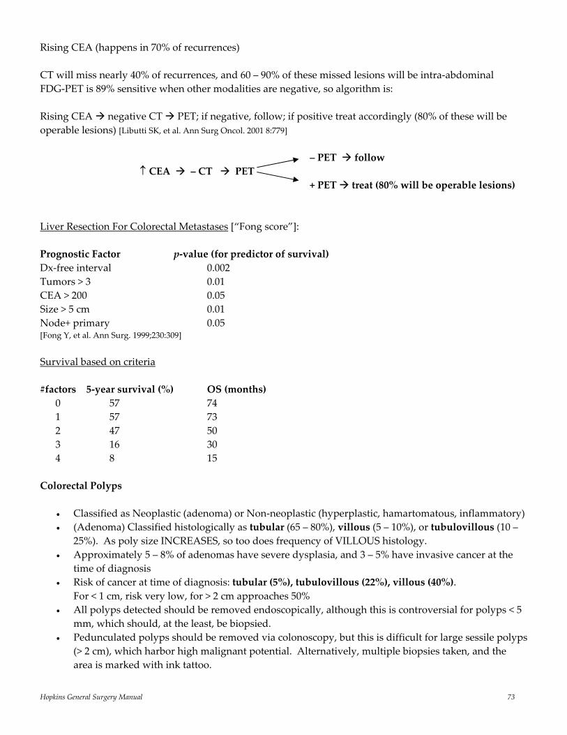
Hopkins General Surgery Manual 73
Rising CEA (happens in 70% of recurrences) CT will miss nearly 40% of recurrences, and 60 – 90% of these missed lesions will be intra‐abdominal FDG‐PET is 89% sensitive when other modalities are negative, so algorithm is: Rising CEA negative CT PET; if negative, follow; if positive treat accordingly (80% of these will be operable lesions) [Libutti SK, et al. Ann Surg Oncol. 2001 8:779] – PET follow ↑ CEA – CT PET + PET treat (80% will be operable lesions) Liver Resection For Colorectal Metastases [“Fong score”]: Prognostic Factor p‐value (for predictor of survival) Dx‐free interval 0.002 Tumors > 3 0.01 CEA > 200 0.05 Size > 5 cm 0.01 Node+ primary 0.05 [Fong Y, et al. Ann Surg. 1999;230:309] Survival based on criteria #factors 5‐year survival (%) OS (months) 0 57 74 1 57 73 2 47 50 3 16 30 4 8 15 Colorectal Polyps
• Classified as Neoplastic (adenoma) or Non‐neoplastic (hyperplastic, hamartomatous, inflammatory) • (Adenoma) Classified histologically as tubular (65 – 80%), villous (5 – 10%), or tubulovillous (10 –
25%). As poly size INCREASES, so too does frequency of VILLOUS histology. • Approximately 5 – 8% of adenomas have severe dysplasia, and 3 – 5% have invasive cancer at the
time of diagnosis • Risk of cancer at time of diagnosis: tubular (5%), tubulovillous (22%), villous (40%). For < 1 cm, risk very low, for > 2 cm approaches 50% • All polyps detected should be removed endoscopically, although this is controversial for polyps < 5
mm, which should, at the least, be biopsied. • Pedunculated polyps should be removed via colonoscopy, but this is difficult for large sessile polyps
(> 2 cm), which harbor high malignant potential. Alternatively, multiple biopsies taken, and the area is marked with ink tattoo.

Hopkins General Surgery Manual 74
• For malignant pedunculated polyps, if there is lymphovascular invasion, poor differentiation, or cancer within 2 mm of resection margin, colon resection is indicated. Sessile polyps with invasive cancer require formal colon resection.
Haggitt Level Well‐differentiated adenocarcinoma in zone 1, 2, or 3 polypectomy; zone 4 formal resection Poorly differentiated in zone 1 polypectomy; zone 2, 3, or 4 formal resection Gardner’s syndrome: (FAP) polyposis, desmoid tumors, osteomas Turcot’s syndrome: polyposis and brain tumors Peutz Jeghers syndrome: polyposis and mucocutaneous pigmentation Muir‐Torre syndrome: polyposis and skin cancer *Note: the above do not have to be colorectal cancer HNPCC (Hereditary Nonpolyposis Colorectal Cancer)
• AD inheritance • Accounts for 2 – 6% of all colorectal cancer • Average age of CRC development 40 – 45 • Begin colonoscopy at 25 • 60% by 60 years old; lifetime risk 80% • Germline mutation in mismatch repair (MMR) genes + somatic mutation in wildtype allele
produces a Micro Satellite Instability (MSI) • 2 genes account for 90% of mutations (hMSH2 and hMLH1) • Predominance of R‐sided cancer (60 – 70% in right/transverse colon) • Increased synchronous and metachronous CRC • “Lynch Syndrome I” CRC only • “Lynch Syndrome II” CRC + other cancer (endometrial, ovarian, stomach, small bowel, UGI) • Follows the adenoma‐carcinoma sequence, just does so quicker • Fare better than staged‐matched non‐HNPCC with CRC (i.e. the cancer is less aggressive)
Surgical Treatment
• Total abdominal colectomy with ileorectal anastomosis recommended for Amsterdam + patients with CRC or MMR carriers
• Prophylactic TAC + IRA as alternative for MMR carries with adenomas or patients with difficult to follow colons

Hopkins General Surgery Manual 75
Revised Amsterdam Criteria (II) 1. HNPCC‐associated caner in 3 relatives, one a first degree of the other two 2. At least 2 successive generations affected 3. At least 1 diagnosed < 50 4. FAP excluded
FAP (Familial Adenomatous Polyposis)
• AD, 100% penetrant • Mutation is in Adenomatous Polyposis Coli (APC) gene, localized to 5q21 • Normal APC protein is localized to basolateral membrane • Truncated, inactive APC appears to allow beta‐catenin accumulation in the cell and nucleus, where
it turns on genes and stimulates cell growth • 80% familial, but 10 – 30% cases are new mutations • Accounts for < 1% of colorectal cancer • Extraintestinal manifestations: desmoid tumors, osteomas, sebaceous cysts (Gardner’s); with brain
tumors (Turcot’s), CHRPE (hyperplastic retinal complication blindness) *Most common extracolonic manifestation is periampullary duodenal malignancy (also pancreatic, biliary, gastric, small intestinal, thyroid) Desmoid tumors appear in 10% of carries by age 30
Most common genetic alterations: p53: ch17; most common (≈ 85%), tumor suppressor APC gene: ch5; sporadic & familial (35 & 75%, respectively); tumor suppressor DCC: ch18; 70% cancers/10% adenoma, tumor suppressor K‐ras: ch12; 50% cancers; oncogene
Hopkins General Surgery Manual 76
Summary:
Genetic Pattern
% of Colorectal Cancer
Clinical Features
LOH (loss of heterozygosity)
1. Sporadic
2. Familial 3. Inherited (polyposis)
FAP
Gardner’s
Turcot’s
60 – 85%
35%
25%
1 – 3 %
Distal tumors (70%); no FH of polyps/CRC; aneuploid DNA; age > 60 Distal tumors; FH of polyps/CRC in several relatives; aneuploid DNA; age 50 – 60 > 100 polyps; early onset disease; mutation of APC ‐Upper GI polyps and CRC; retinal findings ‐Desmoid tumors and bone abnormalities ‐Medulloblastoma
RER (replication error pathway – DNA repair mismatch)
1. Sporadic
2. Familial
3. Inherited Lynch I
Lynch II
20 – 35%
20%
≈6%
≈10%
Proximal tumors (70%); diploid DNA; better prognosis than LOH; age > 60 Proximal tumors; diploid DNA; FH of polyps/CRC; age 50 – 60 CRC only; proximal tumors (70%); 40% with synchronous/ metachronous CRC; age 40 – 45 Lynch I + cancers of endometrium, ovaries, pancreas, stomach, small bowel, urinary tract, bile ducts
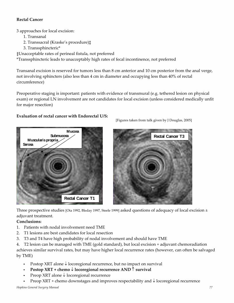
Hopkins General Surgery Manual 77
Rectal Cancer 3 approaches for local excision:
1. Transanal 2. Transsacral (Kraske’s procedure)‡ 3. Transsphincteric*
‡Unacceptable rates of perineal fistula, not preferred *Transsphincteric leads to unacceptably high rates of fecal incontinence, not preferred Transanal excision is reserved for tumors less than 8 cm anterior and 10 cm posterior from the anal verge, not involving sphincters (also less than 4 cm in diameter and occupying less than 40% of rectal circumference) Preoperative staging is important: patients with evidence of transmural (e.g. tethered lesion on physical exam) or regional LN involvement are not candidates for local excision (unless considered medically unfit for major resection) Evaluation of rectal cancer with Endorectal U/S:
Three prospective studies [Ota 1992, Bleday 1997, Steele 1999] asked questions of adequacy of local excision ± adjuvant treatment. Conclusions: 1. Patients with nodal involvement need TME 2. T1 lesions are best candidates for local resection 3. T3 and T4 have high probability of nodal involvement and should have TME 4. T2 lesion can be managed with TME (gold standard), but local excision + adjuvant chemoradiation achieves similar survival rates, but may have higher local recurrence rates (however, can often be salvaged by TME)
Postop XRT alone ↓ locoregional recurrence, but no impact on survival Postop XRT + chemo ↓ locoregional recurrence AND ↑ survival Preop XRT alone ↓ locoregional recurrence Preop XRT + chemo downstages and improves respectability and ↓ locoregional recurrence
Rectal Cancer T1
Rectal Cancer T3 Mucosa
Submucosa Muscularis propria Serosa
[Figures taken from talk given by J Douglas, 2005]
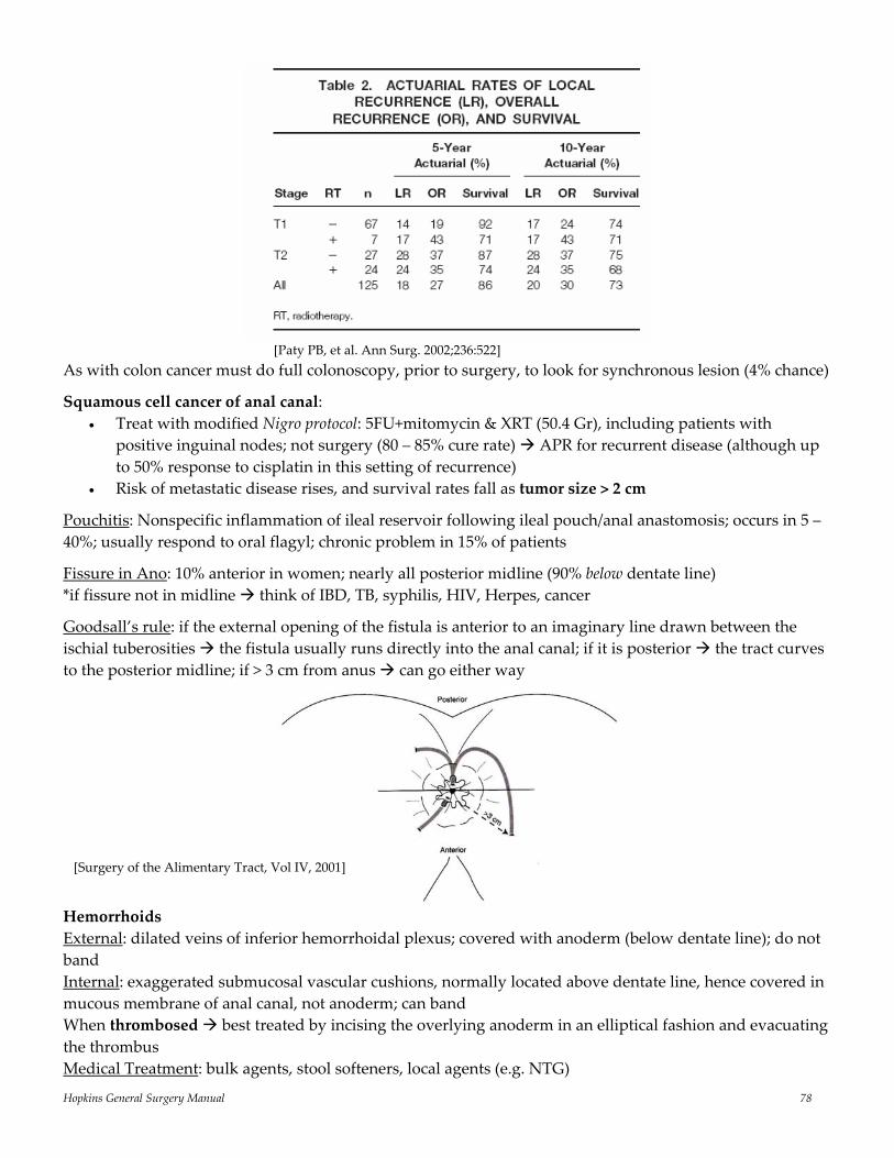
Hopkins General Surgery Manual 78
[Paty PB, et al. Ann Surg. 2002;236:522] As with colon cancer must do full colonoscopy, prior to surgery, to look for synchronous lesion (4% chance)
Squamous cell cancer of anal canal: • Treat with modified Nigro protocol: 5FU+mitomycin & XRT (50.4 Gr), including patients with
positive inguinal nodes; not surgery (80 – 85% cure rate) APR for recurrent disease (although up to 50% response to cisplatin in this setting of recurrence)
• Risk of metastatic disease rises, and survival rates fall as tumor size > 2 cm
Pouchitis: Nonspecific inflammation of ileal reservoir following ileal pouch/anal anastomosis; occurs in 5 – 40%; usually respond to oral flagyl; chronic problem in 15% of patients
Fissure in Ano: 10% anterior in women; nearly all posterior midline (90% below dentate line) *if fissure not in midline think of IBD, TB, syphilis, HIV, Herpes, cancer
Goodsall’s rule: if the external opening of the fistula is anterior to an imaginary line drawn between the ischial tuberosities the fistula usually runs directly into the anal canal; if it is posterior the tract curves to the posterior midline; if > 3 cm from anus can go either way
Hemorrhoids External: dilated veins of inferior hemorrhoidal plexus; covered with anoderm (below dentate line); do not band Internal: exaggerated submucosal vascular cushions, normally located above dentate line, hence covered in mucous membrane of anal canal, not anoderm; can band When thrombosed best treated by incising the overlying anoderm in an elliptical fashion and evacuating the thrombus Medical Treatment: bulk agents, stool softeners, local agents (e.g. NTG)
[Surgery of the Alimentary Tract, Vol IV, 2001]
Hopkins General Surgery Manual 79
Pediatric Surgery Gastroschisis Incidence: 1:10,000 to 1:15,000 (and increasing) Embryology: Mesodermal and ectodermal defects caused by ischemia resulting from premature involution of the right umbilical vein (this is supposed to happen 6 – 7 weeks post‐conception) or a vascular accident involving the right omphalomesenteric artery. Anatomy:
• Full thickness defect of abdominal wall to the right of the umbilical cord; umbilical cord has a normal insertion
• Herniation of bowel loops (uncommonly liver): organs are not covered by a membrane • Meconium stained amniotic fluid common, and may be secondary to intestinal irritation
Associated anomalies (5 – 10%): Not associated with chromosomal abnormalities. Ileal/jejunal atresia is most common associated defect; cardiac anomalies are rare
Outcomes: Mortality ranges from 7 – 25%; if liver herniates mortality increases to 50%:
Management: Vaginal delivery at term, at tertiary care facility. Caesarean may be indicated if liver herniation is present. Primary closure is obtainable in 90% of cases; silo placement and staged reduction necessary in the remaining 10% Omphalocele Incidence: 1:5000 to 1:6000 (and decreasing) Embryology: Improper migration and fusion of lateral embryonic folds. Can be cephalic, caudal, or lateral. Failure of lateral folds to fuse results in isolated omphalocele; failure of cephalic folds results in defects seen in Pentalogy of Cantrell. Anatomy:
• Herniation of the intra‐abdominal contents into the base of the umbilical cord. • Contents are covered with an amnio‐peritoneal membrane. Defect is midline. • Bowel, stomach, and liver most frequently herniated; a membrane made up of peritoneum and
amnion covers the herniated organs. • The umbilical cord inserts into the sac.
Associated Anomalies (40 – 60%): Can be seen with chromosomal abnormalities (including trisomy 18, trisomy 13). Also seen as part of Pentalogy of Cantrell and Beckwith‐Weidemann syndrome (see below). Other anomalies seen occur with the following frequencies: Cardiac defects: 50% (Overall, VSD is most common defect seen with omphalocele) GU anomalies: 40% IUGR reported in 20% of cases
Beckwith‐Weidemann: macroglossia, viceromegaly, hypoglycemia, macrosomia Pentalogy of Cantrell: 1. Cardiac defects, 2. Diaphragmatic defects (2 specific), 3. Sternal defect, 4. Abdominal wall (midline, supraumbilical) defect, and 5. Ectopic cordis
Outcome: overall mortality 40 – 80% (varies depending on presence of associated anomalies; cardiac abnormalities determine mortality to a large extent)
Management: Cardiac echo and karyotype indicated, as well as search for other anomalies. Consideration of anomalies has priority unless sac has ruptured.
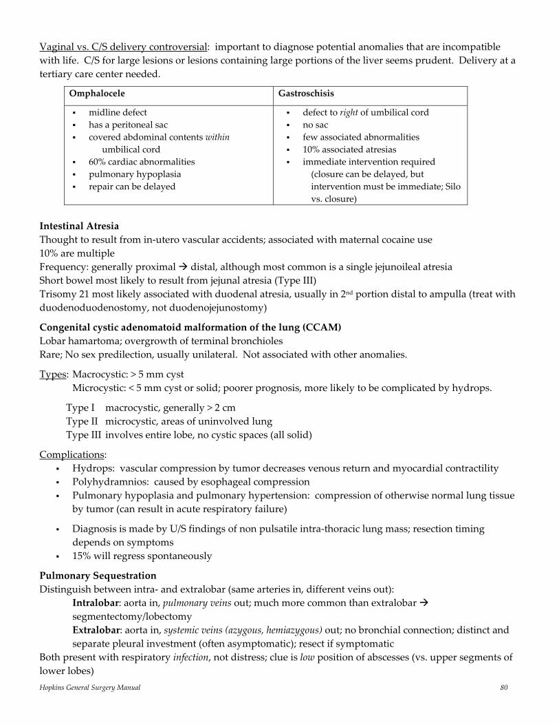
Hopkins General Surgery Manual 80
Vaginal vs. C/S delivery controversial: important to diagnose potential anomalies that are incompatible with life. C/S for large lesions or lesions containing large portions of the liver seems prudent. Delivery at a tertiary care center needed.
Omphalocele
Gastroschisis
midline defect has a peritoneal sac covered abdominal contents within
umbilical cord 60% cardiac abnormalities pulmonary hypoplasia repair can be delayed
defect to right of umbilical cord no sac few associated abnormalities 10% associated atresias immediate intervention required
(closure can be delayed, but intervention must be immediate; Silo vs. closure)
Intestinal Atresia Thought to result from in‐utero vascular accidents; associated with maternal cocaine use 10% are multiple Frequency: generally proximal distal, although most common is a single jejunoileal atresia Short bowel most likely to result from jejunal atresia (Type III) Trisomy 21 most likely associated with duodenal atresia, usually in 2nd portion distal to ampulla (treat with duodenoduodenostomy, not duodenojejunostomy)
Congenital cystic adenomatoid malformation of the lung (CCAM) Lobar hamartoma; overgrowth of terminal bronchioles Rare; No sex predilection, usually unilateral. Not associated with other anomalies.
Types: Macrocystic: > 5 mm cyst Microcystic: < 5 mm cyst or solid; poorer prognosis, more likely to be complicated by hydrops.
Type I macrocystic, generally > 2 cm Type II microcystic, areas of uninvolved lung Type III involves entire lobe, no cystic spaces (all solid)
Complications: Hydrops: vascular compression by tumor decreases venous return and myocardial contractility Polyhydramnios: caused by esophageal compression Pulmonary hypoplasia and pulmonary hypertension: compression of otherwise normal lung tissue
by tumor (can result in acute respiratory failure)
Diagnosis is made by U/S findings of non pulsatile intra‐thoracic lung mass; resection timing depends on symptoms
15% will regress spontaneously
Pulmonary Sequestration Distinguish between intra‐ and extralobar (same arteries in, different veins out):
Intralobar: aorta in, pulmonary veins out; much more common than extralobar segmentectomy/lobectomy Extralobar: aorta in, systemic veins (azygous, hemiazygous) out; no bronchial connection; distinct and separate pleural investment (often asymptomatic); resect if symptomatic
Both present with respiratory infection, not distress; clue is low position of abscesses (vs. upper segments of lower lobes)
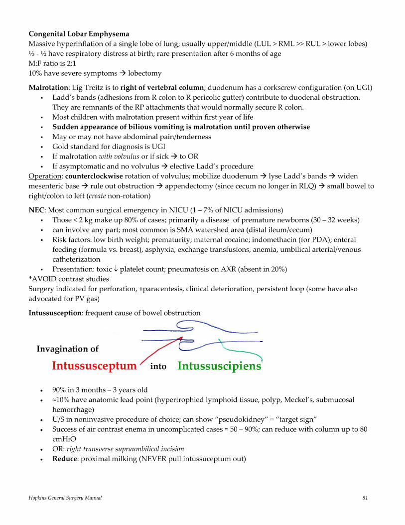
Hopkins General Surgery Manual 81
Congenital Lobar Emphysema Massive hyperinflation of a single lobe of lung; usually upper/middle (LUL > RML >> RUL > lower lobes) ⅓ ‐ ½ have respiratory distress at birth; rare presentation after 6 months of age M:F ratio is 2:1 10% have severe symptoms lobectomy
Malrotation: Lig Treitz is to right of vertebral column; duodenum has a corkscrew configuration (on UGI) Ladd’s bands (adhesions from R colon to R pericolic gutter) contribute to duodenal obstruction.
They are remnants of the RP attachments that would normally secure R colon. Most children with malrotation present within first year of life Sudden appearance of bilious vomiting is malrotation until proven otherwise May or may not have abdominal pain/tenderness Gold standard for diagnosis is UGI If malrotation with volvulus or if sick to OR If asymptomatic and no volvulus elective Ladd’s procedure
Operation: counterclockwise rotation of volvulus; mobilize duodenum lyse Ladd’s bands widen mesenteric base rule out obstruction appendectomy (since cecum no longer in RLQ) small bowel to right/colon to left (create non‐rotation)
NEC: Most common surgical emergency in NICU (1 – 7% of NICU admissions) Those < 2 kg make up 80% of cases; primarily a disease of premature newborns (30 – 32 weeks) can involve any part; most common is SMA watershed area (distal ileum/cecum) Risk factors: low birth weight; prematurity; maternal cocaine; indomethacin (for PDA); enteral
feeding (formula vs. breast), asphyxia, exchange transfusions, anemia, umbilical arterial/venous catheterization
Presentation: toxic ↓ platelet count; pneumatosis on AXR (absent in 20%) *AVOID contrast studies Surgery indicated for perforation, +paracentesis, clinical deterioration, persistent loop (some have also advocated for PV gas)
Intussusception: frequent cause of bowel obstruction
• 90% in 3 months – 3 years old • ≈10% have anatomic lead point (hypertrophied lymphoid tissue, polyp, Meckel’s, submucosal
hemorrhage) • U/S in noninvasive procedure of choice; can show “pseudokidney” = “target sign” • Success of air contrast enema in uncomplicated cases ≈ 50 – 90%; can reduce with column up to 80
cmH2O • OR: right transverse supraumbilical incision • Reduce: proximal milking (NEVER pull intussuceptum out)
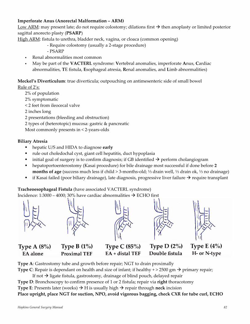
Hopkins General Surgery Manual 82
Imperforate Anus (Anorectal Malformation – ARM) Low ARM: may present late; do not require colostomy; dilations first then anoplasty or limited posterior sagittal anorecto plasty (PSARP) High ARM: fistula to urethra, bladder neck, vagina, or cloaca (common opening) ‐ Require colostomy (usually a 2‐stage procedure) ‐ PSARP
Renal abnormalities most common May be part of the VACTERL syndrome: Vertebral anomalies, imperforate Anus, Cardiac
abnormalities, TE fistula, Esophageal atresia, Renal anomalies, and Limb abnormalities) Meckel’s Diverticulum: true diverticula; outpouching on antimesenteric side of small bowel Rule of 2’s:
2% of population 2% symptomatic < 2 feet from ileocecal valve 2 inches long 2 presentations (bleeding and obstruction) 2 types of (heterotopic) mucosa: gastric & pancreatic Most commonly presents in < 2‐years‐olds
Biliary Atresia
hepatic U/S and HIDA to diagnose early rule out choledochal cyst, giant cell hepatitis, duct hypoplasia initial goal of surgery is to confirm diagnosis; if GB identified perform cholangiogram hepatoportoenterostomy (Kasai procedure) for bile drainage most successful if done before 2 months of age (success much less if child > 3‐months‐old; ⅓ drain well, ⅓ drain ok, ⅓ no drainage)
if Kasai failed (poor biliary drainage), late diagnosis, progressive liver failure require transplant Tracheoesophageal Fistula (have associated VACTERL syndrome) Incidence: 1:3000 – 4000; 30% have cardiac abnormalities ECHO first
Type A: Gastrostomy tube and growth before repair; NGT to drain proximally Type C: Repair is dependant on health and size of infant; if healthy + > 2500 gm primary repair; If not ligate fistula, gastrostomy, drainage of blind pouch, delayed repair Type D: Bronchoscopy to confirm presence of 1 or 2 fistula; repair via right thoracotomy Type E: Presents later (weeks) H is usually high repair through neck incision Place upright, place NGT for suction, NPO, avoid vigorous bagging, check CXR for tube curl, ECHO
Hopkins General Surgery Manual 83
Pyloric Stenosis Present in 3/1000 live births Most common cause of surgically correctable vomiting in newborn Presents with non‐bilious, often projectile, vomiting, usually by 3 – 6 weeks of age; 4:1 M:F Physical exam reveals “olive” about 90% of the time Exam is sufficient to diagnose; UGI if no palpable olive Treatment is Ramstedt pyloromyotomy, but not emergently (fix electrolytes first, fluid balance) Associated malformations: malrotation, hepatic glucuronyl transferase ability (jaundice)
Congenital Diaphragmatic Hernia Bochdaleck (posterolateral‐usually left) and Morgagni (anteriomedial) present shortly after birth (may have few normal hours) CXR shows loops of bowel and/or stomach in chest Resuscitation/stabilization is priority over timing of surgery (including ECMO) as in adults, repair is via abdomen
Hirschsprung’s Disease More common in males (70 – 80%) Lack of ganglion cells from rectum to stomach (although rare proximal to colon) Rectosigmoid is most common location Diagnose with BE (look for sigmoid/rectum ratio > 1), suction rectal biopsy (definitive) Treat with resection and pull‐through (1‐stage vs. 2‐stage); 1‐stage associated with ↓ anastomotic
disruption
Management of undescended testicle: may occur spontaneously during first year of life; if not orchiopexy by 1 year of age (↑risk of infertility, but not risk of cancer: risk 1/4000, i.e. 40‐fold increase) Umbilical hernia: no urgent need for repair, unless very large; if not spontaneously closed when school age elective repair
Inguinal hernia: repair is most common general surgical procedure performed on infants; M > F (3:1), R>L; all should be repaired at the time of diagnosis; bilateral repair is performed by some up to age 5 Pediatric malignancy
#1 overall is leukemia, #2 is CNS (=#1 solid tumor), #3 is neuroblastoma #1 solid organ tumor (excluding intracranial) is neuroblastoma; 90% have ↑ VMA; ↑ HVA worse
prognosis; cells derived from neural crest and may arise anywhere along sympathetic ganglia (adrenal medulla is most common location); 30% cure; associated with N‐myc
Location: 75% adrenal, 20% posterior mediastinum, 4% organ of Zuckerkandel, 1% cervical Favorable prognosis: age < 1 year; stage 1, 2, or 4S, low tumor markers, normal N‐myc, DNA index >
1.0 Wilm’s tumor ≡ nephroblastoma (most common in children > 2‐years‐old) 80% cured with
nephrectomy; chemo regimen is vincristine, D‐actinomycin and doxorubicin (≥ stage III) Hepatoblastoma is most common liver tumor in children; ↑ AFP often; if ↑ β‐HCG can result in
precocious puberty; surgical resection is treatment
Hopkins General Surgery Manual 84
Spleen and Splenectomy Functions:
Filter abnormal RBCs, store platelets, produce Tuftsin and Properdin (opsins), produce Ab (esp. IgM), site of phagocytosis (Does not store RBCs)
White pulp: lymphatic Red pulp: phagocytic
Blood Supply: splenic artery and short gastric veins (gastroepiploic) Drainage: splenic vein and short gastric veins (gastroepiploic) 15 – 20% of people have accessory spleens Spleen is approximately 1% of total body weight receives 5 – 10% of cardiac output
Most common cause of splenic vein thrombosis: Pancreatitis Patients with Ulcerative Colitis develop hyposplenism Definition of Hypersplenism: Hyperfunctioning spleen, loss of blood elements, large spleen (splenomegaly), hyperactive bone marrow trying to keep up with loss of blood elements; splenic cellular sequestration Primary: A diagnosis of exclusion will respond to splenectomy Secondary: e.g. Result of hepatic disease no splenectomy
⅓ of total body platelets are stored in spleen
“Delayed Splenic Rupture”: A subcapsular hematoma may rupture at a later time after blunt trauma up to 2 weeks later. Present with shock ± abdominal pain Signs/Sx of Rupture: Hemoperitoneum and Kehr’s Sign (referred pain to tip of left shoulder), LUQ pain and mass (Ballance’s Sign)
Diagnosis: Abdominal CT if stable; U/S ex lap if unstable Treatment: Nonoperative if: stable, isolated injury without hilar involvement or complete rupture If unstable: splenectomy or splenorrhaphy (salvage operation with wrapping vicral mesh and topical hemostat agents/ partial splenectomy)
Hopkins General Surgery Manual 85
Indications for splenectomy: Hyperslenism, Gaucher’s disease, splenic vein thrombosis, sickle cell disease, thrombocytopenia via drug abuse, sphereocytosis, lymphoma (esp. Hodgkin’s), ITP, TTP, splenic tumors/trauma, Felty’s syndrome, lymphroproliferative disorders (NHL, CLL), Hairy Cell leukemia, Thal major, not G‐6‐PD Deficiency
ITP: (Immune) Autoimmune (usually anti‐platelet Ab) platelet destruction leading to bleeding and
purpura Spontaneous remission occurs in most children; only 25% of adults Splenomegaly is rare #1 cause of failed procedure is missed accessory spleen
Erythrocytes Membrane Abnormalities Hereditary spherocytosis: abnormality of spectrin ↑ osmotic fragility splenectomy is
only effective therapy. Most common indication for splenectomy in US (non‐trauma)
Red Cell Enzyme Defects G‐6‐PD deficiency: most common enzymatic abnormality of RBC; most patients require no
treatment, but some variants improve with splenectomy Autoimmune Hemolytic Anemia (AIHA): If secondary to IgG antibodies (“warm” antibody type) may respond to splenectomy; But if IgM mediated (cold agglutinin disease) liver is sight of RBC sequestration no splenectomy Treatment of choice for TTP (Thrombotic): Plasmapheresis, steroids (splenectomy as last resort); TTP is a disease characterized by occlusion of arterioles and capillaries by hyaline deposits composed of aggregated platelets and fibrin Lab tests following splenectomy: 50% increase in WBC, marked thrombocytosis, Howell‐Jolly bodies in peripheral smear (failure to see HJ bodies following splenectomy missed accessory spleen) Possible complications: Thrombocytosis (treat with ASA if platelets > 1 million), subphrenic abscess, gastric dilation, Overwhelming PostSplenectomy Sepsis (OPSS) OPSS:
• < 1% in adults, more common in children (both incidence and mortality) • More common following splenectomy for hematologic disease (vs. trauma) • Strep pneumo, Meningococcus, HIB, E.coli • Vaccinate preop if possible (Pneumcoccus, Meningococcus, HIB) • Aggressive treatment with PCN for all minor infections
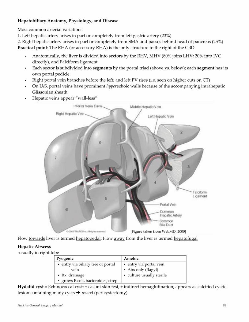
Hopkins General Surgery Manual 86
Hepatobiliary Anatomy, Physiology, and Disease
Most common arterial variations: 1. Left hepatic artery arises in part or completely from left gastric artery (23%) 2. Right hepatic artery arises in part or completely from SMA and passes behind head of pancreas (25%) Practical point: The RHA (or accessory RHA) is the only structure to the right of the CBD
Anatomically, the liver is divided into sectors by the RHV, MHV (80% joins LHV; 20% into IVC directly), and Falciform ligament
Each sector is subdivided into segments by the portal triad (above vs. below); each segment has its own portal pedicle
Right portal vein branches before the left; and left PV rises (i.e. seen on higher cuts on CT) On U/S, portal veins have prominent hyperechoic walls because of the accompanying intrahepatic
Glissonian sheath Hepatic veins appear “wall‐less”
Flow towards liver is termed hepatopedal; Flow away from the liver is termed hepatofugal
Hepatic Abscess ‐usually in right lobe
Pyogenic Amebic entry via biliary tree or portal
vein Rx: drainage grows E.coli, bacteroides, strep
entry via portal vein Abx only (flagyl) culture usually sterile
Hydatid cyst ≡ Echinococcal cyst: + casoni skin test, + indirect hemaglutination; appears as calcified cystic lesion containing many cysts resect (pericystectomy)
[Figure taken from WebMD, 2000]
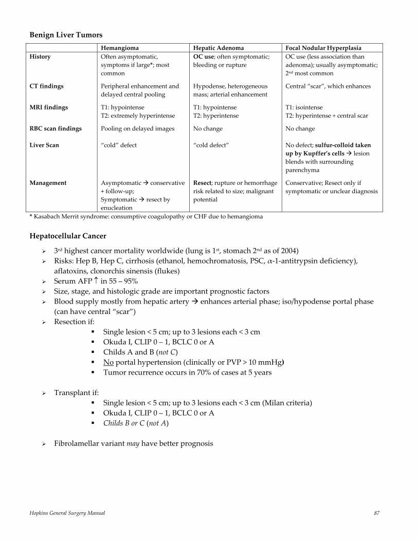
Hopkins General Surgery Manual 87
Benign Liver Tumors
Hemangioma Hepatic Adenoma Focal Nodular Hyperplasia History
CT findings
MRI findings
RBC scan findings Liver Scan
Management
Often asymptomatic, symptoms if large*; most common
Peripheral enhancement and delayed central pooling
T1: hypointense T2: extremely hyperintense
Pooling on delayed images “cold” defect
Asymptomatic conservative + follow‐up; Symptomatic resect by enucleation
OC use; often symptomatic; bleeding or rupture
Hypodense, heterogeneous mass; arterial enhancement
T1: hypointense T2: hyperintense
No change “cold defect”
Resect; rupture or hemorrhage risk related to size; malignant potential
OC use (less association than adenoma); usually asymptomatic; 2nd most common
Central “scar”, which enhances
T1: isointense T2: hyperintense + central scar
No change No defect; sulfur‐colloid taken up by Kupffer’s cells lesion blends with surrounding parenchyma
Conservative; Resect only if symptomatic or unclear diagnosis
* Kasabach Merrit syndrome: consumptive coagulopathy or CHF due to hemangioma Hepatocellular Cancer
3rd highest cancer mortality worldwide (lung is 1st, stomach 2nd as of 2004) Risks: Hep B, Hep C, cirrhosis (ethanol, hemochromatosis, PSC, α‐1‐antitrypsin deficiency), aflatoxins, clonorchis sinensis (flukes)
Serum AFP ↑ in 55 – 95% Size, stage, and histologic grade are important prognostic factors Blood supply mostly from hepatic artery enhances arterial phase; iso/hypodense portal phase (can have central “scar”)
Resection if: Single lesion < 5 cm; up to 3 lesions each < 3 cm Okuda I, CLIP 0 – 1, BCLC 0 or A Childs A and B (not C) No portal hypertension (clinically or PVP > 10 mmHg) Tumor recurrence occurs in 70% of cases at 5 years
Transplant if: Single lesion < 5 cm; up to 3 lesions each < 3 cm (Milan criteria) Okuda I, CLIP 0 – 1, BCLC 0 or A Childs B or C (not A)
Fibrolamellar variant may have better prognosis
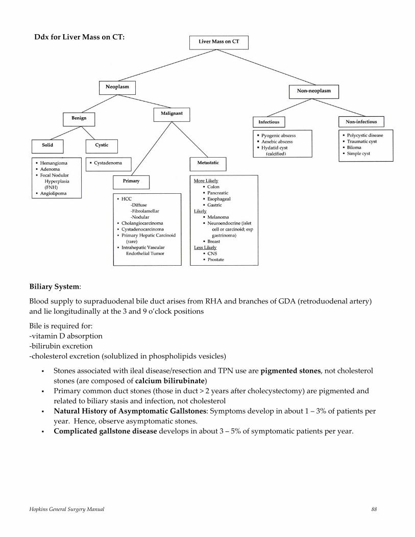
Hopkins General Surgery Manual 88
Biliary System:
Blood supply to supraduodenal bile duct arises from RHA and branches of GDA (retroduodenal artery) and lie longitudinally at the 3 and 9 o’clock positions
Bile is required for: ‐vitamin D absorption ‐bilirubin excretion ‐cholesterol excretion (solublized in phospholipids vesicles)
Stones associated with ileal disease/resection and TPN use are pigmented stones, not cholesterol stones (are composed of calcium bilirubinate)
Primary common duct stones (those in duct > 2 years after cholecystectomy) are pigmented and related to biliary stasis and infection, not cholesterol
Natural History of Asymptomatic Gallstones: Symptoms develop in about 1 – 3% of patients per year. Hence, observe asymptomatic stones.
Complicated gallstone disease develops in about 3 – 5% of symptomatic patients per year.
Ddx for Liver Mass on CT:
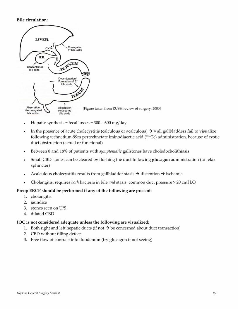
Hopkins General Surgery Manual 89
Bile circulation:
• Hepatic synthesis = fecal losses = 300 – 600 mg/day
• In the presence of acute cholecystitis (calculous or acalculous) ≈ all gallbladders fail to visualize following technetium‐99m pertechnetate iminodiacetic acid (99mTc) administration, because of cystic duct obstruction (actual or functional)
• Between 8 and 18% of patients with symptomatic gallstones have choledocholithiasis
• Small CBD stones can be cleared by flushing the duct following glucagon administration (to relax sphincter)
• Acalculous cholecystitis results from gallbladder stasis distention ischemia
• Cholangitis: requires both bacteria in bile and stasis; common duct pressure > 20 cmH2O
Preop ERCP should be performed if any of the following are present: 1. cholangitis 2. jaundice 3. stones seen on U/S 4. dilated CBD
IOC is not considered adequate unless the following are visualized: 1. Both right and left hepatic ducts (if not be concerned about duct transaction) 2. CBD without filling defect 3. Free flow of contrast into duodenum (try glucagon if not seeing)
[Figure taken from RUSH review of surgery, 2000]
Hopkins General Surgery Manual 90
Gallbladder
Concentrates bile by active absorption of Na+, Cl‐ (H2O follows); cholecystectomy works by eliminating reservoir forces a more continuous source of bile and eliminates chance for “sludge” and stone formation.
70% of patients with EF < 30% (normal is > 35%) on CCK‐HIDA benefit from cholecystectomy, although this may still be controversial
10 – 20% of patients with symptomatic gallstones have choledocholithiasis By definitions: stones in CBD > 2 years after cholecystectomy are primary CBD stones (pigmented,
related to biliary stasis and infection), rather than cholesterol stones; need sphincterotomy and extraction
Porcelain gallbladder has 30 – 65% risk of cancer cholecystectomy indicated Gallbladder adenocarcinoma: 90% have stones. Cholecystectomy adequate if confined to mucosa; if
grossly visible tumor regional lymphadenectomy, wedge segment V, skeletonize portal triad Gallbladder polyps can be malignant; risk is related to size; hence, should remove GB for polyp is
symptomatic or > 10 mm; sessile more likely to be malignant; pedunculated more likely benign Diagnosis of Cholecystitis
Three most sensitive signs of cholecystitis: 1. Sonographic Murphy’s sign 2. Wall thickening > 4 mm 3. Pericholecystic fluid
Postop lap chole patient not doing well, think: Viscous injury (e.g. duodenum) Duct injury Bile leak Retained CBD stone Cystic duct stump leak
Management of gallstone ileus: 1. Remove stone (via enterotomy proximal at site of obstruction) 2. Run entire bowel 3. In acute setting, especially elderly, reserve cholecystectomy for later (risk of recurrence ≈ 5 – 10%) & repair biliary‐enteric fistula
Rates of Positive Bile Cultures
Bile cultures are positive in approximately: 1. 5 – 40% of chronic cholecystitis 2. 30 – 70% acute cholecystitis 3. 60 – 80% of choledocholithiasis 4. 25 – 30% of malignant obstruction 5. 100% of bile duct strictures
Acalculus cholecystitis (pathophysiology involves ischemia) most common in: cocaine use and HIV Sclerosing cholangitis: ERCP to diagnose; multiple strictures/dilations (“beaded” appearance)

Hopkins General Surgery Manual 91
Bismuth classification system of bile duct injury:
Note: Type 5 injury involves a separate inserting sectoral duct with or without injury of the common duct Choledochal Cysts – Todani Class
*20‐fold increase in bile duct malignancy if left untreated; F:M ≈ 4:1
I Solitary fusiform extrahepatic cyst (82%) II Extrahepatic supraduodenal diverticulum “second gallbladder” (3%) III Intraduodenal diverticulum, (choledochocele) (5%) IVA Fusiform extrahepatic & intrahepatic cysts IVB Multiple extrahepatic cysts (A+B = 9%) V Multiple intrahepatic cysts, (Caroli’s Disease) (<1%)
*Current theory is that cysts occur because of bile reflux, which results from an abnormal junction of the biliary and pancreatic ducts; in other words, the congenital aspect of the disease is the ductal abnormality the cysts are a consequence of this
Management:
I Complete cyst excision with hepaticojejunostomy II Excision of cyst with primary choledochorrhaphy III Large sphincteroplasty IV & V Selective management with hepatic resection if localized bile duct cyst, extrahepatic cyst resection, hepaticojejunostomy with large stents, advanced biliary cirrhosis may require hepatic transplantation
[Figure taken from www.uptodate.com]
Hopkins General Surgery Manual 92
Transport of Bilirubin
Bilirubin is synthesized in the RES from biliverdin. Insoluble unconjugated bilirubin, reversibly bound to albumin, is transported to the liver, and into cytoplasm of hepatocytes. The enzyme uridine diphosphate‐glucuronyl transferase conjugates the bili with either one or two molecules of glucuronic acid to form water‐soluble bilirubin mono‐ and diglucuronide. This is secreted into the bile canaliculus.
In the terminal ileum & colon, bilirubin is converted to urobilinogen, 10 – 20% of which is reabsorbed back into portal circulation
Bile is 80% bile salts, 15% lecithin, 5% cholesterol. Gallstones can be classified as cholesterol stones, black stones, and brown stones:
Cholesterol stones form when bile becomes supersaturated with cholesterol Black stones form when bile becomes supersaturated with calcium salts (primarily calcium
bilirubinate) Brown stones from when bile acquires stasis‐induced bacterial contamination
GB concentrates bile by active reabsorption of Na+, Cl‐; H2O then follows Bile pool of 5 g is recirculated q4 hours lose 0.5 g (10%) daily Assessment of Jaundice
Ask why? Excess bili production? Deficient hepatocyte uptake? Deficient conjugation? Deficient hepatocyte secretion? Deficient bili secretion?
Grouped as prehepatic, hepatic, and posthepatic causes
Check fractionated bili levels
1. Predominance of unconjugated (indirect) suggests prehepatic etiology (hemolysis) or hepatic deficiencies of uptake or conjugation
2. Predominance of conjugated (direct) suggests defects in hepatocyte secretion into bile ducts or bile duct secretion into GI tract
3. Combined elevation suggests complex problem, usually acquired liver damage
Hopkins General Surgery Manual 93
Laboratory Investigation of Hyperbilirubinemia
Jaundice may result from overproduction of bilirubin (hemolysis), impaired conjugation (Gilbert’s syndrome), impaired intracellular metabolism or excretion (drug effect), hepatocyte injury (hepatitis), bile duct injury (primary biliary and sclerosing cholangitis), and large duct obstruction (stone, tumor, sclerosing cholangitis)
First investigate by fractionation:
Unconjugated hyperbilirubinemia (hemolysis, Gilbert’s) is usually defined as levels greater than 80% of total bilirubin, which should RARELY exceed 5 mg/dL
Conjugated hyperbilirubinemia (hepatocellular or bile duct disease) exists when the conjugated fraction exceeds 50% of the total level
∆‐bilirubin is an irreversibly (covalently) albumin‐bound form of bilirubin found in the setting of long‐standing conjugated hyperbilirubinemia; not filtered through kidneys; T1/2 = 18 days reason for slow decline of TB following long‐standing hyperbilirubinemia, especially in patients with renal failure
In general, a bilirubin of 2 – 5 mg/dl, of which most is unconjugated, in an otherwise healthy adult is Gilbert’s (2 – 5% of population), hemolysis, or both. The hemolytic component will be more likely LDH and AST (both in RBCs) are also mildly elevated
Alkaline phosphatase comes from liver and bone. Measurement of GGT (gamma‐glutamyl transpeptidase) is often used to indicate the source of the AP. Normal GGT suggests non‐hepatic source, such as osteoblastic bone lesions, or certain other tumors. Spurious elevations in AP may also be seen after albumin infusion if the albumin is derived from placental blood, which is rich in AP Sphincter of Oddi
Regulates flow of bile into duodenum Composed of 4 sphincters containing both circular and longitudinal smooth muscle Length is about 4 – 6 mm Basal (resting) pressure averages 13 mmHg (5 – 15) Undergoes phasic contractions with a frequency of 4/min; each with duration of 8 seconds Pressure increases to 130 ± 15 mmHg (50 – 150) Relaxation occurs with CCK stimulation and parasympathetic stimulation; glucagons (try IV
glucagons to pass small stones) MSO4, sympathetic stimulation increases sphincter tone
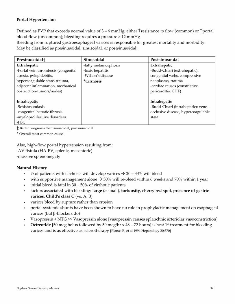
Hopkins General Surgery Manual 94
Portal Hypertension Defined as PVP that exceeds normal value of 3 – 6 mmHg; either ↑resistance to flow (common) or ↑portal blood flow (uncommon); bleeding requires a pressure > 12 mmHg Bleeding from ruptured gastroesophageal varices is responsible for greatest mortality and morbidity May be classified as presinusoidal, sinusoidal, or postsinusoidal: Presinusoidal‡ Sinusoidal Postsinusoidal Extrahepatic ‐Portal vein thrombosis (congenital atresia, pylephlebitis, hypercoagulable state, trauma, adjacent inflammation, mechanical obstruction‐tumors/nodes) Intrahepatic ‐Schistosomiasis ‐congenital hepatic fibrosis ‐myeloprolifertiive disorders ‐PBC
‐fatty metamorphosis ‐toxic hepatitis ‐Wilson’s disease *Cirrhosis
Extrahepatic ‐Budd‐Chiari (extrahepatic): congenital webs, compressive neoplasms, trauma ‐cardiac causes (constrictive pericarditis, CHF) Intrahepatic ‐Budd‐Chiari (intrahepatic): veno‐occlusive disease, hypercoagulable state
‡ Better prognosis than sinusoidal, postsinusoidal * Overall most common cause Also, high‐flow portal hypertension resulting from: ‐AV fistula (HA‐PV, splenic, mesenteric) ‐massive splenomegaly Natural History
½ of patients with cirrhosis will develop varices 20 – 33% will bleed with supportive management alone 30% will re‐bleed within 6 weeks and 70% within 1 year initial bleed is fatal in 30 – 50% of cirrhotic patients factors associated with bleeding: large (> small), tortuosity, cherry red spot, presence of gastric varices, Child’s class C (vs. A, B)
varices bleed by rupture rather than erosion portal‐systemic shunts have been shown to have no role in prophylactic management on esophageal
varices (but β‐blockers do) Vasopressin + NTG >> Vasopressin alone [vasopressin causes splanchnic arteriolar vasoconstriction] Octreotide [50 mcg bolus followed by 50 mcg/hr x 48 – 72 hours] is best 1st treatment for bleeding
varices and is as effective as sclerotherapy [Planas R, et al 1994 Hepatology 20:370]
Hopkins General Surgery Manual 95
Massive Upper GI Bleed: All comers: 40% PUD, 18% gastritis, 13% esophageal varices, 9% Mallory‐Weiss tear, 9% other Known cirrhotic: 53% esophageal varices, 22% gastritis, 20% PUD 4 Main complications of cirrhosis
1. portal hypertension 2. ascites 3. hepatic encephalopathy 4. malignancy (primary hepatic)
SAAG (Serum‐Ascites Albumin Gradient)
If > 1.1 gm/dL indicates portal hypertension. Should be responsive to medical management consisting of sodium restriction (2000 mg/day) + oral diuretics (spironolactone + lasix) Fluid restrict when Na+ falls below 120 mmol/L TIPS Indications Acute variceal hemorrhage is the most common indication for TIPS, followed by refractory ascites and
hepatic hydrothorax. TIPS controls bleeding in 75% to 100% of patients; the efficacy is similar for both esophageal and gastric
varices, and rebleeding does not usually recur unless there is shunt dysfunction. TIPS, however, may not eliminate isolated gastric varices in up to 50% of patients. When rebleeding occurs in spite of an open shunt, angiographic obliteration of the varices may arrest bleeding. Despite good results for control of bleeding, short‐term mortality remains high in patients who have TIPS for variceal bleeding. As a result, better criteria for selection of patients have been sought.
A recent study showed that patients could be stratified by 4 clinical variables before TIPS in order to predict survival after TIPS placement. Overall, all patients undergoing TIPS had a 19% 1‐month and a 48% 1‐year mortality. Of all variables available before TIPS the following:
1. variceal hemorrhage requiring emergent TIPS, 2. bilirubin > 3.0 mg/dL, 3. alanine transaminase > 100 IU/L, and 4. encephalopathy
were associated with a 90% mortality at 1 month. Variceal hemorrhage requiring TIPS was the strongest independent predictor of mortality.
When a patient with TIPS presents with recurrent bleeding/ascites 1st test to perform is Doppler U/S to rule out shunt thrombosis
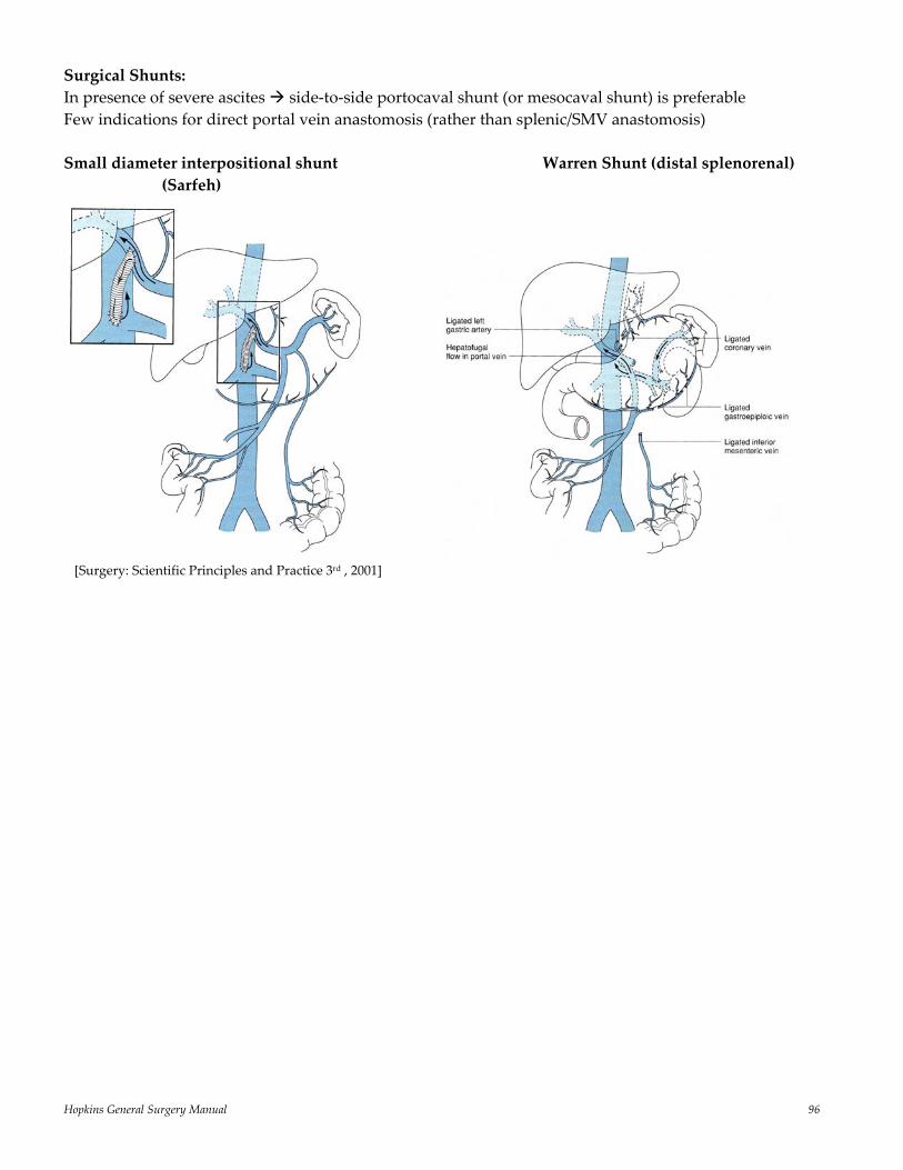
Hopkins General Surgery Manual 96
Surgical Shunts: In presence of severe ascites side‐to‐side portocaval shunt (or mesocaval shunt) is preferable Few indications for direct portal vein anastomosis (rather than splenic/SMV anastomosis) Small diameter interpositional shunt Warren Shunt (distal splenorenal) (Sarfeh)
[Surgery: Scientific Principles and Practice 3rd , 2001]
Hopkins General Surgery Manual 97
Pancreas pan (all) + creas (meat) the only organ that can eat the body Ventral bud uncinate process and inferior portion of head Dorsal bud remainder of gland Islet cells are 1 – 2 % of pancreatic mass, but receive 10 – 25% of arterial blood supply Of the islet cells, β‐cells comprise about 70% and are located at the center of the islet Annular pancreas: “double‐bubble” on AXR; treat obstruction with duodenojejunostomy (no resection of gland) Pancreatic Enzymes and Function
1. Alpha‐amylase: starch digestion, secreted in active form 2. Lipase, phospholipase A, colipase: fat digestion (removes #1, 3 carbon) 3. Proteases (trypsin, chymotrypsin, elastase, carboxypeptidases): protein digestion, secreted as proenzymes 4. Trypsinogen* is converted to active enzyme trypsin by enteropeptidase, a duodenal brush‐border enzyme. Trypsin then activates the other proenzymes and trypsinogen (positive feed‐back) *PRSS1 mutations are related to hereditary pancreatitis Stimulation of Pancreatic Functions:
1. Secretin*: stimulates flow of bicarbonate‐containing fluid 2. Cholecystokinin (CCK)*: major stimulus for zymogen release; weak stimulus for alkaline fluid flow 3. Acetylcholine: major stimulus for zymogen release, poor stimulus for bicarb secretion 4. Somatostatin: inhibits release of gastrin and secretin *secreted from duodenum Secretion Rates
Pancreas: • Basal exocrine: 0.2 – 0.3 mL/min up to 5 mL/min with maximum secretion (i.e. more than gastric H+) • Na+, K+ always ≈ plasma; • At low rates, HCO3, Cl‐ composition ≈ plasma; • At high rates HCO3, Cl‐ reverse
Bile: • 1000 – 1500 mL/day (0.4 – 1 mL/min); gallbladder can only store 60 mL of fluid, but can concentrate
up to 10 – fold* *Keep these numbers in mind for patients with bile leaks to assess nature of ductal injury, i.e. how major an injury?
How ethanol causes pancreatitis: 1. Pancreatic duct hypertrophy ↑ampullary resistance 2. Stimulation of gastric acid secretion ↑secretin ↑exocrine secretion 1+2 enzyme extravasation; exacerbated by ↑acetaldehyde (byproduct of ethanol) ↑membrane permeability, and ↑TG source of cytotoxic free fatty acids
Hopkins General Surgery Manual 98
Ranson’s Criteria – Ethanol‐associated Pancreatitis Initial: During first 48 hours: • Age > 55 • Hct falls > 10% points • WBC > 16,000 per mm3 • BUN increases > 5 mg/dL • Blood glucose > 200 mg/dL • Serum calcium < 8 mg/dL • AST > 250 mg/dL • Arterial PaO2 < 60 mmHg • Serum LDH > 350 mg/dL • Base deficit > 4 mEq/L • Fluid sequestration > 6 L
Mortality: • If 0 – 2 signs, then 2% • If 3 – 4 signs, then 15% • If 5 – 6 signs, then 40% • If 7 – 8 signs, then 100%
Note: all you can really conclude is > 3 signs poor prognosis (in actuality, prognosis is slightly better today because of greatly improved critical care) Distinguish between edematous pancreatitis (resolves) and necrotizing pancreatitis (progresses). Pathogenesis: 1o cell death local inflammatory response systemic inflammatory response via portal circulation to entire body.
CT is helpful for diagnosis because dual phase CT allows comparison of non‐contrast and arterial phase to delineate the ischemic extent of the process and/or to appreciate extralumenal gas Antibiotics should only be used for severe episodes (i.e. > 3 of Ranson’s criteria) 1st line agent is imipenem (crosses blood/pancreas barrier best) 2nd line agent is ciprofloxin Operative debridement if:
1. clinical deterioration, despite maximal medical treatment 2. infection of necrotic pancreas (air in RP, + pancreatic culture) 3. failure to improve after 3 – 4 weeks
If gallstone pancreatitis perform cholecystectomy with IOC: ‐Quick improvement lap chole ± IOC during index admission ‐Severe disease lap chole at interval Pancreatic ascites:
follows ductal disruption; often caused by blunt trauma/pancreatitis abdominal distention with high‐amylase fluid Rx: nonoperative (NPO, TPN, octreotide) if persists > 3 weeks ERCP + sphincterotomy to delineate anatomy and consider transpapillary
stenting; if fails plan Roux‐Y vs. distal pancreatectomy
Site of ductal disruption related to collection: Dorsal rupture RP collection can be sucked into chest (= pancreatic pleural effusion) Ventral rupture inside lesser sac, if not walled off pancreatic ascites
Hopkins General Surgery Manual 99
Pancreatic Pseudocyst
• Encapsulated collection of pancreatic fluid formed by inflammatory fibrosis (NOT epithelial lining) • 1 in 10 after alcoholic pancreatitis; chronic alcoholic pancreatitis is #1 cause in U.S. • Sx: epigastric pain, emesis, fever, weight loss • Signs: palpable epigastric mass; tender epigastrium; ileus • U/S, CT (good for multiple) show fluid collection; MRI/MRCP; ERCP (for treatment): contrast will
fill cyst if communication with duct • Ddx: cystadenocarcinoma, cystadenoma, IPMN, solid pseudopapillary tumor, mesenteric cyst,
adrenal cyst • Complications: infection, bleeding into cyst (most common cause of death), fistula, pancreatic
ascites, gastric outlet obstruction • Treatment: it takes 6 weeks (by definition) to mature and wall off so it can hold sutures; if it’s going
to resolve spontaneously (50% will), it will do so during this time
Most agree that if pseudocyst is > 5 cm it should be drained (especially if symptomatic) Size IS an important predictor of resolution Internal (surgical) drainage is successful in 90% of cases External (surgical) drainage is reserved for thin walled and/or infected cysts External drainage should be used only in cases of sepsis Endoscopic drainage may be appropriate in the setting of chronic pancreatitis
Surgical Drainage:
1. If adherent to posterior wall of stomach: cystogastrostomy 2. If adherent to duodenum: cystoduodenostomy (rare) 3. If not adherent to either: Roux‐en‐Y cystojejunostomy (drain into Roux limb of jejunum) 4. If in tail: resect tail with cyst
*ALWAYS biopsy cyst wall to rule out cystic neoplasia Cystic Neoplasms of the Pancreas Account for < 15% of pancreatic cystic lesions (but incidence increasing)
IPMN* MCN‡ Serous Gender M = F 2:1 0.8:1 Age 70 50 60 – 70 Location Head Tail Uniform Ductal component Yes Rare No Malignant 35% 30% Rare *Associated with chronic pancreatitis (and often mistaken for) ‡ By definition must have underlying ovarian stroma
EUA + FNA for CEA is probably most accurate (optimized sensitivity + specificity) measure of malignancy, BUT resection almost always indicated as it is difficult to exclude malignancy on the basis of biopsies [Brugge WR, et al. Gastroenterology, 2004;126:1330]
Do not perform CT‐guided percutaneous biopsy (often undiagnostic, potential to cause pancreatitis, bleeding, rupture of capsule)
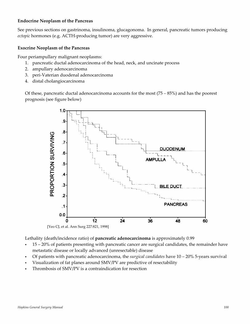
Hopkins General Surgery Manual 100
Endocrine Neoplasm of the Pancreas
See previous sections on gastrinoma, insulinoma, glucagonoma. In general, pancreatic tumors producing ectopic hormones (e.g. ACTH‐producing tumor) are very aggressive. Exocrine Neoplasm of the Pancreas
Four periampullary malignant neoplasms: 1. pancreatic ductal adenocarcinoma of the head, neck, and uncinate process 2. ampullary adenocarcinoma 3. peri‐Vaterian duodenal adenocarcinoma 4. distal cholangiocarcinoma Of these, pancreatic ductal adenocarcinoma accounts for the most (75 – 85%) and has the poorest prognosis (see figure below)
[Yeo CJ, et al. Ann Surg 227:821, 1998]
Lethality (death/incidence ratio) of pancreatic adenocarcinoma is approximately 0.99 15 – 20% of patients presenting with pancreatic cancer are surgical candidates, the remainder have
metastatic disease or locally advanced (unresectable) disease Of patients with pancreatic adenocarcinoma, the surgical candidates have 10 – 20% 5‐years survival Visualization of fat planes around SMV/PV are predictive of resectability Thrombosis of SMV/PV is a contraindication for resection
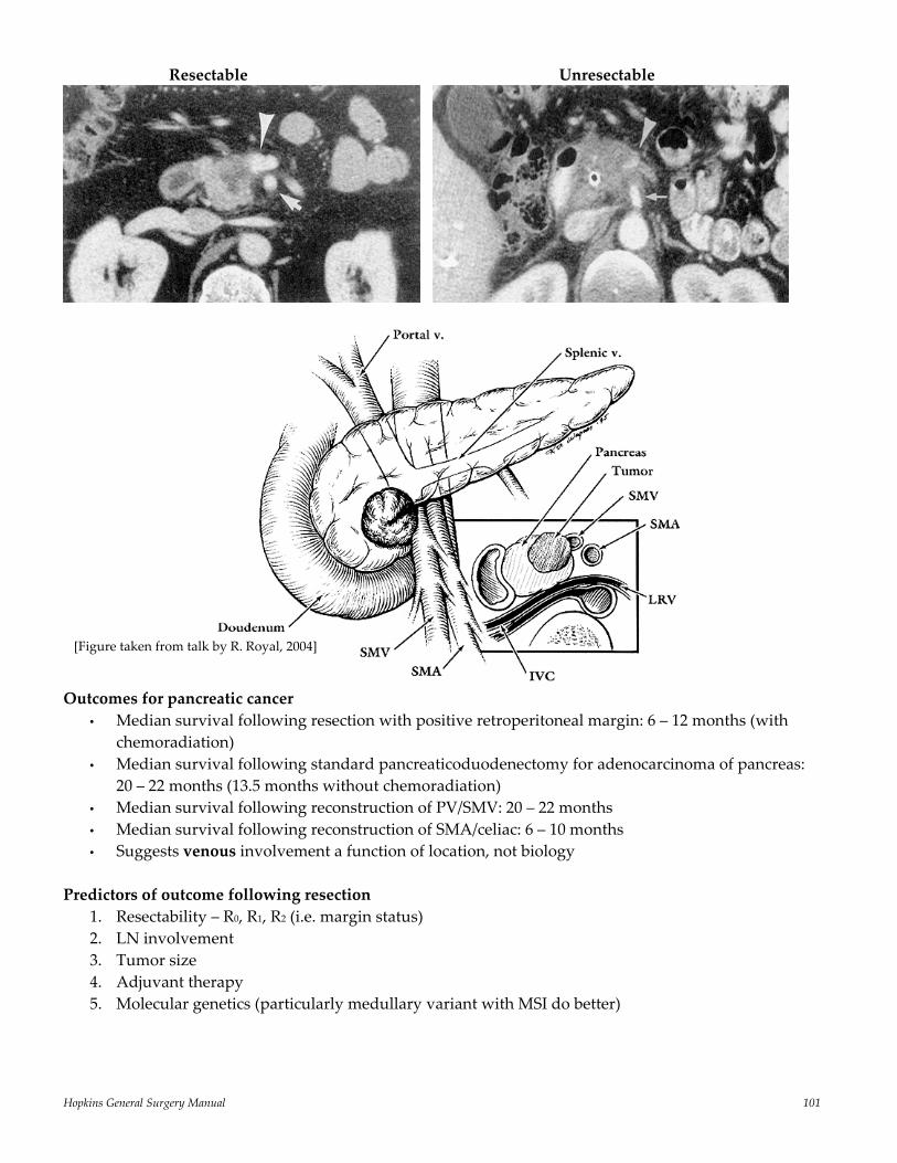
Hopkins General Surgery Manual 101
Resectable Unresectable
Outcomes for pancreatic cancer
• Median survival following resection with positive retroperitoneal margin: 6 – 12 months (with chemoradiation)
• Median survival following standard pancreaticoduodenectomy for adenocarcinoma of pancreas: 20 – 22 months (13.5 months without chemoradiation) • Median survival following reconstruction of PV/SMV: 20 – 22 months • Median survival following reconstruction of SMA/celiac: 6 – 10 months • Suggests venous involvement a function of location, not biology
Predictors of outcome following resection
1. Resectability – R0, R1, R2 (i.e. margin status) 2. LN involvement 3. Tumor size 4. Adjuvant therapy 5. Molecular genetics (particularly medullary variant with MSI do better)
[Figure taken from talk by R. Royal, 2004]
Hopkins General Surgery Manual 102
Sarcoma
8000 cases/year in US; 1% of adult cancers Mesoderm origin No predilection for sex, age, race Overall 5‐year survival approx 50%
Most Common: Liposarcoma, fibrosarcoma, leimyosarcoma (MFH is sort of a “wastebasket” term), essentially no change in treatment or survival
Staging (grade, size/depth, metasteses; not histology): Stage I: low grade, < 5 cm deep or > 5 cm super, no mets 99% OS Stage II: low grade, > 5 cm deep OR high < 5 cm deep/> 5 cm super, no mets 82% OS Stage III: high grade, > 5 cm deep, no mets/nodes 52% OS Stage IV: metasteses (including nodes) 20% OS
Sites: Lower extremity…32% Upper extremity…14% Retroperitoneal…15% Viscera…16% Trunk…11% H&N…12%
Biopsy: < 3 cm, uncomplicated Excisional biopsy If incisional longitudinal Core needle >>> FNA (not recommended)
Prognosis: Grade: low > high Depth: superficial > deep Location: distal > proximal; extremity >> RP/visceral
Treatment:
Low Grade 1. NCI Surgery Branch Prospective Randomized Trial (excluding RP/viscera): surgery + (XRT 6800 rad
vs. observation) XRT effective in preventing recurrence, but no effect on survival 2. MSK Prospective Randomized Trial: surgery + (brachy vs. observe) no difference in local control
or survival
High Grade Narrow (< 1 cm) margin 50 – 90% recurrence Wide (> 1 cm) margin 30 – 50% recurrence Radical resection (entire tissue compartment) < 20% recurrence
Role of XRT:
1. NCI SB PRT: Wide resection + adjuvant chemo (adria + cytoxan) (XRT vs. observe) p = 0.003 (0% vs. 22% recurrence), but no survival difference (p = 0.64)
2. MSK PRT: consistent with above
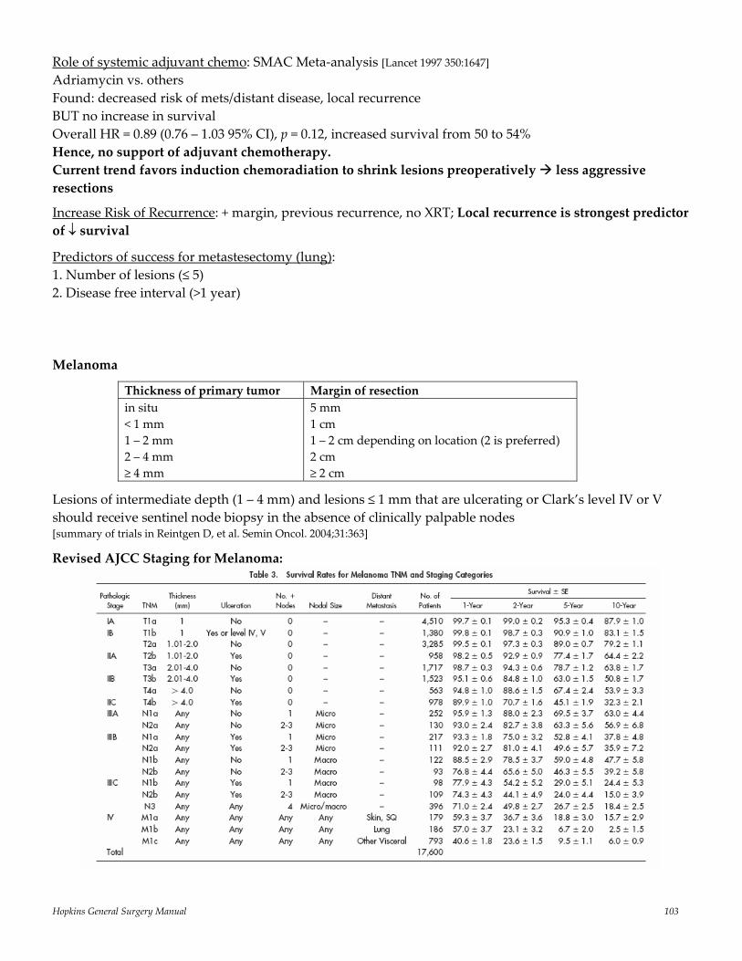
Hopkins General Surgery Manual 103
Role of systemic adjuvant chemo: SMAC Meta‐analysis [Lancet 1997 350:1647] Adriamycin vs. others Found: decreased risk of mets/distant disease, local recurrence BUT no increase in survival Overall HR = 0.89 (0.76 – 1.03 95% CI), p = 0.12, increased survival from 50 to 54% Hence, no support of adjuvant chemotherapy. Current trend favors induction chemoradiation to shrink lesions preoperatively less aggressive resections
Increase Risk of Recurrence: + margin, previous recurrence, no XRT; Local recurrence is strongest predictor of ↓ survival
Predictors of success for metastesectomy (lung): 1. Number of lesions (≤ 5) 2. Disease free interval (>1 year) Melanoma
Thickness of primary tumor Margin of resection in situ < 1 mm 1 – 2 mm 2 – 4 mm ≥ 4 mm
5 mm 1 cm 1 – 2 cm depending on location (2 is preferred) 2 cm ≥ 2 cm
Lesions of intermediate depth (1 – 4 mm) and lesions ≤ 1 mm that are ulcerating or Clark’s level IV or V should receive sentinel node biopsy in the absence of clinically palpable nodes [summary of trials in Reintgen D, et al. Semin Oncol. 2004;31:363]
Revised AJCC Staging for Melanoma:
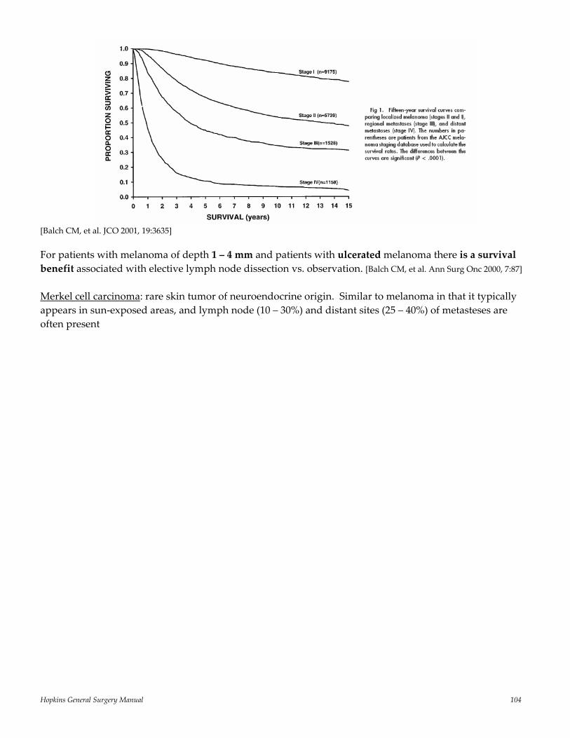
Hopkins General Surgery Manual 104
[Balch CM, et al. JCO 2001, 19:3635] For patients with melanoma of depth 1 – 4 mm and patients with ulcerated melanoma there is a survival benefit associated with elective lymph node dissection vs. observation. [Balch CM, et al. Ann Surg Onc 2000, 7:87] Merkel cell carcinoma: rare skin tumor of neuroendocrine origin. Similar to melanoma in that it typically appears in sun‐exposed areas, and lymph node (10 – 30%) and distant sites (25 – 40%) of metasteses are often present
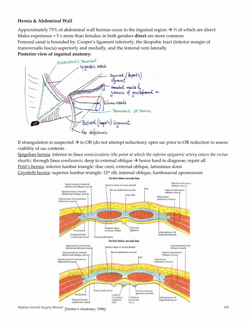
Hopkins General Surgery Manual 105
Hernia & Abdominal Wall
Approximately 75% of abdominal wall hernias occur in the inguinal region ⅔ of which are direct Males experience ≈ 5 x more than females; in both genders direct are more common Femoral canal is bounded by: Cooper’s ligament inferiorly, the ileopubic tract (inferior margin of transversalis fascia) superiorly and medially, and the femoral vein laterally Posterior view of inguinal anatomy:
If strangulation is suspected to OR (do not attempt reduction); open sac prior to OR reduction to assess viability of sac contents Spigelian hernia: inferior to linea semicircularis (the point at which the inferior epigastric artery enters the rectus sheath), through linea semilunaris; deep to external oblique hence hard to diagnose; repair all Petit’s hernia: inferior lumbar triangle: iliac crest, external oblique, latissimus dorsi Grynfeltt hernia: superior lumbar triangle: 12th rib, internal oblique, lumbosacral aponeurosis
[Netter’s Anatomy, 1996]
Hopkins General Surgery Manual 106
Trauma Principles Top 3 causes of prehospital mortality
1. Head injury 2. Hemorrhage 3. Airway obstruction
With massive hemorrhage, the most important factor in predicting outcome is duration of hypotension Critical decision for patient with head injury is whether or not mass lesion is present
Multiple injuries, plus widened mediastinum decompression of mass lesion in head is still first priority.
If patient arrives in shock, with widened mediastinum bleeding most likely in abdomen (go there first)
If patient stable with widened mediastinum CT chest/arterigraphy first, but must RULE OUT abdominal bleeding prior to thoracotomy
The two major injuries associates with widened mediastinum (typically > 8 cm): contained aortic rupture and vertebral body fracture with associated hematoma When assessing circulation must discriminate between pump and volume problems: Pump problems distended neck veins (tamponade, tension pneumothorax, myocardial contusion, air embolus) Finite number of sites of significant internal bleeding: Chest, thigh, abdomen, pelvis, RP Rib fractures, including those of 1st and 2nd ribs, are related to the MAGNITUDE of deformation; Thoracic aorta injury is related to the initial SPEED of deformation; hence, not directly related to each other Amniotic fluid on pelvic exam will be alkaline (deep blue on nitrazine paper); Kleihauer‐Betke (K‐B) blood test detects even small amounts of maternal‐fetal transfusion Low CVP is not a good indicator of hypovolemia, rather its use is helpful when high (tamponade, tension pneumothorax) Zone I: clavicle to cricoid; Zone II: cricoid to angle of mandible; Zone III: angle of mandible to base of skull In general: Zone II unstable secure airway to OR; if stable can do same or conservative approach: carotid arteriogram, barium swallow, rigid esophagoscopy, ± bronchoscopy Brachiocephalic trauma: Zone II to OR without arteriography; Zone 1 & 3 frequently treated conservatively Patients with exsanguinating external hemorrhage, expanding hematomas, or neurologic deficits airway OR (no diagnostic procedures)

Hopkins General Surgery Manual 107
Blunt carotid artery trauma: few signs prior to neurological changes; mortality high (25%) and ½ of survivors have permanent neurological deficit. The lesions are rarely amenable to surgical repair anticoagulation appears to be the mainstay of treatment
In general, try to avoid operating on the vertebral artery, even proximally. Angiography + embolization is always the better option. Carotid artery ligation results in neurologic deficit in only 20% of adults
Use of recombinant Factor VIIa in bleeding trauma patients [Boffard et al. J Trauma 59;8:2005] Randomized nearly 300 patients with either blunt (143 eligible) or penetrating (134 eligible) trauma to receive either 3 x placebo injections or 3 x rFVIIa (200, 100, and 100 μg/kg) in addition to standard treatment with the first dose following the 8th unit of transfused RBC and subsequent doses administered 1 and 3 hours later. In blunt trauma the treatment group had a significant reduction in RBC requirement and “massive” (>20) transfusion requirements. In the penetrating trauma arm, treatment resulted in trends towards fewer transfusions, but the results did not reach statistical significance. Both groups experienced a trend towards reduced mortality. Adverse events (e.g. thromboembolism) were distributed equally between all groups.
[Current Therapy of Trauma, 4th, 1999]
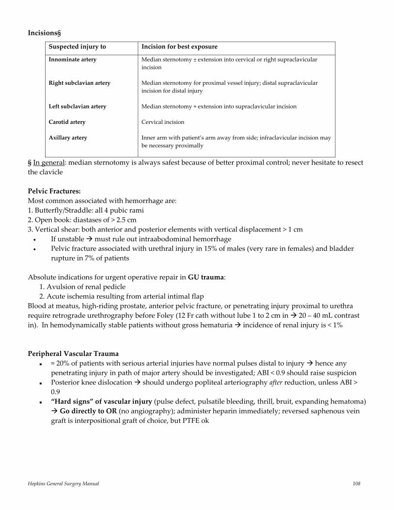
Hopkins General Surgery Manual 108
Incisions§
Suspected injury to
Incision for best exposure
Innominate artery Right subclavian artery Left subclavian artery Carotid artery Axillary artery
Median sternotomy ± extension into cervical or right supraclavicular incision Median sternotomy for proximal vessel injury; distal supraclavicular incision for distal injury Median sternotomy + extension into supraclavicular incision Cervical incision Inner arm with patient’s arm away from side; infraclavicular incision may be necessary proximally
§ In general: median sternotomy is always safest because of better proximal control; never hesitate to resect the clavicle Pelvic Fractures: Most common associated with hemorrhage are: 1. Butterfly/Straddle: all 4 pubic rami 2. Open book: diastases of > 2.5 cm 3. Vertical shear: both anterior and posterior elements with vertical displacement > 1 cm
• If unstable must rule out intraabodominal hemorrhage • Pelvic fracture associated with urethral injury in 15% of males (very rare in females) and bladder
rupture in 7% of patients Absolute indications for urgent operative repair in GU trauma:
1. Avulsion of renal pedicle 2. Acute ischemia resulting from arterial intimal flap
Blood at meatus, high‐riding prostate, anterior pelvic fracture, or penetrating injury proximal to urethra require retrograde urethrography before Foley (12 Fr cath without lube 1 to 2 cm in 20 – 40 mL contrast in). In hemodynamically stable patients without gross hematuria incidence of renal injury is < 1% Peripheral Vascular Trauma
≈ 20% of patients with serious arterial injuries have normal pulses distal to injury hence any penetrating injury in path of major artery should be investigated; ABI < 0.9 should raise suspicion
Posterior knee dislocation should undergo popliteal arteriography after reduction, unless ABI > 0.9
“Hard signs” of vascular injury (pulse defect, pulsatile bleeding, thrill, bruit, expanding hematoma) Go directly to OR (no angiography); administer heparin immediately; reversed saphenous vein
graft is interpositional graft of choice, but PTFE ok
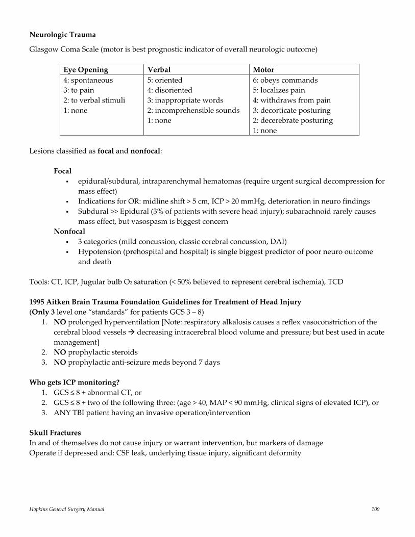
Hopkins General Surgery Manual 109
Neurologic Trauma
Glasgow Coma Scale (motor is best prognostic indicator of overall neurologic outcome)
Eye Opening Verbal Motor 4: spontaneous 3: to pain 2: to verbal stimuli 1: none
5: oriented 4: disoriented 3: inappropriate words 2: incomprehensible sounds 1: none
6: obeys commands 5: localizes pain 4: withdraws from pain 3: decorticate posturing 2: decerebrate posturing 1: none
Lesions classified as focal and nonfocal:
Focal epidural/subdural, intraparenchymal hematomas (require urgent surgical decompression for
mass effect) Indications for OR: midline shift > 5 cm, ICP > 20 mmHg, deterioration in neuro findings Subdural >> Epidural (3% of patients with severe head injury); subarachnoid rarely causes
mass effect, but vasospasm is biggest concern Nonfocal
3 categories (mild concussion, classic cerebral concussion, DAI) Hypotension (prehospital and hospital) is single biggest predictor of poor neuro outcome
and death Tools: CT, ICP, Jugular bulb O2 saturation (< 50% believed to represent cerebral ischemia), TCD 1995 Aitken Brain Trauma Foundation Guidelines for Treatment of Head Injury (Only 3 level one “standards” for patients GCS 3 – 8)
1. NO prolonged hyperventilation [Note: respiratory alkalosis causes a reflex vasoconstriction of the cerebral blood vessels decreasing intracerebral blood volume and pressure; but best used in acute management]
2. NO prophylactic steroids 3. NO prophylactic anti‐seizure meds beyond 7 days
Who gets ICP monitoring?
1. GCS ≤ 8 + abnormal CT, or 2. GCS ≤ 8 + two of the following three: (age > 40, MAP < 90 mmHg, clinical signs of elevated ICP), or 3. ANY TBI patient having an invasive operation/intervention
Skull Fractures In and of themselves do not cause injury or warrant intervention, but markers of damage Operate if depressed and: CSF leak, underlying tissue injury, significant deformity

Hopkins General Surgery Manual 110
C‐Spine Fracture C1 burst fracture (Jefferson’s): caused by axial loading stable (if isolated) treat with collar C2 posterior element fracture (Hangman’s): caused by extension and distraction unstable 3 months in halo Odontoid fracture:
Type I: above base stable; Type II: at the base unstable; < 5 mm displacement 3 months halo; > 5 mm C1/2 fusion or screw fixation Type III: extend into vertebral body halo
3 Columns determine the stability of the spine: 1. Anterior: anterior spinous ligament 2. Middle: vertebral body, posterior spinous ligament 3. Posterior: facet/lamina interface
Instability results when at least ⅔ are interrupted. Penetrating injury rarely results in instability.
T‐ and L‐spine fractures (⅔ occur between T11 and L3): Fractures that involve the middle or posterior columns are by definition unstable and, because of the
narrow spinal canal in this region, can cause severe neurologic injury If finger spreading can be accomplished with symmetry and strength, there is no cord injury above
C8
Cord Injury Most common C6 to T1 If tip of odontiod (dens) is > 4.5 mm above McGregor’s line (hard palate to lowest point on occipital
bone) basilar impression likely OR for compression > 50% height of vertebral body or > 30% narrowing of canal
NASCIS 2 Trial demonstrated that in patients with blunt trauma to the spinal cord high‐dose methylprednisolone (30 mg/kg bolus followed by 5.4 mg/kg/hour for 23 hours), if initiated within 8 hours of injury, resulted in greater neurologic recovery, which remained at one year [NEJM 322;20, 1990]
A follow‐up study further demonstrated that 24 hour treatment was sufficient for patients initiated on treatment within 3 hours, but 48 hour steroid treatment was necessary for patients initiated 3 – 8 hours after injury [JAMA 277;20, 1997].
Neurogenic Shock Not to be confused with flaccid spinal shock Loss of vasomotor tone in viscera and lower extremities; need volume first, peripheral
vasoconstriction (e.g. neo) second. Hypotension should first prompt search of other causes of shock (such as hypovolemia)
May see bradycardia and warm, perfused extremities
Nexus Criteria No films if:
No intoxication, alert, awake, no ∆MS No neuro deficit No neck pain No distracting injury Normal neck exam

Hopkins General Surgery Manual 111
Focused Abdominal Sonogram for Trauma (FAST) Exam: Ultrasound can detect as little as 100 mL of free fluid in the peritoneum. Withhold foley placement until FAST is done to facilitate bladder view 1st View: Subxiphoid, demonstrates a longitudinal cross‐section of the heart and pericardial sac
2nd View: RUQ, demonstrates a sagital view of the liver and right kidney
3rd View: LUQ, demonstrates a sagital view of the diaphragm, spleen, and left kidney (Note: 2 locations for fluid accumulation)
4th view: Pelvis, demonstrates a transverse view of the pelvis
Hopkins General Surgery Manual 112
Chest Wall Injuries Sentinel injuries:
First rib fracture can indicate underlying head and neck injury or great vessel injury Scapular fracture CNS injuries, pulmonary contusion Sternal fracture cardiac contusion, great vessel injury Bilateral rib fracture, lower rib fracture liver, spleen
Flail chest: adjacent ribs, each in two or more places paradoxical movement on respiration (can be overlooked in positive‐pressure ventilation). Three components:
1. Altered chest wall mechanics, 2. Underlying pulmonary contusion (*most significant), 3. Pain reduced tidal volume
If awake, alert, deserve trial of non‐intubation, but adequate IV pain control (consider epidural); if respiratory distress volume‐controlled ventilation Open Pneumothorax: If the defect is more than ⅔ the diameter of the trachea on inspiration air will preferentially pass through chest wall rather than airway; initial management is creating a flutter valve‐type dressing + chest tube at site remote from defect Tracheal Injury:
If penetrating explore via SCM incision Patients presenting with massive subq or mediastinal emphysema should be suspected of having
distal tracheal or bronchus injury; Also, constant “bubbling” after chest tube placement; Perform bronchoscopy ASAP to rule out tracheal/bronchial tear
80% of traumatic tears occur within 2.5 cm of carina; Airway repair done via right PL thoracotomy Pulmonary Contusion: ⅓ do not manifest on CXR until > 24 hours; hypoxia may be first sign; consider intubation if PaO2 < 60 mmHg on > 40% O2 or if PaCO2 > 50 mmHg with normal HCO3 Pulmonary Laceration*: Thoracotomy indicated for: 1. Entire hemithorax opacified, 2. Shock that is persistent or develops or is persistent as hemothorax is evacuated, 3. Rapid removal of > 1500 mL blood, 4. > 250 mL/hour x 4 – 6 hours, 5. Significant hemoptysis *Consider tractotomy with GIA stapler over trauma pneumonectomy (“more papers than survivors”) Air Embolism:
As distributed to end organs, small bubbles cause ischemic damage as they occlude vessels of the microcirculation; Brain and myocardium are most sensitive;
RCA is anterior in supine patient, hence, receives substantial proportion of AE as they exit aortic root
Penetrating thoracic trauma causes ⅔ of traumatic AE Clues to injury: Chest injury, without head injury yet focal neuro signs Treatment: cessation of continued AE; head down, thoracotomy on suspected side with hilar cross‐
clamping; Interventions that increase PaO2, CO, BP enhance air bubble dissolution
Hopkins General Surgery Manual 113
Fat emboli: long bone fracture; petechia, hypoxia, confusion/agitation; Sudan urine stain for fat Cardiac Trauma:
Penetrating heart trauma have 75% prehospital mortality (higher for blunt trauma) RV > LV > RA > LA Major morbidity is coronary artery injury Presentation of tamponade: extreme anxiety, hypotension, distended neck veins Following thoracotomy and aortic cross‐clamping open pericardium (longitudinally from inferior
to superior) Fine fibrillation is a bad sign and won’t convert to course fibrillation until the pH reaches 7.20 s/p blunt cardiac trauma: new murmur should raise suspicion of valvular prolapse (mitral,
tricuspid) or traumatic VSD Esophageal Trauma: most common site is cervical (> 80%) Should be suspected when:
posterior chest wound, transmediastinal injuries, penetrations of platysma, and/or tracheobronchial trauma;
Gastrograffin can miss up to 15% of injuries and should be followed with dilute barium Management: < 24 hours, stable patient: primary closure, buttressed with tissue and drained > 24 hours; unstable patient: Cervical – simple drainage; if large spit fistula; Thoracic – close with Grillo (pleural) patch, extensive debridement, wide drainage Caustic Injuries: scope only to first area of burn, not beyond First Degree (hyperemia and edema): IV until able to handle own saliva barium swallow advance diet as tolerated Second Degree (hemorrhage, exudates, ulcerations): as above, repeat endoscopy in 3 weeks to rule out stricture Third Degree (complete obliteration of mucosa, circum ulceration, eschar): controversial; consider esophagectomy
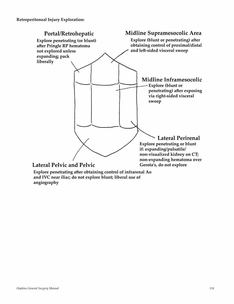
Hopkins General Surgery Manual 114
Retroperitoneal Injury Exploration:
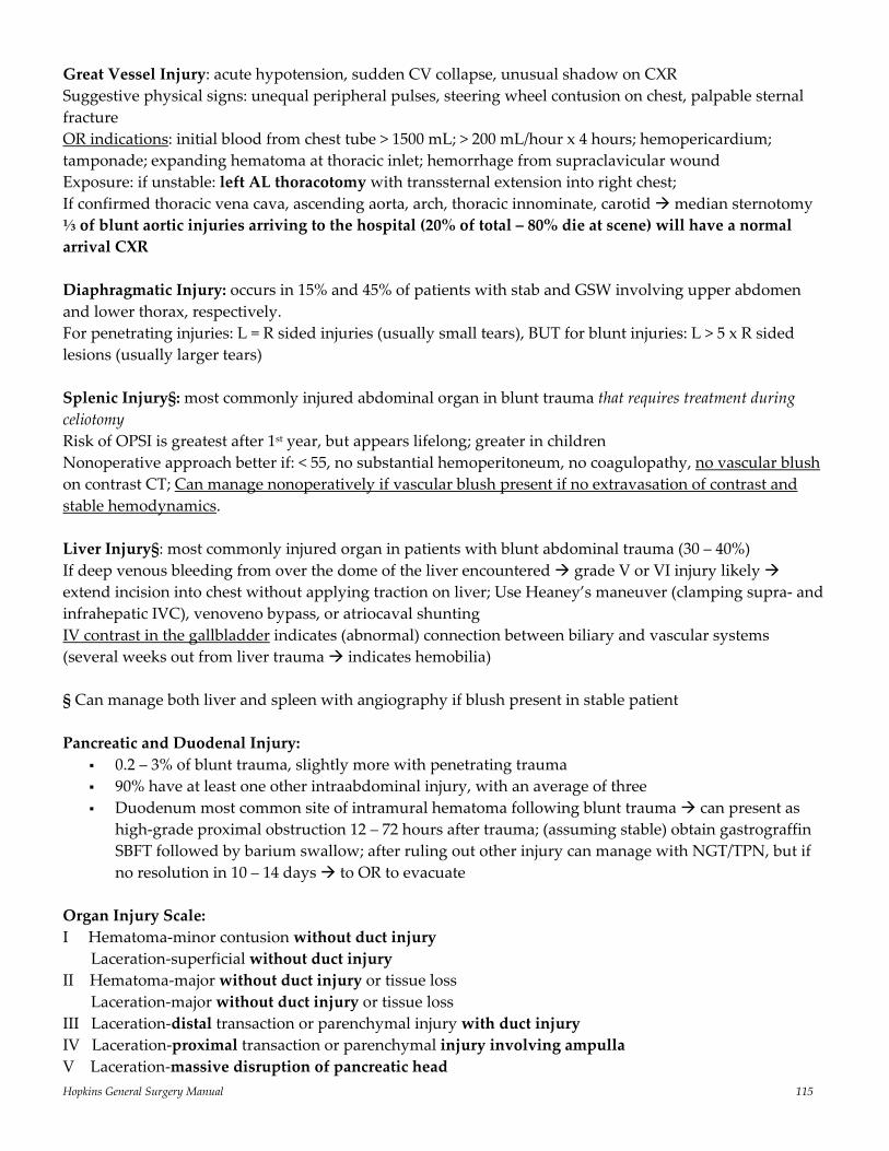
Hopkins General Surgery Manual 115
Great Vessel Injury: acute hypotension, sudden CV collapse, unusual shadow on CXR Suggestive physical signs: unequal peripheral pulses, steering wheel contusion on chest, palpable sternal fracture OR indications: initial blood from chest tube > 1500 mL; > 200 mL/hour x 4 hours; hemopericardium; tamponade; expanding hematoma at thoracic inlet; hemorrhage from supraclavicular wound Exposure: if unstable: left AL thoracotomy with transsternal extension into right chest; If confirmed thoracic vena cava, ascending aorta, arch, thoracic innominate, carotid median sternotomy ⅓ of blunt aortic injuries arriving to the hospital (20% of total – 80% die at scene) will have a normal arrival CXR Diaphragmatic Injury: occurs in 15% and 45% of patients with stab and GSW involving upper abdomen and lower thorax, respectively. For penetrating injuries: L = R sided injuries (usually small tears), BUT for blunt injuries: L > 5 x R sided lesions (usually larger tears) Splenic Injury§: most commonly injured abdominal organ in blunt trauma that requires treatment during celiotomy Risk of OPSI is greatest after 1st year, but appears lifelong; greater in children Nonoperative approach better if: < 55, no substantial hemoperitoneum, no coagulopathy, no vascular blush on contrast CT; Can manage nonoperatively if vascular blush present if no extravasation of contrast and stable hemodynamics. Liver Injury§: most commonly injured organ in patients with blunt abdominal trauma (30 – 40%) If deep venous bleeding from over the dome of the liver encountered grade V or VI injury likely extend incision into chest without applying traction on liver; Use Heaney’s maneuver (clamping supra‐ and infrahepatic IVC), venoveno bypass, or atriocaval shunting IV contrast in the gallbladder indicates (abnormal) connection between biliary and vascular systems (several weeks out from liver trauma indicates hemobilia) § Can manage both liver and spleen with angiography if blush present in stable patient Pancreatic and Duodenal Injury:
0.2 – 3% of blunt trauma, slightly more with penetrating trauma 90% have at least one other intraabdominal injury, with an average of three Duodenum most common site of intramural hematoma following blunt trauma can present as
high‐grade proximal obstruction 12 – 72 hours after trauma; (assuming stable) obtain gastrograffin SBFT followed by barium swallow; after ruling out other injury can manage with NGT/TPN, but if no resolution in 10 – 14 days to OR to evacuate
Organ Injury Scale: I Hematoma‐minor contusion without duct injury Laceration‐superficial without duct injury II Hematoma‐major without duct injury or tissue loss Laceration‐major without duct injury or tissue loss III Laceration‐distal transaction or parenchymal injury with duct injury IV Laceration‐proximal transaction or parenchymal injury involving ampulla V Laceration‐massive disruption of pancreatic head

Hopkins General Surgery Manual 116
Selective Treatment: I External drainage II External drainage; distal pancreatectomy if distal III Distal pancreatectomy IV Extended distal pancreatectomy V Resect (or drain) pancreas, exclude duodenum; consider Whipple Colon/Mesocolon: At laparotomy explore all hematomas or colon wall or mesocolon to identify occult perforations Primary repair of colonic injuries can be performed unless following:
> 50% circumferential injury (i.e. destructive injury) significant associated injuries, ISS > 25 (i.e. unstable) peritonitis significant fecal spillage hemodynamic instability
Rectal Injuries: 3 Principles: 1. Formation of a proximal, completely diverting colostomy 2. Insertion of presacral drains between anal verge and the coccyx 3. Debridement and primary repair of the injury itself, if it is accessible

Hopkins General Surgery Manual 117
Critical Care SIRS (Systemic Inflammatory Response Syndrome): at lease 2 of the following 4 (in the absence of other explanation):
1. Hyperthermia (> 38 0C or 100.4 0F) of hypothermia (< 36 0C or 94 0F) 2. Tachycardia (> 90 bpm) 3. Tachypnea (> 20/min or PaCO2 < 32) 4. WBC > 12,000 or < 4,000 per mm3 (or >10% bands)
Sepsis: Known or suspected presence of infection (bacteremia, toxemia, fungemia, viremia) plus 2 or more SIRS criteria
Severe Sepsis: Sepsis plus evidence of organ dysfunction, hypotension, or evidence of hypoperfusion
Shock: End‐organ hypoperfusion. Period. Often manifested by lactic acidosis, oliguria, mental status changes, and hypotension refractory to fluid administration.
Septic Shock: Severe sepsis leading to shock
Pathophysiology of this cascade leading to shock: It really begins with a “panendothelial organ failure” as a consequence of an inflammatory cascade. Macrophages release TNF (itself a direct myocardial depressant) and IL‐1, which results in two detriments:
1. Increased expression of the adhesion molecules CD11, CD18, ICAM‐1, and ICAM‐2 on endothelial cells and WBCs, resulting in leukoaggregation.
2. Promotion of NO Synthase activity, increasing circulating levels of NO, which does two things: direct myocardial depression, and vasodilation
The endothelium itself becomes the target organ as blood flow is shunted around capillaries (because of obstruction), leading to poor tissue oxygenation
Other etiologies of shock: 1. Cardiogenic 2. Neurogenic 3. Hypovolemic 4. Obstructive 5. Distributive (anaphylaxis)
Catecholamine response to injury is maximal at 24 – 48 hours
CO = HR * (EDV – ESV) Generally, CO↑ as HR↑ (sinus) up to ≈ 160/min Atrial kick provides 15 – 20% of EDV A reduction in Hct by 50% produces an 8‐fold reduction in blood viscosity mechanism whereby
CO↑ in normovolemic anemia
As the arterial waveform propagates distally the systolic pressure increases and diastolic pressure falls slightly so that the MAP remains constant except in certain circumstances like rewarming from CPB or during vasopressor administration in sepsis Aortic MAP and diastolic pressures are slightly higher than distal pressure; BUT systolic pressure rises with distal propagation
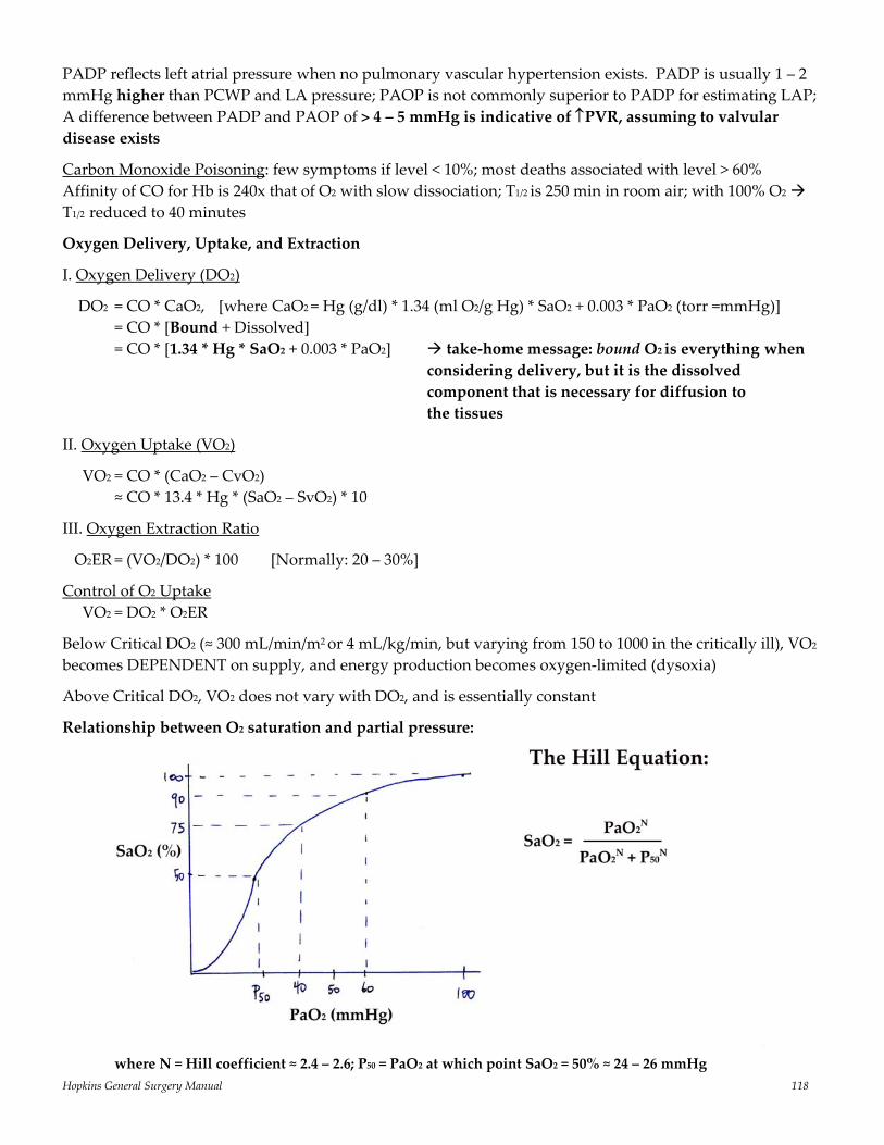
Hopkins General Surgery Manual 118
PADP reflects left atrial pressure when no pulmonary vascular hypertension exists. PADP is usually 1 – 2 mmHg higher than PCWP and LA pressure; PAOP is not commonly superior to PADP for estimating LAP; A difference between PADP and PAOP of > 4 – 5 mmHg is indicative of ↑PVR, assuming to valvular disease exists
Carbon Monoxide Poisoning: few symptoms if level < 10%; most deaths associated with level > 60% Affinity of CO for Hb is 240x that of O2 with slow dissociation; T1/2 is 250 min in room air; with 100% O2 T1/2 reduced to 40 minutes
Oxygen Delivery, Uptake, and Extraction
I. Oxygen Delivery (DO2)
DO2 = CO * CaO2, [where CaO2 = Hg (g/dl) * 1.34 (ml O2/g Hg) * SaO2 + 0.003 * PaO2 (torr =mmHg)] = CO * [Bound + Dissolved] = CO * [1.34 * Hg * SaO2 + 0.003 * PaO2] take‐home message: bound O2 is everything when considering delivery, but it is the dissolved component that is necessary for diffusion to the tissues
II. Oxygen Uptake (VO2)
VO2 = CO * (CaO2 – CvO2) ≈ CO * 13.4 * Hg * (SaO2 – SvO2) * 10
III. Oxygen Extraction Ratio
O2ER = (VO2/DO2) * 100 [Normally: 20 – 30%]
Control of O2 Uptake VO2 = DO2 * O2ER
Below Critical DO2 (≈ 300 mL/min/m2 or 4 mL/kg/min, but varying from 150 to 1000 in the critically ill), VO2 becomes DEPENDENT on supply, and energy production becomes oxygen‐limited (dysoxia)
Above Critical DO2, VO2 does not vary with DO2, and is essentially constant
Relationship between O2 saturation and partial pressure:
where N = Hill coefficient ≈ 2.4 – 2.6; P50 = PaO2 at which point SaO2 = 50% ≈ 24 – 26 mmHg
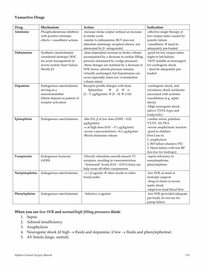
Hopkins General Surgery Manual 119
Vasoactive Drugs
Drug Mechanism Action Indication Amrinone Phosphodiesterase inhibitor
with positive inotropic effects + vasodilator actions
‐increase stroke output without an increase in stroke work. ‐similar to dobutamine, BUT does not stimulate adrenergic receptors (hence, not attenuated by β‐ antagonists)
‐effective single therapy of low‐output states caused by systolic failure. ‐vasodilator must be adequately pre‐loaded
Dobutamine Synthetic catecholamine considered inotropic DOC for acute management of severe systolic heart failure. Mostly β1
‐dose‐dependent increase in stroke volume accompanied by a decrease in cardiac filling pressure (measured by wedge pressure) ‐these changes are matched by a decrease in SVR, hence, arterial pressure remains virtually unchanged, but hypotension can occur especially when low or borderline volume status
‐good for low output states (right or left failure) ‐NOT suitable as monoagent for cardiogenic shock ‐ must be adequately pre‐loaded
Dopamine Endogenous catecholamine serving as a neurotransmitter Effects depend on pattern of receptor activation
Receptor profile changes with dose: Splanchnic β α (2 – 5 μg/kg/min) (5 – 8) (>10)
‐cardiogenic shock, and circulatory shock syndrome associated with systemic vasodilation (e.g. septic shock) ‐High neurogenic shock (above T3/T4, hypo and bradycardic)
Epinephrine Endogenous catecholamine. ‐like DA, β at low does (0.005 – 0.02 μg/kg/min); ‐α at high does (0.01 – 0.1 μg/kg/min) ‐severe vasoconstriction: >0.1 μg/kg/min ‐Blocks histamine release
‐cardiac arrest, pulseless VT/VF, AS, PEA ‐severe anaphylactic reaction ‐good in children First Line in: 1. anaphylaxis 2. RH failure (massive PE) 3. Heart failure with low BP (too low for inotrope)
Vasopressin Endogenous hormone (ADH)
‐Directly stimulates smooth muscle V1 receptors, resulting in vasoconstriction ‐“hormonal” levels (0.01 – 0.03 U/min) can help wean off other vasopressors
‐sepsis refractory to norepinephrine, phenylephrine.
Norepinephrine Endogenous catecholamine ‐ α > β‐agonist often results in reflex bradycardia
‐low SVR, in need of inotropic support ‐drug of choice in severe septic shock ‐improves renal blood flow
Phenylephrine Endogenous catecholamine ‐ Selective α‐agonist ‐low SVR (provided adequate pre‐load); do not use for pump failure
When you see low SVR and normal/high filling pressures think:
1. Sepsis 2. Adrenal insufficiency 3. Anaphylaxis 4. Neurogenic shock (if high → fluids and dopamine; if low → fluids and phenylepherine) 5. AV fistula (large, central)
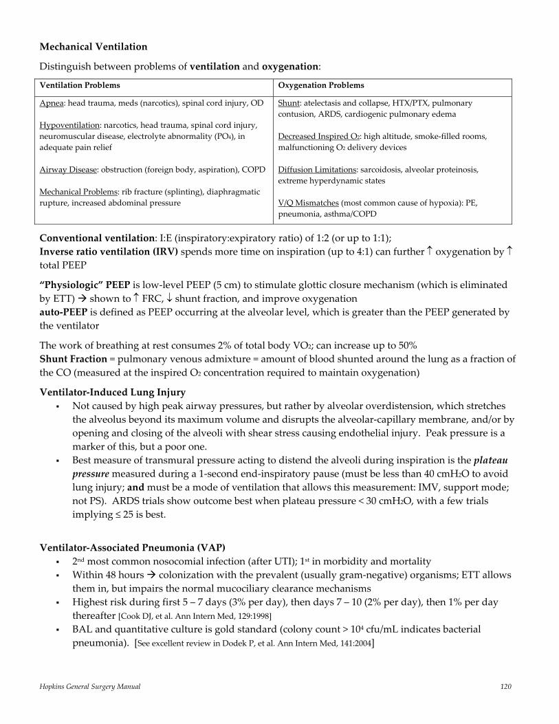
Hopkins General Surgery Manual 120
Mechanical Ventilation
Distinguish between problems of ventilation and oxygenation:
Ventilation Problems
Oxygenation Problems
Apnea: head trauma, meds (narcotics), spinal cord injury, OD Hypoventilation: narcotics, head trauma, spinal cord injury, neuromuscular disease, electrolyte abnormality (PO4), in adequate pain relief Airway Disease: obstruction (foreign body, aspiration), COPD Mechanical Problems: rib fracture (splinting), diaphragmatic rupture, increased abdominal pressure
Shunt: atelectasis and collapse, HTX/PTX, pulmonary contusion, ARDS, cardiogenic pulmonary edema Decreased Inspired O2: high altitude, smoke‐filled rooms, malfunctioning O2 delivery devices Diffusion Limitations: sarcoidosis, alveolar proteinosis, extreme hyperdynamic states V/Q Mismatches (most common cause of hypoxia): PE, pneumonia, asthma/COPD
Conventional ventilation: I:E (inspiratory:expiratory ratio) of 1:2 (or up to 1:1); Inverse ratio ventilation (IRV) spends more time on inspiration (up to 4:1) can further ↑ oxygenation by ↑ total PEEP
“Physiologic” PEEP is low‐level PEEP (5 cm) to stimulate glottic closure mechanism (which is eliminated by ETT) shown to ↑ FRC, ↓ shunt fraction, and improve oxygenation auto‐PEEP is defined as PEEP occurring at the alveolar level, which is greater than the PEEP generated by the ventilator
The work of breathing at rest consumes 2% of total body VO2; can increase up to 50% Shunt Fraction = pulmonary venous admixture = amount of blood shunted around the lung as a fraction of the CO (measured at the inspired O2 concentration required to maintain oxygenation)
Ventilator‐Induced Lung Injury Not caused by high peak airway pressures, but rather by alveolar overdistension, which stretches
the alveolus beyond its maximum volume and disrupts the alveolar‐capillary membrane, and/or by opening and closing of the alveoli with shear stress causing endothelial injury. Peak pressure is a marker of this, but a poor one.
Best measure of transmural pressure acting to distend the alveoli during inspiration is the plateau pressure measured during a 1‐second end‐inspiratory pause (must be less than 40 cmH2O to avoid lung injury; and must be a mode of ventilation that allows this measurement: IMV, support mode; not PS). ARDS trials show outcome best when plateau pressure < 30 cmH2O, with a few trials implying ≤ 25 is best.
Ventilator‐Associated Pneumonia (VAP) 2nd most common nosocomial infection (after UTI); 1st in morbidity and mortality Within 48 hours colonization with the prevalent (usually gram‐negative) organisms; ETT allows
them in, but impairs the normal mucociliary clearance mechanisms Highest risk during first 5 – 7 days (3% per day), then days 7 – 10 (2% per day), then 1% per day
thereafter [Cook DJ, et al. Ann Intern Med, 129:1998] BAL and quantitative culture is gold standard (colony count > 104 cfu/mL indicates bacterial
pneumonia). [See excellent review in Dodek P, et al. Ann Intern Med, 141:2004]
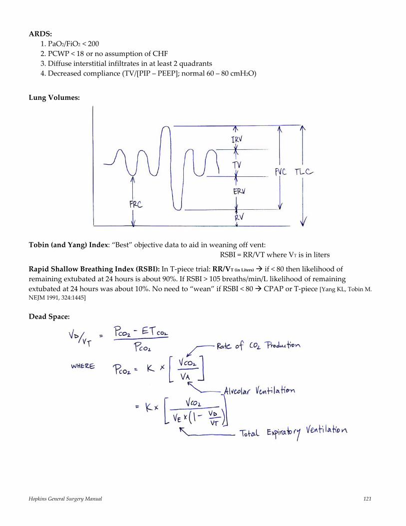
Hopkins General Surgery Manual 121
ARDS: 1. PaO2/FiO2 < 200 2. PCWP < 18 or no assumption of CHF 3. Diffuse interstitial infiltrates in at least 2 quadrants 4. Decreased compliance (TV/[PIP – PEEP]; normal 60 – 80 cmH2O)
Lung Volumes:
Tobin (and Yang) Index: “Best” objective data to aid in weaning off vent: RSBI = RR/VT where VT is in liters
Rapid Shallow Breathing Index (RSBI): In T‐piece trial: RR/VT (in Liters) if < 80 then likelihood of remaining extubated at 24 hours is about 90%. If RSBI > 105 breaths/min/L likelihood of remaining extubated at 24 hours was about 10%. No need to “wean” if RSBI < 80 CPAP or T‐piece [Yang KL, Tobin M. NEJM 1991, 324:1445] Dead Space:
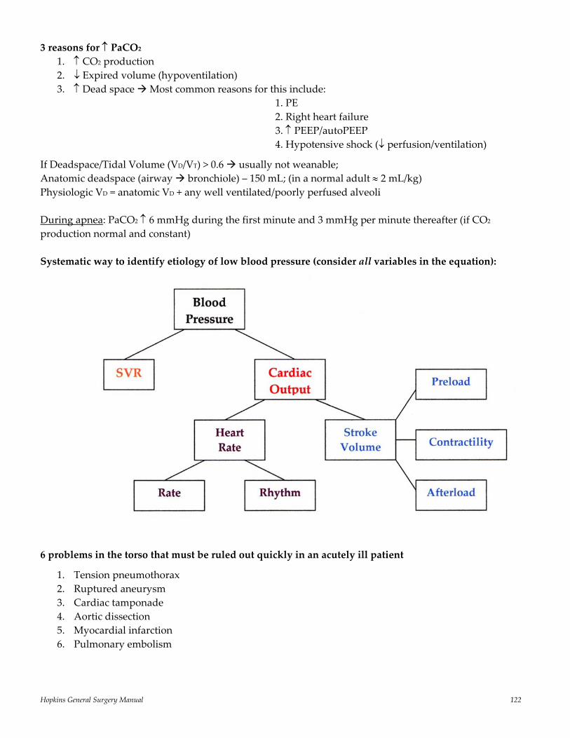
Hopkins General Surgery Manual 122
3 reasons for ↑ PaCO2 1. ↑ CO2 production 2. ↓ Expired volume (hypoventilation) 3. ↑ Dead space Most common reasons for this include: 1. PE 2. Right heart failure 3. ↑ PEEP/autoPEEP 4. Hypotensive shock (↓ perfusion/ventilation)
If Deadspace/Tidal Volume (VD/VT) > 0.6 usually not weanable; Anatomic deadspace (airway bronchiole) – 150 mL; (in a normal adult ≈ 2 mL/kg) Physiologic VD = anatomic VD + any well ventilated/poorly perfused alveoli During apnea: PaCO2 ↑ 6 mmHg during the first minute and 3 mmHg per minute thereafter (if CO2 production normal and constant) Systematic way to identify etiology of low blood pressure (consider all variables in the equation):
6 problems in the torso that must be ruled out quickly in an acutely ill patient
1. Tension pneumothorax 2. Ruptured aneurysm 3. Cardiac tamponade 4. Aortic dissection 5. Myocardial infarction 6. Pulmonary embolism
Hopkins General Surgery Manual 123
Most Influential Recent Trials/Papers in Critical Care Medicine (reverse chronological): Evidence‐Based Clinical Practical Guideline for the Prevention of Ventilator‐Associated Pneumonia [Ann Int Med 141:305, 2004] These guidelines provide excellent evidence‐based recommendations for the prevention and treatment of VAP. Recommendations included: orotracheal intubation, use of closed suction system, heat and moisture exchangers, and semi‐recumbent positioning. Treatments not recommended included: use of sucralfate, use of topical antibiotics. Because of conflicting and/or insufficient data no recommendations were made about: chest PT, timing of tracheostomy, prone positioning, prophylactic IV antibiotics. Eliminating Catheter‐Related Bloodstream Infections in the Intensive Care Unit [Crit Care Med 32;10:2014, 2004] A prospective cohort control trial in a single institution ICU aimed at eliminating CR‐BSI by implementing multifaceted interventions including: hand washing before gloving, using of full sterile technique, and chlorhexidane prep. This study showed a decrease in CR‐BSI rate from 11.3/1000 catheter days to 0/1000 days, which was extrapolated to prevent 43 CR‐BSIs, 8 deaths, and over $1.9 million A Comparison of Albumin and Saline for Fluid Resuscitation in the Intensive Care Unit [NEJM 350;22:2247, 2004] A multi‐center, randomized, double‐blinded trial which compared 28‐day outcomes of patient in the ICU who were administered normal saline or 4% albumin for resuscitation. 3500 patients were randomized into each arm and there were no differences found in deaths, organ failure, number of days spent in ICU, number of days in hospital, requirements for mechanical ventilation, or days of renal replacement. Comparison of 8 vs. 15 Days of Antibiotic Therapy for Ventilator‐Associated Pneumonia in Adults [JAMA 290;19:2588, 2003] A prospective, randomized, multi‐center, double‐blinded study to determine if 8 days of antibiotic therapy is as effective as 15 days in patients with microbiologically proven VAP. The patients treated for 8 days had similar rates of mortality and recurrent infections. However, in patients with non‐fermenting gram‐negative bacilli, including Pseudomonas aeruginosa, higher rates of recurrent pulmonary infection (40.6% vs. 25.4%) were seen. Effect of Treatment with Low Dose of Hydrocortisone and Fludrocortisone on Mortality in Patients with Septic Shock [JAMA 288;7:862, 2002] A placebo‐controlled, randomized, double‐blind, multi‐center trial to assess the role of low dose corticosteroids in the management of patients in septic shock with relative adrenal insufficiency. Over 300 patients were stimulated with corticotripin and responders (appropriate stimulation) and non‐responders (inappropriate stimulation) were randomized to receive either steroids (hydrocortisone 50 mg q6 + fludrocortisone 50 μg qd) or placebo. Amongst non‐responders there were statistically fewer deaths (53% vs. 63%) and statistically less time spent on vasopressors in the steroid treatment group. Amongst responders there were no differences between steroid and placebo treatments.

Hopkins General Surgery Manual 124
Intensive Insulin Therapy in Critically Ill Patients [NEJM 345;19:1359, 2001] A prospective, randomized, controlled study involving mechanically ventilated patients to evaluate the impact of tight glucose control in critically ill patients. Over 1500 patients were randomized receive either tight glucose control (maintenance of blood glucose between 80 and 110 mg/dL) or conventional glucose control (insulin only when blood glucose > 215 mg/dL; maintenance between 180 and 200 mg/dL). At 12 months intensive insulin therapy reduced overall mortality from 8% to 4.6% (p<0.04). In addition to 12 month mortality, intensive insulin therapy also led to decreased in‐hospital mortality, bloodstream infection, acute renal failure, and red cell transfusion requirements. Efficacy and Safety of Recombinant Human Activated Protein C for Severe Sepsis [NEJM 344;10:699, 2001] A randomized, double‐blinded, placebo‐controlled, multi‐center trial evaluating the use of recombinant activated human protein C in the treatment of severe sepsis. Nearly 1700 patients with SIRS and organ failure due to acute infection were randomized to either treatment (24 μg/kg/hr recombinant activated protein C for 96 hours) or placebo. The mortality in the placebo group was 30.8% vs. 24.7% in the treatment group. There was an absolute reduction in the risk of death of 6.1% (p=0.005). The incidence of serious bleeding was higher in the treatment group (3.5% vs. 2.0%, p=0.06). This study is noteworthy in that it is the first agent (of countless agents) to show a decreased mortality in septic patients. Daily Interruption of Sedative Infusions in Critically Ill Patients Undergoing Mechanical Ventilation [NEJM 342;20:1471, 2000] A randomized, controlled trial in a medical intensive care unit involving 128 patients receiving mechanical ventilation and continuous infusions of sedating drugs. In the treatment group the patients were awaken daily by temporary discontinuation of the sedatives. In the control group the sedation was only discontinued at the discretion of the treating physician. The median duration of mechanical ventilation in the treatment group was 4.9 days vs. 7.3 days in the control group (p=0.004) and the median length of stay in the ICU was 6.4 days vs. 9.9 days (p=0.02). There were also fewer diagnostic studies to assess changes in mental status in the treatment group (9% vs. 27%, p=0.02). Low‐Dose Dopamine in Patients with Early Renal Dysfunction: A Placebo‐Controlled Randomised Trial [The Lancet 356:2139, 2000] Over 300 patients were randomized in a placebo‐controlled, double‐blinded study to receive either placebo or “renal‐dose” dopamine (2 μg/kg/min) via continuous infusion upon admission to an ICU. Patients with pre‐existing renal dysfunction were excluded. Use of dopamine did not confer an advantage in peak serum creatinine, need for renal replacement, length of stay in ICU, or overall hospital stay. This study was essentially the final nail in the coffin of the debate over the myth of “renal‐dose” dopamine. Ventilation with Lower Tidal Volumes as Compared with Traditional Tidal Volumes for Acute Lung Injury and the Acute Respiratory Distress Syndrome [NEJM 342;18:1301, 2000] This trial randomized patients across multiple centers with acute lung injury and ARDS to receive either “traditional” ventilatory tidal volumes of 12 mL/kg (with plateau pressures up to 50 cm H2O) or “low tidal volumes” of 6 mL/kg (with plateau pressures up to 30 cm H2O). The trial was stopped after 861 patients were enrolled because mortality was lower in the low tidal volume group (31.0% vs. 39.8%, p=0.007) and the number of days without ventilatory use was also lower.
Hopkins General Surgery Manual 125
A Multicenter, Randomized, Controlled Clinical Trial of Transfusion Requirements in Critical Care [NEJM 340;6:409, 1999] This multi‐center study randomized non‐bleeding, euvolemic, critically ill patients who had a Hb concentration of ≤ 9.0 g/dL within 72 hours of admission to the ICU to one of two transfusion strategies:
• Liberal transfusions: transfusion was initiated when Hb concentration fell below 10.0 g/dL and was subsequently maintained between 10.0 and 12.0 g/dL
• Restrictive transfusion: transfusion was only initiated when Hb concentration fell below 7.0 g/dL and was subsequently maintained between 7.0 and 9.0 g/dL
Overall, the 30‐day mortality was similar between the two groups. However, amongst patients with APACHE II score ≤ 20 (i.e. less ill) mortality was lower in the restrictive group (8.7% vs. 16.1%, p=0.03), as was the case in patients younger than 55 (5.7% vs. 13.0%, p=0.02). There was no difference amongst patients with clinically significant cardiac disease (20.5% vs. 22.9%). A Prospective Study of Indexes Predicting the Outcome of Trials of Weaning From Mechanical Ventilation [NEJM 324; 21:6170, 1991] This study evaluated a number of indexes determined from 36 patients’ successful or unsuccessful extubations and prospectively applied them to a cohort of 64 patients in an effort to predict successful extubation. Of all the parameters studied, the rapid shallow breathing index (RSBI, affectionately referred to as the “Tobin” despite the fact that Karl Yang shared authorship with Martin Tobin on this landmark paper) defined as the ratio of respiratory frequency, f, to tidal volume in liters, Tv, was the most accurate predictor of success (RSBI < 80) or failure (RSBI > 105) of extubation, where “success” was defined as not requiring intubation at 24 hours.
Hopkins General Surgery Manual 126
Hemostasis & Transfusion Three reactions mediate the initial hemostasis response following vascular injury:
1. Vascular response to injury (injury exposes subendothelial components and induces vasoconstriction independent of platelet function)
2. Platelet adherence and aggregation 3. Generation of thrombin
Aspirin, indomethacin, and most other NSAIDs block formation of PGG2 and PGH2 resulting in decreased platelet aggregation “Normal” bleeding time is 5 – 7 minutes. The value of the bleeding time in clinical evaluation is very limited. It can be normal in patients with platelet disorders, even those who have taken aspirin, and can be prolonged in subjects with normal hemostasis. Therefore, it cannot be trusted. Much of the limitation is probably related to technical issues, such as the depth of the cut, the vascularity of the cut tissue, etc. Also, the “normal” range is logarithmically distributed, making interpretation of 7 – 12 minute BTs impossible.
PT and PTT only elevate when factors reduce below approximately 20 to 40% of normal (varies with the individual factor and with the individual laboratory methods/reagents. Generally the tests are adjusted to become “abnormal” when any of the factors is in a range that might not support normal hemostasis. A very common cause of a prolonged aPTT in a patient with a negative bleeding history is a “lupus anticoagulant,” a laboratory artifact not associated with a bleeding tendency.)
20% of normal is usually satisfactory for general hemostasis, but > 50% for major surgery Pre‐operative evaluation:
If a patient has a positive bleeding history and requires minor surgery: PT, PTT, BT, fibrin clot solubility
If a patient has a positive bleeding history and requires major surgery: PT, PTT, BT, fibrin clot solubility, platelet function studies, F VIII levels, F IX level, alpha‐2‐antiplasmin level
As a rule:
1 unit platelets for 2 units RBC. For a total blood volume replacement, expect platelet count of 250,000 to drop to 80,000.
1 mg protamine required for every 100 U of heparin (up to 100 mg total, or 50 mg over 10 min). If the aPTT is measurable (i.e., less than 2 minutes), the plasma concentration of heparin should be low enough (i.e., < 1 U/mL) that 20 – 30 mg protamine will be very effective. However, the protamine may be cleared before the heparin is completely gone. So another dose may be required in an hour or two. Clotting Factors (see figure below): Intrinsic (PTT): exposed collagen + XII XI IX X, which activates thrombin fibrin Extrinsic (PT): TF + VII activated X thrombin fibrin Note: X is common to both VIII is only factor not solely made in liver (made by endothelial cells)

Hopkins General Surgery Manual 127
Bleeding Disorder
Von Willebrand disease is the most common inherited bleeding disorder (1% of population; AD) symptomatic bleeding in 1/1000 long PTT and bleeding time (usually) associated with variable deficiencies in both vWF and factor VIII; platelet defect is also present
(although this is called “platelet type” vWd, it is different from the other types because the defect is in the platelet membrane, not in the vWf).
+ Ristocetin test (Ristocetin “cofactor” [i.e., vWf] activity is the laboratory test for vWf activity, as opposed to antigen level.)
give DDAVP (releases intracellular stores of vWF) for Type I (low vWF) (The effect only lasts for < 12 hours. Repeat doses may be less effective.) (vWf concentrates are available when extended replacement is needed. These are not the same as standard fVIII concentrates, which contain very little, if any, vWf.)
give cryo for Type II (qualitatively poor vWF) and III (low vWF) Hemophilia A (factor VIII deficiency) X‐linked recessive; ≥ 5% normal levels fVIII considered mild
DON’T aspirate hemarthrosis Tx with factor VIII concentrates (to 100% pre‐op levels) ↑ PTT; normal PT
Hemophilia B (factor IX deficiency) X‐linked recessive
Tx with factor IX concentrates ↑ PTT; normal PT (to 50% pre‐op levels)
Glanzman’s thrombasthenia: Platelets have IIb/IIIa deficiency ↓ aggregation abnormalities due to decreased fibrinogen binding. Extremely rare.
Bernard Soulier: Platelets have Ib deficiency ↓ adherence to exposed collagen von Willebrand factor. Extremely rare Hypercoagulable States
APC Resistance: most common inherited hypercoagulable state; AD; 90% associated with FV (Leiden) mutation ≈ 5% prevalence in Caucasian populations, much less in others. Spontaneous DVT typically only occur in thrombophilic families, who probably carry other prothrombotic genes in addition to FVL. In the general population (where FVL is likely to be the only prothrombotic gene in most individuals) FVL is rarely associated with thrombosis.
ATIII Deficiency: rare, 1/5000; don’t respond to heparin unless given FFP (to replace ATIII); can’t treat DVT without giving FFP The most common forms of “AT deficiency” are identified because the AT molecule does not bind heparin normally. These actually are not associated with an increased risk of thrombosis. Specialized tests are necessary to identify the individuals with the dangerous form of AT deficiency.
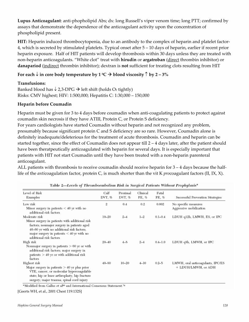
Hopkins General Surgery Manual 128
Lupus Anticoagulant: anti‐phopholipid Abs; dx: long Russell’s viper venom time; long PTT; confirmed by assays that demonstrate the dependence of the anticoagulant activity upon the concentration of phospholipid present.
HIT: Heparin induced thrombocytopenia, due to an antibody to the complex of heparin and platelet factor‐4, which is secreted by stimulated platelets. Typical onset after 5 – 10 days of heparin, earlier if recent prior heparin exposure. Half of HIT patients will develop thrombosis within 30 days unless they are treated with non‐heparin anticoagulants. “White clot” treat with hirudin or argatroban (direct thrombin inhibitor) or danapariod (indirect thrombin inhibitor); dextran is not sufficient for treating clots resulting from HIT
For each ↓ in core body temperature by 1 0C blood viscosity ↑ by 2 – 3%
Transfusions: Banked blood has ↓ 2,3‐DPG left shift (holds O2 tightly) Risks: CMV highest; HIV: 1:500,000; Hepatitis C: 1:30,000 – 150,000
Heparin before Coumadin
Heparin must be given for 3 to 4 days before coumadin when anti‐coagulating patients to protect against coumadin skin necrosis if they have ATIII, Protein C, or Protein S deficiency. For years cardiologists have started Coumadin without heparin and not recognized any problem, presumably because significant protein C and S deficiency are so rare. However, Coumadin alone is definitely inadequate/deleterious for the treatment of acute thrombosis. Coumadin and heparin can be started together, since the effect of Coumadin does not appear till 2 – 4 days later, after the patient should have been theratpeutically anticoagulated with heparin for several days. It is especially important that patients with HIT not start Coumadin until they have been treated with a non‐heparin parenteral anticoagulant. ALL patients with thrombosis to receive coumadin should receive heparin for 3 – 4 days because the half‐life of the anticoagulation factor, protein C, is much shorter than the vit K procoagulant factors (II, IX, X).
[Geerts WH, et al., 2001 Chest 119:132S]

Hopkins General Surgery Manual 129
Hopkins General Surgery Manual 130
Metabolism
A number of vital areas of the body (brain, renal medulla, RBC, WBC, peripheral nerves) are glycolytic tissues (require a glucose source of energy for metabolism) and are unable to utilize fatty acids.
When the limited glycogen stores are depleted, this is accomplished by gluconeogenesis and recycling incompletely metabolized glucose. Primary sources of gluconeogenesis are:
1. Amino acids, derived from the breakdown of muscle proteins, 2. Glycerol, derived from the breakdown of triglycerides in adipose stores
In trauma the hormonal milieu results in catabolism of protein stores beyond that necessary for energy needs alone In starvation body attempts to conserve protein wasting by adapting to allow utilization of fatty acids and ketones for fuel by nonglycolytic tissues In prolonged fasting brain can actually use acetoacetate and β‐hydroxybutyrate in place of glucose
Lactate and pyruvate (derived from incomplete glucose utilization) can be recycled into glucose via energy provided by fatty acid oxidation (the Cori cycle)
Late starvation shift from liver to kidney as the primary source of gluconeogenesis (since alanine is depleted from the liver)
Hemochromatosis: Excessive Fe‐absorption from gut afflicts heart, liver, pancreas, pituitary Earliest test to become abnormal is Fe‐saturation test
Wilson’s Disease: AR defect in copper metabolism most important lab finding is ↓ cerulosplasmin level (< 20 mg/dL) liver copper stores elevated on biopsy
Hopkins General Surgery Manual 131
Transplant Surgery I. Types of Rejection:
1. Hyperacute: preformed anti‐donor Ab. Destruction in 24 – 48 hours. Rarely occurs with present day crossmatching techniques.
2. Accelerated: as above + memory T‐cells in host. Rejection within 5 days
3. Acute: T‐cell mediated. Most common. Weeks to months
4. Chronic: usually humoral response. Months to years. Currently no cure. II. Rejection Prophylaxis, prevention, treatment:
Corticosteriods (Prednisone): block cytokine production (IL‐1, 2, 3, 6, TNF)
Cyclosporin (Neoral): selectively inhibits IL‐2 secretion and proliferation of T cells (calcineurin inhibitor)
Mycophenolate Mofeil (Cellcept): inhibit inosine monophosphate dehydrogenase, (de novo purine synthesis) causing selective antiproliferative effect of T and B cells
Tacrolimus, FK506 (Prograf): inhibition of calcineurin‐dependent signal transduction in T‐cells, inhibiting cytokine production
Sirolimus, (Rapamycin): blocks Ca++‐dependent cytokine‐mediated signal transduction (blocks the
TOR protein, which arrests cell in G1) preventing proliferation of T‐cells
Azathioprine (Imuran): inhibits DNA synthesis and consequent T‐cell activation
IL‐2 Inhibitors (Zenapax, Simulect): monoclonal Abs against IL‐2 receptors; used as induction therapy
CD3 Inhibitors (OKT3): murine monoclonal Abs to CD3 receptor on T‐cells
Thymoglobuline: rabbit polyclonal Ab to multiple T‐cell receptors. Used for both induction and
rejection treatment Long term effects of successful simultaneous kidney/pancreas transplant are: 1. Stabilization of retinopathy 2. Reduced risk of diabetic nephropathy 3. Improvement in nerve conduction velocity 4. No reversal of CAD or PVD Post‐transplant DM is seen in 5 – 20% of renal transplant recipients. Steroids, cyclosporine, FK506 are all diabetogenic
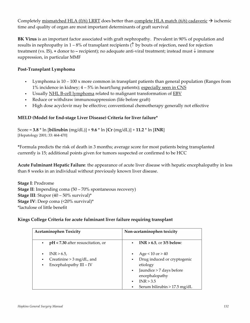
Hopkins General Surgery Manual 132
Completely mismatched HLA (0/6) LRRT does better than complete HLA match (6/6) cadaveric ischemic time and quality of organ are most important determinants of graft survival BK Virus is an important factor associated with graft nephropathy. Prevalent in 90% of population and results in nephropathy in 1 – 8% of transplant recipients (↑ by bouts of rejection, need for rejection treatment (vs. IS), + donor to – recipient); no adequate anti‐viral treatment; instead must ↓ immune suppression, in particular MMF Post‐Transplant Lymphoma
Lymphoma is 10 – 100 x more common in transplant patients than general population (Ranges from 1% incidence in kidney; 4 – 5% in heart/lung patients); especially seen in CNS
Usually NHL B‐cell lymphoma related to malignant transformation of EBV Reduce or withdraw immunosuppression (life before graft) High dose acyclovir may be effective; conventional chemotherapy generally not effective
MELD (Model for End‐stage Liver Disease) Criteria for liver failure* Score = 3.8 * ln [bilirubin (mg/dL)] + 9.6 * ln [Cr (mg/dL)] + 11.2 * ln [INR] [Hepatology 2001; 33: 464‐470] *Formula predicts the risk of death in 3 months; average score for most patients being transplanted currently is 15; additional points given for tumors suspected or confirmed to be HCC Acute Fulminant Hepatic Failure: the appearance of acute liver disease with hepatic encephalopathy in less than 8 weeks in an individual without previously known liver disease. Stage I: Prodrome Stage II: Impending coma (50 – 70% spontaneous recovery) Stage III: Stupor (40 – 50% survival)* Stage IV: Deep coma (<20% survival)* *lactulose of little benefit Kings College Criteria for acute fulminant liver failure requiring transplant
Acetaminophen Toxicity
Non‐acetaminophen toxicity
pH < 7.30 after resuscitation, or INR > 6.5, Creatinine > 3 mg/dL, and Encephalopathy III – IV
INR > 6.5, or 3/5 below: Age < 10 or > 40 Drug induced or cryptogenic
etiology Jaundice > 7 days before
encephalopathy INR > 3.5 Serum bilirubin > 17.5 mg/dL
Hopkins General Surgery Manual 133
Upper limits of acceptable cold ischemic times:
Heart: 6 hours Lung: 4 – 6 hours Liver: 24 hours Kidney: 48 hours Pancreas: 24 – 48 hours
Warm ischemic time must be less than 60 minutes Hepatic arterial thrombosis is main cause of immediate graft loss following liver transplant:
3 – 5 % in adults 5 – 8 % in children
HCC patients are candidates for liver transplant provided§: 1. A single tumor < 5 cm, or 2. Up to three tumors individually < 3 cm §Milan criteria Hepatorenal Syndrome
10% of hospitalized patients with cirrhosis and ascites develop progressive oliguria ↑Cr, ↓CO, ↓BP similar laboratory findings to prerenal azotemia (UOP < 500 mL/24 hours, UNa < 10 mEq/L, Uosm >
Posm) physiology: splanchnic vasodilation (associated with ↑ NO) ↓SVR ↓renal perfusion only effective treatment is hepatic transplantation renal function usually returns to normal
Hopkins General Surgery Manual 134
Nutrition Energy Capacity: (1 calorie = energy to take 1 mL water from 14.5 15.5 0C at 1 atm) Fat: 9 kcal/g Protein: 4 kcal/g (but aqueous, so only 1 – 2 kcal/g when utilized) Carb: 3.4 kcal/g RQ = ratio of CO2 produced to O2 consumed = 1.0 for carbs; 0.7 for fats; >1 for proteins Essential Amino Acids: 2L, 2T, VIP & Me (Leucine, Lysine, Threonine, Tryptophan, Valine, Isoleucine, Phenylalanine, & Methionine) [N]Balance = [N]In – [N]Out, where [N]In = [g protein/6.25 g protein per g N] [N]Out = [UUN (mg N/100 mL urine) * 1000 mL/L * 24 hour urine volume * g N/1000 mg N + 3] Branched Chain Amino Acids: leucine, isoleucine, valine (metabolized in muscle; all essential) Aromatic Amino Acids: tyrosine, tryptophan, phenylalanine (⅔ essential)
Glutamine is #1 AA in body; most rapidly used in stress; fuel of the enterocytes (when depleted brush border breakdown); the addition of glutamine to enteral or parenteral feeds may ↓ sepsis [Houdijk AP, et al. Lancet 1998;352:772] Hydroxybutarate: fuel of colonocyte Arginine: most important for immune function
In hepatic failure Minimize aromatic AA; give branched chain only In renal failure give essential AA only
Fatty Acid Metabolism: ‐medium chain FA can be absorbed directly via portal blood, hence bypassing the lymphatic system ‐long‐chain FA poorly tolerated by patients with compromised gut function; medium chain better, since absorbed directly
Three Main forms of Fat are found in the body: 1. Glycerides (95 – 98% of body stores), essential (see below) or nonessential; most dietary sources are medium (6 C) and long (> 11 C) 2. Phospholipids (mainly in cell membranes and myelin sheaths) 3. Sterols, comprised primarily of cholesterol Essential Fatty Acids: (unsaturated bond within the last 7 carbons of the FA chain at the methyl end) linoleic (TPN mostly consists of this), linolenic, arachidonic; can NOT be synthesized by humans
Ω‐6‐polyunsaturated fats (linoleic) precursors for PGs and leukotrienes Fat digestion: micelles to enterocytes chylomicrons to lymphatics (to junction LIJ/subclavian)
Hopkins General Surgery Manual 135
Energy Storage:
• Fats: 25% BW = fat; so if 70 kg 17 kg fat 160,000 kcal • Carbs: circulating ≈ 80 kcal; Liver glycogen ≈ 300 kcal; • Muscle glycogen ≈ 600 kcal (exhausted in ≈ 24 hours) • Proteins: 12 kg 48,000 kcal; but no access unless late starvation (Main advantage of dextrose in IVF is to obviate the need for protein catabolism)
½ glucose + ½ (fructose + galactose) 40% Liver glycogen + 60% Muscle glycogen Nonprotein calorie to gm Nitrogen ratio of 150:1 generally appropriate (both adults and children) *Patients with major burn (> 25% TBSA) have greatest caloric requirements Assessing Nutritional Status:
Albumin: 18 day T½ Prealbumin: 24 hour T½ Retinol binding protein: 12 hour T½ (most sensitive) Injured patients can maximally oxidize glucose at 5 – 6 mg/kg/min (above this osmotic diuresis, ↑respiratory quotient) Remember: Metabolic Alkalosis require K+ Marasmus = depletion of body fat; relative sparing of visceral protein (simple starvation) Kwashiorkor = acute visceral protein depletion (sparing of fat; acutely ill patients) Deficiencies:
Phosphorus: weakness, paresthesias Zinc: perioral rash, alopecia, poor wound healing, impaired immunity, change in taste Copper: anemia, neutropenia, pancytopenia Iron: anemia Linoleic acid: dermatitis, alopecia, blurred vision, paresthesias Selenium: cardiomyopathy, weakness, alopecia Vitamin A: night blindness, skin keratosis Chromium: glucose intolerance (relative diabetes), peripheral neuropathy Biotin: alopecia, neuritis
Hopkins General Surgery Manual 136
Fluids & Electrolytes
Sodium concentration must be corrected by 2 – 3 mEq per 100 mg/100 mL elevation in blood glucose above 100 [i.e. as BG↑ Na+↓]
Gastric losses (vomiting) usually require hypercholoremic replacement Post‐pyloric losses require balanced salt solution (except pancreatic fistula, which require high
HCO3 replacement) Patients with GI losses initially lose isotonic fluid but the body always tries to protect volume
status (even at the expense of tonicity)
Insensible Losses: Skin > Lung: total 600 – 900 mL/day 0.9% NaCl = 9 gm NaCl per L
HYPO Mg++ and Ca++ both have HYPERexcitability: ↑reflexes, tetany Cannot correct ↓Ca++ with ↓Mg++ since ↓Mg++ induces skeletal resistance to PTH and may impair PTH synthesis
Prolonged vomiting: results in hypokalemia, hypochloremia, metabolic alkalosis; early urine is alkaline…BUT as Na+ is conserved H+/K+ are lost with HCO3 resulting in paradoxical aciduria
Note: The most important treatment for hyperkalemia is Ca++ (vs. insulin, HCO3, etc.) because it is the only agent that actually stabilizes the myocardium
Mechanisms of Diarrhea: Osmotic Diarrhea: accumulation of poorly absorbed solutes in lumen secretion of H2O into lumen Secretory Diarrhea: excessive electrolyte secretion (toxins, neuroendocrine tumors) stimulate cAMP production Inhibition of Absorption: unabsorbed free fatty acids or bile salts decreased H2O absorption
Sweat: normally hypotonic, but can approach isotonicity during periods of high secretion; Na+ secretion parallels Cl‐ (both < plasma); K+ approaches plasma concentration; urea and NH3 >> plasma concentrations
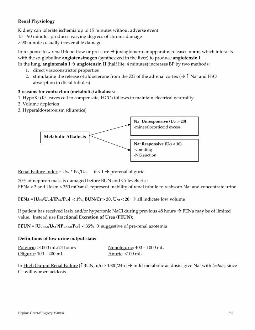
Hopkins General Surgery Manual 137
Renal Physiology
Kidney can tolerate ischemia up to 15 minutes without adverse event 15 – 90 minutes produces varying degrees of chronic damage > 90 minutes usually irreversible damage
In response to ↓ renal blood flow or pressure juxtaglomerular apparatus releases renin, which interacts with the α2‐globuline angiotensinogen (synthesized in the liver) to produce angiotensin I. In the lung, angiotensin I angiotensin II (half life: 4 minutes) increases BP by two methods:
1. direct vasoconstrictor properties 2. stimulating the release of aldosterone from the ZG of the adrenal cortex ( ↑ Na+ and H2O
absorption in distal tubules)
3 reasons for contraction (metabolic) alkalosis: 1. HypoK+ (K+ leaves cell to compensate, HCO3 follows to maintain electrical neutrality 2. Volume depletion 3. Hyperaldosteronism (diuretics)
Renal Failure Index = UNa * PCr/UCr if < 1 prerenal oliguria
70% of nephron mass is damaged before BUN and Cr levels rise FENa > 3 and Uosm < 350 mOsm/L represent inability of renal tubule to reabsorb Na+ and concentrate urine FENa = [UNa/UCr]/[PNa/PCr] < 1%, BUN/Cr > 30, UNa < 20 all indicate low volume If patient has received lasix and/or hypertonic NaCl during previous 48 hours FENa may be of limited value. Instead use Fractional Excretion of Urea (FEUN):
FEUN = [UUREA/UCr]/[PUREA/PCr] < 35% suggestive of pre‐renal azotemia Definitions of low urine output state:
Polyuric: >1000 mL/24 hours Nonoliguric: 400 – 1000 mL Oliguric: 100 – 400 mL Anuric: <100 mL
In High Output Renal Failure [↑BUN, u/o > 1500/24h] mild metabolic acidosis: give Na+ with lactate, since Cl‐ will worsen acidosis
Metabolic Alkalosis Na+ Responsive (UCl < 10) ‐vomiting ‐NG suction
Na+ Unresponsive (UCl > 20) ‐mineralocorticoid excess
Hopkins General Surgery Manual 138
Differential Dx of elevated BUN or Cr (one out of proportion to the other):
↑Cr/BUN ↑BUN/Cr Renal failure Dehydration Muscle breakdown +N‐balance GI bleed Hepatic failure Transtubular K+ Gradient (TTKG)
[Urine K/Plasma K, mEq/L] divided by [Urine osm/Plama osm, mosm/kg]
Normal is 8 – 9; may be up to 1 with potassium loading If K+ is high and TTKG < 7 implies hypoaldostoronism
Appropriate responses Hypokalemia <3 Hyperkalemia >10 If the specific gravity of urine is normal kidney is working, UNLESS artificial osmoles are present including:
• Mannitol • IV contrast • High glucose load • Methanol
Hopkins General Surgery Manual 139
Immunology/Infections IgG: opsonin (along with IgM) to fix complement (2 IgG’s or 1 IgM), #1 in serum; crosses placenta IgM: made first; levels ↓ after splenectomy IgA: in secretions IgE: allergic reactions, type I hypersensitivity IgD: largely unknown Complement: C3a, C5a and anaphylatocins; C5 – 9: MAC (membrane attack complex) Hypersensitivity Type I: immediate; IgE mediated; e.g. anaphylaxis Type II: cytotoxic reactions; IgG or IgM; e.g. ABO/Rh incompatibility Type III: immune‐complex mediated; deposition of complex; e.g. serum sickness, rheumatoid arthritis Type IV: delayed‐type: T Cells (CD4+); e.g. contact dermatitis HIV Patients ‐have increased risk of developing NHL (high‐grade B‐cell) and Kaposi’s sarcoma Advantages of Zosyn
I. vs. 3rd generation cephalosporins ‐enterococci, MSSA ‐pseudomonas, klebsiela ‐4+ anaerobes
II. vs. quinolones ‐enterococci, MSSA ‐pseudomonas ‐4+ anaerobes
III. vs. Unasyn ‐MSSA ‐E.coli, pseudomonas, other G‐ ‐4+ anaerobes
IV. Timentin (better for stenotrophomonas) ‐enterococci, MSSA ‐E.coli, pseudomonas, other G‐ ‐4+ anaerobes Remote infection (e.g. UTI) ↑ the risk of surgical site infection by at least 7%
Hopkins General Surgery Manual 140
Burns 1. Silver nitrate: Broad spectrum, painless, cheap, poor eschar penetration, may cause electrolyte imbalance 2. Silver sulfadiazine (Silvadine): Painless, no electrolyte abnormalities, no occlusive dressing required, little eschar penetration; misses Pseudomonas, idiosyncratic neutropenia; good for small burns 3. Mafenide: Penetrates eschars, broad spectrum (but misses staph); pain and burning on application; 7% have allergic reactions; may cause acid‐base disturb (metabolic acidosis); agent of choice in already‐contaminated burns; water‐soluble Parkland Formula for Burns
*Add Maintenance Fluids to below:*
1. First 24 hours: 4 mL/kg/%BSA. Half over 8 hours, then rest over 16 hours. 2. Second 24 hours: Fluid requirements are 50 – 75% of the first days. Use weight, electrolytes, UOP &
NGT to determine concentration and rate. 3. Withhold K+ for first 48 hours because of large tissue release. 4. Keep UOP @ 0.5 mL/kg/hour
Burn patients initially have drop in CO then are HYPERdynamic Burn Wound Infection
Reduced by: 1. Aggressive resuscitation 2. Early debridement 3. Topical antibiotic therapy To confirm infection need biopsy with quantitative culture (105): must include normal and burned skin (2x2 cm with normal underlying skin)
Hopkins General Surgery Manual 141
Skin & Wound Healing Three major stages of wound healing: 1. Inflammatory Phase (10 minutes – 2 weeks) 2. Proliferative Phase (3 – 6 weeks) 3. Remodeling Phase (up to 1 year) Inflammatory Phase: Hemostasis & Clot formation
Platelet plug mediated by thromboxane A2, thrombin, PF4, C5a (most important complement) Monocytes must be present for normal wound healing Collagen and basement membrane proteins clotting factor activation Vasoconstriction decrease blood loss and allow clot formation (< 24 hours) Platelet degranulation: PDGF and TGF‐β; chemotaxis and proliferation of inflammatory cells Vasodilation (> 24 – 48 hours) supplies cells and substrates for wound repair
Proliferative Phase:
Formation of matrix of fibrin and fibronectin Initiation of collagen formation Proliferation of fibroblasts Growth factors from macrophages initiate angiogenesis (especially FGF) Cross‐linking of collagen requires Vit C Wound is now a scar
Remodeling Phase:
Collagen equilibrium Increase tensile strength (abundance of Type I cross‐linking) Diminishing capillary density and fibroblasts
Tensile Strength of Wound*: Early: fibrin Late: collagen cross‐linking *Tensile strength is never equal to pre‐wound 105 organisms/cm2 is enough to retard wound healing As time progresses: Type III (proliferation) collagen and Type I (mature) Ultimately the ratio of I:III is 8:1 (i.e. that of normal skin)
I Most abundant, found in scar II In cartilage III In would healing (low in Ehler Danlos) IV In basement membrane V Found in cornea
Hopkins General Surgery Manual 142
Pharmacology Clues to drug overdose:
1. Eyes i) Miosis: opiates, org. phos, barbs ii) Mydriasis: amphetamines, cocaine, anti‐chol, ethanol, mushrooms, LSD iii) Nystagmus: PCP, phenytoin, ethanol, VPA
2. Mouth i) Dry: anti‐chol, opiates, SSRIs ii) Very salivary: org. phos
3. Skin look for needle tracks i) Hot, dry: anti‐chol ii) Very pink: CO iii) Very sweaty: org. phos
Pharmacology
↑P–450 ↓P–450 Phenobarb INH Rifampin Cimetidine PTN Benzos Carbamazepine Phenothyazines TMP‐SMX
Lidocaine toxicity: tinnitus, peri‐oral numbness neuro symptoms cardiovascular changes
Local Anesthetics (2 classes)
Amino Esters Amino Amides* Tetracaine Lidocaine Cocaine Mepivacaine Procaine Bupivicaine Chloroprocaine Etidocaine * All have an “i” before ‐caine
Malignant Hyperthermia: can be genetically transferred; triggered by halogenated inhalational agents (1 in 250,000); can also be triggered by succinylcholine (1 in 60,000); earliest sign is rise in CO2; hyperthermia is a relatively late finding; treat with dantrolene.
Ketamine: does not ↓ BP, but does ↑ ICP; good in children; avoid in head trauma, cardiac disease
Propofol: rapid onset and short duration; ideal for patients with altered neuro exam to allow for frequent neuro examinations; does ↓ BP; does not provide analgesia
4 Components of (Informed) Consent: 1. Disclosure 2. Comprehension 3. Competency 4. Voluntariness
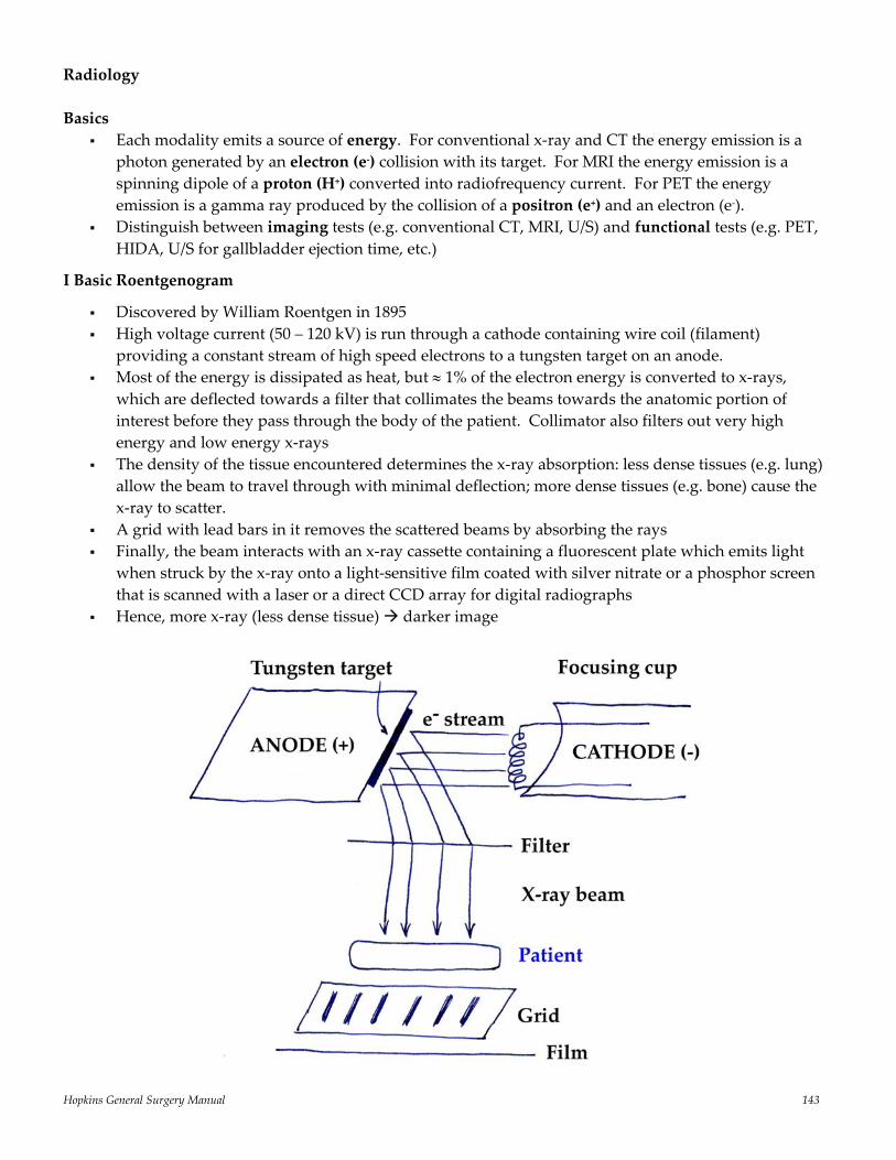
Hopkins General Surgery Manual 143
Radiology Basics
Each modality emits a source of energy. For conventional x‐ray and CT the energy emission is a photon generated by an electron (e‐) collision with its target. For MRI the energy emission is a spinning dipole of a proton (H+) converted into radiofrequency current. For PET the energy emission is a gamma ray produced by the collision of a positron (e+) and an electron (e‐).
Distinguish between imaging tests (e.g. conventional CT, MRI, U/S) and functional tests (e.g. PET, HIDA, U/S for gallbladder ejection time, etc.)
I Basic Roentgenogram
Discovered by William Roentgen in 1895 High voltage current (50 – 120 kV) is run through a cathode containing wire coil (filament)
providing a constant stream of high speed electrons to a tungsten target on an anode. Most of the energy is dissipated as heat, but ≈ 1% of the electron energy is converted to x‐rays,
which are deflected towards a filter that collimates the beams towards the anatomic portion of interest before they pass through the body of the patient. Collimator also filters out very high energy and low energy x‐rays
The density of the tissue encountered determines the x‐ray absorption: less dense tissues (e.g. lung) allow the beam to travel through with minimal deflection; more dense tissues (e.g. bone) cause the x‐ray to scatter.
A grid with lead bars in it removes the scattered beams by absorbing the rays Finally, the beam interacts with an x‐ray cassette containing a fluorescent plate which emits light
when struck by the x‐ray onto a light‐sensitive film coated with silver nitrate or a phosphor screen that is scanned with a laser or a direct CCD array for digital radiographs
Hence, more x‐ray (less dense tissue) darker image
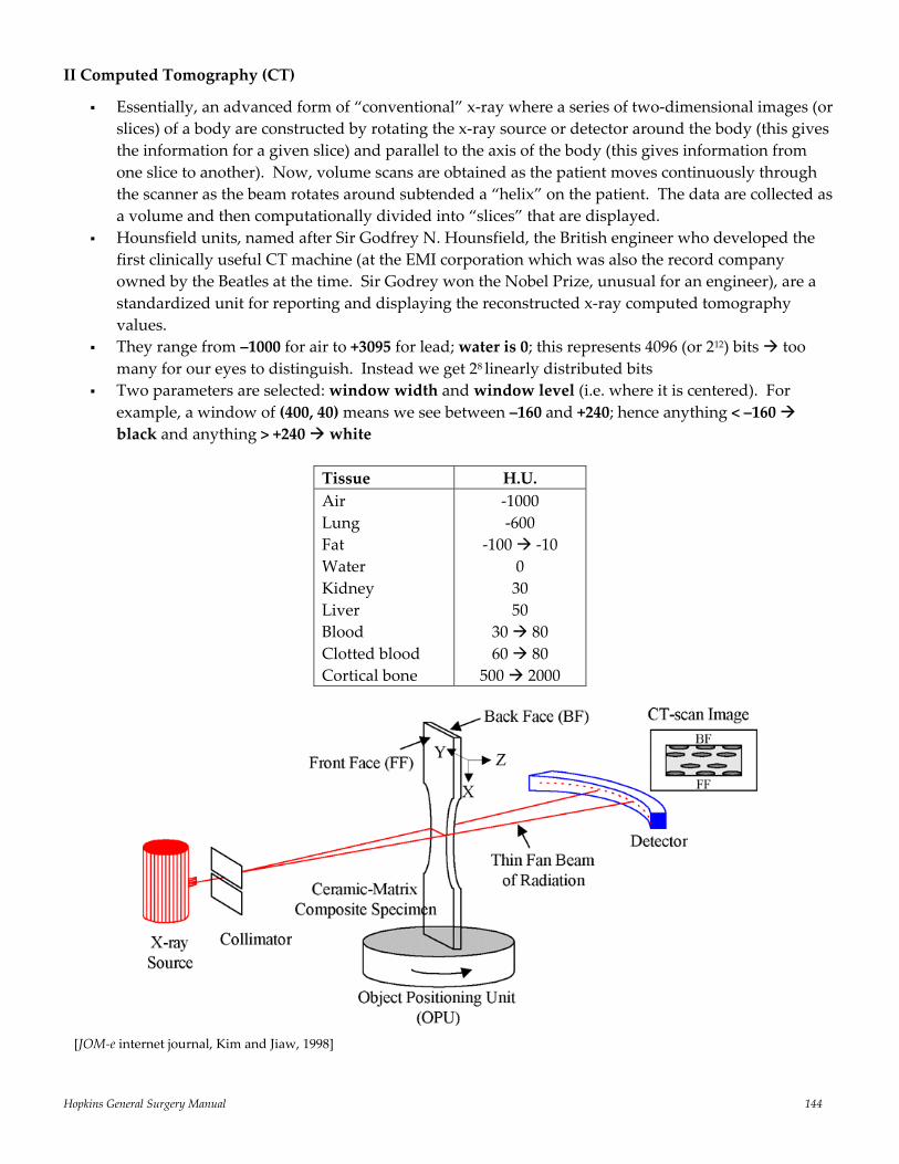
Hopkins General Surgery Manual 144
II Computed Tomography (CT)
Essentially, an advanced form of “conventional” x‐ray where a series of two‐dimensional images (or slices) of a body are constructed by rotating the x‐ray source or detector around the body (this gives the information for a given slice) and parallel to the axis of the body (this gives information from one slice to another). Now, volume scans are obtained as the patient moves continuously through the scanner as the beam rotates around subtended a “helix” on the patient. The data are collected as a volume and then computationally divided into “slices” that are displayed.
Hounsfield units, named after Sir Godfrey N. Hounsfield, the British engineer who developed the first clinically useful CT machine (at the EMI corporation which was also the record company owned by the Beatles at the time. Sir Godrey won the Nobel Prize, unusual for an engineer), are a standardized unit for reporting and displaying the reconstructed x‐ray computed tomography values.
They range from –1000 for air to +3095 for lead; water is 0; this represents 4096 (or 212) bits too many for our eyes to distinguish. Instead we get 28 linearly distributed bits
Two parameters are selected: window width and window level (i.e. where it is centered). For example, a window of (400, 40) means we see between –160 and +240; hence anything < –160 black and anything > +240 white
Tissue H.U. Air Lung Fat Water Kidney Liver Blood Clotted blood Cortical bone
‐1000 ‐600
‐100 ‐10 0 30 50
30 80 60 80
500 2000
[JOM‐e internet journal, Kim and Jiaw, 1998]
Hopkins General Surgery Manual 145
Use the gallbladder as an “internal standard” of 0 H.U. (i.e. water)
If there is contrast in the peritoneum, look for 3 things:
1. Ruptured viscous (general rules) Stomach: ↑ free air, ↑ fluid Small bowel: ↓ free air, ↑ fluid Colon: ↑ free air, ↓ fluid
2. Vessel extravasation: must see clot and contrast (clotted blood will be brighter than “liquid” blood) on pre contrast (but the reverse on post contrast)
3. Ruptured bladder (intraperitoneal): must look at delayed views
The Pouch of Douglass will normally hold ≈300 – 400 mL of blood before overflowing usually to Morrison’s Pouch then to right subphrenic recess III Magnetic Resonance Imaging (MRI)
An H atom contains a single proton and behaves like a spinning bar magnet that will align with a magnetic field created by the solenoid of the MR scanner. Before a radiofrequency (RF) is applied the protons are aligned. The RF deflects them some number of degrees from the Z axis of the magnet they then “relax” back to being aligned, although, the protons are spinning all the time at the Larmor frequency. The coil picks up the spinning magnetic component that is perpendicular to the main magnetic field and induces a current (Faraday’s Law) this is the signal from MRI.
The RF field is applied to the patient to make the bar magnet stand up at 90o (perpendicular) to the spine. A wire (antenna) parallel to the spine, outside of the patient’s body, has an alternating current formed in it, which determines the signal intensity. The magnitude of is proportional to the number of protons and the extent to which their spins are perpendicular to the Z axis. Protons predictably realign with the spine, while continuing to preceess. So there are 2 things at play here:
1. The shortening length of the spinning magnet, and 2. The lengthening magnet parallel to the spine as more and more protons realign.
The longitudinal (or restoration with the main magnetic field) realignment is known as T1 RELAXATION. It is a constant for a given tissue and, by definition, it is the time required for 63% of the protons to realign with the main magnetic field. It’s exponential, so after 1 T1 63% have realigned, after 2 T1’s 86% have realigned, after 3 T1’s 95% have realigned, etc. [i.e. 1 – (0.37)3]
In reality, the spinning magnet shrinks even faster than predicted by the T1 decay because the spinning protons actually lose coherence and cease to spin in unison. This causes some of the remaining protons to cancel out each other’s signal and further reduce the signal generated in the antenna. This process of protons randomly losing coherence and canceling out each other’s magnetic fields is known as T2 RELAXATION. T1 and T2 are independent of each other but simultaneous and T2 relaxation is usually much faster than T1 relaxation. Again, the 63% decay is chosen to describe the constant T2. For example, after 3 T2’s have elapsed the net strength of the magnetic field in the transverse plane will be 5% of original strength.
Hopkins General Surgery Manual 146
MRI exploits the fact that different tissues and states of pathology have different T1 and T2. The most common way to do this is to acquire images using a spin‐echo pulse sequence, where 2 parameters are utilized: TR (repetition time) and TE (echo time). TR is the time between RF pulses. This is the time during which T1 relaxation occurs. At the end of TR, another RF pulse is applied and those protons that have realigned with the magnetic field will be brought up again and produce current in the antenna. So TR adjusts the amount of signal received from a tissue depending on its T1 value (e.g. if TR = 3T1, the signal will be 95% intensity).
TE is the time between the first RF pulse and the observation of signal, that is, the time for T2 relaxation to occur. For example, setting TE = 2T2 would result in 15% net remaining signal intensity produced by the component of the protons in the transverse plane.
Contrast between the two tissues can be produced by the interaction of the two parameters. A very long TR will eliminate difference between tissues with different T1 (e.g. TR = 2500 ms will allow 5T1’s to elapse whether T1 is 250 ms or 500 ms). A very short TE would similarly minimize the difference in T2 decay by different tissues.
• T1 Weighted Image utilizes a short TR (to maximize T1 contrast) and a short TE (to minimize T2 contrast). Anatomic definition is superior to T2 weighted images. Fat is dark and fluid is bright
• T2 Weighted Image utilizes a long TR (to minimize T1 contrast) and a long TE (to maximize T2 contrast). Fat is bright, fluid is dark
• PD (Proton Density) is somewhere between T1 and T2. It utilizes short TE (14 – 20 ms) and long TR (4000 ms) to eliminate differences in T1 and T2 i.e. differences are only due to the proton density of each tissue. Fat and fluid are bright.
HASTE (HAlf‐fourier Single shot Turbo spin‐Echo), FLAIR (FLuid Attenuation Inversion Recovery), STIR (Short T1 Inversion Recovery), etc. are various algorithms based on different TR/TE times and methods of data acquisition. However, the basic principles described above remain the same.
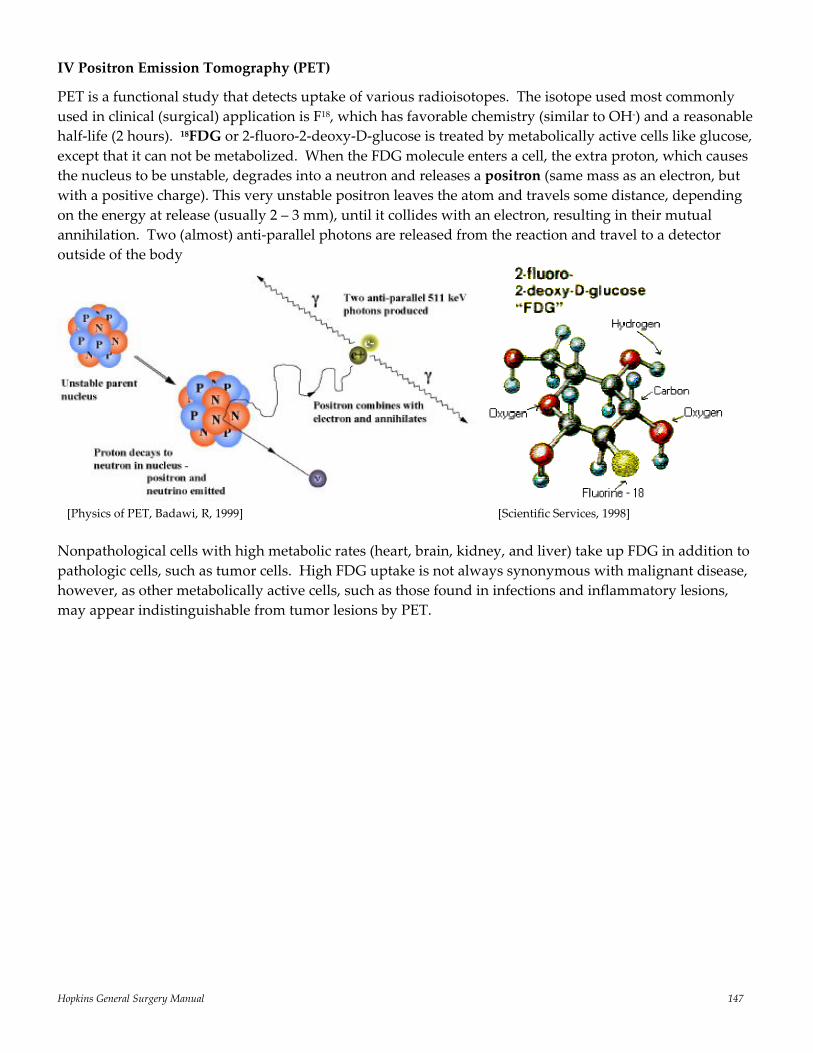
Hopkins General Surgery Manual 147
IV Positron Emission Tomography (PET)
PET is a functional study that detects uptake of various radioisotopes. The isotope used most commonly used in clinical (surgical) application is F18, which has favorable chemistry (similar to OH‐) and a reasonable half‐life (2 hours). 18FDG or 2‐fluoro‐2‐deoxy‐D‐glucose is treated by metabolically active cells like glucose, except that it can not be metabolized. When the FDG molecule enters a cell, the extra proton, which causes the nucleus to be unstable, degrades into a neutron and releases a positron (same mass as an electron, but with a positive charge). This very unstable positron leaves the atom and travels some distance, depending on the energy at release (usually 2 – 3 mm), until it collides with an electron, resulting in their mutual annihilation. Two (almost) anti‐parallel photons are released from the reaction and travel to a detector outside of the body
Nonpathological cells with high metabolic rates (heart, brain, kidney, and liver) take up FDG in addition to pathologic cells, such as tumor cells. High FDG uptake is not always synonymous with malignant disease, however, as other metabolically active cells, such as those found in infections and inflammatory lesions, may appear indistinguishable from tumor lesions by PET.
[Physics of PET, Badawi, R, 1999] [Scientific Services, 1998]
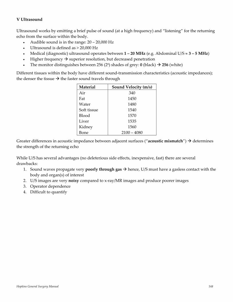
Hopkins General Surgery Manual 148
V Ultrasound Ultrasound works by emitting a brief pulse of sound (at a high frequency) and “listening” for the returning echo from the surface within the body.
• Audible sound is in the range: 20 – 20,000 Hz • Ultrasound is defined as > 20,000 Hz • Medical (diagnostic) ultrasound operates between 1 – 20 MHz (e.g. Abdominal U/S ≈ 3 – 5 MHz) • Higher frequency superior resolution, but decreased penetration • The monitor distinguishes between 256 (28) shades of grey: 0 (black) 256 (white)
Different tissues within the body have different sound‐transmission characteristics (acoustic impedances); the denser the tissue the faster sound travels through
Material Sound Velocity (m/s) Air Fat Water Soft tissue Blood Liver Kidney Bone
340 1450 1480 1540 1570 1535 1560
2100 – 4080
Greater differences in acoustic impedance between adjacent surfaces (“acoustic mismatch”) determines the strength of the returning echo While U/S has several advantages (no deleterious side effects, inexpensive, fast) there are several drawbacks:
1. Sound waves propagate very poorly through gas hence, U/S must have a gasless contact with the body and organ(s) of interest
2. U/S images are very noisy compared to x‐ray/MR images and produce poorer images 3. Operator dependence 4. Difficult to quantify
Hopkins General Surgery Manual 149
Statistics in Medicine Type I Error: Reject the null hypothesis when you shouldn’t (probability α)
Type II Error: Failing to reject the null hypothesis when you should (probability β)
Power: The probability of avoiding Type II error (1 – β). Anther way of saying this: “The ability of your statistical test to detect a difference between 2 populations should a difference exist”. See more on statistical power below. 3 Broad Classes of Statistical Pitfalls
I Sources of Bias Include errors of sampling bias (studied population dose not adequately represent population of interest) and data gathering (questionnaires with leading questions).
II Errors in Methodology Three most common include: designing experiments with insufficient power (see below), failing to pay attention to errors in measurement (understand the difference between reliability and validity, see below), and going on “fishing expeditions” (making multiple comparisons) without appropriately correcting (see the Bonferroni correction, below).
III Interpretation of Results (Misapplication of statistical methods) Include errors of statistical assumptions (e.g. using a method such as ANOVA which relies on assumptions of normality and independence, when such conditions are not met), misunderstandings of statistical significance, and assessing causality (see below). Reliability: the ability of a test to measure the same thing each time it is used (How close are the darts to each other after repeatedly throwing them at the dart board?). Even if the test is meaningless, it should yield the same results over time if used on subjects with the same characteristics.
Validity: the extent to which a test measures the outcome it was designed to measure (How close are the darts to the center of the dart board?). Bonferroni correction: a statistical adjustment for the multiple comparisons often made during statistical “fishing expeditions”. This correction raises the standard of proof needed to justify the significance of a finding when evaluating a wide range of hypotheses simultaneously. If testing n outcomes (instead of 1), divide the α by n. For example, if trying to find the association between body weight and 25 different types of cancer, divided the traditional α of 0.05 by 25 (0.05/25 = 0.002) to ensure an overall risk of Type I error equal to or less than 0.05. Be aware, however, that application of the Bonferroni correction can result in a loss of substantial precision. Causality: Observational studies are very limited in their ability to make causal inferences; doing so requires random assignment. Hence, correlation can be used to infer causation if the interventions are randomly assigned (e.g. dose of drug vs. outcome).
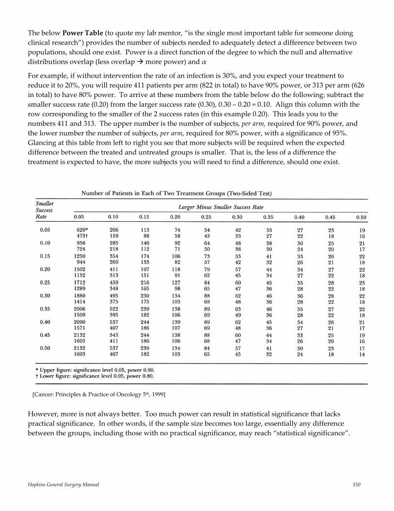
Hopkins General Surgery Manual 150
The below Power Table (to quote my lab mentor, “is the single most important table for someone doing clinical research”) provides the number of subjects needed to adequately detect a difference between two populations, should one exist. Power is a direct function of the degree to which the null and alternative distributions overlap (less overlap more power) and α
For example, if without intervention the rate of an infection is 30%, and you expect your treatment to reduce it to 20%, you will require 411 patients per arm (822 in total) to have 90% power, or 313 per arm (626 in total) to have 80% power. To arrive at these numbers from the table below do the following: subtract the smaller success rate (0.20) from the larger success rate (0.30), 0.30 – 0.20 = 0.10. Align this column with the row corresponding to the smaller of the 2 success rates (in this example 0.20). This leads you to the numbers 411 and 313. The upper number is the number of subjects, per arm, required for 90% power, and the lower number the number of subjects, per arm, required for 80% power, with a significance of 95%. Glancing at this table from left to right you see that more subjects will be required when the expected difference between the treated and untreated groups is smaller. That is, the less of a difference the treatment is expected to have, the more subjects you will need to find a difference, should one exist.
However, more is not always better. Too much power can result in statistical significance that lacks practical significance. In other words, if the sample size becomes too large, essentially any difference between the groups, including those with no practical significance, may reach “statistical significance”.
[Cancer: Principles & Practice of Oncology 5th, 1999]
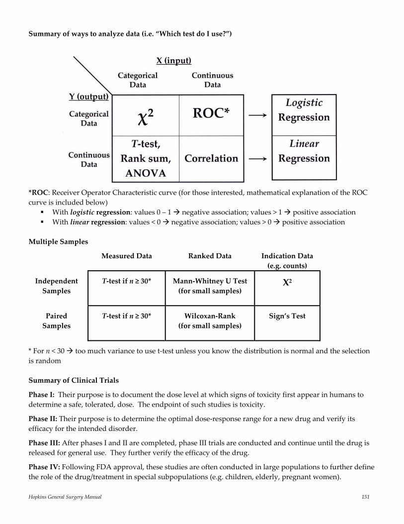
Hopkins General Surgery Manual 151
Summary of ways to analyze data (i.e. “Which test do I use?”)
*ROC: Receiver Operator Characteristic curve (for those interested, mathematical explanation of the ROC curve is included below)
With logistic regression: values 0 – 1 negative association; values > 1 positive association With linear regression: values < 0 negative association; values > 0 positive association
Multiple Samples
Measured Data Ranked Data Indication Data (e.g. counts)
Independent Samples
T‐test if n ≥ 30*
Mann‐Whitney U Test (for small samples)
Χ2
Paired Samples
T‐test if n ≥ 30*
Wilcoxan‐Rank (for small samples)
Sign’s Test
* For n < 30 too much variance to use t‐test unless you know the distribution is normal and the selection is random Summary of Clinical Trials
Phase I: Their purpose is to document the dose level at which signs of toxicity first appear in humans to determine a safe, tolerated, dose. The endpoint of such studies is toxicity.
Phase II: Their purpose is to determine the optimal dose‐response range for a new drug and verify its efficacy for the intended disorder.
Phase III: After phases I and II are completed, phase III trials are conducted and continue until the drug is released for general use. They further verify the efficacy of the drug.
Phase IV: Following FDA approval, these studies are often conducted in large populations to further define the role of the drug/treatment in special subpopulations (e.g. children, elderly, pregnant women).
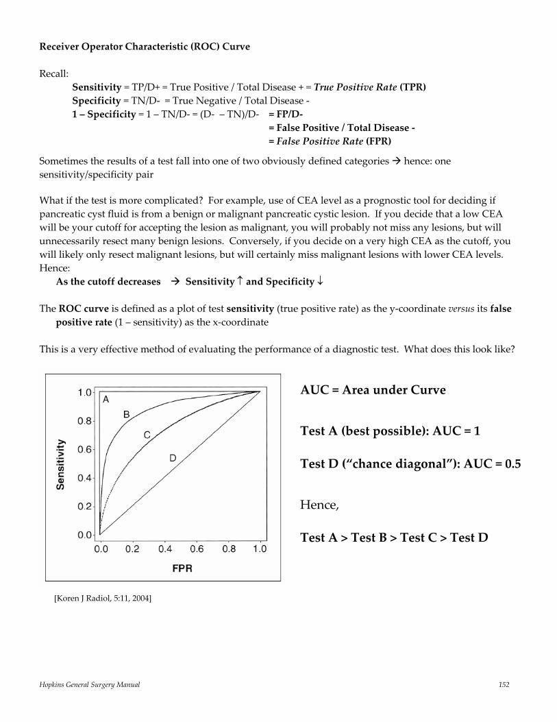
Hopkins General Surgery Manual 152
Receiver Operator Characteristic (ROC) Curve Recall:
Sensitivity = TP/D+ = True Positive / Total Disease + = True Positive Rate (TPR) Specificity = TN/D‐ = True Negative / Total Disease ‐ 1 – Specificity = 1 – TN/D‐ = (D‐ – TN)/D‐ = FP/D‐ = False Positive / Total Disease ‐ = False Positive Rate (FPR)
Sometimes the results of a test fall into one of two obviously defined categories hence: one sensitivity/specificity pair
What if the test is more complicated? For example, use of CEA level as a prognostic tool for deciding if pancreatic cyst fluid is from a benign or malignant pancreatic cystic lesion. If you decide that a low CEA will be your cutoff for accepting the lesion as malignant, you will probably not miss any lesions, but will unnecessarily resect many benign lesions. Conversely, if you decide on a very high CEA as the cutoff, you will likely only resect malignant lesions, but will certainly miss malignant lesions with lower CEA levels. Hence:
As the cutoff decreases Sensitivity ↑ and Specificity ↓
The ROC curve is defined as a plot of test sensitivity (true positive rate) as the y‐coordinate versus its false positive rate (1 – sensitivity) as the x‐coordinate
This is a very effective method of evaluating the performance of a diagnostic test. What does this look like?
AUC = Area under Curve
Test A (best possible): AUC = 1
Test D (“chance diagonal”): AUC = 0.5
Hence,
Test A > Test B > Test C > Test D
[Koren J Radiol, 5:11, 2004]
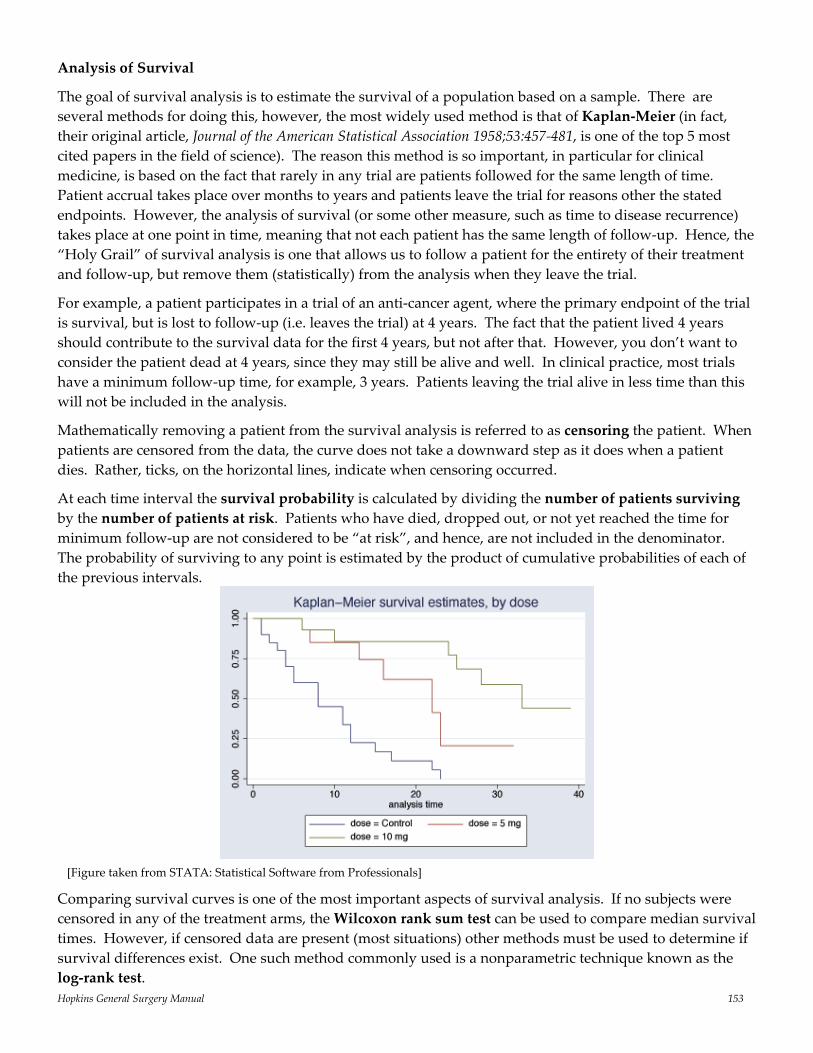
Hopkins General Surgery Manual 153
Analysis of Survival
The goal of survival analysis is to estimate the survival of a population based on a sample. There are several methods for doing this, however, the most widely used method is that of Kaplan‐Meier (in fact, their original article, Journal of the American Statistical Association 1958;53:457‐481, is one of the top 5 most cited papers in the field of science). The reason this method is so important, in particular for clinical medicine, is based on the fact that rarely in any trial are patients followed for the same length of time. Patient accrual takes place over months to years and patients leave the trial for reasons other the stated endpoints. However, the analysis of survival (or some other measure, such as time to disease recurrence) takes place at one point in time, meaning that not each patient has the same length of follow‐up. Hence, the “Holy Grail” of survival analysis is one that allows us to follow a patient for the entirety of their treatment and follow‐up, but remove them (statistically) from the analysis when they leave the trial.
For example, a patient participates in a trial of an anti‐cancer agent, where the primary endpoint of the trial is survival, but is lost to follow‐up (i.e. leaves the trial) at 4 years. The fact that the patient lived 4 years should contribute to the survival data for the first 4 years, but not after that. However, you don’t want to consider the patient dead at 4 years, since they may still be alive and well. In clinical practice, most trials have a minimum follow‐up time, for example, 3 years. Patients leaving the trial alive in less time than this will not be included in the analysis.
Mathematically removing a patient from the survival analysis is referred to as censoring the patient. When patients are censored from the data, the curve does not take a downward step as it does when a patient dies. Rather, ticks, on the horizontal lines, indicate when censoring occurred.
At each time interval the survival probability is calculated by dividing the number of patients surviving by the number of patients at risk. Patients who have died, dropped out, or not yet reached the time for minimum follow‐up are not considered to be “at risk”, and hence, are not included in the denominator. The probability of surviving to any point is estimated by the product of cumulative probabilities of each of the previous intervals.
Comparing survival curves is one of the most important aspects of survival analysis. If no subjects were censored in any of the treatment arms, the Wilcoxon rank sum test can be used to compare median survival times. However, if censored data are present (most situations) other methods must be used to determine if survival differences exist. One such method commonly used is a nonparametric technique known as the log‐rank test.
[Figure taken from STATA: Statistical Software from Professionals]

Hopkins General Surgery Manual 154
Notes

Hopkins General Surgery Manual 155
Notes

Hopkins General Surgery Manual 156
Notes

Hopkins General Surgery Manual 157
Notes

Hopkins General Surgery Manual 158
Notes