atherosclerotic geometries exacerbate pathological ... · atherosclerotic geometries exacerbate...
TRANSCRIPT
Atherosclerotic geometries exacerbate pathologicalthrombus formation poststenosis in a von Willebrandfactor-dependent mannerErik Westeina,b,1,2, Andries D. van der Meerc,1, Marijke J. E. Kuijpersa, Jean-Philippe Frimatc, Albert van den Bergc,and Johan W. M. Heemskerka,2
aDepartment of Biochemistry, Cardiovascular Research Institute Maastricht, Maastricht University, 6200 MD Maastricht, The Netherlands; bDepartmentof Atherothrombosis and Vascular Biology, Baker International Diabetes Institute, Heart and Diabetes Institute, Melbourne, Victoria 8008, Australia;and cBIOS/Lab on a Chip Group, MESA+ Institute for Nanotechnology, University of Twente, 7500 AE Enschede, The Netherlands
Edited by David A. Weitz, Harvard University, Cambridge, MA, and approved December 5, 2012 (received for review June 11, 2012)
Rupture of a vulnerable atherosclerotic plaque causes thrombusformation and precipitates cardiovascular diseases. In addition tothe thrombogenic content of a plaque, also the hemodynamic mi-croenvironment plays a major role in thrombus formation. Howthe altered hemodynamics around a plaque promote pathologicalthrombus formation is not well understood. In this study, weprovide evidence that plaque geometries result in fluid mechanicalconditions that promote platelet aggregation and thrombus forma-tion by increased accumulation and activity of vonWillebrand factor(vWF) at poststenotic sites. Resonant-scanning multiphoton micros-copy revealed that in vivo arterial stenosis of a damaged carotidartery markedly increased platelet aggregate formation in the ste-notic outlet region. Complementary in vitro studies using microflui-dic stenotic chambers, designed to mimic the flow conditions ina stenotic artery, showed enhanced platelet aggregation in the ste-notic outlet region at 60–80% channel occlusion over a range ofinput wall shear rates. The poststenotic thrombus formation wascritically dependent on bloodborne vWF and autocrine plateletstimulation. In stenotic chambers containing endothelial cells, flowprovoked increased endothelial vWF secretion in the stenotic outletregion, contributing to exacerbated platelet aggregation. Taken to-gether, this study identifies a role for the shear-sensitive proteinvWF in transducing hemodynamic forces that are present arounda stenosis to a prothrombogenic microenvironment resulting in spa-tially confined and exacerbated platelet aggregation in the stenosisoutlet region. The developed stenotic microfluidic chamber offersa realistic platform for in vitro evaluation of shear-dependent throm-bus formation in the setting of atherosclerosis.
Atherosclerosis is characterized by progressive growth of ath-erosclerotic plaques in the arterial circulation. In an advanced
stage of atherosclerosis, plaques will become stenotic and causeprogressive obstruction of the arterial lumen. A current theorem isthat rupture or erosion of a stenotic plaque is required for arterialthrombus formation and ensuing thrombotic complications, suchas pulmonary embolism, myocardial infarction, or stroke (1). Bet-ter understanding of the precise regulation of thrombus formationin atherosclerotic vessel segments is critical for the improvement ofcurrent antithrombotic treatments.Biochemical and hemodynamic factors contribute to thrombus
formation at a vulnerable vessel wall (2–4). Thrombogenic com-ponents exposed upon plaque rupture—e.g. tissue factor, colla-gen and von Willebrand factor (vWF)—trigger initial activationof platelets and the coagulation system (5–7). Stable recruitmentof platelets under flow to a growing thrombus involves vWF andfibrinogen as main ligands, which interact with glycoprotein (GP)Ib-V-IX and integrin αIIbβ3, respectively, as well as autocrineagonists produced by platelets themselves (8, 9). There is growingappreciation of the interplay of hemodynamics with this multi-receptor process in plaque-containing areas (10). Intraluminalgrowth of a developing plaque, even in the absence of thrombusformation, will gradually alter the local blood flow dynamics (11).The flow disturbances at sites of severe stenosis may even lead to
fatal occlusive thrombus formation in humans (12). Simulationsindicate that plaque geometries induce gradients in pressure, flowvelocity, and shear stress of the local blood flow, as a consequenceof which autocrine agonists may get trapped that enhance plate-let activation (13). However, the mechanisms dictating shear-dependent platelet aggregation at sites of atherosclerotic stenosisremain poorly understood.In the present study, we used fast resonant-scanning multi-
photon microscopy to investigate stenosis-dependent plateletaggregation in the murine carotid artery in vivo. In addition, wedeveloped a microfluidic platform with stenotic flow channels toinvestigate platelet aggregation at defined positions relative toplaque-like geometries. Our data show that, both in vivo and invitro, the shear stress conditions downstream of a stenotic siteaggravate local platelet aggregation in a strongly vWF-dependentmanner. This work highlights a central role for the shear-sensitiveprotein vWF in converting the fluid forces in the microenviron-ment downstream of a stenotic geometry into prothrombotic re-sponses in a partly occluded artery.
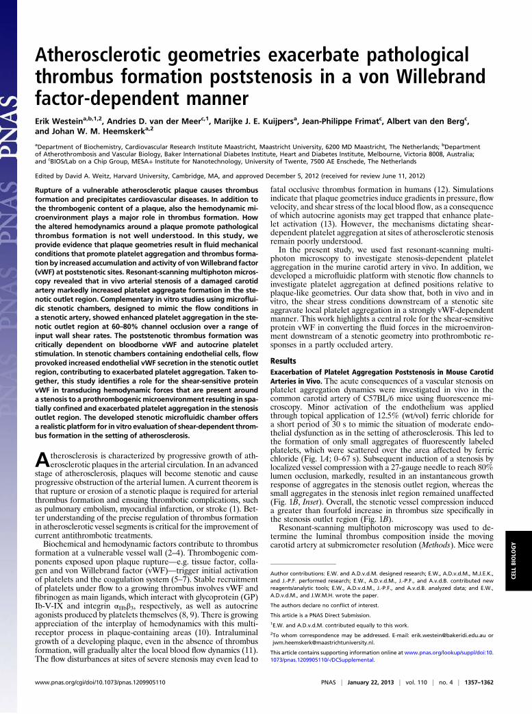
ResultsExacerbation of Platelet Aggregation Poststenosis in Mouse CarotidArteries in Vivo. The acute consequences of a vascular stenosis onplatelet aggregation dynamics were investigated in vivo in thecommon carotid artery of C57BL/6 mice using fluorescence mi-croscopy. Minor activation of the endothelium was appliedthrough topical application of 12.5% (wt/vol) ferric chloride fora short period of 30 s to mimic the situation of moderate endo-thelial dysfunction as in the setting of atherosclerosis. This led tothe formation of only small aggregates of fluorescently labeledplatelets, which were scattered over the area affected by ferricchloride (Fig. 1A; 0–67 s). Subsequent induction of a stenosis bylocalized vessel compression with a 27-gauge needle to reach 80%lumen occlusion, markedly, resulted in an instantaneous growthresponse of aggregates in the stenosis outlet region, whereas thesmall aggregates in the stenosis inlet region remained unaffected(Fig. 1B, Inset). Overall, the stenotic vessel compression induceda greater than fourfold increase in thrombus size specifically inthe stenosis outlet region (Fig. 1B).Resonant-scanning multiphoton microscopy was used to de-
termine the luminal thrombus composition inside the movingcarotid artery at submicrometer resolution (Methods). Mice were
Author contributions: E.W. and A.D.v.d.M. designed research; E.W., A.D.v.d.M., M.J.E.K.,and J.-P.F. performed research; E.W., A.D.v.d.M., J.-P.F., and A.v.d.B. contributed newreagents/analytic tools; E.W., A.D.v.d.M., J.-P.F., and A.v.d.B. analyzed data; and E.W.,A.D.v.d.M., and J.W.M.H. wrote the paper.
The authors declare no conflict of interest.
This article is a PNAS Direct Submission.1E.W. and A.D.v.d.M. contributed equally to this work.2To whom correspondence may be addressed. E-mail: [email protected] [email protected].
This article contains supporting information online at www.pnas.org/lookup/suppl/doi:10.1073/pnas.1209905110/-/DCSupplemental.
www.pnas.org/cgi/doi/10.1073/pnas.1209905110 PNAS | January 22, 2013 | vol. 110 | no. 4 | 1357–1362
CELL
BIOLO
GY
preinjected with platelets labeled with carboxyfluorescein diac-etate succinimidyl ester (CFSE) in combination with Alexa Fluor-labeled markers of platelet activation (labeled anti–P-selectinmAb or fibrinogen), and intraluminal thrombus formation wasmonitored in real time (Fig. 1C). The increased thrombusmass observed in the stenosis outlet section showed high stainingfor P-selectin that was associated with platelets close to thevessel wall. Furthermore, the thrombus in the stenosis outlet wasenriched in fibrin(ogen) associated with the platelets. Jointly,these results demonstrate that stenosis of an injured artery ex-acerbates the thrombotic response immediately downstream ofthe stenosis.
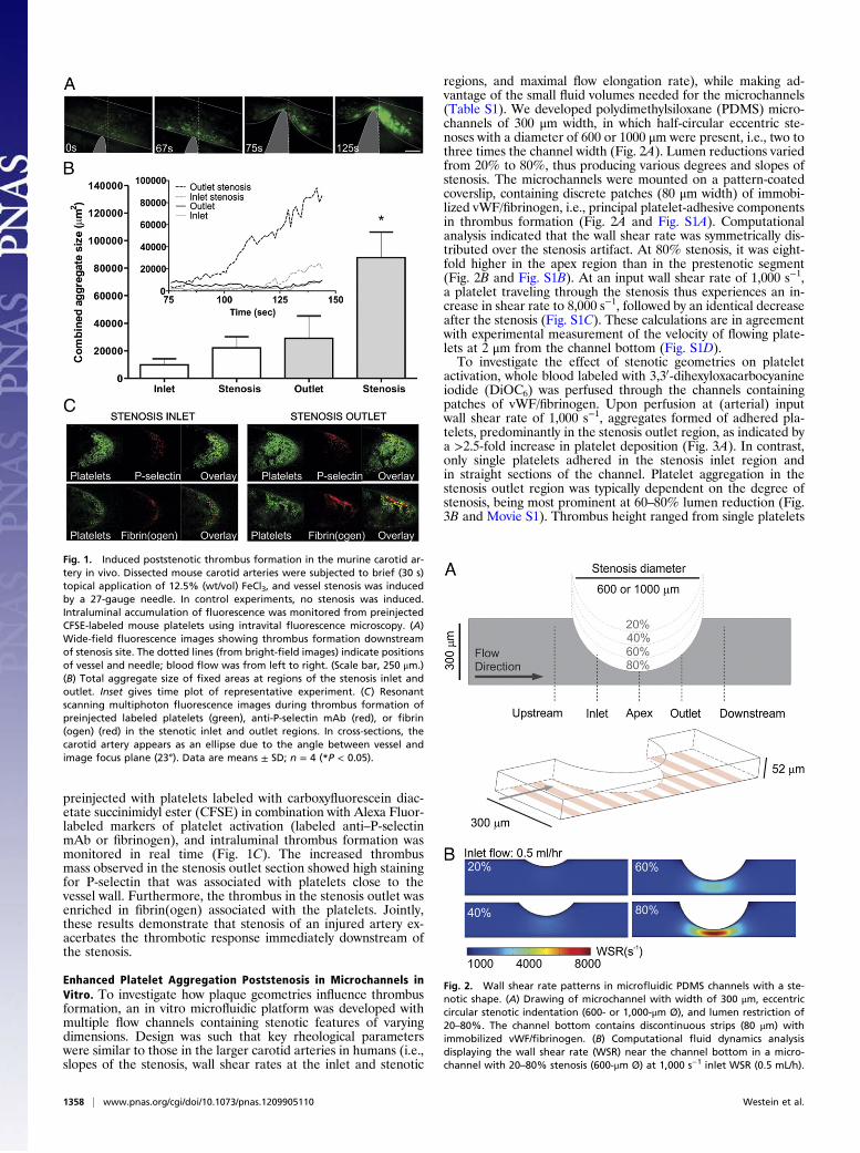
Enhanced Platelet Aggregation Poststenosis in Microchannels inVitro. To investigate how plaque geometries influence thrombusformation, an in vitro microfluidic platform was developed withmultiple flow channels containing stenotic features of varyingdimensions. Design was such that key rheological parameterswere similar to those in the larger carotid arteries in humans (i.e.,slopes of the stenosis, wall shear rates at the inlet and stenotic
regions, and maximal flow elongation rate), while making ad-vantage of the small fluid volumes needed for the microchannels(Table S1). We developed polydimethylsiloxane (PDMS) micro-channels of 300 μm width, in which half-circular eccentric ste-noses with a diameter of 600 or 1000 μm were present, i.e., two tothree times the channel width (Fig. 2A). Lumen reductions variedfrom 20% to 80%, thus producing various degrees and slopes ofstenosis. The microchannels were mounted on a pattern-coatedcoverslip, containing discrete patches (80 μm width) of immobi-lized vWF/fibrinogen, i.e., principal platelet-adhesive componentsin thrombus formation (Fig. 2A and Fig. S1A). Computationalanalysis indicated that the wall shear rate was symmetrically dis-tributed over the stenosis artifact. At 80% stenosis, it was eight-fold higher in the apex region than in the prestenotic segment(Fig. 2B and Fig. S1B). At an input wall shear rate of 1,000 s−1,a platelet traveling through the stenosis thus experiences an in-crease in shear rate to 8,000 s−1, followed by an identical decreaseafter the stenosis (Fig. S1C). These calculations are in agreementwith experimental measurement of the velocity of flowing plate-lets at 2 μm from the channel bottom (Fig. S1D).To investigate the effect of stenotic geometries on platelet
activation, whole blood labeled with 3,30-dihexyloxacarbocyanineiodide (DiOC6) was perfused through the channels containingpatches of vWF/fibrinogen. Upon perfusion at (arterial) inputwall shear rate of 1,000 s−1, aggregates formed of adhered pla-telets, predominantly in the stenosis outlet region, as indicated bya >2.5-fold increase in platelet deposition (Fig. 3A). In contrast,only single platelets adhered in the stenosis inlet region andin straight sections of the channel. Platelet aggregation in thestenosis outlet region was typically dependent on the degree ofstenosis, being most prominent at 60–80% lumen reduction (Fig.3B and Movie S1). Thrombus height ranged from single platelets
Fig. 1. Induced poststenotic thrombus formation in the murine carotid ar-tery in vivo. Dissected mouse carotid arteries were subjected to brief (30 s)topical application of 12.5% (wt/vol) FeCl3, and vessel stenosis was inducedby a 27-gauge needle. In control experiments, no stenosis was induced.Intraluminal accumulation of fluorescence was monitored from preinjectedCFSE-labeled mouse platelets using intravital fluorescence microscopy. (A)Wide-field fluorescence images showing thrombus formation downstreamof stenosis site. The dotted lines (from bright-field images) indicate positionsof vessel and needle; blood flow was from left to right. (Scale bar, 250 μm.)(B) Total aggregate size of fixed areas at regions of the stenosis inlet andoutlet. Inset gives time plot of representative experiment. (C) Resonantscanning multiphoton fluorescence images during thrombus formation ofpreinjected labeled platelets (green), anti-P-selectin mAb (red), or fibrin(ogen) (red) in the stenotic inlet and outlet regions. In cross-sections, thecarotid artery appears as an ellipse due to the angle between vessel andimage focus plane (23°). Data are means ± SD; n = 4 (*P < 0.05).
Fig. 2. Wall shear rate patterns in microfluidic PDMS channels with a ste-notic shape. (A) Drawing of microchannel with width of 300 μm, eccentriccircular stenotic indentation (600- or 1,000-μm Ø), and lumen restriction of20–80%. The channel bottom contains discontinuous strips (80 μm) withimmobilized vWF/fibrinogen. (B) Computational fluid dynamics analysisdisplaying the wall shear rate (WSR) near the channel bottom in a micro-channel with 20–80% stenosis (600-μm Ø) at 1,000 s−1 inlet WSR (0.5 mL/h).
1358 | www.pnas.org/cgi/doi/10.1073/pnas.1209905110 Westein et al.
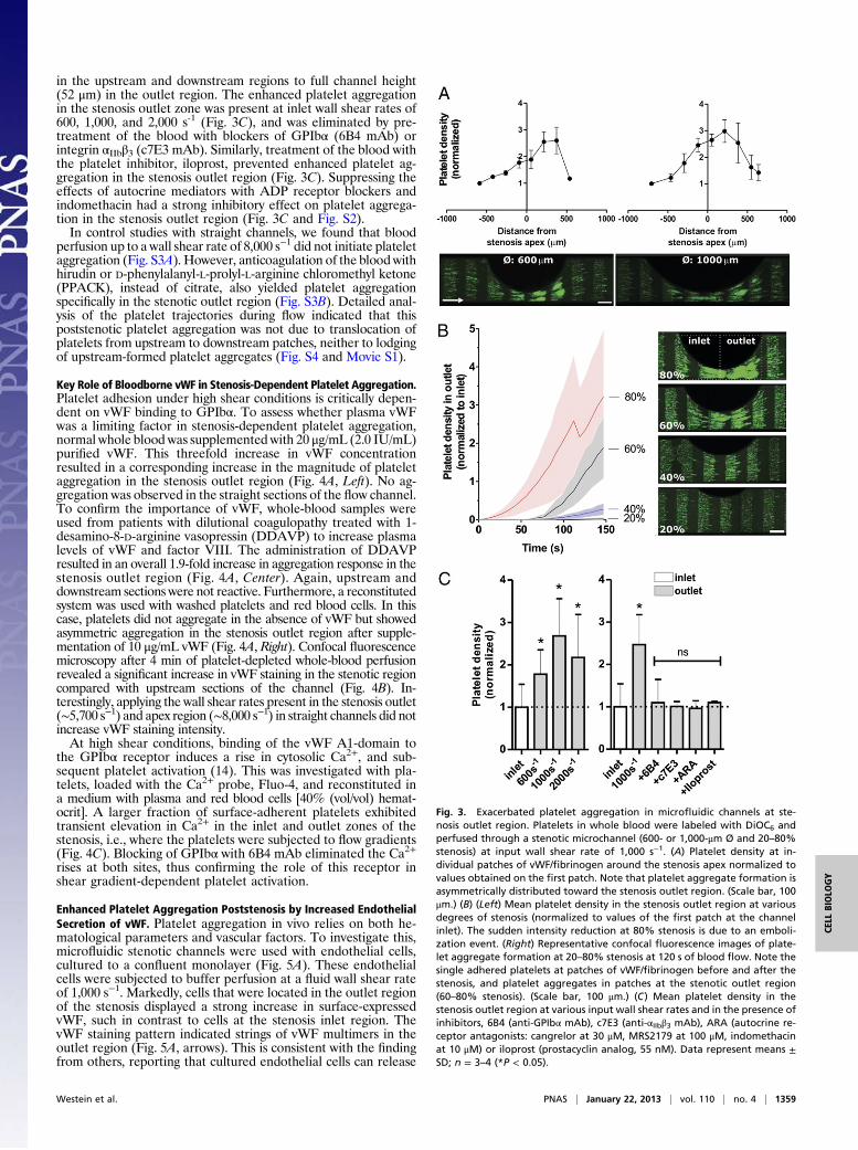
in the upstream and downstream regions to full channel height(52 μm) in the outlet region. The enhanced platelet aggregationin the stenosis outlet zone was present at inlet wall shear rates of600, 1,000, and 2,000 s-1 (Fig. 3C), and was eliminated by pre-treatment of the blood with blockers of GPIbα (6B4 mAb) orintegrin αIIbβ3 (c7E3 mAb). Similarly, treatment of the blood withthe platelet inhibitor, iloprost, prevented enhanced platelet ag-gregation in the stenosis outlet region (Fig. 3C). Suppressing theeffects of autocrine mediators with ADP receptor blockers andindomethacin had a strong inhibitory effect on platelet aggrega-tion in the stenosis outlet region (Fig. 3C and Fig. S2).In control studies with straight channels, we found that blood
perfusion up to a wall shear rate of 8,000 s−1 did not initiate plateletaggregation (Fig. S3A). However, anticoagulation of the blood withhirudin or D-phenylalanyl-L-prolyl-L-arginine chloromethyl ketone(PPACK), instead of citrate, also yielded platelet aggregationspecifically in the stenotic outlet region (Fig. S3B). Detailed anal-ysis of the platelet trajectories during flow indicated that thispoststenotic platelet aggregation was not due to translocation ofplatelets from upstream to downstream patches, neither to lodgingof upstream-formed platelet aggregates (Fig. S4 and Movie S1).
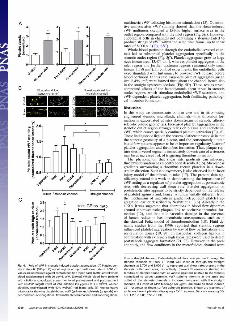
Key Role of Bloodborne vWF in Stenosis-Dependent Platelet Aggregation.Platelet adhesion under high shear conditions is critically depen-dent on vWF binding to GPIbα. To assess whether plasma vWFwas a limiting factor in stenosis-dependent platelet aggregation,normal whole blood was supplemented with 20 μg/mL (2.0 IU/mL)purified vWF. This threefold increase in vWF concentrationresulted in a corresponding increase in the magnitude of plateletaggregation in the stenosis outlet region (Fig. 4A, Left). No ag-gregation was observed in the straight sections of the flow channel.To confirm the importance of vWF, whole-blood samples wereused from patients with dilutional coagulopathy treated with 1-desamino-8-D-arginine vasopressin (DDAVP) to increase plasmalevels of vWF and factor VIII. The administration of DDAVPresulted in an overall 1.9-fold increase in aggregation response in thestenosis outlet region (Fig. 4A, Center). Again, upstream anddownstream sections were not reactive. Furthermore, a reconstitutedsystem was used with washed platelets and red blood cells. In thiscase, platelets did not aggregate in the absence of vWF but showedasymmetric aggregation in the stenosis outlet region after supple-mentation of 10 μg/mL vWF (Fig. 4A, Right). Confocal fluorescencemicroscopy after 4 min of platelet-depleted whole-blood perfusionrevealed a significant increase in vWF staining in the stenotic regioncompared with upstream sections of the channel (Fig. 4B). In-terestingly, applying the wall shear rates present in the stenosis outlet(∼5,700 s−1) and apex region (∼8,000 s−1) in straight channels did notincrease vWF staining intensity.At high shear conditions, binding of the vWF A1-domain to
the GPIbα receptor induces a rise in cytosolic Ca2+, and sub-sequent platelet activation (14). This was investigated with pla-telets, loaded with the Ca2+ probe, Fluo-4, and reconstituted ina medium with plasma and red blood cells [40% (vol/vol) hemat-ocrit]. A larger fraction of surface-adherent platelets exhibitedtransient elevation in Ca2+ in the inlet and outlet zones of thestenosis, i.e., where the platelets were subjected to flow gradients(Fig. 4C). Blocking of GPIbα with 6B4 mAb eliminated the Ca2+rises at both sites, thus confirming the role of this receptor inshear gradient-dependent platelet activation.
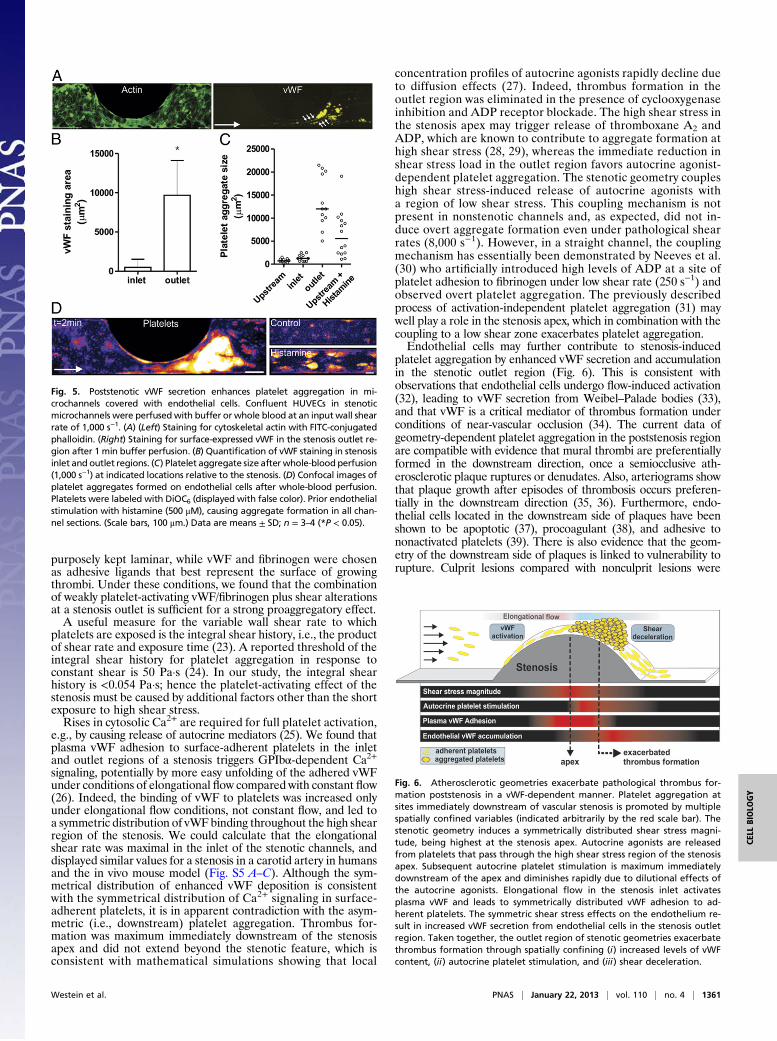
Enhanced Platelet Aggregation Poststenosis by Increased EndothelialSecretion of vWF. Platelet aggregation in vivo relies on both he-matological parameters and vascular factors. To investigate this,microfluidic stenotic channels were used with endothelial cells,cultured to a confluent monolayer (Fig. 5A). These endothelialcells were subjected to buffer perfusion at a fluid wall shear rateof 1,000 s−1. Markedly, cells that were located in the outlet regionof the stenosis displayed a strong increase in surface-expressedvWF, such in contrast to cells at the stenosis inlet region. ThevWF staining pattern indicated strings of vWF multimers in theoutlet region (Fig. 5A, arrows). This is consistent with the findingfrom others, reporting that cultured endothelial cells can release
Fig. 3. Exacerbated platelet aggregation in microfluidic channels at ste-nosis outlet region. Platelets in whole blood were labeled with DiOC6 andperfused through a stenotic microchannel (600- or 1,000-μm Ø and 20–80%stenosis) at input wall shear rate of 1,000 s−1. (A) Platelet density at in-dividual patches of vWF/fibrinogen around the stenosis apex normalized tovalues obtained on the first patch. Note that platelet aggregate formation isasymmetrically distributed toward the stenosis outlet region. (Scale bar, 100μm.) (B) (Left) Mean platelet density in the stenosis outlet region at variousdegrees of stenosis (normalized to values of the first patch at the channelinlet). The sudden intensity reduction at 80% stenosis is due to an emboli-zation event. (Right) Representative confocal fluorescence images of plate-let aggregate formation at 20–80% stenosis at 120 s of blood flow. Note thesingle adhered platelets at patches of vWF/fibrinogen before and after thestenosis, and platelet aggregates in patches at the stenotic outlet region(60–80% stenosis). (Scale bar, 100 μm.) (C) Mean platelet density in thestenosis outlet region at various input wall shear rates and in the presence ofinhibitors, 6B4 (anti-GPIbα mAb), c7E3 (anti-αIIbβ3 mAb), ARA (autocrine re-ceptor antagonists: cangrelor at 30 μM, MRS2179 at 100 μM, indomethacinat 10 μM) or iloprost (prostacyclin analog, 55 nM). Data represent means ±SD; n = 3–4 (*P < 0.05).
Westein et al. PNAS | January 22, 2013 | vol. 110 | no. 4 | 1359
CELL
BIOLO
GY
multimeric vWF following histamine stimulation (15). Quantita-tive analysis after vWF staining showed that the shear-inducedvWF multimers occupied a 15-fold higher surface area in theoutlet region, compared with the inlet region (Fig. 5B). However,endothelial cells in channels not containing a stenosis failed toproduce strings of vWF within the same time frame, up to shearrates of 8,000 s−1 (Fig. S3C).Whole-blood perfusion through the endothelial-covered chan-
nels led to substantial platelet aggregation specifically in thestenotic outlet region (Fig. 5C). Platelet aggregates grew to largesizes (mean area, 13,474 μm2), whereas platelet aggregates in theinlet region and further upstream regions remained only small(mean, 1,378 μm2). In control experiments, the endothelial cellswere stimulated with histamine, to provoke vWF release beforeblood perfusion. In this case, large-size platelet aggregates (meansize, 6,496 μm2) were formed throughout the channel, hence alsoin the straight upstream sections (Fig. 5D). These results revealcompound effects of the hemodynamic shear stress in stenosisoutlet regions, which stimulate endothelial vWF secretion, andvWF-dependent platelet aggregation, both facilitating pathologi-cal thrombus formation.
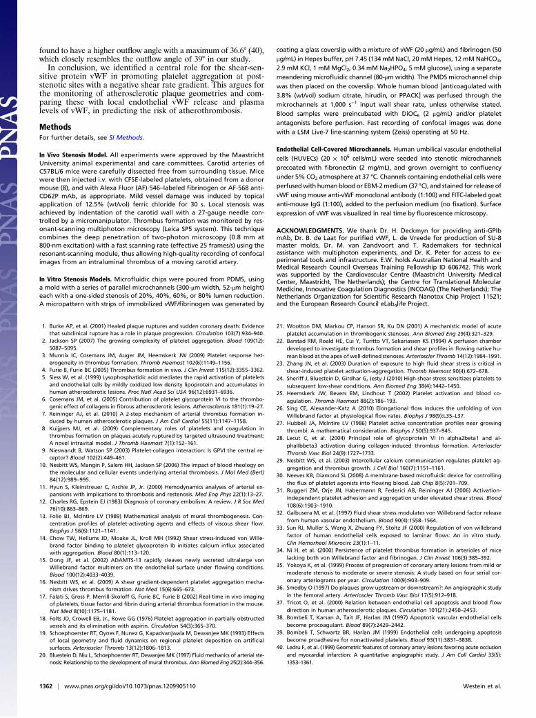
DiscussionIn this study we demonstrate both in vivo and in vitro—usingengineered stenotic microfluidic channels—that thrombus for-mation is exacerbated at sites downstream of stenotic athero-sclerotic plaque geometries. Increased platelet aggregation in thestenotic outlet region strongly relies on plasma and endothelialvWF, which causes spatially confined platelet activation (Fig. 6).These findings shed light on the process of atherothrombosis in thatthe stenotic geometry of a plaque, and the consequently alteredblood flow pattern, appears to be an important regulatory factor ofplatelet aggregation and thrombus formation. Thus, plaque rup-ture sites in vessel segments immediately downstream of a stenosismay be at increased risk of triggering thrombus formation.The phenomenon that shear rate gradients can influence
thrombus formation has recently been described (16). Microsheargradients surrounding a thrombus recruit platelets in a down-stream direction. Such clot asymmetry is also observed in the laserinjury model of thrombosis in mice (17). The present data sig-nificantly extend this work in demonstrating the importance ofvWF acting as a regulator of platelet aggregation at poststenoticsites with decreasing wall shear rate. Platelet aggregation atpoststenotic sites appears to be strictly dependent on the releaseof platelet agonists and, hence, is fundamentally different fromthe mechanism of microshear gradient-dependent platelet ag-gregation, earlier described by Nesbitt et al. (16). Already in the1970s, it was suggested that alterations in blood flow dynamicsaround atherosclerotic plaques link to occlusive thrombus for-mation (12), and that mild vascular damage in the presenceof lumen reduction has thrombotic consequences, such as inthe classical Foltz model of thromboembolism (18). Fluid dy-namic studies from the 1990s reported that stenotic regionsinfluenced platelet aggregation by way of flow perturbations andrecirculation zones (19, 20). In particular, collagen ligands incombination with extremely high shear rates were used to detectpoststenotic aggregate formation (21, 22). However, in the pres-ent study, the flow conditions in the microfluidics channel were
Fig. 4. Role of vWF in stenosis-induced platelet aggregation. (A) Platelet den-sity in stenotic (600-μm Ø) outlet regions at input wall shear rate of 1,000 s−1.Values are normalized against control condition (open bars). (Left) Control wholeblood supplemented with 20 μg/mL vWF. (Center) Whole blood from patientswith dilutional coagulopathy was monitored pretreatment and posttreatmentwith DDAVP. (Right) Effect of vWF addition (10 μg/mL) to 3 × 10⁸/mL washedplatelets, reconstituted with 40% (vol/vol) red blood cells. (B) Representativemicrographs showing platelet-bound vWF (yellow) and platelets (grayscale) un-der conditions of elongational flow in the stenosis channels and nonelongational
flow in straight channels. Platelet-depleted blood was perfused through thestenosis channels at 1,000 s−1 input wall shear or through the straightchannels at 5,700 and 8,000 s−1 to represent wall shear rates present in thestenosis outlet and apex, respectively. (Lower) Fluorescence staining in-tensities of platelet-bound vWF at various positions relative to the stenosisnormalized to values upstream. vWF staining intensity at the apex andoutlet of the stenosis channels is increased compared with the straightchannels. (C) Effect of GPIb blockage (20 μg/mL 6B4 mAb) on shear-inducedCa2+ responses of single, surface-adherent platelets. Shown are fractions ofsurface-adherent platelets displaying a Ca2+ response. Data are means ± SD;n ≥ 3 (*P < 0.05, **P < 0.01).
1360 | www.pnas.org/cgi/doi/10.1073/pnas.1209905110 Westein et al.
purposely kept laminar, while vWF and fibrinogen were chosenas adhesive ligands that best represent the surface of growingthrombi. Under these conditions, we found that the combinationof weakly platelet-activating vWF/fibrinogen plus shear alterationsat a stenosis outlet is sufficient for a strong proaggregatory effect.A useful measure for the variable wall shear rate to which
platelets are exposed is the integral shear history, i.e., the productof shear rate and exposure time (23). A reported threshold of theintegral shear history for platelet aggregation in response toconstant shear is 50 Pa·s (24). In our study, the integral shearhistory is <0.054 Pa·s; hence the platelet-activating effect of thestenosis must be caused by additional factors other than the shortexposure to high shear stress.Rises in cytosolic Ca2+ are required for full platelet activation,
e.g., by causing release of autocrine mediators (25). We found thatplasma vWF adhesion to surface-adherent platelets in the inletand outlet regions of a stenosis triggers GPIbα-dependent Ca2+signaling, potentially by more easy unfolding of the adhered vWFunder conditions of elongational flow compared with constant flow(26). Indeed, the binding of vWF to platelets was increased onlyunder elongational flow conditions, not constant flow, and led toa symmetric distribution of vWF binding throughout the high shearregion of the stenosis. We could calculate that the elongationalshear rate was maximal in the inlet of the stenotic channels, anddisplayed similar values for a stenosis in a carotid artery in humansand the in vivo mouse model (Fig. S5 A–C). Although the sym-metrical distribution of enhanced vWF deposition is consistentwith the symmetrical distribution of Ca2+ signaling in surface-adherent platelets, it is in apparent contradiction with the asym-metric (i.e., downstream) platelet aggregation. Thrombus for-mation was maximum immediately downstream of the stenosisapex and did not extend beyond the stenotic feature, which isconsistent with mathematical simulations showing that local
concentration profiles of autocrine agonists rapidly decline dueto diffusion effects (27). Indeed, thrombus formation in theoutlet region was eliminated in the presence of cyclooxygenaseinhibition and ADP receptor blockade. The high shear stress inthe stenosis apex may trigger release of thromboxane A2 andADP, which are known to contribute to aggregate formation athigh shear stress (28, 29), whereas the immediate reduction inshear stress load in the outlet region favors autocrine agonist-dependent platelet aggregation. The stenotic geometry coupleshigh shear stress-induced release of autocrine agonists witha region of low shear stress. This coupling mechanism is notpresent in nonstenotic channels and, as expected, did not in-duce overt aggregate formation even under pathological shearrates (8,000 s−1). However, in a straight channel, the couplingmechanism has essentially been demonstrated by Neeves et al.(30) who artificially introduced high levels of ADP at a site ofplatelet adhesion to fibrinogen under low shear rate (250 s−1) andobserved overt platelet aggregation. The previously describedprocess of activation-independent platelet aggregation (31) maywell play a role in the stenosis apex, which in combination with thecoupling to a low shear zone exacerbates platelet aggregation.Endothelial cells may further contribute to stenosis-induced
platelet aggregation by enhanced vWF secretion and accumulationin the stenotic outlet region (Fig. 6). This is consistent withobservations that endothelial cells undergo flow-induced activation(32), leading to vWF secretion from Weibel–Palade bodies (33),and that vWF is a critical mediator of thrombus formation underconditions of near-vascular occlusion (34). The current data ofgeometry-dependent platelet aggregation in the poststenosis regionare compatible with evidence that mural thrombi are preferentiallyformed in the downstream direction, once a semiocclusive ath-erosclerotic plaque ruptures or denudates. Also, arteriograms showthat plaque growth after episodes of thrombosis occurs preferen-tially in the downstream direction (35, 36). Furthermore, endo-thelial cells located in the downstream side of plaques have beenshown to be apoptotic (37), procoagulant (38), and adhesive tononactivated platelets (39). There is also evidence that the geom-etry of the downstream side of plaques is linked to vulnerability torupture. Culprit lesions compared with nonculprit lesions were
Fig. 5. Poststenotic vWF secretion enhances platelet aggregation in mi-crochannels covered with endothelial cells. Confluent HUVECs in stenoticmicrochannels were perfusedwith buffer or whole blood at an input wall shearrate of 1,000 s−1. (A) (Left) Staining for cytoskeletal actin with FITC-conjugatedphalloidin. (Right) Staining for surface-expressed vWF in the stenosis outlet re-gion after 1 min buffer perfusion. (B) Quantification of vWF staining in stenosisinlet and outlet regions. (C) Platelet aggregate size after whole-blood perfusion(1,000 s−1) at indicated locations relative to the stenosis. (D) Confocal images ofplatelet aggregates formed on endothelial cells after whole-blood perfusion.Platelets were labeled with DiOC6 (displayed with false color). Prior endothelialstimulation with histamine (500 μM), causing aggregate formation in all chan-nel sections. (Scale bars, 100 μm.) Data are means ± SD; n = 3–4 (*P < 0.05).
Fig. 6. Atherosclerotic geometries exacerbate pathological thrombus for-mation poststenosis in a vWF-dependent manner. Platelet aggregation atsites immediately downstream of vascular stenosis is promoted by multiplespatially confined variables (indicated arbitrarily by the red scale bar). Thestenotic geometry induces a symmetrically distributed shear stress magni-tude, being highest at the stenosis apex. Autocrine agonists are releasedfrom platelets that pass through the high shear stress region of the stenosisapex. Subsequent autocrine platelet stimulation is maximum immediatelydownstream of the apex and diminishes rapidly due to dilutional effects ofthe autocrine agonists. Elongational flow in the stenosis inlet activatesplasma vWF and leads to symmetrically distributed vWF adhesion to ad-herent platelets. The symmetric shear stress effects on the endothelium re-sult in increased vWF secretion from endothelial cells in the stenosis outletregion. Taken together, the outlet region of stenotic geometries exacerbatethrombus formation through spatially confining (i) increased levels of vWFcontent, (ii) autocrine platelet stimulation, and (iii) shear deceleration.
Westein et al. PNAS | January 22, 2013 | vol. 110 | no. 4 | 1361
CELL
BIOLO
GY
found to have a higher outflow angle with a maximum of 36.6° (40),which closely resembles the outflow angle of 39° in our study.In conclusion, we identified a central role for the shear-sen-
sitive protein vWF in promoting platelet aggregation at post-stenotic sites with a negative shear rate gradient. This argues forthe monitoring of atherosclerotic plaque geometries and com-paring these with local endothelial vWF release and plasmalevels of vWF, in predicting the risk of atherothrombosis.
MethodsFor further details, see SI Methods.
In Vivo Stenosis Model. All experiments were approved by the MaastrichtUniversity animal experimental and care committees. Carotid arteries ofC57BL/6 mice were carefully dissected free from surrounding tissue. Micewere then injected i.v. with CFSE-labeled platelets, obtained from a donormouse (8), and with Alexa Fluor (AF)-546–labeled fibrinogen or AF-568 anti-CD62P mAb, as appropriate. Mild vessel damage was induced by topicalapplication of 12.5% (wt/vol) ferric chloride for 30 s. Local stenosis wasachieved by indentation of the carotid wall with a 27-gauge needle con-trolled by a micromanipulator. Thrombus formation was monitored by res-onant-scanning multiphoton microscopy (Leica SP5 system). This techniquecombines the deep penetration of two-photon microscopy (0.8 mm at800-nm excitation) with a fast scanning rate (effective 25 frames/s) using theresonant-scanning module, thus allowing high-quality recording of confocalimages from an intraluminal thrombus of a moving carotid artery.
In Vitro Stenosis Models. Microfluidic chips were poured from PDMS, usinga mold with a series of parallel microchannels (300-μm width, 52-μm height)each with a one-sided stenosis of 20%, 40%, 60%, or 80% lumen reduction.A micropattern with strips of immobilized vWF/fibrinogen was generated by
coating a glass coverslip with a mixture of vWF (20 μg/mL) and fibrinogen (50μg/mL) in Hepes buffer, pH 7.45 (134 mMNaCl, 20 mMHepes, 12 mMNaHCO3,2.9 mM KCl, 1 mMMgCl2, 0.34 mM Na2HPO4, 5 mM glucose), using a separatemeandering microfluidic channel (80-μm width). The PMDS microchannel chipwas then placed on the coverslip. Whole human blood [anticoagulated with3.8% (wt/vol) sodium citrate, hirudin, or PPACK] was perfused through themicrochannels at 1,000 s−1 input wall shear rate, unless otherwise stated.Blood samples were preincubated with DiOC6 (2 μg/mL) and/or plateletantagonists before perfusion. Fast recording of confocal images was donewith a LSM Live-7 line-scanning system (Zeiss) operating at 50 Hz.
Endothelial Cell-Covered Microchannels. Human umbilical vascular endothelialcells (HUVECs) (20 × 106 cells/mL) were seeded into stenotic microchannelsprecoated with fibronectin (2 mg/mL), and grown overnight to confluencyunder 5% CO2 atmosphere at 37 °C. Channels containing endothelial cells wereperfusedwith human blood or EBM-2medium (37 °C), and stained for release ofvWF using mouse anti-vWF monoclonal antibody (1:100) and FITC-labeled goatanti-mouse IgG (1:100), added to the perfusion medium (no fixation). Surfaceexpression of vWF was visualized in real time by fluorescence microscopy.
ACKNOWLEDGMENTS. We thank Dr. H. Deckmyn for providing anti-GPIbmAb, Dr. B. de Laat for purified vWF, L. de Vreede for production of SU-8master molds, Dr. M. van Zandvoort and T. Rademakers for technicalassistance with multiphoton experiments, and Dr. K. Peter for access to ex-perimental tools and infrastructure. E.W. holds Australian National Health andMedical Research Council Overseas Training Fellowship ID 606742. This workwas supported by the Cardiovascular Centre (Maastricht University MedicalCenter, Maastricht, The Netherlands); the Centre for Translational MolecularMedicine, Innovative Coagulation Diagnostics (INCOAG) (The Netherlands); TheNetherlands Organization for Scientific Research Nanotox Chip Project 11521;and the European Research Council eLab4life Project.
1. Burke AP, et al. (2001) Healed plaque ruptures and sudden coronary death: Evidencethat subclinical rupture has a role in plaque progression. Circulation 103(7):934–940.
2. Jackson SP (2007) The growing complexity of platelet aggregation. Blood 109(12):5087–5095.
3. Munnix IC, Cosemans JM, Auger JM, Heemskerk JW (2009) Platelet response het-erogeneity in thrombus formation. Thromb Haemost 102(6):1149–1156.
4. Furie B, Furie BC (2005) Thrombus formation in vivo. J Clin Invest 115(12):3355–3362.5. Siess W, et al. (1999) Lysophosphatidic acid mediates the rapid activation of platelets
and endothelial cells by mildly oxidized low density lipoprotein and accumulates inhuman atherosclerotic lesions. Proc Natl Acad Sci USA 96(12):6931–6936.
6. Cosemans JM, et al. (2005) Contribution of platelet glycoprotein VI to the thrombo-genic effect of collagens in fibrous atherosclerotic lesions. Atherosclerosis 181(1):19–27.
7. Reininger AJ, et al. (2010) A 2-step mechanism of arterial thrombus formation in-duced by human atherosclerotic plaques. J Am Coll Cardiol 55(11):1147–1158.
8. Kuijpers MJ, et al. (2009) Complementary roles of platelets and coagulation inthrombus formation on plaques acutely ruptured by targeted ultrasound treatment:A novel intravital model. J Thromb Haemost 7(1):152–161.
9. Nieswandt B, Watson SP (2003) Platelet-collagen interaction: Is GPVI the central re-ceptor? Blood 102(2):449–461.
10. Nesbitt WS, Mangin P, Salem HH, Jackson SP (2006) The impact of blood rheology onthe molecular and cellular events underlying arterial thrombosis. J Mol Med (Berl)84(12):989–995.
11. Hyun S, Kleinstreuer C, Archie JP, Jr. (2000) Hemodynamics analyses of arterial ex-pansions with implications to thrombosis and restenosis. Med Eng Phys 22(1):13–27.
12. Charles RG, Epstein EJ (1983) Diagnosis of coronary embolism: A review. J R Soc Med76(10):863–869.
13. Folie BJ, McIntire LV (1989) Mathematical analysis of mural thrombogenesis. Con-centration profiles of platelet-activating agents and effects of viscous shear flow.Biophys J 56(6):1121–1141.
14. Chow TW, Hellums JD, Moake JL, Kroll MH (1992) Shear stress-induced von Wille-brand factor binding to platelet glycoprotein Ib initiates calcium influx associatedwith aggregation. Blood 80(1):113–120.
15. Dong JF, et al. (2002) ADAMTS-13 rapidly cleaves newly secreted ultralarge vonWillebrand factor multimers on the endothelial surface under flowing conditions.Blood 100(12):4033–4039.
16. Nesbitt WS, et al. (2009) A shear gradient-dependent platelet aggregation mecha-nism drives thrombus formation. Nat Med 15(6):665–673.
17. Falati S, Gross P, Merrill-Skoloff G, Furie BC, Furie B (2002) Real-time in vivo imagingof platelets, tissue factor and fibrin during arterial thrombus formation in the mouse.Nat Med 8(10):1175–1181.
18. Folts JD, Crowell EB, Jr., Rowe GG (1976) Platelet aggregation in partially obstructedvessels and its elimination with aspirin. Circulation 54(3):365–370.
19. Schoephoerster RT, Oynes F, Nunez G, Kapadvanjwala M, Dewanjee MK (1993) Effectsof local geometry and fluid dynamics on regional platelet deposition on artificialsurfaces. Arterioscler Thromb 13(12):1806–1813.
20. Bluestein D, Niu L, Schoephoerster RT, Dewanjee MK (1997) Fluid mechanics of arterial ste-nosis: Relationship to the development ofmural thrombus.Ann Biomed Eng 25(2):344–356.
21. Wootton DM, Markou CP, Hanson SR, Ku DN (2001) A mechanistic model of acuteplatelet accumulation in thrombogenic stenoses. Ann Biomed Eng 29(4):321–329.
22. Barstad RM, Roald HE, Cui Y, Turitto VT, Sakariassen KS (1994) A perfusion chamberdeveloped to investigate thrombus formation and shear profiles in flowing native hu-man blood at the apex of well-defined stenoses.Arterioscler Thromb 14(12):1984–1991.
23. Zhang JN, et al. (2003) Duration of exposure to high fluid shear stress is critical inshear-induced platelet activation-aggregation. Thromb Haemost 90(4):672–678.
24. Sheriff J, Bluestein D, Girdhar G, Jesty J (2010) High-shear stress sensitizes platelets tosubsequent low-shear conditions. Ann Biomed Eng 38(4):1442–1450.
25. Heemskerk JW, Bevers EM, Lindhout T (2002) Platelet activation and blood co-agulation. Thromb Haemost 88(2):186–193.
26. Sing CE, Alexander-Katz A (2010) Elongational flow induces the unfolding of vonWillebrand factor at physiological flow rates. Biophys J 98(9):L35–L37.
27. Hubbell JA, McIntire LV (1986) Platelet active concentration profiles near growingthrombi. A mathematical consideration. Biophys J 50(5):937–945.
28. Lecut C, et al. (2004) Principal role of glycoprotein VI in alpha2beta1 and al-phaIIbbeta3 activation during collagen-induced thrombus formation. ArteriosclerThromb Vasc Biol 24(9):1727–1733.
29. Nesbitt WS, et al. (2003) Intercellular calcium communication regulates platelet ag-gregation and thrombus growth. J Cell Biol 160(7):1151–1161.
30. Neeves KB, Diamond SL (2008) A membrane-based microfluidic device for controllingthe flux of platelet agonists into flowing blood. Lab Chip 8(5):701–709.
31. Ruggeri ZM, Orje JN, Habermann R, Federici AB, Reininger AJ (2006) Activation-independent platelet adhesion and aggregation under elevated shear stress. Blood108(6):1903–1910.
32. Galbusera M, et al. (1997) Fluid shear stress modulates von Willebrand factor releasefrom human vascular endothelium. Blood 90(4):1558–1564.
33. Sun RJ, Muller S, Wang X, Zhuang FY, Stoltz JF (2000) Regulation of von willebrandfactor of human endothelial cells exposed to laminar flows: An in vitro study.Clin Hemorheol Microcirc 23(1):1–11.
34. Ni H, et al. (2000) Persistence of platelet thrombus formation in arterioles of micelacking both von Willebrand factor and fibrinogen. J Clin Invest 106(3):385–392.
35. Yokoya K, et al. (1999) Process of progression of coronary artery lesions from mild ormoderate stenosis to moderate or severe stenosis: A study based on four serial cor-onary arteriograms per year. Circulation 100(9):903–909.
36. Smedby O (1997) Do plaques grow upstream or downstream?: An angiographic studyin the femoral artery. Arterioscler Thromb Vasc Biol 17(5):912–918.
37. Tricot O, et al. (2000) Relation between endothelial cell apoptosis and blood flowdirection in human atherosclerotic plaques. Circulation 101(21):2450–2453.
38. Bombeli T, Karsan A, Tait JF, Harlan JM (1997) Apoptotic vascular endothelial cellsbecome procoagulant. Blood 89(7):2429–2442.
39. Bombeli T, Schwartz BR, Harlan JM (1999) Endothelial cells undergoing apoptosisbecome proadhesive for nonactivated platelets. Blood 93(11):3831–3838.
40. Ledru F, et al. (1999) Geometric features of coronary artery lesions favoring acute occlusionand myocardial infarction: A quantitative angiographic study. J Am Coll Cardiol 33(5):1353–1361.
1362 | www.pnas.org/cgi/doi/10.1073/pnas.1209905110 Westein et al.