asthma and copd || pulmonary physiology
TRANSCRIPT

P A R T Physiology and Pathology of
Asthma and COPD
2

55
C H A P T E R
Pulmonary Physiology
INTRODUCTION
Asthma is an infl ammatory disease principally of the small airways but current understanding is that all the airways are involved [1] . Th e physi-ological manifestations of lung infl ammation are reversible airfl ow limitation and airfl ow limita-tion that fl uctuates widely with time; asthma can also result in persistent loss of lung function. In contrast, airfl ow obstruction in patients with chronic obstructive pulmonary disease (COPD) presents with both reversible and irreversible air-fl ow limitation, the latter linked to loss of static elastic recoil due in part to the destruction of the architecture of the lung. Th e airfl ow limitation of COPD has much less temporal or periodic variation over the short term but patients with COPD usually exhibit steady consistent losses of lung function over a period of years. In reality the clinical presentation of both COPD and asthma is often quite variable and the pathophysiologi-cal data alone do not allow a defi nitive diagnosis. Th is confusing presentation of patients with fea-tures of both asthma and COPD is in keeping with the overlap observed in the epidemiology of these diseases [2, 3] . Th e focus of this chap-ter is on the pathophysiological presentation of COPD and asthma and the features that distin-guish each disease as well as the similarities. Th is chapter will also examine the physiological proc-esses that characterize airways disease.
LUNG VOLUMES AND ELASTIC RECOIL
Lung volumes and capacities
In diseases of the airway such as asthma and COPD there are characteristic increases in
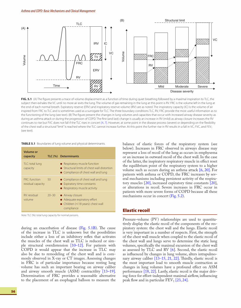
v olume which distinguish these airway disor-ders from restrictive processes of the respiratory system that reduce lung volumes. Lung volumes have important bearing on the degree of dis-ease since increases in the residual volume (RV) occur relatively early even with mild airway dis-ease whereas increases in the total lung capacity (TLC) are usually an indication of more severe or long-standing disease ( Fig. 5.1 ) [4–6].
Th e most useful lung volumes and capaci-ties to measure are those that assess the physical limits of the lung and chest wall and defi ne the extremes of the vital capacity (VC); TLC and RV. Th e volume in the lung at end expiratory position, the functional residual capacity (FRC), is important because of the infl uence that lung volume has on airway caliber and in turn on airfl ow resistance during eupnea. TLC, FRC, and RV are boundary conditions of respiration and accordingly provide the most information about the capacity and function of the respira-tory system [6] . Assessing the changes in these lung volumes to evaluate disease severity and response to therapy allows one to distinguish between obstructive lung disease and restrictive lung disease ( Table 5.1 ) [8, 9] .
Obstructive lung diseases such as chronic bronchitis, asthma, and emphysema all result in increases in lung volume. An increase in TLC usually represents more chronic, severe disease, and in the case of COPD it is related to remodeling of the chest wall leading to the so-called barrel chest. Th e increase in TLC may have another more important role; that is the preservation of forced vital capacity (FVC) in face of a rise in RV that would then result in more pronounced falls in FEV 1 if the rise in TLC had not ameliorated the fall in FVC [4, 7]. Th e increase in TLC in parallel with each increase in RV can occur until the chest wall reaches some structural “ limit ” which probably varies from person to person [7, 10] and acutely
5 Charles G. Irvin
Department of Medicine and Physiology, University of Vermont
College of Medicine, Burlington, VT, USA
Asthma and COPD: Basic Mechanisms and Clinical Management
56
during an exacerbation of disease ( Fig. 5.1B ). Th e cause of the increase in TLC is unknown but the possibilities include either a loss of an inhibitory refl ex that activates the muscles of the chest wall as TLC is reduced or sim-ple structural overdistension [10–12] . For patients with COPD it would appear that the increase in TLC may also be due to remodeling of the chest wall and is com-monly observed in X-ray or CT images. Assessing changes in FRC is of particular importance because resting lung volume has such an important bearing on airway caliber and airway smooth muscle (ASM) contractility [13–19] . Determination of FRC provides a reasonable alternative to the placement of an esophageal balloon to measure the
balance of elastic forces of the respiratory system (see below). Increases in FRC observed in airways disease may represent a loss of recoil of the lung as occurs in emphysema or an increase in outward recoil of the chest wall. In the case of the latter, the inspiratory respiratory muscle in eff ect reset the equilibrium point of the respiratory system to a higher volume such as occurs during an asthma attack [6, 20] . For patients with asthma or COPD, the FRC increases by sev-eral mechanisms including persistent activity of the respira-tory muscles [20] , increased expiratory time constants [20] , or alterations in recoil. Severe increases in FRC occur in patients with more severe forms of COPD because all these mechanisms occur in concert ( Fig. 5.2 ).
Elastic recoil
Pressure–volume (PV) relationships are used to quantita-tively display the elastic recoil of the components of the res-piratory system: the chest wall and the lungs. Elastic recoil is very important in a number of respects. First, the strength of the chest wall muscle when coupled to the elastic recoil of the chest wall and lungs serve to determine the static lung volumes, specifi cally the maximal excursion of the chest wall as assessed by TLC and RV [6] . Second, the elastic recoil as infl uenced by changes in lung volume, alters intrapulmo-nary airway caliber [13–15, 21, 22] . Th irdly, elastic recoil is the most important load to smooth muscle contraction as changes in lung volumes have a profound eff ect on ASM performance [18, 22]. Lastly, elastic recoil is the major driv-ing force for eff ort-independent maximal airfl ow, infl uencing peak fl ow and in particular FEV 1 [23, 24] .
Vol
ume
TLC
FRC
ERV
Time
RV
IRV IC
VC
RV
TV
(A)
FIG. 5.1 (A) The fi gure presents a trace of volume displacement as a function of time during quiet breathing followed by a maximal inspiration to TLC, the
subject then exhales the VC until no more air exits the lung. The volume of gas remaining in the lung at this point is RV. FRC is the volume left in the lung at
the end of each normal breath. Expiratory reserve (ERV) and inspiratory reserve volume (IRV) are as noted. The inspiratory capacity (IC) is the volume of air
inspired from FRC to TLC and is sometimes used as a surrogate for TLC. The three boundary conditions TLC, RV, FRC provide the most useful information as to
the functioning of the lung (see text). (B) The fi gure present the changes in lung volumes and capacities that occur with increased airway disease severity as
during an asthma attack or during the progression of COPD. The fi rst (and last) change is usually an increase in RV (mild) as airway closure increases the RV
continues to rise but FVC does not fall if the TLC rises in concert [4, 7] . However, at some point in the disease process (severe) or depending on the fl exibility
of the chest wall a structural “ limit ” is reached where the TLC cannot increase further. At this point the further rise in RV results in a fall in VC, FVC, and FEV 1
(see text).
(B) Structural limit
VC
FRC
RV
Mild Moderate Severe
Disease severity
Vol
ume
TABLE 5.1 Boundaries of lung volume and physical determinants.
Volume or
capacity TLC (%) Determinants
TLC: total lung
capacity
● Respiratory muscle function
● Structural limits of chest wall distortion
● Compliance of chest wall and lung
FRC: function
residual capacity
50 ● Compliance of chest wall and lung
● Expiratory time constants
● Respiratory muscle activity
RV: residual
volume
20–30 ● Airway closure
● Adequate expiratory eff ort
● Children ( � 18 years): chest wall
compliance
Note : TLC (%): total lung capacity for normal persons.

5Pulmonary Physiology
57
Elastic recoil is diminished in all patients with emphysema whereas patients with asthma or the bronchitic form of COPD exhibit, on average, more normal recoil [25–27] . A caveat is that the loss of static elastic recoil in asthmatic patients has been reported by several work-ers [27–29] . In asthma the loss in elastic recoil can occur acutely [30] , and can be reversed with bronchodilator treat-ment [28, 31] . Unlike emphysema where the loss of elas-tic recoil is thought to be due to microscopic destruction of parenchyma [32] , these more acute losses in elastic recoil in some patients with asthma may relate to alterations in sur-face tension due to extravasation of plasma proteins from the infl ammation or as a result of airway luminal fi brin for-mation within the airway lumen [33] .
Determination of FRC coupled with an assessment of the chest wall (e.g. visual inspection) allows one to assess the likely position of the lung PV relationship without the issues associated with placement of an esophageal balloon. For example, fi nding a low FRC in a patient that was not overweight or obese would indicate a restrictive process; whereas an increase in measured FRC in a patient with nor-mal appearing chest wall is suggestive of either persistent activity of the inspiratory muscles (especially if the patient has labored breathing) or a loss of inward elastic recoil of the lung or chest wall [6, 8] .
Elastic recoil has a profound eff ect on airway caliber. As lung volume increases there is outward pull on intrapul-monary airways since these airways are embedded in the alveolar matrix of the parenchyma [34] . Hence, as lung
volume increases or decreases the airway caliber increases or decreases [13–15] . Since, the contraction of ASM is dependent upon muscle fi ber length and constitutes the load against which ASM must contract, lung volume sets the starting load. Th e observation explains why at low lung volumes the response to an inhaled bronchoconstricting agent is enhanced [16, 18, 19] . Hyperinfl ation diminishes luminal narrowing caused by ASM activation as shown by animal [16, 17] or human studies [18, 19] .
DETERMINANTS OF AIRWAY CALIBER
Th e size of an intrapulmonary airway lumen at any given lung volume (e.g. FRC) is generally thought to depend on: (1) contents of the lumen (such as mucous plugs or liquid accumulation), (2) the structure of the airway both inter-nal and external to the smooth muscle, (3) activation state or bronchomotor tone of the airways smooth muscle, (4) compliance or stiff ness of the airway wall, and (5) tether-ing forces of the attached alveolar wall or lung volume. Transmural pressure ( P tm ) , which is the pressure inside minus pressure outside of the airway, represents the sum-mation of forces working to open or close the airway and is resisted by the structure (or compliance) of the airway wall. In both asthma and COPD there is a considerable change referred to as remodeling of these structural components of the airway wall (Fig. 5.3).
Contents of the lumen
Th e biological processes that lead to obstructive lung disease leave the airway lumen fi lled with liquid, cells, and mucus; this accumulation is even more marked during acute exac-erbations [35] . Indeed, this pathologic feature may be the most physiologically signifi cant as obstruction of the airway causes much of the mismatch of ventilation to perfusion and in turn results in hypoxemia (see below). Moreover removal of such an obstruction is anything but straight-forward as the contents change from being serous to more mucoid in nature. Acutely lumenal obstructions appear in the peripheral branches of the dependent portions of the tracheal–bronchial tree [36, 37] . Th ese pathological changes in the distal lung are associated with 10-fold increase in peripheral resistance [38, 39] that increases further with disease severity [40–42] . Yet even with this 10-fold increase in peripheral resistance in the mild asthmatic, spirometry can still be within normal limits.
Structure of the airway wall
Infl ammation of the airway wall characterizes all forms of obstructive airway disease. Structural alterations include increased extracellular deposits of the extracellular matrix that include fi bronectin, collagen, vitronectin, and a host of other components [43] . Th e apparent thickening of the basement membrane below the airway epithelium is due
Vol
ume
(% P
redi
cted
TLC
)
125
100
B
A
Chestwall
Lung
FRC
FRC
FRC
75
50
25
0 10
Pressure (cmH2O)
20 30
A � B
A or B
FIG. 5.2 Pressure–volume (PV) relationships in a normal subject (heavy
solid lines), asthmatic patient (chestwall shifts to B) and in a patient with
emphysema (where the lung PV shift to A and chest wall to B: A � B).
The heavy solid line to the left is the PV curve of the chest wall showing
outward recoil up until 66% of TLC and the line to the right is the PV curve
of the lung. For the normal person the equilibrium point (FRC) where
the outward recoil is equal but opposite to the inward recoil occurs at
about 50% of the TLC. However, for the patient with loss of recoil of the
lung (dashed line A) or chest (dashed line B) FRC rises because the inward
recoil of the lung is less or the chest is more and a new equilibrium (FRC
A or B) is reached. However, when both the chest wall (B) and lung PV (A)
relationship change (both dashed lines) the FRC is increased further (FRC
A � B).
Asthma and COPD: Basic Mechanisms and Clinical Management
58
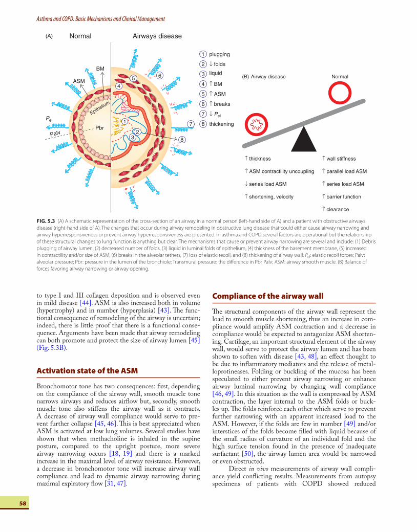
to type I and III collagen deposition and is observed even in mild disease [44] . ASM is also increased both in volume (hypertrophy) and in number (hyperplasia) [43] . Th e func-tional consequence of remodeling of the airway is uncertain; indeed, there is little proof that there is a functional conse-quence. Arguments have been made that airway remodeling can both promote and protect the size of airway lumen [45] ( Fig. 5.3B ).
Activation state of the ASM
Bronchomotor tone has two consequences: fi rst, depending on the compliance of the airway wall, smooth muscle tone narrows airways and reduces airfl ow but, secondly, smooth muscle tone also stiff ens the airway wall as it contracts. A decrease of airway wall compliance would serve to pre-vent further collapse [45, 46] . Th is is best appreciated when ASM is activated at low lung volumes. Several studies have shown that when methacholine is inhaled in the supine posture, compared to the upright posture, more severe airway narrowing occurs [18, 19] and there is a marked increase in the maximal level of airway resistance. However, a decrease in bronchomotor tone will increase airway wall compliance and lead to dynamic airway narrowing during maximal expiratory fl ow [31, 47] .
Compliance of the airway wall
Th e structural components of the airway wall represent the load to smooth muscle shortening, thus an increase in com-pliance would amplify ASM contraction and a decrease in compliance would be expected to antagonize ASM shorten-ing. Cartilage, an important structural element of the airway wall, would serve to protect the airway lumen and has been shown to soften with disease [43, 48] , an eff ect thought to be due to infl ammatory mediators and the release of metal-loprotineases. Folding or buckling of the mucosa has been speculated to either prevent airway narrowing or enhance airway luminal narrowing by changing wall compliance [46, 49] . In this situation as the wall is compressed by ASM contraction, the layer internal to the ASM folds or buck-les up. Th e folds reinforce each other which serve to prevent further narrowing with an apparent increased load to the ASM. However, if the folds are few in number [49] and/or interstices of the folds become fi lled with liquid because of the small radius of curvature of an individual fold and the high surface tension found in the presence of inadequate surfactant [50] , the airway lumen area would be narrowed or even obstructed.
Direct in vivo measurements of airway wall compli-ance yield confl icting results. Measurements from autopsy specimens of patients with COPD showed reduced
FIG. 5.3 (A) A schematic representation of the cross-section of an airway in a normal person (left-hand side of A) and a patient with obstructive airways
disease (right-hand side of A). The changes that occur during airway remodeling in obstructive lung disease that could either cause airway narrowing and
airway hyperresponsiveness or prevent airway hyperresponsiveness are presented. In asthma and COPD several factors are operational but the relationship
of these structural changes to lung function is anything but clear. The mechanisms that cause or prevent airway narrowing are several and include: (1) Debris
plugging of airway lumen, (2) decreased number of folds, (3) liquid in luminal folds of epithelium, (4) thickness of the basement membrane, (5) increased
in contractility and/or size of ASM, (6) breaks in the alveolar tethers, (7) loss of elastic recoil, and (8) thickening of airway wall. P el : elastic recoil forces; Palv:
alveolar pressure; Pbr: pressure in the lumen of the bronchiole; Transmural pressure: the diff erence in Pbr Palv; ASM: airway smooth muscle. (B) Balance of
forces favoring airway narrowing or airway opening.
Normal(A) Airways disease
plugging
↓ folds
liquid
↑ BM
↑ ASM
↑ breaks
↓ Pel
thickening
4
5
6
7
8
1
2
3
Pel
PalvPbr
ASM
BM
Epithelium
3
4
6
7
82
1
5
↑ thickness
↑ ASM contractility uncoupling
↓ series load ASM
↑ shortening, velocity
↑ wall stiffness
↑ parallel load ASM
↑ series load ASM
↑ barrier function
↑ clearance
NormalAirway disease(B)

5Pulmonary Physiology
59
compliance only in very small ( � 1 mm) airways [51] . Normal lungs show age-related decrease in wall compliance which complicates the interpretation of the losses observed in patients with COPD who are normally older [52, 53] . In asthma both increases [53, 54] and decreases [21] in wall stiff ness have been reported. Decreased wall compliance or stiff ening is likely the result of infl ammation and/or wall thickening and hence would be related to asthma severity and disease status. Another potential mechanism of airway narrowing from the increase in wall compliance results from a decrease in forces tethering the airway open. Th ese forces are decreased through two mechanisms. First, there are few alveolar attachments to the outer mucosa of the airway wall and second scar tissue has less tensile strength than normal tissue [55] .
Tethering forces or the coupling of lung volume and resistance
Th e attachments of the alveolar wall to the external wall of the intrapulmonary airway are the anatomical link between the alveoli of the parenchyma and airway. Th ese attachments form the physical linkage system that result in airways wid-ening and the fall in airways resistance as lung volumes increase. Th is tethering force is the single most important factor stabilizing and infl uencing airway lumen integrity [13–15] . Th is fact is evidenced by the profound eff ect lung volume has on airfl ow. As noted above, for a given degree of smooth muscle activation greater airway narrowing occurs at low versus high lung volumes. For example, the simple act of assuming the supine position, which is not associ-ated with a change in smooth muscle activation, markedly increases airway resistance [15, 18] . Th e eff ects of lung vol-ume on airways resistance are very sensitive to even small losses (∼1 cmH 2 O) in lung elastic recoil pressure [16, 17, 35] . Th is process is not limited to changes within the parameter of the airway as denoted by ASM, since changes external to the ASM can also uncouple the lung volume–airway resistance relationship.
Changing from the upright to the supine position leads to airway hyperresponsiveness (AHR) in asthmatics but such changes also occur in normals [18, 56] . Th e mech-anism for this phenomenon is most likely the loss of the tethering forces, as estimated by P el . Th ese forces pull open the airway wall as lung volume increases, thus a loss of out-ward recoil tethering the airway open is termed uncoupling . Airway resistance and lung volume uncoupling occurs rap-idly with sleep onset in the patient with nocturnal asthma and further uncoupling during the night probably as a result of infl ammatory events, resultant airway wall thickening, and mechanical uncoupling [21] . In COPD the loss of elas-tic recoil ( P el ) is both greater and more persistent than the age-dependent loss of recoil that occurs in normal persons, and when considered in regards to loss of P el with aging, accounts for the progressive loss of FEV 1 over the years.
Th e loss of P el , together with breaks in alveolar attach-ments, results in the increased airway narrowing noted on forced expiration in patients with obstructive airway dis-ease [47] . Smooth muscle contraction contributes to severe airway narrowing and loss of bronchodilation response
typically seen in patients with emphysema [57, 58] . For patients with COPD or severe long-standing asthma this profound alteration in the function and structure of the airway accounts for the sustained and therapeutically resist-ant increase in airway resistance or decrease in FEV 1 . Th e dilemma for the clinician is to ascertain what portion of the loss of structure and function is reversible and what portion is not.
AIRFLOW RESISTANCE
Chronic obstructive pulmonary disease
A common feature of the patient with COPD is airfl ow limitation; this is most often assessed by the fall in FEV 1 . Th e current Global Initiative for Chronic Obstructive Lung Disease (GOLD) classifi cation system for COPD uses post-bronchodilator FEV 1 as a major factor in classifi ca-tion of disease severity and in practice this variable makes up a signifi cant part of the case defi nition for COPD [59] . Airfl ow limitation in COPD arises in the small ( � 2 mm diameter at FRC) airways which exhibit neutrophilic infl ammation even in asymptomatic smokers [60, 61] . When patients have mild, asymptomatic disease there are subtle changes in airfl ow limitation [60] with changes in distribution and homogeneity of ventilation. As the severity of COPD increases there is increased involvement of more central airways but even when FEV 1 has fallen to 50–60% of predicted, more than half of the increased resistance can be attributed to narrowed intrapulmonary ( � 3 mm) airways [42] . Airway dimensions in COPD are reduced, even when the lungs are infl ated to a standard infl ation pressure (typi-cally 30 cmH 2 O) so one must conclude that the structural remodeling of collagen, mucous gland hyperplasia, general wall thickening, and loss of tethering elastic forces contrib-utes to the airway narrowing [62] .
Th ese alterations in structure and function lead to profound ventilatory defects and a loss of collateral ven-tilation [63, 64] . In turn there is a trapping of gas due to both long expiratory time constants and check valve-like processes within the obstructed and emphysematous spaces. Re-establishment of collateral pathways with intrabronchial, one-way valves, much like the spiracles of insects, leads to defl ation of these overextended spaces [65] and is currently being explored as a therapeutic option to the COPD patient to relieve this trapped gas [66] .
In patients with severe COPD expiratory airfl ow lim-itation is observed at all lung volumes and often occurs even when there is a minimal expiratory eff ort; in these patients expiratory airfl ow limitation occurs on expiration with each breath [67] . In more mild cases, the airfl ow limitation is observed only on forced expiration because: (1) fl ow and volume during eupnea do not reach their structural limits due to the reserve in the respiratory system and (2) dynamic hyperinfl ation, which increases elastic recoil by increasing lung volume, further prevents reaching these limits during eupnea. Th e pathogenesis of airfl ow limitation in patents
Asthma and COPD: Basic Mechanisms and Clinical Management
60
with COPD or more severe forms of bronchitis and asthma accordingly is quite complex and is a refl ection of the inter-play between airway caliber, respiratory muscle function, lung volume, elastic recoil, and airway wall compliance [6, 14, 23, 24, 27, 62, 68] .
Th e severity of COPD is defi ned as the post-bronchodilator FEV 1 [59] , so it is useful to consider what a fall in FEV 1 signifi es. Th e forced expiratory volume in 1 s – the FEV 1 – is a measurement taken from the volume–time relationship, known as a spirogram, during a forced exhala-tion from TLC. It is important to remember that the FEV 1 is a volume (liters) not a measure of airfl ow (liters per sec-ond) but it refl ects the average airfl ow over the fi rst second of a forced exhalation. A decrease in the FEV 1 , as a percent-age of its predicted value, indicates that the patient is unable to exhale a volume of air in 1 s similar in magnitude to that which could be exhaled by a person of the same sex, age, and height without lung disease. A considerable number of factors infl uence the magnitude of the FEV 1 , but only one of these factors is airway luminal size. Th e magnitude of the FEV 1 is very much infl uenced by the size of the inspira-tion prior to beginning the forced expiration (TLC), elas-tic recoil, airway caliber, airway wall compliance, and size of the lung that is open. In the case of the latter as lung vol-ume is lost by airway closure, fi lling or destruction, the VC, FVC, and hence FEV 1, decreases. It is important to realize that FEV 1 is a polyvalent index; where polyvalent indices are ones in which many factors contribute to the exact value of the index [69] ; indeed most measures of lung function are polyvalent. Accordingly it is unclear what a low FEV 1 might signify, the cause of the decrease or even the best treatment needed to reverse the loss in FEV 1 .
Empirically the FEV 1 is a very useful measure of overall lung function since it relates to morbidity and mor-tality of COPD, even death due to all causes [70] . Th e techniques used to measure FEV 1 have been well codifi ed [71, 72] . Th e general performance and in particular the
variability and reproducibility of the FEV 1 as an outcome measure is robust and for this reason the FEV 1 is a prin-cipal outcome variable for clinical research. Unfortunately a low FEV 1 is not specifi c for any single cause of airfl ow limitation, and other assessments of the loss in airfl ow limi-tation must be made.
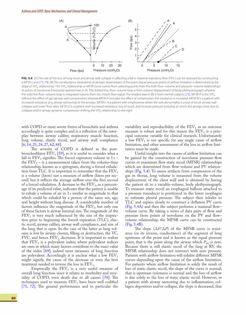
Useful insight into the causes of airfl ow limitation can be gained by the construction of isovolume pressure–fl ow curves or maximum fl ow static recoil (MFSR) relationships which are determined from fl ow–volume and PV relation-ships ( Fig. 5.4) . To assess artifacts from compression of the gas in thorax, lung volume is measured from the volume displacement of the chest wall and is obtained by having the patient sit in a variable-volume, body plethysmograph. To measure static recoil an esophageal balloon attached to a pressure transducer is positioned in the lower esophagus to estimate pleural pressure. Th e subject then inhales to TLC and expires slowly to construct a defl ation PV curve ( Fig. 5.4 A ) and then the subject performs a maximal fl ow–volume curve. By taking a series of data pairs of fl ow and pressure from points of isovolume on the PV and fl ow–volume relationship, the MFSR curve can be constructed ( Fig. 5.4 B ).
Th e slope ( Δ V / Δ P ) of the MFSR curve is resist-ance (or its inverse, conductance) of the segment of lung upstream of the point and is known as the equal pressure point, that is the point along the airway which P tm is zero. Because there is still elastic recoil of the lung at RV, the MFSR relationship does not intersect with zero pressure. Patients with airfl ow limitation will exhibit diff erent MFSR curves depending upon the cause of the airfl ow limitation. For patients where airfl ow limitation is solely the result of loss of static elastic recoil, the slope of the curve is normal; that is upstream resistance is normal and the loss of airfl ow is due solely to the loss of static elastic recoil. However, in a patient with airway narrowing due to infl ammation, col-lagen deposition and/or collapse, the slope is decreased, that
FIG. 5.4 (A) The role of the loss of lung recoil and airway wall collapse in aff ecting a fall in maximal expiratory fl ow (FEV 1 ) can be assessed by constructing
a MFSR curve [73, 74] . (B) The conductance (resistance) of airways downstream of the point (equal pressure point) of airfl ow limitation is determined as the
slope of V / P el relationship. This V / P el relationship or MFSR curve comes from selecting points from the both fl ow–volume and pressure–volume relationships
at points of isovolume (horizontal dashed lines in A). The dotted line, fl ow–volume loop is from volume displacement of body plethysmograph whereas
the solid line fl ow–volume loop is integrated volume from the mouth fl ow signal. The shaded area in (B) is from normal subjects [75] . MFSR A ’ is the V / P el
without the eff ect of gas (airway wall compression) whereas MFSR A includes the eff ect of compression; the resistance is increased. MFSR B is a patient with
increased resistance (e.g. airway narrowing) of the airways. MFSR C is a patient with emphysema where the sole abnormality is a loss of recoil, airway wall
collapse and lower fl ow rates. MFSR D is a patient with increased resistance, loss of recoil, and increase pressure (volume) at which the airways close due to
collapse and/or airway dynamic compression shifting the V / P el relationship to the right.
0
100
10 20 30
Pressure (cmH2O)Flow (lps)
(A)
A
Vol
ume
(%T
LC)
A�
5
4
3
2
1
0 5
Pressure (cmH2O)
Flo
w (
lps)
10 15
(B)A
B
CD
A�

5Pulmonary Physiology
61
is resistance is elevated. By comparing fl ow–volume curves constructed from measuring both lung volumes by integrat-ing the fl ow at the mouth to lung volume measured by the chest wall, the role of airway wall compression on the loss of airfl ow can be assessed [76] . Th e fl ow–volume curve derived from the fl ow and volume exiting the lung is displaced such that peak fl ow occurs at a higher volume but, after this ini-tial peak, fl ow is lower at any given volume due to dynamic airway wall compression. In essence this analysis assesses the delay in gas exiting the lung; the more dynamic com-pression, the more temporal delay.
In most patients with COPD, a combination of fac-tors contributes to airfl ow limitation [73–76] . Determining MSFR relationships can suggest which component might be amenable to treatment; bronchodilation for airway nar-rowing, pursed lip breathing for airway collapse and lung volume reduction surgery for improving static elastic recoil. In the case of the latter several studies using this analysis have shown that the eff ect of lung reduction surgery is a reduction of RV and the resultant increase in the VC [77] or a more complex response where both a change in volume and a change in resistance occurred [78] .
BRONCHIAL RESPONSIVENESS
Bronchial responsiveness includes either the acute response to a one-time treatment with a bronchodilator – usually a β 2 -receptor antagonist – or the response to a constrictor bronchial challenge. Determination of bronchial responsive-ness better defi nes and classifi es patients with airways dis-ease. Bronchial responsiveness assessments are often used as major outcome variables in clinical investigations and treat-ment trials because of the precision of the measure, its asso-ciation to underlying infl ammation, the relevance to asthma triggers, and the changes in bronchial responsiveness as the result of treatment.
Bronchodilator responses
Bronchodilator responsiveness to inhaled bronchodilators is among the factors used to diff erentiate between whether a given patient is to be categorized as COPD or asthma. Professional Society guidelines and position statements for interpretation of spirometric results [72, 79] discuss the various consensus criteria used to derive the currently used bronchodilator criterion that identifi es a signifi cant response as one where a 12% or greater change in the post-bronchodilator FEV 1 or FVC from pre-bronchodilator val-ues and a � 200 ml absolute change occurs ( Table 5.2 ).
Establishing a criterion that indicates a signifi cant bronchodilator response has been controversial because of uncertainty as to just what constitutes a signifi cant response [79, 80] . Various authors have suggested criteria that range from 10% to 15% change from baseline, some suggest using a change in percent of predicted or an exact volume change (e.g. 400 ml). Bronchodilator responses are reported as a percent change from the volumes obtained prior to use of a
β -agonist; however, this enhances the response if the start-ing baseline of FEV 1 or FVC is low. Th e use of a defi ni-tion that includes both an absolute change (e.g. � 200 ml) or change expressed as a percent of predicted addresses this dilemma.
Th ere are several approaches used to deliver bron-chodilators to patients for assessing reversibility but two to four puff s of albuterol with or without a spacer is the most common. However, combinations of bronchodilators with or without an anticholinergic agent or the use of nebulizer treatments are sometimes used. In this case one adminis-ters the bronchodilator until side eff ects are observed or the total dose delivered is used [71, 72, 79–81] . A wait period is then imposed to allow for the drug to exhibit its physiologi-cal eff ects, for example 15–20 min in the case of albuterol. Unfortunately techniques to determine bronchodilator responsiveness are highly variable between studies and labo-ratories making direct comparison of study results diffi cult.
Bronchodilator responsiveness is commonly meas-ured as a change in the FEV 1 but can also be assessed by expiratory fl ow–volume loops, specifi c airway conductance as determined with a body plethysmograph, or with the forced oscillation technique. Each technique measures dif-ferent aspects of lung function and the result obtained may not be exactly equivalent. Moreover the pattern of the bron-chodilator response can vary with some patients having a more central airways response (SGaw), whereas others have a more peripheral lung response as measured with FVC or the FEV 1 [4, 81, 82]. Such fi ndings are of great interest in determining the inferred site of the functional defect such as central airways versus peripheral airways and have obvi-ous ramifi cations to drug delivery.
Bronchoconstriction responses
AHR is either a bronchoconstricting response (fall in FEV 1 ) to a stimulus that normally does not cause a con-striction or is a heightened bronchoconstriction to the stimulus. Examples of the former are exercise, cold air, or bradykinin where examples of the latter are histamine and methacholine. Airways hyperresponsiveness is often used to defi ne the diagnosis of asthma, however, the presence of AHR is not specifi c since many lung diseases are associated with hyperresponsiveness including COPD [83] .
TABLE 5.2 Criteria recommended for acute bronchodilator responses in
adults [79, 81] .
FEV 1 (%) FVC (%) Comments
ACCP 15–25 15–25 % of baseline in 2 of 3 tests
Intermountain
ATS
15 15 % of baseline
ATS 1991 12 12 % of baseline and 200 ml
ATS/ERS 2000 12 12 % of baseline and 200 ml
ACCP: American College of Chest Physicians; ATS: American Thoracic Society [79] ; ERS:
European Respiratory Society [81] .
Asthma and COPD: Basic Mechanisms and Clinical Management
62
Over the last 50 years the proposed mechanisms to explain AHR included anything from enhanced choliner-gic tone of the airways to the current focus on pathologic alterations (remodeling) of the airway wall. While airways smooth muscle usually has a central role in most theories, recent experimental evidence suggests that hypercontrac-tility of the ASM is not always required [84] . Bronchial thermoplasty in which radio frequency energy is applied to airways through the bronchoscope to abate the ASM, improves asthma symptoms with only modest improve-ments in AHR [85] . Th is interesting result suggests that the ASM may not be as essential to AHR as previously thought or may serve other purposes [86] .
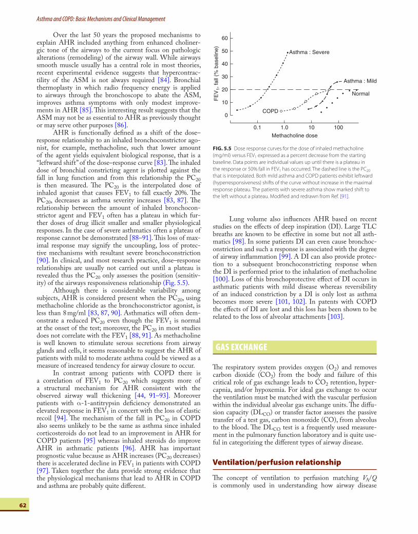
AHR is functionally defi ned as a shift of the dose–response relationship to an inhaled bronchoconstrictor ago-nist, for example, methacholine, such that lower amount of the agent yields equivalent biological response, that is a “ leftward shift ” of the dose–response curve [83] . Th e inhaled dose of bronchial constricting agent is plotted against the fall in lung function and from this relationship the PC 20 is then measured. Th e PC 20 is the interpolated dose of inhaled agonist that causes FEV 1 to fall exactly 20%. Th e PC 20 , decreases as asthma severity increases [83, 87] . Th e relationship between the amount of inhaled bronchocon-strictor agent and FEV 1 often has a plateau in which fur-ther doses of drug illicit smaller and smaller physiological responses. In the case of severe asthmatics often a plateau of response cannot be demonstrated [88–91] . Th is loss of max-imal response may signify the uncoupling, loss of protec-tive mechanisms with resultant severe bronchoconstriction [90] . In clinical, and most research practice, dose–response relationships are usually not carried out until a plateau is revealed thus the PC 20 only assesses the position (sensitiv-ity) of the airways responsiveness relationship ( Fig. 5.5 ).
Although there is considerable variability among subjects, AHR is considered present when the PC 20 , using methacholine chloride as the bronchoconstrictor agonist, is less than 8 mg/ml [83, 87, 90] . Asthmatics will often dem-onstrate a reduced PC 20 even though the FEV 1 is normal at the onset of the test; moreover, the PC 20 in most studies does not correlate with the FEV 1 [88, 91] . As methacholine is well known to stimulate serous secretions from airway glands and cells, it seems reasonable to suggest the AHR of patients with mild to moderate asthma could be viewed as a measure of increased tendency for airway closure to occur.
In contrast among patients with COPD there is a correlation of FEV 1 to PC 20 which suggests more of a structural mechanism for AHR consistent with the observed airway wall thickening [44, 91–93] . Moreover patients with α -1-antitrypsin defi ciency demonstrated an elevated response in FEV 1 in concert with the loss of elastic recoil [94] . Th e mechanism of the fall in PC 20 in COPD also seems unlikely to be the same as asthma since inhaled corticosteroids do not lead to an improvement in AHR for COPD patients [95] whereas inhaled steroids do improve AHR in asthmatic patients [96] . AHR has important prognostic value because as AHR increases (PC 20 decreases) there is accelerated decline in FEV 1 in patients with COPD [97] . Taken together the data provide strong evidence that the physiological mechanisms that lead to AHR in COPD and asthma are probably quite diff erent.
Lung volume also infl uences AHR based on recent studies on the eff ects of deep inspiration (DI). Large TLC breaths are known to be eff ective in some but not all asth-matics [98] . In some patients DI can even cause bronchoc-onstriction and such a response is associated with the degree of airway infl ammation [99] . A DI can also provide protec-tion to a subsequent bronchoconstricting response when the DI is performed prior to the inhalation of methacholine [100] . Loss of this bronchoprotective eff ect of DI occurs in asthmatic patients with mild disease whereas reversibility of an induced constriction by a DI is only lost as asthma becomes more severe [101, 102] . In patents with COPD the eff ects of DI are lost and this loss has been shown to be related to the loss of alveolar attachments [103] .
GAS EXCHANGE
Th e respiratory system provides oxygen (O 2 ) and removes carbon dioxide (CO 2 ) from the body and failure of this critical role of gas exchange leads to CO 2 retention, hyper-capnia, and/or hypoxemia. For ideal gas exchange to occur the ventilation must be matched with the vascular perfusion within the individual alveolar gas exchange units. Th e diff u-sion capacity (DL CO ) or transfer factor assesses the passive transfer of a test gas, carbon monoxide (CO), from alveolus to the blood. Th e DL CO test is a frequently used measure-ment in the pulmonary function laboratory and is quite use-ful in categorizing the diff erent types of airway disease.
Ventilation/perfusion relationship
Th e concept of ventilation to perfusion matching V A / Q is commonly used in understanding how airway disease
Methacholine dose
0.1
60
50
40
30
20
FE
V1,
fall
(% b
asel
ine)
10
0
1.0 10 100
Normal
Asthma : Mild
Asthma : Severe
COPD
FIG. 5.5 Dose response curves for the dose of inhaled methacholine
(mg/ml) versus FEV 1 expressed as a percent decrease from the starting
baseline. Data points are individual values up until there is a plateau in
the response or 50% fall in FEV 1 has occurred. The dashed line is the PC 20
that is interpolated. Both mild asthma and COPD patients exhibit leftward
(hyperresponsiveness) shifts of the curve without increase in the maximal
response plateau. The patients with severe asthma show marked shift to
the left without a plateau. Modifi ed and redrawn from Ref. [91] .
5Pulmonary Physiology
63
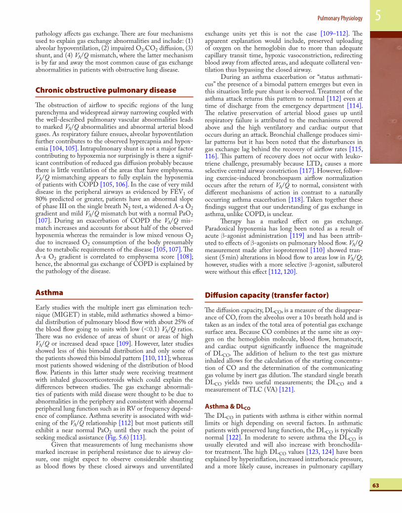
pathology aff ects gas exchange. Th ere are four mechanisms used to explain gas exchange abnormalities and include: (1) alveolar hypoventilation, (2) impaired O 2/ CO 2 diff usion, (3) shunt, and (4) V A / Q mismatch, where the latter mechanism is by far and away the most common cause of gas exchange abnormalities in patients with obstructive lung disease.
Chronic obstructive pulmonary disease
Th e obstruction of airfl ow to specifi c regions of the lung parenchyma and widespread airway narrowing coupled with the well-described pulmonary vascular abnormalities leads to marked V A / Q abnormalities and abnormal arterial blood gases. As respiratory failure ensues, alveolar hypoventilation further contributes to the observed hypercapnia and hypox-emia [104, 105] . Intrapulmonary shunt is not a major factor contributing to hypoxemia nor surprisingly is there a signif-icant contribution of reduced gas diff usion probably because there is little ventilation of the areas that have emphysema. V A / Q mismatching appears to fully explain the hypoxemia of patients with COPD [105, 106] . In the case of very mild disease in the peripheral airways as evidenced by FEV 1 of 80% predicted or greater, patients have an abnormal slope of phase III on the single breath N 2 test, a widened A-a O 2 gradient and mild V A / Q mismatch but with a normal PaO 2 [107] . During an exacerbation of COPD the V A / Q mis-match increases and accounts for about half of the observed hypoxemia whereas the remainder is low mixed venous O 2 due to increased O 2 consumption of the body presumably due to metabolic requirements of the disease [105, 107] . Th e A-a O 2 gradient is correlated to emphysema score [108] ; hence, the abnormal gas exchange of COPD is explained by the pathology of the disease.
Asthma
Early studies with the multiple inert gas elimination tech-nique (MIGET) in stable, mild asthmatics showed a bimo-dal distribution of pulmonary blood fl ow with about 25% of the blood fl ow going to units with low ( � 0.1) V A / Q ratios. Th ere was no evidence of areas of shunt or areas of high V A / Q or increased dead space [109] . However, later studies showed less of this bimodal distribution and only some of the patients showed this bimodal pattern [110, 111] ; whereas most patients showed widening of the distribution of blood fl ow. Patients in this latter study were receiving treatment with inhaled glucocorticosteroids which could explain the diff erences between studies. Th e gas exchange abnormali-ties of patients with mild disease were thought to be due to abnormalities in the periphery and consistent with abnormal peripheral lung function such as in RV or frequency depend-ence of compliance. Asthma severity is associated with wid-ening of the V A / Q relationship [112] but most patients still exhibit a near normal PaO 2 until they reach the point of seeking medical assistance ( Fig. 5.6 ) [113] .
Given that measurements of lung mechanisms show marked increase in peripheral resistance due to airway clo-sure, one might expect to observe considerable shunting as blood fl ows by these closed airways and unventilated
exchange units yet this is not the case [109–112] . Th e apparent explanation would include, preserved uploading of oxygen on the hemoglobin due to more than adequate capillary transit time, hypoxic vasoconstriction, redirecting blood away from aff ected areas, and adequate collateral ven-tilation thus bypassing the closed airway.
During an asthma exacerbation or “ status asthmati-cus ” the presence of a bimodal pattern emerges but even in this situation little pure shunt is observed. Treatment of the asthma attack returns this pattern to normal [112] even at time of discharge from the emergency department [114] . Th e relative preservation of arterial blood gases up until respiratory failure is attributed to the mechanisms covered above and the high ventilatory and cardiac output that occurs during an attack. Bronchial challenge produces simi-lar patterns but it has been noted that the disturbances in gas exchange lag behind the recovery of airfl ow rates [115, 116] . Th is pattern of recovery does not occur with leuko-triene challenge, presumably because LTD 4 causes a more selective central airway constriction [117] . However, follow-ing exercise-induced bronchospasm airfl ow normalization occurs after the return of V A / Q to normal, consistent with diff erent mechanisms of action in contrast to a naturally occurring asthma exacerbation [118] . Taken together these fi ndings suggest that our understanding of gas exchange in asthma, unlike COPD, is unclear.
Th erapy has a marked eff ect on gas exchange. Paradoxical hypoxemia has long been noted as a result of acute β -agonist administration [119] and has been attrib-uted to eff ects of β -agonists on pulmonary blood fl ow. V A / Q measurement made after isoproterenol [110] showed tran-sient (5 min) alterations in blood fl ow to areas low in V A / Q ; however, studies with a more selective β -agonist, salbuterol were without this eff ect [112, 120] .
Diff usion capacity (transfer factor)
Th e diff usion capacity, D L CO , is a measure of the disappear-ance of CO, from the alveolus over a 10 s breath hold and is taken as an index of the total area of potential gas exchange surface area. Because CO combines at the same site as oxy-gen on the hemoglobin molecule, blood fl ow, hematocrit, and cardiac output signifi cantly infl uence the magnitude of D L CO . Th e addition of helium to the test gas mixture inhaled allows for the calculation of the starting concentra-tion of CO and the determination of the communicating gas volume by inert gas dilution. Th e standard single breath D L CO yields two useful measurements; the D L CO and a measurement of TLC (VA) [121] .
Asthma & DLCO
Th e D L CO in patients with asthma is either within normal limits or high depending on several factors. In asthmatic patients with preserved lung function, the D L CO is typically normal [122] . In moderate to severe asthma the D L CO is usually elevated and will also increase with bronchodila-tor treatment. Th e high D L CO values [123, 124] have been explained by hyperinfl ation, increased intrathoracic pressure, and a more likely cause, increases in pulmonary capillary

Asthma and COPD: Basic Mechanisms and Clinical Management
64
blood volume or extravasation of red blood cells into the alveolus. Although not frequently commented on or because current practice is to lyse the lavage sample, the red blood cell content is high in lavage samples of asthmatics,
particularly those who are more severe [125] . Increased vac-ularization of the lung and airways might also increase the apparent pulmonary capillary blood volume and account for the elevated D L CO [43] .
100.0
100
Shunt
Normal
1.00.10.0
1.5
1.0
0.5
0.0100
Episodic asthma
1.00.010.0
0.9
0.6
0.2
0.0100
Chronic severe asthma
1.00.010.0
0.6
0.2
0.4
0.0100
Acute severe asthma
100.10.0
1.2
1.0
0.6
0.4
0.8
1.0
0.2Ven
tilat
ion
and
bloo
d flo
w (
l/min
)
0.0100100.1
COPD type A
1.00.010.0
1.2
0.4
0.8
100
COPD type B
100.10.010.0
Ventilation/blood flow ratio
1.0
1.0
2.0
FIG. 5.6 The V A / Q relationship as determined by the multiple inert gas elimination technique (MIGET). Note the diff erent scaling for both axes in the various
examples. The normal distribution of ventilation (open circles) and blood fl ow (solid circles) is over a narrow range of V A / Q ratios and is unimodal. Patients
with mild or chronic asthma have wider distributions but still exhibit unimodal distributions whereas acute severe asthma is bimodal. In emphysema (COPD,
type A) there is a bimodal distribution in ventilation whereas chronic bronchitis (COPD, type B) exhibits bimodal blood fl ow. Shunt is not present in any of
the examples. Reproduced from Refs [105, 111] with permission.
5Pulmonary Physiology
65
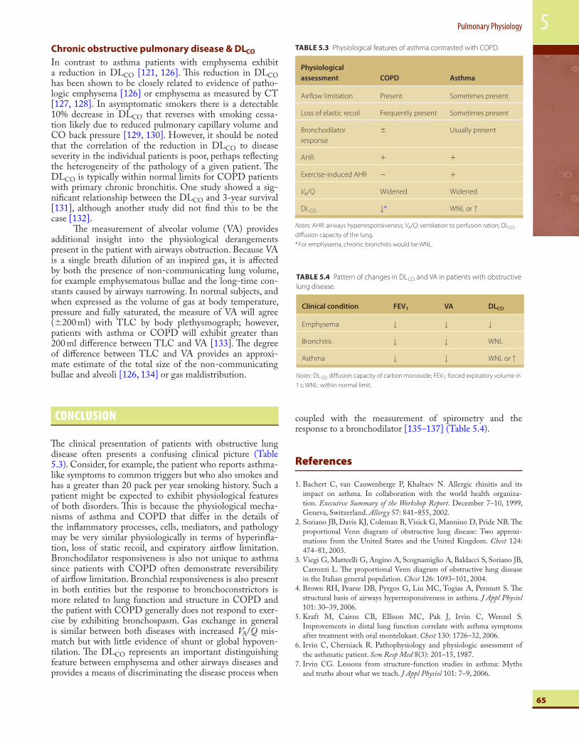
TABLE 5.3 Physiological features of asthma contrasted with COPD.
Physiological
assessment COPD Asthma
Airfl ow limitation Present Sometimes present
Loss of elastic recoil Frequently present Sometimes present
Bronchodilator
response
� Usually present
AHR � �
Exercise-induced AHR � �
V A / Q Widened Widened
D L CO ↓ * WNL or ↑
Notes : AHR: airways hyperresponsiveness; V A / Q : ventilation to perfusion ration; D L CO:
diff usion capacity of the lung.
* For emphysema, chronic bronchitis would be WNL.
Chronic obstructive pulmonary disease & DLCO
In contrast to asthma patients with emphysema exhibit a reduction in D L CO [121, 126] . Th is reduction in D L CO has been shown to be closely related to evidence of patho-logic emphysema [126] or emphysema as measured by CT [127, 128] . In asymptomatic smokers there is a detectable 10% decrease in DL CO that reverses with smoking cessa-tion likely due to reduced pulmonary capillary volume and CO back pressure [129, 130] . However, it should be noted that the correlation of the reduction in D L CO to disease severity in the individual patients is poor, perhaps refl ecting the heterogeneity of the pathology of a given patient. Th e D L CO is typically within normal limits for COPD patients with primary chronic bronchitis. One study showed a sig-nifi cant relationship between the DL CO and 3-year survival [131] , although another study did not fi nd this to be the case [132] .
Th e measurement of alveolar volume (VA) provides additional insight into the physiological derangements present in the patient with airways obstruction. Because VA is a single breath dilution of an inspired gas, it is aff ected by both the presence of non-communicating lung volume, for example emphysematous bullae and the long-time con-stants caused by airways narrowing. In normal subjects, and when expressed as the volume of gas at body temperature, pressure and fully saturated, the measure of VA will agree ( � 200 ml) with TLC by body plethysmograph; however, patients with asthma or COPD will exhibit greater than 200 ml diff erence between TLC and VA [133] . Th e degree of diff erence between TLC and VA provides an approxi-mate estimate of the total size of the non-communicating bullae and alveoli [126, 134] or gas maldistribution.
CONCLUSION
Th e clinical presentation of patients with obstructive lung disease often presents a confusing clinical picture (Table 5.3). Consider, for example, the patient who reports asthma-like symptoms to common triggers but who also smokes and has a greater than 20 pack per year smoking history. Such a patient might be expected to exhibit physiological features of both disorders. Th is is because the physiological mecha-nisms of asthma and COPD that diff er in the details of the infl ammatory processes, cells, mediators, and pathology may be very similar physiologically in terms of hyperinfl a-tion, loss of static recoil, and expiratory airfl ow limitation. Bronchodilator responsiveness is also not unique to asthma since patients with COPD often demonstrate reversibility of airfl ow limitation. Bronchial responsiveness is also present in both entities but the response to bronchoconstrictors is more related to lung function and structure in COPD and the patient with COPD generally does not respond to exer-cise by exhibiting bronchospasm. Gas exchange in general is similar between both diseases with increased V A / Q mis-match but with little evidence of shunt or global hypoven-tilation. Th e DL CO represents an important distinguishing feature between emphysema and other airways diseases and provides a means of discriminating the disease process when
coupled with the measurement of spirometry and the response to a bronchodilator [135–137] (Table 5.4).
References
1 . Bachert C, van Cauwenberge P, Khaltaev N. Allergic rhinitis and its impact on asthma. In collaboration with the world health organiza-tion. Executive Summary of the Workshop Report . December 7–10, 1999, Geneva, Switzerland. Allergy 57: 841–855, 2002.
2 . Soriano JB , Davis KJ , Coleman B , Visick G , Mannino D , Pride NB . Th e proportional Venn diagram of obstructive lung disease: Two approxi-mations from the United States and the United Kingdom . Chest 124 : 474 – 81 , 2003 .
3 . Viegi G , Matteelli G , Angino A , Scognamiglio A , Baldacci S , Soriano JB , Carrozzi L . Th e proportional Venn diagram of obstructive lung disease in the Italian general population . Chest 126 : 1093 – 101 , 2004 .
4 . Brown RH , Pearse DB , Pyrgos G , Liu MC , Togias A , Permutt S . Th e structural basis of airways hyperresponsiveness in asthma . J Appl Physiol 101 : 30 – 39 , 2006 .
5 . Kraft M , Cairns CB , Ellison MC , Pak J , Irvin C , Wenzel S . Improvements in distal lung function correlate with asthma symptoms after treatment with oral montelukast . Chest 130 : 1726 – 32 , 2006 .
6 . Irvin C , Cherniack R . Pathophysiology and physiologic assessment of the asthmatic patient . Sem Resp Med 8 ( 3 ) : 201 – 15 , 1987 .
7 . Irvin CG . Lessons from structure-function studies in asthma: Myths and truths about what we teach . J Appl Physiol 101 : 7 – 9 , 2006 .
TABLE 5.4 Pattern of changes in D L CO and VA in patients with obstructive
lung disease.
Clinical condition FEV 1 VA D L CO
Emphysema ↓ ↓ ↓
Bronchitis ↓ ↓ WNL
Asthma ↓ ↓ WNL or ↑
Notes : D L CO: diff usion capacity of carbon monoxide; FEV 1 : forced expiratory volume in
1 s; WNL: within normal limit.

Asthma and COPD: Basic Mechanisms and Clinical Management
66
8 . Irvin C . Guide to the evaluation of pulmonary function . In: Hamid Q , Shannon J , Martin J , Hamilton J (eds). Physiologic Basis of Respiratory Disease . Ontario : BC Decker , 2005 .
9 . Wanger J , Clausen JL , Coates A , Pedersen OF , Brusasco V , Burgos F , Casaburi R , Crapo R , Enright P , van der Grinten CP et al . Standardisation of the measurement of lung volumes . Eur Respir J 26 : 511 – 22 , 2005 .
10 . Whittaker LA , Irvin CG . Going to extremes of lung volume . J Appl Physiol 102 : 831 – 33 , 2007 .
11 . Loring SH , O’Donnell CR , Butler JP , Lindholm P , Jacobson F , Ferrigno M . Transpulmonary pressures and lung mechanics with glos-sopharyngeal insuffl ation and exsuffl ation beyond normal lung volumes in competitive breath-hold divers . J Appl Physiol 102 : 1 – 846 , 2007 .
12 . Peress L , Sybrecht G , Macklem PT . Th e mechanism of increase in total lung capacity during acute asthma . Am J Med 61 : 165 – 69 , 1976 .
13 . Butler J , Caro CG , Alcala R , Dubois AB . Physiological factors aff ect-ing airway resistance in normal subjects and in patients with obstruc-tive respiratory disease . J Clin Invest 39 : 584 – 91 , 1960 .
14 . Vincent NJ , Knudson R , Leith DE , Macklem PT , Mead J . Factors infl uencing pulmonary resistance . J Appl Physiol 29 : 236 – 43 , 1970 .
15 . Briscoe WA , Dubois AB . Th e relationship between airway resistance, airway conductance and lung volume in subjects of diff erent age and body size . J Clin Invest 37 : 1279 – 85 , 1958 .
16 . Bates JH , Schuessler TF , Dolman C , Eidelman DH . Temporal dynam-ics of acute isovolume bronchoconstriction in the rat . J Appl Physiol 82 : 55 – 62 , 1997 .
17 . Nagase T , Martin JG , Ludwig MS . Comparative study of mechanical interdependence: Eff ect of lung volume on raw during induced con-striction . J Appl Physiol 75 : 2500 – 5 , 1993 .
18 . Ding DJ , Martin JG , Macklem PT . Eff ects of lung volume on maximal methacholine-induced bronchoconstriction in normal humans . J Appl Physiol 62 : 1324 – 30 , 1987 .
19 . Meinero M , Coletta G , Dutto L , Milanese M , Nova G , Sciolla A , Pellegrino R , Brusasco V . Mechanical response to metha-choline and deep inspiration in supine men . J Appl Physiol 102 : 269 – 75 , 2007 .
20 . Martin J , Powell E , Shore S , Emrich J , Engel LA . Th e role of respira-tory muscles in the hyperinfl ation of bronchial asthma . Am Rev Respir Dis 121 : 441 – 47 , 1980 .
21 . Irvin CG , Pak J , Martin RJ . Airway-parenchyma uncoupling in noc-turnal asthma . Am J Respir Crit Care Med 161 : 50 – 56 , 2000 .
22 . Irvin CG . Lung volume: A principle determinant of airway smooth muscle function . Eur Respir J 22 : 3 – 5 , 2003 .
23 . Mead J , Turner JM , Macklem PT , Little JB . Signifi cance of the rela-tionship between lung recoil and maximum expiratory fl ow . J Appl Physiol 22 : 95 – 108 , 1967 .
24 . Black LF , Hyatt RE , Stubbs SE . Mechanism of expiratory airfl ow limitation in chronic obstructive pulmonary disease associated with 1-antitrypsin defi ciency . Am Rev Respir Dis 105 : 891 – 99 , 1972 .
25 . Gibson GJ , Pride NB , Davis J , Schroter RC . Exponential descrip-tion of the static pressure–volume curve of normal and diseased lungs . Am Rev Respir Dis 120 : 799 – 811 , 1979 .
26 . Finucane KE , Colebatch HJ . Elastic behavior of the lung in patients with airway obstruction . J Appl Physiol 26 : 330 – 38 , 1969 .
27 . Schlueter DP , Immekus J , Stead WW . Relationship between maximal inspiratory pressure and total lung capacity (coeffi cient of retraction) in normal subjects and in patients with emphysema, asthma, and diff use pulmonary infi ltration . Am Rev Respir Dis 96 : 656 – 65 , 1967 .
28 . Gold WM , Kaufman HS , Wadd JA . Elastic recoil of the lungs in chronic asthmatic patients before and after therapy . J Appl Physiol 23 : 433 – 38 , 1967 .
29 . Woolcock AJ , Read J . Th e static elastic properties of the lungs in asthma . Am Rev Respir Dis 98 : 788 – 94 , 1968 .
30 . Mansell A , Dubrawsky C , Levison H , Bryan AC , Langer H , Collins-Williams C , Orange RP . Lung mechanics in antigen-induced asthma . J Appl Physiol 37 : 297 – 301 , 1974 .
31 . De Troyer A , Yernault JC , Rodenstein D . Infl uence of beta-2 agonist aerosols on pressure–volume characteristics of the lungs . Am Rev Respir Dis 118 : 987 – 95 , 1978 .
32 . Eidelman DH , Ghezzo H , Kim WD , Hyatt RE , Cosio MG . Pressure–volume curves in smokers. Comparison with alpha-1-antitrypsin defi -ciency . Am Rev Respir Dis 139 : 1452 – 58 , 1989 .
33 . Wagers SS , Norton RJ , Rinaldi LM , Bates JH , Sobel BE , Irvin CG . Extravascular fi brin, plasminogen activator, plasminogen activator inhibitors, and airway hyperresponsiveness . J Clin Invest 114 : 104 – 11 , 2004 .
34 . Mead J , Takishima T , Leith D . Stress distribution in lungs: A model of pulmonary elasticity . J Appl Physiol 28 : 596 – 608 , 1970 .
35 . Carroll NG , Mutavdzic S , James AL . Increased mast cells and neu-trophils in submucosal mucous glands and mucus plugging in patients with asthma . Th orax 57 : 677 – 82 , 2002 .
36 . Lundblad LK , Th ompson-Figueroa J , Allen GB , Rinaldi L , Norton RJ , Irvin CG , Bates JH . Airway hyperresponsiveness in allergi-cally infl amed mice: Th e role of airway closure . Am J Respir Crit Care Med 175 : 768 – 74 , 2007 .
37 . Venegas JG , Winkler T , Musch G , Vidal Melo MF , Layfi eld D , Tgavalekos N , Fischman AJ , Callahan RJ , Bellani G , Harris RS . Self-organized patchiness in asthma as a prelude to catastrophic shifts . Nature 434 : 777 – 82 , 2005 .
38 . Wagner EM , Liu MC , Weinmann GG , Permutt S , Bleecker ER . Peripheral lung resistance in normal and asthmatic subjects . Am Rev Respir Dis 141 : 584 – 88 , 1990 .
39 . Kaminsky DA , Irvin CG , Gurka DA , Feldsien DC , Wagner EM , Liu MC , Wenzel SE . Peripheral airways responsiveness to cool, dry air in normal and asthmatic individuals . Am J Respir Crit Care Med 152 : 1784 – 90 , 1995 .
40 . Kraft M , Pak J , Martin RJ , Kaminsky D , Irvin CG . Distal lung dys-function at night in nocturnal asthma . Am J Respir Crit Care Med 163 : 1551 – 56 , 2001 .
41 . Ohrui T , Yanai M , Sekizawa K , Morikawa M , Sasaki H , Takishima T . Eff ective site of bronchodilation by beta-adrenergic and anticholinergic agents in patients with chronic obstructive pulmonary disease: Direct measurement of intrabronchial pressure with a new catheter . Am Rev Respir Dis 146 : 88 – 91 , 1992 .
42 . Yanai M , Sekizawa K , Ohrui T , Sasaki H , Takishima T . Site of airway obstruction in pulmonary disease: Direct measurement of intrabron-chial pressure . J Appl Physiol 72 : 1016 – 23 , 1992 .
43 . James AL , Wenzel S . Signifi cance of airway remodeling in airway dis-eases . Eur J Respir Dis 30 : 1420 – 41 , 2007 .
44 . Roche WR , Beasley R , Williams JH , Holgate ST . Subepithelial fi brosis in the bronchi of asthmatics . Lancet 1 : 520 – 24 , 1989 .
45 . McParland BE , Macklem PT , Pare PD . Airway wall remodeling: Friend or foe? J Appl Physiol 95 : 426 – 34 , 2003 .
46 . Wiggs BR , Hrousis CA , Drazen JM , Kamm RD . On the mechanism of mucosal folding in normal and asthmatic airways . J Appl Physiol 83 : 1814 – 21 , 1997 .
47 . Krowka MJ , Enright PL , Rodarte JR , Hyatt RE . Eff ect of eff ort on measurement of forced expiratory volume in one second . Am Rev Respir Dis 136 : 829 – 33 , 1987 .
48 . Tandon MK , Campbell AH . Bronchial cartilage in chronic bronchitis . Th orax 24 : 607 – 12 , 1969 .
49 . Lambert RK , Codd SL , Alley MR , Pack RJ . Physical determinants of bronchial mucosal folding . J Appl Physiol 77 : 1206 – 16 , 1994 .
50 . Yager D , Butler JP , Bastacky J , Israel E , Smith G , Drazen JM . Amplifi cation of airway constriction due to liquid fi lling of airway interstices . J Appl Physiol 66 : 2873 – 84 , 1989 .
51 . Wilson AG , Massarella GR , Pride NB . Elastic properties of airways in human lungs post mortem . Am Rev Respir Dis 110 : 716 – 29 , 1974 .
52 . Maisel JC , Silvers GW , Mitchell RS , Petty TL . Bronchial atrophy and dynamic expiratory collapse . Am Rev Respir Dis 98 : 988 – 97 , 1968 .
53 . Brackel HJ , Pedersen OF , Mulder PG , Overbeek SE , Kerrebijn KF , Bogaard JM . Central airways behave more stiffl y during forced expiration
5Pulmonary Physiology
67
in patients with asthma . Am J Respir Crit Care Med 162 : 896 – 904 , 2000 .
54 . Wilson JW , Li X , Pain MC . Th e lack of distensibility of asthmatic air-ways . Am Rev Respir Dis 148 : 806 – 9 , 1993 .
55 . Saetta M , Ghezzo H , Kim WD , King M , Angus GE , Wang NS , Cosio MG . Loss of alveolar attachments in smokers. A morphometric cor-relate of lung function impairment . Am Rev Respir Dis 132 : 894 – 900 , 1985 .
56 . Skloot G , Permutt S , Togias A . Airway hyperresponsiveness in asthma: A problem of limited smooth muscle relaxation with inspiration . J Clin Invest 96 : 2393 – 403 , 1995 .
57 . Opazo Saez AM , Seow CY , Pare PD . Peripheral airway smooth mus-cle mechanics in obstructive airways disease . Am J Respir Crit Care Med 161 : 910 – 17 , 2000 .
58 . Pare PD , Wiggs BR , James A , Hogg JC , Bosken C . Th e compara-tive mechanics and morphology of airways in asthma and in chronic obstructive pulmonary disease . Am Rev Respir Dis 143 : 1189 – 93 , 1991 .
59 . Pauwels RA , Buist AS , Calverley PM , Jenkins CR , Hurd SS . Global strategy for the diagnosis, management, and prevention of chronic obstructive pulmonary disease. NHLBI/WHO global initiative for chronic obstructive lung disease (GOLD) workshop summary . Am J Respir Crit Care Med 163 : 1256 – 76 , 2001 .
60 . Cosio M , Ghezzo H , Hogg JC , Corbin R , Loveland M , Dosman J , Macklem PT . Th e relations between structural changes in small airways and pulmonary-function tests . N Engl J Med 298 : 1277 – 81 , 1978 .
61 . Niewoehner DE , Kleinerman J , Rice DB . Pathologic changes in the peripheral airways of young cigarette smokers . N Engl J Med 291 : 755 – 58 , 1974 .
62 . Hogg JC , Chu F , Utokaparch S , Woods R , Elliott WM , Buzatu L , Cherniack RM , Rogers RM , Sciurba FC , Coxson HO et al . Th e nature of small-airway obstruction in chronic obstructive pulmonary disease . N Engl J Med 350 : 2645 – 53 , 2004 .
63 . Hogg JC , Macklem PT , Th urlbeck WM . Th e resistance of collateral channels in excised human lungs . J Clin Invest 48 : 421 – 31 , 1969 .
64 . Terry PB , Traystman RJ , Newball HH , Batra G , Menkes HA . Collateral ventilation in man . N Engl J Med 298 : 10 – 15 , 1978 .
65 . Choong CK , Macklem PT , Pierce JA , Lefrak SS , Woods JC , Conradi MS , Yablonskiy DA , Hogg JC , Chino K , Cooper JD . Transpleural ventilation of explanted human lungs . Th orax 62 : 623 – 30 , 2007 .
66 . Wood DE , McKenna RJ Jr. , Yusen RD , Sterman DH , Ost DE , Springmeyer SC , Gonzalez HX , Mulligan MS , Gildea T , Houck WV et al . A multicenter trial of an intrabronchial valve for treatment of severe emphysema . J Th orac Cardiovasc Surg 133 : 65 – 73 , 2007 .
67 . Eltayara L , Becklake MR , Volta CA , Milic-Emili J . Relationship between chronic dyspnea and expiratory fl ow limitation in patients with chronic obstructive pulmonary disease . Am J Respir Crit Care Med 154 : 1726 – 34 , 1996 .
68 . Hogg JC , Macklem PT , Th urlbeck WM . Site and nature of airway obstruction in chronic obstructive lung disease . N Engl J Med 278 : 1355 – 60 , 1968 .
69 . Mead J . Problems in interpreting common tests of pulmonary mechan-ical function . In: Macklem P , Permutt S (eds). Th e Lung in Transition Between Death and Disease . New York : Marcel Dekker Inc , 1979 .
70 . Ferguson GT , Enright PL , Buist AS , Higgins MW . Offi ce spirometry for lung health assessment in adults: A consensus statement from the national lung health education program . Chest 117 : 1146 – 61 , 2000 .
71 . Enright PL , Johnson LR , Connett JE , Voelker H , Buist AS . Spirometry in the lung health study. I: Methods and quality control . Am Rev Respir Dis 143 : 1215 – 23 , 1991 .
72 . Miller MR , Hankinson J , Brusasco V , Burgos F , Casaburi R , Coates A , Crapo R , Enright P , van der Grinten CP , Gustafsson P et al . Standardisation of spirometry . Eur Respir J 26 : 319 – 38 , 2005 .
73 . Fry DL , Hyatt RE . Pulmonary mechanisms: A unifi ed analysis of the relationship between pressure, volume and gas fl ow in the lungs of nor-mal and diseased subjects . Am J Physiol 26 : 672 – 89 , 1960 .
74 . Pride NB , Permutt S , Riley RL , Bromberger-Barnea B . Determinants of maximal expiratory fl ow from the lungs . J Appl Physiol 23 : 646 – 62 , 1967 .
75 . Leaver DG , Tatterfi eld AE , Pride NB . Contributions of loss of lung recoil and of enhanced airways collapsibility to the airfl ow obstruc-tion of chronic bronchitis and emphysema . J Clin Invest 52 : 2117 – 28 , 1973 .
76 . Ingram RH Jr. , Schilder DP . Eff ect of gas compression on pulmonary pressure, fl ow, and volume relationship . J Appl Physiol 21 : 1821 – 26 , 1966 .
77 . Ingenito EP , Evans RB , Loring SH , Kaczka DW , Rodenhouse JD , Body SC , Sugarbaker DJ , Mentzer SJ , DeCamp MM , Reilly JJ Jr. . Relation between preoperative inspiratory lung resistance and the out-come of lung-volume-reduction surgery for emphysema . N Engl J Med 338 : 1181 – 85 , 1998 .
78 . Gelb AF , Brenner M , McKenna RJ Jr. , Fischel R , Zamel N , Schein MJ . Serial lung function and elastic recoil 2 years after lung volume reduc-tion surgery for emphysema . Chest 113 : 1497 – 506 , 1998 .
79 . ATS . Lung function testing: Selection of reference values and interpre-tive strategies . Am Rev Respir Dis 144 : 1202 – 18 , 1991 .
80 . Pellegrino R , Viegi G , Brusasco V , Crapo RO , Burgos F , Casaburi R , Coates A , van der Grinten CP , Gustafsson P , Hankinson J et al . Interpretative strategies for lung function tests . Eur Respir J 26 : 948 – 68 , 2005 .
81 . Smith HR , Irvin CG , Cherniack RM . Th e utility of spirometry in the diagnosis of reversible airways obstruction . Chest 101 : 1577 – 81 , 1992 .
82 . O’Donnell DE , Lam M , Webb KA . Spirometric correlates of improve-ment in exercise performance after anticholinergic therapy in chronic obstructive pulmonary disease . Am J Respir Crit Care Med 160 : 542 – 49 , 1999 .
83 . Crapo RO , Casaburi R , Coates AL , Enright PL , Hankinson JL , Irvin CG , MacIntyre NR , McKay RT , Wanger JS , Anderson SD et al . Guidelines for methacholine and exercise challenge testing-1999. Th is offi cial statement of the American thoracic society was adopted by the ATS board of directors, July 1999 . Am J Respir Crit Care Med 161 : 309 – 29 , 2000 .
84 . Wagers S , Lundblad LK , Ekman M , Irvin CG , Bates JH . Th e allergic mouse model of asthma: Normal smooth muscle in an abnormal lung? J Appl Physiol 96 : 2019 – 27 , 2003 .
85 . Cox G , Th omson NC , Rubin AS , Niven RM , Corris PA , Siersted HC , Olivenstein R , Pavord ID , McCormack D , Chaudhuri R et al . Asthma control during the year after bronchial thermoplasty . N Engl J Med 356 : 1327 – 37 , 2007 .
86 . Solway J , Irvin CG . Airway smooth muscle as a target for asthma ther-apy . N Engl J Med 356 : 1367 – 69 , 2007 .
87 . Hargreave FE , Ryan G , Th omson NC , O’Byrne PM , Latimer K , Juniper EF , Dolovich J . Bronchial responsiveness to histamine or meth-acholine in asthma: Measurement and clinical signifi cance . J Allergy Clin Immunol 68 : 347 – 55 , 1981 .
88 . Ryan G , Latimer KM , Dolovich J , Hargreave FE . Bronchial respon-siveness to histamine: Relationship to diurnal variation of peak fl ow rate, improvement after bronchodilator, and airway calibre . Th orax 37 : 423 – 29 , 1982 .
89 . Gibbons WJ , Sharma A , Lougheed D , Macklem PT . Detection of excessive bronchoconstriction in asthma . Am J Respir Crit Care Med 153 : 582 – 89 , 1996 .
90 . Sterk PJ , Bel EH . Th e shape of the dose–response curve to inhaled bronchoconstrictor agents in asthma and in chronic obstructive pulmo-nary disease . Am Rev Respir Dis 143 : 1433 – 37 , 1991 .
91 . Woolcock AJ , Anderson SD , Peat JK , Du Toit JI , Zhang YG , Smith CM , Salome CM . Characteristics of bronchial hyperresponsive-ness in chronic obstructive pulmonary disease and in asthma . Am Rev Respir Dis 143 : 1438 – 43 , 1991 .
92 . Taylor RG , Joyce H , Gross E , Holland F , Pride NB . Bronchial reactiv-ity to inhaled histamine and annual rate of decline in FEV 1 in male smokers and ex-smokers . Th orax 40 : 9 – 16 , 1985 .

Asthma and COPD: Basic Mechanisms and Clinical Management
68
93 . Tashkin DP , Altose MD , Bleecker ER , Connett JE , Kanner RE , Lee WW , Wise R . Th e lung health study: Airway responsiveness to
inhaled methacholine in smokers with mild to moderate airfl ow limi-tation. Th e Lung Health Study Research Group . Am Rev Respir Dis 145 : 301 – 10 , 1992 .
94 . Cheung D , Schot R , Zwinderman AH , Zagers H , Dijkman JH , Sterk PJ . Relationship between loss in parenchymal elastic recoil pressure and maximal airway narrowing in subjects with alpha1-antitrypsin defi ciency . Am J Respir Crit Care Med 155 : 135 – 40 , 1997 .
95 . Watson A , Lim TK , Joyce H , Pride NB . Failure of inhaled corticos-teroids to modify bronchoconstrictor or bronchodilator responsive-ness in middle-aged smokers with mild airfl ow obstruction . Chest 101 : 350 – 55 , 1992 .
96 . Sont JK , Willems LN , Bel EH , van Krieken JH , Vandenbroucke JP , Sterk PJ . Clinical control and histopathologic outcome of asthma when using airway hyperresponsiveness as an additional guide to long-term treatment. Th e AMPUL study group . Am J Respir Crit Care Med 159 : 1043 – 51 , 1999 .
97 . Tashkin DP , Altose MD , Connett JE , Kanner RE , Lee WW , Wise RA . Methacholine reactivity predicts changes in lung function over time in smokers with early chronic obstructive pulmonary dis-ease. Th e Lung Health Study Research Group . Am J Respir Crit Care Med 153 : 1802 – 11 , 1996 .
98 . Orehek J , Charpin D , Velardocchio JM , Grimaud C . Bronchomotor eff ect of bronchoconstriction-induced deep inspirations in asthmatics . Am Rev Respir Dis 121 : 297 – 305 , 1980 .
99 . Lim TK , Pride NB , Ingram RH Jr. . Eff ects of volume history dur-ing spontaneous and acutely induced air-fl ow obstruction in asthma . Am Rev Respir Dis 135 : 591 – 96 , 1987 .
100 . Scichilone N , Kapsali T , Permutt S , Togias A . Deep inspiration-induced bronchoprotection is stronger than bronchodilation . Am J Respir Crit Care Med 162 : 910 – 16 , 2000 .
101 . Assefa D , Amin N , Dozor AJ . Eff ect of deep inspiration on airway caliber in children with asthma . Pediatr Pulmonol 38 : 406 – 12 , 2004 .
102 . Scichilone N , Marchese R , Soresi S , Interrante A , Togias A , Bellia V . Deep inspiration-induced changes in lung volume decrease with severity of asthma . Respir Med 101 : 951 – 56 , 2007 .
103 . Scichilone N , Bruno A , Marchese R , Vignola AM , Togias A , Bellia V . Association between reduced bronchodilatory eff ect of deep inspira-tion and loss of alveolar attachments . Respir Res 6 : 55 , 2005 .
104 . Begin P , Grassino A . Inspiratory muscle dysfunction and chronic hypercapnia in chronic obstructive pulmonary disease . Am Rev Respir Dis 143 : 905 – 12 , 1991 .
105 . Agusti AG , Barbera JA . Contribution of multiple inert gas elimi-nation technique to pulmonary medicine. 2. Chronic pulmonary diseases: Chronic obstructive pulmonary disease and idiopathic pulmonary fi brosis . Th orax 49 : 924 – 32 , 1994 .
106 . Hlastala MP , Robertson HT . Inert gas elimination characteristics of the normal and abnormal lung . J Appl Physiol 44 : 258 – 66 , 1978 .
107 . Barbera JA , Roca J , Ferrer A , Felez MA , Diaz O , Roger N , Rodriguez-Roisin R . Mechanisms of worsening gas exchange dur-ing acute exacerbations of chronic obstructive pulmonary disease . Eur Respir J 10 : 1285 – 91 , 1997 .
108 . Barbera JA , Roca J , Ramirez J , Wagner PD , Ussetti P , Rodriguez-Roisin R . Gas exchange during exercise in mild chronic obstructive pulmonary disease. Correlation with lung structure . Am Rev Respir Dis 144 : 520 – 25 , 1991 .
109 . Wagner PD , Dantzker DR , Iacovoni VE , Tomlin WC , West JB . Ventilation–perfusion inequality in asymptomatic asthma . Am Rev Respir Dis 118 : 511 – 24 , 1978 .
110 . Wagner PD , Hedenstierna G , Bylin G . Ventilation–perfusion ine-quality in chronic asthma . Am Rev Respir Dis 136 : 605 – 12 , 1987 .
111 . Rodriguez-Roisin R , Roca J . Contributions of multiple inert gas elimination technique to pulmonary medicine. III: Bronchial asthma . Th orax 49 : 1027 – 33 , 1994 .
112 . Ballester E , Roca J , Ramis L , Wagner PD , Rodriguez-Roisin R . Pulmonary gas exchange in severe chronic asthma. Response to
100% oxygen and salbutamol . Am Rev Respir Dis 141 : 558 – 62 , 1990 .
113 . Mountain RD , Sahn SA . Clinical features and outcome in patients with acute asthma presenting with hypercapnia . Am Rev Respir Dis 138 : 535 – 39 , 1988 .
114 . Roca J , Ramis L , Rodriguez-Roisin R , Ballester E , Montserrat JM , Wagner PD . Serial relationships between ventilation–perfusion ine-quality and spirometry in acute severe asthma requiring hospitaliza-tion . Am Rev Respir Dis 137 : 1055 – 61 , 1988 .
115 . Rodriguez-Roisin R , Ferrer A , Navajas D , Agusti AG , Wagner PD , Roca J . Ventilation–perfusion mismatch after methacholine chal-lenge in patients with mild bronchial asthma . Am Rev Respir Dis 144 : 88 – 94 , 1991 .
116 . Echazarreta AL , Gomez FP , Ribas J , Achaval M , Barbera JA , Roca J , Chung KF , Rodriguez-Roisin R . Eff ects of inhaled furosemide on platelet-activating factor challenge in mild asthma . Eur Respir J 14 : 616 – 21 , 1999 .
117 . Echazarreta AL , Dahlen B , Garcia G , Agusti C , Barbera JA , Roca J , Dahlen SE , Rodriguez-Roisin R . Pulmonary gas exchange and spu-tum cellular responses to inhaled leukotriene D-4 in asthma . Am J Respir Crit Care Med 164 : 202 – 6 , 2001 .
118 . Young IH , Corte P , Schoeff el RE . Pattern and time course of ventilation–perfusion inequality in exercise-induced asthma . Am Rev Respir Dis 125 : 304 – 11 , 1982 .
119 . Knudson RJ , Constantine HP . An eff ect of isoproterenol on ventila-tion–perfusion in asthmatic versus normal subjects . J Appl Physiol 22 : 402 – 6 , 1967 .
120 . Ballester E , Reyes A , Roca J , Guitart R , Wagner PD , Rodriguez-Roisin R . Ventilation–perfusion mismatching in acute severe asthma: Eff ects of salbutamol and 100% oxygen . Th orax 44 : 258 – 67 , 1989 .
121 . Cotton DJ , Graham BL . Single-breath carbon monoxide diff usion capacity or transfer factor . In: Hamid Q , Shannon J , Martin J (eds). Physiologic Basis of Respiratory Disease . Hamilton : BC Decker , 2005 .
122 . Saydain G , Beck KC , Decker PA , Cowl CT , Scanlon PD . Clinical signifi cance of elevated diff using capacity . Chest 125 : 446 – 52 , 2004 .
123 . Keens TG , Mansell A , Krastins IR , Levison H , Bryan AC , Hyland RH , Zamel N . Evaluation of the single-breath diff using capacity in asthma and cystic fi brosis . Chest 76 : 41 – 44 , 1979 .
124 . Collard P , Njinou B , Nejadnik B , Keyeux A , Frans A . Single breath diff using capacity for carbon monoxide in stable asthma . Chest 105 : 1426 – 29 , 1994 .
125 . Diaz P , Galleguillos FR , Gonzalez MC , Pantin CF , Kay AB . Bronchoalveolar lavage in asthma: Th e eff ect of disodium cromogly-cate (cromolyn) on leukocyte counts, immunoglobulins, and comple-ment . J Allergy Clin Immunol 74 : 41 – 48 , 1984 .
126 . McLean A , Warren PM , Gillooly M , MacNee W , Lamb D . Microscopic and macroscopic measurements of emphysema: Relation to carbon monoxide gas transfer . Th orax 47 : 144 – 49 , 1992 .
127 . Gould GA , MacNee W , McLean A , Warren PM , Redpath A , Best JJ , Lamb D , Flenley DC . CT measurements of lung density in life can quantitate distal airspace enlargement – An essential defi n-ing feature of human emphysema . Am Rev Respir Dis 137 : 380 – 92 , 1988 .
128 . Gevenois PA , De Vuyst P , de Maertelaer V , Zanen J , Jacobovitz D , Cosio MG , Yernault JC . Comparison of computed density and micro-scopic morphometry in pulmonary emphysema . Am J Respir Crit Care Med 154 : 187 – 92 , 1996 .
129 . Sansores RH , Pare P , Abboud RT . Eff ect of smoking cessation on pulmonary carbon monoxide diff using capacity and capillary blood volume . Am Rev Respir Dis 146 : 959 – 64 , 1992 .
130 . Watson A , Joyce H , Hopper L , Pride NB . Infl uence of smoking habits on change in carbon monoxide transfer factor over 10 years in middle aged men . Th orax 48 : 119 – 24 , 1993 .
131 . Dubois P , Machiels J , Smeets F , Delwiche JP , Lulling J . CO trans-fer capacity as a determining factor of survival for severe hypoxae-mic COPD patients under long-term oxygen therapy . Eur Respir J 3 : 1042 – 47 , 1990 .

5Pulmonary Physiology
69
132 . Anthonisen NR , Wright EC , Hodgkin JE . Prognosis in chronic obstructive pulmonary disease . Am Rev Respir Dis 133 : 14 – 20 , 1986 .
133 . Graham BL , Mink JT , Cotton DJ . Eff ect of breath-hold time on DL CO (sb) in patients with airway obstruction . J Appl Physiol 58 : 1319 – 25 , 1985 .
134 . Wade JF 3rd , Mortenson R , Irvin CG . Physiologic evaluation of bul-lous emphysema . Chest 100 : 1151 – 54 , 1991 .
135 . Gelb AF , Gold WM , Wright RR , Bruch HR , Nadel JA . Physiologic diagnosis of subclinical emphysema . Am Rev Respir Dis 107 : 50 – 63 , 1973 .
136 . Klein JS , Gamsu G , Webb WR , Golden JA , Muller NL . High-resolution CT diagnosis of emphysema in symptomatic patients with normal chest radiographs and isolated low diff using capacity . Radiology 182 : 817 – 21 , 1992 .
137 . Sciurba FC . Physiologic similarities and diff erences between COPD and asthma . Chest 126 : 117S – 124S , 2004 , discussion 159S–161S.