association of tsh receptor antibody, thyroid stimulating...
TRANSCRIPT

114
ORIGINAL ARTICLE
Acta Medica Indonesiana - The Indonesian Journal of Internal Medicine
Association of TSH Receptor Antibody, Thyroid Stimulating Antibody, and Thyroid Blocking Antibody with Clinical Activity Score and Degree of Severity of Graves Ophthalmopathy
Imam Subekti1, Asman Boedisantoso1, Nila Djuwita F. Moeloek2, Sarwono Waspadji1, Muchtaruddin Mansyur3
1 Department of Internal Medicine, Faculty of Medicine, University of Indonesia - Cipto Mangunkusumo Hospital. Jl. Diponegoro 71, Jakarta Pusat 10430, Indonesia. Correspondence mail: [email protected]. 2 Department of Ophthalmology, Faculty of Medicine, University of Indonesia. 3 Department of Public Health, Faculty of Medicine, University of Indonesia.
ABSTRAKTujuan: mempelajari hubungan TSH Receptor Antibody (TRAb), Thyroid Stimulating Antibody (TSAb), Thyroid
Stimulation Blocking Antibody (TBAb) dan rasio TSAb/TBAb dengan skor aktivitas klinis (clinical activity score = CAS) dan derajat keparahan oftalmopati, pada OG klinis dan scan. Metode: studi potong lintang ini melibatkan 75 subjek Graves yang diperoleh secara konsekutif di RSUPN-CM Jakarta sejak Desember 2009 hingga Januari 2011. Oftalmopati klinis ditegakkan berdasarkan kriteria Bartley, dan oftalmopati scan ditegakkan berdasarkan penebalan otot ekstraokular dan peningkatan volume lemak retroorbita pada CT scan orbita. TSH, FT4, FT3 dan TRAb diperiksa dengan cara Elisa. Sedangkan TSAb dan TBAb diperiksa dengan cara RIA. Tingkat aktivitas klinis ditentukan berdasarkan clinical activity score (CAS), dan derajat keparahan OG ditentukan berdasarkan sistem NOSPECS modifikasi Eckstein. Hasil: subjek Graves berasal dari berbagai suku bangsa yang ada di Indonesia dengan rentang 20-63 tahun, yang memenuhi kriteria inklusi. Diperoleh 28 (37,3%) subjek OG klinis dan 61 (83,6%) subjek OG scan. Proporsi TSAb sebesar 70 (93,3%) dan TBAb sebesar 13 (17,3%) subjek Graves. Tidak ada korelasi antara TRAb dengan CAS dan derajat keparahan OG klinis dan OG scan. Korelasi TSAb dengan CAS OG klinis, CAS OG scan dan derajat keparahan OG scan juga tidak bermakna. Sedangkan korelasi TSAb dengan derajat keparahan OG klinis bermakna (r=0,274; p=0,009). Korelasi TBAb dengan CAS OG klinis, derajat keparahan OG klinis dan OG scan tidak bermakna. Terdapat korelasi terbalik kuat bermakna TBAb dengan CAS OG scan (r= -0,565, p=0,035). Rasio TSAb/TBAb berkorelasi kuat dengan semua parameter, berturut-turut dengan CAS OG klinis (r=0,730; p=0,031), CAS OG scan (r=0,607; p=0,024), derajat keparahan OG klinis (r=0,563; p=0,023) dan derajat keparahan OG scan (r=0,762; p=0,001). Kesimpulan: TRAb tidak berkorelasi dengan CAS dan derajat keparahan baik OG klinis maupun OG scan, sedangkan TSAb berkorelasi dengan derajat keparahan OG klinis. TBAb berkorelasi negatif dengan CAS OG scan, sementara rasio TSAb/TBAb berkorelasi dengan CAS dan derajat keparahan baik pada OG klinis dan OG scan.
Kata kunci: clinical activity score (CAS), oftalmopati Graves, TBAb, TRAb, dan TSAb.
ABSTRACTAim: to evaluate the association between TSH receptor antibody (TRAb), thyroid stimulating antibody
(TSAb), and thyroid stimulation blocking antibody (TBAb) and TSAb/TBAb ratio with clinical activity score (CAS) and degree of severity of GO. Methods: this was a cross sectional study involving 75 consecutive Graves’ disease patients admitted at Cipto Mangunkusumo hospital between December 2009 until January 2011. Bartley criteria were used as clinical criteria for GO. In addition to clinical criteria, orbital CT scan and measurement of extraocular muscle thickness and increment of retroorbital fat volume were carried out. Furthermore, TSH,

Vol 44 • Number 2 • April 2012 Association of TSH Receptor Antibody, Thyroid Stimulating Antibody
115
FT4, FT3 and TRAb were measured using ELISA method, TSAb and TBAb using RIA method. Clinical activity of GO was measured using clinical activity score (CAS). Degree of severity of GO was measured using Eckstein modified NOSPECS system. Results: subjects consisted of various different ethnic groups in Indonesia, with the range of age between 20-63 years old. There were 28 (37%) and 61 (83.6%) GO subjects with clinical-based and CT scan-based respectively. Among all GD subjects, the proportion of TSAb were 70 (93.3%) and TBAb were 13 (17.3%). There was no correlation between TRAb and CAS nor the severity of GO, either clinical-based and CT scan-based GO. There was also no correlation between TSAb and CAS. No correlation between TSAb and severity of CT scan-based GO, but significant correlation between TSAb and severity of clinical-based GO (r=0.274; p=0.009) was noted. There was no correlation between TBAb and severity of GO. We also found no correlation between TBAb and CAS of clinical-based GO, but we found strong negative correlation with CAS CT scan-based GO (r=-0.565; p=0.035). The TSAb/TBAb ratio was also strongly correlated with all parameters evaluated, CAS of clinical-based GO (r=0.730; p=0.031), CAS of CT scan-based GO (r=0.607; p=0.024), degree of severity of clinical-based GO (r=0.563; p=0.023), and degree of severity of CT scan-based GO (r=0.762; p=0.001). Conclusion: TRAb was not correlated with CAS and degree of severity of clinical-based and CT scan-based GO, while TSAb was correlated with degree of severity of GO. There was negative correlation between TBAb and CAS of clinical-based GO, whereas TSAb/TBAb ratio was correlated with CAS and degree of severity of both clinical-based and CT scan-based GO.
Key words: clinical activity score (CAS), Graves’ ophthalmo-pathy, TBAb, TRAb, TSAb.
INTRODUCTIONOphthalmopathy is the most common extra-
thyroid manifestation of Graves disease (GD).1 The pathogenesis of graves ophthalmopathy (GO) is multifactorial, which involves autoimmune process.2 GD is characterized by the presence of antibody to TSH-R (thyroid stimulating hormone receptor), which is known as TRAb (TSH receptor antibody). TRAbs in GD can be divided into 2 major functions: thyroid-stimulating antibody (TSAb) and thyroid stimulation blocking antibody (TBAb).3 TRAb can be measured using two methods. First measurement is based on binding competition, which is known as thyroid-binding inhibitory immunoglobulin (TBII); and secondly, by measuring the production of cyclic adenosine monophosphate (cAMP) as an outcome from interaction between antibody and the TSH-R.4,5
The degree of GO can be measured by two methods: clinical activity and severity. Clinical activity is determined by using clinical activity score (CAS), which consists of 7 variables of orbital signs and symptoms, and each variable is scored of one and the sum of the score from 7 variables can reflect the level of disease activity.6 Eckstein suggested the NOSPECS system to evaluate degree of severity of GO by evaluating retraction, soft tissue inflammation, proptosis, clinical extraocular muscles involvement, corneal
defect and optic neuropathy. The degree of severity of GO can be detected earlier using an orbital CT scan, because it can measure extra ocular muscle thickness, retro orbital fat volume and the presence or absence of proptosis.7 Theoretically, the clinical manifestation of GO will correlate with TRAb, but previous studies regarding those issues were inconsistent. Studies in Europe (Caucasian) and Asian (Oriental) showed that TSAb activity had a positive correlation with GO. However, the correlation of TRAb (measurement using TBII) and GO had been inconsistent, correlation is noted in Europe7-9 but not in Asia.10,11 Among those studies, the role of TBAb had never been evaluated. In GD patients (and also GO patients) who had those two antibodies (TSAb and TBAb), the TBAb will block the action and activity of TSAb, and vice versa the TSAb will also influence TBAb activity. On that condition, the clinical manifestations might depend on the balanced ratio of TSAb and TBAb, leading an assumption that TSAb/TBAb ratio might be correlated with GO. This study tried to determine the association between TRAb, TSAb, TBAb and TSAb/TBAb ratio with CAS and severity of GO.
METHODS
This was an observational cross sectional study with consecutive sampling performed at

Imam Subekti Acta Med Indones-Indones J Intern Med
116
Cipto Mangunkusumo Hospital Thyroid Clinic from December 2009 until January 2011. Subject criteria were newly diagnosed naive GD patients or previously diagnosed GD patients who were still in toxic condition, either under treatment or not. Subjects were of any ethnic groups in Indonesia which belong to Proto Malay or Deutro Malay group12-14 or mixed from the two groups, and were willing to participate in this study. Subjects were non pregnant woman, no thyroid crisis, no heart failure and no other severe diseases. The number of sample was calculated using Rifffenburgh formula15 and found that this study needed 16 GO patients with positive TBAb activity. Bartley et al. criteria was used for clinical-based GO.16 CT scan-based GO was defined from clinical-based GO combined with orbital CT scan. Data collection included history taking; general and specific (eye) physical examination; TSHs, FT4, FT3, TRAb, TSAb, TBAb, and orbital CT scan to measure extra ocular muscles (superior, inferior, lateralis and medialis rectus muscles), retro orbital fat volume, and the presence or absence of proptosis by measuring the distance between basis and top of cornea.17-21 Hormone measurements used ELISA while TSAb and TBAb used the RIA method. Clinical activity was measured using clinical activity score (CAS). The degree of severity was measured using Eckstein modified NOSPECS system. Statistical analysis used parametric test for normal distributed variables using Kolmogorov Smirnov test (FT4, TBAb and TSAb/TBAb ratio). Data transformation to logaritm-10 (log-10) among variables (FT3, TRAb and TSAb) who did not have normal distribution, was performed. After transformed to log-10 data, Kolmogorov Smirnov test for normality was repeated, and all variables were then normally distributed. Correlation analysis among different antibodies (TRAb, TSAb, TBAb and TSAB/TBAb ratio) with CAS and the degree of severity of GO used Spearman correlation test.
RESULTSDuring the study, based on the TRAb levels
from 30 subjects with normal thyroid function as reference, there were 75 subjects who were eligible for the study. There were 56 females (74.7%) and 19 males (25.3%). There were variable subject ethnic groups from Proto Malay, Deutero Malay and mixed groups. From all
subjects, 70 (93.3%) of them had TSAb activity ≥180%, and 13 (17.3%) subjects with TBAb activity ≥45.6%.
On clinical examination, based on Bartley criteria, there were 28 (37.3%) subjects with GO. However, when clinical examination combined with orbital CT scan, there were 61 (83.6%) subjects with GO. From all 28 subjects with clinical-based GO, there were 21 (75%) female (37.5% from all female GD patients) and 7 (25%) male (36.8% from all male GD patients), so the female to male ratio was similar to sex ratio of GD patients which was 3:1. There were no significant differences in sex proportion between subjects with and without GO (p=0.959). Regarding the age group, there were also no significant differences of proportion between subjects with or without GO (p=0.223).
From all CT scan-based GO, there were 46 (75.4%) female subjects (83.6% from all female GD subjects) and 15 (24.6%) male subjects (83.3% from all male GD subjects), so the female to male ratio was similar to sex ratio of GD patients which was 3:1. There were no significant differences of sex proportion between subjects with and without GO (p=0.616). Regarding the age group, there was also no significant difference in proportion between subjects with or without GO (p=0.574).
Characteristics of subjects can be seen in Table 1.
Table 2 shows the results of the correlation analysis between antibodies related to Graves’ disease with CAS and degree of severity of GO.
Significant correlation among different antibodies related to Graves’ disease with CAS and severity of GO could be seen in Figure 1-6.
DISCUSSIONThis study was specifically done in an
Indonesian population in order to highlight the characteristics of GD and GO among Indonesians. Actually, there are no native Indonesian people genetically; nevertheless, this study was trying to homogenize by choosing subjects from ethnic groups in the archipelago, which were Proto Malay and Deutro Malay group.
The prevalence of clinical-based GO in this study was 37.3%, which was quite the same as previous study from Lim et al.22 In a multi ethnic Malaysia population (Malay, Chinese, and Indian). Lim et al.22 reported the prevalence

Vol 44 • Number 2 • April 2012 Association of TSH Receptor Antibody, Thyroid Stimulating Antibody
117
Table 1. Characteristics of Graves’ subjects, clinical-based GO and scan-based GO
Variables Graves’ Clinical-based GO Scan-based GO p
Subjek 75 (100%) 28 (37.3%) 61 (83.6%)
Age (year)
- Women 37.7 (SD 9.7) 39.7 (SD 10.2) 37.6 (SD 9.8) 0.810
- Men 34.5 (SD 8.8) 32.1 (SD 8.3) 34.8 (SD 9.0)
Sex
- Women 56 (74.7%) 21 (37.5%) 46 (83.6%)
- Men 19 (25.3%) 7 (36.8%) 15 (83.3%)
Duration of Illness (mo) 12 15 12 0.948
Smoking 14 (18.7%) 4 (14.3%) 10 (18.2%)
FT4 (ng/dL) 4.76 (SD 2.5) 4.314 (SD 2.0) 4.72 (SD 2.5) 0.685
FT3 (pg/mL) 11.1 11.3 11.1 0.812
TRAb (U/L) 8.08 8.24 8.08 0.689
TSAb (%) 646.5 972.4 659.6 0.443
TBAb (%) 68.93 (SD 17.7) 68.7 (SD 16.1) 68.93 (SD 17.7) 0.972
Ratio (%) 21.31 (SD 1.3) 25 (SD 1.1) 21.31 (SD 12.8) 0.806
Tabel 2. Correlation between Antibodies related to Graves’ disease with CAS and Degree of Severity of GO
CAS Degree of severityClinical based-GO (28)
Scan based-GO (61)
Clinical based-GO (28)
Scan based-GO (61)
TRAb r = 0,277 r = 0,109 r = -0,078 r = -0,037
p = 0,077 p = 0,202 p = 0,254 p = 0,377
TSAb r = 0,104 r = 0,139 r = 0,274 r = 0,127
p = 0,300 p = 0,142 p = 0,009 p = 0,141
TBAb r = -0,468 r = -0,565 r = 0,169 r = -0,179
p = 0,145 p = 0,035 p = 0,290 p = 0,280
TSAb/TBAb r = 0,730 r = 0,607 r = 0,563 r = 0,762
ratio p = 0,031 p = 0,024 p = 0,023 p = 0,001
3.400
3.200
3.000
2.800
2.600
2.400
2.200
2.000
0 1 2 3 4 5 6
The severity of clinical-based GO
TSAb
r = 0,274; p = 0,009
Figure 1. Correlation between TSAb activity and severity of clinical-based GO
100.0
90.0
80.0
70.0
60.0
50.0
40.0
0 1 2 3 4
TBAb
The CAS of scan-based GO
r = -0,565p = 0,035
Figure 2. Correlation between TBAb activity and CAS of scan-based GO
of GO was 34.7% among GD patients. On the other hand, Marcocci et al.23 and Brent24 reported variable GO prevalance, ranging from 25%-50%. Upper lid retraction, which was one of GO criteria in this study, was found in 22.7% of GD subjects, or 60.7% of GO subjects. This number is higher than the study by Lim et al (53.4%), but still lower as compared to the study by Mourits et al.25 (70%) who reported that lid retraction is the most common sign in GO.
After performing an orbital CT scan to diagnose GO, the prevalence of GO was increased from 37.3% to 83.6%. The CT-scan-based GO prevalence is the same as previous study reporting the prevalence around 60-90%, if
using CT scan, MRI or USG B scan.26,27 Moreover Krassas also noted that with the help of CT scan, GO can be detected in almost all GD cases.28

Imam Subekti Acta Med Indones-Indones J Intern Med
118
There were 9%-10.7% subjects with severe GO according to modified NOSPECS system by Eckstein et al.7 but those subjects did not have pronounced eye complaints. This is opposite to Caucasian population, in which 3%-5% GO patients had severe GO with accompanying intense pain symptoms, corneal ulceration and compression optic neuropathy.29 The mechanisms underlying those different findings is still unclear. As previously genetic study revealed that genetic predisposition for GD is variable between ethnic groups. HLA study on GO progression in Europe, Asia and America yielded different characteristics. These findings might underline the variability in GO clinical manifestation.
In a correlation analysis among different antibodies, there are moderate correlation between TRAb and TSAb. It showed us that TRAb concentration, which was measured all antibodies bind to TSH-receptor (TBII), being affected by other factors, including TBAb. Tada et al.30 reported that TRAb in GD patients who also had TBAb was more heterogenous than in GD patients without TBAb. Strong correlation between TSAb and TSAb/TBAb ratio makes impression that TBAb did not strongly affect TSAb activity.
TRAb and TSAb were correlated with FT4 and FT3, in which TRAb correlation was stronger than TSAb. This finding was in line with study from Noh et al.31 TSAb will induce intracellular cAMP production, but does not have role in the production of cAMP-dependent inositol triphosphate. The two systems in transcellular signal transduction are being activated by two different antibodies. The TRAb measurement method measured those two antibodies, while the TSAb measurement method measured only the cAMP, consequently the correlation of TRAb to thyroid function is stronger than TSAb. The findings of significant correlation between TRAb and TSAb to FT3 indicates that TRAb and TSAb have a role in clinical manifestation of GD, recalling that FT3 is an active hormone. These conditions were consistent with the findings that there were a significant mean TSAb difference among toxic, subtoxic and non toxic GD patients.
In a correlation analysis between antibodies and the CAS, revealed TRAb and TSAb activity was not correlated with the CAS of scan and clinical-based GO. Conversely, there was a strong negative correlation between TBAb and the CAS
50.00
40.00
30.00
20.00
10.00
0.00
0 1 2 3 4
Ratio
The severity of clinical based-GO
r = 0,607;p = 0,031
Figure 4. Correlation between TSAb/TBAb ratio and severity of clinical-based GO
0 1 2 3 4
r = 0,607;p = 0,024
50
40
30
20
10
0
Ratio
The CAS of scan based-GO
Figure 5. Correlation between TSAb/TBAb ratio and CAS of scan-based GO
0 1 2 3
The severity of scan based-GO
50
40
30
20
10
0
Ratio
r = 0,762p = 0,001
Figure 6. Correlation between TSAb/TBAb ratio and severity of scan-based GO
r = -0,565p = 0,035
30.0
20.0
10.0
40.0
50.0
0 1 2 3 4
The CAS of scan-based GO
Ratio
r = 0,730p = 0,031
Figure 3. Correlation between TSAb/TBAb ratio and CAS of clinical-based GO
Vol 44 • Number 2 • April 2012 Association of TSH Receptor Antibody, Thyroid Stimulating Antibody
119
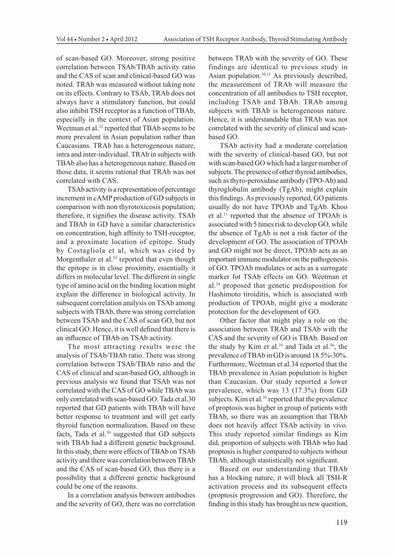
of scan-based GO. Moreover, strong positive correlation between TSAb/TBAb activity ratio and the CAS of scan and clinical-based GO was noted. TRAb was measured without taking note on its effects. Contrary to TSAb, TRAb does not always have a stimulatory function, but could also inhibit TSH receptor as a function of TBAb, especially in the context of Asian population. Weetman et al.32 reported that TBAb seems to be more prevalent in Asian population rather than Caucasians. TRAb has a heterogeneous nature, intra and inter-individual. TRAb in subjects with TBAb also has a heterogeneous nature. Based on those data, it seems rational that TRAb was not correlated with CAS.
TSAb activity is a representation of percentage increment in cAMP production of GD subjects in comparison with non thyrotoxicosis population; therefore, it signifies the disease activity. TSAb and TBAb in GD have a similar characteristics on concentration, high affinity to TSH-receptor, and a proximate location of epitope. Study by Costagliola et al, which was cited by Morgenthaler et al.33 reported that even though the epitope is in close proximity, essentially it differs in molecular level. The different in single type of amino acid on the binding location might explain the difference in biological activity. In subsequent correlation analysis on TSAb among subjects with TBAb, there was strong correlation between TSAb and the CAS of scan GO, but not clinical GO. Hence, it is well defined that there is an influence of TBAb on TSAb activity.
The most attracting results were the analysis of TSAb/TBAb ratio. There was strong correlation between TSAb/TBAb ratio and the CAS of clinical and scan-based GO, although in previous analysis we found that TSAb was not correlated with the CAS of GO while TBAb was only correlated with scan-based GO. Tada et al.30 reported that GD patients with TBAb will have better response to treatment and will get early thyroid function normalization. Based on these facts, Tada et al.30 suggested that GD subjects with TBAb had a different genetic background. In this study, there were effects of TBAb on TSAb activity and there was correlation between TBAb and the CAS of scan-based GO, thus there is a possibility that a different genetic background could be one of the reasons.
In a correlation analysis between antibodies and the severity of GO, there was no correlation
between TRAb with the severity of GO. These findings are identical to previous study in Asian population.10,11 As previously described, the measurement of TRAb will measure the concentration of all antibodies to TSH receptor, including TSAb and TBAb. TRAb among subjects with TBAb is heterogeneous nature. Hence, it is understandable that TRAb was not correlated with the severity of clinical and scan-based GO.
TSAb activity had a moderate correlation with the severity of clinical-based GO, but not with scan-based GO which had a larger number of subjects. The presence of other thyroid antibodies, such as thyro-peroxidase antibody (TPO-Ab) and thyroglobulin antibody (TgAb), might explain this findings. As previously reported, GO patients usually do not have TPOAb and TgAb. Khoo et al.11 reported that the absence of TPOAb is associated with 5 times risk to develop GO, while the absence of TgAb is not a risk factor of the development of GO. The association of TPOAb and GO might not be direct, TPOAb acts as an important immune modulator on the pathogenesis of GO. TPOAb modulates or acts as a surrogate marker fot TSAb effects on GO. Weetman et al.34 proposed that genetic predisposition for Hashimoto tiroiditis, which is associated with production of TPOAb, might give a moderate protection for the development of GO.
Other factor that might play a role on the association between TRAb and TSAb with the CAS and the severity of GO is TBAb. Based on the study by Kim et al.35 and Tada et al.36, the prevalence of TBAb in GD is around 18.5%-30%. Furthermore, Weetman et al.34 reported that the TBAb prevalence in Asian population is higher than Caucasian. Our study reported a lower prevalence, which was 13 (17.3%) from GD subjects. Kim et al.35 reported that the prevalence of proptosis was higher in group of patients with TBAb, so there was an assumption that TBAb does not heavily affect TSAb activity in vivo. This study reported similar findings as Kim did, proportion of subjects with TBAb who had proptosis is higher compared to subjects without TBAb, although stastistically not significant.
Based on our understanding that TBAb has a blocking nature, it will block all TSH-R activation process and its subsequent effects (proptosis progression and GO). Therefore, the finding in this study has brought us new question,

Imam Subekti Acta Med Indones-Indones J Intern Med
120
how big were the effects of TBAb on GO progression. In this study, the effects of TBAb on GO had been inconsistent. There was correlation between TBAb and the CAS of scan-based GO but there was no correlation with the severity of GO. Kim et al.35 described that actually the in vivo blocking effect of TBAB on stimulation effects of TRAb was not strong.
The calculated TSAb/TBAb activity ratio was strongly correlated with the severity of both clinical and scan-based GO. Based on extensive literature search, we found no journal which reported correlation between TSAb/TBAb activity ratio and GO. There was also strong correlation between TSAb/TBAb activity ratio with TSAb. Meanwhile we found non stastitically significant negative correlation between TSAb/TBAb activity ratio and TBAb; and TSAb with TBAb. On subsequent analysis among subjects with TBAb, there was strong correlation between TSAb and the CAS of scan-based GO, between TSAb with the severity of clinical and scan-based GO. Based on those findings, there are two possible explanations.
The first possibility is the presence of TBAb which has blocking nature. In previous study by Morgenthaler et al.33 TSAb and TBAb have the same affinity and epitop binding site on TSH-R. While Kim et al.35 reported that the blocking effects of TBAb on TRAb stimulation effects is not strong. Consequently, there is a need for doing further study regarding TBAb, especially if we look at the concept that TBAb has a blocking nature to TSAb stimulation effects.
The second possible explanation is the subject characteristics itself. As stated before, Tada et al.30 reported that GD subjects with TBAb responded to treatment better and had earlier normalization of thyroid function. Previous studies reported different characteristics of GD who had TBAb, which lead us to propose that there are other factors such as genetic differences which underline the treatment response among subjects with or without TBAb.
CONCLUSIONFrom all above discussion, it can be concluded
that TRAb was not correlated with CAS and severity clinical and scan-based GO, similair to previous studies in Asia. TSAb activity was correlated with the severity of clinical-based GO while TBAb was correlated with CAS of scan-
based GO. TSAb/TBAb activity ratio had a strong correlation with CAS and severity of clinical and scan-based GO.
REFERENCES1. Bartalena L, Pinchera A, Marcocci C. Management
of Graves ophthalmopathy: reality and perspectives. Endocr Rev. 2000;21:168-99.
2. Kazim M, Goldberg RA, Smith TJ. Insight into the pathogenesis of thyroid associated orbitopathy: evolving rationale for therapy. Arch Ophthalmol. 2002;120:380-6.
3. Schott M, Scherbaum WA, Morgenthaler NG. Thyrotropin receptor autoanti-bodies in Graves’ disease. Trends Endocrinol Metab. 2005;16(5):243-8.
4. Vitti P, Elisei R, Tonacchera M, Chiovato L, Mancusi F, Rago T, et al. Detection of thyroid-stimulating antibody using Chinese hamster ovary cells transfected with cloned human thyrotropin receptor. J Clin Endocrinol Metab. 1993;76:499-503.
5. Ochi Y, Yamashiro K, Takasu N, Kajita Y, Nagata A. Sensitive assay to detect thyroid stimulating antibody (TSAb) in the presence of thyroid blocking antibody (TSBAb) in serum. Horm Metab Res. 2001;33:115-20.
6. Pinchera A, Wiersinga WM, Glinoer D, Kendall-Taylor P, Koornneef L, Marcocci C, et al. Classification of eye changes of Graves’ disease. Thyroid. 1992;2:235-6.
7. Eckstein AK, Plicht M, Lax H, Neuha¨user M, Mann K, Lederbogen S, Heckmann C, Esser J, and Morgenthaler NG. Thyrotropin receptor autoantibodies are independent risk factors for Graves’ ophthalmopathy and help to predict severity and outcome of the disease. J Clin Endocrinol Metab. 2006;91:3464-70.
8. Vos XG, Smit N, Endert E, Tijssen JGP, Wiersinga WM. Frequency and characteristics of TBII-seronegative patients in a population with untreated Graves’ hyperthyroidism: a prospective study. Clin Endocrinol. 2008;69:311-7.
9. Gerding MN, van der Meer JW, Broenink M, Bakker O, Wiersinga WM, Prummel MF. Association of thyrotrophin receptor antibodies with the clinical features of Graves’ ophthalmopathy. Clin Endocrinol (Oxf). 2000;52:267-71.
10. Goh SY, Ho SC, Seah LL, Fong KS, Khoo DHC. Thyroid autoantibody profiles in ophthalmic dominant and thyroid dominant Graves’ disease differ and suggest ophthalmopathy is a multiantigenic disease. Clin Endocrinol. 2004; 60:60-7.
11. Khoo DHC, Ho SC, Seah LL, et al. The combination of absent thyroid peroxidase antibodies and high thyroid-stimulating immunoglobulin levels in Graves’ disease identifies a group at markedly increased risk of ophthalmopathy. Thyroid. 1999;9:1175-80.
12. Lim D. Sejarah Indonesia [online]. E-mail to Media Care 2007 March 14 [cited 2009 July 10]. ([email protected]).
13. Koentjaraningrat. Pendahuluan. Manusia dan Kebudayaan di Indonesia. Editor Koentjaraningrat. Penerbit Djambatan, Jakarta. 1997:1-36.
14. Penduduk Indonesia tertua dan persebaran bangsa-bangsa zaman prehistori [internet] 2009 August 26 [cited 2011 July 1]. Available from: http://ekohero.blogspot.com/2009/08/penduduk-indonesia-tertua-dan.html

Vol 44 • Number 2 • April 2012 Association of TSH Receptor Antibody, Thyroid Stimulating Antibody
121
15. Riffenburgh RH. Statistics in Medicine. 2nd Edition. London Elsevier 2006:413-5.
16. Bartley GB, Gorman CA. Diagnostic criteria for Graves’ ophthalmopathy. Am J Ophthalmol. 1995;119:792-5.
17. Fidor-Mikita E, Krupski W. Computed tomography imaging of orbits in thyroid orbitopathy. JPCCR. 2008;2:59-63.
18. Forbes G, Gehnng DG, Gorman CA, Brennan MD. Volume measurements of nor-mal orbital structures by computer tomographic analysis. AJR. 1985;145:149-54.
19. Regensburg NI, Wiersinga WM, Velthove EJV. Age and gender-specific refe-rence values of orbital fat and muscle volumes in Caucasians. Br J Ophthlamol. Published online December 2, 2009.
20. Kwon J, Barrera JF, Jung YT, Most SP. Measurements of orbital volume change using computed topmography in isolated orbital blowout fractures. Arch Facial Surg. 2009;11(6):395-8.
21. Marcocci C, Pinchera A, Marino M. A treatment strategy for Graves' orbitopathy. Nat Clin Pract Endocrinol Metab. 2007;3:330–6.
22. Lim SL, Lim AKE, Mumtaz M, Hussein E, Bebakar WMW, Khir AS. Prevalence, risk factor, and clinical features of thyroid-associated opthalmopathy in multiethnic Malaysian patients with Graves’ disease. Thyroid. 2008;18:1297-301.
23. Marcocci C, Bartalena L, Bogazzi F, Panicucci M, Pinchera A. Studies on the occurrence of ophthalmopathy in Graves’ disease. Acta Endocrinol. 1989;120(4):473-8.
24. Brent GA. Graves’ disease. N Engl J Med. 2008;358: 2594-605.
25. Mourits MP. Diagnosis and differential diagnosis of Graves’ orbitopathy. Graves’ orbitopathy: a multidisciplinary approach. In: Wiersinga WM, Kahaly GJ, Karger AG, eds. Switzerland: Basel; 2007. p. 66-77.
26. Weetman AP. Graves' disease. N Eng J Med. 2000;343(17):1236-48.
27. Noth D, Gebauer M, Muller B, Burgi U, Diem P. Graves’ ophthalmopathy: natural history and treatment outcome. Swiss Med Wkly. 2001;131:603-9.
28. Krassas GE, Wiersinga WM. Thyroid eye disease: current concepts and the EUGOGO perspective. Thyroid Int. 2005;4:1-21.
29. Wiersinga WM, Bartalena L. Epidemiology and prevention of Graves ophthalmo-pathy. Thyroid. 2002;12:855-60.
30. Tada H, Mizuta I, Takano T, Tatsumi K, Izumi Y, Hidaka Y, et al. Blocking-type anti-TSH receptor antibodies and relation to responsiveness to antithyroid drug therapy and remission in Graves’ disease. Clin Endocrinol. 2003;58:403-8.
31. Noh JY, Hamada N, Inoue Y, Abe Y, Ito K, Ito K. Thyroid-stimulating antibody is related to Graves’ ophthalmopathy, but thyrotropin-binding inhibitor immuno-globulin is related to hyperthyroidism in patients with Graves’ disease. Thyroid. 2000;10(9):809-13.
32. Weetman AP, McGregor AM. Autoimmune thyroid disease: further developments in our understanding. Endocr Rev. 1994;15:788-832.
33. Morgenthaler NG, Ho SC, Minich WB. Stimulating and blocking thyroid-stimulating hormone (TSH) receptor antibodies from patients with Graves’ disease and autoimmune hypothyroidism have very similar concentration, TSH receptor affinity, and binding site. J Clin Endocrinol Metab. 2007;92:1058-65.
34. Weetman AP. Thyroid associated eye disease: pathophysiology [see comments]. Lancet. 1991;338:25-8.
35. Kim WB, Chung HK, Park YJ, Park DJ, Tahara K, Kohn LD, Cho BY. The prevalence and clinical significance of blocking thyrotropin receptor antibodies in untreated hyperthyroid Graves’ disease. Thyroid. 2000;10(7):579-86.
36. Tada H, Izumi Y, Watanabe Y, Takano T, Fukata S, Kuma K, et al. Blocking type anti-tSH receptor antibodies detected by radireceptor assay in Graves’ disease. Endoc J. 2001;48(6):703-10.