arvo 2016 annual meeting abstracts - pdfs.semanticscholar.org · arvo 2016 annual meeting abstracts...
TRANSCRIPT
ARVO 2016 Annual Meeting Abstracts
These abstracts are licensed under a Creative Commons Attribution-NonCommercial-No Derivatives 4.0 International License. Go to http://iovs.arvojournals.org/ to access the versions of record.
474 Retinal Detachment and PVR BasicWednesday, May 04, 2016 3:45 PM–5:30 PMExhibit/Poster Hall Poster SessionProgram #/Board # Range: 5363–5386/B0034–B0057Organizing Section: Retina
Program Number: 5363 Poster Board Number: B0034Presentation Time: 3:45 PM–5:30 PMFOXS1 drives EMT in retinal pigment, mammary and hepatic epithelial cells through p38 activation by TGFΒ1 and TNFaTimothy A. Blenkinsop1, Tomasz Swigut2, Nathan Boles3, Rajini Srinivasan2, Qingjie Wang3, Jeffrey Stern3, Joanna Wysocka2, Sally Temple3. 1Black Family Stem Cell Institute, Icahn School of Medicine at Mount Sinai, New York, NY; 2Stanford University, Stanford, CA; 3Neural Stem Cell Institute, Albany, NY.Purpose: To reveal novel factors in the development of Proliferative vitreoretinopathy by examining Retinal Pigment Epithelial (RPE) cells undergoing an Epithelial to Mesenchymal Transition (EMT). We tested the hypothesis that the combination of TGFb1 and TNFa induces a more severe transition in RPE towards acquiring mesenchymal properties, which are two factors RPE are exposed to during rhegmatogenous retinal detachment.Methods: Using cultured adult human RPE we compared the effect of TGFb1 and/or TNFa on inducing EMT in RPE. RPE were cultured in the presence or absence of TGFb1 and/or TNFa. Human globes from donors varied from 51-89 years of age. All experiments conducted using human RPE from at least three individual donors. Comparison of relative gene expression was analyzed using a cutoff of 2-fold expression change for identifying differentially expressed genes.Results: We found that TGFb1 and TNFa pathways synergistically activate an EMT program in adult human RPE, resulting in manifestations similar to those observed in PVR. To characterize the molecular mechanism underlying this cellular transition we mapped epigenomic and transcriptional changes and identified a set of transcription factors that are upregulated upon EMT induction in the adult RPE. Among those, we found that FOXS1, while not expressed in normal RPE, is highly induced in response to TGFb1 and TNFa in combination. In turn, FOXS1 is necessary and sufficient for SNAIL and SLUG expression, two master EMT transcription factors. Furthermore, consistent with a more general role in promoting EMT in various biological contexts, FOXS1 also drives EMT in human mammary and hepatic epithelial cells, and has increased copy numbers in a large number of metastatic cancers.Conclusions: Using an unbiased transcriptomic and epigenetic approach, we identify FOXS1 to be a key regulator in EMT generally, and more specifically, we found FoxS1 may play a role in RPE driven PVRCommercial Relationships: Timothy A. Blenkinsop; Tomasz Swigut, None; Nathan Boles, None; Rajini Srinivasan, None; Qingjie Wang, None; Jeffrey Stern, None; Joanna Wysocka, None; Sally Temple, None
Program Number: 5364 Poster Board Number: B0035Presentation Time: 3:45 PM–5:30 PMIncreased ocular levels of microRNA-148a in cases of retinal detachment promote epithelial–mesenchymal transitionKei Takayama, Hiroki Kaneko, Fuxiang Ye, Shiang-Jyi Hwang, Takeshi Iwase, Tetsu Asami, Yasuki Ito, Shinji Ueno, Shunsuke Yasuda, Hiroko Terasaki. Ophthalmology, Nagoya University Graduate School of Medicine, Nagoya, Japan.Purpose: The purpose of this study was to determine microRNA expression in vitreous and sub-retinal fluid (SRF) samples from
patients with retinal detachment (RD). The pathological importance of the identified microRNA transcript levels was analyzed in vitro.Methods: Vitreous fluid was collected from 10 patients with macular hole (MH), vitreomacular traction syndrome (VMTS), or foveoschisis and from 11 patients with RD. SRF was collected from 7 patients with RD. Of these, blood serum was collected in 4 patients. MicroRNA microarray profiling was performed to identify microRNA transcripts that were present in vitreous fluid, and more redundantly detected in SRF, from the patients with RD, but not detected in control eyes. Western blotting and scratch assays were performed in ARPE-19 cells and primary human RPE cell lines transfected with microRNA to elucidate the effect of identified microRNA transcripts on epithelial–mesenchymal transition (EMT).Results: MicroRNA microarray profiling revealed hsa-miR-148a-3p was the most redundantly detected transcript in SRF and vitreous fluid from patients with RD, but not those with the other diseases. Expression levels of hsa-miR-148a-3p were higher in SRF samples than in blood serum samples in 3 out of 4 patients. Following hsa-miR-148a-3p mimic transfection, ARPE-19 and human RPE cells demonstrated increased expression of alpha-smooth muscle actin by western blotting and increased migration ability during scratch assays.Conclusions: The results of the present study indicate hsa-miR-148a-3p was specifically detected in RD and promotes EMT in RPE.Commercial Relationships: Kei Takayama, None; Hiroki Kaneko, None; Fuxiang Ye, None; Shiang-Jyi Hwang, None; Takeshi Iwase, None; Tetsu Asami, None; Yasuki Ito, None; Shinji Ueno, None; Shunsuke Yasuda, None; Hiroko Terasaki, None
Program Number: 5365 Poster Board Number: B0036Presentation Time: 3:45 PM–5:30 PMEffects of Substance P on Mouse Model of Proliferative Vitreoretinopathy Induced by DispaseSeung-Young Yu1, Eung Suk Kim2, Bo Kwon Son1, Hyung-Woo Kwak1. 1Department of Ophthalmology, KyungHee University Hospital, Seoul, Korea (the Republic of); 2Department of Ophthalmology, Eul-ji University Hospital, Seoul, Korea (the Republic of).Purpose: To evaluate the effects of substance P (SP) on mouse model of proliferative vitreoretinopathy (PVR) induced by dispase.Methods: Total 12 C57BL/6J mice were conducted in this study. A PVR-like condition induced by intravitreal dispase injection. SP-treated group (6 of 12 C57BL/6J mice) were intravenously administered immediately and 24 h after intravitreal dispase injection and vehicle-treated group (6 of 12 C57BL/6J mice) were administered with sterile phosphate buffer saline (PBS). The effect of SP was evaluated by analyzing retinal structure via histologic analysis, cytokine related to systemic inflammation, and cell survival within retina of mice 1 week after injection.Results: At 1 week post injection, the vehicle-treated group showed developing of retinal folds, but the SP-treated group showed more intact retinal structure. Viable cells were increased in each retina layer in the SP-treated group compared to the PBS treated group. The ONL had 48.176 nuclei in the PBS-treated group, and 60.764 nuclei in the SP-treated group (PBS vs. SP groups, p<0.05). The INL had 19.294 nuclei in the PBS-treated group, and 22.470 nuclei in the SP treated group (PBS vs. SP groups, p<0.05). In addition, SP treatment alleviated the systemic inflammatory response. SP treatment caused a decrease in tumor necrosis factor-alpha (PBS-treated group: 82.200; SP-treated group: 45.828 pg/mL; PBS vs. SP groups, p<0.05) and an increase in interleukin-10 (PBS-treated group: 21.309; SP-treated group: 52.033 pg/mL; PBS vs. SP groups, p<0.05).Conclusions: The injection of SP suppressed the expression of inflammatory markers and disease progression in PVR model.
ARVO 2016 Annual Meeting Abstracts
These abstracts are licensed under a Creative Commons Attribution-NonCommercial-No Derivatives 4.0 International License. Go to http://iovs.arvojournals.org/ to access the versions of record.
Therefore, this study highlights the potential for the SP as a treatment to prevent conditions such as PVR.Commercial Relationships: Seung-Young Yu; Eung Suk Kim, None; Bo Kwon Son, None; Hyung-Woo Kwak, Allergan (F), Bayer (F), Zeiss (F), Novartis (F)
Program Number: 5366 Poster Board Number: B0037Presentation Time: 3:45 PM–5:30 PMThe importance of Caveolin-1 in the eyes with proliferative vitreoretinopathyYosuke Nagasaka2, 1, Hiroki Kaneko2, Fuxiang Ye2, Kei Takayama2, Hiroko Terasaki2. 1Ohthalmology, Tajimi Prefectural Hospital, Tajimi, Japan; 2Ohthalmology, Nagoya Univercity Graduate school of Medicine, Nagoya, Japan.Purpose: Previous studies showed strong involvement of epithelial-mesenchymal transition (EMT) of the retinal pigment epithelial (RPE) cells in the pathogenesis of proliferative vitreoretinopathy (PVR). On the other hand, Caveolin-1, principal protein component of caveolae, has been reported to play an important role in EMT. In this study, we investigated the involvement of Caveolin-1 in EMT and PVR pathogenesis, using tissue samples from PVR eyes, mice with surgically induced PVR and different cell line of RPE.Methods: Fibro-vascular membrans excised from 3 eyes with PVR were immunostained with anti-Caveolin-1 antibody. C57BL6J and Caveolin-1 knock out (Cav-1-/-) mice were obtained, and PVR was induced surgically. The retina/RPE lysates obtained from PVR eyes were analyzed with Western Blot (WB). Primary human RPE (hRPE) cells and ARPE-19 cells transfected with siRNA_CAV-1, and primary mouse RPE cells collected from C57BL6J and Cav-1-/- mice were analyzed by WB with anti-αSMA antibody and scratch assay.Results: All tissue samples from PVR eyes were specifically stained with Caveolin-1. Retina/RPE samples from PVR eyes with Cav-1-/-
mice strongly expressed αSMA compared to those with C57BL6J mice. hRPE with siRNA_ CAV-1 and primary mouse RPE cells from Cav-1-/- mice showed larger number of migrating cells in the scratch assay.Conclusions: Caveolin-1 is expressed in the PVR tissue and prevents PVR by negatively regulating EMT in RPE cells.Commercial Relationships: Yosuke Nagasaka, None; Hiroki Kaneko; Fuxiang Ye, None; Kei Takayama, None; Hiroko Terasaki, None
Program Number: 5367 Poster Board Number: B0038Presentation Time: 3:45 PM–5:30 PMThe CRISPR/Cas9-created MDM2 T309G contributes to the vitreous-induced cellular responses intrinsic to proliferative vitreoretinopathyGaoen Ma, Yajian Duan, Xionggao Huang, Patricia A. D’Amore, Hetian Lei. schepens eye reseach insstitute, Boston, MA.Purpose: The oncogene protein murine double minute 2(MDM2) is a key negative regulator of the p53 tumor suppressor. The G allele singe nucleotide polymorphisms (SNPs) (rs2279744) in the MDM2 promoter locus is associated with a higher risk of proliferative vitreoretinopathy (PVR). We used CRISPR/Cas9 to create the SNP G309 in the primary retinal pigment epithelial (PRPE) to determine whether the SNP G309 contributes to the pathogenesis of PVR.Methods: For SpCas9 targets and generation of single guide (sg) RNAs, two 20-nucleotide targeted sequences were chosen preceding a 5’-NGG protospacer adjacent motif (PAM) sequence. Control sgRNA sequence was designed to target lacZ gene. The plasmids of pAAV-SpCas9D10A, pAAV-MDM2-sgRNA 1+2 (or pAAV-LacZ sgRNA) and a single strand homology directed repair (HDR) template together were transfected into PRPE cells by
electroporation. The amplified DNA fragments around the designed mutation were subjected to Sanger DNA sequencing, surveyor nuclease assay and next generation sequencing. The expression of MDM2 and p53 in the modified and control cells was examined by western blot. Cellular responses to vitreous that are involved in PVR such as cell proliferation, contraction were assessed.Results: Sanger DNA sequencing and surveyor nuclease assay demonstrated that the introduction of the MDM2 T309G SNP in the PRPE cells. Next generation sequencing indicated that there 42.52% MDM2 G309 and 57.19% MDM2 T309 in the PRPE cells transfected with pAAV-SpCas9D10A, pAAV-MDM2 sgRNA 1+2 plus the HDR template, but 100% MDM2 T309 in the lacZ sgRNA-transfected cells. Vitreous from experimental rabbits (RV) enhanced MDM2 expression(1.8±0.2), resulting a decrease(70±9%) in p53 in the PRPE cells with the MDM2 T309G compared to those with MDM2 T309T. Furthermore, RV promoted greater cell proliferation and contraction in the PRPE cells with the MDM2 T309G than those with MDM2 T309T.Conclusions: The MDM2 T309G SNP in the PRPE cells leads to enhanced vitreous-induced expression of MDM2 and cellular responses involved to PVR, and may contribute to the pathogenesis of PVR.Commercial Relationships: Gaoen Ma; Yajian Duan, None; Xionggao Huang, None; Patricia A. D'Amore, None; Hetian Lei, NoneSupport: NIH R01 EY012509
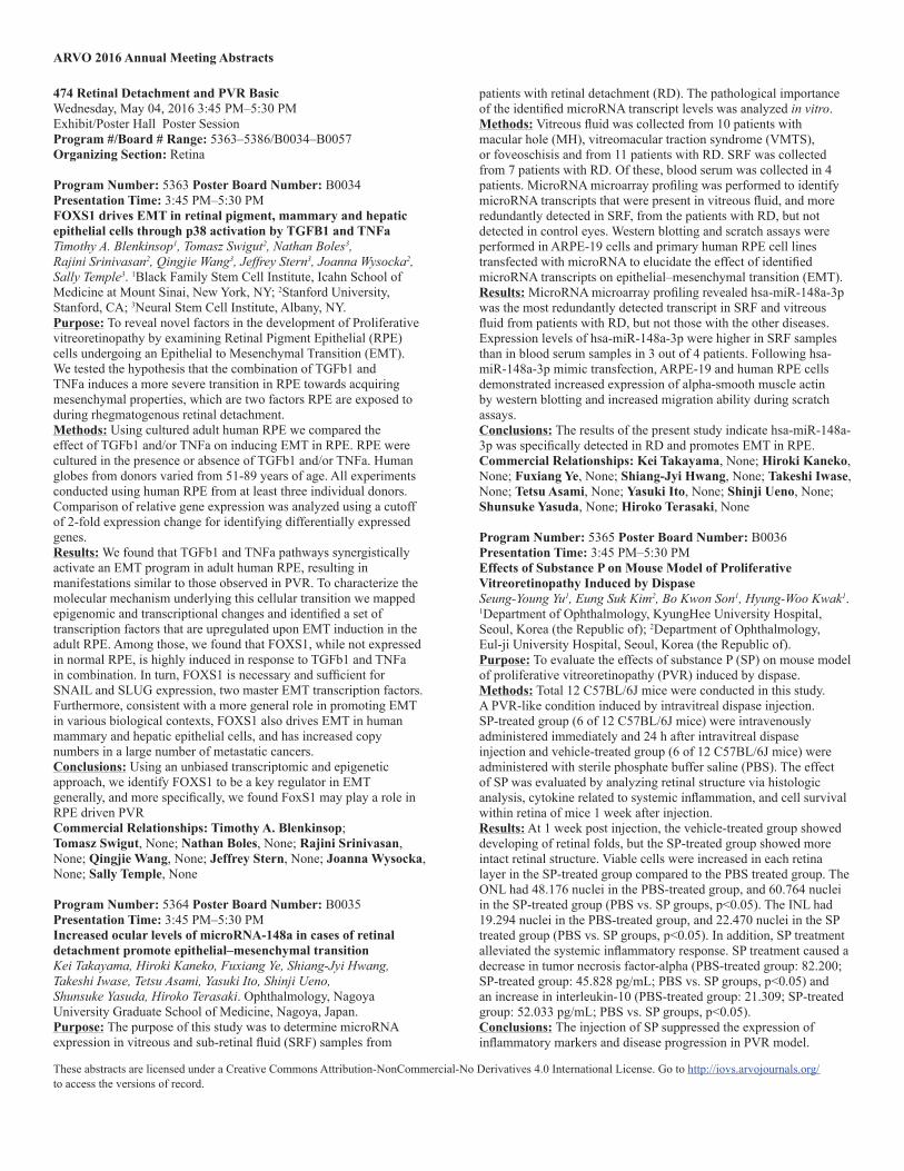
Program Number: 5368 Poster Board Number: B0039Presentation Time: 3:45 PM–5:30 PMPolarized Secretion of MMPs and TIMPs by Retinal Pigment Epithelium during Wound HealingWhitney Greene, Ramesh Kaini, Heuy-Ching H. Wang. Ophthalmology, United States Army Inst of Surgical Rsrch, San Antonio, TX.Purpose: To characterize the secretion of matrix metalloproteases (MMPs) and tissue inhibitors of matrix metalloproteases (TIMPs) by induced pluripotent stem cell- derived Retinal Pigment Epithelium (iPS-RPE) during wound healing. iPS-RPE displays the phenotype and functions of in vivo retinal pigment epithelium (RPE), and provides a physiologically relevant cell type to study disease mechanisms. iPS-RPE was used to develop an in vitro wound healing model. We hypothesize that iPS-RPE secretes mediators of tissue remodeling such as MMPs and TIMPs that promote migration and proliferation of cells during wound healing.Methods: iPS-RPE was grown on transwells until fully confluent and pigmented. The monolayers were scratched to induce a wound. Cell culture media were collected from both the apical and basolateral sides of the transwells every 72 hours for 12 days. The media were analyzed by multiplex ELISA assays to detect secreted MMPs and TIMPS. Activity assays were performed to detect activated forms of MMP-2 and TIMP-1 in the conditioned media from the wounded monolayers.Results: MMP-2 and TIMP-1, -2, -3, and -4 were detected in the culture media from iPS-RPE. The proteins were found to be secreted in a polarized manner. Increased levels of MMP-2 were detected in media from the apical side of wounded cells. Increased levels of TIMPs were detected in culture media from both the apical and basolateral sides of wounded cells. The secretion of MMP-2 was elevated from Days 3-12 after wounding of the monolayer. MMP-2 activity was also increased from Days 3 -12 after wounding. Secretion of all four TIMPs increased within 3 days after wounding and peaked at Day 6. TIMP-1 activity was increased from Days 3-12 after wounding.
ARVO 2016 Annual Meeting Abstracts
These abstracts are licensed under a Creative Commons Attribution-NonCommercial-No Derivatives 4.0 International License. Go to http://iovs.arvojournals.org/ to access the versions of record.
Conclusions: These results indicate that iPS-RPE secretes MMP-2 and all four TIMPS in a polarized manner. After wounding of the monolayer, apical secretion of MMP-2 was significantly higher compared to control. After wounding of the monolayer, apical secretion of all four TIMPs increased compared to control, while only TIMP-1 and -3 showed increased basolateral secretion compared to control.Commercial Relationships: Whitney Greene, None; Ramesh Kaini; Heuy-Ching H. Wang, None
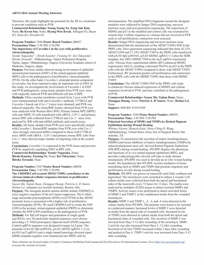
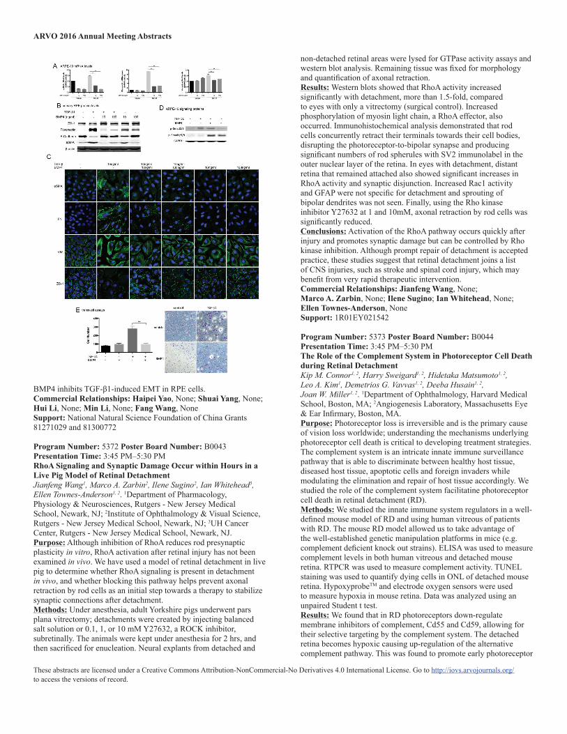
Program Number: 5369 Poster Board Number: B0040Presentation Time: 3:45 PM–5:30 PMLong non-coding RNA MALAT1 mediates transforming growth factor beta1-induced epithelial to mesenchymal transition in retinal pigment epithelial cellsShuai Yang, Haipei Yao, Min Li, Hui Li, Fang Wang. Department of Ophthalmology, Tenth People’s Hospital of Tongji University, Shanghai, China.Purpose: Epithelial-mesenchymal transition (EMT) of retinal pigment epithelial (RPE) cells is a hallmark in the development of proliferative vitreoretinopathy (PVR). Currently, long non-coding RNAs (lncRNAs) are emerging as key regulators in various biological processes. However, the role of lncRNAs in EMT of RPE cells remains largely unknown. This study investigated the role of lncRNA-MALAT1 in human RPE cells upon TGFβ1 induction of EMT.Methods: ARPE-19 cells was cultured and exposed to TGF-β1. EMT was confirmed by morphological change, as well as the increased expression of alpha-smooth muscle actin (αSMA) and fibronectin, and the down-regulation of E-cadherin and Zona occludin-1(ZO-1) at both mRNA and protein levels. Expression of lncRNA MALAT1 was evaluated by real-time PCR (RT-PCR) at 0, 12, 24, 48, and 72 hours after TGFβ1 stimulation. SiRNA targeting MALAT1 was designed and the silencing efficiency was determined by RT-PCR. The biological effect of MALAT1 siRNA on EMT, migration, and viability of RPE cells were evaluated by detecting EMT-related genes, transwell assay and MTS assay, respectively. The effect of MALAT1 silencing on phosphorylation of Smad2/3 and p-38 were detected by western blot. We also determined MALAT1 expression in primary RPE cells incubated with PVR vitreous samples.Results: MALAT1 is dramatically up-regulated in ARPE-19 cells incubated with TGFβ1 and reaches the apex at 48 hours (fold change=2.80±0.16, P=0.003). Transfection of MALAT1-SiRNA decreased the expression of MALAT1 by 67%. MALAT1 silencing attenuated TGFβ1-induced morphological transition of RPE cells. Western blot, immunofluorescence, and RT-PCR results show that MALAT1 is required for the TGFβ1-induced upregulating of αSMA and fibronectin, as well as down-regulating of ZO-1 and E-Cadherin. Inhibition of MALAT1 attenuates TGFβ1-induced migration and proliferation of RPE cells. Besides, MALAT1 activates the canonical Smad2/3, but not the non-canonical p38 signaling of TGFβ. Furthermore, MALAT1 is increased by 3.14±0.78 times (n=4, P=0.038) in human primary RPE cells incubated with PVR vitreous samples compared with cells exposed to normal vitreous.Conclusions: LncRNA MALAT1 is involved in TGFβ1-induced EMT of human RPE cells and provided new understanding for the pathogenesis of PVR.
Knockdown of MALAT1 attenuates TGF-β1 induced EMT in RPE cellsCommercial Relationships: Shuai Yang, None; Haipei Yao, None; Min Li, None; Hui Li, None; Fang Wang, NoneSupport: National Natural Science Foundation of China (No. 81300772 and No. 81271029)
Program Number: 5370 Poster Board Number: B0041Presentation Time: 3:45 PM–5:30 PMIntravitreal pirfenidone inhibits post traumatic proliferative vitreoretinopathySarbani Hazra1, B.N.M Khalida Khatun1, Sudip Nandi2, RAJDEEP GUHA2, Sushovan Chowdhury2, Samar Basak3, Subramanian Krishnakumar4, Aditya Konar2. 1Veterinary Surgery & Radiology, WBUAFS, Kolkata, India; 2CSIR-IICB, Kolkata, India; 3Disha Eye Hspital, Barrackpore, India; 4Sankara Nethralaya, Chennai, India.Purpose: To evaluate the efficacy and safety of intravitreal pirfenidone for inhibition of proliferative vitreoretinopathy in a model of penetrating ocular injury.Methods: Penetrating trauma was induced on the retina of rabbit and treated either with 0.1 ml of PBS or 0.1 ml of 0.5% pirfenidone, and development of PVR was evaluated clinically and graded after one month. Histopathology and immunohistochemistry with α SMA and collagen-1 were performed to validate further the clinical assessment. A similar injury was inflicted in rat eye and treated either with 10 µl of PBS or 10 µl of 0.5% pirfenidone, expression of cytokine at different time points were examined by RT-PCR.
ARVO 2016 Annual Meeting Abstracts
These abstracts are licensed under a Creative Commons Attribution-NonCommercial-No Derivatives 4.0 International License. Go to http://iovs.arvojournals.org/ to access the versions of record.
Availability of pirfenidone in the vitreous of rat at various time points was determined by HPLC following injection of 10 µl of 0.5% pirfenidone. In normal rabbit eye, 0.1 ml of 0.5% pirfenidone was injected to evaluate any toxic effect.Results: Prevention of PVR formation was observed clinically in animals injected with pirfenidone, and the animals obtained significantly (p<0.05) lesser PVR grade than the PBS-treated control animals. Pirfenidone inhibited increased expression of cytokines as observed in control eyes. Pirfenidone could be detected up to 96 hrs in the vitreous of rat eye following single intravitreal injection. Pirfenidone did not show any adverse effect following intravitreal injection; eyes were devoid of any abnormal clinical sign, IOP and ERG did not show any significant change and histology of retina remained normal.Conclusions: Pirfenidone can provide a potential and safe therapy for PVRCommercial Relationships: Sarbani Hazra, None; B.N.M Khalida Khatun, None; Sudip Nandi, None; RAJDEEP GUHA, None; Sushovan Chowdhury, None; Samar Basak, None; Subramanian Krishnakumar, None; Aditya Konar, None
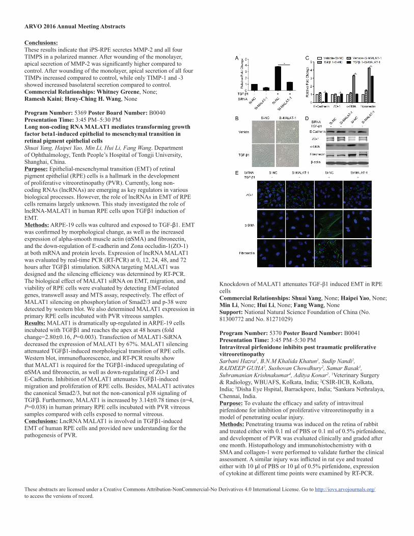
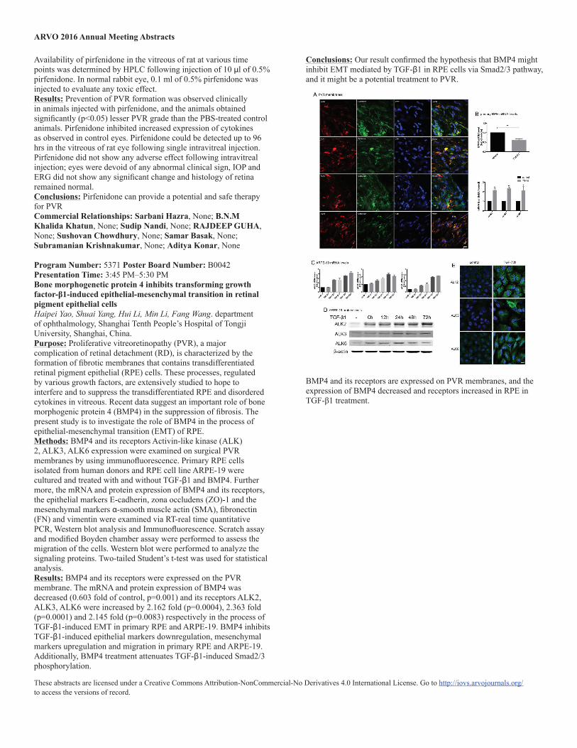
Program Number: 5371 Poster Board Number: B0042Presentation Time: 3:45 PM–5:30 PMBone morphogenetic protein 4 inhibits transforming growth factor-β1-induced epithelial-mesenchymal transition in retinal pigment epithelial cellsHaipei Yao, Shuai Yang, Hui Li, Min Li, Fang Wang. department of ophthalmology, Shanghai Tenth People’s Hospital of Tongji University, Shanghai, China.Purpose: Proliferative vitreoretinopathy (PVR), a major complication of retinal detachment (RD), is characterized by the formation of fibrotic membranes that contains transdifferentiated retinal pigment epithelial (RPE) cells. These processes, regulated by various growth factors, are extensively studied to hope to interfere and to suppress the transdifferentiated RPE and disordered cytokines in vitreous. Recent data suggest an important role of bone morphogenic protein 4 (BMP4) in the suppression of fibrosis. The present study is to investigate the role of BMP4 in the process of epithelial-mesenchymal transition (EMT) of RPE.Methods: BMP4 and its receptors Activin-like kinase (ALK) 2, ALK3, ALK6 expression were examined on surgical PVR membranes by using immunofluorescence. Primary RPE cells isolated from human donors and RPE cell line ARPE-19 were cultured and treated with and without TGF-β1 and BMP4. Further more, the mRNA and protein expression of BMP4 and its receptors, the epithelial markers E-cadherin, zona occludens (ZO)-1 and the mesenchymal markers α-smooth muscle actin (SMA), fibronectin (FN) and vimentin were examined via RT-real time quantitative PCR, Western blot analysis and Immunofluorescence. Scratch assay and modified Boyden chamber assay were performed to assess the migration of the cells. Western blot were performed to analyze the signaling proteins. Two-tailed Student’s t-test was used for statistical analysis.Results: BMP4 and its receptors were expressed on the PVR membrane. The mRNA and protein expression of BMP4 was decreased (0.603 fold of control, p=0.001) and its receptors ALK2, ALK3, ALK6 were increased by 2.162 fold (p=0.0004), 2.363 fold (p=0.0001) and 2.145 fold (p=0.0083) respectively in the process of TGF-β1-induced EMT in primary RPE and ARPE-19. BMP4 inhibits TGF-β1-induced epithelial markers downregulation, mesenchymal markers upregulation and migration in primary RPE and ARPE-19. Additionally, BMP4 treatment attenuates TGF-β1-induced Smad2/3 phosphorylation.
Conclusions: Our result confirmed the hypothesis that BMP4 might inhibit EMT mediated by TGF-β1 in RPE cells via Smad2/3 pathway, and it might be a potential treatment to PVR.
BMP4 and its receptors are expressed on PVR membranes, and the expression of BMP4 decreased and receptors increased in RPE in TGF-β1 treatment.
ARVO 2016 Annual Meeting Abstracts
These abstracts are licensed under a Creative Commons Attribution-NonCommercial-No Derivatives 4.0 International License. Go to http://iovs.arvojournals.org/ to access the versions of record.
BMP4 inhibits TGF-β1-induced EMT in RPE cells.Commercial Relationships: Haipei Yao, None; Shuai Yang, None; Hui Li, None; Min Li, None; Fang Wang, NoneSupport: National Natural Science Foundation of China Grants 81271029 and 81300772
Program Number: 5372 Poster Board Number: B0043Presentation Time: 3:45 PM–5:30 PMRhoA Signaling and Synaptic Damage Occur within Hours in a Live Pig Model of Retinal DetachmentJianfeng Wang1, Marco A. Zarbin2, Ilene Sugino2, Ian Whitehead3, Ellen Townes-Anderson1, 2. 1Department of Pharmacology, Physiology & Neurosciences, Rutgers - New Jersey Medical School, Newark, NJ; 2Institute of Ophthalmology & Visual Science, Rutgers - New Jersey Medical School, Newark, NJ; 3UH Cancer Center, Rutgers - New Jersey Medical School, Newark, NJ.Purpose: Although inhibition of RhoA reduces rod presynaptic plasticity in vitro, RhoA activation after retinal injury has not been examined in vivo. We have used a model of retinal detachment in live pig to determine whether RhoA signaling is present in detachment in vivo, and whether blocking this pathway helps prevent axonal retraction by rod cells as an initial step towards a therapy to stabilize synaptic connections after detachment.Methods: Under anesthesia, adult Yorkshire pigs underwent pars plana vitrectomy; detachments were created by injecting balanced salt solution or 0.1, 1, or 10 mM Y27632, a ROCK inhibitor, subretinally. The animals were kept under anesthesia for 2 hrs, and then sacrificed for enucleation. Neural explants from detached and
non-detached retinal areas were lysed for GTPase activity assays and western blot analysis. Remaining tissue was fixed for morphology and quantification of axonal retraction.Results: Western blots showed that RhoA activity increased significantly with detachment, more than 1.5-fold, compared to eyes with only a vitrectomy (surgical control). Increased phosphorylation of myosin light chain, a RhoA effector, also occurred. Immunohistochemical analysis demonstrated that rod cells concurrently retract their terminals towards their cell bodies, disrupting the photoreceptor-to-bipolar synapse and producing significant numbers of rod spherules with SV2 immunolabel in the outer nuclear layer of the retina. In eyes with detachment, distant retina that remained attached also showed significant increases in RhoA activity and synaptic disjunction. Increased Rac1 activity and GFAP were not specific for detachment and sprouting of bipolar dendrites was not seen. Finally, using the Rho kinase inhibitor Y27632 at 1 and 10mM, axonal retraction by rod cells was significantly reduced.Conclusions: Activation of the RhoA pathway occurs quickly after injury and promotes synaptic damage but can be controlled by Rho kinase inhibition. Although prompt repair of detachment is accepted practice, these studies suggest that retinal detachment joins a list of CNS injuries, such as stroke and spinal cord injury, which may benefit from very rapid therapeutic intervention.Commercial Relationships: Jianfeng Wang, None; Marco A. Zarbin, None; Ilene Sugino; Ian Whitehead, None; Ellen Townes-Anderson, NoneSupport: 1R01EY021542
Program Number: 5373 Poster Board Number: B0044Presentation Time: 3:45 PM–5:30 PMThe Role of the Complement System in Photoreceptor Cell Death during Retinal DetachmentKip M. Connor1, 2, Harry Sweigard1, 2, Hidetaka Matsumoto1, 2, Leo A. Kim1, Demetrios G. Vavvas1, 2, Deeba Husain1, 2, Joan W. Miller1, 2. 1Department of Ophthalmology, Harvard Medical School, Boston, MA; 2Angiogenesis Laboratory, Massachusetts Eye & Ear Infirmary, Boston, MA.Purpose: Photoreceptor loss is irreversible and is the primary cause of vision loss worldwide; understanding the mechanisms underlying photoreceptor cell death is critical to developing treatment strategies. The complement system is an intricate innate immune surveillance pathway that is able to discriminate between healthy host tissue, diseased host tissue, apoptotic cells and foreign invaders while modulating the elimination and repair of host tissue accordingly. We studied the role of the complement system facilitatine photoreceptor cell death in retinal detachment (RD).Methods: We studied the innate immune system regulators in a well-defined mouse model of RD and using human vitreous of patients with RD. The mouse RD model allowed us to take advantage of the well-established genetic manipulation platforms in mice (e.g. complement deficient knock out strains). ELISA was used to measure complement levels in both human vitreous and detached mouse retina. RTPCR was used to measure complement activity. TUNEL staining was used to quantify dying cells in ONL of detached mouse retina. HypoxyprobeTM and electrode oxygen sensors were used to measure hypoxia in mouse retina. Data was analyzed using an unpaired Student t test.Results: We found that in RD photoreceptors down-regulate membrane inhibitors of complement, Cd55 and Cd59, allowing for their selective targeting by the complement system. The detached retina becomes hypoxic causing up-regulation of the alternative complement pathway. This was found to promote early photoreceptor
ARVO 2016 Annual Meeting Abstracts
These abstracts are licensed under a Creative Commons Attribution-NonCommercial-No Derivatives 4.0 International License. Go to http://iovs.arvojournals.org/ to access the versions of record.
death. Preventing complement production using knockout mice or through pharmacologic inhibition, ameliorates much of the photoreceptor cell death observed in RD.Conclusions: This study identified a new role for the alternative complement pathway in photoreceptor death in RD. Understanding the mechanism by which the innate immune system facilitates photoreceptor cell death will provide new therapeutic targets for this retinal condition and other neurodegenerative conditions.Commercial Relationships: Kip M. Connor; Harry Sweigard, Novartis; Hidetaka Matsumoto, None; Leo A. Kim, None; Demetrios G. Vavvas, None; Deeba Husain, None; Joan W. Miller, ONL Therapeutics (P), Valeant via Mass. Eye and Ear (P), ONL Therapeutics (C), Valeant via Mass. Eye and Ear (R), Maculogix (C), KalVista (C), Alcon (C), Amgen (C)Support: National Institutes of Health (NIH) grants R01EY022084–01/S1, Research to Prevent Blindness Special Research Scholar Award
Program Number: 5374 Poster Board Number: B0045Presentation Time: 3:45 PM–5:30 PMModulation of matrix metalloproteinase and endoplasmic reticulum stress may alleviate photoreceptor cell death in rhegmatogenous retinal detachmentYoon Jeon Kim1, Jeong A Choi2, Woon Hyung Ghim1, Jae-Young Koh1, 2, Young Hee Yoon1. 1Asan medical center, Seoul, Korea (the Republic of); 2Asan institute for Life Sciences, Seoul, Korea (the Republic of).Purpose: Rhegmatogenous retinal detachment (RD) may cause the photoreceptor degeneration and irreversible visual loss. Previous studies reported the correlation between elevated matrix metalloproteinase (MMP) and endoplasmic reticulum (ER) stress in photoreceptor degeneration. This study is to investigate the role of MMP, ER stress and zinc homeostasis, in the patho-mechanism of RD-related photoreceptor degeneration.Methods: RD mice model was created by injecting 2 µl of balanced salt solution (BSS) through self-sealing scleral incision. Pathological endpoints examined in vivo included photoreceptor cell death and MMP2, MMP9, and zinc level in RD mice. In addition, the induction of ER stress and unfolded protein response (UPR) were assessed in RD mice and changes of ER stress after modulation of MMP were examined.Results: Photoreceptor cell death was peaked on 3 day and then dropped down. MMP 2 and MMP 9 were elevated by two-fold in RD mice eyes compared to control eyes (P < 0.05). The expressions of ER stress markers, GRP78, pPERK, and peIF2α were increased in RD mice. When MMP activator, 4-aminophenylmercuric acetate (APMA) 10 µM was treated into the subretinal space, retina remained to be detached longer and photoreceptor cell death was more increased compared to APMA untreated RD eyes. When MMP inhibitor, GM6001 1 mM was treated, on the other hands, MMP2, MMP9, and ER stress markers were decreased significantly. Regarding the level of zinc, total amount of zinc were decreased significantly in RD mice eyes (192.5 ± 12.4 µg/dl) compared to those in control fellow eyes (227.9 ± 9.6 µg/dl, P < 0.05). When chelating extracellular zinc of subretinal space with CaEDTA 100 µM, TUNEL positive photoreceptor cell were decreased.Conclusions: RD model induced zinc release from retina to subretinal space and it was correlated with increased MMP activation and ER stress expression. This suggests that modulating MMP and ER stress may alleviate photoreceptor cell death in RD.Commercial Relationships: Yoon Jeon Kim, None; Jeong A Choi, None; Woon Hyung Ghim, None; Jae-Young Koh, None; Young Hee Yoon, Bayer (C), Alcon (R), Allergan (C)
Support: NRF-2013R1A2A2A10168457
Program Number: 5375 Poster Board Number: B0046Presentation Time: 3:45 PM–5:30 PMNOX2 induces photoreceptor apoptosis through endoplasmic reticulum stress pathway in a rat model of retinal detachmentHong Zhu, Quan Yan, Fenghua Wang, Xueting Luo, Xiaodong Sun. Opthalmology, Shanghai First People’s Hospital, Shanghai, China.Purpose: To investigate the expression of NOX2, a NADPH oxidase, and its regulation on endoplasmic reticulum stress (ERS)-mediated apoptosis and photoreceptor damage using a rat model of retinal detachment (RD).Methods: Animal model of RD was created in Brown Norway rats by subretinal injection of 1% sodium hyaluronate. Lentivirus NOX2 shRNA (LV-NOX2-sh) was constructed to inhibit NOX2 expression in vivo. The rats were randomly divided into four groups: normal control group (non-detachment), RD control group, negative control group (vehicle+RD) and LV-NOX2-sh+RD group. Expression of NOX2 and biomarkers of ERS pathway (CHOP, TRB3 and Caspase-12) were detected using quantitative real-time PCR and western blotting on day 1, 3, 5 and 7 after RD in each group. Non-detachment animals served as control. TdT-mediated fluorescein-16-dUTP nick-end labeling (TUNEL) assay was used to stain the apoptosis cells in histological sections. Retinal outer nuclear layer (ONL) thickness was measured to assess retina damage in each group.Results: NOX2 expression was significantly increased after RD. CHOP and TRB3 were over-expressed in RD control group. In LV-NOX2-sh group, the expression of NOX2 was notably inhibited in vivo. And the expressions of CHOP and TRB3 were decreased compared with those RD rats and vehicle+RD rats at the same time points. The ratio of TUNEL-positive photoreceptors in LV-NOX2-sh+RD group was significantly reduced accompany with less expression of Caspase-12. Meanwhile, the ONL thickness in LV-NOX2-sh+RD group was thicker than that both in RD group and vehicle+RD group.Conclusions: NOX2 expression is up-regulated in retinas and induces ERS pathway after RD. Inhibition of NOX2 protects photoreceptors through ERS-mediated apoptosis pathway in RD rat model. NOX2 may be a crucial regulatory molecule in ERS-mediated photoreceptor apoptosis after RD.Commercial Relationships: Hong Zhu, None; Quan Yan, None; Fenghua Wang, None; Xueting Luo, None; Xiaodong Sun, None
Program Number: 5376 Poster Board Number: B0047Presentation Time: 3:45 PM–5:30 PMVitreous and subretinal fluid ATP concentrations in rhegmatogenous retinal detachmentTakashi Tachibana, Toshio Hisatomi, Shoji Notomi, Shunji Nakatake, Yusuke Murakami, Akihito Sengoku, Yasuhiro Ikeda, Shigeo Yoshida, Tatsuro Ishibashi, Koh-Hei Sonoda. Ophthalmology, Kyushu University, Fukuoka, Japan.Purpose: Adenosine triphosphate (ATP) plays an important role in cell energy metabolism. During the course of apoptosis, dying cells release ATP into extracellular space. We previously reported that extracellular ATP accelerates photoreceptor cell apoptosis in subretinal hemorrhage. However, role of ATP in vitreous is not fully understood. The purpose of this study is to determine the vitreous levels of ATP and its correlation with clinical data in rhegmatogenous retinal detachment (RRD).Methods: Vitreous samples were obtained from 58 eyes of 58 patients with RRD (n = 41), macular hole (MH; n=13), and epiretinal membrane (ERM; n =14) during pars plana vitrectomy.
ARVO 2016 Annual Meeting Abstracts
These abstracts are licensed under a Creative Commons Attribution-NonCommercial-No Derivatives 4.0 International License. Go to http://iovs.arvojournals.org/ to access the versions of record.
In RRD patient, subretinal fluids were also corrected from retinal tear during vitrectomy. We measured ATP concentration using luciferin-luciferase assay.Results: Compared with MH and ERM patient, RRD patient have higher vitreous ATP concentration (RRD: 1.74ng/ml, MH: 0.11ng/ml, ERM: 0.065ng/ml, p < 0.005, respectively). In RRD patient, subretinal fluid contains higher ATP than vitreous fluid (vitreous fluid: 1.74ng/ml, subretinal fluid: 13.72ng/ml, p<0.005).Conclusions: RRD patient have high ATP concentration in vitreous. High ATP concentration in vitreous and subretinal fluid of RRD may be associated with photoreceptor apoptotic cell death.Commercial Relationships: Takashi Tachibana, None; Toshio Hisatomi, None; Shoji Notomi, None; Shunji Nakatake, None; Yusuke Murakami, None; Akihito Sengoku, None; Yasuhiro Ikeda, None; Shigeo Yoshida, None; Tatsuro Ishibashi, None; Koh-Hei Sonoda, None
Program Number: 5377 Poster Board Number: B0048Presentation Time: 3:45 PM–5:30 PMSubretinal injection of poly(IC) causes acute photoreceptor cell death in the mouse retinal detachment modelSymantas Ragauskas1, 2, Sabine Grüner3, Ludovic Collin3, Giedrius Kalesnykas1. 1Experimentica ltd, Kuopio, Finland; 2State Research Institute Centre for Innovative Medicine, Vilnius, Lithuania; 3Roche Pharma Research and Early Development, NORD DTA, Roche Innovation Center Basel, F. Hoffmann-La Roche Ltd, Basel, Switzerland.Purpose: To determine the effect of poly(IC) subretinal injection on programmed retinal cell death.Methods: Male 6-8 weeks old C57Bl/6J mice (n=30) were used. Subretinal injections were performed using poly(IC) (polyinosinicpolycytidylic acid/potassium salt) or phosphate buffer saline (PBS). The animals were followed for 2 days, 5 days and 7 days after subretinal injection. In vivo spectral domain-optical coherence tomography (SD-OCT) was employed to verify successful subretinal injection at the baseline. SD-OCT was also performed at the end of the follow-up period prior to animal sacrifice. The mice were transcardially perfused and the eyes were enucleated and cryosectioned. Ocular sections were systematically chosen and stained for hematoxylin and eosin (H&E) and for terminal deoxynucleotidyl transferase dUTP nick end labeling (TUNEL). The number of TUNEL-positive cells as well as the thickness of outer nuclear layer (ONL) were estimated using StereoInvestigator software (v. 10, MicroBrightField Inc., USA).Results: Both injections of PBS and poly(IC) caused retinal detachment. However, polyIC-injected group with a 2-day follow-up showed a significant increase in TUNEL-positive cells (5.2±1.48 cells/100 µm of ONL length, mean±SD) found in ONL as compared to other poly (IC) time-points (5-day group: 1.2±0.82 cells/100 µm of ONL length; 7-day group: 0.37±0.48 cells/100 µm of ONL length) or PBS-injected eyes (2-day group: 0.65±0.82 cells/100 µm of ONL length; 5-day group: 0.38±0.40 cells/100 µm of ONL length; 7-day group: 0.34±0.29 cells/100 µm of ONL length).Conclusions: Subretinal injection of poly(IC) causes acute programmed cell death of photoreceptor cells. The use of SD-OCT is particularly important to verify and follow-up this preclinical model.Commercial Relationships: Symantas Ragauskas; Sabine Grüner, Roche Innovation Center Basel (F); Ludovic Collin, Roche Innovation Center Basel (F); Giedrius Kalesnykas, Experimentica ltd (S), Experimentica ltd (I), Experimentica ltd
Program Number: 5378 Poster Board Number: B0049Presentation Time: 3:45 PM–5:30 PMRegulatory roles of glycolytic enzymes in photoreceptor survivalThomas Wubben, Heather Knupp, Kevin Toolan, Cagri G. Besirli. Ophthalmology, University of Michigan, Ann Arbor, MI.Purpose: To examine the regulatory roles of hexokinase II (HKII) and pyruvate kinase muscle isozyme 2 (PKM2) in pro- and anti-apoptotic pathways in photoreceptors following apoptotic stress.Methods: Retina-retinal pigment epithelium (RPE) separation was created in Brown-Norway rats by subretinal injection of 1% hyaluronic acid. Retinas were harvested and assayed with Western blot analysis and immunohistochemistry. Cultured 661W photoreceptor cells were subjected to hypoxic and glucose-deprived conditions and assayed with Western blot analysis and quantitative PCR.Results: Retina-RPE separation resulted in an increase in the expression of HKII in the outer retina with HKII predominantly localized in the inner segments of photoreceptors at 24 hours post-detachment. Similarly, retinal detachment led to increased PKM2 expression in the outer nuclear layer and a global decrease in PKM2 phosphorylation. We developed an in vitro cell culture model to mimic hypoxic and hypoglycemic stress induced by photoreceptor-RPE separation. Cultured 661W photoreceptor cells subjected to hypoxic and glucose-deprived conditions showed increased protein levels of HKII as well as increased gene expression. These conditions were also observed to regulate PKM2 phosphorylation in 661W cells.Conclusions: Our results demonstrate that the expression of glycolytic proteins HKII and PKM2, which are enriched in the outer nuclear layer of the retina, are regulated during apoptotic stress. We developed an in vitro system using 661W cone-like cells to replicate the hypoxic and hypoglycemic stress experienced by photoreceptors after retina-RPE separation. Similar to in vivo changes, we showed that hypoxic and hypoglycemic stress leads to changes in the expression and post-translational modification of glycolytic proteins, providing an in vitro platform to investigate the regulatory roles of glycolytic proteins in photoreceptor survival.Commercial Relationships: Thomas Wubben, None; Heather Knupp; Kevin Toolan, None; Cagri G. Besirli, NoneSupport: NEI-1-K08-EY-023982-01
Program Number: 5379 Poster Board Number: B0050Presentation Time: 3:45 PM–5:30 PMThe role of microglial expression of microRNA-155 on a murine model of retinal detachmentYoko Okunuki1, 2, Eiichi Hasegawa1, 2, Ryo Mukai1, 2, Clifford Kim1, 2, Garrett Klokman1, 2, Saori Inafuku1, 2, Kip M. Connor1, 2. 1Angiogenesis laboratory, Massachusetts Eye and Ear Infirmary, Boston, MA; 2Department of Ophthalmology, Harvard Medical School, Boston, MA.Purpose: Retinal detachment (RD) is a sight threatening disorder characterized by the physical separation of the photoreceptors from the retinal pigment epithelium. As a result, the detached photoreceptors undergo apoptosis and programmed cell death. It has been reported that retinal microglia become activated during RD however, the role of microglia in the pathophysiology of photoreceptor cell death is not well understood. The association of pro-inflammatory microRNA-155 (miR-155), abundantly expressed in microglia, is suggested to be associated with worsening of neurodegenerative brain diseases. In this study, we examined the role of miR-155 on photoreceptor cell death in RD.Methods: RD was induced by subretinal injection of 4 ul of sodium hyaluronate in miR-155 -/- mice and C57BL/6J mice. Microglia were isolated from intact retinas and 24hr post RD retinas of C57BL/6J
ARVO 2016 Annual Meeting Abstracts
These abstracts are licensed under a Creative Commons Attribution-NonCommercial-No Derivatives 4.0 International License. Go to http://iovs.arvojournals.org/ to access the versions of record.
mice by fluorescence-activated cell sorting, and miR-155 expression in microglia was measured by RT-PCR. RD-induced photoreceptor cell death was compared between miR-155 -/- mice and C57BL/6J mice by TUNEL staining. Results: MiR-155 expression in microglia was significantly increased in 24hr post RD retinas compared to intact retinas (p<0.001). The number of TUNEL positive cells in miR-155 -/- retinas was significantly decreased 24hrs after RD (p=0.008, n=8).Conclusions: We demonstrated that miR-155 expression in microglia was increased after RD in mice. Systemic defect of miR-155 suppressed photoreceptor cell death caused by RD. These results suggest that miR-155 defect protects photoreceptor cells from cell death, although the exact role of microglial expression of miR-155 with retinal damage still needs to be explored.Commercial Relationships: Yoko Okunuki, None; Eiichi Hasegawa; Ryo Mukai, None; Clifford Kim, None; Garrett Klokman, None; Saori Inafuku, None; Kip M. Connor, NoneSupport: R01 EY022084
Program Number: 5380 Poster Board Number: B0051Presentation Time: 3:45 PM–5:30 PMA protein kinase C theta mutation causes early-onset exudative retinal detachmentXiaojie Ji1, 2, Ye Liu1, Ron Hurd1, Jieping Wang1, Bernard FitzMaurice1, Jürgen Naggert1, Patsy M. Nishina1, Bo Chang1. 1The Jackson Laboratory, Bar Harbor, ME; 2Graduate School of Biomedical Science and Engineering, University of Maine, Orono, ME.Purpose: The mechanisms underlying retinal detachment (RD) remain largely unknown. To study RD, we screened for novel murine models of early-onset exudative RD. Here we identified a model, rpea1, with a mutation in the Prkcq gene, which encodes protein kinase C, theta (PKCθ).Methods: The chromosomal location of the causal mutation was mapped by recombinant analysis, and the mutation identified through comparison of whole exome sequences of the mutant and WT controls. A complementation of the Prkcqrpea1 allele and the targeted null allele confirmed the causative mutation. The impact of Prkcq deficiency was determined by clinical evaluation, electroretinography (ERG), light microscopy, marker and western blot analyses.Results: We found that PKCθ is predominantly expressed in the lateral plasma membrane of the retinal pigment epithelium (RPE), co-localizing with both tight and adherens junction proteins. The strong PKCθ localization in RPE cells suggests that RD is likely to be caused by pathology or dysfunction in RPE cells. In line with this hypothesis, aberrant F-actin perijunctional rings were observed in RPE flat mounts with PKCθ deficiency. Although PKCθ was previously reported to be the kinase responsible for the phosphorylation of ezrin/radixin/moesin (ERM) proteins, which links actin filaments to membrane-associated proteins, the level of phosphorylated ERM proteins was increased in PKCθ deficient RPE/choroid cells. This indicates that PKCθ is unlikely to phosphorylate ERM proteins directly in RPE cells in vivo, but may negatively and indirectly regulate their phosphorylation and activation.Conclusions: Previous studies have shown that PKCθ and the actin cytoskeleton, especially the perijunctional actin ring, may play an important role in maintaining barrier function. Our results demonstrate that PKCθ deficiency-induced aberrant F-actin perijunctional rings in RPE cells may be in part due to the change of ERM protein phosphorylation, and the consequent weakening of the barrier function and defective fluid homeostasis in the posterior retina. Aberrant RPE barrier function that leads to suboptimal
regulation of subretinal fluid homeostasis may be a common theme for RD.Commercial Relationships: Xiaojie Ji, None; Ye Liu, None; Ron Hurd, None; Jieping Wang, None; Bernard FitzMaurice, None; Jürgen Naggert, None; Patsy M. Nishina, None; Bo Chang, None
Program Number: 5381 Poster Board Number: B0052Presentation Time: 3:45 PM–5:30 PMPrss56 Mutant Mouse: a Model for Nanophthalmos-Related Retinal DetachmentSeyyedhassan Paylakhi, Cassandre LabelleDumais, Michael Sellarole, Saidas Nair. Ophtalmologie, University of California, San Francisco (UCSF), San Francisco, CA.Purpose: Mutations in human PRSS56 cause nanophthalmos/posterior microphthalmia. Nanophthalmic eyes exhibit severely reduced ocular axial length and high hyperopia. Moreover, these eyes are susceptible to developing uveal effusion characterized by suprachroidal fluid accumulation due to trans-scleral drainage defects. Leakage of the accumulated fluid into the subretinal space induces retinal detachment. Mice with a mutation in Prss56 recapitulate features of human nanophthalmos (hyperopia and reduced ocular axial length). Here, we assess if Prss56 mutant mice are susceptible to developing retinal detachment. Furthermore, we employ a genetic strategy to rescue the ocular size reduction in Prss56 mutant mice and utilize this new model to study the link between ocular size and retinal detachment.Methods: 1) To detect retinal detachment, we examined the retina of Prss56 mutant and control littermates between 2 and 12 months of age, using funduscopy, optical coherence tomography (OCT), fluorescein angiography and histology. 2) We bred Egr1 knockout mouse, a previously reported model for ocular axial elongation/myopia, to a Prss56 mutant mouse to study the genetic interaction between Egr1 and Prss56 for ocular size regulation and retinal detachment. Ocular dimensions and refraction were measured using a digital vernier caliper and mouse photo-refractor respectively.Results: 1) Fundus examination revealed the presence of retinal lesions in Prss56 mutant eyes starting at 5 months of age. OCT examination suggests that these lesions correspond to areas of retinal detachment. Histological analysis confirmed the presence of focal retinal detachment in Prss56 mutant eyes. The retinal detachment exacerbates with age. The retinal detachment is accompanied by vascular leakage and gliosis. 2) Inactivation of Egr1 partially rescues the Prss56 mutant mediated reduction in ocular axial length and corrects hyperopia. In addition, inactivation of Egr1 rescues retinal detachment observed in Prss56 mutant eyes.Conclusions: In addition to ocular size reduction and hyperopia, Prss56 mutant mice develop progressive, age-related retinal detachment. Importantly, inactivation of Egr1 rescues the retinal detachment observed in Prss56 mutant mice, likely through its effect on partially reversing the reduction in ocular size. Thus, Prss56 mutant mice constitute a valuable model to study the role of ocular size and related defects in retinal detachment.Commercial Relationships: Seyyedhassan Paylakhi, None; Cassandre LabelleDumais, None; Michael Sellarole, None; Saidas Nair, NoneSupport: This work was supported in part by NIH P30 core grant for vision research (UCSF, Ophthalmology), Research to Prevent Blindness, and NEI grants EY022891.
ARVO 2016 Annual Meeting Abstracts
These abstracts are licensed under a Creative Commons Attribution-NonCommercial-No Derivatives 4.0 International License. Go to http://iovs.arvojournals.org/ to access the versions of record.
Program Number: 5382 Poster Board Number: B0053Presentation Time: 3:45 PM–5:30 PMIL6 Signaling in acute retinal detachment: Anti-inflammatory or Neuroprotective?Xinlei Wang1, 3, Eric Miller4, Mayank Goswami2, Robert J. Zawadzki1, 2, Marie E. Burns1, 4. 1Ophthalmology & Vision Science, UC Davis, Davis, CA; 2Cell Biology and Human Anatomy, UC Davis RISE Eye-Pod Laboratory, Davis, CA; 3Cell Biology & Human Anatomy, UC Davis, Davis, CA; 4Center for Neuroscience, UC Davis, Davis, CA.Purpose: Interleukin-6 has been implicated in most vitreoretinal diseases, but its role is unclear because IL6 can be both a pro- and anti-inflammatory cytokine. Retinal detachment (RD) elevates IL6 levels in a manner correlated with the extent of the detachment and the resulting retinal atrophy. Here we determined the time course and identity of the primary immune cells responding to acute RD in mice, and tested whether IL6 signaling following RD is anti-inflammatory or neuroprotective.Methods: RD was induced in wild-type and IL6-deficient mice by subretinal injection of sodium hyaluronate. The RD location, extent and evolution was monitored longitudinally in-vivo by optical coherence tomography (OCT) imaging. The numbers and morphology of microglia (Iba1(hi), Cd11b(lo)) and monocytes (Iba1-, Cd11b(hi)) were assessed by flow cytometry and immunohistochemistry. Photoreceptor death and retinal atrophy was quantified by Tunel staining and retinal thickness measurement using optical coherence tomography. To prevent IL6 signaling in wild-type mice, RD was preceeded by intravitreal injection of IL6 receptor α (IL6Rα) antibody or IL6 receptor β (gp130) antibody.Results: All RD groups showed increased numbers of monocytes and resident microglia above control levels within 1 day after RD. Diffuse infiltration of monocytes was found in vitreous, nerve fiber layer and inner plexiform layer, while ameboid microglial cells populated the outer retinal layers. Retinal atrophy followed RD in all groups in days 3-7, and was found to be worse in both IL6-KO mice and following IL6Rα inhibition. However, blocking gp130 function reduced acute monocyte recruitment and photoreceptor death.Conclusions: Monocytes infiltrate surprisingly rapidly following retinal detachment. These studies suggest the neuroprotective role of IL6 signaling in retinal detachment may be through IL6Rα, while more destructive cytokines coincidently signal through the soluble receptor gp130. Thus, future work more specifically targeting gp130 signaling may prove beneficial in mitigating the damaging consequences of neuroinflammation.Commercial Relationships: Xinlei Wang; Eric Miller, None; Mayank Goswami, None; Robert J. Zawadzki, None; Marie E. Burns, NoneSupport: National Eye Institute R01-EY24320, the UC Davis NEI Core Grant (P30- EY012576), and Prop. 63, the Mental Health Services Act and the Behavioral Health Center of Excellence at UC Davis.
Program Number: 5383 Poster Board Number: B0054Presentation Time: 3:45 PM–5:30 PMDeletion of interleukin-33 (IL33) results in persistent Müller cell activation during retinal detachmentSofia Pavlou1, Josy Augustine1, Sarah Doyle2, Alan W. Stitt1, Matthew Campbell2, Heping Xu1, Mei Chen1. 1Queen’s University Belfast, Belfast, United Kingdom; 2Trinity College Dublin, Dublin, United Kingdom.Purpose: IL-33, a newly identified member of the IL-1 family, has diverse roles in immunity and inflammation. In the retina, IL-33 is exclusively expressed by Müller cells under physiological
conditions. However, the role of IL-33 in Müller cell function and in retinal patho-physiology remains largely unknown. In this study, we investigated the role of IL-33 in Müller cell activation in a mouse model of retinal detachment (RD).Methods: Retinal detachment was induced in adult wild-type (WT) C57BL/6J mice and IL-33-/- mice by subretinal injection of sodium hyaluronate (2μl/eye). At different times after induction of RD, eyes were collected and processed for immunohistochemistry. Müller cell activation was evaluated by upregulation of GFAP expression, while photoreceptor degeneration was assessed by immunostaining of cone arrestin and rhodopsin.Results: After retinal detachment for 24 hours, Müller cells became activated in both WT and IL-33-/- mice as evidenced by strong GFAP expression, which persisted for up to 7 days post-detachment. In WT mice subjected to retinal detachment, GFAP upregulation was accompanied by an increased intra-nuclear IL-33 expression in Müller cells. At 28 days post-detachment, GFAP expression was significantly reduced in WT mice when compared to IL-33-/- mice (p<0.05). In line with the higher GFAP expression in IL-33-/- mice, photoreceptor degeneration was also more severe in these retinas when compared to the WT mice at day 28 after retinal detachment.Conclusions: The deletion of IL33 resulted in persistent Müller gliosis and more severe photoreceptor degeneration in our model of retinal detachment. Our results suggest that IL-33 negatively regulates Müller cell activation and protects photoreceptors from inflammation-mediated damage occurring as a consequence of retinal detachment.Commercial Relationships: Sofia Pavlou, None; Josy Augustine, None; Sarah Doyle; Alan W. Stitt, None; Matthew Campbell, None; Heping Xu, None; Mei Chen, None
Program Number: 5384 Poster Board Number: B0055Presentation Time: 3:45 PM–5:30 PMInhibition of Proliferation and Epithelial Mesenchymal Transition via Wnt and TGF-β Signaling Pathway in an in vitro Cell Culture Based-PVR Model by HC-HA/PTX3 Purified from Amniotic MembraneHua He1, Ajay E. Kuriyan2, Chen-Wei Su1, Yuan Zhang3, Megha Mahabole1, Ying-Ting Zhu1, Esdras A. Quintero2, Nidhi Rehlan2, Harry W. Flynn2, Jean-Marie A. Parel2, Scheffer C. Tseng1, 3. 1Research and Development, TissueTech, Inc., Miami, FL; 2Department of Ophthalmology, Bascom Palmer Eye Institute, University of Miami Miller School of Medicine, Miami, FL; 3Ocular Surface Center and Ocular Surface Research Education Foundation, Miami, FL.Purpose: Proliferative vitreoretinopathy (PVR) is the main cause of failure of rhegmatogenous retinal detachments and characterized by proliferation and epithelial mesenchymal transition (EMT) of retinal pigment epithelial (RPE) cells under the influence of vitreous growth factors. Recently, we have purified a heavy chain-hyaluronic acid/pentraxin 3 (HC-HA/PTX3) complex from human amniotic membrane (AM) which retains AM’s anti-inflammatory, anti-scarring, and anti-angiogenic activity. Herein, we investigate whether HC-HA/PTX3 can prevent PVR by inhibiting proliferation and EMT of RPE cells in vitro.Methods: EGF(10 ng/ml) and FGF-2 (10 ng/ml) or together with TGF-β were used to induce proliferation and EMT in human ARPE-19 cells with or without HC-HA/PTX3. The cell viability was determined by MTT and WST-1, the proliferation was measured by BrdU ELISA, WST-1 assay as well as BrdU and β-catenin immunostaining, while EMT was examined by immunostaining of phosphorylated Smad2/3 and a-smooth muscle actin. The migration
ARVO 2016 Annual Meeting Abstracts
These abstracts are licensed under a Creative Commons Attribution-NonCommercial-No Derivatives 4.0 International License. Go to http://iovs.arvojournals.org/ to access the versions of record.
and collagen gel contraction in ARPE-19 and human primary RPE cells were also assayed.Results: We have established an in vitro cell culture-based PVR model by defining the cell density, growth factors, and measurement methods. HC-HA/PTX3 dose-dependently inhibited proliferation and EMT of RPE cells stimulated by EGF/FGF-2 and TGF-βs, respectively, without any cytotoxic effect on the normal RPE cells. In addition, HC-HA/PTX3 and HA inhibited migration of EGF+FGF-2+TGF-β1 stimulated ARPE-19 cells but HC-HA/PTX3 but not HA inhibited collagen gel contraction in both primary human RPE and cell line ARPE-19 cells. The inhibition of proliferation of EMT by HC-HA/PTX3 is mediated by down-regulation of canonical signaling pathways of Wnt (β-catenin) and TGF-β (Smad2/3), respectively.Conclusions: HC-HA/PTX3 is a non-toxic, potent inhibitor of RPE cell proliferation and EMT in vitro. Hence, its efficacy in preventing PVR can be tested in our established rabbit PVR model.Commercial Relationships: Hua He, TissueTech Inc.; Ajay E. Kuriyan, None; Chen-Wei Su, TissueTech; Yuan Zhang, None; Megha Mahabole, TissueTech; Ying-Ting Zhu, TissueTech Inc.; Esdras A. Quintero, None; Nidhi Rehlan, None; Harry W. Flynn, None; Jean-Marie A. Parel, None; Scheffer C. Tseng, TissueTech Inc. (I)Support: NIH grants of R43 EY025447, R43 EY021045, R44 EY017497, and P30EY014801; an award from Bayer Global Ophthalmology Awards Program Grant, Heed Ophthalmic Foundation; the Department of Defense (DOD Grant #W81XWH-09-1-0675); a research grant from TissueTech, Inc. and an unrestricted grant from Ocular Surface Research & Education Foundation, Miami, FL.
Program Number: 5385 Poster Board Number: B0056Presentation Time: 3:45 PM–5:30 PMPI3K/Akt signaling is critical for photoreceptor survival and regulates multiple cell death and cell survival pathwaysBoris Busov, Mercy Pawar, Cagri Besirli. Ophthalmology and Visual Sciences, University of Michigan, Ann Arbor, MI.Purpose: To study the cell survival and death mechanisms regulated by the phosphatidylinositol 3-Kinase/Akt (PI3K) signaling in photoreceptor cells.Methods: 661W photoreceptor cells were treated with PI3K inhibitor LY294002 in combination with 3-methylamphetamine (3-MA), Bafilomycin (Baf-1), pan-caspase inhibitor (Z-VAD-FMK) and Necrostatin-1 (Nec-1) and cell viability, Caspase 3/7 and 8 activities were analyzed. Immunohistochemistry and annexin staining were performed to elucidate the pathways. Expression of proteins involved in the pathways were analyzed with Western blotting. The role of PI3K/Akt signaling in regulating photoreceptor neuroprotective protein Faim2 was evaluated using immunoprecipitation and immunohistochemistry. Microarray analysis of cell death-associated genes was done on LY294002 treated 661W cells using Mouse Cell Death Pathway Finder RT2 Profiler PCR Array. Microarray analysis of expression of Akt genes using PI3K-AKT Signaling PCR Array was done on detached and attached extract of wild type and Faim2 knockout mice.Results: Treatment of cells with various concentrations of LY294002 inhibitor demonstrated increased cell death in a dose-dependent manner. Combined treatment of inhibitor LY294002 with Z-VAD-FMK, Nec-1, Baf-1, and 3-MA did not prevent cell death. Increased levels of caspase 3/7 activity with high levels of cell death were seen in LY-treated cells but no caspase 8 activation was observed. LY-treated cells showed an increased LC3 II/LC3I ratio and decreased p62 levels in Westerns. The Cell Death Profiler Assay showed marked fold increases in the levels of many apoptotic and autophagy markers.
Stress-induced Akt phosphorylation in the retina was diminished in Faim2 knockout animals. Direct association of Faim2 and Akt was detected by immunoprecipitation.Conclusions: PI3K signaling is critical in the survival of photoreceptors. Inhibition of baseline PI3K activity led to increased apoptosis and autophagy, but blocking these pathways did not prevent cell death. In addition, blocking necroptosis did not provide protection. These results indicate that in addition to apoptosis, necroptosis, and autophagy, alternative cell death pathways are dis-inhibited in photoreceptors when baseline PI3K signaling is turned off. Faim2 is important for Akt activation in the retina and may be regulating PI3K-mediated Akt phosphorylation by direct association.Commercial Relationships: Boris Busov, None; Mercy Pawar, None; Cagri Besirli, NoneSupport: Career Development Award, Research to Prevent Blindness (CGB); National Eye Institute, NEI-1-K08-EY-023982-01 (CGB)
Program Number: 5386 Poster Board Number: B0057Presentation Time: 3:45 PM–5:30 PMHyperreflective points and cytokines as clinical and molecular markers in retinal detachmentGoran Petrovski1, Natasha Josifovska1, Kata Varga1, Xhevat Lumi2, Mária Szatmari-Tóth3, Endre Kristóf3, Attila Kovács1, Domagoj Ivastinovic4, Rózsa Dégi1, Á Szabó1, Andrea Facskó1. 1Ophthalmology, University of Szeged, Szeged, Hungary; 2University of Ljubljana, Ljubljana, Slovenia; 3University of Debrecen, Debrecen, Hungary; 4University of Graz, Graz, Austria.Purpose: Retinal detachment (RD) is one of the most frequently diagnosed ophthalmologic conditions requiring prompt surgical intervention. Combination of proper surgical technique and new diagnostic markers, both clinical and molecular, can help improve the diagnosis and prognosis of RD.Methods: 10 rhegmatogenous RD patients were included in the study after obtaining patient consent and Regional Ethical Approval (average age: 64.1 ± 13.7). OCT was performed in all RDs before surgery and a day after 23G vitrectomy. Subretinal fluid (SRF) was collected during surgery and analyzed by ELISA for presence of inflammatory factors (IL-6, IL-8), while the effect of SRF upon human macrophages-led phagocytosis of apoptotic retinal pigment epithelial (RPE) cells was studied ex vivo and quantified by flow cytometry. Immunohistochemistry (IHC) of an eye enucleated due to phthisis bulbi following several RD surgeries due to PVR was performed to determine presence of markers for microglial cells (CD34) and macrophages (CD14, CD68).Results: OCT of fresh RD patients contained hyperreflective points (HRPs) at the detached neuroretina border and proximal to the RPE layer - their size and number decreased following successful reattachment surgery. IHC of the phthitic eye showed presence of cell conglomerates at the detached neuroretina border which were positive for CD68 and negative for CD34 and CD14. The SRF had higher concentration of IL-6 and IL-8 compared to vitreous from non-RD pathology; when given to macrophages, the SRF doubled their capacity for engulfing dying RPE cells ex vivo.Conclusions: Fresh RD can be hallmarked by a presence of HRPs at the detached neuroretina border on OCT; the HRPs disappear after successful reattachment surgery, and likely resemble the macrophage conglomerates seen by IHC. The SRF contains pro-inflammatory factors which increase the ability of professional phagocytes to engulf dying RPE, or for that matter, other dying cells in the retina.Commercial Relationships: Goran Petrovski, None; Natasha Josifovska, None; Kata Varga, None; Xhevat Lumi, None; Mária Szatmari-Tóth, None; Endre Kristóf, None;

ARVO 2016 Annual Meeting Abstracts
These abstracts are licensed under a Creative Commons Attribution-NonCommercial-No Derivatives 4.0 International License. Go to http://iovs.arvojournals.org/ to access the versions of record.
Attila Kovács, None; Domagoj Ivastinovic, None; Rózsa Dégi, None; Ágnes Szabó, None; Andrea Facskó, None