arthroscopic versus open synovectomy in rheumatoid knee
TRANSCRIPT

بسم هللا الرحمن الرحيم
{ ك ل
سبحان
وا
ال
ا إ ق
ن
م ل
ما عل
ل
ان
مت
نعل
ك أ
ح إن
عليم ال
ال
{كيمت
يم صدق هللا العظ(32سورة البقرة اآلية )
Arthroscopic Versus Open
Synovectomy In Rheumatoid
Knee
AcknowledgementAt first, thanks to ALLAH for all his gifts
Words stand short when they come to express my gratefulness to
my supervisors. I would like to express my deep gratitude and
appreciation to Prof.Dr. MOHAMMED SALAH ELDEEN SHAWKY,
Professor of Orthopedic Surgery, Faculty of Medicine, Benha University,
for his great supervision, great help, available advice, continuous
encouragement and without his support it was impossible for this
study to be achieved in this form. I had the privilege to benefit from his
great knowledge .It is an honor to work under his guidance and
supervision.
I also sincerely express my great appreciation to Prof.Dr. MAMDOUH
MOHAMMED EL-KARAMANY, Assistant Professor of orthopaedic
surgery, Faculty of Medicine, Benha University, for his advice and all
efforts he offered to make this work possible.
Next I would like to express my deep thanks and grateful
appreciation to Dr. SAMEER MOHAMAD ABDALLAH , Lecturer of
orthopaedic surgery, faculty of Medicine, Benha University for his
constant encouragement, continuous support and everlasting skillful
help. Their favors will never be forgotten.
Last but not least, I dedicate this work to my family whom without
their sincere emotional support this work could not have been
completed.

Anatomy of Synovial Joints

Synovial joints contain the following structures : Synovial cavity Articular capsule Articular cartilage* Many, but not all, synovial joints also containadditional structures:

o Articular discs or menisci
o Articular fat pads
o Bursa
o Accessory ligaments
o Tendons
Blood supply:
The blood supply of a synovial joint is derived
from the arteries sharing in the anastomosis around
the joint.
Synovial membrane and synovial fluid:
Synovial membrane is the soft tissue found
between the articular capsule and the joint cavity
of synovial joints.

Synovial membrane of the knee
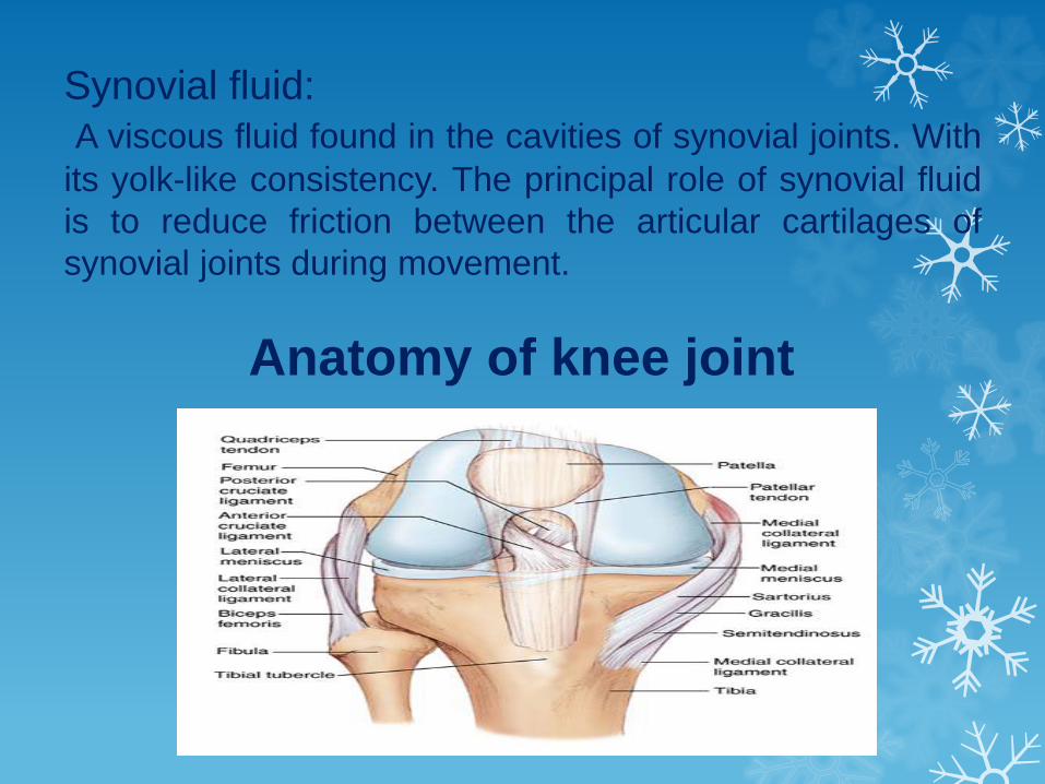
Synovial fluid:
A viscous fluid found in the cavities of synovial joints. With
its yolk-like consistency. The principal role of synovial fluid
is to reduce friction between the articular cartilages of
synovial joints during movement.
Anatomy of knee joint

Pathology of R.A.
•Joints and tendons:
Stage 1 – pre-clinical :
- Raised ESR, C-reactive protein (CRP)
and RF may be detectable years before
the first diagnosis.
Stage 2 – synovitis:
- Proliferation of synoviocytes.
- There is thickening of the capsular
structures.
- Villous formation of the synovium and a
cell-rich effusion into the joints and tendon
sheaths. the disorder is potentially
reversible.

Stage 3 – destruction:
- Articular cartilage is eroded and granulation
tissue creeps over the articular surface.
- A synovial effusion, often containing copious
amounts of fibrinoid material, produces
swelling of the joints, tendons and bursae.
Stage 4 – deformity:
- The combination of articular destruction,
capsular stretching and tendon rupture leads to
progressive instability and deformity of the
joints.

• Extra-articular tissues:
Rheumatoid nodules:
Nodules occur under the skin (especially over
bony prominences), in the synovium, on
tendons, in the sclera and in many of the
viscera.
Diagnosis- X-rays :
There may be no changes in the early stages of the
disease.
The x-ray may demonstrate juxta-articular osteopenia
There may be bony erosions and subluxation.
- MRI :MRI has proved itself as a valuable technique to detect
changes in all components of the joints affected by RA.
Synovitis volume, bone marrow edema and bone erosions
are suitable for serial measurement.
- Ultrasonography:
It is more sensitive in detecting synovial and tendon
inflammation than clinical examination alone
Management:Treatment Goals:
The ultimate treatment goal is remission and complete
suppression of disease activity.
Other treatment goals which include control synovitis,
Relieve Pain, maintain functional ability, improve and
maintain quality of life and minimize adverse events,
particularly from pharmacological therapy.
Cost effective treatment:There is no cure for RA, but treatments can improve
symptoms and slow the progress of the disease.

Surgical Treatment
Aims of surgical treatment options:- Diagnosis by taking arthroscopic synovial biopsy as part of
arthroscopic treatment.
- Debulking the diseased tissues as synovectomy can be
performed both arthroscopically and open
- Regaining motion by Capsulectomy, removal of secondary
spurs and resurfacing.
- Pain relief is done through synovectomy and joint resurfacing.
- In early phases of the disease, an arthroscopic or open
synovectomy may be performed. It consists of the removal of
the inflamed synovium and prevents a quick destruction of the
affected joints.
- Severely affected joints may require joint replacement surgery.

Synovectomy of the knee
- Synovectomy is the surgical removal of a part of the
synovial membrane of a synovial joint.
- Surgical synovectomy is recommended for patients who do
not experience substantial pain relief in response to medical
therapy for 6 months.
- During synovectomy, part of the synovium is left intact so
that it can still perform its function of releasing synovial fluid,
which serves as a lubricant in the joint.
- Synovectomy can be performed by making a large incision
that exposes the entire joint or it can be done using
arthroscopic methods.
- The choice of approach depends on the extent of repair
required.

Knee Synovectomy Indications
It is indicated generally in chronic synovitis. The indication
remains the same weather the synovectomy performed
arthroscopically or by open surgery.
Synovectomy should be performed when the disease is
limited to the synovium before the involvement of articular
cartilage and bone and when there has been a failure of trial of
adequate conservative treatment for at least 6 months.
Knee Synovectomy Contraindications
- Advanced arthritis.
- Extensive joint instability with bone destruction.

Knee Synovectomy Complications
Iatrogenic chondral injury.
Hemoarthrosis.
DVT and Pulmonary embolism.
Stiffness.
Infection.
Fluid Extravasation and Compartment Syndrome.
Instrument failure and breakage.
Arthrofibrosis, Patella infra and Loss of motion.
Ligament Injuries.
Fractures.
Synovitis and synovial fistula.
Neurovascular injuries.
Results of Synovectomy in Rheumatoid knee
- The immediate :
Operative removal of synovial membrane from the knee
in rheumatoid arthritis will give immediate pain relief in some
two-thirds of patients.
- Short term results :
The success of the operation is measured by the loss of
pain, swelling of pain, range of motion achieved, stability and
strength of quadriceps muscle.
- Long term results:
The patients were initially seen at intervals of six
months and later at yearly intervals depending on their
condition. the continuing success of a synovectomy can be
judged from the way the joint reacts during a general flare-up
of the disease involving several joints.

Open surgical synovectomy
Indications:
Persistent pain and swelling of the knee despite adequate
medical treatment for a minimum of six to twelve months.
Contraindications:
- End-stages with signs of bony destruction.
- Deformity and instability of the joint.
- The stiff, dry, painful joint.
- A flexion contracture greater than 25 degree.

Advantages:
- The major advantage of this procedure is that with the
open operation one is confident that nearly all the synovium
has been removed.
- In late stage disease open operation is preferable as it
allows removal of the menisci as well as the proliferative
synovium from the articular margins of the tibia.
- General debridement of the joint with trimming of
osteophytes and removal of pannus can also be readily
achieved.
- Open knee synovectomy is standard and allows an
inspection of all compartments.

Disadvantages:
- In open procedure the posterior compartment was not
approachable due to proximity to neurovascular structures
and the synovium present in intercondylar notch and
under and over the meniscus was difficult to take out and
so it never became total synovectomy.
- With open synovectomy there is a significant morbidity
such as knee stiffness due to arthrofibrosis.
- The other major problem with the open operation is the
length of postoperative rehabilitation as patient usually
needs to be an in-patient for up to 14 days and requires
regular physiotherapy for up to three months before gaining
maximal restoration of function.
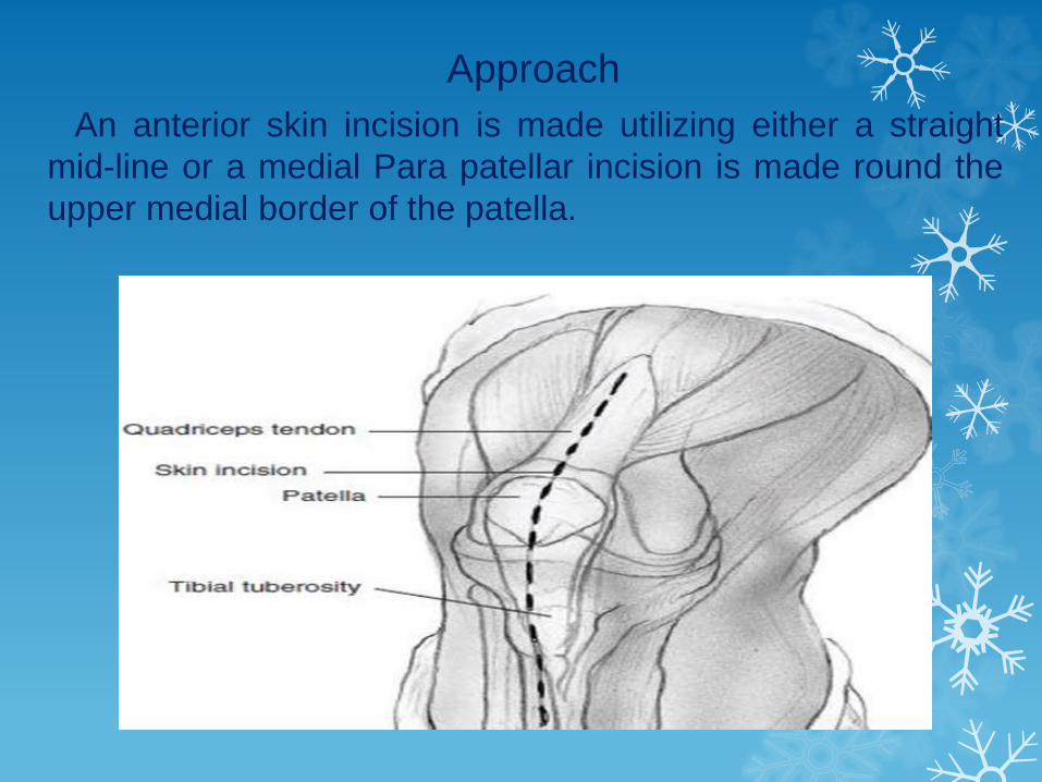
Approach
An anterior skin incision is made utilizing either a straight
mid-line or a medial Para patellar incision is made round the
upper medial border of the patella.
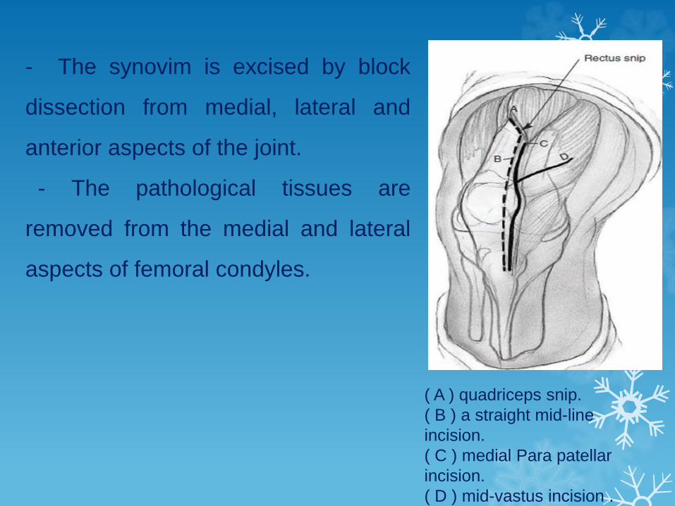
- The synovim is excised by block
dissection from medial, lateral and
anterior aspects of the joint.
- The pathological tissues are
removed from the medial and lateral
aspects of femoral condyles.
( A ) quadriceps snip.
( B ) a straight mid-line
incision.
( C ) medial Para patellar
incision.
( D ) mid-vastus incision .
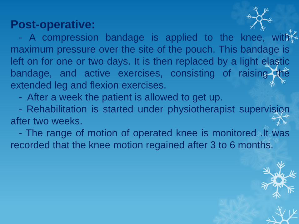
Post-operative:- A compression bandage is applied to the knee, with
maximum pressure over the site of the pouch. This bandage is
left on for one or two days. It is then replaced by a light elastic
bandage, and active exercises, consisting of raising the
extended leg and flexion exercises.
- After a week the patient is allowed to get up.
- Rehabilitation is started under physiotherapist supervision
after two weeks.
- The range of motion of operated knee is monitored .It was
recorded that the knee motion regained after 3 to 6 months.

- Return to Work:
Limited work loading of the affected joint is an appropriate
restriction. This may include no lifting, carrying, twisting,
pushing or pulling, standing, squatting, or kneeling, depending
on the joint involved.
Individuals may be required to use devices to assist with
ambulation such as crutches, canes, or walkers.
Possible complications of surgery
- Accidental damage to the knee joint.
- Bleeding.
- Nerve damage.
- Deep Vein Thrombosis.
- Pulmonary embolism.
- Persistent or recurrent pain.
- Scar formation and adhesions.

Arthroscopic synovectomy
Indication of knee arthroscopy:
After conservative modalities have proven ineffective over a
reasonable period of time and the individual remains
disabled, consideration for an arthroscopic evaluation of the
knee is warranted for purposes of diagnosis and treatment.
Patient education plays a critical role in the outcome of
arthroscopic surgery.
Contraindication:
- Unsatisfactory skin conditions and history of knee reflex
sympathetic dystrophy.
- End-stages with signs of bony destruction.
- Deformity and instability of the joint.

Advantages of arthroscopic synovectomy
Arthroscopic synovectomy is a surgical procedure with
minimal morbidity, which does not require open arthrotomy,
and leaves less joint capsule and ligament damage, thus
allowing immediate mobilization and reducing hospital stays.
As compared with open arthrotomy, the arthroscopic
technique offers superior views, easier access to knee
compartments and facilitates the effective removal of
pathologic synovium.
Incision is minimal, Quadriceps muscle remains intact,
Incidence of infection is decreased, Incidence of hemarthrosis
is decreased, Range of motion is maintained or increased.
Postoperative physical therapy is minimal or none. Menisci
are spared. Patient acceptance is high.

Operative Technique
Arthroscope insertion:
- Extend the knee and make a small stab wound superior
and medial to the patellar tendon. Introduce the inflow
cannula into the joint utilizing the blunt obturator.
- Flex the knee. Identify the “soft spot” for the inferior
lateral portal. Introduce the cannula for the arthroscope
through this portal.
- Insert the arthroscope into the knee joint through the
cannula. Extend the knee and position the arthroscope in
the suprapatellar pouch.
- Inspect the patellofemoral joint.
- Inspect the lateral gutter.
- Inspect the medial gutter.
- Inspect the lateral compartment

Postoperative Care Issues
A compressive dressing should be placed at the end of
surgery and is normally removed approximately 48 hours
after the procedure.
The stitches will be removed at clinic if the patient is seen 2
weeks or less post operatively.
patients can weight-bear as tolerated after surgery.
Range-of-motion and strengthening exercises can be
initiated immediately after the procedure.
Most patients can successfully rehabilitate with a home
exercise program.
The patients began physical therapy the same day to
achieve maximum range of motion, strengthen the
quadriceps and hamstring muscles, and use modalities to
decrease the swelling, pain, and inflammation in the acute
postoperative period.

Complications: Infection.
Nerve injury.
Vessel injury.
Tourniquet palsy.
Bleeding.
Persistent or recurrent pain.
Knee ligament injury.
Broken instruments.
Synovial fistula.
Equipment failure.

Common Occurrences: Some patients will note bruising around the knee.
Anterior knee pain.
Persistence of arthritic symptoms.
Portal discomfort.
Swelling.
Skin itching.
Return to work:
If patients’ job involves sitting for the majority of the day
they can return after 3 days.
If their job is physically demanding and involves heavy
manual work or standing for long periods, 1-2 weeks off
work may be necessary.
Driving:
Patients should not return to driving until their knee is pain
free and they have full knee flexion.
Summary and Conclusion
• surgical synovectomy is recommended for patients who do
not experience substantial pain relief in response to medical
therapy for 6 months.
• When there is structural damage to a joint or the tissues
around it, medicines can't fix it, and surgery may help.
• In early phases of the disease, an arthroscopic or open
synovectomy may be performed. It consists of the removal
of the inflamed synovia and prevents a quick destruction of
the affected joints.
• Although there has been an increase in the popularity of
less invasive methods of synovectomy such a radiation
synovectomy and arthroscopic synovectomy ,the open
synovectomy remains the procedure of choice in the
management of sever synovitis of the knee joint even quite
late in the disease process .
• The major advantage of open synovectomy is that with the
open operation one is confident that nearly all the synovium
has been removed.
• With open synovectomy there is a significant morbidity such
as knee stiffness due to arthrofibrosis and, rarely, wound
and joint infection. The other major problem with the open
operation is the length of postoperative rehabilitation .

• Arthroscopic synovectomy offers several theoretical
advantages, including decreased invasiveness of surgery,
potential for faster recovery, and reduced hospital stay.
• Patients undergoing arthroscopic synovectomy had similar
pain reduction, but more frequent recurrences of synovitis
than patients with open synovectomy.
• After open synovectomy range of motion of operated knee is
monitored .It was recorded that the knee motion regained
after 3 to 6 months.
• After arthroscopic synovectomy range of motion regained
quickly as the approach minimally invasive with no affection
of muscles and no adhesions.