april 30, 2018 archives • 2018 • vol.1 209-230 g-protein ... · g-protein couple receptor...
TRANSCRIPT

April 30, 2018
Archives • 2018 • vol.1 • 209-230
http://pharmacologyonline.silae.it
ISSN: 1820-8620
G-PROTEIN COUPLE RECEPTOR MEDIATED TRANSGENE EXPRESSION OF A LYS-ALA-LEU-ALA REPEATED PEPTIDE EMBEDDED WITH LIPID-DNA NANOPARTICLES IN DENDRITIC CELL
Sharif M. Shaheen*1, 2, , Md. Jashim Uddin3, A. K. Azad, 2, M. Sarowar Hossain, 2, M. Mustafezur Rahman, 2
1Faculty of Pharmaceutical Science, Hokkaido University, Sapporo city, Kita 12, Nishi
6, Hokkaido 060-0812, JAPAN
2Daffodil International University, Daffodil Tower (DT)-054/2 Sobahanbag, Mirpur Road Dhanmondi, Dhaka-1207, Bangladesh.
3Department of Biochemistry, Vanderbuilt Univesity, 850 RRB, TN, USA
Abstract
No information was reported to date about the uptake process of repeated cationic arginine or lysine followed by leucine or alanine with lipid bilayer based DNA nanocargoes. Here we orchestrated the concept of repeated lys-ala-leu-ala (KALA) peptide modified DNA-lipid nanoparticle, proclaimed a promising transfection activity in dendritic cell, following GPCR mediated endocytosis. Chemical inhibitors studies of chlorpromazine, Fillipin, and Amiloride did not support the concept of macropinocytosis, clathrin mediated endocytosis and cavaeola mediated endocytosis. Cetirizine hydrochloride, a GPCR blocker shut down the KALA mediated transgene expression. Confocal studies showed no liposomal uptake in KALA modification rather than R8 modification, which implies to receptor mediated endocytosis. Low concentration of KALA pretreatment allowed a R8 MEND (usually non-expressed) to that of the same expression of KALA MEND and vice versa of high concentration KALA pretreatment. A PKA inhibitor, rottlerin and PKA terminator (p-300/CREB inhibitor), curcumin halted the gene expression of KALA modified MEND, where octa-arginine modified MEND did not respond to the inhibitory effect by curcumin. Collectively, our data suggest that a repeated lys-ala-leu-ala (KALA) modification in lipid embedded DNA nanoparticle approached a GPCR induction both in endocytosis and activation, which might be a potential reason of boom transgene expression in dendritic cell.
Keywords: Macropinocytosis, Stearyl Octa-arginine, Stearyl-KALA, MEND, G-Protein Couple Receptor, Endocytosis, Lysine-leucine repeated peptide, Leucine repeated rich peptide

PhOL Sharif, et al. 210 (pag 209-230)
http://pharmacologyonline.silae.it
ISSN: 1824-8620
Introduction Nano-cargoes containing gene of interest or anticancer drug needs promisingly uptake and momentously moving to the cytosol. Recent many studies reported about the uptake process of non-viral vector, containing surface modification with cationic amino acid enriched oligo peptide like octa-arginine [1, 2] as well as octa-histidine [3, 4]. However, the cellular uptake process of arginine, lysine and even histidine enriched peptide are still contradictory [5, 6, 7]. Low concentration of octa-arginine modification in nano particles follows Clathrin mediated endocytosis (CME), whereas high concentration of octa-arginine modification follows the macropinocytosis of the same nano-cargoes [1]. Although the cellular uptake process of arginine and lysine enriched peptides have been reported somewhere else however their exact phenomena of the internalization of nano cargoes are still not clear. There are many factors that can affect uptake process of nano cargoes containing DNA and thereby transgene expression, include mostly the type of peptide, nature of the linker between the peptide and lipid bilayers. One peptide named TAT, containing both the arginine and lysine showed controversial mechanism of internalization of nanocargoes. As per previous studies, neither the classic receptor-transporter nor the endocytosis-mediated processes involved in the uptake of TAT and also other peptides like TAT [3, 4]. Inverted micelle-driven delivery and direct penetration had been proposed as possible internalization [8, 9]. And the TAT sequence consists of several type arginine and lysine residues, which is crucial for translocation. There are some homopolymers of arginine, resembling to the TAT peptide [10] made them probable candidates to follow the TAT. One of the candidates was established for the better internalization as an optimized number of eight (R8) [10]. The octa-arginine succeeded in the internalization of nano cargoes containing plasmid DNA but it depended on the type of cells like cancer cell and fibroblast cell [11]. The high density octa-arginine modified liposome showed the mechanism of uptake through macropinocytosis by blocking the increased gene expression in NIH3T3 cell line [1]. Nevertheless this R8 peptide modified DNA cargoes has no gene expression in dendritic cell line [12], where the uptake pathway of R8 liposome was mediated by micropinocytosis
[12, 13]. Later we replaced STR-R8 peptide with stearylated repeat lysine-leucine peptide, STR-KALA has got very high gene expression in dendritic cell line (JAWS) [14, 15, 16]. KALA and GALA peptides were reported elsewhere for their fusogenic activity in lipid membrane interface [17, 18] and we previously used cholesteryl-GALA [19, 5] as a membrane fusion inducer with endosome, because its conformation
changes from a random coil to -helix in acidic environment. But with GALA modification into octaarginine modified MEND (Multi-functional envelope type of nano device) containing the GL3 plasmid, had no effect in transgene expression (supplementary Figure S1) after a transfection studies in the dendritic cells. However, considering the environment in the cytoplasm, peptides that possess fusogenic activity in a neutral pH environment are essential for inducing fusion to the nuclear membrane. And KALA was used as a pH-independent fusion inducer [20]. Stearyl-KALA modified MEND raised the gene expression around three orders of magnitude in both dendritic cell line (JAWs-II) and the bone marrow derived dendrtic cell, BMDC [16-18, 20]. And the mechanism how stearyl-KALA peptide promotes gene expression is unknown. Here we gave our effort to find out the reasons on behind of the mechanism of uptake of a repeated lys-ala-leu-ala (KALA) peptide modification in lipid embedded DNA nanoparticles, followed by the possible reason of high transgene expression.
Results Multiple mechanisms for endocytosis have been reported to date [21-25] mainly: Clathrin mediated endocytosis, Macropinocytosis, Caveola mediated endocytosis and Clathrin-caveola independent endocytosis. TAT peptide and Octa-arginine modified nanoparticle so far followed the macropinocytosis pathway [26-28, 15]. Figure 1 showed also the macro pinocytosis when octa-arginine modified nanoparticle delivered plasmid DNA in dendritic cell line (JAWs-II) was inhibited by amiloride. And previously it has been reported that both the octa-arginine and octa-lysine modified nanoparticles followed macropinocytosis [2, 29]. But lysine-leucine repeated moiety instead of only lysine modified nanoparticle did not respond to the chemical inhibitor amiloride, which showed that KALA peptide modified nanoparticle didn’t follow macropinocytosis (Figure 1). The other chemical inhibitors like chlorpromazine and fillipin couldn’t

PhOL Sharif, et al. 211 (pag 209-230)
http://pharmacologyonline.silae.it
ISSN: 1824-8620
inhibit the KALA modified nanoparticle mediated transgene expression (Supplementary Figure S1). It revealed that lysine-leucine repeated peptide neither followed the clathrin mediated endocytosis nor the caveola mediated pathway. To verify inhibition of gene expression by chemical inhibitors for the pathway of endocytosis we investigated further the fluorescence uptake when dendritic cells were transfected with the NBD-DOPE 1% labeled KALA liposome and also KALA DNA condensed particle (Figure 2). Octa-arginine modified liposome (R8 liposome) and DNA condensed particle (R8 MEND) were inhibited by both CCD and amiloride significantly (Supplementary Figure S2a and S2b). One more control was performed using dextran-rhodamine treatment on mock dendritic cell and CCD treated dendritic cells (Supplementary Figure S2c) to evaluate the fluid phase inhibition. Octa-arginine modified nanoparticles were significantly inhibited by amiloride with a concentration of 1mM. Usually macropinocytosis can be inhibited by amiloride and its analogs, which inhibit the Na+/H+ exchange protein in the plasma membrane [30]. Nevertheless, though octa-lysine based nanoparticle was internalized by macropinocytosis but the uptake of lysine-leucine repeated peptide modified nanoparticles were neither inhibited by amiloride nor CCD (Figure 2b and Supplementary Figure S2b.), which means macropinocytosis does not work in lysine-leucine enriched peptide modification with MEND or liposome. So there might be some other component which is responsible for the uptake of nanoparticles and independent on CME, Cavaeola and macropinocytosis. And the cell surface membrane receptor might be one issue to consider. Usually there are many ways of ligand-receptor interaction such as hydrophilic interaction through ionic and polar bond, and hydrophobic interaction through hydrophobic and van der Waals’ bond. Here we have used repeated lysine-leucine (STR-KALA), which has got subsequent hydrophilic and hydrophobic, can protrude to the ligand-receptor interaction. Keeping this assumption we pretreated the dendritic cell with three different concentration of STR-KALA (7μM, 13.8μM and 27.6 μM final) at 37°C only 10 min. Thereafter, we transfected the cell with STR-KALA MEND, containing plasmid DNA of reporter luciferase for 24 h. we found a dose-dependent transgene inhibition, significantly (Figure 3a). Confocal
microscopic studies also showed that there was no uptake in the pretreated dendritic cell (Supplementary Figure 3b). But when the cells were incubated with low concentration of KALA (3 μM) only for 5-10 min and thereafter R8 MEND, which had no transgene expression exhibited a dramatic promising gene expression as if gene expression by KALA modified DNA nanoparticles (Figure 4). A triangular transgene stimulation by stearyl KALA, where the peak pointed gene expression happened by the low concentration of KALA and there after an increased in KALA made a decrease in gene expression but still significantly higher than that of control R8 MEND. Usually at low temperature vesicular transport should be stopped but we found many red dots of aqueous phase marker sulfo-rhodamine loaded STR-KALA nanoparticles, binding on the cell surface just after 10 min at 4°C (Supplementary Figure S2d.). It shows also that KALA peptide modified nanoparticle doesn’t follow macropinocytosis /or phagocytosis. Whenever we pretreated dendritic cell with stearyl KALA peptide (23μM) as previously and then we applied sulfo-rhodamine loaded KALA liposome to the pretreated cell, we found no red dot signal on the cell surface after 60 min (Figure 3b, upper panel in comparison to the lower panel). Figure 3b (lower panel) shows the clear many red dots of sulfo-rhodamine loaded stearyl-KALA liposome, which seems to be endocytosed at 37°C. A high concentration of stearyl-KALA might be inhibitory effect, where a low concentration might be stimulatory effect, which might be an interaction with a receptor and thereafter follow-up some signaling cascade of up-regulation and down-regulation of transgene expression of the delivered gene. So it seems KALA modified nano cargo might be internalized by a receptor binding process. The chemical structure of KALA and secretin hormone resemble to the point of hydrophobic and hydrophilic amino acid combinations. Both are of 27 amino acids, where secretin is repeated arginine leucine [31] and KALA is repeated lysine leucine in their paradigm dogma. Secretin is a potent GPCR agonist and accordingly, KALA seems to be a GPCR agonist. If it is a GPCR agonist then there should be either stimulatory or inhibitory effect depending on the concentration of agonist [32]. Here we got a magic like boom gene expression (positive feedback) of R8 MEND

PhOL Sharif, et al. 212 (pag 209-230)
http://pharmacologyonline.silae.it
ISSN: 1824-8620
while dendritic cells was incubated with low concentration stearyl-KALA peptide (3µM) for 10 min before the addition of R8 MEND (Figure 4). After certain concentration of KALA (7µM) the expression reduced, which seems to be a negative feedback of gene expression usually hormones, are showing in the biological system [33]. Of note, there is a kind of GPCR blocker, cetirizine hydrochloride (also known as H1 blocker) was used with low to high concentration and found remarkable blocking the transgene expression (Figure 5), where an scramble peptide receptor blocker, sulfanilamide [34, 35] didn’t block the transgene expression of KALA modified nano cargo (supplementary Figure S5a.). Considering an optimum concentration of cetirizine hydrochloride (0.9 mM) was used to visualize the fluorescence signal under confocal microscope, when labeled liposomes were transfected into the dendritic cells (Figure 5). Upper panel left and right are the KALA liposomes loaded with sulfo-rhodamine showed almost no uptake (no red signal) after treatment with cetirizine hydrochloride. But the lower panel right showed clear visualization of many red spots, showed not much inhibition by cetirizine hydrochloride. Figure 6a, showed significant shutdown of transgene expression when a PKA inhibitor, rottlerin was used. Thereafter, cells were incubated with p-300/CREB inhibitors like Curcumin 10µM (Fig. 6b). Curcumin did not show any significant transgene expression difference between curcumin treated R8 MEND and a control R8 MEND (Fig. 6c).
Discussion Here we reflected the mechanistic pathway of stearyl-KALA modified nanoparticles containing CpG free DNA and its transgene expression, which differed from that of TAT and arginine, followed so far. From our laboratory previously we have shown that both the octa-arginine and octa-lysine modified nano particles were internalized in NIH3T3 cell by macropinocytosis [2]. In dendritic cell octa-arginine (R8) modified nano particles (R8 MEND) also followed the macropinocytosis (Figure 1 and Figure 2). Whenever the lysine was followed with hydrophobic alanine and leucine the mechanism might be changed to a new way. Continuous cationic amino acids like arginine and lysine (higher positive charge density) orchestrated the actin processing of the cell to be engulfed as endocytosis [11, 2, 36-38]. And the hydrophobic moiety inclusion with that of
hydrophilic moiety repeatedly in DNA nanoparticles is experienced by the cell to the parts of its own to respond rather than the signal of actin processing. Especially, the leucine or alanine, hydrophobic moiety might be the important amino acid of changing the mode of uptake in dendritic cell. Of note, one group described leucine in conserved inter subunit interactions involving the hydorophobic packing as the highly stabilized conformational state of macrophage migration inhibitory factor (MIF) in solution they studied by generating point mutation and characterization, using a battery of biophysical methods and X-ray crystallography [39]. And whenever we incubated the cells with leucine amino acid for few minutes (10 min) and thereafter transfected the cell by stearyl-KALA modified nano particles of CpG free GL3 plasmid it increased transgene expression significantly (Supplemental figure S5b). In this regard, Leucine-rich repeats are protein-protein interaction domains amino acids residues in length found in proteins with diverse structure and functions [40]. G-protein couple receptor (GPCRs) are characterized by a relatively large ectodomain (ECD) containing leucine-rich-repeats [41-43]. So the ligand-receptor might be the hydrophobic interaction between leucine-rich repeat of GPCR ectodomain and leucine-rich-repeat peptide of stearyl-KALA nano particles. And we got a significantly increased gene expression when the dendritic cells were free incubated with the amino acid leucine (Supplemental Figure S3b.). Since KALA i.e. lysine-leucine repeated peptide resembles to the hormone secretin, might be a GPCR agonist. The secretin receptor is a prototypic member of family B G protein-coupled receptor. The carboxyl-terminal region of this peptide assumes a helical conformation that occupies the peptide-binding cleft within the structurally complex disulphide-bonded amino-terminal domain of this receptor. The amino terminus of secretin is directed toward the core helical bundle domain of this receptor that seems to be structurally distinct from the analogous region of family A G protein-coupled receptors [34]. Since the KALA peptide goes to the alfa helical conformation within a wide range of pH (5 to 7.4) [20], there is a possibility of the same pattern of peptide binding cleft as that of secretin with amino-terminal ecto-domain of the GPCR. And consequently a GPCR-mediated endocytosis

PhOL Sharif, et al. 213 (pag 209-230)
http://pharmacologyonline.silae.it
ISSN: 1824-8620
pathway of nano cargo internalization by stearyl-KALA may persist in dendritic cell. GPCR blockers supported in respect of transgene expression and rhodamine fluorescence intensity in confocal live images (Figure 5). A PKA inhibitor, rottlerin deleted the promising transgene expression of KALA MEND (Figure 6a). And thereafter significant reduction of transgene expression by curcumin, a PKA terminator, further, indicated to the GPCR activation by stearyl-KALA modified DNA nanoparticles (Figure 6b). Curcumin is well known as p-300/CREB inhibitor and acts as PKA terminator [44]. Intracellular signal inhibitors like cetirizine hydrochloride, rottlerin, curcumin consequently, proposes a GPCR oriented endocytosis and transgene expression pathways (Figure 6c). In summary, we herein, described the pathway of transgene expression of a peptide of repeated lys-ala-leu-ala modified DNA nanoparticles (KALA MEND) in dendritic cell, where DNA cargoes was capsized inside the cell through a GPCR mediated endocytosis rather than conventional micropinocytosis, and GPCR activation has got a potential role on increased transcription via p-300/CREB signaling pathways for higher transgene expression. .However, further intensive studies confirmatory with GPCR receptor expression would be performed in the days ahead. Experimental Materials 1,2-dioleoyl-sn-glycero-3-phosphoethanolamine-N-(lissamine rhodamine B sulfonyl) (Rh-DOPE), 1,2-dioleoyl-sn-glycero-3- phosphoethanolamine-N-(7-nitro-2-1,3-benzoxadiazol-4-yl) (NBD-DOPE) (From chicken egg, monosodium salt), and dioleoylphosphatidyl ethanolamine (DOPE) were purchased from Avanti Polar Lipids (Alabaster, AL). Sulfo-rhodamine B was purchased from Molecular Probes (Eugene, OR). Chlorpromazine, Fillipin and amiloride were purchased from Sigma. HEPES were obtained from Dojindo (Japan). Stearylated octaarginine (STR-R8) was synthesized as described before [12]. Cetirizine hydrochloride, ranitidine hydrochloride, curcumin were purchased from Wako chemical, Osaka, Japan. We have synthesized a CpG free plasmid of firefly luciferase [18, 22]. Fmoc-amino acid derivatives were purchased from Novabiochem (Läufelfingen, Switzerland). The link amide resin (TGS-RAM) was purchased from Shimadzu (Kyoto, Japan). Other reagents were purchased from Sigma Aldrich (St.
Louis, MO, USA) and the Wako Chemical (Osaka, Japan). JAWS II cells derived from murine dendritic cells were purchased from the ATCC. Methods Synthesis of Lys-Ala-Leu-Ala repeated peptide (a truncated version from KALA) and stearylation We synthesized Lys-Ala-Leu-Ala repeated -peptide following the sequence of KALA peptide [20]. KALA peptide has been syncopated from its original sequence to a truncated version [16]. The peptide chain was manually assembled using Fmoc solid-phase peptide synthesis on a link amide resin using a diisopropylcarbodiimide (DICDI)/1-hydroxybenzotriazole (HOBT) coupling system. Stearylation of the peptide resin was conducted using stearic acid in the presence of DICDI/HOBt, as reported previously [12]. Lys-Ala-Leu-Ala repeated peptide (stearyl KALA) and stearyl octa-arginine (R8) modified nanoparticles (both liposome and MEND, multi envelope type nano device) KALA MEND was prepared according to the stearyl R8 MEND reported elsewhere with some modification [13, 16]. At first plasmid DNA (CpGfree) core was prepared with protamine sulfate condenser at N/P ratio 2.2 in 10mM HEPES buffer of pH 7.4. The core solution was hydrtated in the previously dried lipid film of DOPE/CHEMS (9:2) along with stearyl-KALA (5 mol% of total lipid). The film was prepared by solvent evaporation with the help of nitrogen gas. After lipid film hydration, the system was sonicated in a bath for around one minute. The MEND was post modified with another 5mol% of stearyl-KALA peptide to make whole system positive. Because the DNA-protamine core, which was positively charged was sonicated with the negatively charged film, so the system was further modified with the stearyl-KALA. In case of R8 MEND, stearyl-octa argininie 5mol% of total lipid was post modified with the MEND. Liposomes of stearyl-KALA and stearyl-octa ariginine were prepared as per the procedure previously reported elsewhere with some modification [14, 15]. First a lipid mixture of DOPE and CHEMS of 9:2 molar ratio was taken in a test tube and then a 5 mol% stearyl-KALA of total lipid was incorporated with the mixture and allowed for 10 min incubation at room temperature. Thereafter 200 μl chloroform was added to the mixture and dried under nitrogen gas stream. After solvent evaporation the lipid film was further washed by the same amount of chloroform and dried under

PhOL Sharif, et al. 214 (pag 209-230)
http://pharmacologyonline.silae.it
ISSN: 1824-8620
same process. The lipid was then hydrated by adding 250 μl, 10 mM HEPES buffer with 1 mol% of 1mM sulfo-rhodamine as aqueous phase marker, when required and incubating the mixture for 10 min at room temperature with gentle shaking. Then, a bath sonication was performed around for one minute to prepare multi-lamellar liposome. After liposome preparation one more 5 mol% stearyl-KALA peptide was added as post modification make the system positive charge (+ve) and allowed the system for at least 10 min. To remove aggregated particle 1000 x g for 5 min was carried out and the sup was carefully taken up. In case of octa-arginine liposome, stearyl octa-arginine 5mol% of the total lipid was post modified with MEND and incubated the system at RT for 30min. The diameter and zeta-potential of liposomes were measured (Table 1) using an electrophoretic light-scattering spectrophotometer (Zetasizer Nanoseries, Malvern Instruments, Malvern UK). The size and zeta potential of stearyl KALA nano particles were little bit lower in comparison to that of octa-arginine modified nano particle. Encapsulation efficiency or entrapment efficiency or percent quenching was measured for stearyl-KALA nanoparticle containing sulforhodamine, followed the equation (1-fb/fa) x100, where fb and fa were fluorescence intensity before and after SDS (8mM) treatment for 1 h, respectively. And it was around 62% for stearyl-KALA liposomes, studied here. Transfection studies Dendritic cell line, JAWS-II was cultured in alfa-modification of Minimum essential medium (MEM) containing 20% FCS, Penicillin 50 IU/ml, streptomycin 50mg/ml, 4mM L-glutamate, 1mM sodium pyruvate and 5 ng/ml of GMCSF. The cells were wake up from the 5% DMSO cryopreservation and passage 3-4 has been used for transfection studies. 5 x 10⁴ cells/well were cultured in 24 well dish for 24 h and then cells were washed with PBS(-) and 0.5ml serum free medium containing 0.4 μg plasmid DNA (CpG free) packaged in KALA and octa-arginine nano cargoes were incubated for 3 h at 37°C in CO2 humidifier. Thereafter, medium was replaced with fresh medium containing 20% serum, and incubated for an additional 21 h. The cells were washed by PBS and lysed using 75 μl of reporter lysis buffer (Promega, Madison, WI). Luciferase activity in cell lysates was measured using a luminometer (Luminescencer-PSN, ATTO, Japan). The amount of protein in the cell lysate
was determined using a BCA protein assay kit (Pierce). Luciferase activity is expressed as relative light units (RLU) per mg of protein per ml of lysate, where 106 RLU correspond to 5 ng luciferase. To examine the mechanism of octa-arginine and STR-KALA nano cargoes internalization, cells were incubated in the absence or presence of amiloride (1 mM final concentration), Fillipin (1μg/ml final concentration), and chlorpromazine (1μg/ml) for 1 h at 37°C in CO2 humidifier. Cetirizine HCl, a GPCR blocker also known as H1 blocker, with an optimized concentration 0.9 mM (final) was used to the DC population for 1 h before transfection. Rottlerin, a PKA inhibitor around 30µM as well as PKA terminator (p-300/CREB), curcumin 10µM were treated before transfection. Thereafter transfection was followed up by applying nano cargoes containing CpGfree firefly luciferase plasmid for gene expression. Fluorescence measurement studies for cellular uptake In order to find out the uptake of the nanocargoes, we labeled the nanocargoes with 1mol% NBD-DOPE of total lipid. And the cells were transfected with the same amount of the nano cargoes for 1 h at 37 °C in CO2 humidifier. Following incubation, the medium was removed, and the cells were washed once with ice-cold phosphate-buffered saline (PBS) containing heparin (20 U/ml). The inhibition of the uptake of labeled octa-arginine and stearyl KALA-nanocargoes and/or liposome by the chemical inhibitors were performed using the same concentration of inhibitors at the same conditions previously used in the transgene inhibition. The cells were then washed and lysed using 75 μl of reporter lysis buffer and allowed to freeze at -80°C overnight and thaw at room temperature and thereafter cells were scrapped and fluorescence was measured using a fluorescence spectroscopy (Spectrofluorometry JASCO, F-3600). Confocal microscopic studies For the analysis of cellular binding we performed in situ transfection in confocal microscope maintaining a device to keep constant at low temperature (4 °C) in connection with a confocal laser scanning microscope (LSM META 510, Carl Zeiss Col. Ltd., Jena, Germany). Stearyl KALA modified liposome containing sulfo-rhodamine was applied at a final lipid concentration of 0.01mM in serum free medium and after 10 min of the liposome addition the confocal image was

PhOL Sharif, et al. 215 (pag 209-230)
http://pharmacologyonline.silae.it
ISSN: 1824-8620
captured using a confocal laser microscope equipped with an oil-immersion objective lens (Plan-Apochromat x40/NA). And to check the receptor binding of KALA-liposome, we pretreated dendritic cell (5x10⁴/well seeded one day before) with stearyl KALA peptide 23μM for 5 min, then sulfo-rhodamine loaded KALA-liposome were applied in serum free media of the same condition we used previously and incubated in humidifier chamber at 37 °C for another 10 min. Thereafter cells were examined under confocal microscope (Olympus Fluoview FV1Oi) instantly keeping temperature 37°C. To investigate cellular endocytosis, cells were treated with stearyl KALA liposome loaded with Sulfo-Rhodamine, at a final concentration of 0.01 mM lipid, in serum-free medium at 37 °C for 1.5h. Cells were washed once with medium and the incubation was continued for an additional 1 h with serum-medium. Cells were then washed with PBS containing heparin 20U/ml two times and examined using the same confocal microscope in HEPES buffer. The same observation was followed after a pretreatment of stearyl KALA of 23 μM for 5 min. One confirmatory study of macropinocytosis was performed by the treatment of Dextran-rhodamine 5μM final concentration for 1h. After a treatment of CCD 60μM final concentration for 1h and thereafter dextran-rhodamine was applied to confirm the inhibitory effect of CCD against pinocytosis. Acknowledgement: This work was supported by Funding Program for Next Generation World-Leading Researchers (NEXT program), in part Grant-in-Aid for Scientific Research (S) from the Ministry of Education, Culture, Sports, Science and Technology (MEXT), JAPAN and Daffodil International University, Bangladesh. In this aspect the authors would like to thank Prof. Harashima Hideyoshi and Prof. Hidetaka Akita for giving facilities of the work. The authors are also grateful to Mr. Ariful I. of Cadbury College, Barmingham, UK for proofreading. No conflict of interest was declared so far.
References [1]. A. El-Sayed, I. A. Khalil, K. Kogure, S. Futaki, H. Harashima, Octaarginine- and octalysine-modified nanoparticles have different modes of endosomal escape. J. Biol. Chem.2008, 283, 23450-23461.
[2]. E. Vives, P. Brodin, and B. Lebleu, A truncated HIV-1 Tat protein basic domain rapidly translocates through the plasma membrane and accumulates in the cell nucleu, J. Biol. Chem.1997, 272, 16010-16017. [3]. D. Derossi, S. Calvet, A. Trembleau, A. Brunissen, G. Chassaing. and A. Prochiantz, Cell internalization of the third helix of the Antennapedia homeodomain is receptor-independent, J. Biol. Chem.1996, 271, 18188–18193. [4]. N. Toriyabe, Y. Hayashi, H. Harashima, The transfection activity of R8-modified nanoparticles and siRNA condensation using pH sensitive stearylated-octahistidine.Biomaterials 2013, 34(4), 1337-1343. [5]. F. Duchardt, M. Fotin-Mleczek, H. Schwarz, R. Fischer, R. Brock, A comprehensive model for the cellular uptake of cationic cell-penetrating peptides, Traffic.2007, 8(7), 848-866. [6]. D. A. Mann, A. D. Frankel, Endocytosis and targeting of exogenous HIV-1 Tat protein.EMBO J. 1991, 10(7), 1733-1739. [7]. F. Madani, S. Lindberg, U. Langel, S. Futaki, A. Gräslund,Endocytosis and targeting of exogenous HIV-1 Tat protein.EMBO J. 2011, 10(7),1733-1739. [8]. R. Trehin, and H. P. Merkle, Chances and pitfalls of cell penetrating peptides for cellular drug delivery. Eur. J. Pharm. Biopharm. 2004, 58, 209-223. [9]. S. Futaki, T. Suzuki, W. Ohashi, T. Yagami, S. Tanaka, K. Ueda, and Y. Sugiura, Arginine-rich peptides. An abundant source of membrane-permeable peptides having potential as carriers for intracellular protein delivery,J. Biol. Chem.2001a, 276 , 5836-5840. [10] S. Futaki, W. Ohashashi, T. Suzuki, M. Niwa, S. Tanaka, K. Ueda, H. Harashima, Y. Sugiura, Stearylated arginine-rich peptides: a new class of transfection systems. Bioconjug. Chem.2001b, 12, 1005-1011. [11] K. Kogure, R. Moriguchi, K. Sasaki, M. Ueno, S. Futaki, H. Harashima, Development of a non-viral multifunctional envelope-type nano device by a

PhOL Sharif, et al. 216 (pag 209-230)
http://pharmacologyonline.silae.it
ISSN: 1824-8620
novel lipid film hydration method, J. Contr. Rel. 2004, 98, 317-323. [12] T. Nakamura, R. Moriguchi, K. Kogure, N. Shastri, H. Harashima, Efficient MHC class I presentation by controlled intracellular trafficking of antigens in octaarginine-modified liposomes. Mol. Ther.2008, 16, 1507-14. [13] A. Homhuan, K. Kogure, T. Nakamura, N. Shastri, H. Harashima, Enhanced antigen presentation and CTL activity by transduction of mature rather than immature dendritic cells with octa-arginine modified liposomes. J. Contr. Rel. 2009, 136, 79-85. [14] S. M. Shaheen, H. Akita, T. Nakamura, S. Takayama, S. Futaki, A. Yamashita, R. Katoono, N. Yui,and H. Harashima, KALA-modified multi-layered nanoparticles as gene carriers for MHC Class-I mediated antigen presentation for a DNA vaccine. Biomaterials 2011, 32 , 6342-6350. [15] S. M. Shaheen, H. Akita, I. Souchirou, N. Miura, T. Nakamura, H. Harashima, A potential non-viral vector to transfect Bone Marrow Derived Dendritic Cell (BMDC) and thereby MHC-Class I antigen presentation might be a potential use in DNA vaccine for carcinoma. Cancer Res. 2012, 72(24 Suppl.3), P4-04-08. [16] H. Harashima, A. Hidetaka, S.M. Shaheen, T. Nakamura, S. Ishii, S. Futaki, US Patent8981044 B2, 20130122054 A1, CN103037840A, EP2572706A1, EP2572706A4, O2011132713A1, Lipid membrane structure having intranuclear migrating property.March 17, 2015. [17] L.Weijun, F. Nicol, Jr. S. C. Francis, GALA: a designed synthetic pH-responsive amphipathic peptide with applications in drug and gene delivery. Adv. Drug Deli. Rev. 2004,56, 967- 985. [18] T. B. Wyman, F. Nicol, O. Zelphati, P.V. Scaria, C. Plank, Jr. F. C. Szoka, Design, synthesis, and characterization of a cationic peptide that binds to nucleic acids and permeabilizes bilayers. Biochemistry.1997, 36 , 3008-17. [19] T. Kakudo, S. Chaki, S. Futaki, I. Nakase, K. Akaji, T. Kawakami, K. Maruyama, H. Kamiya, H. Harashima, Transferrinmodified liposomes
equipped with a pH-sensitive fusogenic peptide: an artificial viral-like delivery system. Biochemistry2004, 43, 5618-28. [20] N. Miura, S. M. Shaheen, H. Akita, T. Nakamura, H. Harashima. A KALA-modified lipid nanoparticle containing CpG-free plasmid DNA as a potential DNA vaccine carrier for antigen presentation and as an immune-stimulative adjuvant.Nucleic Acids Res.2015, 43(3), 1317-1331. [21] D. S. Friend, D. Papahadjopoulos, and R. J. Debs, Endocytosis and intracellular processing accompanying transfection mediated by cationic liposomes. Biochem. Biophys. Acta.1996, 1278 , 41-50. [22] F. Labat-Moleur, A. M. Steffan, C. Brisson, H. Perron, O. Feugeas, P. Furstenberger, F. Oberling, E. Brambilla, and J. P. Behr, An electron microscopy study into the mechanism of gene transfer with lipopolyamines. Gene. Ther.1996, 3, 1010-1017. [23] I. S. Zuhorn, R. Kalicharan, and D. Hoekstra, Lipoplex-mediated transfection of mammalian cells occurs through the cholesterol-dependent clathrin-mediated pathway of endocytosis. J. Biol. Chem.2002, 277, 18021-18028. [24] C. Lamaze, and S.L. Schmid, S.L., The emergence of clathrin-independent pinocytic pathways. Curr. Opin. Cell Biol. 1995, 7, 573–580. [25] S. D. Conner, and S. L. Schmid, Regulated portals of entry into the cell. Nature 2003, 422,37- 44. [26] S.Wadia, R.V. Stan, and Dowdyx, S.F. Transducible TAT-HA fusogenic peptide enhances escape of TAT-fusion proteins after lipid raft macropinocytosis. Nat Med. 2004, 10, 310-315. [27] I. M. Kaplan, J. S. Wadia, and S. F. Dowdy, Cationic TAT peptide transduction domain enters cells by macropinocytosis. J. Control. Rel.2005, 102, 247–253. [28] I. Nakase, M. Niwa, T. Takeuchi, K. Sonomura, N. Kawabata, Y. Koike, M. Takehashi, S.Tanaka, K. Ueda, J. C. Simpson, A. T. Jones, Y. Sugiura, S. Futaki, Cellular uptake of arginine-rich

PhOL Sharif, et al. 217 (pag 209-230)
http://pharmacologyonline.silae.it
ISSN: 1824-8620
peptides: roles for macropinocytosis and actin rearrangement. Mol. Ther.2004,10, 1011-1022. [29] M. A. I. Soraj, L. He, K. Peynshaert, J.Cousaert, D.Vercauteren, K.Braeckmans, S. C. De Smedt, A. T. Jones, siRNA and pharmacological inhibition of endocytic pathways to characterize the differential role of macropinocytosis and the actin cytoskeleton on cellular uptake of dextran and cationic cell penetrating peptides octaarginine (R8) and HIV-Tat.J. Contr. Rel. 2012, 16, 132-41. [30] L. J. Hewlett, A. R. Prescott, and C. Watts, The coated pit and macropinocytic pathways serve distinct endosome populations. J. Cell. Biol. 1994, 124, 689-703. [31] J. M. Laurence, M. Dong, K. G. Harikumar, Ligand binding and activation of the secretin receptor, a prototypic family B G protein-coupled receptor. British J. Pharm. 2012, 166, 18–26.
[32] G. B. Jillian and J. H. Stephen, Multiple GPCR conformations and signalling pathways: implications for antagonist affinity estimates, Trends Pharmacol. Sci. 2007, 28(8), 374-381.
[33] P. Raven, G. Johnson, S. Singer, J. Losos, and K. Mason, In. Chapter 46. The Endocrine system: The positive and negative feedback, Biology, Eight Edition McGraw-Hill Publishing Company, NY 2007. [34] Y.Wang, X. Y. Wang, J. R. Subjeck, H. L. Kim, The carbonic anhydrase (CA) family of zinc metalloen Carbonic anhydrase IX has chaperone-like functions and is an immunoadjuvant. Mol. Cancer. Ther.2008, 7, 3867. [35] S. Watkins, S. and W. J. Pichler, Sulfamethoxazole Induces a Switch Mechanism in T Cell Receptors Containing TCRVβ20-1, Altering pHLA Recognition. PLoS One2013, 8 (10), e76211.
[36]J. Tilstra, K. K. Rehman, T. Hennon, S. E. Plevy, P. Clemens, and P. D. Robbins, Protein transduction: identification, characterization and optimization. Biochem. Soc. Trans. 2007, 35, 811-815.
[37] S. Abes, D. Williams, P. Prevot, A. Thierry, M. J. Gait, B. Lebleu, Endosome trapping limits the efficiency of splicing correction by PNA-oligolysine conjugates.J. Contr. Rel.2006, 110, 595-604. [38] A. Koshkaryev, A. Piroyan, V. P. Torchilin, Bleomycin in octaarginine-modified fusogenic liposomes results in improved tumor growth inhibition. Cancer Lett.2013, 334(2), 293-301. [39] F. El-Turk, B. Fauvet, A. Ashrafi, H. Ouertatani-Sakouhi, M. K. Cho, M. Neri, R. M. Cascella, U. Pojer, F. Zweckstetter, M., Lashuel, Characterization of molecular determinants of the conformational stability of macrophage migration inhibitory factor: leucine 46 hydrophobic pocket. PLoS One 2012, 7(9), e45024. [40] Y. Chen, S. Aulia, L. Li, and B. L. Tang, AMIGO and friends: an emerging family of brain-enriched, neuronal growth modulating, type I transmembrane proteins with leucine-rich repeats (LRR) and cell adhesion molecule motifs.Brain Res. Rev.2006, 51, 265-74. [41] S. Y. Hsu, M. Kudo, T. Chen, K. Nakabayashi, A. Bhalla, van der Spek, P.J., van Duin, M., A. J. Hsueh, The three subfamilies of leucine-rich repeat-containing G protein-coupled receptors (LGR): identification of LGR6 and LGR7 and the signaling mechanism for LGR7. Mol. Endocrinol.2000, 14, 1257-71. [42] M. Fukuma, K. Tanese, K. Effendi, Y. Yamazaki, Y. Masugi, M. Suda , M. Sakamoto, Leucine-rich repeat-containing G protein-coupled receptor 5 regulates epithelial cell phenotype and survival of hepatocellular carcinoma cells. Exp. Cell Res. 2013, 319, 113-21. [43] D. Puett, Y. Li, G. DeMars, K. Angelova, F. Fanelli, F., A functional transmembrane complex: the luteinizing hormone receptor with bound ligand and G protein. Mol. Cell. Endocrinol.2007, 260-262, 126-36.

PhOL Sharif, et al. 218 (pag 209-230)
http://pharmacologyonline.silae.it
ISSN: 1824-8620
[44] K. Balasubramanyam, A. V. Radhika, A. Mohammed, V. Swaminathan,, N. B. Siddappa, Udaykumar Ranga, and T. K. Kundu, Curcumin, a Novel p300/CREB-binding Protein-specific Inhibitor of Acetyltransferase, Represses the Acetylation of Histone/Non-histone Proteins and Histone Acetyltransferase-dependent Chromatin Transcription.J. Biol. Chem. 2004, 279 ( 49), 51163–51171, [45] R.R. Hudgins, and M. F. Jarrold, Helix formation in unsolvated alanine-based peptides: helical monomers and helical dimers. J. Am. Chem. Soc.1999, 121, 3494-501.

PhOL Sharif, et al. 219 (pag 209-230)
http://pharmacologyonline.silae.it
ISSN: 1824-8620
Table 1. Characterization of stearyl KALA and stearyl R8 nanoparticles (MEND and liposomes)
Packaging system Size dnm PDI zeta mV
NBD KALA MEND 119.3 ± 5.9 0.173 ± 0.008 40.1 ± 2
NBD R8 MEND 190 ± 9.5 0.197 ± 0.009 46.3 ± 2.3
NBD KALA liposome 116.8 ± 5.84 0.493 ± 0.02 31.6 ± 1.58
NBD R8 liposome 185.5 ± 9.3 0.231 ± 0.01 52.7 ± 2.6
NBD DOPE/chems liposome 172.8 ± 8.6 0.135 ± 0.006 (-ve) 58.5 ± 2.9
R8 liposome sulfo rhodamine 207.5 ± 10.7 0.284 ± 0.01 40.6 ± 2.03
KALA liposome sulfo rhodamine 92.5 ± 4.6 0.33 ± 0.02 33 ± 1.65
*Data represents three independent experiments, N=3, Mean ± SD

PhOL Sharif, et al. 220 (pag 209-230)
http://pharmacologyonline.silae.it
ISSN: 1824-8620
Figure 1. Effect of amiloride on transgene expression of firefly luciferase encoded in CpGfree plasmid DNA:
The left light bars are the stearyl octa-arginine modified nano particles (stearyl-R8 MENDof both the controland amiloride treated dencritic cells, which were transfected with R8 MEND after treatment with the
inhibitors). And the right dark bars: stearyl lysine-alanine-leucine-alanine repeated (KALA) peptide unit modification of DNA-nano particles of both the control (no amiloride treatment but only KALA MEND) and
amiloride treated dencritic cells, transfected with KALA MEND. Amiloride (1mM final concentration for 1h) was used as macropinocytosis inhibitor and applied on the cells before the transfection by the respective MENDS.
Amiloride reduced transgene expression of R8 MEND significantly but surprisingly it had no significant reduction in transgene expression of KALA MEND. *P<0.05 unpaired two tailed student t test, followed by
Mann-Whitney test.
Tra
nsg
ene
exp
ress
ion
(L
uci
. R
LU
/mg
pro
tein
)
*
ns
Figure 1
108
104
105
106
107
103
KALA MEND after
Amiloride treatment
KALA MEND
Control R8 MEND after
Amiloride treatment
R8 MEND
Control

PhOL Sharif, et al. 221 (pag 209-230)
http://pharmacologyonline.silae.it
ISSN: 1824-8620
Figure 2. Effect of Amiloride on the uptake of fluorescently labeled DNA nanoparticles: NBD-DOPE labeled
octa-arginine modified nanoparticles (both the R8 liposome and R8 MEND) and stearyl KALA modified
nanoparticles of both the liposome and MEND.
a) Dendritic cells treated with amiloride 1mM in serum free media and thereafter NBD 1% labeled R8
nanoparticles (both the liposome and MEND) were transfected.There were significant inhibition of the uptake
of both the fluorescently labeled R8 liposome and R8 MEND. b) The same procedure was followed for the
KALA modified nanoparticles. But there were non-significant inhibition by amiloride treated cells, revealing
macropinocytosis-independent in case of KALA modified fluorescently labeled liposome and also
MEND.*P<0.05 and **P<0.03 unpaired two tailed student t test, followed by Mann-Whitney test.
0
500
1000
1500
2000
R8 lip Amiloride,R8 lip
R8 MEND Amiloride,R8 MEND
***
Flu
ore
scen
ce I
nte
nsi
ty a
.u.
/mg
pro
tein
Figure 2
a)
0
500
1000
1500
2000
KALAliposome
Amilo. KALAlip
KALA MEND Amilo. KALAMEND
ns
ns
Flu
ore
scen
ce I
nte
nsi
ty a
.u.
/mg
pro
tein
b)

PhOL Sharif, et al. 222 (pag 209-230)
http://pharmacologyonline.silae.it
ISSN: 1824-8620
Figure 3. Pretreatment effect of stearyl-KALA on the transgene expression of KALA MEND and also on the
uptake of KALA liposome.a) Dose dependent decrease in transgene expression of KALA MEND: 7μM, 13.8μM
and 27.6 μM KALA peptide concentrations were pretreated for 10 min before the transfection of KALA MEND.
It showed a decreased transgene expression with an increase in dose of KALA concentration. *P<0.05,
**P<0.03 and ***P<0.01 unpaired two tailed student t test, followed by Mann-Whitney test.
b) Confocal microscopic studies of KALA pretreated and non-treated dendritic cells transfected with KALA
liposome, containing aqueous phase marker (sulfo-rhodamine). KALA (23 µM) was pretreated for 10 min at
37C and thereafter KALA liposomes of aqueous phase marker was incubated at the same temperature for one
hour (upper panel) and the control of only KALA liposomes of the same at the same conditions were treated
(lower panel) in non-treated dendritic cells. Sulfo-rhodamine loaded KALA liposomes were applied to the cells
in serum free medium with a final lipid concentration (0.01mM final). After one hour cells were washed by
PBS(-) and heparin 20U/ml as well as 5mM HEPES buffer pH 7.4 containing NaCl 135mM, KCl l 5.4mM,
MgCl2.6H2O 1mM, CaCl2. 2H2O 1.8mM. Nucleus was stained by Hoechest 33343 (x5) 1 mg/ml for 15 min just
before PBS washing and examined under an Olympus confocal microscope. Clear rhodamine signal indicated
the uptake of liposome showed by yellow arrow.
prot. CoreCpGF
KALA MENDCpGF
7 μM 13.8 μM 27.6 μM
***
**
Figure 3
108
104
105
106
107
a)
STR-KALA conc.
Tra
nsg
ene
exp
ress
ion
(L
uci
. R
LU
/mg
pro
tein
)
b)
60 min 37°C
60 min 37°C
Stearyl KALA pretreatment

PhOL Sharif, et al. 223 (pag 209-230)
http://pharmacologyonline.silae.it
ISSN: 1824-8620
Figure 4. Effect of KALA on a non-responded MEND (R8 MEND) in dendritic cell for transgene expression: a
triangle dose depended transgene expression.Lower concentration of KALA (3μM) exaggerated the
transgene expression of a poorly responded R8 MEND (a positive feedback effect). Thereafter with a
comparatively higher concentration made also significantly increased gene expression of R8 MEND but the
extent of gene expression reduced with the increase of KALA concentration (a negative feedback effect) than
that of lower concentration of KALA. It seems a triangular transgene stimulation by stearyl KALA in R8
modified MEND though it was a non-responsive non-viral vector in immune cell like dendritic cell. ***P<0.01
unpaired two tailed student t test, followed by Mann-Whitney test.
STR-KALA conc.
Tra
nsg
ene
exp
ress
ion
(L
uci
. R
LU
/mg
pro
tein
)
R8 MEND dope/chems0.4ug DNA
KALA treat 3μM 10m R8 MEND
KALA treat 7μM 10m R8 MEND
KALA treat 13.8μM 10m R8 MEND
KALA treat 27.6μM 10m R8 MEND
108
103
105
106
107
104
***
Figure 4

PhOL Sharif, et al. 224 (pag 209-230)
http://pharmacologyonline.silae.it
ISSN: 1824-8620
Figure 5. Effect of GPCR blocker on transgene expression and cell viability aftertransfection by stearyl KALA
modified nanoparticles containing CpG free GL3. a) Gene expression blocked by GPCR blocker cetirizine hydrochloride (H1 blocker). There was very clear significant shutdown of the transgene expression of KALA modified nanoparticles by using GPCR blockers. *P<0.05 and **P<0.03 unpaired two tailed student t test, followed by Mann-Whitney test. b) Confocal microscopy studies of the fate of stearyl KALA liposome and
stearyl octa-arginnine liposome after treatment of GPCR blocker. Upper panel is the control KALA liposome containing sulforhodamine and the lower panel is the GPCR blocker cetirizine hydrochloride (0.9 mM)
pretreated cell followed by the transfection of stearyl KALA liposome containing sulforhodamine. Endosome was stained by the Lysotracker green DND 189 just 30 min before the washing and nucleus was stained by Hochest 33343 after washing the Lysotracker green. And staining was allowed for 15 min further and then
washed again by PBS-heparin-HEPES
KALA MEND H1 blocker 0.9 mM
a)
108
106
107
Tra
nsg
ene
exp
ress
ion
(L
uci
. R
LU
/mg
pro
tein
)
Figure 5
***109
KALA liposome
KALA liposome after H1 blocker 0.9 mM treatment
b)

PhOL Sharif, et al. 225 (pag 209-230)
http://pharmacologyonline.silae.it
ISSN: 1824-8620
.
Figure 6. Effect of PKA inhibitor, rottlerin and PKA terminator, p-300/CREB inhibitor, Curcumin on transgene
expression by KALA MEND and R8 MEND. a) KALA MEND was halted by a concentration of 30 µM rottlerin
treatment. b) Significant decreasing of transgene expression of KALA MEND by using 10µM curcumin; c) Effect
of curcumin on transgene expression by R8 MEND. Octa arginine modification in MEND did not response to the
same concentration of curcumin treatment. ***P<0.001 and **P<0.03 unpaired two tailed student t test,
followed by Mann-Whitney test.
KALA MEND--
Cur. 10 μM + KALA MEND
DMSO 2.5% + KALA
MEND
R8 MEND -- Curc. 10 μM
Figure 6
103
105
106
107
Tra
nsg
ene
exp
ress
ion
(L
uci
. R
LU
/mg
pro
tein
)
104
103
105
106
Tra
nsg
ene
exp
ress
ion
(L
uci
. R
LU
/mg
pro
tein
)
104
107
**
ns
a) c)
KALA MEND--
Rottlerin 30 μM
b)
103
105
106
107
Tra
nsg
ene
exp
ress
ion
(L
uci
. R
LU
/mg
pro
tein
)
104
***
PhOL Sharif, et al. 226 (pag 209-230)
http://pharmacologyonline.silae.it
ISSN: 1824-8620
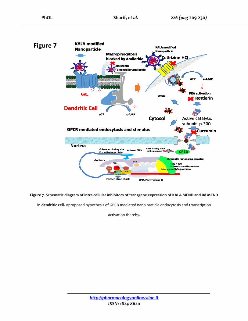
Figure 7. Schematic diagram of intra cellular inhibitors of transgene expression of KALA MEND and R8 MEND
in dendritic cell. Aproposed hypothesis of GPCR mediated nano particle endocytosis and transcription
activation thereby.
Figure 7
PhOL Sharif, et al. 227 (pag 209-230)
http://pharmacologyonline.silae.it
ISSN: 1824-8620
Kala MEND CpGFDNA
Chlorpro. 1μg/ml, 1h
Fillipin 1μg/ml, 1h
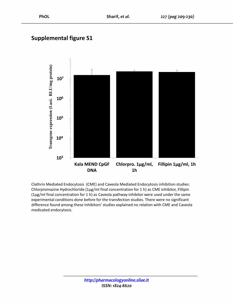
Supplemental figure S1
104
105
106
107
103
Tra
nsg
ene
exp
ress
ion
(L
uci
. R
LU
/mg
pro
tein
)
Clathrin Mediated Endocytosis (CME) and Caveola Mediated Endocytosis inhibition studies:Chlorpromazine Hydrochloride (1μg/ml final concentration for 1 h) as CME inhibitor, Fillipin(1μg/ml final concentration for 1 h) as Caveola pathway inhibitor were used under the same experimental conditions done before for the transfection studies. There were no significant difference found among these inhibitors’ studies explained no relation with CME and Caveolamedicated endocytosis.

PhOL Sharif, et al. 228 (pag 209-230)
http://pharmacologyonline.silae.it
ISSN: 1824-8620
0.0E+00
2.0E+02
4.0E+02
6.0E+02
8.0E+02
1.0E+03
1.2E+03
1.4E+03
1.6E+03
1.8E+03
NBD-dope1%
liposome
R8 NBD-dope 1%liposome
R8 NBD-dope 1%
MEND
CCD, R8NBD-dope
1% lip
CCD, R8MEND NBD-
dope 1%
Flu
ore
scen
ce I
nte
nsi
ty a
.u.
/mg
pro
tein
Cytochalasin D (CCD)
0
500
1000
1500
2000
2500
KALA NBD-dope 1%liposome
CCD KALAliposome
NBD-dope1%
KALA NBD-dope 1%
MEND
CCD KALAMEND
NBD 1%
nsns
22
Dextran - Rhodamine
CCD treated Dextran - Rhodamine
***
**
Flu
ore
scen
ce I
nte
nsi
ty a
.u.
/mg
pro
tein
Supplementary Figure S2
Dendritic cells treated with Cytochalasin D (CCD) 60 μM in serum free media and thereafter NBD 1%
labeled R8 nanoparticles were transfected. Dextran-Rhodamine 5μM final concentration was applied to
the dendritic cells at 37°C for 1h in serum free condition. CCD treated dendritic cell for 1h and
thereafter dextran-Rhodamine was applied as before for 1 h. **P<0.01, *** P<0.001 unpaired student t
test followed by Mann-whitney test (non parametric)
210 min at 4 ° C
Sulpho-rhodamine loaded stearyl KALA liposome in situ application on the dendritic cell at 4°C
after10 min under a LSM confocal microscope. There are clear rhodamine signal in many dotted spot
indicated by red arrow reveals the possibility of receptor mediated binding.
S2a.
S2b.
S2d.
S2c.

PhOL Sharif, et al. 229 (pag 209-230)
http://pharmacologyonline.silae.it
ISSN: 1824-8620
Cel
l v
iab
ilit
y (
%)
as
a f
un
ctio
n o
f p
rote
in c
on
c.
0
20
40
60
80
100
120
KALA MEND H1 blocker 0.9 mM
Supplementary figure S3
Total protein concentration of dendritic cell population after transfection with the control KALA MEND and H1 blocker (Cetirizine HCl) treated (0.9mM for 1h) dendritic cells, those were subsequently exposed to KALA MEND. H1 blocker with 0.9 mM showed no cytotoxicity.

PhOL Sharif, et al. 230 (pag 209-230)
http://pharmacologyonline.silae.it
ISSN: 1824-8620
R8 liposome after H1 blocker 0.9 mM treatment
R8 liposome
Supplementary figure S4
Confocal microscopic studies of R8 liposome treated dendritic cell as control. It showed no remarkable reduction like KALA liposome by H1 blocker. Red = liposome rhodamine leveled, green = endosome.