appendix g1 emf research updates - bpa.gov g1 health... · 1003741.000 - 1234 research on extremely...
TRANSCRIPT
Appendix G1
EMF Research Updates

Health Sciences Practice
Research on Extremely Low Frequency Electric and Magnetic Fields and Health
1003741.000 - 1234
Research on Extremely Low Frequency Electric and Magnetic Fields and Health Prepared for: The Bonneville Power Administration Prepared by: Exponent 149 Commonwealth Drive Menlo Park, California 94025 July 20, 2015 Exponent, Inc.
July 20, 2015
1003741.000 - 1234 ii
Contents Page
List of Figures iii
List of Tables iv
Acronyms and Abbreviations v
Introduction vii
1 Scientific Methods 1
Weight-of-evidence review 1 Scientific reviews on ELF EMF 4 Dissenting opinion on ELF EMF 6
Basics of epidemiology 6 IARC classifications 9
2 Human Health Research 12
Cancer 12 Childhood leukemia 12 Childhood brain cancer 24 Breast cancer 26 Other adult cancers 30 In vivo studies of carcinogenesis 36 In vitro studies of carcinogenesis 41
Reproductive and developmental effects 44 Neurodegenerative disease 52
3 Other Areas of Research 59
Pacemakers and implanted cardiac devices 59 Flora 63 Fauna 65
Dairy cattle and deer 65 Wild bees and honey bees 67 Birds 69
Marine life 70
4 Standards and Guidelines 73
5 References 76
Appendix 1 – World Health Organization International EMF Project Summary of ELF EMF Conclusions in EHC 238
Appendix 2 – WHO Fact Sheet Appendix 3 – Comment on the BioInitiative Report
July 20, 2015
1003741.000 - 1234 iii
List of Figures
Page
Figure 1. Weight-of-evidence reviews consider three types of research 3
Figure 2. Basic design of cohort and case-control studies 7
Figure 3. Interpretation of an odds ratio in a case-control study 8
Figure 4. Basic IARC method for classifying exposures based on potential carcinogenicity 11
July 20, 2015
1003741.000 - 1234 iv
List of Tables
Page
Table 1. Relevant studies of childhood leukemia, October 2010 – April 2015 23
Table 2. Relevant studies of childhood brain cancer, October 2010 – April 2015 26
Table 3. Relevant studies of breast cancer, October 2010 – April 2015 29
Table 4. Relevant studies of adult brain cancer, October 2010 – April 2015 34
Table 5. Relevant studies of adult leukemia/lymphoma, October 2010 – April 2015 36
Table 6. Relevant in vivo studies of carcinogenesis, October 2010 – April 2015 41
Table 7. Relevant in vitro studies of carcinogenesis, October 2010 – April 2015 43
Table 8. Relevant studies of reproductive and developmental effects, October 2010 – April 2015 49
Table 9. Relevant in vivo studies of reproductive and developmental effects, October 2010 ‒ April 2015 51
Table 10. Relevant studies of neurodegenerative diseases, October 2010 – April 2015 58
Table 11. Reference levels for whole body exposure to 60-Hz fields: general public 74
July 20, 2015
1003741.000 - 1234 v
Acronyms and Abbreviations
AC Alternating current
ACGIH American Conference of Governmental Industrial Hygienists
ACRBR Australian Centre for Radiofrequency Bioeffects Research
ALL Acute lymphoblastic leukemia
BPA Bonneville Power Administration
CI Confidence interval
DMBA 7, 12-dimethylbenz[a]anthracene
ELF Extremely low frequency
EMF Electric and magnetic fields
EMI Electromagnetic interference
EPRI Electric Power Research Institute
G Gauss
GD Gestational day
HCN Health Council of the Netherlands
Hz Hertz
IARC International Agency for Research on Cancer
ICD Implanted cardiac device
ICNIRP International Commission on Non-Ionizing Radiation Protection
IGF-1 Insulin-like growth factor 1
m Meter
mg Milligram
mg/kg Milligram per kilogram
mG Milligauss
NRPB National Radiological Protection Board
NIEHS National Institute of Environmental Health Sciences
OR Odds ratio
PND Post-natal day
RF Radiofrequency
ROW Right-of-way
RR Relative risk
July 20, 2015
1003741.000 - 1234 vi
SCENIHR Scientific Committee on Emerging and Newly Identified Health Risks
SES Socioeconomic status
SSI Swedish Radiation Protection Authority
SSM Swedish Radiation Safety Authority
WHO World Health Organization
July 20, 2015
1003741.000 - 1234 vii
Introduction
Electrical objects produce two field types—electric fields and magnetic fields. The term field is
used to describe the way an object influences its surrounding area. A temperature field, for
example, surrounds a warm object, such as a space heater or campfire. Electric and magnetic
fields (EMF) surround any object that generates, transmits, or uses electricity, including
appliances, electrical wiring, office equipment, generators, and any other electrical devices.
These fields are invisible, and they cannot be felt or heard.
Electric fields occur as a result of the electric potential (i.e., voltage) on these objects, and
magnetic fields occur as a result of current flow through these objects.1 Just like a temperature
field, both electric fields and magnetic fields can be measured, and their levels depend on the
properties of the source of the field (e.g., voltage, current, and configuration) and the distance
from the source of the field, among other things.
Both electric fields and magnetic fields decrease rapidly with distance from the source. For
example, a magnetic field of 300 milligauss (mG) within 6 inches of a vacuum cleaner
diminishes to 1 mG at 4 feet (NIEHS, 2002). This is similar to the way that the heat generated
by a space heater or a campfire diminishes as a person moves farther away from it. Although
ordinary objects do not block magnetic fields, objects such as trees and buildings easily block
electric fields.
The electrical power system in the United States produces alternating current (AC) EMF that
changes direction and intensity 60 times per second—i.e., a frequency of 60 Hertz (Hz).2 This
frequency is in the extremely low frequency (ELF) range of the electromagnetic spectrum.
Electricity produced by generating stations flows as 60-Hz current through transmission and
distribution lines and provides power to the many appliances and electrical devices that we use
in our homes, schools, and workplaces. Magnetic fields are found throughout our environment
1 The electric field is expressed in measurement units of volts per meter (V/m) or kilovolts per meter (kV/m);
1 kilovolt per meter is equal to 1,000 V/m. The strength of magnetic fields is expressed as magnetic flux density in units called gauss (G), or in milligauss (mG), where 1 G is equal to 1,000 mG.
2 Europe’s electrical system produces 50-Hz EMF. Since 50-Hz EMF is also in the ELF range, research on 50-Hz EMF is relevant to questions on 60-Hz EMF.
July 20, 2015
1003741.000 - 1234 viii
because electricity is needed for many things in our daily lives, from lighting, heating, and
cooling our homes to powering our refrigerators and computers.
Questions about whether these ubiquitous exposures could affect our health began to be raised
in the 1970s. Since then, researchers from many different scientific disciplines have
investigated this question, and hundreds of studies have been conducted. The public frequently
expresses concern about ELF EMF, particularly in the context of new transmission lines. The
intent of this report is to provide an update to previously prepared reports that had summarized
the large body of scientific research on ELF EMF and health (Exponent, 2007, 2011). The
current report also provides a summary of recent evaluations and recommendations with respect
to the precautions, if any, of public health agencies
In July 2007, Exponent provided a report to the Bonneville Power Administration (BPA) that
described the conclusions of a comprehensive, weight-of-evidence review published by the
World Health Organization (WHO) in June 2007; the portion of Exponent’s 2007 report that
describes the conclusions of the WHO report is attached as Appendix 1 for reference.3 The
WHO review still represents the most recent comprehensive review of the literature by a
multidisciplinary scientific panel. The WHO assembled a multidisciplinary Task Group of 21
scientists from around the world to draft a Monograph that summarized the research and
provided conclusions as to whether there are risks associated with ELF EMF and, if so, at what
exposure levels (WHO, 2007a). The report concluded that the only established effects of ELF
EMF exposure are acute neurostimulatory effects (i.e., shock-like effects) that occur at very
high levels of exposure; these exposure levels are not encountered in ordinary residential or
occupational environments. The fact sheet from the WHO review is attached as Appendix 2
(WHO, 2007b).
In January 2011, Exponent provided an update to BPA on scientific research related to ELF
EMF and potential health effects. The 2011 report gave a detailed overview of the relevant
body of research published between January 1, 2006, and October 1, 2010.4 In light of
3 Exponent. Assessment of Research Regarding EMF and Health and Environmental Effects. Olympic Peninsula
Reinforcement Transmission Line Project. Exponent, Inc., July 2007. 4 Exponent. Research on Extremely Low Frequency Electric and Magnetic Fields and Health. Exponent, Inc.,
January 2011.
July 20, 2015
1003741.000 - 1234 ix
additional research results since 2010, the conclusion remains that the scientific evidence does
not confirm the existence of long-term health consequences of exposure to ELF EMF.
Research is a constantly evolving process. Despite the volume of research available on ELF
EMF and the large reduction in uncertainty that research has achieved over the years, scientists
continue research in this area with the goal of clarifying and replicating old findings and testing
new hypotheses. New studies on ELF EMF are published every month. While the WHO
review provides a comprehensive and relatively up-to-date summary of the status of research on
this topic, new research has the potential to modify or strengthen conclusions. The BPA has,
therefore, requested an update on the research with regard to ELF EMF and health. This report
provides an overview of the cumulative body of research published since our previous update
and covers the relevant scientific literature published during the period between October 1,
2010, and April 1, 2015.
A summary of the methods scientists use to conduct studies and make decisions about health
risks is included in Section 1 as a framework for understanding later discussions. In Section 2,
the discussion of new research is broadly grouped by health outcome—cancer, reproductive
effects, developmental effects, and neurodegenerative diseases. This discussion summarizes
two types of research—epidemiologic studies and experimental studies in animals (in vivo)—
within each health outcome category. Experimental studies in cells and tissues (in vitro) of
carcinogenesis are discussed briefly in Section 2. Other areas of research not reviewed by
WHO are discussed in Section 3, including the possible effects of ELF EMF on the functioning
of pacemakers, on flora and fauna, and on marine life. Finally, guidelines for ELF EMF
exposure developed by scientific organizations to prevent against established health effects are
summarized in Section 4.
July 20, 2015
1003741.000 - 1234 1
1 Scientific Methods
Weight-of-evidence review
Most of what we encounter in our everyday environment has no effect on our health. Other
exposures, however, may affect our health in either a beneficial or a harmful way, including
such ubiquitous interactions with our environment as the air we breathe, the water we drink, and
our exposure to sunlight. Much time and money is spent by scientists around the world
designing, conducting, and publishing research to determine what factors may affect our health,
including environmental exposures (like ELF EMF), infectious agents, and our genetics. The
process for arriving at a conclusion about whether there is a health risk associated with any of
these factors often is not straightforward or definitive. Rather, it is a long process that requires
repeated hypothesis generation and testing.
The process begins when a scientist forms a hypothesis and conducts a study to test that
hypothesis. Studies are conducted by scientists at academic universities and scientific
institutions around the world. Once a study is complete, the authors submit it to a scientific
journal for publication, where it undergoes peer review prior to publication. The evidence to
evaluate any health risk includes all of the relevant studies published in the peer-reviewed
literature.
These individual research studies can be thought of as puzzle pieces. When all of the research is
placed together, we have some understanding of possible health effects; no conclusions can be
reached, however, by looking at only one study, just as no picture can be formed with just one
puzzle piece. Each study provides a different piece of information to the puzzle because of its
unique strengths and weaknesses—if the study used valid methods and had no obvious sources
of bias, it may provide a wealth of information or, if the study was not well conducted, it may
add little or no information to our understanding.
This process of evaluating all of the research together to determine whether something poses
either a health benefit or health risk is referred to as a weight-of-evidence review. There are
three types of research that are considered in a weight-of-evidence review: epidemiology studies

July 20, 2015
1003741.000 - 1234 2
of people, experimental studies in animals (in vivo research), and experimental studies in cells
and tissues (in vitro research). It is important to consider all three types of research together
because they provide complementary information:
Epidemiologic studies collect observational data about human populations in their
everyday environments to determine whether there are patterns between exposures
and diseases. These studies measure statistical associations to evaluate whether a
disease and exposure occur together more often than expected. An important
limitation of these studies is that, if an association is measured, they do not tell
scientists how the exposure is truly related to the disease, and whether the exposure
is causally related to the disease. That conclusion can only be reached by
considering the entire body of research. Most of the studies evaluating ELF EMF
examine whether people with a particular disease have had higher estimates of ELF
EMF exposure in the past compared to people without that disease.
Experimental studies in which scientists expose animals (in vivo) to varying levels of
electric or magnetic fields (some as high as 50,000 mG) are an important source of
information. These studies compare the amount of disease they observe in exposed
animals to the amount of disease they observe in animals that have not been exposed.
The strength of animal studies is that scientists are able to control all aspects of the
animals’ lives to minimize the potential confounding effects of factors other than the
exposure of interest. The most valuable experimental studies for understanding
disease are those in which the animals receive life-long exposures. The main
limitation of laboratory animal studies, however, is that they are conducted in a
species other than human, and thus require interspecies extrapolation for a human
health risk assessment.
Experimental studies in vitro involve the exposure of isolated cells and tissues to the
agent of interest, in this case ELF EMF, and compare the characteristics of exposed
and unexposed samples to look for differences that are indicative of a disease
process. These studies are limited because what occurs to exposed cells or tissues
July 20, 2015
1003741.000 - 1234 3
outside of a human body may not be the same as what occurs to cells and tissues
inside a body.
The weight-of-evidence approach is the standard process used worldwide by scientists,
scientific organizations, and regulatory agencies to assess the possible health benefits and risks
associated with exposures. A weight-of-evidence review begins with a systematic identification
and review of relevant published, peer-reviewed epidemiologic, in vivo, and in vitro research.
The weight that individual studies provide to the overall conclusions is not equal—studies vary
widely in terms of the sophistication and validity of their methods. Therefore, each study from
each discipline must be evaluated critically and assigned a weight. A final conclusion is then
reached by considering the cumulative body of research, giving more weight to studies of higher
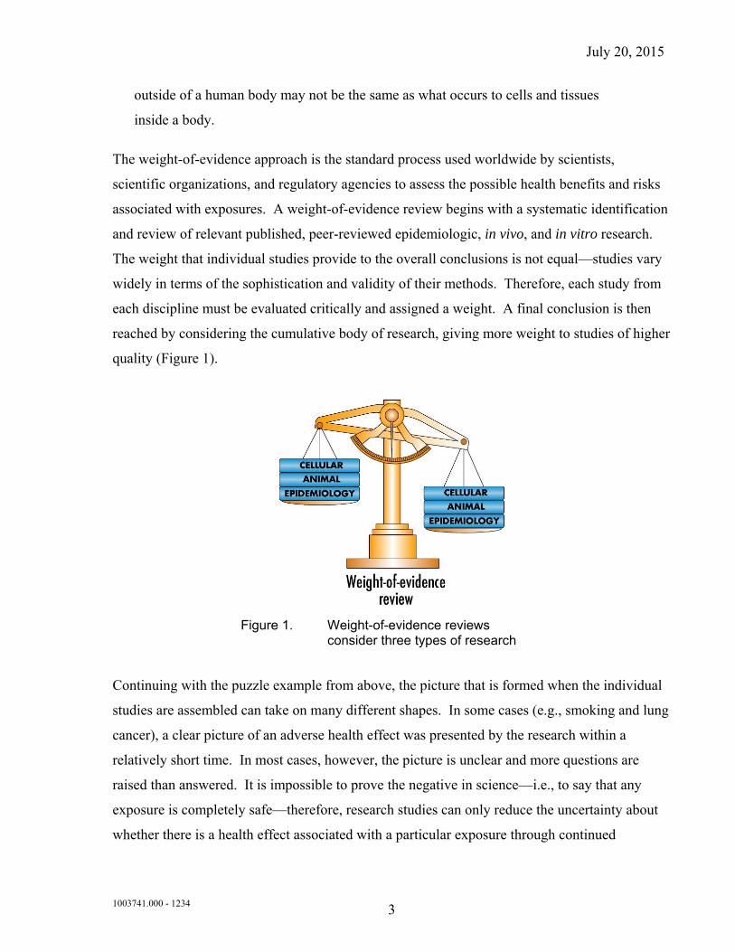
quality (Figure 1).
Figure 1. Weight-of-evidence reviews consider three types of research
Continuing with the puzzle example from above, the picture that is formed when the individual
studies are assembled can take on many different shapes. In some cases (e.g., smoking and lung
cancer), a clear picture of an adverse health effect was presented by the research within a
relatively short time. In most cases, however, the picture is unclear and more questions are
raised than answered. It is impossible to prove the negative in science—i.e., to say that any
exposure is completely safe—therefore, research studies can only reduce the uncertainty about
whether there is a health effect associated with a particular exposure through continued
July 20, 2015
1003741.000 - 1234 4
research. The only way to reduce this uncertainty is to conduct high quality studies with
meaningful results that are replicated across study populations (in the case of epidemiologic
studies) and by different laboratories (in the case of in vivo and in vitro research). Thus, in most
areas of research, unless the data clearly indicate an increased risk at defined exposure levels,
scientific panels will conclude that the research is inadequate or limited and requires further
study until the uncertainty has been reduced below an acceptable level. While the public may
interpret this conclusion as indicating concern, it is natural for scientists to recommend future
research to reduce uncertainty around a largely negative body of research or to replicate findings
that appear positive.
Scientific and health organizations put together panels of scientists to conduct weight-of-
evidence reviews. These panels consist of experts from around the world in the areas of interest
(e.g., epidemiology, neurophysiology, toxicology, etc.) and they follow standard scientific
methods for arriving at conclusions about possible health risks. The conclusions of these
reviews are looked to for the current scientific consensus on a particular topic and form the basis
of recommendations made by organizations and governments on exposure standards and
precautionary measures.
Scientific reviews on ELF EMF
Numerous national and international organizations responsible for public health have convened
multidisciplinary panels of scientists to conduct weight-of-evidence reviews and arrive at
conclusions about the possible risks associated with ELF EMF. These organizations include the
following (in ascending, chronological order of their most recent publication):
The National Institute for Environmental Health Sciences (NIEHS) in the United States
assembled a 30-person Working Group to review the cumulative body of epidemiologic and
experimental data on ELF EMF and provide conclusions and recommendations to the
government (NIEHS, 1998, 1999).
The International Agency for Research on Cancer (IARC) completed a full carcinogenic
evaluation of ELF EMF in 2002 (IARC, 2002).
July 20, 2015
1003741.000 - 1234 5
The WHO released a review in June 2007 as part of its International EMF Program to assess
the scientific evidence related to ELF EMF in the frequency range from 0 to 300 GHz
(WHO, 2007a). Appendix 1 summarizes the conclusions of this review.
The Swedish Radiation Protection Authority (SSI), using other major scientific reviews
as a starting point, evaluated new studies in consecutive annual reports (SSI, 2007; SSI,
2008). The Swedish Radiation Safety Authority (SSM) superseded the SSI on June 30,
2008, and continued to publish reports on ELF EMF (SSM 2010, 2013, 2014, 2015).
The National Radiological Protection Board (NRPB)5 of Great Britain issued full
evaluations of the research in 1992, 2001, and 2004 (NRPB 1992, 2001a, 2004), with
supplemental updates (NRPB, 1993; NRPB, 1994a) and topic-specific reports (NRPB,
1994b; NRPB, 2001b; HPA, 2006) published in the interim. In a letter addressing a related
topic, the Director of the Health Protection Agency of Great Britain (HPA) reiterated their
position on ELF EMF and appropriate precautionary measures (HMG, 2009).
The International Commission on Non-Ionizing Radiation Protection (ICNIRP), the
formally recognized organization for providing guidance on standards for non-ionizing
radiation exposure for the WHO, published a review of the cumulative body of
epidemiologic and experimental data on ELF EMF in 2003. The ICNIRP released draft
exposure guidelines for ELF EMF in July 2009 (ICNIRP, 2009). While the ICNIRP panel
stated that they relied heavily on previous reviews of the literature related to long-term ELF
EMF exposures, they provided relevant conclusions as part of the drafting of these
guidelines. Final guidelines for ELF EMF exposure were issued in late 2010 (ICNIRP,
2010).
The Scientific Committee on Emerging and Newly Identified Health Risks (SCENIHR),
a scientific committee commissioned by the European Commission issued its most recent
report in March 2015 (SCENIHR, 2015) updating its previous reports and conclusions
(SCENIHR, 2007; SCENIHR, 2009) to the Health Directorate of the European Commission.
5 The NRPB merged with the HPA in April 2005 to form its new Radiation Protection Division, and
subsequently, the HPA merged into Public Health England in 2013.
July 20, 2015
1003741.000 - 1234 6
Dissenting opinion on ELF EMF
In August 2007, an ad hoc, self-organized group of 14 scientists and public health and policy
consultants published an on-line report titled “The BioInitiative Report: A Rationale for a
Biologically-based Public Exposure Standard for Electromagnetic Fields (ELF and RF).” An
updated version of the report was subsequently published on-line in 2012. The updated report
incorporates several new sections into the 2007 report. The 2012 version was authored by some
of the same individuals, with some additional authors. The group’s objective was to “assess
scientific evidence on health impacts from electromagnetic radiation below current public
exposure limits and evaluate what changes in these limits are warranted now to reduce possible
public health risks in the future” (BioInitiative 2012, p. 4). The original report was followed by
several publications related to ELF EMF that summarized some of the on-line report’s
conclusions (Hardell and Sage, 2008; Davanipour and Sobel, 2009; Johansson 2009). The
individuals who comprised this group did not represent any well-established regulatory agency
nor were they convened by a recognized scientific authority.
The report has been criticized by scientific agencies because it did not follow the methods of a
standard weight-of-evidence review. The main criticisms included, among others, the lack of
consideration of study quality when evaluating individual studies, selective referencing of
positive studies in support of a specific conclusion, and heavy reliance on in vitro studies, which
typically play a secondary role in proper human health risk assessments due to their limitations
discussed above (ACRBR, 2008; HCN, 2008). Contrary to proper health risk evaluations
conducted by other health and scientific agencies, neither of the BioInitiative Reports
represented a consensus opinion, but included the conclusions of the individual authors of the
various chapters. For these reasons, its conclusions and recommendations are not considered
further in this report. Appendix 3 provides a detailed scientific commentary on the report.
Basics of epidemiology
This section briefly describes the main types of epidemiologic studies and the major issues that
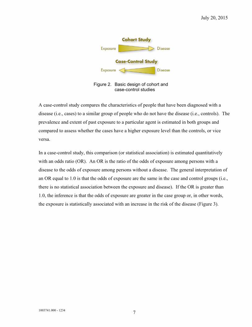
are relevant to evaluating their results. The two, main types of epidemiologic studies are cohort
studies and case-control studies (Figure 2).
July 20, 2015
1003741.000 - 1234 7
Figure 2. Basic design of cohort and case-control studies
A case-control study compares the characteristics of people that have been diagnosed with a
disease (i.e., cases) to a similar group of people who do not have the disease (i.e., controls). The
prevalence and extent of past exposure to a particular agent is estimated in both groups and
compared to assess whether the cases have a higher exposure level than the controls, or vice
versa.
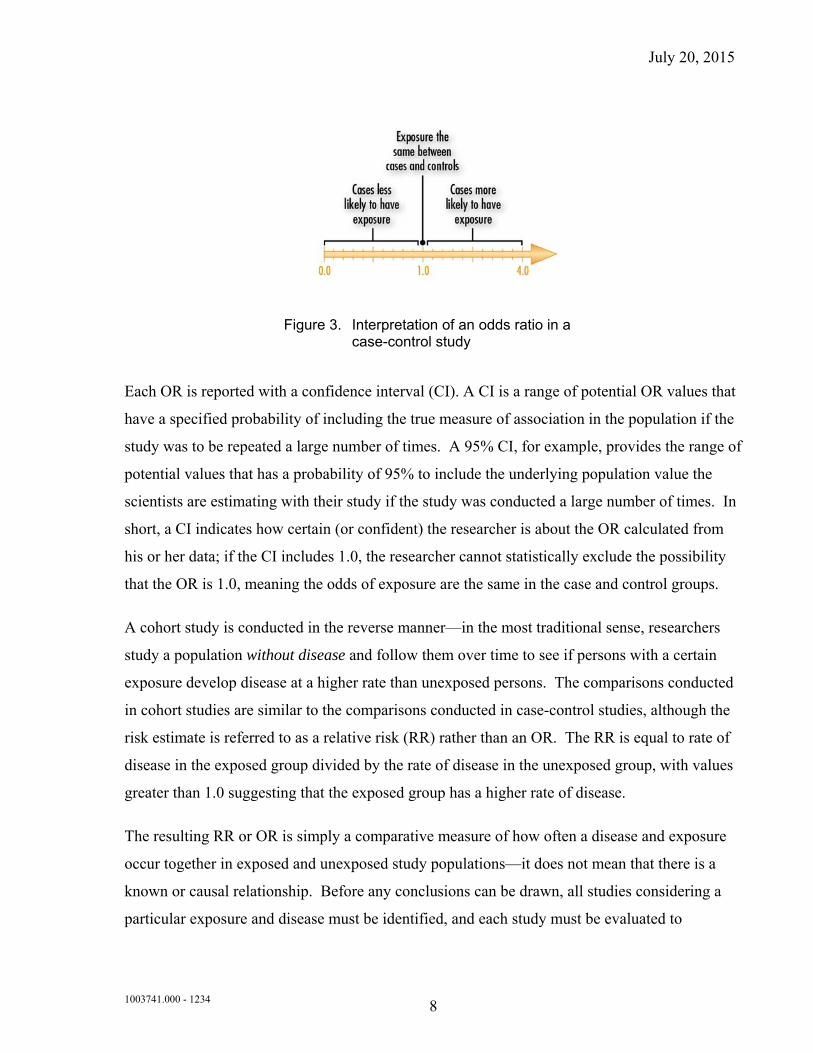
In a case-control study, this comparison (or statistical association) is estimated quantitatively
with an odds ratio (OR). An OR is the ratio of the odds of exposure among persons with a
disease to the odds of exposure among persons without a disease. The general interpretation of
an OR equal to 1.0 is that the odds of exposure are the same in the case and control groups (i.e.,
there is no statistical association between the exposure and disease). If the OR is greater than
1.0, the inference is that the odds of exposure are greater in the case group or, in other words,
the exposure is statistically associated with an increase in the risk of the disease (Figure 3).
July 20, 2015
1003741.000 - 1234 8
Figure 3. Interpretation of an odds ratio in a case-control study
Each OR is reported with a confidence interval (CI). A CI is a range of potential OR values that
have a specified probability of including the true measure of association in the population if the
study was to be repeated a large number of times. A 95% CI, for example, provides the range of
potential values that has a probability of 95% to include the underlying population value the
scientists are estimating with their study if the study was conducted a large number of times. In
short, a CI indicates how certain (or confident) the researcher is about the OR calculated from
his or her data; if the CI includes 1.0, the researcher cannot statistically exclude the possibility
that the OR is 1.0, meaning the odds of exposure are the same in the case and control groups.
A cohort study is conducted in the reverse manner—in the most traditional sense, researchers
study a population without disease and follow them over time to see if persons with a certain
exposure develop disease at a higher rate than unexposed persons. The comparisons conducted
in cohort studies are similar to the comparisons conducted in case-control studies, although the
risk estimate is referred to as a relative risk (RR) rather than an OR. The RR is equal to rate of
disease in the exposed group divided by the rate of disease in the unexposed group, with values
greater than 1.0 suggesting that the exposed group has a higher rate of disease.
The resulting RR or OR is simply a comparative measure of how often a disease and exposure
occur together in exposed and unexposed study populations—it does not mean that there is a
known or causal relationship. Before any conclusions can be drawn, all studies considering a
particular exposure and disease must be identified, and each study must be evaluated to
July 20, 2015
1003741.000 - 1234 9
determine the possible role that factors such as chance, bias, and confounding may have played
in the study’s results.
Chance refers to a random event (i.e., a coincidence). An association can be observed
between an exposure and disease that simply is the result of a chance occurrence. Statistics,
such as the CI, are calculated to determine whether chance is a likely explanation for the
findings. The CI does not include other sources of variability in the data other that those
related to statistical sampling error such as might arise from bias and confounding, discussed
below.
Bias refers to any systematic error in the design, conduct, or analysis of a study that would
cause a distorted estimate of an exposure’s effect on the risk of disease. There are many
different types of bias; for example, selection bias may occur if the characteristics of persons
that participate in a study differ in a meaningful way from the characteristics of those
subjects that do not participate (e.g., cases living near power lines might be more likely to
participate than controls because the cases are concerned about this possible exposure).
Confounding is a situation in which an association is distorted because the exposure being
studied is associated with other risk factors for the disease. For example, a link between
coffee drinking in mothers and low birth weight babies may be observed in a study, but
some women who drink coffee also smoke cigarettes. When the smoking habits of mothers
are taken into account, coffee drinking may not be associated with low birth weight babies
because the confounding effect of smoking has been removed.
As part of the weight-of-evidence review process, each study’s design and methods are
evaluated critically to determine if and how chance, bias, and confounding may have affected
the results and, subsequently, the weight that should be placed on the study’s findings.
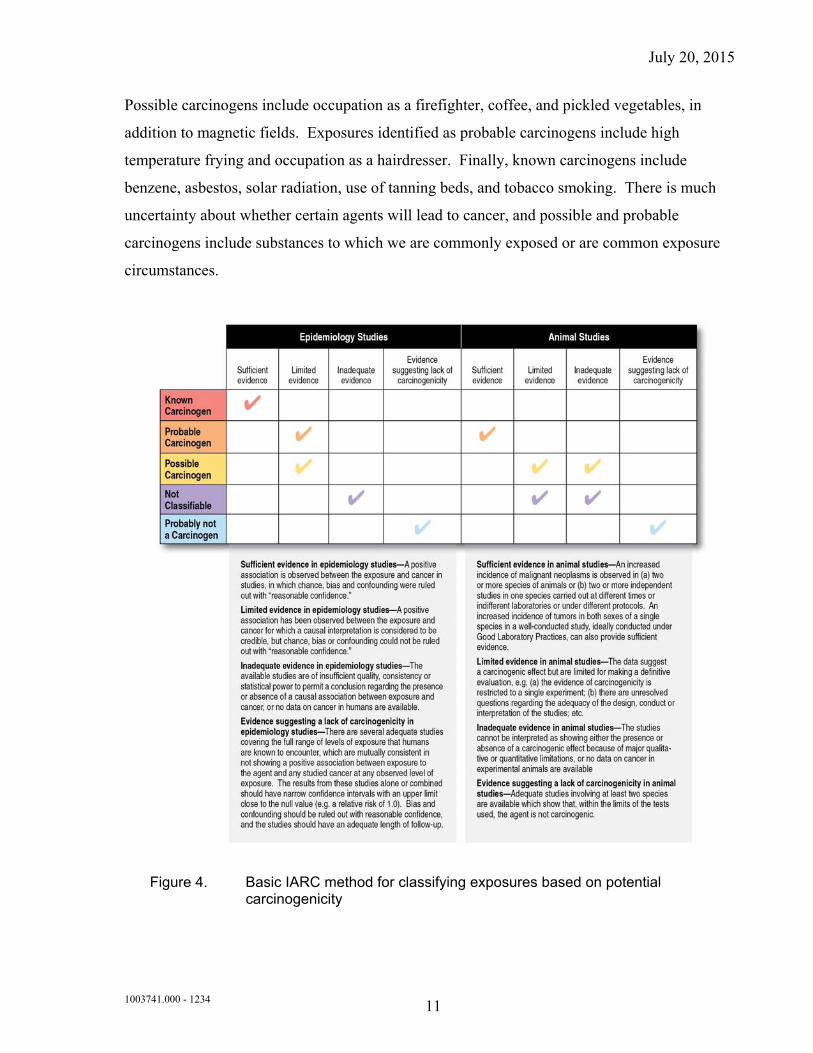
IARC classifications
This section briefly describes the method that the IARC uses following a weight-of-evidence
review to classify exposures based on the evidence in support of carcinogenicity. The WHO
adopted this method for both cancer and non-cancer health outcomes in their 2007 review on
ELF EMF, and other scientific agencies refer to this classification system, as well.
July 20, 2015
1003741.000 - 1234 10
First, each research type (epidemiology, in vivo, and in vitro) is evaluated to determine the
strength of evidence in support of carcinogenicity (as defined in Figure 4). Epidemiologic
studies are characterized as having sufficient evidence for carcinogenicity if an association is
found and chance, bias, and confounding can be ruled out with “reasonable confidence.”
Limited evidence is used to describe a body of research where the findings are inconsistent or
where an association is observed but there are outstanding questions about study design or other
methodological issues that preclude making strong conclusions. Inadequate evidence describes
a body of research where it is unclear whether the data is supportive or unsupportive of
causation because there is a lack of data or there are major quantitative or qualitative issues.
The same overall categories apply for in vivo research. In vitro research is not described in
Figure 4 because it provides ancillary information and, therefore, is used to a lesser degree in
evaluating carcinogenicity and is classified simply as strong, moderate, or weak.
Agents are then classified into five overall categories using the combined categories from
epidemiologic, in vivo, and in vitro research (listed from highest to lowest risk): (1) known
carcinogen, (2) probable carcinogen, (3) possible carcinogen, (4) non-classifiable, and (5)
probably not a carcinogen.
As summarized in Figure 4, the category possible carcinogen typically denotes exposures for
which there is limited evidence of carcinogenicity in epidemiologic studies, and in vivo studies
provide limited or inadequate evidence of carcinogenicity.
The IARC has reviewed close to 1,000 substances and exposure circumstances to evaluate their
potential carcinogenicity. About 80% of exposures fall in the categories possible carcinogen
(29%) or non-classifiable (51%). This occurs because, as described above, it is nearly
impossible to prove that something is completely safe and few exposures show a clear-cut or
probable risk, so most agents will end up in either of these two categories. Throughout the
history of the IARC, only one agent has been classified as probably not a carcinogen, which
illustrates the conservatism of the evaluations and the difficulty in proving the absence of an
effect beyond all doubt.
Over half of the agents are non-classifiable in terms of carcinogenicity, i.e., it is unclear whether
they can cause cancer—hair coloring products, jet fuel, and tea are included in this category.
July 20, 2015
1003741.000 - 1234 11
Possible carcinogens include occupation as a firefighter, coffee, and pickled vegetables, in
addition to magnetic fields. Exposures identified as probable carcinogens include high
temperature frying and occupation as a hairdresser. Finally, known carcinogens include
benzene, asbestos, solar radiation, use of tanning beds, and tobacco smoking. There is much
uncertainty about whether certain agents will lead to cancer, and possible and probable
carcinogens include substances to which we are commonly exposed or are common exposure
circumstances.
Figure 4. Basic IARC method for classifying exposures based on potential carcinogenicity
July 20, 2015
1003741.000 - 1234 12
2 Human Health Research
The following sections provide an overview of peer-reviewed research published between
October 1, 2010, and April 1, 2015. A literature review was conducted to identify new
epidemiologic and in vivo research published on 50 or 60-Hz ELF EMF. A large number of
search strings referencing the exposure and diseases of interest, as well as authors who regularly
publish in this area, were included as search terms in the PubMed database, a service of the U.S.
National Library of Medicine that includes over 17 million citations from MEDLINE and other
life science journals for biomedical articles dating to the 1950s.6 Scientists with experience in
this area reviewed the search results to identify relevant studies.
This report focuses on the diseases that have received the most attention—cancer, reproductive
effects, developmental effects, and neurodegenerative diseases. Other health effects have been
studied (i.e., rare cancer types, suicide, depression, electrical hypersensitivity, and
cardiovascular effects), but because research on these topics evolves slowly, these topics are not
separately summarized here. The WHO review provides a good resource for the status of
research on these additional health effects.
This update focuses on identifying and summarizing new epidemiologic and major in vivo
research, since these study types are the most informative for risk assessment in this field.
Cancer
Childhood leukemia
What was previously known about childhood leukemia?
While authoritative scientific panels have not concluded that the overall evidence confirms the
existence of any adverse health effects, these panels consistently recognized the limited
evidence from childhood leukemia epidemiologic studies, which provided the basis for the
“possibly carcinogenic to humans” classification of ELF magnetic fields. Since 1979, several
dozen epidemiologic studies have been conducted in the United States, Canada, Europe, New 6 PubMed includes links to full-text articles and other related resources (http://www.ncbi.nlm.nih.gov/PubMed/).
July 20, 2015
1003741.000 - 1234 13
Zealand, and Asia that evaluated the relationship between childhood leukemia and magnetic
fields using various methods to estimate exposure. These methods have included long-term (48-
hour) personal monitoring; spot or long-term (24- or 48-hour) measurements indoors and
outdoors; calculations using loading, line configuration, and distance of nearby power
installations to estimate historical, residential exposure; and wire code categories.7 As a group
of independent studies, they did not show a clear or consistent association between magnetic
fields and childhood leukemia. The largest and most methodologically sound case-control
studies to estimate personal magnetic field exposure directly did not report a consistent
relationship (Linet et al., 1997; McBride et al., 1999; UKCCS, 2000). When two independent
pooled analyses combined the data from these case-control studies, however, a statistically
significant association was observed between rare average magnetic-field exposure above 3-
4 mG and childhood leukemia (Ahlbom et al., 2000; Greenland et al., 2000). Both pooled
analyses indicated that children with leukemia were about two times more likely to have had
estimated magnetic-field exposures above 3-4 mG. Average exposures at this level are
uncommon; according to the WHO, results from several extensive surveys showed that
approximately 0.5–7.0% of children had time-averaged exposures in excess of 3 mG and 0.4–
3.3% had time-averaged exposures in excess of 4 mG (WHO, 2007a). While these analyses
provide a valuable quantitative summary of the data, pooled analyses are limited by the
disparate methods used to collect the underlying data in the individual studies included in the
pooled analyses. Questions have been raised as to whether the original studies, particularly
those that are large and estimated exposure directly, provide a more valid estimate of the
association than the pooled analyses (Elwood, 2006).
Despite the association observed in these pooled analyses, health agencies have not concluded
that magnetic fields are a known or probable cause of childhood leukemia. The studies are of
insufficient strength to rule out with “reasonable confidence” the role that chance, bias, and
confounding may have played in the observed statistical association. In other words,
researchers do not have enough confidence in the way these studies were conducted to conclude
that the measured statistical association represents a true relationship between magnetic fields
and childhood leukemia. Furthermore, experimental data do not provide evidence for a risk in 7 Wire code categories are categories used to classify the potential magnetic field exposures at residences based
on the characteristics of and distance to nearby power installations.
July 20, 2015
1003741.000 - 1234 14
the more highly-controlled in vivo studies, and in vitro studies do not provide evidence of a
plausible biological mechanism whereby magnetic fields lead to carcinogenesis.
Since chance, bias, and confounding could not be ruled out as an explanation for the association,
the IARC concluded in 2002 that the data on childhood leukemia provided limited evidence of
carcinogenicity (IARC, 2002). In 2007, the WHO reviewed studies on childhood leukemia and
magnetic-field exposure published since the 2002 IARC review (WHO, 2007a). They
concluded that the new epidemiologic studies were consistent with previous results and they did
not provide new evidence to alter the classification of limited epidemiologic evidence in support
of carcinogenicity. Taken together with the largely negative in vivo and in vitro research, the
WHO confirmed the previous IARC classification of magnetic fields as a possible carcinogen
based upon IARC criteria (Figure 4).8
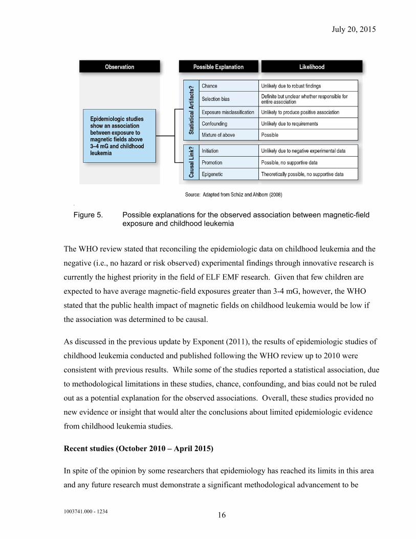
Since it is unclear whether the association is real, the WHO review evaluated other factors that
might be partially, or fully, responsible for the association, including chance, control selection
bias, confounding from hypothesized or unknown risk factors, and misclassification of magnetic
field exposure (Figure 5). The following is a summary of their evaluation:
The WHO review concluded that chance is an unlikely explanation since the pooled
analyses had a large sample size and decreased variability.
Control selection bias occurs when the controls that are selected from the population
and decide to participate in the study do not represent the true exposure experience of the
entire non-diseased population. In the case of magnetic fields, the WHO speculates that
controls with a higher socioeconomic status (SES) may participate in studies more often
than controls with a lower SES. Since persons with a higher SES may have lower
magnetic-field exposures or tend to live farther from transmission lines, the control
group’s magnetic-field exposure may be artificially low. Thus, when the exposure
experience of the control group is compared to the case group, there may be a difference
in exposure distribution between the case and control groups in the study that does not
8 The WHO concluded the following: “Consistent epidemiological evidence suggests that chronic low intensity
ELF magnetic field exposure is associated with an increased risk of childhood leukaemia. However, the evidence for a causal relationship is limited, therefore exposure limits based upon epidemiological evidence are not recommended, but some precautionary measures are warranted” (WHO, 2007a, pp. 355-356).
July 20, 2015
1003741.000 - 1234 15
exist in the general population. The WHO concluded that control selection bias is
probably occurring in these studies and would result in an overestimate of the true
association, but may not explain the entire observed statistical association
The WHO panel concluded that confounding is less likely to be causing the observed
association than other factors, although the possibility that some yet-to-be identified
confounder is responsible for the association cannot be excluded completely. Suggested
risk factors that may be confounding the relationship include SES, residential mobility,
contact currents, and traffic density.9
The WHO stated that the possible effects of exposure misclassification are the most
difficult to predict. EMF presents unique challenges in exposure assessment because it
is ubiquitous, imperceptible, and has many sources (Kheifets and Oksuzyan, 2008). No
target exposure or exposure window has been identified, and the numerous methods of
estimating exposure likely result in a different degree of error within and between
studies. Most reviews have concluded that exposure misclassification would likely
result in an underestimate of the true association, meaning the association we observe is
lower than the true value; however, the extent to which this might occur varies widely
and is difficult to assess (Greenland et al., 2000). The WHO concluded that exposure
misclassification is likely present in these studies, but is unlikely to entirely explain the
observed association.
9 For example, if dwellings near power lines encounter higher traffic density and pollution from traffic density
causes childhood leukemia, traffic density may cause an association between magnetic-field exposure and childhood leukemia, where a relationship does not truly exist.
July 20, 2015
1003741.000 - 1234 16
.
Figure 5. Possible explanations for the observed association between magnetic-field exposure and childhood leukemia
The WHO review stated that reconciling the epidemiologic data on childhood leukemia and the
negative (i.e., no hazard or risk observed) experimental findings through innovative research is
currently the highest priority in the field of ELF EMF research. Given that few children are
expected to have average magnetic-field exposures greater than 3-4 mG, however, the WHO
stated that the public health impact of magnetic fields on childhood leukemia would be low if
the association was determined to be causal.
As discussed in the previous update by Exponent (2011), the results of epidemiologic studies of
childhood leukemia conducted and published following the WHO review up to 2010 were
consistent with previous results. While some of the studies reported a statistical association, due
to methodological limitations in these studies, chance, confounding, and bias could not be ruled
out as a potential explanation for the observed associations. Overall, these studies provided no
new evidence or insight that would alter the conclusions about limited epidemiologic evidence
from childhood leukemia studies.
Recent studies (October 2010 – April 2015)
In spite of the opinion by some researchers that epidemiology has reached its limits in this area
and any future research must demonstrate a significant methodological advancement to be
July 20, 2015
1003741.000 - 1234 17
justified (Schmiedel and Blettner, 2010), childhood leukemia has continued to be the main focus
of ELF EMF epidemiologic research due to the previously reported and unexplained
associations. A number of studies investigating childhood leukemia and magnetic fields have
been published since the previous Exponent report that provided an update of the literature up to
October 2010 (Table 1). While some of the recent studies continue to support a weak
association between elevated magnetic-field levels and childhood leukemia, some recently
published large and methodologically advanced studies showed no association. The previously
reported association, however, remains unexplained. Overall, the results of the new studies
published since 2010 have not resulted in a change of the overall evidence that would alter the
classification of the epidemiologic data as limited. Similar conclusions were expressed in the
most recent SCENIHR report (SCENIHR, 2015).
A review of the EMF and childhood leukemia literature by Calvente et al. (2010) that included
studies related to both ELF and radiofrequency exposures concluded that “studies to date have
not convincingly confirmed or ruled out an association between non-ionizing radiation and the
risk of childhood leukemia.” The authors also note that inconsistencies between studies may be
explained by the influence of confounding factors, selection bias, and misclassification. A
general review of childhood leukemia etiology highlighted genetic factors and ionizing radiation
as factors known to contribute to leukemia development among children (Eden, 2010). While
the review also mentioned the potential for EMF to be a contributor, the author notes that if
EMF has any effect, it “would account for only a small percentage of cases,” and that a
plausible biological mechanism has not been identified to explain any potential carcinogenic
effects. Two recent papers have also evaluated the potential impact of EMF (Teepen and van
Dijck, 2012; Grellier et al., 2014). Both reviews emphasize the lack of biological support for a
potential carcinogenic effect and the importance that various sources of bias may play in the
studies that report the association. They conclude that even if the association with EMF was
causal, it would have limited public health impact. The authors also continue to emphasize that
further improvement in our understanding may only be gained by studies with improved
methodology and reduced potential for bias such as can be achieved by international and
interdisciplinary collaborations (Ziegelberger et al., 2011; Teepen and van Dijck, 2012).
July 20, 2015
1003741.000 - 1234 18
A case-control study of childhood leukemia from Brazil (Wünsch-Filho et al., 2011) that
included 162 cases recruited between 2003 and 2009 and 565 matched controls identified
through the regional birth registry utilized two approaches for exposure assessment; the
researchers conducted spot and 24-hour measurements in the children’s homes and also
determined the distance of the homes to the closest power line with voltages of 88 kV, 138 kV,
230 kV, 345 kV, and 440 kV. The authors reported no statistically significant associations
between leukemia and magnetic-field exposure >3 mG (OR, 1.09; CI, 0.33-3.61) or living
within 100 meters of a transmission line (OR, 1.54; CI, 0.26-9.12). The authors also discussed
several sources of potential bias, most notably selection bias, which in their assessment may
have potentially influenced the results. A small case-control study from the Czech Republic
(Jirik et al., 2012) included 79 cases of childhood leukemia and 79 controls and reported no
statistically significant association between leukemia and measured magnetic-field exposure in
the children’s homes (OR for >0.4mG, 0.90; CI, 0.37-2.22).
A case-control study reported from Northern California included 245 children under the age of 8
years diagnosed with leukemia between 2000 and 2007 and 269 matched control children
without leukemia. The researchers assessed the potential for exposure to magnetic fields and
contact currents in the homes of the participating children. No associations were reported for
any of the investigated exposure metrics (Does et al., 2011).
In recent years, several large case-control studies on EMF and childhood leukemia were
published from France, Denmark, the United Kingdom, and Italy (Sermage-Faure et al., 2013;
Bunch et al., 2014; Pedersen et al., 2014; Magnani et al., 2014; Salvan et al., 2015). Sermage-
Faure et al. (2013) collected geocoded information on residential addresses and power line
locations in France to examine the risk of childhood leukemia in association with distance to
power lines. The study included 2,779 cases of childhood leukemia, diagnosed between 2002
and 2007, and 30,000 control children. Overall, the authors reported no statistically significant
increase in leukemia risk with distance to power lines. The authors, however, noted a
statistically non-significant association of distance with childhood leukemia in a sub-analysis
within 50 meters of 225-kV ‒ 400-kV lines. This estimate was based on only nine leukemia
cases, rendering the association imprecise.
July 20, 2015
1003741.000 - 1234 19
Pedersen et al. (2014) reported results of a similar study from Denmark. The Danish study
included 1,698 cases of childhood leukemia and 3,396 healthy control children. The authors
reported no risk increases for childhood leukemia with residential distance to power lines (living
<200 meters from a transmission line, OR, 0.76; CI, 0.40-1.45). Exposure prevalence, however,
was very low in the study; less than 1% of the cases (n=13) lived within 200 meters of a
transmission line. Bunch et al. (2014) updated and extended an earlier study by Draper et al.
(2005) in the United Kingdom. The update extended the study period by 13 years, included
Scotland in addition to England and Wales, and included 132-kV lines in addition to 275-kV
and 400-kV transmission lines.
The Bunch et al. study, which included over 53,000 childhood cancer cases diagnosed between
1962 and 2008, and over 66,000 healthy children as controls, is the largest study to date
conducted on this subject. The authors reported no statistically significant association with
residential distance to power lines with any of the voltage categories. The statistical association
reported in the earlier study (Draper et al., 2005) was no longer apparent in the updated analysis
by Bunch et al. (2014). According to an analysis by calendar time, the association was only
present in the earlier decades (1960s and 1970s) but not in the later decades starting from the
1980s (Bunch et al., 2014). This pattern of diminishing association with calendar time weakens
the argument that the associations observed in the earlier decades are due to magnetic-fields.
The strengths of the French, Danish, and British studies include their large size and their
population-based design that minimized the potential for selection bias. These studies, however,
relied on a poor proxy of the actual residential magnetic-field exposure and relied on distance to
the nearest transmission lines as their main exposure metric. The limitations of distance as an
exposure proxy have been discussed in the scientific literature by several observers in the
context of the French study (Bonnet-Belfais et al., 2013; Clavel et al., 2013). Chang et al.
(2014) recently provided a detailed discussion of the limitations of exposure assessment
methods based on geographical information systems.
Italian researchers have also published the methods and results of a large childhood leukemia
case-control study (Magnani et al., 2014; Salvan et al., 2015). The investigators included 412
leukemia cases under the age of 10 years diagnosed in Italy between 1998 and 2001 along with
July 20, 2015
1003741.000 - 1234 20
587 controls in their study of leukemia and residential exposure to 50-Hz magnetic fields. Long
term (24 ‒ 48-hr) measurements in the children’s bedroom were conducted to assess exposure.
Conditional logistic regression was used to calculate RR and adjust for potentially confounding
variables. The researchers evaluated the influence of various exposure metrics used in the
analyses (measures of central tendency or peak-exposure measures, continuous or categorical
exposures), the influence of time of measurements (nighttime, weekend, entire measurements),
and the effect of residential mobility on the observed associations. No consistent exposure-
response patterns were observed in any of the analyses. Restrictions on study subject eligibility,
potential for participation bias, and low prevalence of highly-exposed subjects (particularly
exposure above 3 mG), as also discussed by the authors, represent shortcomings in the studies
that limit their potential interpretation.
British researchers examined the relationships between the fathers’ occupational exposures to 33
various factors (including potential exposure to EMF on the job) and the likelihood of exposure
of their offspring with childhood leukemia to EMF (Keegan et al., 2012). The authors studied a
total of 15,785 childhood leukemia cases that were diagnosed between 1962 and 2006 and a
similar number of matched controls. Paternal exposure to EMF in relation to leukemia in their
children was not statistically significant when all types of leukemia or the two most common
subtypes, lymphoid leukemia and myeloid leukemia, were considered. The authors reported a
statistically significant increase for leukemia classified as “other types,” which included only
7% of the leukemia cases. This observed association may be attributable to the small number of
cases in this sub-analysis or the large number comparisons made in the overall analyses, which
could falsely identify associations as statistically significant.
The potential association between exposure to magnetic fields and survival of children
following a leukemia diagnosis has also been investigated by a recent pooled analysis (Schüz et
al., 2012), which aimed to follow up on earlier observations. Two previous studies, based on
very small number of cases, reported poorer survival among childhood leukemia cases with
increased average exposure to magnetic fields, suggesting the magnetic fields may play a role in
the progression in the disease following diagnosis (Foliart et al., 2006; Svendsen et al., 2007).
The Schüz et al. (2012) study included exposure and clinical data on more than 3,000 cases of
childhood leukemia from Canada, Denmark, Germany, Japan, the United Kingdom, and the
July 20, 2015
1003741.000 - 1234 21
United States. The authors reported no association between magnetic-field exposure and overall
survival or relapse of disease after diagnosis in children with leukemia.
Chinese scientists (Zhao et al., 2014) summarized the data from nine EMF-childhood leukemia
epidemiologic studies published between 1997 and 2013 in a meta-analysis. The authors
reported a statistically significant association between average exposure above 4 mG and all
types of childhood leukemia (OR, 1.57, 95% CI, 1.03-2.4). The studies included in the meta-
analysis largely overlapped with studies included in previous pooled analyses, thus they
provided little new insight.
The potential role corona ions near power lines may play in childhood cancer development was
investigated in a large childhood cancer epidemiologic study in the United Kingdom (Swanson
et al., 2014). Corona ion distribution around power lines was modeled with consideration of
meteorological data on wind conditions, in addition to power line characteristics and proximity
to residential address. The results, in the authors’ assessment, provided no empirical support for
the corona ion hypothesis.
Potential non-causal, alternative explanations for the observed epidemiologic association
between magnetic fields and childhood leukemia were examined in several methodological
studies. Swanson and Kheifets (2012) hypothesized that if free radicals are involved in the
biological mechanism that explains the epidemiologic association then, due to the small
timescale of the reactions, the effects of ELF EMF and the earth’s geomagnetic fields would be
similar. The authors evaluated whether the magnitude of the earth’s geomagnetic field modifies
the effects reported by ELF EMF childhood leukemia studies from various parts of the world.
The results were not fully supportive of the hypothesis. In another study, Swanson (2013)
examined whether differences in residential mobility among residents who lived at varying
distances from power lines may explain the statistical association of leukemia with residential
proximity to power lines. The study reported some variations in residential mobility, “but only
small ones, and not such as to support the hypothesis.”
As part of the Northern California Childhood Leukemia study, researchers evaluated the role
selection bias may play in the association between childhood leukemia and residential magnetic-
field exposure (Slusky et al., 2014). Wire code categories were used to assess exposure among
July 20, 2015
1003741.000 - 1234 22
participant and nonparticipant subjects in the study. While systematic differences between
participant and nonparticipant subjects were reported in both wire code categories and SES,
these differences did not appreciably influence the association between childhood leukemia and
magnetic-field exposure estimates. Wire code categories are poor surrogates of actual
magnetic-field exposure, and the study showed no association between magnetic fields and
childhood leukemia among the participant subjects, thus the study findings warrant cautious
interpretation.
In addition to continued interest in EMF exposure distribution in the general population
(Karipidis, 2015), a number of exposure assessment studies have evaluated various exposure
scenarios, where highly exposed populations can reliably be identified without requiring
participation of the study subjects (e.g., Okokon et al., 2014; Hareuveny et al., 2010; Zaryabova
et al., 2013). Such exposure scenarios without the need for subject participation would enable a
more accurate assessment of the importance of selection bias, which remains a key limitation of
most epidemiologic studies.
In summary, while a number of new studies have been published since 2010, recent
epidemiologic studies on childhood leukemia have not provided new evidence that would alter
the overall conclusion; the epidemiologic evidence continues to be limited and remains
unsupported by laboratory animal studies and by the lack of known biological mechanisms that
could explain a carcinogenic effect.
July 20, 2015
1003741.000 - 1234 23
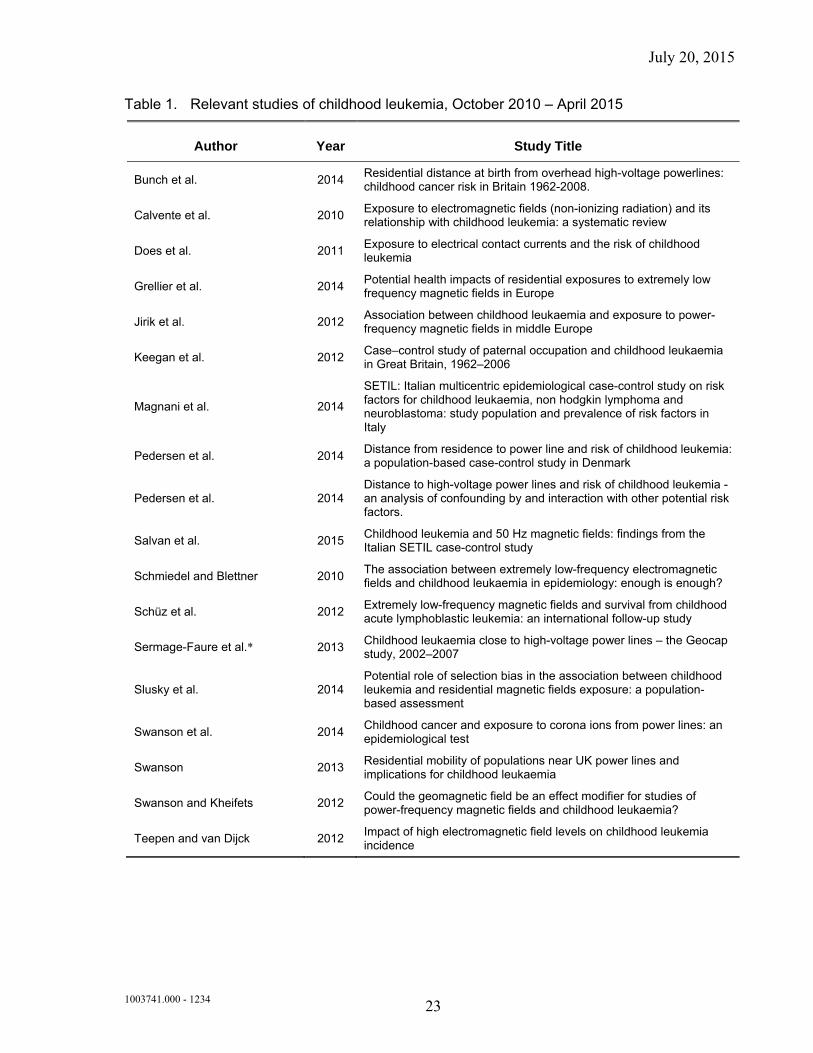
Table 1. Relevant studies of childhood leukemia, October 2010 – April 2015
Author Year Study Title
Bunch et al. 2014 Residential distance at birth from overhead high-voltage powerlines: childhood cancer risk in Britain 1962-2008.
Calvente et al. 2010 Exposure to electromagnetic fields (non-ionizing radiation) and its relationship with childhood leukemia: a systematic review
Does et al. 2011 Exposure to electrical contact currents and the risk of childhood leukemia
Grellier et al. 2014 Potential health impacts of residential exposures to extremely low frequency magnetic fields in Europe
Jirik et al. 2012 Association between childhood leukaemia and exposure to power-frequency magnetic fields in middle Europe
Keegan et al. 2012 Case–control study of paternal occupation and childhood leukaemia in Great Britain, 1962–2006
Magnani et al. 2014
SETIL: Italian multicentric epidemiological case-control study on risk factors for childhood leukaemia, non hodgkin lymphoma and neuroblastoma: study population and prevalence of risk factors in Italy
Pedersen et al. 2014 Distance from residence to power line and risk of childhood leukemia: a population-based case-control study in Denmark
Pedersen et al. 2014 Distance to high-voltage power lines and risk of childhood leukemia - an analysis of confounding by and interaction with other potential risk factors.
Salvan et al. 2015 Childhood leukemia and 50 Hz magnetic fields: findings from the Italian SETIL case-control study
Schmiedel and Blettner 2010 The association between extremely low-frequency electromagnetic fields and childhood leukaemia in epidemiology: enough is enough?
Schüz et al. 2012 Extremely low-frequency magnetic fields and survival from childhood acute lymphoblastic leukemia: an international follow-up study
Sermage-Faure et al.* 2013 Childhood leukaemia close to high-voltage power lines – the Geocap study, 2002–2007
Slusky et al. 2014 Potential role of selection bias in the association between childhood leukemia and residential magnetic fields exposure: a population-based assessment
Swanson et al. 2014 Childhood cancer and exposure to corona ions from power lines: an epidemiological test
Swanson 2013 Residential mobility of populations near UK power lines and implications for childhood leukaemia
Swanson and Kheifets 2012 Could the geomagnetic field be an effect modifier for studies of power-frequency magnetic fields and childhood leukaemia?
Teepen and van Dijck 2012 Impact of high electromagnetic field levels on childhood leukemia incidence
July 20, 2015
1003741.000 - 1234 24
Childhood brain cancer
What was previously known about childhood brain cancer?
The research related to magnetic fields and childhood brain cancer has been less consistent than
that observed for childhood leukemia and the WHO review concluded that the evidence was
inadequate to support a carcinogenic effect. In order to provide a more definitive answer, the
WHO review recommended a combined analysis of childhood brain cancer studies similar to
those completed for childhood leukemia:
As with childhood leukaemia, a pooled analysis of childhood brain cancer
studies should be very informative and is therefore recommended. A
pooled analysis of this kind can inexpensively provide a greater and
improved insight into the existing data, including the possibility of
selection bias and, if the studies are sufficiently homogeneous, can offer
the best estimate of risk (WHO 2007a, p. 18).
In response to the WHO recommendation above, a meta-analysis (Mezei et al., 2008) and a
pooled analysis (Kheifets et al., 2010) of studies on childhood brain tumors and residential
magnetic-field exposure were conducted. In the meta-analysis, 13 epidemiologic studies were
identified that used various proxies of magnetic-field exposure (distance, wire codes, calculated
magnetic fields, and measured magnetic fields). The combined effect estimate was close to 1.0
and not statistically significant, indicating no association between magnetic-field exposure and
childhood brain tumors. A sub-group of five studies, however, with information on childhood
brain tumors and calculated or measured magnetic fields greater than 3 ‒ 4 mG reported a
combined OR that was elevated but not statistically significant (OR, 1.68, 95% CI, 0.83-3.43).
The authors suggested two explanations for this elevated OR. First, they suggested that an
increased risk of childhood brain tumors could not be excluded at high exposure levels (i.e., >3
‒ 4 mG). Second, they stated that the similarity of this result to the findings of the pooled
analyses of childhood leukemia suggests that control selection bias is operating in both
analyses. Similar to the meta-analysis, some categories of high exposure in the pooled analysis
of studies with measured or calculated magnetic-field levels had an OR >1.0, but none of the
July 20, 2015
1003741.000 - 1234 25
findings were statistically significant and enhanced calculations showed inconsistency in the
results of subgroup analyses and no dose-response pattern (Kheifets et al., 2010b). The main
analysis reported no association between childhood brain cancer and magnetic-field exposure
>4 mG, compared to magnetic-field exposure <1 mG (OR=1.14, 95% CI=0.61-2.13). Both the
authors of the meta-analysis and the pooled analysis concluded that their results provide little
evidence for an association between magnetic fields and childhood brain tumors.
The pooled analysis included two case-control studies published after the WHO 2007 review
(Kroll et al., 2010; Saito et al., 2010). In their study of 55 cases of childhood brain cancer, Saito
et al. (2010) reported that children with brain cancer were more likely to have average
magnetic-field exposure levels greater than 4 mG, compared to children without brain cancer.10
The association was based on three cases and one control; interpretations of the data were,
therefore, limited by small numbers in the upper exposure category. The strength of this study
is the exposure assessment; measurements were taken continuously over a weeklong period in
the child’s bedroom approximately 1 year after diagnosis. An important limitation, however, is
the very poor participation rates among study subjects; poor participation rates introduce the
possibility of selection bias, among other biases. As described above, Kroll et al. (2010)
included 6,584 cases of brain cancer diagnosed over a 33-year period in the United Kingdom.
No associations were reported in any analysis of brain cancer, including calculated magnetic
fields >1 ‒ 2 mG, 2 ‒ 4 mG, and 4 mG.
Thus, the combined analyses of childhood brain cancer epidemiologic studies provided no
support for an association.
Recent studies (October 2010 – April 2015)
Given the observed general absence in the epidemiologic literature of associations between ELF
EMF exposure and childhood brain cancer, there has been only limited interest in this area for
further studies. Hug et al. (2010) investigated the potential relationship between estimated
parental exposure to ELF EMF and cancer in their offspring in Germany. The study included
444 children with cancer of the central nervous system diagnosed between 1992 and 1997.
Potential exposure to ELF EMF of the parents was estimated from their job titles, the industry in
10 The unpublished results of this study were included in Mezei et al. (2008).
July 20, 2015
1003741.000 - 1234 26
which they worked, and the time period, as reported in their occupational history. Neither
paternal nor maternal exposure to ELF EMF was associated with the occurrence of brain cancer
in the offspring. The updated and extended British study by Bunch et al. (2014), discussed in
the childhood leukemia section, also included 11,968 cases of childhood brain cancer diagnosed
in the United Kingdom between 1962 and 2008. Similar to previous results reported from the
earlier stud (Draper et al., 2005; Kroll et al., 2010), no association was observed between
estimates of residential exposure to EMF from high-voltage power lines and childhood brain
cancer.
In summary, the recent data do not alter the classification of the epidemiologic data in this field
as inadequate.
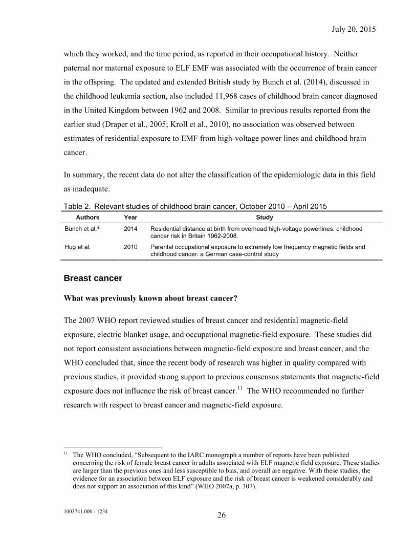
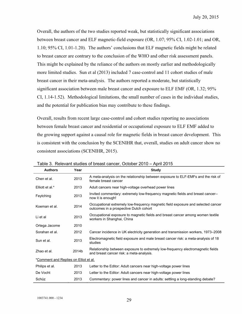
Table 2. Relevant studies of childhood brain cancer, October 2010 – April 2015
Breast cancer
What was previously known about breast cancer?
The 2007 WHO report reviewed studies of breast cancer and residential magnetic-field
exposure, electric blanket usage, and occupational magnetic-field exposure. These studies did
not report consistent associations between magnetic-field exposure and breast cancer, and the
WHO concluded that, since the recent body of research was higher in quality compared with
previous studies, it provided strong support to previous consensus statements that magnetic-field
exposure does not influence the risk of breast cancer.11 The WHO recommended no further
research with respect to breast cancer and magnetic-field exposure.
11 The WHO concluded, “Subsequent to the IARC monograph a number of reports have been published
concerning the risk of female breast cancer in adults associated with ELF magnetic field exposure. These studies are larger than the previous ones and less susceptible to bias, and overall are negative. With these studies, the evidence for an association between ELF exposure and the risk of breast cancer is weakened considerably and does not support an association of this kind” (WHO 2007a, p. 307).
Authors Year Study
Bunch et al.* 2014 Residential distance at birth from overhead high-voltage powerlines: childhood cancer risk in Britain 1962-2008.
Hug et al. 2010 Parental occupational exposure to extremely low frequency magnetic fields and childhood cancer: a German case-control study
July 20, 2015
1003741.000 - 1234 27
Recent studies (October 2010 – April 2015)
A case-control study from Brazil investigated environmental exposures as potential risk factors
for breast cancer including 110 cases of breast cancer diagnosed between 1999 and 2002 among
women aged 20-35 years (Ortega Jacome et al., 2010). Information on environmental
exposures, including residential proximity to electrical power transformers, was collected with
the use of questionnaires. No statistically significant association was reported between breast
cancer and residence within 20 meters of a power transformer; however, very limited, if any,
inference can be drawn from the study as the study was small with limited statistical power, the
exposure assessment was severely limited for EMF exposure, and no description of control
selection was provided.
A large case-control study in the United Kingdom investigated estimated exposure to ELF EMF
due to residential proximity to high-voltage transmission lines and cancers among adults (Elliott
et al., 2013). Among other cancer outcomes, the study included 29,202 newly diagnosed female
breast cancer cases from England and Wales diagnosed between 1974 and 2008, and a total of
over 79,000 controls between the age of 15 and 74 years. Data from geographical information
systems were used to identify location of power lines and residential addresses for cases and
controls. Magnetic-field exposure was calculated for each control address and for each case
address for the year of and 5 years prior to diagnosis. Breast cancer risk among women showed
no association with residential distance to power lines or with estimated magnetic-field
exposure. The study was criticized by several researchers following publication with respect to
its exposure assessment, exposure categorization, and the potential for confounding (de Vocht,
2013; Philips et al., 2013; Schüz, 2013).
Three large cohort epidemiologic studies from the United Kingdom, China, and the Netherlands
reported on occupational exposure to ELF EMF and breast cancer development. Close to
80,000 British workers from electricity generation and transmission facilities were included in a
study by Sorahan (2012). Cancer incidence within the cohort was studied between 1973 and
2008. Standardized registration rates were calculated among the workers and compared to rates
observed in the general population. Based on these comparisons, no statistically significant
increases were reported for breast cancer among either male or female workers. There was no
July 20, 2015
1003741.000 - 1234 28
trend for breast cancer incidence with year of hire, years of being employed, or years since
leaving employment. The longitudinal follow up of the cohort and its large size are among the
main strengths of the study. The study, however, is limited in its exposure assessment; cancer
risk was not calculated by magnetic-field exposure levels, and incidence rates were compared to
an external reference group.
Breast cancer incidence was studied among more than 267,000 female textile workers in
Shanghai (Li et al., 2013). Between 1989 and 2000, a total of 1,687 incidence breast cancer
cases were identified in the cohort. The authors conducted a case-cohort analysis, in which they
compared the estimated exposure to ELF EMF among the cases to that among 4,702 non-cases
selected from the cohort. Exposure assessment was based on complete work history and a job-
exposure matrix was developed specifically for the cohort. No association was observed
between cumulative exposure and breast cancer regardless of age, histological type, and whether
lag period was used or not. According to an editorial that accompanied the paper, the study
added additional evidence against a link between ELF EMF and breast cancer and is consistent
with the previous “consistently negative” literature in this area (Feychting, 2013). The editorial
also suggested that further studies on breast cancer “have little new knowledge to add,”
considering the substantial improvement in study quality over time in breast cancer
epidemiologic research.
In a cohort of about 120,000 men and women in the Netherlands Cohort study, the relationship
between occupational exposure to ELF magnetic fields and cancer incidence was investigated
(Koeman et al., 2014). A case-cohort approach was used to analyze the data. A total of 2,077
breast cancer cases among women and no breast cancer cases among men were identified.
Exposure to ELF magnetic fields was assigned based on job title using a job-exposure matrix.
Breast cancer showed no association with the level of estimated ELF magnetic-field exposure,
or the length of employment, or cumulative exposure in the exposed jobs.
Several meta-analyses of breast cancer studies were conducted by Chinese investigators for both
female and male breast cancers (Chen et al., 2013; Sun et al., 2013; Zhao et al., 2014). For
female breast cancer, 23 case-control studies, published between 1991 and 2007 (Chen et al.,
2013), and 16 studies, published between 2000 and 2007 (Zhao et al., 2014), were combined.
July 20, 2015
1003741.000 - 1234 29
Overall, the authors of the two studies reported weak, but statistically significant associations
between breast cancer and ELF magnetic-field exposure (OR, 1.07; 95% CI, 1.02-1.01; and OR,
1.10; 95% CI, 1.01-1.20). The authors’ conclusions that ELF magnetic fields might be related
to breast cancer are contrary to the conclusion of the WHO and other risk assessment panels.
This might be explained by the reliance of the authors on mostly earlier and methodologically
more limited studies. Sun et al (2013) included 7 case-control and 11 cohort studies of male
breast cancer in their meta-analysis. The authors reported a moderate, but statistically
significant association between male breast cancer and exposure to ELF EMF (OR, 1.32; 95%
CI, 1.14-1.52). Methodological limitations, the small number of cases in the individual studies,
and the potential for publication bias may contribute to these findings.
Overall, results from recent large case-control and cohort studies reporting no associations
between female breast cancer and residential or occupational exposure to ELF EMF added to
the growing support against a causal role for magnetic fields in breast cancer development. This
is consistent with the conclusion by the SCENIHR that, overall, studies on adult cancer show no
consistent associations (SCENIHR, 2015).
Table 3. Relevant studies of breast cancer, October 2010 – April 2015 Authors Year Study
Chen et al. 2013 A meta-analysis on the relationship between exposure to ELF-EMFs and the risk of female breast cancer
Elliott et al.* 2013 Adult cancers near high-voltage overhead power lines
Feytching 2013 Invited commentary: extremely low-frequency magnetic fields and breast cancer--now it is enough!
Koeman et al. 2014 Occupational extremely low-frequency magnetic field exposure and selected cancer outcomes in a prospective Dutch cohort
Li et al 2013 Occupational exposure to magnetic fields and breast cancer among women textile workers in Shanghai, China
Ortega Jacome 2010
Sorahan et al. 2012 Cancer incidence in UK electricity generation and transmission workers, 1973–2008
Sun et al. 2013 Electromagnetic field exposure and male breast cancer risk: a meta-analysis of 18 studies
Zhao et al. 2014b Relationship between exposure to extremely low-frequency electromagnetic fields and breast cancer risk: a meta-analysis.
*Comment and Replies on Elliot et al.
Philips et al. 2013 Letter to the Editor: Adult cancers near high-voltage power lines
De Vocht 2013 Letter to the Editor: Adult cancers near high-voltage power lines
Schüz 2013 Commentary: power lines and cancer in adults: settling a long-standing debate?
July 20, 2015
1003741.000 - 1234 30
Other adult cancers
What was previously known about adult cancers (other than breast cancer)?
In general, scientific panels have concluded that there is not a strong or consistent relationship
between other types of cancers that occur in adults (primarily, leukemia, lymphoma, and brain
cancer was the focus of epidemiologic studies) and exposure to magnetic fields; however, the
possibility cannot be entirely ruled out because the findings have been inconsistent (IARC,
2002; WHO, 2007a). Stronger findings have not been observed in studies with better exposure
assessment methods, which have led scientific panels to conclude that the evidence for an
association is weak. The IARC classified the epidemiologic data with regard to adult leukemia,
lymphoma, and brain cancer as “inadequate” in 2002, and the WHO confirmed this
classification in 2007, with much of the remaining uncertainty attributed to limitations in
exposure assessment methods.
Much of the research on EMF and adult cancers is related to occupational exposures, given the
higher range of exposures encountered in the occupational environment. The main limitation of
these studies, however, has been the methods used to assess exposure, with early studies relying
simply on a person’s occupational title (often taken from a death certificate) and later studies
linking a person’s full or partial occupational history to representative average exposures for
each occupation (i.e., a job-exposure matrix). The latter method, while advanced, still has some
important limitations, as highlighted in a review summarizing an expert panel’s findings by
Kheifets et al. (2009).12 While a person’s occupation may provide some indication of the
overall magnitude of their occupational magnetic-field exposure, it does not take into account
the possible variation in exposure due to different job tasks within occupational titles, the
frequency and intensity of contact to relevant exposure sources, or variation by calendar time.
Furthermore, since scientists do not know any mechanism by which magnetic fields could lead
to cancer, an appropriate exposure metric is unknown.
12 Kheifets et al. (2009) reports on the conclusions of an independent panel organized by the Energy Networks
Association in the United Kingdom in 2006 to review the current status of the science on occupational EMF exposure and identify the highest priority research needs.
July 20, 2015
1003741.000 - 1234 31
In order to reduce the remaining uncertainty about whether there is an association between
magnetic fields and these cancers, researchers have recommended (1) meta-analyses to clarify
inconsistencies and (2) better exposure assessment methods that incorporate a greater level of
detail on tasks and exposure characteristics such as spark discharge, contact current, and
harmonics (WHO, 2007a; Kheifets et al., 2009). As described in a previous Exponent report
(2011), a meta-analysis of occupational epidemiologic studies on adult brain cancer and
leukemia has been conducted and published in 2008, partially in response to the WHO’s
recommendation (Kheifets et al., 2008). Based on all relevant publications of occupational
EMF exposure and adult leukemia or brain cancer, summary estimates of association were
calculated using various schemes to weight and categorize the study data. The authors reported
a small and statistically significant association between leukemia and brain cancer in relation to
the highest estimate of magnetic-field exposure in the individual studies. Several findings,
however, led the authors to conclude that magnetic-field exposure is not responsible for the
observed associations, including the lack of a consistent pattern among leukemia subtypes when
the past and new meta-analyses were compared. In addition, for brain cancer, the recent meta-
analysis reported a weaker association than the previous meta-analysis, whereas a stronger
association would be expected since the quality of studies has increased over time. The authors
concluded “the lack of a clear pattern of EMF exposure and outcome risk does not support a
hypothesis that these exposures are responsible for the observed excess risk” (Kheifets et al.,
2008, p. 677).
Adult brain cancer
What was previously known about adult brain cancer?
As described above, the IARC (2002) and the WHO (2007) reviews classified the epidemiologic
evidence on adult brain cancer as inadequate. Results of studies published following these
reviews, up to October 2010, as reviewed in the 2011 Exponent report, did not provide evidence
that would alter the conclusions of IARC and WHO. While the potential for exposure
misclassification were reduced by improved exposure assessment methods in some more recent
studies and combined analyses have attempted to clarify inconsistencies, the epidemiologic
literature, overall, provided no consistent support for an association between EMF exposure and
July 20, 2015
1003741.000 - 1234 32
adult brain cancer. The then most recent report by the SCENIHR described the data on brain
cancers as “uncertain” (SCENIHR, 2009, p. 43).
Recent studies (October 2010 – April 2015)
Gomes et al. (2011) conducted a systematic review of the scientific literature on occupational
and environmental risk factors and primary adult brain cancer. Among other risk factors, the
review also included exposure to EMF. Based on their review, the authors concluded that the
“[s]tudies on electromagnetic field exposures and the risk of brain neoplasm are inconsistent,”
any potential link is “inconclusive,” and the “biological plausibility is not strong” (Gomes et al.,
2011, p. 95). The authors made recommendations that future studies should concurrently
evaluate the effects of multiple potential risk factors, improve exposure assessment to include
lifetime exposure, and consider genetic and other potentially confounding factors.
Case-control epidemiologic studies from Brazil (Marcilio et al., 2011) and the United Kingdom
(Elliott et al., 2013) have investigated EMF exposure due to residential proximity to high-
voltage transmission lines and adult brain cancer. The Brazilian study included 2,357 cases of
adult brain cancer deaths and 4,706 controls from the Metropolitan Region of São Paulo, Brazil.
The authors reported no association between brain cancer mortality and living near a
transmission line at death or calculated magnetic-field levels from these transmission lines at the
last residential address (Marcilio et al., 2011).
Elliott et al. (2013), in their case-control epidemiologic study of adult cancers and residential
proximity to power lines in the United Kingdom, discussed above, also included 6,781 brain
cancer cases. The authors reported no statistically significant elevation in brain cancer risk in
association with either distance to the nearest power line from the subjects’ homes or estimated
magnetic-field levels due to the nearest transmission lines.
In addition to studies of residential EMF exposure, a number of epidemiologic studies have been
published since 2010 that investigated the potential relationship between occupational exposure
to EMF and brain cancer. Baldi et al. (2011) conducted a case-control study in southwestern
France to study the association between brain cancer and estimates of both residential and
occupational exposure to EMF. They included 221 brain cancer cases diagnosed between 1999
and 2001 and 442 matched controls from the general population. The authors reported
July 20, 2015
1003741.000 - 1234 33
statistically non-significant associations for occupational exposure to ELF EMF based on a
crude job-exposure matrix and residential distance within 100 meters of a high-voltage power
line. The association with occupational exposure, but not with distance to power lines, became
statistically significant when restricted to a subset of brain tumors (meningiomas) (Baldi et al.,
2011). Meningiomas, however, constituted only less than one third of brain tumors in the study
and the association may have been identified only by post hoc analyses.
In the study of cancer incidence among about 80,000 electricity generation and transmission
workers in the United Kingdom (discussed earlier), no risk increases for brain cancer was
reported either among male or female workers (Sorahan, 2012). Results of a more in-depth
analysis focusing on brain cancers in the same population were reported in a separate paper
(Sorahan, 2014a). Neither of the two main subtypes of brain tumors (gliomas and
meningiomas) showed statistically significant increases with estimated occupational exposure to
EMF, even when overall exposure, or exposure during either the more recent time period
(within the last 10 years) or earlier time period (more than 10 years ago) was considered. An
overall comparison of the cohort with the general population showed no risk increases for brain
cancer within the cohort either.
In the Netherlands Cohort Study of about 120,000 adults, 160 and 84 cases of brain cancer were
identified among men and women, respectively, during a 17-year period between 1986 and 2003
(Koeman et al., 2014). No statistically significant risk increase or trend was observed for
cumulative occupational exposure to ELF EMF either among men or women.
An occupational historical cohort study of 201,784 jet engine manufacturing workers, employed
between 1952 and 2001, has examined the development of central nervous system neoplasms in
relation to individual level estimates of exposure to 11 physical and chemical agents during
work (Marsh et al., 2013). Exposure to EMF, dichotomized as exposed vs. unexposed, was
among the investigated occupational exposures. The authors analyzed the data using several
statistical approaches: a nested case-control analysis, and a cohort analysis with both internal
and external comparisons. None of the investigated occupational exposures, including EMF,
showed an association with brain cancer in any of the analyses conducted by the authors.
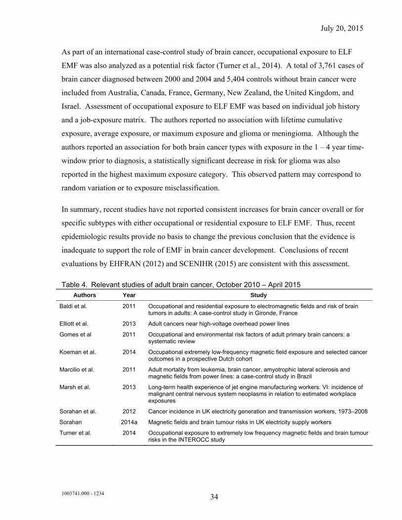
July 20, 2015
1003741.000 - 1234 34
As part of an international case-control study of brain cancer, occupational exposure to ELF
EMF was also analyzed as a potential risk factor (Turner et al., 2014). A total of 3,761 cases of
brain cancer diagnosed between 2000 and 2004 and 5,404 controls without brain cancer were
included from Australia, Canada, France, Germany, New Zealand, the United Kingdom, and
Israel. Assessment of occupational exposure to ELF EMF was based on individual job history
and a job-exposure matrix. The authors reported no association with lifetime cumulative
exposure, average exposure, or maximum exposure and glioma or meningioma. Although the
authors reported an association for both brain cancer types with exposure in the 1 ‒ 4 year time-
window prior to diagnosis, a statistically significant decrease in risk for glioma was also
reported in the highest maximum exposure category. This observed pattern may correspond to
random variation or to exposure misclassification.
In summary, recent studies have not reported consistent increases for brain cancer overall or for
specific subtypes with either occupational or residential exposure to ELF EMF. Thus, recent
epidemiologic results provide no basis to change the previous conclusion that the evidence is
inadequate to support the role of EMF in brain cancer development. Conclusions of recent
evaluations by EHFRAN (2012) and SCENIHR (2015) are consistent with this assessment.
Table 4. Relevant studies of adult brain cancer, October 2010 – April 2015 Authors Year Study
Baldi et al. 2011 Occupational and residential exposure to electromagnetic fields and risk of brain tumors in adults: A case-control study in Gironde, France
Elliott et al. 2013 Adult cancers near high-voltage overhead power lines
Gomes et al 2011 Occupational and environmental risk factors of adult primary brain cancers: a systematic review
Koeman et al. 2014 Occupational extremely low-frequency magnetic field exposure and selected cancer outcomes in a prospective Dutch cohort
Marcilio et al. 2011 Adult mortality from leukemia, brain cancer, amyotrophic lateral sclerosis and magnetic fields from power lines: a case-control study in Brazil
Marsh et al. 2013 Long-term health experience of jet engine manufacturing workers: VI: incidence of malignant central nervous system neoplasms in relation to estimated workplace exposures
Sorahan et al. 2012 Cancer incidence in UK electricity generation and transmission workers, 1973–2008
Sorahan 2014a Magnetic fields and brain tumour risks in UK electricity supply workers
Turner et al. 2014 Occupational exposure to extremely low frequency magnetic fields and brain tumour risks in the INTEROCC study
July 20, 2015
1003741.000 - 1234 35
Adult leukemia and lymphoma
What was previously known about adult leukemia/lymphoma?
The same issues discussed above with regard to adult brain cancer are relevant to research on
adult leukemia and lymphoma. The WHO classified the epidemiologic evidence as
“inadequate” for all adult cancers (WHO 2007a). Studies that were published up to October
2010 and were reviewed in the previous report (Exponent 2011) did not materially change the
overall evidence following the WHO report.
Recent studies (October 2010 – April 2015)
The case-control epidemiologic studies of residential proximity to transmission lines in Brazil
and the United Kingdom also investigated leukemia (Marcilio et al., 2011; Elliott et al., 2013).
The Brazilian study included 1,857 cases of adult leukemia deaths and 4,706 controls. The
authors reported a statistically non-significant association with leukemia mortality for subjects’
who resided within 50 meters of a transmission line at the time of death. The interpretation of
this finding remains unclear because this association was reported to be stronger for lower
voltage transmission lines (under 200 kV), where the magnetic-field levels are anticipated to be
lower. A slightly elevated, but statistically non-significant, association was reported in the
Marcilio et al. study between calculated magnetic fields and mortality due to leukemia, as well.
Elliott et al (2013) included 7,823 cases of adult leukemia diagnosed in the United Kingdom
between 1974 and 2008 along with close to 80,000 controls. The authors reported no elevated
risk or trend for leukemia incidence to diminish with distance or lower estimated magnetic-field
exposure from high-voltage power lines.
In a Spanish epidemiologic study of hematologic cancers (Rodriguez-Garcia and Ramos, 2012),
the authors reported inverse correlations between acute myeloid leukemia, acute lymphoblastic
leukemia (ALL), and distance to thermoelectric power plants and high-density power line
networks. No firm conclusion can be drawn from the study, however, because it was severely
limited due to the use of ecologic data and rudimentary exposure assessment methods and also
lacked an appropriate comparison group.
July 20, 2015
1003741.000 - 1234 36
In the cohort of electricity power plant and transmission workers in the United Kingdom,
Sorahan (2012) reported no increase in risk for leukemia either among men or women, and no
increasing trend was observed with length of employment. In a more in-depth analysis that
focused on leukemia in the same electricity worker cohort, Sorahan (2014b) reported that
neither estimated exposure to EMF in the more recent period nor in or more distant periods is
related to leukemia development. Although there was a statistical association reported in a sub-
analysis for acute lymphocytic leukemia, the author opined that this was most likely due to
chance based on the inconsistent overall pattern. In the Netherlands Cohort Study discussed
earlier, Koeman et al. (2014) identified 761 and 467 hematopoietic malignancies among men
and women, respectively. No increases in risk or trend were observed in association with
cumulative exposure to ELF magnetic fields among either men or women.
Results of recent epidemiologic studies did not provide consistent evidence in support of an
association of adult leukemia and other lymphohematopoietic malignancies with exposure to
EMF either in occupational or residential environments. Therefore, the earlier conclusions as
expressed by IARC and WHO, with respect to the evidence being inadequate for adult cancers,
remain unchanged. This is consistent with the assessment recently conducted by EFHRAN
(2012) and SCENIHR (2015).
Table 5. Relevant studies of adult leukemia/lymphoma, October 2010 – April 2015
Authors Year Study
Elliott et al. 2013 Adult cancers near high-voltage overhead power lines
Koeman et al. 2014 Occupational extremely low-frequency magnetic field exposure and selected cancer outcomes in a prospective Dutch cohort
Marcilio et al. 2011 Adult mortality from leukemia, brain cancer, amyotrophic lateral sclerosis and magnetic fields from power lines: a case-control study in Brazil
Rodriguez-Garcia and Ramos
2012 High incidence of acute leukemia in the proximity of some industrial facilities in El Bierzo, northwestern Spain
Sorahan et al. 2012 Cancer incidence in UK electricity generation and transmission workers, 1973–2008
Sorahan 2014b Magnetic fields and leukaemia risks in UK electricity supply workers
In vivo studies of carcinogenesis
What was previously known from in vivo studies of carcinogenesis?
It is standard procedure to conduct studies on laboratory animals to determine whether exposure
to a specific agent leads to the development of cancer (USEPA, 2005). This approach is used
July 20, 2015
1003741.000 - 1234 37
because all known human carcinogens that were adequately tested also cause cancer in
laboratory animals (IARC, 2006). In the field of ELF EMF research, a number of research
laboratories have exposed rodents, including those with a particular genetic susceptibility to
cancer, to high levels of magnetic fields over the course of the animals’ lifetime and performed
tissue evaluations to assess the incidence of cancer in many organs. In these studies, magnetic-
field exposure has been administered alone (to test for the ability of magnetic fields to act as a
complete carcinogen), in combination with a known carcinogen (to test for a promotional or co-
carcinogenetic effect), or in combination with a known carcinogen and a known promoter (to
test for a co-promotional effect).
Numerous whole animal studies published prior to the WHO review and shortly afterward found
that magnetic-field exposure alone does not increase the occurrence of cancer or promote the
effects of other known carcinogens (Bernard et al., 2008; Boorman et al., 1999a, 1999b; Chung
et al., 2008, 2010; Harris et al., 1998; Mandeville et al., 1997; McCormick et al., 1999; Negishi
et al., 2008; Sommer and Lerchel, 2004, 2006; Yasui et al., 1997).
A series of studies, however, from a single German research group reported that the incidence of
7, 12-dimethylbenz[a]anthracene (DMBA)-induced mammary tumors was increased in rats with
magnetic field exposure (Löscher et al., 1993, 1994, 1997; Baum et l., 1995; Löscher and
Mevissen, 1995; Mevissen et al., 1993a, 1993b, 1996a, 1996b, 1998). Others were unable to
replicate these findings (Anderson et al., 1999; Boorman et al., 1999a, 1999b) and further
research has suggested that the Fischer 344 (F344) rat used in the German studies is a sensitive
sub-strain, while other strains are not (Fedrowitz et al., 2004).
Overall, the WHO concluded the following with respect to in vivo research: “There is no
evidence that ELF exposure alone causes tumours. The evidence that ELF field exposure can
enhance tumour development in combination with carcinogens is inadequate” (WHO 2007a, p.
322). Recommendations for future research included the development of a rodent model for
childhood ALL and the continued investigation of whether magnetic fields can act as a promoter
or co-carcinogen. Studies published following the WHO review and discussed in the previous
Exponent report (2011), overall, provided further evidence against a role for magnetic fields as a
July 20, 2015
1003741.000 - 1234 38
co-carcinogenic agent, and strengthened the conclusion that there is inadequate evidence of
carcinogenicity from in vivo research.
Recent studies (October 2010 – April 2015)
Since 2010, no new animal studies have been published that examine the potential of magnetic-
field exposure—alone or in combination with known carcinogens—to have an adverse effect on
tumor growth and development.
To further investigate the sensitivity of the F344 rat to the promotional effect of magnetic field
exposure in DMBA-induced mammary tumors, however, Fedrowitz and Löscher (2012)
examined gene expression in the mammary gland tissues of female F344 rats and female Lewis
rats that had been continuously exposed short-term to 1,000 mG, 50-Hz magnetic fields for 14
days. Controls were sham-exposed. All analyses were done in a blinded manner and a 2.5-fold
change in expression was selected a priori as the cut-off for a response. Of 31,100 genes
examined, magnetic-field exposure increased expression of nine genes in the Lewis rat; in
contrast, eight genes were increased in expression and six were decreased in the F344 rat. Only
one of these genes was affected in both rat sub-strains, but in opposite directions. The gene for
α-amylase showed the greatest response to magnetic-field exposure in the F344 rats. In a
follow-up study, the same researchers further examined the response of mammary tissue α-
amylase to magnetic-field exposure using tissues collected from previous experiments
(Fedrowitz et al., 2012). Again, controls were sham-exposed and analyses conducted in a
blinded fashion. Both rat sub-strains showed increased α-amylase expression in either the
caudal or the cranial mammary tissue in response to magnetic-field exposure, depending on the
experiment. Control α-amylase activity, however, was found to vary considerably from one
experiment to the next and the authors cautioned that α-amylase activity is highly stress-
sensitive, limiting the conclusions that can be drawn.
Five animal studies were recently published that addressed the ability of ELF EMF to damage
genetic material (i.e., DNA) (Alcaraz et al., 2014; Borhani et al., 2011; Miyakoshi et al., 2012;
Saha et al., 2014; Wilson et al., 2015). Genotoxicity is conceptually linked to carcinogenesis as
gene mutation is considered one of the initiating steps in the process by which a cell becomes
cancerous. Alcaraz et al. (2014) exposed male mice to 2,000 mG, 50-Hz magnetic fields for 7,
July 20, 2015
1003741.000 - 1234 39
14, 21, or 28 days. Controls were not sham-exposed; however, additional mice were exposed to
50 centi-Gray of X-rays as positive controls and all analyses were conducted in a blinded
manner. Although the study investigators reported an increase in micronuclei (i.e., small
nucleus-like structures containing DNA indicative of a chromosomal break) in bone marrow
erythrocytes 24 hours after magnetic-field exposure, the increase was not duration-dependent
and was substantially lower than that induced with X-irradiation. In a similar study, Wilson et
al. (2015) examined the effect of exposure to 100 mG, 1,000 mG, or 3,000 mG, 50-Hz magnetic
fields for 2 or 15 hours on the gene mutation frequency in sperm and blood cells. Negative
controls were sham-exposed; positive controls were exposed to 1 Gray of X-irradiation. Twelve
weeks after exposure (the necessary latency to ensure that the sperm had been exposed at the
time of DNA synthesis and cell division), the blood mutation frequency after magnetic-field
exposure was similar to that of the sham-treated controls; the sperm mutation frequency was
slightly, but significantly, increased although the finding was not dose-dependent. In contrast,
X-irradiation significantly increased the mutation frequency in both cell types. Borhani et al.
(2011) reported increased DNA damage (i.e., fragmentation) in 4-day old mouse embryos of
female mice exposed to 5,000 mG, 50-Hz magnetic fields for 4 hours per day, 6 days per week
for 2 weeks prior to mating. Controls were sham exposed, but analyses were not conducted in a
blinded manner. In contrast, Saha et al. (2014) did not find increased DNA double-strand
breaks in the embryonic neuronal stem cell compartment of mouse embryos exposed to 1,000
mG, 50-Hz magnetic fields for 2 hours on gestational day (GD) 13.5 or with continuous or
intermittent (5 minutes on, 10 minutes off) exposure to a 3,000 mG magnetic field for 15 hours
starting on GD 12.5. This study included a sham-exposed control as well as multiple positive
control groups exposed to 10-200 milli-Gray of X-irradiation on GD 13.5. Additionally,
appropriate statistical methods were used to account for litter effects. Because of these
methodological strengths, greater weight should be given to the Saha et al. study. Finally,
Miyakoshi et al. (2012) examined the effect of co-exposure to bleomycin and a 100,000 mG,
50-Hz magnetic field for 3 days on DNA damage in newborn male rats. Controls were sham-
exposed, but analyses were not reported to have been conducted in a blinded manner. In the
absence of bleomycin treatment or with 5 milligrams per kilogram (mg/kg) of bleomycin,
magnetic-field exposure had no effect on the observed frequency of micronuclei, a marker of
July 20, 2015
1003741.000 - 1234 40
DNA double strand breaks. Magnetic-field exposure, however, appeared to increase the
micronuclei frequency observed in conjunction with exposure to 10 mg/kg bleomycin.
Three recent studies examined the potential therapeutic use of 50-Hz electromagnetic-field
exposure to treat tumors (Berg et al., 2010; El-Bialy and Rageh, 2013; Mahna et al., 2014). In
Berg et al. (2010), male SCID-mice were transplanted with 5 x 10⁵ MX-1 tumor cells, then
treated with 0.1 milligram (mg) of bleomycin injected directly into the tumor, exposed to
200,000 mG, 50-Hz magnetic fields for 8 days (3 hour per day), or both. A thermostat was used
during the exposure to ensure a constant temperature of 22 degrees Celsius. Controls were not
sham exposed and analyses were not blinded. Compared to untreated controls, all three
treatments reduced relative tumor volume and tumor weight; the combination treatment was
most effective. Other experiments reported in this study looked at the effect of magnetic-field
exposure on tumor cells and lymphocytes in vitro. In a similar study (El-Bialy and Rageh,
2013), female Balb mice were injected with Ehrlich ascites carcinoma cells, then treated with
the anti-tumor agent, cisplatin, for 3 days, exposed to 100,000 mG, 50-Hz magnetic fields for 2
weeks (1 hour per day), or both. Again, controls were not sham exposed and the analyses were
not blinded. All three treatments were reported to increase DNA fragmentation, an early
indicator of apoptosis. Cell proliferation and subsequent tumor volume were also reduced.
Again, the greatest response came with the combined treatment. Finally, Mahna et al. (2014)
injected female Balb/c mice with spontaneous mouse mammary tumor cells, and then exposed
them to 150,000 mG, 50-Hz magnetic fields for 10 minutes per day for 12 days. Other groups
were exposed to the magnetic field and also treated to electrochemotherapy with bleomycin,
electroporation, or bleomycin injection. A sham-exposed control group was included, but
analyses were not reported to have been conducted in a blinded manner. As in the other studies,
magnetic-field exposure alone or in combination with the other treatments was reported to
reduce tumor volume. These studies, although limited, suggest that high magnetic-field
exposure may have therapeutic potential in the treatment of tumors.
Overall, the results of these in vivo studies are difficult to interpret with regard to carcinogenesis
as the animal models, magnetic-field strengths, and exposure durations used vary considerably
across the body of studies. Further, none specifically assessed tumor development, although a
few looked at the therapeutic potential of high magnetic-field strengths for tumor treatment.
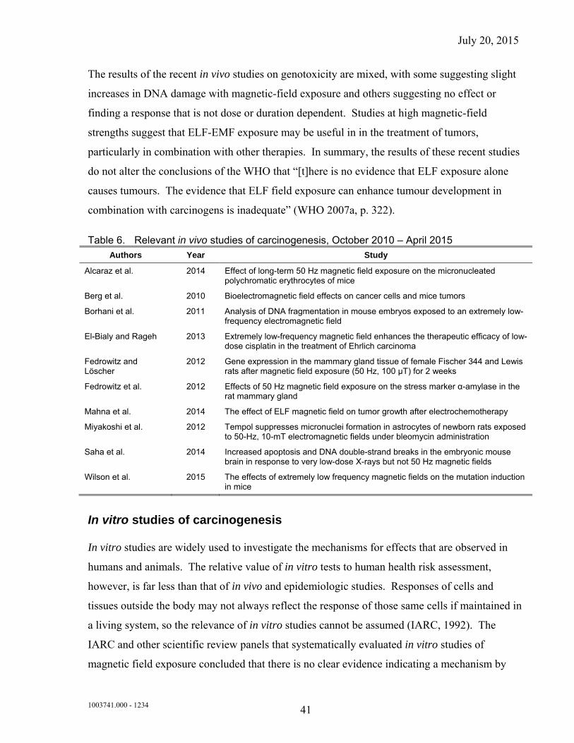
July 20, 2015
1003741.000 - 1234 41
The results of the recent in vivo studies on genotoxicity are mixed, with some suggesting slight
increases in DNA damage with magnetic-field exposure and others suggesting no effect or
finding a response that is not dose or duration dependent. Studies at high magnetic-field
strengths suggest that ELF-EMF exposure may be useful in in the treatment of tumors,
particularly in combination with other therapies. In summary, the results of these recent studies
do not alter the conclusions of the WHO that “[t]here is no evidence that ELF exposure alone
causes tumours. The evidence that ELF field exposure can enhance tumour development in
combination with carcinogens is inadequate” (WHO 2007a, p. 322).
Table 6. Relevant in vivo studies of carcinogenesis, October 2010 – April 2015
Authors Year Study
Alcaraz et al. 2014 Effect of long-term 50 Hz magnetic field exposure on the micronucleated polychromatic erythrocytes of mice
Berg et al. 2010 Bioelectromagnetic field effects on cancer cells and mice tumors
Borhani et al. 2011 Analysis of DNA fragmentation in mouse embryos exposed to an extremely low-frequency electromagnetic field
El-Bialy and Rageh 2013 Extremely low-frequency magnetic field enhances the therapeutic efficacy of low-dose cisplatin in the treatment of Ehrlich carcinoma
Fedrowitz and Löscher
2012 Gene expression in the mammary gland tissue of female Fischer 344 and Lewis rats after magnetic field exposure (50 Hz, 100 μT) for 2 weeks
Fedrowitz et al. 2012 Effects of 50 Hz magnetic field exposure on the stress marker α-amylase in the rat mammary gland
Mahna et al. 2014 The effect of ELF magnetic field on tumor growth after electrochemotherapy
Miyakoshi et al. 2012 Tempol suppresses micronuclei formation in astrocytes of newborn rats exposed to 50-Hz, 10-mT electromagnetic fields under bleomycin administration
Saha et al. 2014 Increased apoptosis and DNA double-strand breaks in the embryonic mouse brain in response to very low-dose X-rays but not 50 Hz magnetic fields
Wilson et al. 2015 The effects of extremely low frequency magnetic fields on the mutation induction in mice
In vitro studies of carcinogenesis
In vitro studies are widely used to investigate the mechanisms for effects that are observed in
humans and animals. The relative value of in vitro tests to human health risk assessment,
however, is far less than that of in vivo and epidemiologic studies. Responses of cells and
tissues outside the body may not always reflect the response of those same cells if maintained in
a living system, so the relevance of in vitro studies cannot be assumed (IARC, 1992). The
IARC and other scientific review panels that systematically evaluated in vitro studies of
magnetic field exposure concluded that there is no clear evidence indicating a mechanism by
July 20, 2015
1003741.000 - 1234 42
which ELF magnetic fields could adversely affect biological processes in cells (IARC, 2002;
ICNIRP, 2003; NRPB, 2004). The WHO panel reviewed the in vitro research published since
the time of those reviews and reached the same conclusion. The WHO noted that previous
studies have not indicated a genotoxic effect of ELF magnetic fields on mammalian cells;
however, a series of experiments reported DNA damage in human fibroblasts exposed
intermittently to 50-Hz magnetic fields (Ivancsits et al., 2002a, 2002b; Ivancsits et al., 2003a,
2003b). These findings were not replicated by other laboratories (Scarfi et al., 2005; Burdak-
Rothkamm et al., 2009), and the WHO recommended continued research in this area (WHO,
2007a).
Additional in vitro studies investigating the potential of ELF EMF to damage DNA have been
conducted in recent years (Buldak et al., 2012; Cho et al., 2014; Duan et al., 2015; Focke et al.,
2010; Jin et al., 2012, 2014, 2015; Kim et al., 2010, 2012; Luukkonen et al., 2011, 2014;
Mizuno et al., 2014; Yoon et al., 2014). Most of these studies exposed human- or mouse-
derived cells to 10,000 or 20,000 mG (50-60 Hz) magnetic fields, although some used fields as
low as 100 mG or as high as 70,000 mG. The exposure durations ranged from 10 minutes to
72 hours. Some of these studies found an association between magnetic-field exposure and
DNA damage, primarily DNA double strand breaks (Buldak et al., 2012; Duan et al., 2015;
Focke et al., 2010; Kim et al., 2010, 2012; Luukkonon et al., 2011, 2014; Yoon et al., 2014). In
some cases, this damage was only observed after repeated exposures (Kim et al., 2010) or with
intermittent, but not continuous, exposures (Focke et al., 2010) and at higher, rather than lower,
magnetic-field strengths (Duan et al., 2015; Yoon et al., 2014). Other studies, however, found
no effect of magnetic-field exposure on the observed frequency of DNA damage (Jin et al.,
2012, 2014, 2015; Mizuno et al., 2014). Recent studies were also contradictory with regard to
the ability of magnetic fields to alter the cellular response to known genotoxicants, with some
reporting increased DNA damage with co-exposure (Luukkonen et al., 2011, 2014; Yoon et al.,
2014) and others reporting no effect (Jin et al., 2012, 2014, 2015) or a reduction in DNA
damage with co-exposure (Buldak et al., 2012). Many of these in vitro studies also examined
the effects of magnetic-field exposure on other biological mechanisms, including oxidative
stress and apoptosis, the results of which are not reported here. Overall, the results of these
studies with regard to genotoxicity are conflicting and many suffer from significant design
flaws. None of these studies reported genotyping the cells lines used in order to verify their
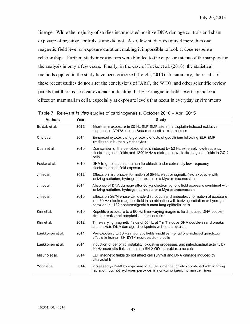
July 20, 2015
1003741.000 - 1234 43
lineage. While the majority of studies incorporated positive DNA damage controls and sham
exposure of negative controls, some did not. Also, few studies examined more than one
magnetic-field level or exposure duration, making it impossible to look at dose-response
relationships. Further, study investigators were blinded to the exposure status of the samples for
the analysis in only a few cases. Finally, in the case of Focke et al. (2010), the statistical
methods applied in the study have been criticized (Lerchl, 2010). In summary, the results of
these recent studies do not alter the conclusions of IARC, the WHO, and other scientific review
panels that there is no clear evidence indicating that ELF magnetic fields exert a genotoxic
effect on mammalian cells, especially at exposure levels that occur in everyday environments
Table 7. Relevant in vitro studies of carcinogenesis, October 2010 – April 2015 Authors Year Study
Buldak et al. 2012 Short-term exposure to 50 Hz ELF-EMF alters the cisplatin-induced oxidative response in AT478 murine Squamous cell carcinoma cells
Cho et al. 2014 Enhanced cytotoxic and genotoxic effects of gadolinium following ELF-EMF irradiation in human lymphocytes
Duan et al. 2015 Comparison of the genotoxic effects induced by 50 Hz extremely low-frequency electromagnetic fields and 1800 MHz radiofrequency electromagnetic fields in GC-2 cells
Focke et al. 2010 DNA fragmentation in human fibroblasts under extremely low frequency electromagnetic field exposure
Jin et al. 2012 Effects on micronuclei formation of 60-Hz electromagnetic field exposure with ionizing radiation, hydrogen peroxide, or c-Myc overexpression
Jin et al. 2014 Absence of DNA damage after 60-Hz electromagnetic field exposure combined with ionizing radiation, hydrogen peroxide, or c-Myc overexpression
Jin et al. 2015 Effects on G2/M phase cell cycle distribution and aneuploidy formation of exposure to a 60 Hz electromagnetic field in combination with ionizing radiation or hydrogen peroxide in L132 nontumorigenic human lung epithelial cells
Kim et al. 2010 Repetitive exposure to a 60-Hz time-varying magnetic field induced DNA double-strand breaks and apoptosis in human cells
Kim et al. 2012 Time-varying magnetic fields of 60 Hz at 7 mT induce DNA double-strand breaks and activate DNA damage checkpoints without apoptosis
Luukkonen et al. 2011 Pre-exposure to 50 Hz magnetic fields modifies menadione-induced genotoxic effects in human SH-SY5Y neuroblastoma cells
Luukkonen et al. 2014 Induction of genomic instability, oxidative processes, and mitochondrial activity by 50 Hz magnetic fields in human SH-SY5Y neuroblastoma cells
Mizuno et al. 2014 ELF magnetic fields do not affect cell survival and DNA damage induced by ultraviolet B
Yoon et al. 2014 Increased γ-H2AX by exposure to a 60-Hz magnetic fields combined with ionizing radiation, but not hydrogen peroxide, in non-tumorigenic human cell lines
July 20, 2015
1003741.000 - 1234 44
Reproductive and developmental effects
What was previously known about reproductive and developmental effects?
The epidemiologic literature on reproductive health outcomes and EMF provide no consistent
evidence for an association. Two studies published in 2002 received considerable attention
because of a reported association between peak magnetic-field exposure greater than
approximately 16 mG and miscarriage (Li et al., 2002; Lee et al., 2002). While at the time these
two studies relied on improved methodology for exposure assessment, using 24-hour personal
magnetic-field measurements (early studies on miscarriage were limited because they used
surrogate measures of exposure, including visual display terminal use, electric blanket use, or
wire code data), limitations in design of the two studies precluded drawing firm conclusions. It
has been proposed that the observed association may be the result of behavioral differences
between women with “healthy” pregnancies that went to term (less physically active) and
women who miscarried (more physically active) (Savitz, 2002). Furthermore, nearly half of the
miscarriages reported in the cohort by Li et al. had magnetic-field measurements taken after
miscarriage occurred, when changes in physical activity may have already occurred, and all
measurements in Lee et al. occurred post-miscarriage; thus there was high potential for exposure
misclassification in the study.
Later exposure assessment studies supported an association between physical activity and peak
exposure to EMF (Savitz et al., 2006; Mezei et al., 2006; Lewis et al., 2015).
The scientific panels that have considered these two studies concluded that the possibility of
these biases preclude making any conclusions about the effect of magnetic fields on miscarriage
(NRPB, 2004; FPTRPC, 2005; WHO, 2007a). The WHO concluded, “There is some evidence
for increased risk of miscarriage associated with measured maternal magnetic field exposure,
but this evidence is inadequate” (WHO 2007a, p. 254). The WHO stated that, given the
potentially high public health impact of such an association, further epidemiologic research is
recommended.
Epidemiologic research published following the WHO report and up to 2010, as summarized in
the previous report (Exponent, 2011) did not provide sufficient evidence to alter the conclusion
July 20, 2015
1003741.000 - 1234 45
that the evidence for reproductive or developmental effects is inadequate. Similarly, studies of
animals in vivo did not provide evidence to change the conclusions expressed by the WHO.
Recent studies (October 2010 – April 2015)
There is a great variability in the quality in the epidemiologic research that has continued to
focus on reproductive and developmental outcomes. Using data from an earlier cohort study of
pregnant women and their offspring (Li et al., 2002), researchers studied the association
between EMF exposure experienced in the mother’s womb during pregnancy and various health
outcomes, such as asthma (Li et al., 2011) and obesity(Li et al., 2012), during childhood. The
first of these studies (Li et al., 2011) analyzed data on children from 626 pregnancies and
reported that that asthmatic children were more likely to have mothers with median magnetic-
field exposures >2 mG during pregnancy, compared to children without asthma. The reported
association was statistically significant and consistent with an exposure-response pattern. The
paper triggered criticism in the scientific literature and it was suggested that unmeasured
confounders may be responsible for the observed association (e.g., SES and other indoor and
ambient environmental exposures), as many known risk factors for asthma were not controlled
for in the analysis and no mechanism is known to explain the association between EMF and
asthma (Villeneuve, 2012; Brain et al., 2012). This study, being the first of this kind, requires
independent confirmation in studies with more detailed information on and control for known
risk factors for childhood asthma.
Li et al. (2012) also published results on childhood obesity and exposure during pregnancy of
the mothers using the same data set. The mothers’ EMF exposure during pregnancy was related
to the weight of their children. The children of mothers with average magnetic-field exposures
>1.5 mG were significantly more likely to be over the 97.5 percentile of age-specific weight
than children of mothers with exposures ≤ 1.5 mG. A statistical trend between weight and EMF
exposure was also reported. Methodological concerns related to the underlying cohort study of
pregnant women pertain to all the analyses using the resulting data set. These concerns include
the potential for selection bias, a low compliance rate, and the apparent selection of exposure
categories after inspection of the data (NRPB, 2002). Because analysis related to asthma and
July 20, 2015
1003741.000 - 1234 46
obesity was not part of the original study, and these analyses were conducted on a post-hoc
basis, it raises potential concerns with multiple comparisons and chance findings.
Canadian researchers investigated the relationship between residential proximity to high-voltage
transmission lines and adverse birth outcomes and stillbirth (Auger et al., 2011; Auger et al.,
2012). In the first study, the authors identified over 700,000 singleton live births occurring
between 1990 and 2004 through provincial birth files. The relative occurrence of adverse birth
outcomes, including preterm birth, low birth weight, and being small for gestational age, was
investigated in relation to residential distance to transmission lines. The distance of the birth
residence to transmission lines was categorized using cut-points at 50, 75, 100, 150, and 400
meters. No voltage information was reported for the transmission lines and no EMF levels were
estimated in the study. No associations were observed with residential distance of the mothers
for any of the investigated outcomes.
The second study included more than half a million live births along with 2,033 stillbirths in
Québec (Auger et al., 2012) occurring between 1998 and 2007. The authors reported that
residential proximity to transmission lines was not associated with the likelihood of stillbirth
overall and stratified by early preterm (<28 weeks) and late preterm (28-36 weeks) births. No
exposure-response pattern was observed overall and by term of stillbirth, although an
association was reported for residences within 25 meters of power transmission lines in a sub-
analysis. Considering their results, the authors concluded that “residential proximity is unlikely
to be associated with stillbirth.” The use of distance as a proxy for ELF EMF exposure without
information on the level of EMF exposure from the lines is a main limitation of this study, and
potential bias may also result due to in- and out-migration from the study area.
The risk of birth defects in relation to residential exposure magnetic fields from high-voltage
power lines was investigated in a population-based case-control study in Northern Italy
(Malagoli et al., 2012). The authors identified 228 cases of congenital malformations diagnosed
in live births, stillbirths, and induced abortions during the period between 1998 and 2006. The
authors reported no associations between birth defects and estimated exposure, and concluded
that their results do not support the hypothesis that in utero exposure of the fetus to EMF is
related to birth defects. We note, however, that the study had low statistical power to detect an
July 20, 2015
1003741.000 - 1234 47
association because of small number of cases and controls in the study who lived close to
transmission lines.
Mahram and Ghazavi (2013) studied the fetal growth and development of children in Iran born
to 222 women who lived in close proximity to high-voltage lines during pregnancy (defined as
exposed) and 158 women with no exposure during pregnancy. The authors reported no
significant differences between the exposed and unexposed groups for any of the investigated
outcomes, including pregnancy duration, birth weight, birth length, head circumference, and
congenital malformation. The study was, however, severely limited in its exposure assessment
and in its control of potential confounding variables. Another study from Iran was a hospital-
based case-control study (Shamsi Mahmoudabadi et al., 2013) that included 58 women with
spontaneous abortion and 58 pregnant women. The measured magnetic-field levels were
reported to be higher among the cases than among controls. The study, however, provides very
limited information on major study characteristics, such as subject recruitment, exposure
assessment, exposure metric used, and assessment of potential confounders. Thus no firm
conclusions can be drawn from the study and it contributes little weight, if any, to an overall
assessment of EMF effects on adverse pregnancy outcomes.
Wang et al. (2013) identified 413 pregnant women in China at 8 weeks of gestation between
2010 and 2012. The researchers reported no statistically significant associations between
miscarriage and still birth and magnetic-field levels measured at the front door of the
participants’ homes. The authors reported an association, however, with maximum fields
measured in the alleys in front of the homes. Because magnetic-field levels measured at the
front door or in the alley in front of the residences are very poor predictors of personal exposure,
the study provides a very limited contribution, if any, to our understanding of associations of
EMF and birth outcomes.
Residential proximity to power lines during pregnancy and birth outcomes were investigated in
Northwest England between 2004 and 2008 (de Vocht et al., 2014a). Hospital records for over
140,000 births were scrutinized and distance to the nearest power lines for the homes during
pregnancy were determined using geographical information systems by the researchers. The
authors reported moderately lower birth weight within 50 meters of power lines, but observed
July 20, 2015
1003741.000 - 1234 48
no statistically significant increase in risk of any adverse clinical birth outcomes (such as
preterm birth, small for gestational age, or low birth weight). The limitations of the study
include its reliance on distance for exposure assessment and the potential for confounding by
SES, as also discussed by the authors. A follow-up investigation to the study indicated that
minimizing residual confounding due to missing data for most potentially confounding variables
by using multiple imputations and the use of propensity scores reduced the estimated effect size
(de Vocht and Lee, 2014).
Bellieni et al. (2012) estimated the effect of ELF EMF present in incubators on the blood
melatonin levels on newborns. The authors reported a statistically significant increase of blood
melatonin levels among 28 newborns 48 hours after being taken out of the incubators with
assumed elevated ELF EMF exposure, but not among 28 control newborns who were not in
incubators. Neither the before nor the after values, however, were statistically different from
each other in the two groups (incubator vs. control), thus the clinical significance of the
findings, if any, is unclear.
The potential influence of magnetic-field exposure on male fertility measures, such as semen
quality and abnormalities, was evaluated in a population-based case-control study of healthy
semen donors in China (Li et al., 2010). A statistically significant association was reported
between a measure of peak magnetic-field levels and poor sperm quality. Personal magnetic-
field exposures were measured during a 24-hour period for the included subjects, which could
be considered a strength of the study. The authors, however, were not able to control for
potential exposure to chemicals and other factors that are known to have a major influence on
sperm quality.
Overall, recent epidemiologic research on reproductive and developmental effects has not
changed the conclusion that the evidence is inadequate to support an association for any of the
investigated adverse reproductive or developmental outcomes. The most recent review by
SCENIHR (2015) concluded that “recent results do not show an effect of ELF MF [magnetic
field] exposure on reproductive function in humans.”
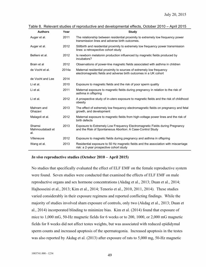
July 20, 2015
1003741.000 - 1234 49
Table 8. Relevant studies of reproductive and developmental effects, October 2010 – April 2015
Authors Year Study
Auger et al. 2011 The relationship between residential proximity to extremely low frequency power transmission lines and adverse birth outcomes.
Auger et al. 2012 Stillbirth and residential proximity to extremely low frequency power transmission lines: a retrospective cohort study
Bellieni et al. 2012 Is newborn melatonin production influenced by magnetic fields produced by incubators?
Brain et al 2012 Observations of power-line magnetic fields associated with asthma in children
de Vocht et al. 2014a Maternal residential proximity to sources of extremely low frequency electromagnetic fields and adverse birth outcomes in a UK cohort
de Vocht and Lee 2014
Li et al. 2010 Exposure to magnetic fields and the risk of poor sperm quality
Li et al. 2011 Maternal exposure to magnetic fields during pregnancy in relation to the risk of asthma in offspring
Li et al. 2012 A prospective study of in-utero exposure to magnetic fields and the risk of childhood obesity
Mahram and Ghazavi
2013 The effect of extremely low frequency electromagnetic fields on pregnancy and fetal growth, and development
Malagoli et al. 2012 Maternal exposure to magnetic fields from high-voltage power lines and the risk of birth defects
Shamsi Mahmoudabadi et al.
2013 Exposure to Extremely Low Frequency Electromagnetic Fields during Pregnancy and the Risk of Spontaneous Abortion: A Case-Control Study
Villeneuve 2012 Exposure to magnetic fields during pregnancy and asthma in offspring
Wang et al. 2013 Residential exposure to 50 Hz magnetic fields and the association with miscarriage risk: a 2-year prospective cohort study
In vivo reproductive studies (October 2010 – April 2015)
No studies that specifically evaluated the effect of ELF EMF on the female reproductive system
were found. Seven studies were conducted that examined the effects of ELF EMF on male
reproductive organs and sex hormone concentrations (Akdag et al., 2013; Duan et al., 2014;
Hajhosseini et al., 2013; Kim et al., 2014; Tenorio et al., 2010, 2011, 2014). These studies
varied considerably in their exposure regimens and reported conflicting findings. While the
majority of studies involved sham exposure of controls, only two (Akdag et al., 2013; Duan et
al., 2014) incorporated blinding to minimize bias. Kim et al. (2014) found that exposure of
mice to 1,000 mG, 50-Hz magnetic fields for 6 weeks or to 200, 1000, or 2,000 mG magnetic
fields for 8 weeks did not affect testes weights, but was associated with reduced epididymal
sperm counts and increased apoptosis of the spermatogonia. Increased apoptosis in the testes
was also reported by Akdag et al. (2013) after exposure of rats to 5,000 mg, 50-Hz magnetic
July 20, 2015
1003741.000 - 1234 50
fields (but not 1,000 mG) for 2 hours per day for 10 months, but the specific cell type affected
was not identified. Epididymal sperm counts, however, were unaffected. Hajhosseini et al.
(2013) reported increased apoptosis after 6 weeks exposure to 80,000 mG, 50-Hz magnetic
fields; in this case, however, the apoptotic cells were identified as Sertoli cells, which help to
support development of sperm in the seminiferous tubules. These investigators also found that
exposure was associated with reduced serum testosterone levels. In contrast, Duan et al. (2014)
found no effects of exposure to 5,000 mG magnetic fields for 4 hours per day for 4 or 8 weeks
on multiple parameters related to spermatogenesis in the rat, including testes and epididymal
weights, epididymal sperm count, serum testosterone, testicular histology, and the various stages
of spermatogenesis. They also saw no effect of exposure on the rate of apoptosis in the testes.
Two studies by the same group of investigators (Tenorio et al., 2010, 2011) examined the
effects of ELF EMF exposure during late gestation and the early postnatal period on testicular
development in rats. Exposures to 10,000 mG, 60-Hz magnetic fields for three 30-minute
sessions per day began on GD 13 and continued until postnatal day (PND) 21 or PND 90.
Plasma testosterone levels were unaffected by exposure. Although the investigators reported
alterations in a number of testicular morphometric measures with exposure on PND 21 (Tenorio
et al., 2010), these measures seemed to recover somewhat by PND 90 (Tenorio et al., 2011).
Testicular degeneration was highly variable but evident in some rats from both exposure
groups. Because littermates are known to be more similar than offspring derived from separate
litters, the litter should be used as the statistical unit for analysis, but this was not done in these
studies. Finally, Tenorio et al. (2014) conducted another study of adult rats involving similar
exposures for 15, 30, or 60 days after thermal heating of the testes. In this case, the authors
reported that magnetic-field exposure interfered with recovery from the reversible testicular
damageproduced by heating. Overall, many of the studies of ELF EMF exposure in male rats
suggest possible reproductive system alterations. The results are highly variable, however, and
conflicting across the body of studies. Further, it is not evident if any of the reported changes
translate to decrements in reproductive capacity or health.
Three studies examined the effects of ELF EMF exposure during prenatal development
(Lahijani et al., 2011; Roshanger et al., 2014; Vallejo and Hidalgo, 2011). Lahijani et al. (2011)
reported that a 24-hour exposure of chicken embryos to a 73,200 mG, 50-Hz magnetic field
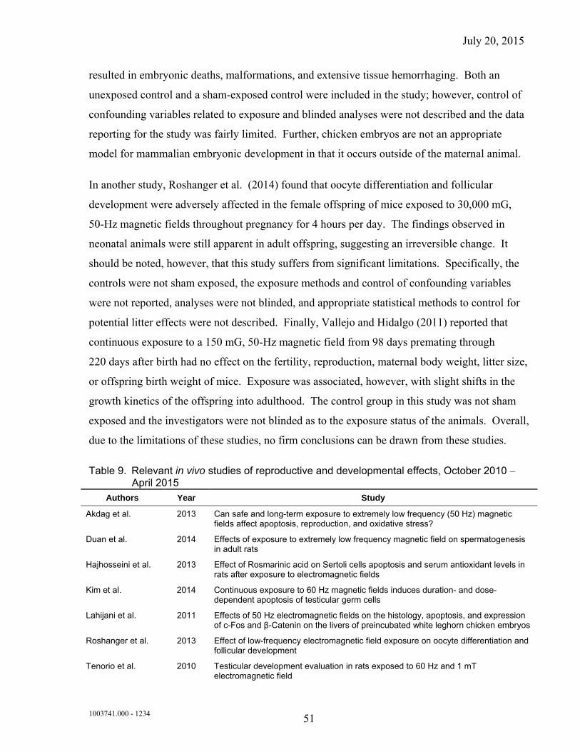
July 20, 2015
1003741.000 - 1234 51
resulted in embryonic deaths, malformations, and extensive tissue hemorrhaging. Both an
unexposed control and a sham-exposed control were included in the study; however, control of
confounding variables related to exposure and blinded analyses were not described and the data
reporting for the study was fairly limited. Further, chicken embryos are not an appropriate
model for mammalian embryonic development in that it occurs outside of the maternal animal.
In another study, Roshanger et al. (2014) found that oocyte differentiation and follicular
development were adversely affected in the female offspring of mice exposed to 30,000 mG,
50-Hz magnetic fields throughout pregnancy for 4 hours per day. The findings observed in
neonatal animals were still apparent in adult offspring, suggesting an irreversible change. It
should be noted, however, that this study suffers from significant limitations. Specifically, the
controls were not sham exposed, the exposure methods and control of confounding variables
were not reported, analyses were not blinded, and appropriate statistical methods to control for
potential litter effects were not described. Finally, Vallejo and Hidalgo (2011) reported that
continuous exposure to a 150 mG, 50-Hz magnetic field from 98 days premating through
220 days after birth had no effect on the fertility, reproduction, maternal body weight, litter size,
or offspring birth weight of mice. Exposure was associated, however, with slight shifts in the
growth kinetics of the offspring into adulthood. The control group in this study was not sham
exposed and the investigators were not blinded as to the exposure status of the animals. Overall,
due to the limitations of these studies, no firm conclusions can be drawn from these studies.
Table 9. Relevant in vivo studies of reproductive and developmental effects, October 2010 ‒ April 2015
Authors Year Study
Akdag et al. 2013 Can safe and long-term exposure to extremely low frequency (50 Hz) magnetic fields affect apoptosis, reproduction, and oxidative stress?
Duan et al. 2014 Effects of exposure to extremely low frequency magnetic field on spermatogenesis in adult rats
Hajhosseini et al. 2013 Effect of Rosmarinic acid on Sertoli cells apoptosis and serum antioxidant levels in rats after exposure to electromagnetic fields
Kim et al. 2014 Continuous exposure to 60 Hz magnetic fields induces duration- and dose-dependent apoptosis of testicular germ cells
Lahijani et al. 2011 Effects of 50 Hz electromagnetic fields on the histology, apoptosis, and expression of c-Fos and β-Catenin on the livers of preincubated white leghorn chicken embryos
Roshanger et al. 2013 Effect of low-frequency electromagnetic field exposure on oocyte differentiation and follicular development
Tenorio et al. 2010 Testicular development evaluation in rats exposed to 60 Hz and 1 mT electromagnetic field
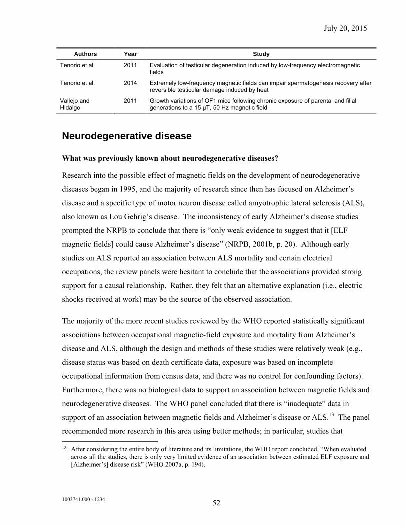
July 20, 2015
1003741.000 - 1234 52
Authors Year Study
Tenorio et al. 2011 Evaluation of testicular degeneration induced by low-frequency electromagnetic fields
Tenorio et al. 2014 Extremely low-frequency magnetic fields can impair spermatogenesis recovery after reversible testicular damage induced by heat
Vallejo and Hidalgo
2011 Growth variations of OF1 mice following chronic exposure of parental and filial generations to a 15 μT, 50 Hz magnetic field
Neurodegenerative disease
What was previously known about neurodegenerative diseases?
Research into the possible effect of magnetic fields on the development of neurodegenerative
diseases began in 1995, and the majority of research since then has focused on Alzheimer’s
disease and a specific type of motor neuron disease called amyotrophic lateral sclerosis (ALS),
also known as Lou Gehrig’s disease. The inconsistency of early Alzheimer’s disease studies
prompted the NRPB to conclude that there is “only weak evidence to suggest that it [ELF
magnetic fields] could cause Alzheimer’s disease” (NRPB, 2001b, p. 20). Although early
studies on ALS reported an association between ALS mortality and certain electrical
occupations, the review panels were hesitant to conclude that the associations provided strong
support for a causal relationship. Rather, they felt that an alternative explanation (i.e., electric
shocks received at work) may be the source of the observed association.
The majority of the more recent studies reviewed by the WHO reported statistically significant
associations between occupational magnetic-field exposure and mortality from Alzheimer’s
disease and ALS, although the design and methods of these studies were relatively weak (e.g.,
disease status was based on death certificate data, exposure was based on incomplete
occupational information from census data, and there was no control for confounding factors).
Furthermore, there was no biological data to support an association between magnetic fields and
neurodegenerative diseases. The WHO panel concluded that there is “inadequate” data in
support of an association between magnetic fields and Alzheimer’s disease or ALS.13 The panel
recommended more research in this area using better methods; in particular, studies that 13 After considering the entire body of literature and its limitations, the WHO report concluded, “When evaluated
across all the studies, there is only very limited evidence of an association between estimated ELF exposure and [Alzheimer’s] disease risk” (WHO 2007a, p. 194).
July 20, 2015
1003741.000 - 1234 53
enrolled incident Alzheimer’s disease cases (rather than ascertaining cases from death
certificates) and studies that estimated electrical shock history in ALS cases were recommended.
Some of the studies published following the WHO review and until 2010, as summarized in the
previous report (Exponent, 2011), had indicated an association for Alzheimer disease and ALS,
but the study results overall indicated no consistent pattern. The main limitations of these
studies include the reliance on death certificates for case identification and diagnosis; the
difficulty of identifying a relevant exposure window given the long and nebulous course of this
disease; the difficulty of estimating magnetic-field exposure prior to the appearance of the
disease; the under-reporting of Alzheimer’s disease on death certificates; crude exposure
evaluations that are often based on the recollection of occupational histories by friends and
family given the cognitive impairment of the study participants; and the lack of consideration of
both residential and occupational exposures or confounding variables. Thus, overall,
epidemiologic studies published until 2010 did not alter the conclusion that there is inadequate
data on Alzheimer’s disease or ALS. There was no body of in vivo research to suggest an effect
and two studies reported no effect of magnetic fields on ALS progression (Seyhan and
Canseven, 2006; Poulletier de Gannes et al., 2009). These conclusions were consistent with the
then most recent review by the SCENIHR (SCENIHR, 2009).
Recent studies (October 2010 – April 2015)
Occupational exposure estimates remained the focus of the majority of epidemiologic research
related to ELF EMF. As part of the previously described occupational cohort study of British
electricity supply workers, mortality due to Alzheimer disease, motor neuron disease, and
Parkinson’s disease were also investigated (Sorahan and Mohammed, 2014). The authors
included over 73,000 workers in their analyses, for which the follow-up period included almost
40 years between 1973 and 2010. Exposure to power-frequency magnetic fields were calculated
for all workers throughout their working years. Relative risks for the neurodegenerative
diseases studied were calculated using Poisson regression. Various exposure categories were
considered in the analyses based on cumulative lifetime exposures, and also by considering only
the recent or more distant time periods of exposure. The authors reported no statistically
July 20, 2015
1003741.000 - 1234 54
significant trends in any of their analyses that would indicate an association between estimated
magnetic-field exposure and neurodegenerative disease development.
Mortality due to motor neuron disease in relation to occupational exposure to magnetic fields
was evaluated using data from the National Longitudinal Mortality Study in the United States
(Parlett et al., 2011). The study included more than 300,000 participants, who provided
information on their job at the time of the interview. An EMF job-exposure matrix was used to
estimate magnetic-field exposure levels in the participants’ jobs. Mortality within the cohort
was observed using the National Death Index and cause of death was determined based on death
certificates. Over the 2.7 million years of follow-up time, 40 deaths due to motor neuron
diseases were observed in the cohort. After adjustment for age, sex, and education, the authors
reported no increase in the risk of motor neuron disease in relation to magnetic-field exposure.
From the previously described Netherlands Cohort Study that included about 120,000 male and
female study subjects and a follow-up time of about 17 years, analyses on occupational
exposures and mortality due to Parkinson’s disease have also been reported (Brouwer et al.,
2015). In total, 402 deaths among males and 207 deaths among females, due to Parkinson’s
disease, were observed. The study reported an association between Parkinson’s disease
mortality and work exposure to EMF categorized as “ever exposed to high EMF,” in one
analysis. No association, however, was observed for EMF exposure or electric shocks when
cumulative exposure was evaluated. Similarly, no statistical trends were observed between any
of these two exposures. The authors concluded that their study “does not support the hypothesis
that the investigated occupational exposures increase PD [Parkinson’s disease] mortality,
although we cannot exclude that small risks may exist.”
Davanipour et al. (2014) conducted a study of severe cognitive dysfunction and occupational
ELF magnetic-field exposure among “3,050 Mexican Americans, aged 65+, enrolled in Phase I
of the Hispanic Established Population for the Epidemiologic Study of the Elderly (H-EPESE)
study.” In-home personal interviews were used to obtain information on occupational history,
and socio-demographic variables. Occupational exposure to magnetic fields was classified as
low, medium, and high. Cognitive function was evaluated with the use of the mini-mental state
exam and cognitive dysfunction was defined as an exam score below 10. Although the authors
July 20, 2015
1003741.000 - 1234 55
describe their study as a population-based case-control study, the study is a cross-sectional
survey. The authors report a statistically significant association between estimated occupational
magnetic-field exposure and severe cognitive dysfunction. The reported association is,
however, difficult to interpret due to the number of severe limitations of the study; including the
cross-sectional design, the lack of clear clinical diagnosis for case-definition, and the
rudimentary assessment of exposure to occupational EMF.
A hospital-based case-control study was conducted by Dutch researchers to evaluate the
association between Parkinson’s disease and occupational exposure to electric shocks and ELF
magnetic fields (van der Mark et al., 2014). The authors included 444 cases of Parkinson’s
disease and 876 matched controls in their study. Occupational history was determined based on
telephone interviews. JEMs were used to categorize jobs for exposure to both electric shocks
and magnetic fields. The authors reported no risk increases with any of the estimated exposure
metrics for electric shocks or magnetic fields and concluded that their results suggest no
association with Parkinson’s disease.
Other studies also have addressed the issue of whether electric shocks may explain the observed
association reported for neurodegenerative diseases. A few earlier studies, although limited in
size and methodology, provided no overall support for an association (Das et al., 2012; Grell et
al., 2012). More recent and larger studies have also failed to observe an association with
electric shocks. Researchers in the United States conducted a mortality case-control study
analyzing death certificates between 1991 and 1999 (Vergara et al., 2015). In total 5,886 ALS
deaths were identified during the study period and their exposure experience was compared to
10-times as many matched control deaths. Exposure to electric shocks and ELF magnetic fields
was classified based on job titles reported on the death certificates and using corresponding job-
exposure matrices. Similar to previous results, a statistically significant association was
reported for “electrical occupations,” but no consistent associations were observed for either
magnetic fields or electric shocks. The main limitation of the study is the use of death
certificates for case ascertainment and exposure assessment potentially resulting in disease and
exposure misclassifications.
July 20, 2015
1003741.000 - 1234 56
Data from the Swiss National Cohort between 2000 and 2008 was used to assess the relationship
between occupational electric shock and magnetic-field exposures and ALS mortality. A total
of 2.2 million workers were included in the cohort. Job-exposure matrices for magnetic fields
and shocks were used to classify to workers’ exposure as high, medium, or low for both
exposures based on their occupations reported during the 1990 and 2000 censuses. The authors
reported a statistically significant association of ALS mortality with estimated medium or high
occupational magnetic-field exposure based at both censuses, but not with estimates of electric
shock exposure. The main limitations of the study include the use of mortality data, which may
result in disease misclassification, and the use of census data for exposure assessment, which
may result in exposure misclassification.
Residential exposure to EMF due to residential proximity to high-voltage transmission lines and
neurodegenerative diseases were also examined in epidemiologic studies in Brazil, Switzerland,
and the Netherlands. Researchers in Brazil (Marcilio et al., 2011) identified 367 deaths due to
ALS between 2001 and 2005 in the city of Sao Paulo and compared their residential proximity
to transmission lines to that of 4,706 controls. The authors reported no associations between
ALS and either distance from transmission lines or calculated magnetic-field levels.
A Swiss population-based case-control study (Frei et al., 2013) identified cases of
neurodegenerative diseases between 1994 and 2010 from hospital discharge records in the
whole of Denmark. Six controls from the general population on Denmark were selected for
each case. The authors determined the distance from the nearest power line to the residential
address for all newly-reported cases and matched controls using geographical information
systems. Overall, none of the investigated diseases, including Alzheimer disease and other
types of dementia, ALS, Parkinson’s disease, or multiple sclerosis was related to residential
proximity to power lines. The inclusion of newly-diagnosed cases from hospital discharge
records represents a significant methodological improvement over mortality studies. The study,
however, was limited by the methods used for the exposure assessment that, as in previous
studies, relied on distance to power lines.
A large population-based case-control study of ALS and residential proximity to high-voltage
power lines was also conducted in the Netherlands (Seelen et al., 2014). The authors identified
July 20, 2015
1003741.000 - 1234 57
1,139 ALS cases diagnosed between 2006 and 2013. A total of 2,864 controls from general
practitioners’ rosters were frequency matched to cases from the same time period. The
Municipal Personal Records Database was used to determine lifetime residential history for all
cases and controls. Addresses were geocoded and the shortest distance to a high-voltage power
lines (50 ‒ 380 kV) was determined for each address. No statistically significant association
was reported between residential proximity to power lines of any voltage, and ALS. The
authors combined their results with the two previously mentioned studies (Marcilio et al., 2011;
Frei et al., 2013) and reported no overall associations for living within 200 meters of a high
voltage power line (OR 0.9; 95% CI 0.7-1.1). As in other power-line studies, the main
limitation of the Dutch study is the use of distance to power lines as an estimate for magnetic-
field exposure. By reconstructing lifetime residential history, however, the authors achieved a
methodological improvement compared to previous studies.
Two meta-analyses were published that assessed the relationship between occupational
exposure to ELF EMF and ALS (Zhou et al., 2012) or neurodegenerative diseases (Vergara et
al., 2013). Both of these reported weak to no associations with the investigated outcomes.
Vergara et al. (2013) reported evidence for publication bias, and both analyses reported
consistent variation in results by study characteristics. Thus, the authors of both analyses
concluded that potential within-study biases, evidence of publication bias, and uncertainties in
the various exposure assessments greatly limit the ability to infer an association, if any, between
occupational exposure to magnetic fields and neurodegenerative disease. In conclusion, the
results presented by the two meta-analyses provided no convincing evidence of a relationship
between ELF EMF and neurodegenerative disease.
Overall, results from recent epidemiologic literature does not provide sufficient new insight or
evidence that would alter the conclusion that the evidence is currently inadequate to support a
causal link between exposure to ELF magnetic fields and any of the neurodegenerative diseases.
Most of the recent studies provided no support for a potential association. The potential
etiologic role of electric shocks in the development of neurodegenerative diseases has also been
investigated in several recent studies (Das et al., 2012; Grell et al., 2012; Vergara et al., 2013;
van der Mark et al., 2014), but none of these studies reported associations that would support
such a hypothesis. The main limitations of most of the recent studies are similar to those in
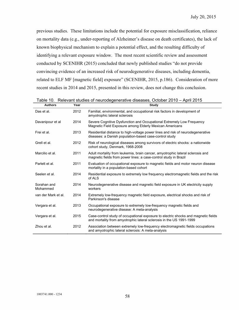
July 20, 2015
1003741.000 - 1234 58
previous studies. These limitations include the potential for exposure misclassification, reliance
on mortality data (e.g., under-reporting of Alzheimer’s disease on death certificates), the lack of
known biophysical mechanism to explain a potential effect, and the resulting difficulty of
identifying a relevant exposure window. The most recent scientific review and assessment
conducted by SCENIHR (2015) concluded that newly published studies “do not provide
convincing evidence of an increased risk of neurodegenerative diseases, including dementia,
related to ELF MF [magnetic field] exposure” (SCENIHR, 2015, p.186). Consideration of more
recent studies in 2014 and 2015, presented in this review, does not change this conclusion.
Table 10. Relevant studies of neurodegenerative diseases, October 2010 – April 2015 Authors Year Study
Das et al. 2012 Familial, environmental, and occupational risk factors in development of amyotrophic lateral sclerosis
Davanipour et al 2014 Severe Cognitive Dysfunction and Occupational Extremely Low Frequency Magnetic Field Exposure among Elderly Mexican Americans
Frei et al. 2013 Residential distance to high-voltage power lines and risk of neurodegenerative diseases: a Danish population-based case-control study
Grell et al. 2012 Risk of neurological diseases among survivors of electric shocks: a nationwide cohort study, Denmark, 1968-2008
Marcilio et al. 2011 Adult mortality from leukemia, brain cancer, amyotrophic lateral sclerosis and magnetic fields from power lines: a case-control study in Brazil
Parlett et al. 2011 Evaluation of occupational exposure to magnetic fields and motor neuron disease mortality in a population-based cohort
Seelen et al. 2014 Residential exposure to extremely low frequency electromagnetic fields and the risk of ALS
Sorahan and Mohammed
2014 Neurodegenerative disease and magnetic field exposure in UK electricity supply workers
van der Mark et al. 2014 Extremely low-frequency magnetic field exposure, electrical shocks and risk of Parkinson's disease
Vergara et al. 2013 Occupational exposure to extremely low-frequency magnetic fields and neurodegenerative disease: A meta-analysis
Vergara et al. 2015 Case-control study of occupational exposure to electric shocks and magnetic fields and mortality from amyotrophic lateral sclerosis in the US 1991-1999
Zhou et al. 2012 Association between extremely low-frequency electromagnetic fields occupations and amyotrophic lateral sclerosis: A meta-analysis
July 20, 2015
1003741.000 - 1234 59
3 Other Areas of Research
Pacemakers and implanted cardiac devices
The sensing system of pacemakers and other implanted cardiac devices (ICD) is designed to be
responsive to the heart’s electrical signal. For this reason, other electrical signals potentially can
interfere with the normal functioning of pacemakers and ICDs, a phenomenon called
electromagnetic interference (EMI). Most sources of EMF are too weak to affect a pacemaker
or ICD; however, EMF from certain sources (e.g., some appliances and industrial equipment),
may cause interference. This section considers potential EMI with implanted cardiac devices
such as pacemakers and defibrillators.
In the presence of electromagnetic fields, pacemakers and ICDs can respond in different ways,
defined as modes. The probability of interference occurring and the mode of the response
depend on the strength and nature of the interference signal, the patient’s orientation in the
electromagnetic field, the exact location of the device, and the variable parameters of the device
that are specific to a patient.
There are a number of experimental studies dating back to the 1990s that were conducted to
assess whether interference may occur when currents are induced in the patient’s body by ELF
electric or magnetic fields (e.g., Toivonen et al., 1991; Astridge et al., 1993; Scholten and Silny,
2001). In general, pacing abnormalities in these tests occurred at magnetic-field levels that are
much higher than the levels a person would encounter on a daily basis. Electric fields did
produce interference at levels that can be produced by certain electrical sources, but most
pacemakers were not affected by high levels of electric fields (up to 20 kV/m) and did not
exhibit any pacing abnormalities. Unipolar (single lead) pacemakers tended to be more
sensitive to electric fields compared to bipolar (two lead) devices, which are designed
specifically to reduce the effects of EMI.
A recent study by Joosten et al. (2009) confirmed earlier work by Scholten and Silny (2001).
Both studies found that the performance of a pacemaker in the presence of external ELF electric
fields varied considerably based on anatomical and physiological conditions. The 15 study
July 20, 2015
1003741.000 - 1234 60
subjects in Joosten et al., (2009) experienced a variance of up to 200% when the interference
voltage was applied at the input of their cardiac pacemakers. This variance was due to personal
factors such as state of respiration, systole and diastole of the heart, filling of the stomach, and
muscle activity. The authors’ analyses further suggested that for a 50-Hz electric field to affect
the function of the most sensitive unipolar pacemaker, the field levels would have to be greater
than 4.3 kV/m and 6.2 kV/m during inhalation and exhalation, respectively. Unipolar
pacemakers are less and less common today; the study authors found that in Germany, only 6%
of the pacemakers in use have a unipolar sensing system.
In the absence of specific recommendations from medical device manufacturers, two
organizations, the American Conference of Governmental Industrial Hygienists (ACGIH) and
the Electric Power Research Institute (EPRI), have suggested exposure levels to prevent
pacemaker EMI. Both organizations suggest that exposures be kept below 1.5-2 kV/m for
electric fields and the ACGIH recommends an exposure level not to exceed 1 Gauss (G) for
magnetic fields (ACGIH 2001, EPRI 2004). These recommendations are general in nature and
do not address that classes of pacemakers from some manufacturers are quite immune to
interference even at levels much greater than these recommended guidelines. Both the ACGIH
and EPRI recommend that patients consult their physicians and the respective pacemaker
manufacturers before following these organizations’ guidelines.
The Food and Drug Administration’s Center for Devices and Radiological Health has also
issued guidelines for both the development of pacemakers and the design of new electrical
devices to minimize susceptibility to electrical interference from any source. Pacemakers are
designed to filter out electrical stimuli from sources other than the heart (e.g., the muscles of the
chest, currents encountered from touching household appliances, or currents induced by external
electric or magnetic fields). Used in both temporary and permanent pacemakers, these electrical
filters increase the pacemaker’s ability to distinguish extraneous signals from legitimate cardiac
signals (Toivonen et al., 1991). Furthermore, most circuitry of modern pacemakers is
encapsulated by titanium metal, which insulates the device by shielding the pacemaker’s pulse
generator from electric fields. Some pacemakers also may be programmed to pace the heart
automatically if interference from electric or magnetic fields is detected (fixed pacing mode).
July 20, 2015
1003741.000 - 1234 61
This supports cardiac function and allows the subject to feel the pacing and move away from the
source.
Many pacemakers currently in use would not be susceptible to electric fields typically
encountered in the daily environment electric fields due to recent design improvements. There
remains a very small possibility that some pacemakers, particularly those of older design and
with single-lead electrodes, may sense potentials induced on the electrodes and leads of the
pacemaker and provide unnecessary stimulation to the heart.
In the past 5 years, scientific research to evaluate potential interference with pacemakers and
other implanted cardiac devices has continued. Much of the published literature, however,
focused on possible interference from dental, medical, surgical, diagnostic, and therapeutic
equipment (e.g., Zaphiratos et al., 2013; Stoopler et al., 2011; Maheshwari et al., 2015; Magnani
et al., 2012; Magnani et al., 2014; Yoshida et al., 2014) or personal electronic devices (e.g.,
Misiri et al., 2012; Tiikkaja et al., 2012a; Kozik et al., 2014). While some of these reports
indicate the possibility of interference in certain scenarios with these equipment devices, these
interference scenarios are not relevant to transmission line environments due to differences in
many factors, including, most importantly, the proximity of the interfering signal sources and
the intensity and frequency of the interfering electromagnetic fields.
Several recent studies tested the functioning of cardiac pacemakers and ICDs in high power-
frequency (50-60 Hz) EMF. Korpinen and colleagues reported on testing 31 pacemakers placed
in human-shaped phantoms directly under a 400-kV transmission line (Korpinen et al., 2012).
The results showed no interference with bipolar sensing and interference with only one unipolar
pacemaker. The electric-field level was 6.7 ‒ 7.5 kV/m at the time of this interference. The
same research team reported on testing 10 different ICD devices placed in a human-shaped
phantom in a similar experimental setting under a 400-kV transmission line (Korpinen et al.,
2014). Anomalous behavior in one ICD device was reported when the electric-field level
exceeded 5.1 kV/m; in a subsequent test, however, the same ICD exhibited no disturbances at an
electric field level of 7.5 kV/m.
Souques et al. (2011) investigated electric utility workers with ICDs at electric substations in
France. No interference with ICDs was observed with a magnetic field as high as 650
July 20, 2015
1003741.000 - 1234 62
microtesla (µT) [6,500 mG] and electric fields as high as 12.2 kV/m. Tiikkaja et al. (2013)
tested 11 volunteers with pacemakers and 13 volunteers with ICDs in an experimental setting at
ELF magnetic-field levels up to 300 µT (3,000 mG). Frequencies tested in the experimental
setting ranged from 2 to 200 Hz. No interference was observed with ICDs or pacemakers with
bipolar sensing, while three pacemakers with unipolar sensing experienced some form of
interference. The authors note that magnetic-field intensities used in their study are rare even in
industrial environments, and the public is unlikely to encounter such high magnetic fields.
Similar findings were reported in earlier reports by the same research group (Tiikkaja et al.,
2012b; Tiikkaja et al., 2012c).
French researchers developed an exposure system for testing ICD devices at high power-
frequency magnetic fields up to 4,000 µT [40,000 mG] at 50 Hz and up to 3,900 µT at 60 Hz.
Four ICDs were tested in these settings and no dysfunctions were reported (Katrib et al., 2013).
Numerical modeling performed by the same researchers indicated that exposure to magnetic
field levels above 5,000 µT [50,000 mG] or 1,400 µT [14,000 mG] are required for potential
interference inhomogeneous or anatomical models, respectively (Katrib et al., 2011).
Napp et al. (2014) evaluated interference thresholds for 110 patients with ICDs in an
experimental setting. Patients were exposed to single and combined 50-Hz electric fields and
magnetic fields with strength of up 30 kV/m and 2.55 millitesla (mT) [25,500 mG],
respectively. Tests were conducted with ICD devices set to maximum and normal sensitivities.
No interference was detected for either electric or magnetic fields below European Union
(1999/519/EC) exposure limits for the general public (5 kV/m and 100 µT [1,000 mG]). With
normal sensitivity, no interference was detected with any of the ICD devices in fields up to
about 0.5 mT [5,000 mG] and about 9 ‒ 10 kV/m. The authors concluded that ELF EMF fields
typically encountered in daily life do no not interfere with ICDs. High fields that may be
present in some occupational environments, however, may cause inappropriate sensing in some
ICD devices.
French scientists conducted a survey of almost 1,000 physicians who dealt with patients with
active implanted medical devices in France (Hours et al., 2014). About 16% of the physicians
reported that they were aware of at least one incident of electromagnetic interference between

July 20, 2015
1003741.000 - 1234 63
the implanted device and an EMF source. The main sources for interference were electronic
security systems (anti-theft gate of 21% and airport security gate 10%) and medical
electromagnetic devices (14%) and in only two instances (<1%) were power lines suspected as
the source of the underlying issue. However, no detail was provided on what types of implanted
device (e.g., cardiac, urologic, neurologic, or ENF devices) were involved in the latter two
instances.
Recent searches of the Manufacturer and User Facility Device Experience database maintained
by the United States Food and Drug Administration, the Recalls and Safety Alerts Database of
Health Canada’s MedEffect™, and the Medicines and Healthcare Products Regulatory Agency,
the relevant regulatory body in the United Kingdom, have not identified any reports in these
databases as of 2014, that would suggest episodes where EMI occurred with ICDs due to
exposure to electric fields or magnetic fields from power lines.
In summary, interference with the functioning of pacemakers or ICDs from strong electric and
magnetic fields is theoretically possible under certain circumstances. The likelihood of
interference occurring is low, however, particularly with modern devices and sources that
produce low levels of EMF. Transmission-line magnetic fields are too weak to affect
pacemakers, and electric-field strength decreases with distance and is shielded by trees,
buildings, vehicles, fences, and other objects. Most modern ICDs are now designed and
constructed with features that make these devices more immune to extraneous electrical signals
and significantly reduce the potential for interference. It is recommended that concerned
patients contact their doctors to discuss the make and model of their implanted device, their
clinical condition, and any lifestyle factors that put them in close contact with strong electric or
magnetic fields.
Flora
Electric currents are involved in cell to cell communication in plants (Framm and Lautner,
2007). For this reason, numerous laboratory and on-site studies over the past 35 years have
been conducted to assess the possible effects of exposure to ELF EMF from transmission lines
on flora—including agricultural crops, trees, and forest and woodland vegetation (e.g., Hodges
July 20, 2015
1003741.000 - 1234 64
et al., 1975; Bankoske et al., 1976; McKee et al., 1978; Miller et al., 1979; Rogers et al., 1980;
Lee and Clark, 1981; Warren et al., 1981; Rogers et al., 1982; Greene 1983; Hilson et al., 1983;
Hodges and Mitchell, 1984; Brulfert et al., 1985; Parsch and Norman, 1986; Conti et al., 1989;
Krizaj and Valencic 1989; Ruzic et al., 1992; Reed et al., 1993; Smith et al., 1993; Mihai et al.,
1994; Davies 1996; Zapotosky et al., 1996). Researchers have found no adverse effects on plant
responses from exposure to EMF levels comparable to that produced by high-voltage
transmission lines, including seed germination, seedling emergence and growth, leaf area per
plant, flowering, seed production, longevity, and biomass production. The one confirmed
adverse effect was damage to the tops of trees growing under or within 40 feet of an
experimental transmission line operating at a voltage of 1,200 kV, attributable to corona-
induced damage to branch tips. The right-of-way (ROW) clearance on operational transmission
lines is typically a 100 to 200 foot clearance on each side of the line; this area would be cleared
of trees or the branches trimmed back sufficiently to prevent flashover and other interference.
This effect is not relevant to trees growing at greater distances from the ROW clearance area.
Experimental studies of plants have suggested that magnetic fields increased plant size and
weight for radish and barley but not mustard plants (Davies, 1996). A group of studies
evaluated the influence of ELF EMF on germination, seedling growth, and subsequent yield.
Huang and Wang (2008) evaluated the effects of magnetic fields on the early seed germination
of mung beans. The exposures from an inverter system were applied at six different frequencies
between 10 Hz and 60 Hz, producing magnetic-field levels from 6 mG to 20 mG. The authors
found that magnetic-field exposure at frequencies of 20 and 60 Hz enhanced early mung bean
growth, while magnetic fields induced by 10, 30, 40, and 50 Hz frequencies had an inhibitory
effect on early mung bean growth. Costanzo (2008) performed a similar study of soy beans
exposed in vitro to 50-Hz electric fields at strengths of 1.3 kV/m and 2.5 kV/m (root mean
square). The author found that this exposure increased soy bean growth in length. In addition,
this same study reported that direct current (DC) electric fields of the same peak to peak value
had no effect (Costanzo, 2008). A later study by the same investigator reported both increases
and decreases in growths of mung beans at varying levels of exposure to ELF electric fields
(Costanzo, 2011). A study of 60 Hz magnetic-field treatments of 80,000-200,000 mG on
tomato seeds found exposure significantly improved seed performance in vitro and plant yield in
the soil (De Souza et al., 2010). Researchers in Russia reported that exposure to 50 Hz
July 20, 2015
1003741.000 - 1234 65
magnetic field levels at 5,000 mG had a stimulating effect on lipid synthesis in the chloroplast,
mitochondrial and cell membranes of radish seedlings (Novitskii et al., 2014). A study of 60-Hz
magnetic field exposure at 20,000 mG reported that exposed coffee plants showed significant
increase in photosynthesis compared to unexposed plants (Isaac Aleman et al., 2014).
Thus, researchers have found no adverse effects on plant responses at the levels of EMF
produced by typical high- or low-voltage transmission lines.
Fauna
Since the 1970s, research has been conducted on the possible effect of EMF on wild and
domestic animals in response to concerns about the effects of high-voltage and ultra-high-
voltage transmission lines in the vicinity of farms and the natural habitat of wild animals.
National agencies and universities have conducted research on an assortment of fauna using a
variety of study designs including observational studies of animals in their natural habitats and
highly-controlled experimental studies. The research to date does not suggest that AC magnetic
or electric fields (or any other aspect of high-voltage transmission lines, such as audible noise)
result in adverse effects on the health, behavior, or productivity of fauna, including livestock
(e.g., dairy cows, sheep, and pigs) and a variety of other species (e.g., small mammals, deer, elk,
birds, and bees).
Dairy cattle and deer
Burchard et al. (2007) is the most recent publication in a long-term series of controlled studies
conducted at McGill University (e.g., Rodriquez et al., 2002, 2003, 2004; Burchard et al., 2003;
2004) on the possible effects of strong and continuous EMF exposure on the health, behavior,
and productivity of dairy cattle. The broad goal of this research program was to assess whether
EMF exposure could mimic the effect of days with long periods of light and increase milk
production and feed intake through a hormonal pathway involving melatonin. In previous
studies, some differences were reported between EMF-exposed and unexposed cows; however,
they were not reported consistently between studies, the changes were still within the range of
what is considered normal, and it did not appear that the changes were adverse in nature.
July 20, 2015
1003741.000 - 1234 66
The study by Burchard et al. in 2007 differed from previous studies in that the exposure was
restricted to magnetic fields; the outcomes evaluated included the hormones progesterone,
melatonin, prolactin, and insulin-like growth factor 1 (IGF-1), as well as feed consumption. No
significant differences in melatonin levels, progesterone levels, or feed intake were reported.
Significant decreases in prolactin and IGF-1 levels were reported. Thus, similar to the previous
studies by this group of investigators, Burchard et al. (2007) did not report findings that suggest
magnetic fields cause changes in the melatonin pathway that could result in effects on
reproduction or milk production.
The research does indicate that some species of animals are able to detect and orient to DC
magnetic fields at levels associated with the earth’s static geomagnetic field (~ 500 mG), and
this detection may be important for navigational purposes (in particular for species such as
birds). Based upon the characteristics of the major hypothesized detection mechanisms and
testing in some species, it seems unlikely that a weak 60-Hz magnetic field would be detected or
that it would perturb navigational functions.
Along these lines, two studies, both of which received considerable press attention, published
analyses of the orientation of cattle and deer using satellite images and field observations that
identify a possible geomagnetic component influencing the animals’ behavior. A report by
Begall et al. (2008) found that domestic cattle and red and roe deer tend to orient their bodies
pointing in a northerly direction. The authors’ hypothesize that this body orientation is related
to the earth’s static geomagnetic field because in areas where the earth’s magnetic North Pole
can be distinguished more easily from the geographic North Pole’s high magnetic declination,
body orientation appeared to point more towards the magnetic north rather than the geographic
north. This northerly body orientation was not correlated with time of day or the position of the
sun, and although the authors speculated that the orientation of the animals was not influenced
by wind, no analyses were presented. Based on these limited and indirect data the authors
raised the possibility that these species can detect the earth’s geomagnetic field.
In the second study, Burda et al. (2009) also explored the possible magnetic basis for the
northerly orientation of cattle and deer by analyzing their behavior in the vicinity of high-
voltage power lines. They report that cattle within 150 m and deer within 50 m of high-voltage
July 20, 2015
1003741.000 - 1234 67
power lines exhibit a random body orientation with respect to magnetic north. Some of the
effect might be attributed to the deflection of the geomagnetic field by steel towers close to the
line, but the authors did not test this possibility. Other analyses indicated that the orientation of
cattle differed around power lines running in an east-west or north-south direction, which
suggests that neither sun nor wind cues explain the orientation of these animals with respect to
magnetic north. If the observed orientations of cattle and deer are attributable to the earth’s
geomagnetic field, the biological significance is not clear and the authors suggest additional
experimental study. With respect to deer, the authors commented that deer prefer to locate near
power lines, perhaps because of the browse or shelter afforded.
These observational studies were later criticized by researchers who performed similar analyses
but were unable to replicate the initial findings (Hert et al., 2011). Hert et el. (2011) pointed out
several methodological limitations in the original studies, including the limited quality of the
publicly available satellite images, the un-blinded nature of herd and animal selection and
evaluation, and that potential alternative explanations to magnetoreception were ignored. As
Hert et al. (2011) concluded these limitations “could easily have led to an unsubstantiated
positive conclusion about the existence of magnetoreception” (Hert et al., p. 677). In their
response to these criticisms, the authors of the original studies disputed the conclusions of Hert
et al., and pointed to potential differences in the analyses conducted by the two groups (Begall
et al., 2011). Another recently published study aimed at replicating the original findings
reported mixed results (Slaby et al., 2013). They concluded that while solitary cows or cows in
small groups showed a tendency for north-south alignment, cows in larger herds did not.
Wild bees and honey bees
Wild bees have an important role in natural plant and forest ecosystems. Research on wild bees
was conducted at a site near a United States Navy communications system in Northern
Michigan where two species of honeybees were observed living in the vicinity of this facility.
The researchers studied the bees’ exposure to 76-Hz electric and magnetic fields produced by
the facility’s communications system and compared the mortality, foraging behavior, and nest
architecture to a group of honeybees living at a distance from the facility. A few differences
were found in nesting parameters, although the effects were small, inconsistent, and likely due
July 20, 2015
1003741.000 - 1234 68
to other factors. Although a small increase in the overwinter mortality was reported in one of
the two species studied, the researchers concluded that since the reported differences were small
and inconsistent between experiments, there were no findings that raised concern about ELF
EMF exposures to wild bees (Zapotosky et al., 1996). This conclusion was confirmed in a
review by the United States National Academy of Sciences (NAS, 1997).
More research has focused on commercial honeybees since farmers often place hives on fields
near transmission lines. Greenberg et al. (1981) studied the effect of a 765-kV transmission line
on honeybee colonies placed at varying distances from the transmission line’s centerline, with
some hives exposed to EMF from the line and some shielded. Differences between the shielded
and unshielded hives were reported at exposures above 4.1 kV/m, including decreases in hive
weight, abnormal amounts of propolis at hive entrances, increased mortality and irritability, loss
of the queen in some hives, and a decrease in the hive’s overwinter survival.
These adverse effects were reported only in the unshielded group. Since the shielding only
prevented exposure to electric fields, not magnetic fields, the results indicate that these adverse
effects are attributable to electric field exposure. These results have been replicated by other
investigators (Rogers et al., 1980, 1981, 1982). Further studies indicated that the effects were
indirect, i.e., the electric fields were not affecting the bees directly, and that field levels greater
than 200 kV/m were required to affect the behavior of free-flying bees. Thus, heating of the
hive by induced currents caused some of the adverse effects and the rest were attributed to
shocks within the hive (Bindokas et al., 1988a, 1988b, 1989). Prevention is easily accomplished
by placing a grounded metal cover on top of the hive.
Since the nests of wild bees in the ground or in trees contain no metal or highly conductive
materials, there appears to be little relevance of such effects to wild bees. At these locations,
wild bees also are naturally shielded from electric fields. Laboratory studies indicate that bees
are unable to discriminate 60-Hz magnetic fields reliably at intensities less than 4,300 mG,
although they can detect fluctuations in the earth’s static geomagnetic field as weak as 0.26 mG
(Kirschvink et al., 1997). The difference in the sensitivity of honey bees is an illustration that a
sensory mechanism has developed to detect static magnetic fields that effectively rejects
extraneous signals, in this case AC (60-Hz) magnetic fields.
July 20, 2015
1003741.000 - 1234 69
More recent research on bees and magnetoreception continues to focus on the role that static
magnetic fields, like the geomagnetic field of the earth, may play in orientation of various bee
species (e.g., Wajnberg et al., 2010; Dovey et al., 2013; Dittmar et al., 2014; Esquivel et al.,
2014). The role of static electric fields in communication of bees has also been investigated
(e.g., Greggers et al., 2013). Given the varying sensitivity of bees to static and time-varying
fields, this large body of literature is not directly relevant to AC transmission lines.
The use of right-of-way corridors under transmission lines has been proposed to play a role in
pollinator conservation. A recent review of studies in this area concluded that vegetation in
these corridors provides habitat for pollinating species, and when managed properly, it may
exert a strong positive effect of native pollinator diversity and local abundance (Wojcik and
Buchmann, 2012). These beneficial effects were similar to those reported near edges of
cultivated fields (Sydenham et al., 2014). Similar to bee findings, power-line corridors have
also been reported to provide alternative habitat for butterflies (Lensu et al., 2011).
A recent study examined bees in power-line corridors in Maryland, Wisconsin, and Oregon
(Russell, 2013). The authors reported that integrated vegetation management and planting of
native vegetation significantly increased the number of native bee species. The authors also
reported that measured EMF levels did not have a demonstrable effect on bee abundance,
diversity or behavior.
Birds
A recent study by Dell’omo et al. (2009) analyzed the effects of exposure to magnetic fields
from high-voltage power lines during the embryonic and post-hatching period of kestrel
nestling. The authors found that exposure does not have any significant short-term
physiological effects on these birds.
The ability of birds to detect and use of the earth’s geomagnetic field during migration does not
translate to a capability to detect 60-Hz magnetic fields. Scientists have hypothesized that the
mechanism for detection of the earth’s geomagnetic field by birds (and bees), for which there is
the most evidence, indicates they would be far less sensitive to 60-Hz magnetic fields. The
WHO suggested that power frequency fields at intensities much less than the earth’s
July 20, 2015
1003741.000 - 1234 70
geomagnetic field of around 500 mG are unlikely to be of much biological significance in
relation to birds’ navigational abilities because the changes produced by ELF magnetic fields
and static magnetic fields are similar (WHO, 2007).
Finally, in a study by Elmusharaf et al. (2007), veterinarians in the Netherlands noted the
beneficial effects of AC magnetic fields in poultry. The researchers infected broiler chickens
with coccidiosis and reported that exposure to a 50 mG AC magnetic field for 30 minutes each
day for a course of 15 days prior to infection provided significant protection against intestinal
lesions and reduced growth characteristic of this disease.
Overall, the research over the course of the past 35 to 40 years does not suggest that electric or
magnetic fields result in any adverse effects on the health, behavior, or productivity of fauna,
including livestock, small mammals, deer, elk, birds, and bees.
Marine life
Although transmission lines mostly traverse the land they also frequently cross water bodies as
well. Therefore, the potential for effects on certain marine ecological systems are evaluated
regarding the potential impact of EMF on aquatic species in rivers and creeks. To date, there is
little or no evidence that fish, mammals, or birds exhibit any harmful effects when exposed to
EMF of frequencies close to or at power frequencies (50-60 Hz) at levels found under
transmission lines, even for a prolonged period of time (e.g., NRC, 1997a, 1997b; NIEHS 1998;
WHO, 2007a). Thus, there is no concern that EMF would have any direct toxic effects on the
marine biota.
A number of fish species, however, are reported to make use of the earth’s geomagnetic field in
navigation and migration, including Pacific salmon (Oncorhynchus spp.); the chinook salmon
(O. tschawytscha) and the steelhead (O. mykiss) species particularly spend their adult lives in
estuarine or oceanic environments and are well known for their annual spawning runs into
freshwater, returning to the home streams and rivers where they were spawned and spent the
first few months of their lives (Groot and Margolis, 1998). Pacific salmon are an important part
of the history, ecology, and economy of the Pacific Northwest region.
July 20, 2015
1003741.000 - 1234 71
Transmission lines will be a source of potential exposure to 60-Hz magnetic fields in rivers and
streams below the conductors, but not electric field exposure because the water shields the fish
from electric fields. Since the level of EMF decreases with distance from the source, maximum
magnetic-field exposures of fish will occur when they are directly under the lines. The
magnetic field levels in rivers and streams below transmission lines would be expected to be
significantly lower than for spans on land because clearances for river and stream crossings are
usually much higher. Additionally, prolonged exposure is not a critical issue as the fish species
of most interest are migratory by nature and will only be exposed to magnetic fields during the
relatively short time they take to spawn or travel down or up the river during their life cycle.
The Pacific salmon have been thought to navigate by several mechanisms: detecting and
orienting to the earth’s geomagnetic field, using a celestial compass (i.e., based on the position
of the sun in the sky), and using their innate ability to imprint on their home stream by odor
(Groot and Margolis, 1998, Quinn et al, 1981).
Generally, scientific studies have reported that, along with other cues or biological mechanisms,
certain species of birds, bees, and fish may have magnetite in certain organs in their bodies, and
use magnetite crystals as an aid in navigation (Bullock, 1977; Wiltschko and Wiltschko, 1991,
Kirschvink et al, 1993, Walker et al. 1988). Crystals of magnetite have been found in Pacific
salmon (Mann et al, 1998; Walker et al, 1998). These magnetite crystals are believed to serve
as a compass that orients to the earth’s magnetic field. Other studies, however, have not found
magnetite in sockeye salmon (Oncorhynchus nerka) fry (Quinn et al., 1981). While salmon can
apparently detect the geomagnetic field, their behavior is governed by multiple stimuli as
demonstrated by the ineffectiveness of magnetic field stimuli in the daytime (Quinn et al., 1982)
and the inability of strong magnetic fields from permanent magnets attached to sockeye salmon
(Ueda et al., 1998) or other salmon (Yano et al., 1997) to alter their migration behavior. A
review of the relevant research on potential effects of marine renewable energy facilities, which
also included potential exposure to EMF from associated cables, concluded that although the
available evidence is limited, it currently does not indicate that there are biologically significant
effects as a result of exposure to EMF (Gill et al., 2012). This conclusion is consistent with a
review of magnetic fields from submarine cable and research on interactions of magnetic fields
with marine species (BOEMRE, 2011).
July 20, 2015
1003741.000 - 1234 72
An important consideration is that the earth’s geomagnetic field is static (0 Hz), in contrast to
the oscillating magnetic field created by AC transmission lines, which produce currents that
change direction and intensity 60 times per second. Static magnetic fields have fixed polarity,
i.e. the earth’s magnetic north and south poles. AC transmission lines produce magnetic fields
that do not have fixed polarity.
No studies have been conducted to date that specifically examine the effects of AC magnetic
fields on the salmon’s ability to orient to the earth’s geomagnetic field. Theoretical calculations
do not suggest that 60-Hz magnetic fields could affect magnetite at levels less than 50 mG
(Adair 1994). Studies on the response of other organisms that also use magnetite crystals as one
means of navigation can, however, provide useful insight regarding salmon. Kirschvink et al.
(1993) reports studies of the effects of AC magnetic fields on honey bees, which use magnetite
crystals to navigate. In this study, the honey bees only oriented to an AC magnetic field when it
was one million times greater in intensity than the DC field needed to elicit the same orientation
response. This difference in intensity indicates that the AC magnetic field is less influential
than the DC magnetic field in the navigation of honey bees and potentially other organisms that
orient to the earth’s geomagnetic field using magnetite crystals (Kirschvink et al., 1993). The
level of AC magnetic fields under transmission lines are well below the levels reported in that
study.
The scientific literature does not support the conclusion that the EMF associated with the
proposed transmission line will have an adverse impact on the survival, growth, and
reproduction of organisms in a marine ecosystem. There are no data on the effects of AC EMF
on salmon navigation, but based on a study with honey bees, it appears that organisms that use
magnetite crystals to orient to the earth’s geomagnetic field would be affected only when the
field levels are very much greater than the levels expected from a transmission line. Given this
evidence and the salmon’s ability to navigate using multiple sensory cues, overhead
transmission lines are unlikely to have an adverse impact on these species of interest and the
aquatic ecosystems of these creeks.
July 20, 2015
1003741.000 - 1234 73
4 Standards and Guidelines
Scientific agencies develop exposure standards and guidelines to protect against known health
effects following a thorough review of the relevant research. One of the main objectives of
weight-of-evidence reviews is to identify the lowest exposure level below which no health
hazards have been found (i.e., a threshold level). Exposure limits are then set well below the
threshold level established by these reviews to take into account individual variability and
sensitivity that may exist in susceptible populations.
The only effects known to be produced in humans by exposure to ELF EMF are seen at very
high field levels to which the average person is not typically exposed. The effects are short-
term, immediate, perceptible reactions to the electrical stimulation of the muscle and the
nervous system. These effects are neither severe nor life-threatening.
Two international scientific organizations, ICNIRP and ICES, have published guidelines for
limiting public exposure to ELF EMF to protect against these effects (ICNIRP, 1998, 2010;
ICES, 2002). ICNIRP is an independent organization of scientists from various disciplines with
expertise in the field of non-ionizing radiation assembled from around the world. It is the
formally recognized, non-governmental organization that develops safety guidance for non-
ionizing radiation for the WHO, the International Labour Organization, and the European
Union.
The ICES is sponsored by the American National Standards Institute and IEEE. The mandate
for ICES is the “[d]evelopment of standards for the safe use of electromagnetic energy in the
range of 0 Hz to 300 GHz relative to the hazards of exposure to man … to such energy.” 14 The
ICES encourages a balanced international volunteer participation from several sectors: the
interested general public; the scientific, health and engineering communities; agencies of
governments; energy producers; and energy users.
14 The ICES is a 50-year-old internationally recognized, EMF standard-setting organization, which is sponsored by
the IEEE that itself was established in 1884. The ICES should not be confused with a group of scientists who have acted together as an advocacy group and banded together in 2003 under the similar name of the International Commission for Electromagnetic Safety.
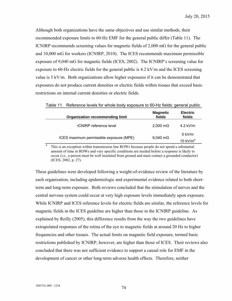
July 20, 2015
1003741.000 - 1234 74
Although both organizations have the same objectives and use similar methods, their
recommended exposure limits to 60-Hz EMF for the general public differ (Table 11). The
ICNIRP recommends screening values for magnetic fields of 2,000 mG for the general public
and 10,000 mG for workers (ICNIRP, 2010). The ICES recommends maximum permissible
exposure of 9,040 mG for magnetic fields (ICES, 2002). The ICNIRP’s screening value for
exposure to 60-Hz electric fields for the general public is 4.2 kV/m and the ICES screening
value is 5 kV/m. Both organizations allow higher exposures if it can be demonstrated that
exposures do not produce current densities or electric fields within tissues that exceed basic
restrictions on internal current densities or electric fields.
Table 11. Reference levels for whole body exposure to 60-Hz fields: general public
Organization recommending limit Magnetic
fields Electric fields
ICNIRP reference level 2,000 mG 4.2 kV/m
ICES maximum permissible exposure (MPE) 9,040 mG 5 kV/m
10 kV/ma a This is an exception within transmission line ROWs because people do not spend a substantial
amount of time in ROWs and very specific conditions are needed before a response is likely to occur (i.e., a person must be well insulated from ground and must contact a grounded conductor) (ICES, 2002, p. 27).
These guidelines were developed following a weight-of-evidence review of the literature by
each organization, including epidemiologic and experimental evidence related to both short-
term and long-term exposure. Both reviews concluded that the stimulation of nerves and the
central nervous system could occur at very high exposure levels immediately upon exposure.
While ICNIRP and ICES reference levels for electric fields are similar, the reference levels for
magnetic fields in the ICES guideline are higher than those in the ICNIRP guideline. As
explained by Reilly (2005), this difference results from the way the two guidelines have
extrapolated responses of the retina of the eye to magnetic fields at around 20 Hz to higher
frequencies and other tissues. The actual limits on magnetic field exposure, termed basic
restrictions published by ICNIRP, however, are higher than those of ICES. Their reviews also
concluded that there was not sufficient evidence to support a causal role for EMF in the
development of cancer or other long-term adverse health effects. Therefore, neither
July 20, 2015
1003741.000 - 1234 75
organization found a basis to recommend quantitative exposure guidelines to prevent effects at
lower exposure levels.
Following the publication of their 1998 guidelines, the ICNIRP published an evaluation of the
epidemiologic literature (ICNIRP, 2001) and a full weight-of-evidence evaluation of health
research on EMF (ICNIRP, 2003), concluding again that there is no basis for exposure
restrictions for long-term health effects. In June 2009, the ICNIRP published an updated review
of the scientific literature related to potential short- and long-term adverse effects, and draft
guidelines to replace their 1998 ELF EMF exposure guidelines (ICNIRP, 2009). The final
guideline was published in December 2010 and those screening vales are listed in Table 11.
There are no national or state standards in the United States limiting exposures to ELF EMF
based on health effects. Two states, Florida and New York, have enacted standards to limit
magnetic fields at the edge of transmission line ROWs (150 mG and 200 mG, respectively)
(NYPSC, 1978, 1990; FDER, 1989; FDEP, 1996). The basis for limiting magnetic fields from
transmission lines was to maintain the status quo so that fields from new transmission lines
would be no higher than those produced by existing transmission lines.
July 20, 2015
1003741.000 - 1234 76
5 References
Adair RK. Biological responses to weak 60-Hz electric and magnetic fields must vary as the square of the field strength. Proc Natl Acad Sci 91: 9422-9425, 1994.
Ahlbom A, Day N, Feychting M, Roman E, Skinner J, Dockerty J, Linet M, McBride M, Michaelis J, Olsen JH, Tynes T, Verkasalo PK. A pooled analysis of magnetic fields and childhood leukaemia. Br J Cancer 83: 692-698, 2000.
Akdag MZ, Dasdag S, Uzunlar AK, Ulukaya E, Oral AY, Celik N, Aksen F. Can safe and long-term exposure to extremely low frequency (50 Hz) magnetic fields affect apoptosis, reproduction, and oxidative stress? Int J Radiat Biol 89: 1053-1060, 2013.
Alcaraz M, Olmos E, Alcaraz-Saura M, Achel DG, Castillo J. Effect of long-term 50 Hz magnetic field exposure on the micronucleated polychromatic erythrocytes of mice. Electromagn Biol Med 33: 51-57, 2014.
American Conference of Government Industrial Hygienists (ACGIH). Documentation of the Threshold Limit Values and Biological Exposure Indices, 7th edition. Publication No. 0100. Cincinnati, OH: American Conference of Government Industrial Hygienists, 2001.
Anderson LE, Boorman GA, Morris JE, Sasser LB, Mann PC, Grumbein SL, Hailey JR, McNally A, Sills RC, Haseman JK. Effect of 13 week magnetic field exposures on DMBA-initiated mammary gland carcinomas in female Sprague-Dawley rats. Carcinogenesis 20: 1615-1620, 1999.
Astridge PS, Kaye GC, Whitworth S, Kelly P, Camm AJ, Perrins EJ. The response of implanted dual chamber pacemakers to 50 Hz extraneous electrical interference. Pacing Clin Electrophysiol 16: 1966-1974, 1993.
Auger N, Joseph D, Goneau M, Daniel M. The relationship between residential proximity to extremely low frequency power transmission lines and adverse birth outcomes. J Epidemiol Community Health 65: 83-85, 2011.
Auger N, Park AL, Yacouba S, Goneau M, Zayed J. Stillbirth and residential proximity to extremely low frequency power transmission lines: a retrospective cohort study. Occup Environ Med 69: 147-149, 2012.
Australian Centre for Radiofrequency Bioeffects Research (ACRBR). ACRBR Position Statement on BioInitiative Report. December 18, 2008.
Baldi I, Coureau G, Jaffre A, Gruber A, Ducamp S, Provost D, Lebailly P, Vital A, Loiseau H, Salamon R. Occupational and residential exposure to electromagnetic fields and risk of brain tumors in adults: a case-control study in Gironde, France. Int J Cancer 129: 1477-1484, 2011.
July 20, 2015
1003741.000 - 1234 77
Bankoske JW, Graves HB, McKee GW. The Effects of High Voltage Electric Fields on the Growth and Development of Plants and Animals. In Proceedings of the First National Symposium on Environmental Concerns in Rights-of-Way Management, edited by Tillman R. Mississippi: Mississippi State University, 1976.
Baum A, Mevissen M, Kamino K, Mohr U, Löscher W. A histopathological study on alterations in DMBA-induced mammary carcinogenesis in rats with 50 Hz, 100 µT magnetic field exposure. Carcinogenesis 16: 119-125, 1995.
Begall S, Cerveny J, Neef J, Vojtech O, Burda H. Magnetic alignment in grazing and resting cattle and deer. Proc Natl Acad Sci 105: 13451-13455, 2008.
Bellieni CV, Tei M, Iacoponi F, Tataranno ML, Negro S, Proietti F, Longini M, Perrone S, Buonocore G. Is newborn melatonin production influenced by magnetic fields produced by incubators? Early Hum Dev 88: 707-710, 2012.
Berg H, Gunther B, Hilger I, Radeva M, Traitcheva N, Wollweber L. Bioelectromagnetic field effects on cancer cells and mice tumors. Electromagn Biol Med 29: 132-143, 2010.
Bindokas V, Gauger J, Greenberg B. Exposure scheme separates effects of electric shock and electric field for honey bees, Apis mellifera, L. Bioelectromagnetics 9: 275-284, 1988a.
Bindokas V, Gauger J, Greenberg B. Mechanism of biological effects observed in honey bees (Apis mellifera, L.) hived under extra-high-voltage transmission lines: implications derived from bee exposure to simulated intense electric fields and shocks. Bioelectromagnetics 9: 285-301, 1988b.
Bindokas V, Gauger J, Greenberg B. Laboratory investigations of the electrical characteristics of honey bees and their exposure to intense electric fields. Bioelectromagnetics 10: 1-12, 1989.
BioInitiative Working Group (BWG). Cindy Sage and David O. Carpenter Editors. BioInitiative Report: A Rationale for Biologically-based Public Exposure Standard for Electromagnetic Fields (ELF and RF). August 31, 2007.
BioInitiative Working Group (BWG). Cindy Sage and David O. Carpenter Editors. BioInitiative Report: A Rationale for Biologically-based Exposure Standards for Low-Intensity Electromagnetic Radiation at www.bioinitiative.org. December 31, 2012.
Bureau of Ocean Energy Management, Regulation and Enforcement (BOEMRE). Effects pf EMFs from Undersea Power Cables on Elasmobranchs and Other Marine Species. Camarillo, CA: U.S. Department of the Interior, Bureau of Ocean Energy Management, Regulation and Enforcement, 2011.
Bonnet-Belfais M, Lambrozo J, Aurengo A. Comment: childhood leukaemia and power lines--the Geocap study: is proximity an appropriate MF exposure surrogate? Br J Cancer 109: 1382-1383, 2013.
July 20, 2015
1003741.000 - 1234 78
Boorman GA, Anderson LE, Morris JE, Sasser LB, Mann PC, Grumbein SL, Hailey JR, McNally A, Sills RC, Haseman JK. Effect of 26 week magnetic field exposures in a DMBA initiation-promotion mammary gland model in Sprague-Dawley rats. Carcinogenesis 20: 899-904, 1999.
Boorman GA, Anderson LE, Morris JE, Sasser LB, Mann PC, Grumbein SL, Hailey JR, McNally A, Sills RC, Haseman JK. Effect of 26 week magnetic field exposures in a DMBA initiation-promotion mammary gland model in Sprague-Dawley rats. Carcinogenesis 20: 899-904, 1999a.
Boorman GA, McCormick DL, Findlay JC, Hailey JR, Gauger JR, Johnson TR, Kovatch RM, Sills RC, Haseman JK. Chronic toxicity/oncogenicity evaluation of 60 Hz (power frequency) magnetic fields in F344/N rats. Toxicol Pathol 27: 267-278, 1999b.
Borhani N, Rajaei F, Salehi Z, Javadi A. Analysis of DNA fragmentation in mouse embryos exposed to an extremely low-frequency electromagnetic field. Electromagn Biol Med 30: 246-252, 2011.
Brain JD, Kavet R, Valberg PA. Observations on power-line magnetic fields associated with asthma in children. Arch Pediatr Adolesc Med 155: 97-98, 2012.
Brouwer M, Koeman T, van den Brandt PA, Kromhout H, Schouten LJ, Peters S, Huss A, Vermeulen R. Occupational exposures and Parkinson's disease mortality in a prospective Dutch cohort. Occup Environ Med 72: 448-455, 2015.
Buldak RJ, Polaniak R, Buldak L, Zwirska-Korczala K, Skonieczna M, Monsiol A, Kukla M, Dulawa-Buldak A, Birkner E. Short-term exposure to 50 Hz ELF-EMF alters the cisplatin-induced oxidative response in AT478 murine squamous cell carcinoma cells. Bioelectromagnetics 33: 641-651, 2012.
Bullock TH. Electromagnetic sensing in fish. Neuro sci Res Prog Bull 15: 17-22, 1977.
Bunch KJ, Keegan TJ, Swanson J, Vincent TJ, Murphy MF. Residential distance at birth from overhead high-voltage powerlines: childhood cancer risk in Britain 1962-2008. Br J Cancer 110: 1402-1408, 2014.
Burdak-Rothkamm S, Rothkamm K, Folkard M, Patel G, Hone P, Lloyd D, Ainsbury L, Prise KM. DNA and chromosomal damage in response to intermittent extremely low-frequency magnetic fields. Mutat Res 672: 82-89, 2009.
Calvente I, Fernandez MF, Villalba J, Olea N, Nunez MI. Exposure to electromagnetic fields (non-ionizing radiation) and its relationship with childhood leukemia: a systematic review. Sci Total Environ 408: 3062-3069, 2010.
Chang ET, Adami HO, Bailey WH, Boffetta P, Krieger RI, Moolgavkar SH, Mandel JS. Validity of geographically modeled environmental exposure estimates. Crit Rev Toxicol 44: 450-466, 2014.
July 20, 2015
1003741.000 - 1234 79
Chen G and Xu Z. Global protein expression in response to extremely low frequency magnetic fields. Adv Exp Med Biol 990: 107-110, 2013.
Cho S, Lee Y, Lee S, Choi YJ, Chung HW. Enhanced cytotoxic and genotoxic effects of gadolinium following ELF-EMF irradiation in human lymphocytes. Drug Chem Toxicol 37: 440-447, 2014.
Conti R, Nicolini P, Cerretelli P, Margonato V, Veicsteinas A, Floris C. Enel's research activity on possible biological effects of 50 Hz electromagnetic fields - results and plans of a large research programme. Alta Frequenza 58: 395-402, 1989.
Costanzo E. The influence of an electric field on the growth of soy seedlings. J Electrostat 55: 417-420, 2008.
Das K, Nag C, Ghosh M. Familial, environmental, and occupational risk factors in development of amyotrophic lateral sclerosis. N Am J Med Sci 4: 350-355, 2012.
Davanipour Z and Sobel E. Long-term exposure to magnetic fields and the risks of Alzheimer's disease and breast cancer: Further biological research. Pathophysiology 16: 149-156, 2009.
Davanipour Z, Tseng CC, Lee PJ, Markides KS, Sobel E. Severe cognitive dysfunction and occupational extremely low frequency magnetic field exposure among elderly Mexican Americans. Br J Med Med Res 4: 1641-1662, 2014.
Davies MS. Effects of 60 Hz electromagnetic fields on early growth in three plant species and a replication of previous results. Bioelectromagnetics 17: 154-161, 1996.
De Souza A, Sueiro L, Garcia D, Porras E. Extremely low frequency non-uniform magnetic fields improve tomato seed germination and early seedling growth. Seed Sci Tech 38: 61-72, 2010.
de Vocht F. Adult cancers near high-voltage power lines. Epidemiology 24: 782, 2013.
de Vocht F, Hannam K, Baker P, Agius R. Maternal residential proximity to sources of extremely low frequency electromagnetic fields and adverse birth outcomes in a UK cohort. Bioelectromagnetics 35: 201-209, 2014.
de Vocht F and Lee B. Residential proximity to electromagnetic field sources and birth weight: Minimizing residual confounding using multiple imputation and propensity score matching. Environ Int 69: 51-57, 2014.
Dell'omo G, Costantini D, Lucini V, Antonucci G, Nonno R, Polichetti A. Magnetic fields produced by power lines do not affect growth, serum melatonin, leukocytes and fledging success in wild kestrel. Comp Biochem Physiol Toxicol Pharmacol 150: 372-376, 2009.
Dittmar L, Stürzl W, Jetzschke S, Mertes M, Boeddeker N. Out of the box: how bees orient in an ambiguous environment. Animal Behaviour 89: 13-21, 2014.
July 20, 2015
1003741.000 - 1234 80
Does M, Scelo G, Metayer C, Selvin S, Kavet R, Buffler P. Exposure to electrical contact currents and the risk of childhood leukemia. Radiat Res 175: 390-396, 2011.
Dovey KM, Kemfort JR, Towne WF. The depth of the honeybee's backup sun-compass systems. J Exp Biol 216: 2129-2139, 2013.
Draper G, Vincent T, Kroll ME, Swanson J. Childhood cancer in relation to distance from high voltage power lines in England and Wales: a case-control study. BMJ 330: 1290, 2005.
Duan W, Liu C, Wu H, Chen C, Zhang T, Gao P, Luo X, Yu Z, Zhou Z. Effects of exposure to extremely low frequency magnetic fields on spermatogenesis in adult rats. Bioelectromagnetics 35: 58-69, 2014.
Duan W, Liu C, Zhang L, He M, Xu S, Chen C, Pi H, Gao P, Zhang Y, Zhong M, Yu Z, Zhou Z. Comparison of the genotoxic effects induced by 50 Hz extremely low-frequency electromagnetic fields and 1800 MHz radiofrequency electromagnetic fields in GC-2 cells. Radiat Res 183: 305-314, 2015.
Eden T. Aetiology of childhood leukaemia. Cancer Treat Rev 36: 286-297, 2010.
El-Bialy NS and Rageh MM. Extremely low-frequency magnetic field enhances the therapeutic efficacy of low-dose cisplatin in the treatment of Ehrlich carcinoma. Biomed Res Int 2013: 189352, 2013.
Electric Power Research Institute (EPRI). Electromagnetic Interference with Implanted Medical Devices: 1997-2003. Report No. 1005570. Palo Alto, CA: EPRI, 2004.
Elliott P, Shaddick G, Douglass M, de Hoogh K, Briggs DJ, Toledano MB. Adult cancers near high-voltage overhead power lines. Epidemiology 24: 184-190, 2013.
Elmusharaf M, Cuppen J, Grooten N, Beynen A. Antagonistic effect of electromagnetic field exposure on coccidiosis infection in broiler chickens. Poultry Sci 86: 2139-2143, 2007.
Elwood JM. Childhood leukemia and residential magnetic fields: are pooled analyses more valid than original studies? Bioelectromagnetics 27: 112-118, 2006.
Esquivel DMS, Correa AAC, Vaillant OS, Bandeira de Melo V, Gouva GS, Ferreira CG, Ferreira TA, Wajnberg E. A time-compressed simulated geomagnetic storm influences the nest-exiting flight angles of the stingless bee Tetragonisca angustula. Naturwissenschaften 101: 245-249, 2014.
European Health Risk Assessment Network on Electromagnetic Fields Exposure (EFHRAN). Risk Analysis of Human Exposure to Electromagnetic Fields (Revised). Report D2 of the EFHRAN Project. Milan, Italy: European Health Risk Assessment Network on Electromagnetic Fields Exposure, 2012.
July 20, 2015
1003741.000 - 1234 81
Exponent, Inc. Assessment of Research Regarding EMF and Health and Environmental Effects. Olympic Peninsula Reinforcement Transmission Line Project, Prepared for Bonneville Power Administration. Exponent, Inc., July 2007.
Exponent, Inc. Research on Extremely Low Frequency Electric and Magnetic Fields and Health, Prepared for the Bonneville Power Administration. Exponent, Inc., January 2011.
Federal-Provincial-Territorial Radiation Protection Committee (FPTRPC). Health Effects and Exposure Guidelines Related to Extremely Low Frequency Electric and Magnetic Fields - An Overview. Ottawa: Health Canada, 2005.
Fedrowitz M, Kamino K, Löscher W. Significant differences in the effects of magnetic field exposure on 7,12-dimethylbenz(a)anthracene-induced mammary carcinogenesis in two substrains of Sprague-Dawley rats. Cancer Res 64: 243-251, 2004.
Fedrowitz M and Löscher W. Gene expression in the mammary gland tissue of female Fischer 344 and Lewis rats after magnetic field exposure (50 Hz, 100 muT) for 2 weeks. Int J Radiat Biol 88: 425-429, 2012.
Fedrowitz M and Lӧscher W. Gene expression in the mammary gland tissue of female Fischer 344 and Lewis rats after magnetic field exposure (50 Hz, 100 muT) for 2 weeks. Int J Radiat Biol 88: 425-429, 2012.
Feychting M, Ahlbom A, Kheifets L. EMF and health. Ann Rev Public Health 26: 165-189, 2005.
Feychting M. Invited commentary: extremely low-frequency magnetic fields and breast cancer--now it is enough! Am J Epidemiol 178: 1046-1050, 2013.
Florida Department of Environmental Protection (FDEP). Electric and Magnetic Fields. Chapter 62-814: FDEP, 1996.
Florida Department of Environmental Regulation (FDER). Electric and Magnetic Fields. Chapter 17-274: FDER, 1989.
Focke F, Schuermann D, Kuster N, Schar P. DNA fragmentation in human fibroblasts under extremely low frequency electromagnetic field exposure. Mutat Res 683: 74-83, 2010.
Foliart DE, Pollock BH, Mezei G, Iriye R, Silva JM, Ebi KL, Kheifets L, Link MP, Kavet R. Magnetic field exposure and long-term survival among children with leukaemia. Br J Cancer 94: 161-164, 2006.
Framm J and Lautner S. Electrical signals and their physiological significance in plants. Plant Cell Environ 30: 249-257, 2007.
Frei P, Poulsen AH, Mezei G, Pedersen C, Cronberg Salem L, Johansen C, Rӧӧsli M, Schüz J. Residential distance to high-voltage power lines and risk of neurodegenerative diseases: a Danish population-based case-control study. Am J Epidemiol 177: 970-978, 2013.
July 20, 2015
1003741.000 - 1234 82
Gill AB, Bartlett M, Thomsen F. Potential interactions between diadromous fishes of U.K. conservation importance and the electromagnetic fields and subsea noise from marine renewable energy developments. J Fish Biol 81: 664-695, 2012.
Gomes J, Al Zayadi A, Guzman A. Occupational and environmental risk factors of adult primary brain cancers: a systematic review. Int J Occup Environ Med 2: 82-111, 2011.
Greenberg B, Bindokas VP, Gauger Jr. Biological effects of 765 kilovolt transmission line exposure and thresholds in honey bee colonies. Bioelectromagnetics 2: 315-328, 1981.
Greenland S, Sheppard AR, Kaune WT, Poole C, Kelsh MA. A pooled analysis of magnetic fields, wire codes, and childhood leukemia. Childhood Leukemia-EMF Study Group. Epidemiology 11: 624-634, 2000.
Greggers U, Koch G, Schmidt V, Duerr A, Floriou-Servou A, Piepenbrock D, Goepfert MC, Menzel R. Reception and learning of electric fields in bees. Proc Royal Soc Biol Sci B 280: 1-8, 2013.
Grell K, Meersohn A, Schüz J, Johansen C. Risk of neurological diseases among survivors of electric shocks: a nationwide cohort study, Denmark, 1968-2008. Bioelectromagnetics 33: 459-465, 2012.
Grellier J, Ravazzani P, Cardis E. Potential health impacts of residential exposures to extremely low frequency magnetic fields in Europe. Environ Int 62: 55-63, 2014.
Groot C and Margolis L, eds. Pacific Salmon Life Histories. Vancouver: UBC Press, 1998.
Hajhosseini L, Khaki A, Merat E, Ainehchi N. Effect of rosmarinic acid on sertoli cells apoptosis and serum antioxidant levels in rats after exposure to electromagnetic fields. Afr J Tradit Complement Altern Med 10: 477-480, 2013.
Hardell L and Sage C. Biological effects from electromagnetic fields exposure and public exposure standards. Biomed Pharmacother 62: 104-109, 2008.
Hareuveny R, Kandel S, Yitzhak NM, Kheifets L, Mezei G. Exposure to 50 Hz magnetic fields in apartment buildings with indoor transformer stations in Israel. J Expo Sci Environ Epidemiol 21: 365-371, 2011.
Harris AW, Basten A, Gebski V, Noonan D, Finnie J, Bath ML, Bangay MJ, Repacholi MH. A test of lymphoma induction by long-term exposure of E mu-Pim1 trangenic mice to 50 Hz magnetic fields. Radiat Res 149: 300-307, 1998.
Health Council of the Netherlands (HCN). Electromagnetic Fields: Annual Update 2003. Publication No. 2004/01. The Hague: Health Council of the Netherlands, 2004.
Health Council of the Netherlands (HCN). BioInitiative Report. The Hague: Health Council of the Netherlands, 2008.
July 20, 2015
1003741.000 - 1234 83
Health Protection Agency (HPA). Power frequency electromagnetic fields, melatonin and the risk of breast cancer: report of an independent advisory group on non-ionising radiation. Documents of the Health Protection Agency. Series B: Radiation, Chemical and Environmental Hazards. Oxfordshire: Health Protection Agency, 2006.
Her Majesty's Government (HMG). Department of Health, Department for Communities & Local Government, Department of Energy & Climate Change. Government Response to the Stakeholder Advisory Group on Extremely Low Frequency Electric and Magnetic Fields (ELF EMFs) (SAGE) Recommendations2009.
Hilson D, Noggle J, Burns E. Effects of Electric Field on Plants Growing Under High Voltage Transmission Lines. Chattanooga, TN: Office of Natural Resources, Air Quality Branch. Tennessee Valley Authority, 1983.
Hodges T, Mitchell C, Heydt G. Effect of Electric Fields from 765-kV Transmission Lines on Plant Growth. New York, NY: Purdue University for Indiana and Michigan Electric Company, a Subsidiary of American Electric Power, 1975.
Hodges T and Mitchell C. Influence on High Intensity Electric Fields on Yield of Sweet Corn and Dent Corn. North Liberty, IN: A report for the American Electric Power Service Corporation, 1984.
Hours M, Khati I, Hamelin J. Interference between active implanted medical devices and electromagnetic field emitting devices is rare but real: results of an incidence study in a population of physicians in France. Pacing Clin Electrophysiol 37: 290-296, 2014.
Huang H-H and Wang S-R. The effects of inverter magnetic fields on early seed germination of mung beans. Bioelectromagnetics 29: 649-657, 2008.
Hug K, Grize L, Seidler A, Kaatsch P, Schüz J. Parental occupational exposure to extremely low frequency magnetic fields and childhood cancer: a German case-control study. Am J Epidemiol 171: 27-35, 2010.
International Agency for Research on Cancer (IARC). Mechanisms for Carcinogenesis in Risk Identification. Lyon, France: IARC Press, 1992.
International Agency for Research on Cancer (IARC). IARC Monographs on the Evaluation of Carcinogenic Risks to Humans. Volume 80: Static and Extremely Low-Frequency (ELF) Electric and Magnetic Fields. Lyon, France: IARC Press, 2002.
International Agency for Research on Cancer (IARC). IARC Monographs on the Evaluation of Carcinogenic Risks to Humans, Preamble. Lyon, France: 2006
International Commission for Non-Ionizing Radiation Protection (ICNIRP). Review of the epidemiologic literature on EMF and health. Environ Health Perspect 109: S911-S933, 2001.
July 20, 2015
1003741.000 - 1234 84
International Commission for Non-Ionizing Radiation Protection (ICNIRP). Draft Guidelines for limiting exposure to time-varying electric and magnetic fields (1 Hz to 100 kHz). Health Phys 96: 504-514, 2009.
International Commission on Non-Ionizing Radiation Protection (ICNIRP). Guidelines for limiting exposure to time-varying electric, magnetic, and electromagnetic fields (up to 300 GHz). Health Phys 74: 494-522, 1998.
International Commission on Non-ionizing Radiation Protection (ICNIRP). Exposure to Static and Low Frequency Electromagnetic Fields, Biological Effects and Health Consequences (0-100 kHz). ICNIRP 13/2003. Oberschleissheim, Germany: International Commission on Non-ionizing Radiation Protection, 2003.
International Commission on Non-ionizing Radiation Protection (ICNIRP). Guidelines for limiting exposure to time-varying electric and magnetic fields (1 Hz to 100 kHz). Health Phys 99: 818-836, 2010.
Isaac Aleman E, Oliveira Moreira R, Almeida Lima A, Chaves Silva S, Gonzalez-Olmedo JL, Chalfun-Junior A. Effects of 60 Hz sinusoidal magnetic field on in vitro establishment, multiplication, and acclimatization phases of Coffea arabica seedlings. Bioelectromagnetics 35: 414-425, 2014.
Ivancsits S, Pilger A, Diem E, Schaffer A, Rüdinger H. Vanadate induces DNA strand breaks in cultured human fibroblasts at doses relevant to occupational exposure. Mutat Res 519: 25-35, 2002a.
Ivancsits S, Diem E, Pilger A, Rudiger HW, Jahn O. Induction of DNA strand breaks by intermittent exposure to extremely-low-frequency electromagnetic fields in human diploid fibroblasts. Mutat Res 519: 1-13, 2002b.
Ivancsits S, Diem E, Jahn O, Rudiger HW. Intermittent extremely low frequency electromagnetic fields cause DNA damage in a dose-dependent way. Int Arch Occup Environ Health 76: 431-436, 2003a.
Ivancsits S, Diem E, Jahn O, Rudiger HW. Age-related effects on induction of DNA strand breaks by intermittent exposure to electromagnetic fields. Mech Ageing Dev 124: 847-850, 2003b.
Jin YB, Kang GY, Lee JS, Choi JI, Lee JW, Hong SC, Myung SH, Lee YS. Effects on micronuclei formation of 60-Hz electromagnetic field exposure with ionizing radiation, hydrogen peroxide, or c-Myc overexpression. Int J Radiat Biol 88: 374-380, 2012.
Jin YB, Choi SH, Lee JS, Kim JK, Lee JW, Hong SC, Myung SH, Lee YS. Absence of DNA damage after 60-Hz electromagnetic field exposure combined with ionizing radiation, hydrogen peroxide, or c-Myc overexpression. Radiat Environ Biophys 53: 93-101, 2014.
July 20, 2015
1003741.000 - 1234 85
Jin H, Yoon HE, Lee JS, Kim JK, Myung SH, Lee YS. Effects on g2/m phase cell cycle distribution and aneuploidy formation of exposure to a 60 Hz electromagnetic field in combination with ionizing radiation or hydrogen peroxide in l132 nontumorigenic human lung epithelial cells. Korean J Physiol Pharmacol 19: 119-124, 2015.
Jirik V, Pekarek L, Janout V, Tomaskova H. Association between childhood leukaemia and exposure to power-frequency magnetic fields in Middle Europe. Biomed Environ Sci 25: 597-601, 2012.
Johansson O. Disturbance of the immune system by electromagnetic fields - A potentially underlying cause for cellular damage and tissue repair reduction which could lead to disease and impairment. Pathophysiology 16: 157-177, 2009.
Joosten S, Pammler K, Silny J. The influence of anatomical and physiological parameters on the interference voltage at the input of unipolar cardiac pacemakers in low frequency electric fields. Phys Med Biol 54: 591-609, 2009.
Kabuto M, Nitta H, Yamamoto S, Yamaguchi N, Akiba S, Honda Y, Hagihara J, Isaka K, Saito T, Ojima T, Nakamura Y, Mizoue T, Ito S, Eboshida A, Yamazaki S, Sokejima S, Kurokawa Y, Kubo O. Childhood leukemia and magnetic fields in Japan: a case-control study of childhood leukemia and residential power-frequency magnetic fields in Japan. Int J Cancer 119: 643-650, 2006.
Karipidis KK. Survey of residential power-frequency magnetic fields in Melbourne, Australia. Radiat Prot Dosimetry 163: 81-91, 2015.
Katrib J, Schmitt P, Magne I, Kourtiche D, Souques M, Nadi M. Implantable cardioverter-defibrillators exposed to low frequency magnetic fields. In General Assembly and Scientific Symposium, 2011 XXXth URSI 2011.
Katrib J, Nadi M, Kourtiche D, Magne I, Schmitt P, Souques M, Roth P. In vitro assessment of the immunity of implantable cardioverter-defibrillators to magnetic fields of 50/60 Hz. Physiol Meas 34: 1281-1292, 2013.
Keegan TJ, Bunch KJ, Vincent TJ, King JC, O'Neill KA, Kendall GM, MacCarthy A, Fear NT, Murphy MF. Case-control study of paternal occupation and childhood leukaemia in Great Britain, 1962-2006. Br J Cancer 107: 1652-1659, 2012.
Kheifets L, Monroe J, Vergara X, Mezei G, Afifi AA. Occupational electromagnetic fields and leukemia and brain cancer: an update to two meta-analyses. J Occup Environ Med 50: 677-688, 2008.
Kheifets L and Oksuzyan S. Exposure assessment and other challenges in non-ionizing radiation studies of childhood leukaemia. Radiat Prot Dosimetry 132: 139-147, 2008.
July 20, 2015
1003741.000 - 1234 86
Kheifets L, Bowman JD, Checkoway H, Feychting M, Harrington JM, Kavet R, Marsh G, Mezei G, Renew DC, van Wijngaarden E. Future needs of occupational epidemiology of extremely low frequency electric and magnetic fields: review and recommendations. Occup Environ Med 66: 72-80, 2009.
Kheifets L, Ahlbom A, Crespi CM, Feychting M, Johansen C, Monroe J, Murphy MF, Oksuzyan S, Preston-Martin S, Roman E, Saito T, Savitz D, Schüz J, Simpson J, Swanson J, Tynes T, Verkasalo P, Mezei G. A pooled analysis of extremely low-frequency magnetic fields and childhood brain tumors. Am J Epidemiol 172: 752-761, 2010.
Kim J, Ha CS, Lee HJ, Song K. Repetitive exposure to a 60-Hz time-varying magnetic field induces DNA double-strand breaks and apoptosis in human cells. Biochem Biophys Res Commun 400: 739-744, 2010.
Kim J, Yoon Y, Yun S, Park GS, Lee HJ, Song K. Time-varying magnetic fields of 60 Hz at 7 mT induce DNA double-strand breaks and activate DNA damage checkpoints without apoptosis. Bioelectromagnetics 33: 383-393, 2012.
Kirschvink J, Diaz Ricci J, Nesson M, Kirschvink S. Magnetite-based magnetoreceptors in animals: structural, behavioral, and biophysical studies. Palo Alto, CA: Electric Power Research Institute, 1993.
Kirschvink JL, Padmanabha S, Boyce CK, Oglesby J. Measurement of the threshold sensitivity of honeybees to weak, extremely low-frequency magnetic fields. Journal of Experimental Biology 200: 1363-1368, 1997.
Koeman T, van den Brandt PA, Slottje P, Schouten LJ, Goldbohm RA, Kromhout H, Vermeulen R. Occupational extremely low-frequency magnetic field exposure and selected cancer outcomes in a prospective Dutch cohort. Cancer Causes Control 25: 203-214, 2014.
Korpinen L, Kuisti H, Elovaara J, Virtanen V. Cardiac pacemakers in electric and magnetic fields of 400-kV power lines. Pacing Clin Electrophysiol 35: 422-430, 2012.
Korpinen L, Kuisti H, Elovaara J. Current densities and total contact currents for 110 and 220 kV power line tasks. Bioelectromagnetics 35: 531-535, 2014.
Kozik TM, Chien G, Connolly TF, Grewal GS, Liang D, Chien W. iPad2(R) use in patients with implantable cardioverter defibrillators causes electromagnetic interference: the EMIT Study. J Am Heart Assoc 3: e000746, 2014.
Krizaj D and Valencic V. The effects of ELF magnetic fields and temperature on differential plant growth. J Bioelectricity 8: 159-165, 1989.
Kroll ME, Swanson J, Vincent TJ, Draper GJ. Childhood cancer and magnetic fields from high-voltage power lines in England and Wales: a case-control study. Br J Cancer 103: 1122-1127, 2010.
July 20, 2015
1003741.000 - 1234 87
Lahijani MS, Farivar S, Khodaeian M. Effects of 50 Hz electromagnetic fields on the histology, apoptosis, and expression of c-Fos and beta-catenin on the livers of preincubated white Leghorn chicken embryos. Electromagn Biol Med 30: 158-169, 2011.
Lai H and Singh NP. Magnetic-field-induced DNA strand breaks in brain cells of the rat. Environ Health Perspect 112:687-694, 2004.
Lee GM, Neutra RR, Hristova L, Yost M, Hiatt RA. A nested case-control study of residential and personal magnetic field measures and miscarriages. Epidemiology 13: 21-31, 2002.
Lee J and Clark C. Ecological Effects of EHV and UHV Transmission Lines Current Issues. In: CIRGE Symposium on Transmission Lines and the Environment. pp. 223-307. Stockholm, Sweden, June 23-25, 1981.
Lensu T, Komonen A, Hiltula O, Paivinen J, Saari V, Kotiaho JS. The role of power line rights-of-way as an alternative habitat for declined mire butterflies. J Environ Manage 92: 2539-2546, 2011.
Lerchl A. Are these data real? Comments on "No effects of intermittent 50 Hz EMF on cytoplasmic free calcium and on the mitochondrial membrane potential in human diploid fibroblasts." by Pilger et al. (Radiat Environ Biophys 43:203-7 (2004)). Radiat Environ Biophys 49: 491-493; author reply 495-497, 2010b.
Lewis RC, Evenson KR, Savitz DA, Meeker JD. Temporal variability of daily personal magnetic field exposure metrics in pregnant women. J Expo Sci Environ Epidemiol 25: 58-64, 2015.
Li DK, Odouli R, Wi S, Janevic T, Golditch I, Bracken TD, Senior R, Rankin R, Iriye R. A population-based prospective cohort study of personal exposure to magnetic fields during pregnancy and the risk of miscarriage. Epidemiology 13: 9-20, 2002.
Li DK, Yan B, Li Z, Gao E, Miao M, Gong D, Weng X, Ferber JR, Yuan W. Exposure to magnetic fields and the risk of poor sperm quality. Reprod Toxicol 29: 86-92, 2010.
Li DK, Chen H, Odouli R. Maternal Exposure to Magnetic Fields During Pregnancy in Relation to the Risk of Asthma in Offspring. Arch Pediatr Adolesc Med 165: 945-950, 2011.
Li DK, Ferber JR, Odouli R, Quesenberry CP, Jr. A prospective study of in-utero exposure to magnetic fields and the risk of childhood obesity. Sci Rep 2: 540, 2012.
Li W, Ray RM, Thomas DB, Yost M, Davis S, Breslow N, Gao DL, Fitzgibbons ED, Camp JE, Wong E, Wernli KJ, Checkoway H. Occupational exposure to magnetic fields and breast cancer among women textile workers in Shanghai, China. Am J Epidemiol 178: 1038-1045, 2013.
Linet MS, Hatch EE, Kleinerman RA, Robison LL, Kaune WT, Friedman DR, Severson RK, Haines CM, Hartsock CT, Niwa S, Wacholder S, Tarone RE. Residential exposure to magnetic fields and acute lymphoblastic leukemia in children. N Engl J Med 337: 1-7, 1997.
July 20, 2015
1003741.000 - 1234 88
Löscher W, Mevissen M, Lehmacher W, Stamm A. Tumor promotion in a breast cancer model by exposure to a weak alternating magnetic field. Cancer Lett 71: 75-81, 1993.
Löscher W, Wahnschaffe U, Mevissen M, Lerchl A, Stamm A. Effects of weak alternating magnetic fields on nocturnal melatonin production and mammary carcinogenesis in rats. Oncology 51: 288-295, 1994.
Löscher W and Mevissen M. Linear relationship between flux density and tumor co-promoting effect of prolonged magnetic field exposure in a breast cancer model. Cancer Lett 96: 175-180, 1995.
Löscher W, Mevissen M, Haussler B. Seasonal influence on 7,12-dimethylbenz[a]anthracene-induced mammary carcinogenesis in Spague-Dawley rats under controlled laboratory conditions. Pharmacol Toxicol 81: 265-270, 1997.
Luukkonen J, Liimatainen A, Hoyto A, Juutilainen J, Naarala J. Pre-exposure to 50 Hz magnetic fields modifies menadione-induced genotoxic effects in human SH-SY5Y neuroblastoma cells. PLoS One 6: e18021, 2011.
Luukkonen J, Liimatainen A, Juutilainen J, Naarala J. Induction of genomic instability, oxidative processes, and mitochondrial activity by 50Hz magnetic fields in human SH-SY5Y neuroblastoma cells. Mutat Res 760: 33-41, 2014.
Magnani A, Matheoud R, Brambilla M, Valzano S, Occhetta E, Marino P, Balbo P. In vitro tests of electromagnetic interference of electromagnetic navigational bronchoscopy to implantable cardioverter defibrillators. Europace 14: 1054-1059, 2012.
Magnani C, Mattioli S, Miligi L, Ranucci A, Rondelli R, Salvan A, Bisanti L, Masera G, Rizzari C, Zambon P, Cannizzaro S, Gafa L, Luzzatto LL, Benvenuti A, Michelozzi P, Kirchmayer U, Cocco P, Biddau P, Galassi C, Celentano E, Guarino E, Assennato G, de Nichilo G, Merlo DF, Bocchini V, Pannelli F, Mosciatti P, Minelli L, Chiavarini M, Cuttini M, Casotto V, Torregrossa MV, Valenti RM, Forastiere F, Haupt R, Lagorio S, Risica S, Polichetti A. SETIL: Italian multicentric epidemiological case-control study on risk factors for childhood leukaemia, non hodgkin lymphoma and neuroblastoma: study population and prevalence of risk factors in Italy. Ital J Pediatr 40: 103, 2014.
Maheshwari KR, Nikdel K, Guillaume G, Letra AM, Silva RM, Dorn SO. Evaluating the Effects of Different Dental Devices on Implantable Cardioverter Defibrillators. J Endod, 2015.
Mahna A, Firoozabadi SM, Shankayi Z. The effect of ELF magnetic field on tumor growth after electrochemotherapy. J Membr Biol 247: 9-15, 2014.
Mahram M and Ghazavi M. The effect of extremely low frequency electromagnetic fields on pregnancy and fetal growth, and development. Arch Iran Med 16: 221-224, 2013.

July 20, 2015
1003741.000 - 1234 89
Malagoli C, Crespi CM, Rodolfi R, Signorelli C, Poli M, Zanichelli P, Fabbi S, Teggi S, Garavelli L, Astolfi G, Calzolari E, Lucenti C, Vinceti M. Maternal exposure to magnetic fields from high-voltage power lines and the risk of birth defects. Bioelectromagnetics 33: 405-409, 2012.
Mandeville R, Franco E, Sidrac-Ghall S, Paris-Nadon L, Rocheleau N, Mercier G, Desy M, Gaboury L. Evaluation of the potential carcinogenicity of 60 Hz linear sinusoidal continuous-wave magnetic fields in Fisher F344 rats. FAESB Journal 11: 1127-1136, 1997.
Mann S, Sparks N, Walker MM, Kirschvink JL. Ultrastructure, morphology and organization of biogenic magnetitie from Sockeye Salmon, Onchorhynchus nerka: Implications for magnetoreception. J Exper Biol 140: 35-49, 1988.
Marcilio I, Gouveia N, Pereira Filho ML, Kheifets L. Adult mortality from leukemia, brain cancer, amyotrophic lateral sclerosis and magnetic fields from power lines: a case-control study in Brazil. Rev Bras Epidemiol 14: 580-588, 2011.
Marsh GM, Youk AO, Buchanich JM, Xu H, Downing S, Kennedy KJ, Esmen NA, Hancock RP, Lacey SE, Fleissner ML. Long-term health experience of jet engine manufacturing workers: VI: incidence of malignant central nervous system neoplasms in relation to estimated workplace exposures. J Occup Environ Med 55: 654-675, 2013.
McBride ML, Gallagher RP, Theriault G, Armstrong BG, Tamaro S, Spinelli JJ, Deadman JE, Fincham S, Robson D, Choi W. Power-frequency electric and magnetic fields and risk of childhood leukemia in Canada. Am J Epidemiol 149: 831-842, 1999.
McCormick DL, Ryan B, Findlay JC, Gauger JR, Johnson TR, Morrissey R, Boorman GA. Exposure to 60 Hz magnetic fields and risk of lymphoma in PIM transgenic and TSG-p53 (p53 knockout) mice. Carcinogenesis 19: 1649-1653, 1998.
McCormick DL, Boorman GA, Findlay JC, Hailey JR, Johnson TR, Gauger JR, Pletcher JM, Sills RC, Haseman JK. Chronic toxicity/oncogenicity evaluation of 60 Hz (power frequency) magnetic fields in B6C3F1 mice. Toxicol Pathol 27: 279-285, 1999.
McKee GW, Knievel DP, Poznaniak DT, Bankoske JW. Effect of 60-Hz high intensity electric fields on living plants. IEEE Trans Power App Sys PAS-97: 1177-1181, 1978.
Mevissen M, Stamm A, Buntenkotter S, Zwingleberg R, Wahnschaffe U, Löscher W. Effects of magnetic fields on mmmary tumor development induced by 7,12-dimethylbenz(a)anthracene in rats. Bioelectromagnetics 14: 131-143, 1993a.
Mevissen M, Stamm A, Buntenkotter S, Zwingleberg R, Wahnschaffe U, Löscher W. Effects of AC Magnetic Field on DMBA-induced Mammary Carcinogenesis in Sprague-Dawley Rats. In Electricity and Magnetism in Biology and Medicine, edited by Blank M. San Francisco, CA: San Francisco Press, 1993b.
July 20, 2015
1003741.000 - 1234 90
Mevissen M, Lerchl A, Löscher W. Study on pineal function and DMBA-induced breast cancer formation in rats during exposure to a 100-mg, 50-Hz magnetic field. J Toxicol Environ Health 48: 169-185, 1996a.
Mevissen M, Lerchl A, Szamel M, Löscher W. Exposure of DMBA-treated female rats in a 50-Hz, 50 microTesla magnetic field: effects on mammary tumor growth, melatonin levels, and T lymphocyte activation. Carcinogenesis 17: 903-910, 1996b.
Mevissen M, Haussler M, Lerchl A, Löscher W. Acceleration of mammary tumorigenesis by exposure of 7,12-dimethylbenz[a]anthracene-treated female rats in a 50-Hz, 100-microT magnetic field: replication study. J Toxicol Environ Health 53: 401-418, 1998.
Mezei G, Bracken T, Senior R, Kavet R. Examination of Magnetic-Field Peak-Exposure Measures. In 27th Annual Meeting of the Bioelectromagnetics Society. Dublin, Ireland, 2005.
Mezei G, Bracken TD, Senior R, Kavet R. Analyses of magnetic-field peak-exposure summary measures. J Expo Sci Environ Epidemiol 16: 477-485, 2006.
Mezei G, Gadallah M, Kheifets L. Residential magnetic field exposure and childhood brain cancer: a meta-analysis. Epidemiology 19: 424-430, 2008.
Mihai R, Cogalniceanu G, Brezeanu A. Control of nicotiana tabacum callus growth by alternating and pulsed electric field. Elec Magnetobiol 13: 195-201, 1994.
Miller M, Carstensen E, Kaufman G, Robertson D. 60-Hz electric field parameters associated with the perturbation of a eukaryotic system. In: Biological effects of extremely low frequency electromagnetic fields. In Proceedings of the Eighteenth Annual Hanford Life Sciences Symposium at Richland, Washington, October 16-18, 1978: Technical Information Center, U.S. Department of Energy, 1979.
Misiri J, Kusumoto F, Goldschlager N. Electromagnetic interference and implanted cardiac devices: the nonmedical environment (part I). Clin Cardiol 35: 276-280, 2012.
Miyakoshi Y, Kajihara C, Shimizu H, Yanagisawa H. Tempol suppresses micronuclei formation in astrocytes of newborn rats exposed to 50-Hz, 10-mT electromagnetic fields under bleomycin administration. Mutat Res 747: 138-141, 2012.
Mizuno K, Narita E, Yamada M, Shinohara N, Miyakoshi J. ELF magnetic fields do not affect cell survival and DNA damage induced by ultraviolet B. Bioelectromagnetics 35: 108-115, 2014.
Napp A, Joosten S, Stunder D, Knackstedt C, Zink M, Bellmann B, Marx N, Schauerte P, Silny J. Electromagnetic interference with implantable cardioverter-defibrillators at power frequency: an in vivo study. Circulation 129: 441-450, 2014.
National Academy of Sciences (NAS) National Research Council. Possible Effects of Electromagnetic Fields on Biologic Systems. Washington, D.C.: National Academy of Sciences Press, 1997.
July 20, 2015
1003741.000 - 1234 91
National Institute of Environmental Health Sciences (NIEHS). Assessment of Health Effects from Exposure to Power-line Frequency Electric and Magnetic Fields: Working Group Report. Research Triangle Park, NC: National Institute of Environmental Health Sciences of the U.S. National Institutes of Health, 1998.
National Institute of Environmental Health Sciences (NIEHS). Health Effects from Exposure to Power Line Frequency Electric and Magnetic Fields. NIH Publication No. 99-4493. Research Triangle Park, NC: National Institute of Environmental Health Sciences of the U.S. National Institute of Health, 1999.
National Institute of Environmental Health Sciences (NIEHS). EMF Questions & Answsers (NIH Publication 02-4493). Research Triangle Park, NC: NIEHS, 2002.
National Radiological Protection Board (NRPB). Electromagnetic Fields and the Risk of Cancer. Report of an Advisory Group on Non-ionizing Radiation. Volume 3, Number 1. Chilton, UK: National Radiological Protection Board, 1992.
National Radiological Protection Board (NRPB). Electromagnetic Fields and the Risk of Cancer. Supplementary Report by the Advisory Group on Non-ionising Radiation. Volume 4, Number 5. Chilton, UK: National Radiological Protection Board, 1993.
National Radiological Protection Board (NRPB). Electromagnetic Fields and the Risk of Cancer. Supplementary Report by the Advisory Group on Non-ionising Radiation. Volume 5, Number 2. Chilton, UK: National Radiological Protection Board, 1994a.
National Radiological Protection Board (NRPB). Health Effects Related to the Use of Visual Display Units. Report by the Advisory Group on Non-ionising Radiation. Volume 5, Number 2. Chilton, UK: National Radiological Protection Board, 1994b.
National Radiological Protection Board (NRPB). ELF Electromagnetic Fields and the Risk of Cancer: Report of an Advisory Group on Non-ionising Radiation. Volume 12, No. 1. Chilton: National Radiological Protection Board, 2001a.
National Radiological Protection Board (NRPB). ELF Electromagnetic Fields and Neurodegenerative Disease. Volume 12, Number 4. Chilton, UK: National Radiological Protection Board, 2001b.
National Radiological Protection Board (NRPB). Review of the Scientific Evidence for Limiting Exposure to Electromagnetic Fields (0-300 GHz). Chilton, UK: National Radiological Protection Board, 2004.
National Radiological Protection Board (NRPB). Magnetic fields and miscarriage. Radiol Prot Bull No 1 (The eBulliten), June 2002, http://www.hpa.org.uk/webc/HPAwebFile/HPAweb_C/1211528166233.
National Research Council (NRC). Possible Health Effects of Exposure to Residential Electric and Magnetic Fields. Washington, D.C.: National Academy Press, 1997a.
July 20, 2015
1003741.000 - 1234 92
National Research Council (NRC). An Evaluation of the U.S. Navy's Extremely Low Frequency Communications System Ecological Monitoring Program. Washington, D.C.: National Academy Press, 1997b.
New York Public Service Commission (NYPSC). Opinion No. 78-13. Opinion and Order Determining Health and Safety Issues, Imposing Operating Conditions, and Authorizing, in Case 26529, Operation Pursuant to those Conditions: NYPSC, 1978.
New York Public Service Commission (NYPSC). Statement of Interim Policy on Magnetic Fields of Major Transmission Facilities. Cases 26529 and 26559 Proceeding on Motion of the Commission: NYPSC, 1990.
Novitskii YI, Novitskaya GV, Serdyukov YA. Lipid utilization in radish seedlings as affected by weak horizontal extremely low frequency magnetic field. Bioelectromagnetics 35: 91-99, 2014.
Okokon EO, Roivainen P, Kheifets L, Mezei G, Juutilainen J. Indoor transformer stations and ELF magnetic field exposure: use of transformer structural characteristics to improve exposure assessment. J Expo Sci Environ Epidemiol 24: 100-104, 2014.
Ortega Jacome GP, Koifman RJ, Rego Monteiro GT, Koifman S. Environmental exposure and breast cancer among young women in Rio de Janeiro, Brazil. J Toxicol Environ Health A 73: 858-865, 2010.
Parlett LE, Bowman JD, van Wijngaarden E. Evaluation of occupational exposure to magnetic fields and motor neuron disease mortality in a population-based cohort. J Occup Environ Med 53: 1447-1451, 2011.
Pedersen C, Raaschou-Nielsen O, Rod NH, Frei P, Poulsen AH, Johansen C, Schüz J. Distance from residence to power line and risk of childhood leukemia: a population-based case-control study in Denmark. Cancer Causes Control 25: 171-177, 2014.
Philips A, O'Carroll M, Henshaw D, Lamburn G. Adult cancers near high-voltage power lines. Epidemiology 24: 782-783, 2013.
Poulletier de Gannes F, Ruffie G, Taxile M, Ladeveze E, Hurtier A, Haro E, Duleu S, Charlet de Sauvage R, Billaudel B, Geffard M, Veyret B, Lagroye I. Amyotrophic lateral sclerosis (ALS) and extremely-low frequency (ELF) magnetic fields: a study in the SOD-1 transgenic mouse model. Amyotroph Lateral Scler 10: 370-373, 2009.
Quinn T, Merrill R, Brannon E. Magnetic field detection in Sockeye salmon. J Exp Zool 217: 137-142, 1981.
Reed D, Jones E, Mroz G, Liechty H, Cattelino P, Jurgensen M. Effects of 76 Hz electromagnetic fields on forest ecosystems in northern Michigan: tree growth. Int J Biometeorol 37: 229-234, 1993.

July 20, 2015
1003741.000 - 1234 93
Reilly JP. An analysis of differences in the low-frequency electric and magnetic field exposure standards of ICES and ICNIRP. Health Phys 89: 71-80, 2005.
Rodriguez M, Petitclerc D, Burchard JF, Nguyen DH, Block E, Downey BR. Responses of the estrous cycle in dairy cows exposed to electric and magnetic fields (60 Hz) during 8-h photoperiods. Anim Reprod Sci 77: 11-20, 2003.
Rodriguez-Garcia JA and Ramos F. High incidence of acute leukemia in the proximity of some industrial facilities in El Bierzo, northwestern Spain. Int J Occup Med Environ Health 25: 22-30, 2012.
Rogers LE, Warren JL, Gano KA, Hinds NR, Fitzner RE, Gilbert RO. Environmental Studies of a 1100-kV Prototype Transmission Line. An Interim Report. Richland, Washington: Batelle Pacific Northwest Laboratories, Prepared for Department of Energy-Bonneville Power Administration, 1980.
Rogers LE, Warren JL, Hinds NR, Gano KA, Fitzner RE, Piepel GF. Environmental Studies of a 1110 kV Prototype Transmission Line. An Annual Report for the 1980 Study Period. Richland, Washington: Batelle Pacific Northwest Laboratories, Prepared for Department of Energy-Bonneville Power Administration, 1981.
Rogers LE, Warren JL, Hinds NR, Gano KA, Fitzner RE, Piepel GF. Environmental Studies of a 1100 kV Prototype Transmission Line. An Annual Report for the 1981 Study Period. Richland, Washington: Batelle Pacific Northwest Laboratories, Prepared for Department of Energy-Bonneville Power Administration, 1982.
Roshanger L, Hamdi BA, Khaki A, Soleimani-Rad S, Soleimani-Rad J. Effect of low-frequency electromagnetic field exposure on oocyte differentiation and follicular development. Adv Biomed Res 3: 1-7, 2014.
Russell KN, Ikerd H, Droege S. The potential conservation value of unmowed powerline strips for native bees. Biological Conservation 124: 133-148, 2005.
Russell K. Use of Transmission Line Easements for the Benefit of Native Bees. Electric Power Research Institute, Palo Alto, 2013.
Ruzic R, Jerman I, Jeglic A, Fefer D. Electromagnetic stimulation of buds of Castanea sativa, mill. In tissue culture. Electromagn Biol Med 11: 145-153, 1992.
Saha S, Woodbine L, Haines J, Coster M, Ricket N, Barazzuol L, Ainsbury E, Sienkiewicz Z, Jeggo P. Increased apoptosis and DNA double-strand breaks in the embryonic mouse brain in response to very low-dose X-rays but not 50 Hz magnetic fields. J R Soc Interface 11: 20140783, 2014.
Saito T, Nitta H, Kubo O, Yamamoto S, Yamaguchi N, Akiba S, Honda Y, Hagihara J, Isaka K, Ojima T, Nakamura Y, Mizoue T, Ito S, Eboshida A, Yamazaki S, Sokejima S, Kurokawa Y, Kabuto M. Power-frequency magnetic fields and childhood brain tumors: a case-control study in Japan. J Epidemiol 20: 54-61, 2010.
July 20, 2015
1003741.000 - 1234 94
Salvan A, Ranucci A, Lagorio S, Magnani C. Childhood leukemia and 50 Hz magnetic fields: findings from the Italian SETIL case-control study. Int J Environ Res Public Health 12: 2184-2204, 2015.
Savitz DA. Magnetic fields and miscarriage. Epidemiology 13: 1-4, 2002.
Savitz DA, Herring AH, Mezei G, Evenson KR, Terry JW, Jr., Kavet R. Physical activity and magnetic field exposure in pregnancy. Epidemiology 17: 222-225, 2006.
Scarfi MR, Sannino A, Perrotta A, Sarti M, Mesirca P, Bersani F. Evaluation of genotoxic effects in human fibroblasts after intermittent exposure to 50 Hz electromagnetic fields: a confirmatory study. Radiat Res 164: 270-276, 2005.
Schmiedel S and Blettner M. The association between extremely low-frequency electromagnetic fields and childhood leukaemia in epidemiology: enough is enough? Br J Cancer 103: 931-932, 2010.
Scholten A and Silny J. The interference threshold of cardiac pacemakers in electric 50 Hz fields. J Med Eng Technol 25: 1-11, 2001.
Schüz J, Grell K, Kinsey S, Linet MS, Link MP, Mezei G, Pollock BH, Roman E, Zhang Y, McBride ML, Johansen C, Spix C, Hagihara J, Saito AM, Simpson J, Robison LL, Dockerty JD, Feychting M, Kheifets L, Frederiksen K. Extremely low-frequency magnetic fields and survival from childhood acute lymphoblastic leukemia: an international follow-up study. Blood Cancer J 2: e98, 2012.
Schüz J. Commentary: power lines and cancer in adults: settling a long-standing debate? Epidemiology 24: 191-192, 2013.
Scientific Committee of Emerging and Newly Identified Health Risks (SCENIHR) for the Directorate-General for Health & Consumers of the European Commission. Possible Effects of Electromagnetic Fields (EMF) on Human Health. Brussels, Belgium: European Commission, 2007.
Scientific Committee on Emerging and Newly Identified Health Risks (SCENIHR). Health Effects of Exposure to EMF. Brussels, Belgium: European Commission, 2009.
Scientific Committee on Emerging and Newly Identified Health Risks (SCENIHR). Opinion on Potential Health Effects of Exposure to Electromagnetic Fields (EMF). Brussels, Belgium: European Commission, 2015.
Seelen M, Vermeulen RC, van Dillen LS, van der Kooi AJ, Huss A, de Visser M, van den Berg LH, Veldink JH. Residential exposure to extremely low frequency electromagnetic fields and the risk of ALS. Neurology 83: 1767-1769, 2014.
Sermage-Faure C, Demoury C, Rudant J, Goujon-Bellec S, Guyot-Goubin A, Deschamps F, Hemon D, Clavel J. Childhood leukaemia close to high-voltage power lines--the Geocap study, 2002-2007. Br J Cancer 108: 1899-1906, 2013.
July 20, 2015
1003741.000 - 1234 95
Seyhan N and Canseven AG. In vivo effects of ELF MFs on collagen synthesis, free radical processes, natural antioxidant system, respiratory burst system, immune system activities, and electrolytes in the skin, plasma, spleen, lung, kidney, and brain tissues. Electromagn Biol Med 25: 291-305, 2006.
Shamsi Mahmoudabadi F, Ziaei S, Firoozabadi M, Kazemnejad A. Exposure to extremely low frequency electromagnetic fields during pregnancy and the risk of spontaneous abortion: a case-control study. J Res Health Sci 13: 131-134, 2013.
Slusky DA, Does M, Metayer C, Mezei G, Selvin S, Buffler PA. Potential role of selection bias in the association between childhood leukemia and residential magnetic fields exposure: a population-based assessment. Cancer Epidemiol 38: 307-313, 2014.
Smith SD, McLeod BR, Liboff AR. Effects of CR-tuned 60 Hz magnetic fields on sprouting and early growth of Raphanus sativus. Bioelectrochem and Bioenergetics 32: 67-76, 1993.
Sommer AM and Lerchl A. The risk of lymphoma in AKR/J mice does not rise with chronic exposure to 50 Hz magnetic fields (1 microT and 100 microT). Radiat Res 162: 194-200, 2004.
Sorahan T. Cancer incidence in UK electricity generation and transmission workers, 1973-2008. Occup Med (Lond) 62: 496-505, 2012.
Sorahan T and Mohammed N. Neurodegenerative disease and magnetic field exposure in UK electricity supply workers. Occup Med (Lond) 64: 454-460, 2014.
Sorahan T. Magnetic fields and brain tumour risks in UK electricity supply workers. Occup Med (Lond) 64: 157-165, 2014a.
Sorahan T. Magnetic fields and leukaemia risks in UK electricity supply workers. Occup Med (Lond) 64: 150-156, 2014b.
Souques M, Magne I, Lambrozo J. Implantable cardioverter defibrillator and 50-Hz electric and magnetic fields exposure in the workplace. Int Arch Occup Environ Health 84: 1-6, 2011.
Stoopler ET, Sia YW, Kuperstein AS. Does ultrasonic dental equipment affect cardiovascular implantable electronic devices? J Can Dent Assoc 77: b113, 2011.
Sun JW, Li XR, Gao HY, Yin JY, Qin Q, Nie SF, Wei S. Electromagnetic field exposure and male breast cancer risk: a meta-analysis of 18 studies. Asian Pac J Cancer Prev 14: 523-528, 2013.
Svendsen AL, Weihkopf T, Kaatsch P, Schüz J. Exposure to magnetic fields and survival after diagnosis of childhood leukemia: a German cohort study. Cancer Epidemiol Biomarkers Prev 16: 1167-1171, 2007.
Swanson J and Kheifets L. Could the geomagnetic field be an effect modifier for studies of power-frequency magnetic fields and childhood leukaemia? J Radiol Prot 32: 413-418, 2012.
July 20, 2015
1003741.000 - 1234 96
Swanson J. Residential mobility of populations near UK power lines and implications for childhood leukaemia. J Radiol Prot 33: N9-N14, 2013.
Swanson J, Bunch KJ, Vincent TJ, Murphy MF. Childhood cancer and exposure to corona ions from power lines: an epidemiological test. J Radiol Prot 34: 873-889, 2014b.
Swedish Radiation Protection Authority (SSI). Fourth annual report from SSI’s Independent Expert Group on Electromagnetic Fields, 2006: Recent Research on EMF and Health Risks. SSI Rapport 2007:04. Stockholm, Sweden: Swedish Radiation Protection Authority, 2007.
Swedish Radiation Protection Authority (SSI). Fifth annual report from SSI’s Independent Expert Group on Electromagnetic Fields, 2007. SSI Rapport 2008:12. Stockholm, Sweden: Swedish Radiation Protection Authority 2008.
Swedish Radiation Safety Authority (SSM). Recent Research on EMF and Health Risks: Sixth Annual Report from SSM's independent Expert Group on Electromagnetic Fields 2009. Report Number: 2009:36. Stockholm, Sweden: Swedish Radiation Safety Authority, 2009.
Swedish Radiation Safety Authority (SSM). Recent Research on EMF and Health Risks: Seventh annual report from SSMs independent Expert Group on Electromagnetic Fields 2010. Report Number: 2010:44. Stockholm, Sweden: Swedish Radiation Safety Authority, 2010.
Swedish Radiation Safety Authority (SSM). Research 2013:19. Eighth Report from SSM:s Scientific Council on Electromagnetic Fields. Stockholm, Sweden: Swedish Radiation Safety Authority, 2013.
Swedish Radiation Safety Authority (SSM). Research 2014:16. Recent Research on EMF and Health Risk. Ninth report from SSM's Scientific Council on Electromagnetic Fields. Stockholm, Sweden: Swedish Radiation Safety Authority 2014.
Swedish Radiation Safety Authority (SSM). Research 2015:19. Recent Research on EMF and Health Risk - Tenth report from SSM's Scientific Council on Electromagnetic Fields. Stockholm, Sweden: Swedish Radiation Safety Authority 2015.
Syndeham M, Eldegard K, Totland O. Spatio-temporal variation in species assemblages in field edges: seasonally distinct responses of solitary bees to local habitat characteristics and landscape conditions. Biodivers Conserv 23: 2393-2414, 2014.
Teepen JC and van Dijck JA. Impact of high electromagnetic field levels on childhood leukemia incidence. Int J Cancer 131: 769-778, 2012.
Tenorio BM, Jimenez GC, Morais RN, Torres SM, Albuquerque Nogueira R, Silva Junior VA. Testicular development evaluation in rats exposed to 60 Hz and 1 mT electromagnetic field. J Appl Toxicol 31: 223-230, 2011.
Tenorio BM, Jimenez GC, Morais RN, Peixoto CA, Albuquerque Nogueira R, Silva Junior VA. Evaluation of testicular degeneration induced by low-frequency electromagnetic fields. J Appl Toxicol, 2011b.
July 20, 2015
1003741.000 - 1234 97
Tenorio BM, Ferreira Filho MB, Jimenez GC, de Morais RN, Peixoto CA, Nogueira Rde A, da Silva Junior VA. Extremely low-frequency magnetic fields can impair spermatogenesis recovery after reversible testicular damage induced by heat. Electromagn Biol Med 33: 139-146, 2014.
Tiikkaja M, Aro A, Alanko T, Lindholm H, Hietanen M. Inappropriate implantable cardioverter-defibrillator magnet-mode switch induced by a laptop computer. Pacing Clin Electrophysiol 35: e177-178, 2012a.
Tiikkaja M, Alanko T, Lindholm H, Hietanen M, Toivonen L, Hartikainen J. Interference of low frequency magnetic fields with implantable cardioverter-defibrillators. Scand Cardiovasc J 46: 308-314, 2012b.
Tiikkaja M, Alanko T, Lindholm H, Hietanen M, Hartikainen J, Toivonen L. Experimental study on malfunction of pacemakers due to exposure to different external magnetic fields. J Interv Card Electrophysiol 34: 19-27, 2012c.
Tiikkaja M, Aro AL, Alanko T, Lindholm H, Sistonen H, Hartikainen JE, Toivonen L, Juutilainen J, Hietanen M. Electromagnetic interference with cardiac pacemakers and implantable cardioverter-defibrillators from low-frequency electromagnetic fields in vivo. Europace 15: 388-394, 2013.
Toivonen L, Valjus J, Hongisto M, Metso R. The influence of elevated 50 Hz electric and magnetic fields on implanted cardiac pacemakers: The role of the lead configuration and programming of the sensitivity. Pacing Clin Electrophysiol 14: 2114-2122, 1991.
Turner MC, Benke G, Bowman JD, Figuerola J, Fleming S, Hours M, Kincl L, Krewski D, McLean D, Parent ME, Richardson L, Sadetzki S, Schlaefer K, Schlehofer B, Schüz J, Siemiatycki J, Van Tongeren M, Cardis E. Occupational exposure to extremely low frequency magnetic fields and brain tumour risks in the INTEROCC study. Cancer Epidemiol Biomarkers Prev 23: 1863-1872, 2014.
Ueda H, Kaeriyama M, Mukasa K, Urano A, Kudo H, Shoji T, Tokumitsu Y, Yamauchi K, Kurihara K. Lacustrine sockeye salmon return straight to their natal area from open water using both visual and olfactory cues. Chem Senses 23: 207-212, 1998.
UK Childhood Cancer Study Investigators (UKCCS). Exposure to power-frequency magnetic fields and the risk of childhood cancer. Lancet 354: 1925-1931, 1999.
UK Childhood Cancer Study Investigators (UKCCS). Childhood cancer and residential proximity to power lines. Br J Cancer 83: 1573-1580, 2000.
US Environmental Protection Agency (USEPA). Guidelines for Carcinogen Risk Assessment and Supplemental Guidance for Assessing Susceptibility from Early-Life Exposure to Carcinogens. EPA/630/P-03/001F. Washington, D.C.: USEPA, 2005.
July 20, 2015
1003741.000 - 1234 98
Vallejo D and Hidalgo MA. Growth variations in OF1 mice following chronic exposure of parental and filial generations to a 15 muT, 50 Hz magnetic field. Electromagn Biol Med 31: 19-33, 2012.
van der Mark M, Vermeulen R, Nijssen PC, Mulleners WM, Sas AM, van Laar T, Kromhout H, Huss A. Extremely low-frequency magnetic field exposure, electrical shocks and risk of Parkinson's disease. Int Arch Occup Environ Health 88: 227-234, 2014.
Vergara X, Kheifets L, Greenland S, Oksuzyan S, Cho YS, Mezei G. Occupational exposure to extremely low-frequency magnetic fields and neurodegenerative disease: a meta-analysis. J Occup Environ Med 55: 135-146, 2013.
Vergara X, Mezei G, Kheifets L. Case-control study of occupational exposure to electric shocks and magnetic fields and mortality from amyotrophic lateral sclerosis in the US, 1991-1999. J Expo Sci Environ Epidemiol 25: 65-71, 2015.
Villeneuve PJ. Exposure to magnetic fields during pregnancy and asthma in offspring. Arch Pediatr Adolesc Med 166: 97, 2012.
Wajnberg E, Acosta-Avalos D, Alves OC, de Oliveira JF, Srygley RB, Esquivel DMS. Magnetoreception in eusocial insects: an update. J Roy Soc Interface 7: S207-S225, 2010.
Walker MM, Quinn KJ, Kirschvink JL, Groot C. Production of single-domain magnetite throughout life by sockeye salmon, oncorhynchus nerka. J Exp Biol 140: 51-63, 1988.
Wang Q, Cao Z, Qu Y, Peng X, Guo S, Chen L. Residential exposure to 50 Hz magnetic fields and the association with miscarriage risk: a 2-year prospective cohort study. PLoS One 8: e82113, 2013.
Warren N, Hinds K, Gano R, Fitzner R, Piepel G. Environmental Studies of a 1100-kV Prototype Transmission Line: an Annual Report for the 1980 Study Period. Portland, OR: Prepared by Battelle Pacific Northwest Laboratories for Bonneville Power Administration, 1981.
Wertheimer N and Leeper E. Electrical wiring configurations and childhood cancer. Am J Epidemiol 109: 273-284, 1979.
Wilson JW, Haines J, Sienkiewicz Z, Dubrova YE. The effects of extremely low frequency magnetic fields on mutation induction in mice. Mutat Res 773: 22-26, 2015.
Wiltschko W and Whiltchko R. Orientation by the earth's magnetic field in migrating birds and homing pigeons. In Effects of Atmospheric and Geophysical Variables in Biology and Medicine: Biometeorology. The Hague: SPB Academic Publishing, 1991.
Wojcik V and Buchmann S. Pollinator conservation and management on electric transmission and roadside rights-of-way: A review. J Pollinator Ecol 7: 16-26, 2012.
July 20, 2015
1003741.000 - 1234 99
World Health Organization (WHO). Fact Sheet No. 322: Electromagnetic Fields and Public Health – Exposure to Extremely Low Frequency Fields. Geneva, Switzerland: World Health Organization, 2007.
World Health Organization (WHO). Environmental Health Criteria 238: Extremely Low Frequency (ELF) Fields. Geneva, Switzerland: World Health Organization, 2007.
Wunsch-Filho V, Pelissari DM, Barbieri FE, Sant'Anna L, de Oliveira CT, de Mata JF, Tone LG, Lee ML, de Andrea ML, Bruniera P, Epelman S, Filho VO, Kheifets L. Exposure to magnetic fields and childhood acute lymphocytic leukemia in Sao Paulo, Brazil. Cancer Epidemiol 35: 534-539, 2011.
Yano A, Ogura M, Sato Y, Sakaki Y, Shimizu Y, Baba N, Nagasawa K. Effect of modified magnetic field on the ocean migration of maturing chum salmon, Oncorhynchus keta. Marine Biol 129: 523-530, 1997.
Yasui M, Kikuchi T, Ogawa M, Otaka Y, Tsuchitani M, Iwata H. Carcinogenicity test of 50 Hz sinusoidal magnetic fields in rats. Leuk Lymphoma 49: 531-540, 1997.
Yoon HE, Lee JS, Myung SH, Lee YS. Increased gamma-H2AX by exposure to a 60-Hz magnetic fields combined with ionizing radiation, but not hydrogen peroxide, in non-tumorigenic human cell lines. Int J Radiat Biol 90: 291-298, 2014.
Yoshida S, Fujiwara K, Kohira S, Hirose M. Electromagnetic interference of implantable cardiac devices from a shoulder massage machine. J Artif Organs 17: 243-249, 2014.
Zaphiratos V, Donati F, Drolet P, Bianchi A, Benzaquen B, Lapointe J, Fortier LP. Magnetic interference of cardiac pacemakers from a surgical magnetic drape. Anesth Analg 116: 555-559, 2013.
Zapotosky J, Gauger J, Haradem D. ELF Communications System Ecological Monitoring Program: Final Summary Report. Space and Naval Warfare Systems Command, Report No. D06214-6. Washington, D.C.: IIT Research Institute, 1996.
Zaryabova V, Shalamanova T, Israel M. Pilot study of extremely low frequency magnetic fields emitted by transformers in dwellings. Social aspects. Electromagn Biol Med 32: 209-217, 2013.
Zhao G, Lin X, Zhou M, Zhao J. Relationship between exposure to extremely low-frequency electromagnetic fields and breast cancer risk: a meta-analysis. Eur J Gynaecol Oncol 35: 264-269, 2014.
Zhao L, Liu X, Wang C, Yan K, Lin X, Li S, Bao H, Liu X. Magnetic fields exposure and childhood leukemia risk: a meta-analysis based on 11,699 cases and 13,194 controls. Leuk Res 38: 269-274, 2014.
Zhou H, Chen G, Chen C, Yu Y, Xu Z. Association between extremely low-frequency electromagnetic fields occupations and amyotrophic lateral sclerosis: A meta-analysis. PLoS One 7: e48354 doi: 10.1371/journal.pone.0048354, 2012.
July 20, 2015
1003741.000 - 1234 100
Ziegelberger G, Dehos A, Grosche B, Hornhardt S, Jung T, Weiss W. Childhood leukemia--risk factors and the need for an interdisciplinary research agenda. Prog Biophys Mol Biol 107: 312-314, 2011.
Appendix 1 World Health Organization International EMF Project Summary of ELF EMF Conclusions in EHC 238
July 20, 2015
1-1 1003741.000 - 1234
Overview
The World Health Organization (WHO) is a scientific organization within the United Nations
system whose mandate includes providing leadership on global health matters, shaping the health
research agenda, and setting norms and standards. The WHO established the International EMF
Project in 1996, in response to public concerns about exposure to electric and magnetic fields
(EMF) and possible adverse health effects. The Project’s membership includes 8 international
organizations, 8 collaborating institutions, and over 54 national authorities. The overall purpose
of the Project is to assess health and environmental effects of exposures in the EMF frequency
range of 0 ‒ 100 gigahertz (GHz): static fields (0 Hz); extremely low frequency (ELF) fields (up to
100 kilohertz [kHz]); and radiofrequency fields (100 kHz ‒ 300 GHz). A key objective is to
evaluate the scientific literature and make a status report on health effects, to be used as the basis
for a coherent international response, including the identification of important research gaps and
the development of internationally acceptable standards for EMF exposure, the results of which
are published as part of the WHO’s Environmental Health Criteria (EHC) Programme. Their
Monograph on ELF EMF (EHC 238) was published in June 2007
The EHC 238 report used standard scientific procedures, as outlined in the Preamble, to conduct
its weight-of-evidence review.1 The Task Group responsible for the report’s overall conclusions
consisted of 21 scientists from around the world with expertise in a wide range of disciplines. The
Task Group relied on the conclusions of previous weight-of-evidence reviews, where possible, and
(with regard to cancer) mainly focused on evaluating studies published after the IARC review in
2002. Specific terms were used by the Task Group to describe the strength of the evidence in
support of causality. Limited evidence describes a body of research where the findings are
inconsistent or there are outstanding questions about study design or other methodological issues
that preclude making strong conclusions. Inadequate evidence describes a body of research where
it is unclear whether the data are supportive or unsupportive of causation because there is a lack of
data or there are major quantitative or qualitative issues.
1 The term weight-of-evidence review is used in this report to denote a systematic review process by a multidisciplinary,
scientific panel involving experimental and epidemiologic research to arrive at conclusions about possible health risks, as described in Section 1 above. The WHO Monograph on EMF does not specifically describe their report as a weight-of-evidence review. Rather, they describe conducting a health risk assessment. Although the two terms are similar, a health risk assessment differs from a weight-of-evidence review in that it also incorporates an exposure assessment and an exposure-response assessment.
July 20, 2015
1-2 1003741.000 - 1234
The following sections describe the conclusions of the WHO by health outcome (cancer,
reproductive effects, and neurodegenerative diseases). The conclusions and perspectives of
weight-of-evidence reviews conducted by other scientific organizations are discussed, where
appropriate, to highlight consistencies and inconsistencies in conclusions.
Data Considered in EHC 238 and the WHO’s Conclusions
Cancer
The overwhelming majority of health research related to ELF EMF has focused on the possibility
of a relationship with cancer, including leukemia, lymphoma, breast cancer, and brain cancer. The
vast majority of epidemiologic studies in this field enrolled persons with a specific cancer type
(cases); selected a group of individuals similar to the cancer cases (controls); estimated past
magnetic- or electric-field exposures, or both; and compared these exposures between the cases
and controls to test for statistical differences. Some of these studies looked for statistical
associations of these diseases with magnetic fields produced by nearby power lines (estimated
through calculations or distance) or appliances, while other studies actually measured magnetic-
field levels in homes or estimated personal magnetic-field exposures from all sources. In studies
of adult cancers, occupational magnetic-field exposures were estimated in some studies as well. In
vivo studies in this field exposed animals to high levels of magnetic fields (up to 50,000 milligauss
[mG]) over the course of their entire lifetime to observe whether exposed animals had higher rates
of cancer than unexposed animals. Some of these studies exposed animals to magnetic fields in
tandem with a known carcinogen to test whether magnetic-field exposure promoted
carcinogenesis. Since the energy level associated with ELF EMF is extremely low, researchers
believe it is highly unlikely that electric or magnetic fields can directly damage DNA. Therefore,
in vitro studies in this field have largely focused on investigating whether ELF EMF could
promote damage from other known carcinogens or cause cancer through a pathway other than
DNA damage (e.g., hormonal or immune effects or alterations in signal transduction).
The International Agency for Research on Cancer (IARC) is the division of the WHO with
responsibility to coordinate and conduct research on the causes of human cancer and the
mechanisms of carcinogenesis and to develop scientific strategies for cancer control. The IARC
convened a scientific panel in 2001 to conduct an extensive review and arrive at conclusions about
July 20, 2015
1-3 1003741.000 - 1234
the possible carcinogenicity of EMF (IARC, 2002). The IARC has a standard method for
classifying exposures based on the strength of the scientific research in support of carcinogenicity.
Categories include (from highest to lowest risk): carcinogenic to humans, probably carcinogenic to
humans, possibly carcinogenic to humans, unclassifiable, and probably not carcinogenic to
humans. As a result of two pooled analyses reporting an association between high, average
magnetic-field exposure and childhood leukemia, the epidemiologic data were classified as
providing “limited evidence of carcinogenicity”2 in relation to childhood leukemia. With regard to
all other cancer types, the epidemiologic evidence was classified as inadequate. The IARC panel
also reported that there was “inadequate evidence of carcinogenicity” in studies of experimental
animals. Overall, magnetic fields were evaluated as “possibly carcinogenic to humans.” The
IARC usage of “possibly” denotes an exposure in which epidemiologic evidence points to a
statistical association, but other explanations cannot be ruled out as the cause of that statistical
association (e.g., bias and confounding)3 and experimental evidence does not support a cause-and-
effect relationship. Considering the published epidemiologic, in vivo, and in vitro research since
the IARC review, the WHO concluded that the IARC’s classification of possibly carcinogenic to
humans remains accurate (WHO, 2007a).
Childhood Leukemia
The issue that has received the most attention is childhood leukemia. Research in this area was
prompted by an epidemiologic study of children in the United States that reported a statistical
association between childhood leukemia and a higher predicted magnetic-field level in the home
based on characteristics of nearby distribution and transmission lines (Wertheimer and Leeper,
1979). Subsequently, some epidemiologic studies reported that children with leukemia were more
likely to live closer to power lines or have higher estimates of magnetic-field exposure (compared
to children without leukemia), while other epidemiologic studies did not report this statistical
2 Each type of evidence is categorized based on the strength of the evidence in support of carcinogenicity. The categories
include: sufficient evidence of carcinogenicity, limited evidence of carcinogenicity, inadequate evidence of carcinogenicity, and evidence suggesting lack of carcinogenicity. If a positive association between an exposure and cancer is found (although factors such as chance, bias and confounding cannot be ruled out with reasonable confidence), the epidemiologic evidence is rated as “limited evidence of carcinogenicity.” If chance, bias and confounding can be ruled out with reasonable confidence, then the evidence is classified as “sufficient evidence of carcinogenicity.” The in vivo studies are ranked using a similar system, and the totality of the evidence is then considered to reach a conclusion about a particular exposure’s carcinogenicity.
3 Bias refers to any systematic error in the design, implementation, or analysis of a study that results in a mistaken estimate of an exposure’s effect on the risk of disease. A confounder is something that is related to both the disease under study and the exposure of interest such that we cannot be sure what causes the observed association, the confounder or the exposure of interest.
July 20, 2015
1-4 1003741.000 - 1234
association. Of note, the largest epidemiologic studies of childhood leukemia that actually
measured personal magnetic field exposure (as opposed to estimating exposure through
calculations or distance) did not report evidence to support a causal relationship, nor did they
report a dose-response relationship with exposure to higher magnetic-field levels (Linet et al.,
1997; McBride et al., 1999; UKCCS, 1999).
In 2000, researchers combined the data from previously published epidemiologic studies of
magnetic fields and childhood leukemia that met specified criteria (Ahlbom et al., 2000;
Greenland et al., 2000). The researchers pooled the data on the individuals from each of the
studies, creating a study with a much larger number of subjects and, as a result, greater statistical
power to detect an effect (should one exist) than any single study. In both pooled analyses, a weak
association was reported between childhood leukemia and estimates of average magnetic-field
exposures greater than 3-4 mG. The authors were appropriately cautious in the interpretation of
their analyses, and noted the uncertainty related to pooling estimates of exposure obtained by
different methods from studies of diverse design, as did other researchers (e.g., Elwood, 2006).
Because of the inherent uncertainty associated with observational epidemiologic studies, the
results of these pooled analyses were not considered to provide strong epidemiologic support for a
causal relationship. Furthermore, in vivo studies have not found that magnetic fields induce or
promote cancer in animals exposed under highly controlled conditions for their entire lifespan, nor
have in vitro studies found a cellular mechanism by which magnetic fields could induce
carcinogenesis. As discussed above, these findings resulted in the classification of magnetic fields
as a possible carcinogen (IARC, 2002).
The WHO evaluated two subsequently-published studies related to childhood leukemia and
magnetic fields (Draper et al, 2005; Kabuto et al., 2006). Draper et al. conducted a case-control
study of childhood cancer, which included 9,700 children with leukemia (i.e., cases) and an equal
number of children that did not have leukemia (i.e., controls). The study compared the distance of
birth address to high-voltage transmission lines among cases and controls and reported a weak
association between childhood leukemia and birth addresses within 600 feet of high-voltage
transmission lines. Kabuto et al. conducted a smaller case-control study in Japan that measured
the average weekly magnetic field in the bedrooms of 312 children with leukemia and 603
children without leukemia. The investigators reported that children with leukemia were more
likely to have average magnetic-field levels >4 mG compared to children without leukemia.
July 20, 2015
1-5 1003741.000 - 1234
The WHO did not assign a high weight or significance to these studies in their overall evaluation,
stating that the low participation rate in Kabuto et al. and the use of distance as a proxy for
magnetic-field exposure in Draper et al. were important limitations. Less weight should be placed
on these studies relative to studies that used good exposure assessment techniques and had high
participation rates. The WHO described the results of these two studies as consistent with the
classification of limited epidemiologic evidence in support of carcinogenicity and, together with
the largely negative in vivo and in vitro research, consistent with the classification of magnetic
fields as a possible carcinogen.
The WHO concluded that several factors might be fully, or partially, responsible for the consistent
association observed between high, average magnetic-fields and childhood leukemia, including
misclassification of magnetic-field exposure due to poor exposure assessment methods,
confounding from unknown risk factors, and selection bias.4 The WHO concluded that
reconciling the epidemiologic data on childhood leukemia and the negative (i.e., no hazard)
experimental findings through innovative research is currently the highest priority in the field of
ELF EMF research. Given that few children are expected to have average magnetic-field
exposures greater than 3-4 mG, however, the WHO stated that the public health impact of
magnetic fields on childhood leukemia would be low if the association were causal.
Breast Cancer
Research on breast cancer has examined the possible effects of ELF EMF from three sources:
workplace exposures, residential exposure from power lines, and electric blankets. Some of the
early epidemiologic studies in this field reported a weak association between breast cancer and
higher magnetic-field exposures, while others did not; however, the conclusions that could be
drawn from this initial body of research were limited because of study quality issues (e.g., poor
exposure assessment, inadequate control for confounding variables, and small sample sizes within
subgroups with reported associations). Review panels evaluating this initial body of research
4 Selection bias arises if there are differences in the persons who participate in a study compared to the persons who do not
participate in a study that are related to the exposure and differential by case/control status. For example, if the parents of a child with leukemia were informed that the study was investigating magnetic-field exposure and they resided close to a transmission line, they may be more likely to participate than a family that lived far from a transmission line. As a result, children with leukemia that lived closer to transmission lines (and with a presumably higher magnetic-field exposure) would be over-represented in the study population compared to the source population. In this scenario, the study may report that children with leukemia are more likely to have higher magnetic-field exposure when, if the entire source population of leukemia cases were to be considered, there would be no difference in the exposure levels between leukemia cases and controls.
July 20, 2015
1-6 1003741.000 - 1234
concluded that the evidence in support of an association was weak, but should be further evaluated
with higher quality studies (NRPB, 2001a; IARC, 2002; ICNIRP, 2003).
A large number of studies on breast cancer and magnetic-field exposure have been conducted
since the publication of the IARC review in 2002. These studies were systematically reviewed by
the WHO and included seven studies that estimated residential magnetic-field exposure, four
studies reporting associations with electric blanket usage, and nine studies that estimated
occupational magnetic-field exposure. No consistent associations between magnetic-field
exposure and breast cancer were reported in these studies. The WHO concluded that this recent
body of research was higher in quality compared with previous studies, and, for that reason,
provides strong support to previous consensus statements that magnetic-field exposure does not
influence the risk of breast cancer. In summary, the WHO stated “With these [recent] studies, the
evidence for an association between ELF magnetic field exposure and the risk of female breast
cancer is weakened considerably and does not support an association of this kind” (WHO, 2007a,
p. 9). The WHO recommended no further research with respect to breast cancer and magnetic-
field exposure.
Breast cancer has received additional attention because of some initial epidemiologic and
experimental findings suggesting that magnetic fields may depress levels of the hormone
melatonin (which is believed to have anti-carcinogenetic effects), leading to the development of
breast cancer. A comprehensive weight-of-evidence review by the Health Protection Agency of
Great Britain (HPA) in 2006 concluded that the evidence to date did not support the hypothesis
that exposure to magnetic fields affects melatonin levels, or the risk of breast cancer in general
(HPA, 2006). The WHO also considered this body of research, concluding “Overall, these data do
not indicate that ELF electric and/or magnetic fields affect the neuroendocrine system in a way
that would have an adverse impact on human health and the evidence is thus considered
inadequate” (WHO, 2007a, p. 186).
Adult leukemia and brain cancer
A large number of studies of variable quality and using a wide range of techniques have been
conducted in both occupational and residential settings to explore the possible relationship
between EMF exposure and adult brain cancer and leukemia. The scientific committees
July 20, 2015
1-7 1003741.000 - 1234
assembled by the IARC, the International Commission on Non-Ionizing Radiation Protection
(ICNIRP), and the National Radiological Protection Board of Great Britain (NRPB) concluded
that the evidence is weak and does not support a role for electric or magnetic fields in the etiology
of brain cancer or leukemia among adults (NRPB, 2001a; IARC, 2002; ICNIRP, 2003). The
WHO reviewed the body of research published since the time of these reviews, including three
studies estimating residential exposure, four cohort studies estimating occupational exposure, and
eight case-control studies reporting on occupation and brain cancer or leukemia risk. The WHO
concluded, “[i]n the case of adult brain cancer and leukaemia, the new studies published after the
IARC monograph do not change the conclusion that the overall evidence for an association
between ELF [EMF] and the risk of these diseases remains inadequate” (WHO, 2007a, p. 307).
The WHO panel recommended updating the existing European cohorts of occupationally exposed
individuals and then pooling the epidemiologic data on brain cancer and adult leukemia to confirm
the absence of an association.
In vivo and in vitro research on carcinogenesis
It is standard procedure to conduct studies of laboratory animals to determine whether exposure to
a specific agent leads to the development of cancer (USEPA, 2005). This approach is used
because all known human carcinogens cause cancer in laboratory animals. In the field of ELF
EMF research, a number of research laboratories have exposed rodents with a particular genetic
susceptibility to cancer to high levels of magnetic fields over the course of their lifetime and
performed tissue evaluations to assess the incidence of cancer in many organs. In these studies,
magnetic-field exposure has been administered alone (to test for the ability of magnetic fields to
act as a complete carcinogen), in combination with a known carcinogen (to test for a promotional
or co-carcinogenetic effect), or in combination with a known carcinogen and a known promoter
(to test for a co-promotional effect). The WHO described four large-scale, long-term studies of
rodents exposed to magnetic fields over the course of their lifetime that did not report increases in
any type of cancer (Mandeville et al., 1997; Yasui et al., 1997; Boorman et al., 1999a, 1999b;
McCormick et al., 1999). No directly relevant animal model for childhood acute lymphoblastic
leukemia (ALL) currently exists. Some animals, however, develop a type of lymphoma similar to
childhood ALL and studies exposing transgenic mice predisposed to this lymphoma to power-
frequency magnetic fields have not reported an increased incidence of lymphoma associated with
exposure (Harris et al., 1998; McCormick et al., 1998; Sommer and Lerchel 2004). Based on this
July 20, 2015
1-8 1003741.000 - 1234
body of research, the WHO panel concluded that exposure to ELF magnetic fields, does not
appear to cause cancer alone, although it is a high priority to identify and perform studies on an
animal model that is more directly relevant to childhood ALL.
Studies investigating whether exposure to magnetic fields can promote cancer or act as a co-
carcinogen used known cancer-causing agents, such as ionizing radiation, ultraviolet radiation, or
other chemicals. No effects were observed for studies on chemically-induced preneoplastic liver
lesions, leukemia/lymphoma, skin tumors, or brain tumors; however, the incidence of DMBA-
induced mammary tumors was increased with magnetic-field exposure in a series of experiments
(Löscher et al., 1993, 1994, 1997; Baum et al., 1995; Löscher and Mevissen, 1995; Mevissen et
al., 1993a, 1993b, 1996a, 1996b, 1998), suggesting that magnetic-field exposure increased the
proliferation of mammary tumor cells. These results were not replicated in a subsequent series of
experiments in another laboratory (Anderson et al., 1999; Boorman et al.1999; NTP, 1999),
possibly due to differences in experimental protocol and the species strain (Fedrowitz et al., 2004).
Some studies have reported an increase in genotoxic effects among exposed animals (e.g., DNA
strand breaks in the brains of mice [Lai and Singh, 2004]), although the results have not been
replicated.
In summary, the WHO concluded with respect to in vivo research, “[t]here is no evidence that ELF
exposure alone causes tumours. The evidence that ELF field exposure can enhance tumour
development in combination with carcinogens is inadequate” (WHO, 2007, p. 322)
Recommendations for future research include the development of a rodent model for childhood
ALL and the continued investigation of whether magnetic fields can act as a co-carcinogen.
In vitro studies are widely used to investigate the mechanisms for effects that are observed in
humans and animals. The relative value of in vitro tests to human health risk assessment,
however, is much less than that of in vivo and epidemiologic studies. Responses of cells and
tissues outside the body may not always reflect the response of those same cells if maintained in a
living system, so the relevance of in vitro studies cannot be assumed (IARC, 1992).
The IARC and other scientific review panels that systematically evaluated in vitro studies
concluded that there is no clear evidence indicating how ELF magnetic fields could adversely
affect biological processes in cells (IARC, 2002; ICNIRP, 2003; NRPB, 2004). The WHO panel
reviewed the in vitro research published since the time of these reviews and reached the same
July 20, 2015
1-9 1003741.000 - 1234
conclusion. The WHO noted that previous studies have not indicated a genotoxic effect of ELF
magnetic fields on mammalian cells, however a series of experiments reported DNA damage in
human fibroblasts exposed intermittently to 50-Hz magnetic fields (Ivancsits et al., 2002a, 2002b;
Ivancsits et al., 2003a, 2003b). These findings have not been replicated by other laboratories
(Scarfi et al., 2005), and the WHO recommended continued research in this area. Research in the
field of in vitro genotoxicity of magnetic fields combined with known DNA-damaging agents is
also recommended, following suggestive findings from several laboratories. As noted by the
Swedish Radiation Protection Authority, the levels at which these effects were observed are much
higher than the levels we are exposed to in our everyday environments and therefore are not
directly relevant to questions about low-level, chronic exposures (SSI, 2007). In vitro studies
investigating other possible mechanisms, including gene activation, cell proliferation, apoptosis,
calcium signaling, intercellular communication, heat shock protein expression and malignant
transformation, have produced “inconsistent and inconclusive” results (WHO, 2007a, p. 347).
Reproductive Effects
Epidemiologic studies have been conducted to observe whether maternal or paternal EMF
exposures are associated with adverse reproductive effects, including effects on fertility,
reproduction, miscarriage, and prenatal and postnatal growth and development. A body of in vivo
literature is also available on this topic. Early studies on the potential effect of EMF exposures on
reproductive outcomes were limited because the majority of the studies used surrogate measures
of exposure (including visual display terminal use, electric blanket use, or wire code data) or
assessed exposure retrospectively.
Two studies related to miscarriage improved exposure assessment by directly measuring
magnetic-field exposure. These two studies reported a positive association between miscarriage
and exposure to high maximum, or instantaneous, peak magnetic fields (Li et al., 2002; Lee et al.,
2002). No consistent associations were reported, however, with high, average magnetic-field
levels, the typical method for assessing magnetic-field exposure. The WHO noted several issues
that have been raised by other investigators and scientific review panels concerning the validity of
these associations (HCN, 2004; NRPB, 2004; Feychting et al., 2005; Mezei et al., 2005; Savitz et
al., 2006). First, the studies had a low response rate, which means that the case and control groups
may not be comparable because those who participated in the study may have differed from those
July 20, 2015
1-10 1003741.000 - 1234
who declined (i.e., selection bias). Second, in the study by Lee et al. (2002), magnetic-field
measurements were taken 30 weeks after a woman’s last menstrual period. Some of these women
had already miscarried at 30 weeks when magnetic-field exposure was measured. This introduces
the possibility for bias because pregnancy may alter physical activity levels and physical activity
may be associated with magnetic-field exposure in pregnant women, as confirmed in a study by
Savitz et al. (2006). It is possible that the women who miscarried prior to 30 weeks in the study
by Lee et al. (2002) subsequently increased their physical activity levels (i.e., returned to work or
their normal routine), which resulted in greater opportunities to encounter higher peak magnetic-
field levels. Furthermore, there is no biological basis to indicate that EMF increases the risk of
reproductive effects. In vivo studies exposed animals to high levels of EMF and reported no
significant, adverse developmental effects. The WHO stated that in vivo studies on other
reproductive outcomes are inadequate at this time.
The WHO concluded that, overall, the body of research does not suggest that maternal or paternal
exposures to ELF EMF cause adverse reproductive outcomes. The evidence from epidemiologic
studies on miscarriage is inadequate, and the WHO recommended further research on this possible
association, although they gave it a low priority.
Neurodegenerative Diseases
Research into the possible effect of magnetic fields on the development of neurodegenerative
diseases began in 1995, and the majority of research since then has focused on Alzheimer’s
disease and a specific type of motor neuron disease called amyotrophic lateral sclerosis (ALS) or
Lou Gehrig’s disease. The inconsistency of the Alzheimer’s studies prompted the NRPB5 to
conclude that there is “only weak evidence to suggest that it [extremely low frequency magnetic
fields] could cause Alzheimer’s disease” (NRPB, 2001b, p. 20). Early studies on ALS, which had
no obvious biases and were well conducted, reported an association between ALS mortality and
estimated occupational magnetic-field exposure. The review panels, however, were hesitant to
conclude that the associations provided strong support for a causal relationship between ALS and
occupational magnetic-field exposure. The scientific panels felt that an alternative explanation
(i.e., electric shocks received at work) may be the source of the observed association. The NRPB
5 The NRPB merged with the Health Protection Agency in April 2005 to form its new Radiation Protection Division
and subsequently became part of Public Health England in 2013.
July 20, 2015
1-11 1003741.000 - 1234
concluded: “[i]n summary, the epidemiological evidence suggests that employment in electrical
occupations may increase the risk of ALS, possibly, however, as a result of the increased risk of
receiving an electric shock rather than from the increased exposure to electromagnetic fields”
(NRPB, 2001b, p. 20).
Most recent studies reported associations between occupational magnetic-field exposure and
mortality from Alzheimer’s disease and ALS, although the design and methods of these studies
were relatively weak (disease status based on death certificate data, exposure based on incomplete
occupational information from census data, and no control for confounding factors). There is
currently no biological data to support an association between magnetic fields and
neurodegenerative diseases. The WHO concluded that there is inadequate data in support of an
association between magnetic fields and Alzheimer’s disease or ALS. The WHO highly
recommended that further studies be conducted in this area, particularly studies where the
association between magnetic fields and ALS is estimated while controlling for the possible
confounding effect of electric shocks.

Appendix 2 WHO Fact Sheet
July 20, 2015
2-1 1003741.000 - 1234
Fact sheet N°322 June 2007
Electromagnetic fields and public health Exposure to extremely low frequency fields
The use of electricity has become an integral part of everyday life. Whenever electricity flows, both electric and magnetic fields exist close to the lines that carry electricity, and close to appliances. Since the late 1970s, questions have been raised whether exposure to these extremely low frequency (ELF) electric and magnetic fields (EMF) produces adverse health consequences. Since then, much research has been done, successfully resolving important issues and narrowing the focus of future research.
In 1996, the World Health Organization (WHO) established the International Electromagnetic Fields Project to investigate potential health risks associated with technologies emitting EMF. A WHO Task Group recently concluded a review of the health implications of ELF fields (WHO, 2007).
This Fact Sheet is based on the findings of that Task Group and updates recent reviews on the health effects of ELF EMF published in 2002 by the International Agency for Research on Cancer (IARC), established under the auspices of WHO, and by the International Commission on Non-Ionizing Radiation Protection (ICNIRP) in 2003.
ELF field sources and residential exposures
Electric and magnetic fields exist wherever electric current flows - in power lines and cables, residential wiring and electrical appliances. Electric fields arise from electric charges, are measured in volts per metre (V/m) and are shielded by common materials, such as wood and metal. Magnetic fields arise from the motion of electric charges (i.e. a current), are expressed in tesla (T), or more commonly in millitesla (mT) or microtesla (µT). In some countries another unit called the gauss, (G), is commonly used (10,000 G = 1 T). These fields are not shielded by most common materials, and pass easily through them. Both types of fields are strongest close to the source and diminish with distance.
Most electric power operates at a frequency of 50 or 60 cycles per second, or hertz (Hz). Close to certain appliances, the magnetic field values can be of the order of a few hundred microtesla. Underneath power lines, magnetic fields can be about 20 µT and electric fields can be several thousand volts per metre. However, average residential power-frequency magnetic fields in homes are much lower - about 0.07 µT in Europe and 0.11 µT in North America. Mean values of the electric field in the home are up to several tens of volts per metre.
Task group evaluation
In October 2005, WHO convened a Task Group of scientific experts to assess any risks to health that might exist from exposure to ELF electric and magnetic fields in the frequency range >0 to 100,000 Hz (100 kHz). While IARC examined the evidence regarding cancer in 2002, this Task Group reviewed evidence for a number of health effects, and updated the evidence regarding cancer. The conclusions and recommendations of the Task Group are presented in a WHO Environmental Health Criteria (EHC) monograph (WHO, 2007).
Following a standard health risk assessment process, the Task Group concluded that there are no substantive health issues related to ELF electric fields at levels generally encountered by members of the public. Thus the remainder of this fact sheet addresses predominantly the effects of exposure to ELF magnetic fields.
July 20, 2015
2-2 1003741.000 - 1234
Short-term effects
There are established biological effects from acute exposure at high levels (well above 100 µT) that are explained by recognized biophysical mechanisms. External ELF magnetic fields induce electric fields and currents in the body which, at very high field strengths, cause nerve and muscle stimulation and changes in nerve cell excitability in the central nervous system.
Potential long-term effects
Much of the scientific research examining long-term risks from ELF magnetic field exposure has focused on childhood leukaemia. In 2002, IARC published a monograph classifying ELF magnetic fields as "possibly carcinogenic to humans". This classification is used to denote an agent for which there is limited evidence of carcinogenicity in humans and less than sufficient evidence for carcinogenicity in experimental animals (other examples include coffee and welding fumes). This classification was based on pooled analyses of epidemiological studies demonstrating a consistent pattern of a two-fold increase in childhood leukaemia associated with average exposure to residential power-frequency magnetic field above 0.3 to 0.4 µT. The Task Group concluded that additional studies since then do not alter the status of this classification.
However, the epidemiological evidence is weakened by methodological problems, such as potential selection bias. In addition, there are no accepted biophysical mechanisms that would suggest that low-level exposures are involved in cancer development. Thus, if there were any effects from exposures to these low-level fields, it would have to be through a biological mechanism that is as yet unknown. Additionally, animal studies have been largely negative. Thus, on balance, the evidence related to childhood leukaemia is not strong enough to be considered causal.
Childhood leukaemia is a comparatively rare disease with a total annual number of new cases estimated to be 49,000 worldwide in 2000. Average magnetic field exposures above 0.3 μT in homes are rare: it is estimated that only between 1% and 4% of children live in such conditions. If the association between magnetic fields and childhood leukaemia is causal, the number of cases worldwide that might be attributable to magnetic field exposure is estimated to range from 100 to 2400 cases per year, based on values for the year 2000, representing 0.2 to 4.95% of the total incidence for that year. Thus, if ELF magnetic fields actually do increase the risk of the disease, when considered in a global context, the impact on public health of ELF EMF exposure would be limited.
A number of other adverse health effects have been studied for possible association with ELF magnetic field exposure. These include other childhood cancers, cancers in adults, depression, suicide, cardiovascular disorders, reproductive dysfunction, developmental disorders, immunological modifications, neurobehavioural effects and neurodegenerative disease. The WHO Task Group concluded that scientific evidence supporting an association between ELF magnetic field exposure and all of these health effects is much weaker than for childhood leukaemia. In some instances (i.e. for cardiovascular disease or breast cancer) the evidence suggests that these fields do not cause them.
International exposure guidelines
Health effects related to short-term, high-level exposure have been established and form the basis of two international exposure limit guidelines (ICNIRP, 1998; IEEE, 2002). At present, these bodies consider the scientific evidence related to possible health effects from long-term, low-level exposure to ELF fields insufficient to justify lowering these quantitative exposure limits.
July 20, 2015
2-3 1003741.000 - 1234
WHO's guidance
For high-level short-term exposures to EMF, adverse health effects have been scientifically established (ICNIRP, 2003). International exposure guidelines designed to protect workers and the public from these effects should be adopted by policy makers. EMF protection programs should include exposure measurements from sources where exposures might be expected to exceed limit values.
Regarding long-term effects, given the weakness of the evidence for a link between exposure to ELF magnetic fields and childhood leukaemia, the benefits of exposure reduction on health are unclear. In view of this situation, the following recommendations are given:
Government and industry should monitor science and promote research programmes to further reduce the uncertainty of the scientific evidence on the health effects of ELF field exposure. Through the ELF risk assessment process, gaps in knowledge have been identified and these form the basis of a new research agenda.
Member States are encouraged to establish effective and open communication programmes with all stakeholders to enable informed decision-making. These may include improving coordination and consultation among industry, local government, and citizens in the planning process for ELF EMF-emitting facilities.
When constructing new facilities and designing new equipment, including appliances, low-cost ways of reducing exposures may be explored. Appropriate exposure reduction measures will vary from one country to another. However, policies based on the adoption of arbitrary low exposure limits are not warranted.
Further reading
WHO - World Health Organization. Extremely low frequency fields. Environmental Health Criteria, Vol. 238. Geneva, World Health Organization, 2007.
IARC Working Group on the Evaluation of Carcinogenic Risks to Humans. Non-ionizing radiation, Part 1: Static and extremely low-frequency (ELF) electric and magnetic fields. Lyon, IARC, 2002 (Monographs on the Evaluation of Carcinogenic Risks to Humans, 80).
ICNIRP - International Commission on Non-Ionizing Radiation Protection. Exposure to static and low frequency electromagnetic fields, biological effects and health consequences (0-100 kHz). Bernhardt JH et al., eds. Oberschleissheim, International Commission on Non-ionizing Radiation Protection, 2003 (ICNIRP 13/2003).
ICNIRP – International Commission on Non-Ionizing Radiation Protection (1998). Guidelines for limiting exposure to time varying electric, magnetic and electromagnetic fields (up to 300 GHz). Health Physics 74(4), 494-522.
IEEE Standards Coordinating Committee 28. IEEE standard for safety levels with respect to human exposure to electromagnetic fields, 0-3 kHz. New York, NY, IEEE - The Institute of Electrical and Electronics Engineers, 2002 (IEEE Std C95.6-2002).
For more information contact:
WHO Media centre Telephone: +41 22 791 2222 E-mail: [email protected]

Appendix 3 Comment on the BioInitiative Report
July 20, 2015
3-1 1003741.000 - 1234
Background
In August 2007, an ad hoc group of 14 scientists and public health and policy consultants
published an on-line report titled “The BioInitiative Report: A Rationale for a Biologically-based
Public Exposure Standard for Electromagnetic Fields (ELF and RF).” The group’s objective was
to “assess scientific evidence on health impacts from electromagnetic radiation below current
public exposure limits and evaluate what changes in these limits are warranted now to reduce
possible public health risks in the future” (p. 4). The individuals who comprised this group did not
represent any well-established regulatory agency, nor were they convened by a recognized
scientific authority. The report is a collection of 17 sections on various topics each authored by 1
to 3 persons from the working group. The research on both ELF and radio frequency (RF) EMF
was addressed, with major portions of the report focused largely or entirely on RF research.
Epidemiologic literature related to ELF EMF and childhood cancers, Alzheimer’s disease, and
breast cancer was discussed, as well as experimental data for a number of mechanistic hypotheses.
In 2012 an updated version of the BioInitiative Report was posted on the internet. The updated
report, along with the previously published reviews, also considered scientific research published
on ELF and RF EMF in the 5 years since the first report. The 2012 BioInitiative Report (1,479
pages in total) is a compilation of 24 chapters written by an expanded group of 29 different
authors. The 2012 BioInitiative Report includes chapters from the 2007 BioInitiative Report. The
methods and approach the authors of the 2012 update employed were similar to those in the
original report. The main conclusions of the 2012 BioInitiative Report are very similar to the
conclusion expressed in the earlier version; namely, that existing standards that set exposure limits
to ELF and RF EMF are insufficient.
Reviews of the BioInitiative Report by Scientific Organization
Following the publication of the BioInitiative Report in 2007, several scientific agencies
commented on its methods and conclusions. These included the Health Council of the
Netherlands (HCN), the Australian Centre for Radiofrequency Bioeffects Research (ACRBR), the
EMF-NET Steering Committee of the European Commission, and the IEEE’s Committee on Man
and Radiation. The main comment and criticism of all of these agencies were that the
BioInitiative Report did not follow the generally accepted methods of a standard weight-of-
July 20, 2015
3-2 1003741.000 - 1234
evidence review and, for this reason, its conclusions and recommendations were not convincing
(EMF-NET, 2007; HCN, 2008; ACRBR, 2008; COMAR, 2009). The ACRBR wrote, “As it
stands it [the BioInitiative Report] merely provides a set of views that are not consistent with the
consensus of science, and it does not provide an analysis that is rigorous-enough to raise doubts
about the scientific consensus” (ACRBR, 2008, p. 2).
EMF-NET, a committee funded by the European Commission, opined that the summary for the
public is “written in an alarmist and emotive language and the arguments have no scientific
support from well-conducted EMF research” and “There is a lack of balance in the report; no
mention is made in fact of reports that do not concur with authors’ statements and conclusions”
(EMF-NET, 2007, p. 1).
The HCN also questioned the motives of the authors, asking why the BioInitiative report was
written, in the first place, and they stated, “Upfront, therefore, the reason for writing the report was
not to give an objective analysis of the current state of science, that would subsequently lead to
recommendations. Instead, the aim was to present information to demonstrate why current
standards are inadequate” (HCN, 2008, p. 3).
Conclusions and comments
The authors of the BioInitiative Report contended that the standard procedure for developing
exposure guidelines―i.e., to set guidelines where adverse health effects have been established by
using a weight-of-evidence approach―is not appropriate and should be replaced by a process that
sets guidelines at exposure levels where biological effects have been reported in some studies, but
not substantiated in a rigorous review of the science or linked to adverse health effects.
Based on this argument, the main conclusion of the BioInitiative Report was that existing
standards for exposure to ELF EMF are insufficient because “effects are now widely reported to
occur at exposure levels significantly below most current national and international limits” (Table
1-1). Specifically, the authors concluded that there was strong evidence to suggest that magnetic
fields were a cause of childhood leukemia based on epidemiologic findings.
The report recommended the following:
July 20, 2015
3-3 1003741.000 - 1234
ELF limits should be set below those exposure levels that have been linked in childhood leukemia studies to increased risk of disease, plus an additional safety factor … While new ELF limits are being developed and implemented, a reasonable approach would be a 1 mG (0.1 µT) planning limit for habitable space adjacent to all new or upgraded power lines and a 2 mG (0.2 µT) limit for all other new construction. It is also recommended that a 1 mG (0.1 µT) limit be established for existing habitable space for children and/or women who are pregnant (WHO, 2007a, p. 22)
The recommendations made in the BioInitiative Report are not based on appropriate scientific
methods and, therefore, do not warrant any changes to the conclusions from the numerous
scientific agencies that have already considered this issue. These organizations are consistent in
their conclusions that the research does not support the setting of exposure standards at these low
levels of magnetic field exposure.
The World Health Organization (WHO) published the most recent weight-of-evidence review in
June 2007 and concluded the following:
Everyday, low-intensity ELF magnetic field exposure poses a possible increased risk of childhood leukaemia, but the evidence is not strong enough to be considered causal and therefore ELF magnetic fields remain classified as possibly carcinogenic (WHO 2007a, p. 357)
The report continued:
Given the weakness of the evidence for a link between exposure to ELF magnetic fields and childhood leukaemia and the limited potential impact on public health, the benefits of exposure reduction on health are unclear and thus the cost of reducing exposure should be very low (WHO 2007a, p. 372)
The WHO made no recommendations for exposure standards at the magnetic field levels where an
association has been reported in some epidemiologic studies of childhood leukemia. In a fact
sheet created for the general public and published on their website, the WHO stated,
When constructing new facilities and designing new equipment, including appliances, low-cost ways of reducing exposures may be explored…However, policies based on the adoption of arbitrary low exposure limits are not warranted (WHO 2007b).
As stated, the conclusions in the BioInitiative Report deviate substantially from those of reputable
scientific organizations because they were not based on standard, scientific methods. Valid
July 20, 2015
3-4 1003741.000 - 1234
scientific conclusions are based on weight-of-evidence reviews, which entail a systematic
evaluation of the entire body of scientific evidence in three areas of research (i.e., epidemiology,
in vivo research, and in vitro research), by panels of experts in these relevant disciplines. The
report by the BioInitiative working group does not represent a valid weight-of-evidence review for
the following key reasons:
1. Review panels should consist of a multidisciplinary team of experts that reach consensus
statements by collaboratively contributing to and reviewing the final work product. This
process ensures that overall conclusions represent a valid and balanced view of each relevant
area of research. The document released by the BioInitiative working group was a compilation
of sections, with each authored by one to three members of the group. It does not appear that
the report was developed collaboratively or reviewed in its entirety by each member.
2. Valid conclusions about causality are based on systematic evaluations of three lines of
evidence―epidemiology, in vivo research, and in vitro research. The conclusions in the
BioInitiative Report are not based on this multidisciplinary approach. In particular, little
attention is provided to the results from in vivo studies on cancer and disproportionate weight
is given to the results of in vitro studies reporting biological effects.
3. The entire body of evidence to date should be considered when drawing conclusions
regarding the strength of evidence in support of a hypothesis. The BioInitiative Report is
not a comprehensive review of the cumulative evidence. Rather, results from specific studies
are cited, but no rationale is provided for their inclusion relative to the many other relevant,
published studies.
4. The evidence from each study must be evaluated critically to determine its validity and
the degree to which it is relevant and able to support or refute the hypothesis under
question. The significance of the results reported in any study depends on the validity of the
methods used in that study, so weight-of-evidence reviews must include an evaluation of the
strengths and limitations of each study. In some discussions, the report claimed to use a
weight-of-evidence approach, but the individual sections of the report provide little evidence
that the strengths and limitations of individual studies (e.g., the quality of exposure
assessment, sample size, biases, and confounding factors) were evaluated systematically.
July 20, 2015
3-5 1003741.000 - 1234
5. Support for a causal relationship is based on consistent findings from methodologically
sound epidemiology studies that are coherent with the results reported from in vivo and
in vitro studies. The BioInitiative group often arrived at conclusions about causality by
considering only a few studies from one discipline, with no consideration of the significance
and validity of the study’s results.
In summary, the authors of this report largely ignored basic scientific methods that should be
followed in the review and evaluation of scientific evidence. These methods are fundamental to
scientific inquiry and are not, as the BioInitiative Report states, “unreasonably high.”
The policy responses proposed in the report are cast as consistent with the precautionary principle,
i.e., taking action in situations of scientific uncertainty before there is strong proof of harm. A
central tenet of the precautionary principle is that precautionary recommendations are proportional
to the perceived level of risk and that this perception is founded largely on the weight of the
available scientific evidence. The BioInitiative Report recommends precautionary measures on
the basis of argument, rather than the basis of sound peer-reviewed scientific evidence.
Unlike the BioInitiative Report, the WHO review was the product of a multidisciplinary scientific
panel assembled by an established public health agency that followed appropriate scientific
methods, including the systematic and critical examination of all the relevant evidence. The
recommendations from the WHO report (WHO, 2007a, pp. 372-373) are presented below:
Policy-makers should establish guidelines for ELF field exposure for both the general public and workers. The best source of guidance for both exposure levels and the principles of scientific review are the international guidelines.
Policy-makers should establish an ELF EMF protection programme that includes measurements of fields from all sources to ensure that the exposure limits are not exceeded either for the general public or workers.
Provided that the health, social and economic benefits of electric power are not compromised, implementing very low-cost precautionary procedures to reduce exposures is reasonable and warranted.
July 20, 2015
3-6 1003741.000 - 1234
Policy-makers and community planners should implement very low-cost measures when constructing new facilities and designing new equipment including appliances.
Changes to engineering practice to reduce ELF exposure from equipment or devices should be considered, provided that they yield other additional benefits, such as greater safety, or involve little or no cost.
When changes to existing ELF sources are contemplated, ELF field reduction should be considered alongside safety, reliability and economic aspects.
Local authorities should enforce wiring regulations to reduce unintentional ground currents when building new or rewiring existing facilities, while maintaining safety. Proactive measures to identify violations or existing problems in wiring would be expensive and unlikely to be justified.
National authorities should implement an effective and open communication strategy to enable informed decision-making by all stakeholders; this should include information on how individuals can reduce their own exposure.
Local authorities should improve planning of ELF EMF-emitting facilities, including better consultation between industry, local government, and citizens when siting major ELF EMF-emitting sources.
Government and industry should promote research programmes to reduce the uncertainty of the scientific evidence on the health effects of ELF field exposure.