appendix a: background information for chlorpyrifos appendix a: background information for...
TRANSCRIPT
64
Appendix A: Background Information for Chlorpyrifos
Chlorpyrifos is an organophosphorus insecticide. It belongs to the phosphorothioate (also called
phosphorothionate) group, composed of organophosphorus compounds that contain the P=S substructure.
Other organophosphorus insecticides in this group include diazinon, methyl parathion, parathion,
fenthion, and fenitrothion. These compounds require metabolic activation to their oxon analogs
(compounds in which the =S is replaced by =O) for anticholinesterase activity. The structures of
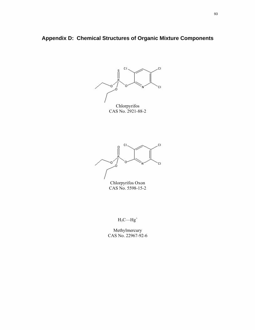
chlorpyrifos and its toxic metabolite, chlorpyrifos oxon, are provided in Appendix D.
As of 2000, chlorpyrifos was one of the most widely used organophosphorus insecticides in the United
States, for both agricultural and residential purposes. Registered uses included food crops, turf and
ornamental plants, indoor pest control (including crack and crevice treatment), termite control, mosquito
control, and pet collars. It was registered for use in a wide variety of buildings, including residential,
commercial, schools, daycare centers, restaurants, hospitals, hotels, and food manufacturing plants (EPA
2000c). Many uses of chlorpyrifos are being phased out (see Section A.4).
A study of urinary pesticide metabolites during the third trimester in 386 pregnant women from East
Harlem indicated that exposure to chlorpyrifos was prevalent (42% of the women had detectable levels of
the chlorpyrifos metabolite), was higher than the median in NHANES III, did not show seasonal
variation, and did not change during the time period of the study (1998–2001) (Berkowitz et al. 2003).
The exposure of these women was thought to be primarily indoor, due to household use and pesticide
exterminator application, and also dietary. A study of nine homes and 18 adult residents, 2/home,
conducted in the Lower Rio Grande Valley concluded that indoor dust and air were the primary exposure
media for the residents of those households, based on monitoring of those media, as well as outdoor soil
and air, food, and a characteristic urinary metabolite of chlorpyrifos (as a biomarker of exposure)
(Buckley et al. 1997).
A.1 Toxicokinetics
Chlorpyrifos is known to be absorbed through the respiratory tract in humans and animals, but
quantitative estimates were not available (ATSDR 1997; FAO/WHO 1999).
65
Chlorpyrifos is readily absorbed from the gastrointestinal tract. Based on the percent of administered
dose excreted in the urine, absorption of chlorpyrifos in humans was about 70% in a single-dose oral
study (Nolan et al. 1984), and in rats ranged from 84–90% in single-dose gavage studies (ATSDR 1997;
EPA 2000b).
Dermal absorption of chlorpyrifos (in dipropylene glycol methyl ether) in humans was about 1.3% of the
administered dose within 48 hours (Nolan et al. 1984). Dermal absorption in animals was much higher,
but results were confounded by dermal irritation, and even blistering, which compromised the integrity of
the skin.
The main features of chlorpyrifos metabolism (ATSDR 1997; Buratti et al. 2003; FAO/WHO 1999; Tang
et al. 2001) are:
• activation of chlorpyrifos by cytochrome P450 oxidative desulfuration of the P=S moiety to P=O,
resulting in the toxic intermediate, chlorpyrifos oxon;
• detoxification by cytochrome P450 dearylation of chlorpyrifos, resulting in 3,5,6-trichloro
2-pyridinol, and diethyl thiophosphate;
• detoxification by A-esterases (including paraoxonases) that hydrolyze the phosphate ester bonds
of chlorpyrifos and chlorpyrifos oxon to form 3,5,6-trichloro-2-pyridinol, and also diethyl
thiophosphate (from chlorpyrifos), or diethylphosphate (from chlorpyrifos oxon); and
• formation of glucuronide and sulfate conjugates of 3,5,6-trichloro-2-pyridinol (TCP).
Metabolic activation occurs predominantly in the liver. Detoxification occurs predominantly in liver and
plasma (ATSDR 1997; FAO/WHO 1999).
Metabolism of chlorpyrifos is rapid and extensive; the parent compound and the oxon are not detected or
are found only in trace concentrations in blood or urine, except following very high exposures. The
metabolite TCP is the principal form found in the circulation (ATSDR 1997; FAO/WHO 1999). The
elimination half-life for this metabolite in humans following oral or dermal exposure was approximately
27 hours (Nolan et al. 1984). Chlorpyrifos metabolites are excreted primarily in the urine (ATSDR 1997;
FAO/WHO 1999).
In humans, the cytochrome P450 isozymes that activate chlorpyrifos are CYP1A2 and CYP2B6 at low
chlorpyrifos concentrations (environmentally relevant) (Buratti et al. 2003), and CYP3A4 and also
66
CYP2B6 at higher concentrations (Burati et al. 2003; Tang et al. 2001). Isozymes involved in dearylation
of chlorpyrifos are reported to be CYP2C19 and CYP3A4 (Tang et al. 2001).
A.2 Health Effects
The principal toxic effect of chlorpyrifos in humans, experimental animals, and insects is acetylcholinest
erase inhibition. Acetylcholine is a neurotransmitter in the central and peripheral neurons. Inhibition of
acetylcholinesterase, the enzyme that breaks down and terminates the action of acetylcholine, results in
the accumulation of acetylcholine at acetylcholine receptors leading to continued stimulation.
In humans and experimental animals, the accumulation of acetylcholine results in cholinergic responses in
the peripheral (muscarinic and nicotinic) and central nervous system and neuromuscular junctions. These
cholinergic responses, seen in severe acetylcholinesterase inhibition, include excessive glandular
secretions (salivation, lacrimation, rhinitis), miosis, bronchoconstriction, vasodilation, hypotension,
diarrhea, nausea, vomiting, urinary incontinence, and bradycardia associated with muscarinic receptor
stimulation. Tachycardia, mydriasis (dilation of the pupil), muscle fasciculations, cramping, twitching,
muscle weakness, muscle paralysis, and hypertension are associated with nicotinic receptor stimulation.
Central nervous system toxicity includes respiratory depression, anxiety, insomnia, headache, apathy,
drowsiness, dizziness, loss of concentration, confusion, tremors, convulsions, and coma. These effects
usually appear within a few minutes to 24 hours after exposure, depending on the extent and route of
exposure. In nonfatal exposures, the effects are usually transient, with rapid and complete recovery
following cessation of exposure. Recovery from chlorpyrifos poisoning results from increased
availability of active acetylcholinesterase either from synthesis of new enzyme, the spontaneous
hydrolysis of the enzyme-phosphate ester complex, or treatment with atropine, a competitive antagonist
of acetylcholine at muscarinic and central nervous system receptors, and with pralidoxime (2-PAM), a
drug that regenerates inhibited acetylcholinesterase enzyme by displacing the diethylphosphoester bond
that chlorpyrifos oxon forms at the active site (Aaron and Howland 1998; ATSDR 1997).
Transient, delayed polyneuropathy has been reported in humans in case reports of acute- or intermediate-
duration exposure to chlorpyrifos; these reports did not adequately characterize exposure (ATSDR 1997;
FAO/WHO 1999). Chlorpyrifos has been tested for organophosphate-induced delayed neurotoxicity in
chickens; results were negative in these oral studies except at doses 4–6 times the LD50, which required
aggressive antidotal treatment (ATSDR 1997).
67
Acetylcholinesterase activity is also present in erythrocytes where it is known as erythrocyte acetyl
cholinesterase. Both forms of acetylcholinesterase are produced by the same gene and are kinetically
identical. In in vitro assays, erythrocyte and neural acetylcholinesterase are inhibited to roughly the same
extent by exposure to diazinon and many other organophosphorus compounds with insecticidal activity;
measurement of erythrocyte acetylcholinesterase can be used as a surrogate indicator of the extent of
inhibition of neural acetylcholinesterase (ATSDR 1997).
A cholinesterase capable of hydrolyzing acetylcholine and butrylcholine is produced by the liver and
circulates in the blood. This enzyme, referred to as serum cholinesterase, plasma cholinesterase, pseudo
cholinesterase, or butyrylcholinesterase, is also inhibited by chlorpyrifos and is often used as a marker for
exposure (ATSDR 1997). This enzyme is present in some nonneural cells in the central and peripheral
nervous systems as well as in plasma and serum, the liver, and other organs. Its physiologic function is
not known, but is hypothesized to be the hydrolysis of esters ingested from plants (Lefkowitz et al. 1996).
Plasma cholinesterases are also inhibited by organophosphate compounds through irreversible binding;
this binding can act as a detoxification mechanism as it affords some protection to acetylcholinesterase in
the nervous system (Parkinson 1996; Taylor 1996). In general, this enzyme is inhibited at lower levels of
organophosphate exposure than required to inhibit neural or erythrocyte acetylcholinesterase. For
chlorpyrifos, plasma cholinesterase is inhibited earlier in the time course of events following a single dose
or exposure, followed by acetylcholinesterase (ATSDR 1997).
Developing children (including infants and fetuses) are predicted to be more sensitive than adults to the
neurotoxicity of chlorpyrifos, based on studies in animals (ATSDR 1997; EPA 2000b).
Chlorpyrifos was evaluated for carcinogenicity in 2-year feeding studies in rats, mice, and dogs; results
were negative (ATSDR 1997; EPA 2000b).
A.3 Mechanisms of Action
Chlorpyrifos and chlorpyrifos oxon inhibit acetylcholinesterase by reacting with the active site to form a
stable dialkylphosphorylated enzyme that cannot hydrolyze acetylcholine. Chlorpyrifos oxon, the active
metabolic intermediate of chlorpyrifos, is much more potent than chlorpyrifos in inhibiting
acetylcholinesterase (ATSDR 1997; FAO/WHO 1999).
68
A.4 Health Guidelines
ATSDR (1997) did not derive inhalation MRLs for chlorpyrifos because of the lack of suitable
information for any exposure duration.
ATSDR (1997) derived acute and intermediate oral MRLs of 0.003 mg/kg/day for chlorpyrifos based on a
no-observed-adverse-effect level (NOAEL) of 0.03 mg/kg/day for plasma cholinesterase inhibition in
adult male volunteers who ingested chlorpyrifos by capsule for 20 days (Coulston et al. 1972). An
uncertainty factor of 10 was used for human variability. The lowest-observed-adverse-effect level
(LOAEL) (65% mean decrease in plasma cholinesterase; and symptoms possibly associated with
exposure in one of four volunteers) was 0.1 mg/kg/day for 9 days.
ATSDR (1997) derived a chronic oral MRL of 0.001 mg/kg/day based on a NOAEL of 0.1 mg/kg/day for
plasma, erythrocyte, and brain cholinesterase inhibition in rats fed chlorpyrifos in their diet for 2 years.
Plasma and erythrocyte cholinesterase were inhibited at 1 and 3 mg/kg/day, and brain cholinesterase was
inhibited at 3 mg/kg/day. An uncertainty factor of 100 was used to extrapolate from animals to humans
and to account for human variability.
EPA (IRIS 2004) derived a chronic RfD for chlorpyrifos based on the human study that ATSDR (1997)
also used for oral MRL derivation (referenced as Dow Chemical Company 1972 by IRIS 2004). EPA
applied an uncertainty factor of 10 to the no-observed-effect level (NOEL) of 0.03 mg/kg/day, resulting in
a chronic oral RfD of 0.003 mg/kg/day. This RfD was verified in 1986.
More recently, the EPA (2000c) Office of Pesticide Programs (OPP) reevaluated the use of the human
data as the basis for its acute and chronic RfDs because of a joint Science Advisory Panel/Science
Advisory Board meeting in December 1998 that discussed issues regarding the scientific and ethical
concerns for human toxicity testing. There was a concern that a regulatory decision cannot be based on a
human study until a formal decision has been made concerning the ethical aspects of this use (EPA
2000c). Since the ethics decision had not yet been made, and as part of the reevaluations conducted for
reregistration and under the FQPA, the EPA (2000c) OPP has reevaluated the human and animal data,
concluding that the human data provided useful information that can be used as supportive data, and
derived new RfDs based on the animal data. An acute RfD of 0.005 mg/kg/day, based on plasma
cholinesterase inhibition in an acute oral study in rats, and a chronic RfD of 3x10⁻⁴ mg/kg/day, based on
the weight of evidence for plasma and erythrocyte cholinesterase inhibition from five oral studies in dogs
69
and rats, were derived. Although not on in the Integrated Risk Information System (IRIS), these
derivations include a consideration of toxicological and mechanistic data that have become available
since the RfD on IRIS was derived (by OPP). These newer RfDs have been subjected to extensive
review, including public comment, and are available online (EPA 2000c). The FQPA safety factor of 10
(EPA 2003) was applied to these RfDs to estimate population adjusted doses (PADs) for children and
females 13–50 when assessing dietary (food + drinking water) exposures, resulting in a acute PAD of
5x10⁻⁴ mg/kg/day and a chronic PAD of 3x10⁻⁵ mg/kg/day (EPA 2000c).
In June of 2000, EPA announced an agreement with chlorpyrifos registrants to eliminate certain uses of
this pesticide (EPA 2000a, 2002a). Uses on foods frequently eaten by children (apples, grapes, tomatoes),
uses by homeowners (except for ant baits in child resistant containers), and uses in settings such as
schools and parks where children may be exposed, are being canceled, or phased out, or limited to
minimize exposure. Residential uses by licensed applicators are being phased out or limited to lower
application concentrations or rates. Reduced application rates for other agricultural uses and golf courses
also are being instituted to protect workers and wildlife.
The National Toxicology Program (NTP) (2003) and the International Agency for Research on Cancer
(IARC) (2003) do not include chlorpyrifos in their listings. The EPA (2002b) Office of Water classified
chlorpyrifos in Cancer Group D (not classifiable as to human carcinogenicity).
A.5 Derivation of Target-Organ Toxicity Dose (TTD) Values
The relevant endpoint for chlorpyrifos in this mixture is neurological. The intermediate oral MRL of
0.003 mg/kg/day for chlorpyrifos is based on neurological effects in humans. Chlorpyrifos is not known
to be a cumulative or persistent toxin, so this MRL is appropriate for the screening level assessment of
neurological effects of intermediate to chronic oral exposure to chlorpyrifos. Derivation of this MRL was
described in the Section A.4.
Summary (TTD for Chlorpyrifos)
MRLNEURO = 0.003 mg/kg/day
70
A.6 References
Aaron CK, Howland MA. 1998. Insecticides: Organophosphates and carbamates. In: Goldfrank LR, Flomenbaum NE, Lewin NA, et al., eds. Goldfrank’s toxicological emergencies. Stamford, CT: Appleton & Lange, 1429–1449.
ATSDR. 1997. Toxicological profile for chlorpyrifos. Atlanta, GA: Agency for Toxic Substances and Disease Registry.
Berkowitz GS, Obel J, Deych E, et al. 2003. Exposure to indoor pesticides during pregnancy in a multiethnic, urban cohort. Environ Health Perspect 111(1):79–84.
Buckley TJ, Liddle J, Ashley DL, et al. 1997. Environmental and biomarker measurements in nine homes in the lower Rio Grande valley: Multimedia results for pesticides, metals, PAHs, and VOCs. Environ Int 23(5):705–732.
Buratti FM, Volpe MT, Meneguz A, et al. 2003. CYP-specific bioactivation of four organophosphorothioate pesticides by human liver microsomes. Toxicol Appl Pharmacol 186(3):143– 154
Coulston F, Goldberg L, Abraham, R, et al. 1972. Final report on safety evaluation and metabolic studies on Dow co. 179(IN151). Inst Exp Pathol Toxicol, Albany Medical College. (As cited in ATSDR 1997.)
EPA. 2000a. Chlorpyrifos revised risk assessment and agreement with registrants. U.S. Environmental Protection Agency. Prevention, pesticides and toxic substances (7506C). http://www.epa.gov/pesticides/op/chlorpyrifos/agreement.pdf. June 2000.
EPA. 2000b. Toxicology chapter for chlorpyrifos. Washington, DC: U.S. Environmental Protections Agency. Office of Prevention, pesticides and toxic substances. http://www.epa.gov/oppsrrd1/op/chlorpyrifos/rev_tox.pdf. April 18, 2000.
EPA. 2000c. Human health risk assessment. Chlorpyrifos. Washington, DC: U.S. Environmental Protection Agency. http://www.epa.gov/oppsrrd1/op/chlorpyrifos/hedrra. June 8, 2000.
EPA. 2002a. Pesticides: Organophosphates. Chlorpyrifos facts. Washington, DC: U.S. Environmental Protections Agency. http://www.epa.gov/oppsrrd1/REDs/factsheets/chlorpyrifos_fs.htm.
EPA. 2000b. Toxicology chapter for chlorpyrifos. Washington, DC: U.S. Environmental Protections Agency. Office of Prevention, Pesticides and Toxic Substances. http://www.epa.gov/oppsrrd1/op/chlorpyrifos/rev_tox.pdf. April 18, 2000.
EPA. 2003. Pesticides. Regulating pesticides. The Food Quality Protection Act (FQPA). http://www.epa.gov/oppfead1/fqpa/backgrnd.htm.
FAO/WHO. 1999. Pesticide residues in food. Toxicological evaluations. International Programme on Chemical Safety. Food and Agriculture Organization & World Health Organization, 1–61. http://www.inchem.org/documents/jmpr/jmpmono/v99pr03.htm.
71
IARC. 2003. Overall evaluations of carcinogenicity to humans: As evaluated in IARC Monographs volumes 1–82 (a total of 885 agents, mixtures and exposures). International Agency for Research on Cancer. http://193.51.164.11/moneeval/crthall.html. January 2004.
IRIS. 2004. Integrated Risk Information Systems. U.S. Environmental Protection Agency. http://www.epa.gov.iris. January 2004.
Lefkowitz RJ, Hoffman BB, Taylor P. 1996. Neurotransmission: The autonomic and somatic motor nervous systems. In: Goodman LS, Gilman A, Hardman JG, et al., eds., Goodman & Gilman's the pharmacological basis of therapeutics. New York, NY: McGraw-Hill: Health Professions Division, 105–139.
Nolan RJ, Rick DL, Freshour NL. 1984. Chlorpyrifos: Pharmacokinetic in human volunteers. Toxicol Appl Pharmacol 73(1):8–15.
NTP. 2003. 10th report on carcinogens. U.S. Department of Health and Human Services. National Toxicology Program. http://ehp.niehs.nih.gov/roc/toc10.htm.
Parkinson A. 1996. Biotransformation of xenobiotics. In: Klassen CD, ed. Cassarett and Doull’s Toxicology Program. http://ehp.niehs.nih.gov/roc/toc10.htm.
Tang J, Cao Y, Rose RL, et al. 2001. Metabolism of chlorpyrifos by human cytochrome P450 isoforms and human, mouse, and rat liver microsomes. Drug Metab Dispos 29(9):1201–1204.
Taylor P. 1996. Anticholinesterase agents. In: Goodman LS, Gilman A, Hardman JG, et al., eds. Goodmand & Gilman’s the pharmacological basis of therapeutics. New York, NY: McGraw-Hill: Health Professions Division, 161–176.
72
Appendix B: Background Information for Lead
Lead is present in the environment primarily as divalent lead compounds. Contamination of the
environment was ubiquitous, even in residential areas, due to the use of leaded gasoline and lead paint.
Both of these uses have been phased out, but lead paint remains a problem in residences, and lead from
paint and gasoline remains in soil and household dust. Other sources of lead emissions or exposure
include mining, smelting, industrial activities, and hazardous waste sites (ATSDR 2005).
B.1 Toxicokinetics
Gastrointestinal absorption of soluble lead salts in adult humans can be high during fasting (40–50%), but
is about 3–15% when ingested with food. On the basis of dietary balance studies, gastrointestinal
absorption of lead in children appears to be higher and may account for 40–50% of the ingested dose.
Studies in animals also provide evidence that gastrointestinal absorption of lead is much higher in
younger organisms. Absorption is strongly affected by nutritional status, with higher absorption of lead
in children who are iron deficient. Calcium deficiency also may increase lead absorption, based on
studies in children. Co-administration of calcium with lead decreases lead absorption in adults, and in
animal studies. Vitamin D administration has been shown to enhance lead absorption in animal studies.
The distribution of lead appears similar across routes of exposure. Initially, lead is distributed to the
blood plasma and soft tissues, but under steady-state conditions, 99% of the lead in blood is found in the
erythrocyte, where much of it is bound to hemoglobin. Lead accumulates in blood, such that bone lead
accounts for approximately 73% of the body burden in children, increasing to 94% in adults. Inorganic
lead is not known to be metabolized, but lead ions are complexed by macromolecules. Unabsorbed lead
is excreted in the feces; absorbed lead that is not retained is excreted through the urine and bile (ATSDR
2005).
B.2 Health Effects
The effects of lead are similar across inhalation and oral routes of exposure. Lead has been shown to
affect virtually every organ and system in the body in both humans and animals. The most sensitive
effects of lead appear to be neurological (particularly in children), hematological, and cardiovascular.
Epidemiological studies provide evidence for an association between prenatal and postnatal exposure to
73
lead and adverse effects on neurodevelopment in infants and young children, and support the use of PbB
as an index of toxicological effect. The neurological effects included impaired cognitive ability and IQ
deficits in children. On the basis of several meta-analyses, it appears that a highly significant IQ
decrement of 1–3 points is associated with a change in PbB from 10 to 20 μg/dL. In addition,
associations between biomarkers of lead exposure and increased problem behavior in the classroom have
been reported (ATSDR 2005; Marlowe et al. 1985). In adult humans, slowing of nerve conduction
velocity occurs at PbBs of 30 μg/dL; peripheral nerve function appears to be affected in children at
similar PbBs. Oral studies in animals support the human evidence regarding neurobehavioral toxicity of
lead to infants and children from prenatal and postnatal exposure. In animals, lead has been shown to
alter a number of neurotransmitter systems including dopamine, norepinephrine, serotonin, and gamma
aminobutyric acid systems (ATSDR 2005).
Lead interferes with the synthesis of heme, resulting in accumulation of aminolevulinic acid
(ALA) in tissues and elevated excretion of ALA in urine, elevation of zinc protoporphyrin in erythrocyte,
reductions in blood hemoglobin, and in a hypochromic, normocytic anemia at higher levels of exposure.
Many epidemiological studies have found increases in blood pressure to be associated with increases in
PbB. The contribution of lead, as compared with other factors, is relatively small, and whether the
observed associations represent causality is controversial. Animal data demonstrate that oral exposure to
lead increases blood pressure. At higher levels of exposure in humans, lead produces cardiac lesions and
electrocardiographic abnormalities. Chronic nephropathy in humans is associated with PbB levels of 40–
>100 μg/dL. Oral exposure of animals to lead causes renal damage; histopathology is similar in humans
and animals and includes intranuclear inclusion bodies, swollen mitochondria, and tubular damage.
Adverse effects on the testes and sperm have been seen in occupationally exposed men with PbBs of 40–
50 μg/dL, and the more recent literature suggest that PbB concentrations <40 μg/dL also may be
associated with adverse effects on sperm counts and morphology (ATSDR 2005).
B.3 Mechanisms of Action
Lead can affect virtually every organ or system in the body through mechanisms that involve fundamental
biochemical processes. These mechanisms include the ability of lead to inhibit or mimic the action of
calcium and to interact with proteins. In the interaction with proteins, lead binds with virtually every
available functional group, including sulfhydryl, amine, phosphate, and carboxyl groups, with sulfhydryl
having the highest affinity. In its binding with sulfhydryl groups, lead may interfere with the activity of
74
zinc metalloenzymes, as zinc binds to a sulfhydryl group at the active site. Lead also binds to
metallothionein, a sulfhydryl-rich protein, but does not appear to displace cadmium or zinc. Metallo
thionein is induced by cadmium, zinc, and arsenic, but apparently not by lead, although metallothionein
sequesters lead in the cell. Another lead-binding protein is an acidic, carboxyl-rich protein found in the
kidney and brain (ATSDR 2005).
Lead interferes with heme synthesis by altering the activity of several mitochondrial and cytosolic
enzymes. One of the most sensitive hematological effects is inhibition of the cytosolic enzyme
aminolevulinic acid dehydratase (ALAD), with no threshold apparent through the lowest PbB levels
(3 μg/dL). Lead’s inhibition of ALAD occurs through binding of lead to vicinal sulfhydryls at the active
site of ALAD, where zinc is normally bound to a single sulfhydryl. Lead stimulates the mitochondrial
enzyme delta-aminolevulinic acid synthetase (ALAS), through feedback derepression, with a threshold in
human leukocytes at a PbB of about 40 μg/dL. As a result of the inhibition of ALAD and stimulation of
ALAS, ALA accumulates in blood, urine, and soft tissues, including brain. ALA is structurally similar to
gamma-aminobutyric acid (GABA), an inhibitory neurotransmitter. ALA appears to act as a GABA
agonist at the presynaptic GABA receptors, causing negative-feedback inhibition of GABA release. In
addition, ALA undergoes autooxidation, generating free radicals that may contribute to toxicity, and ALA
promotes oxyhemoglobin oxidation. At relatively high levels of lead exposure, anemia may occur due to
the interference with heme synthesis and also to red cell destruction. Decreases in tissue heme pools can
have deleterious effects throughout the body, not only because heme is a constituent of hemoglobin, but
also because heme is a prosthetic group of cytochrome P450 and the cytochromes of cellular energetics
(ATSDR 2005; EPA 1986). Lead inhibits the insertion of iron into protoporphyrin by the mitochondrial
enzyme ferrochelatase, possibly through binding of lead to the sulfhydryl groups of the active site or
indirectly through disruption of mitochondrial structure. Inhibition of ferrochelatase results in elevation
of zinc protoporphyrin (ZPP) in erythrocytes; ZPP is a sensitive indicator of lead exposure, occurring in
children at PbBs of about 25 μg/dL. Effects on heme synthesis are not restricted to the erythrocyte. A
number of studies suggest that lead-impaired heme production itself may be a factor in lead's neuro
toxicity (ATSDR 2005). Other potential mechanisms of neurotoxicity include lead acting as a calcium
agonist in a number of processes (ATSDR 2005), and lead inhibition of receptor binding to the NMDA
receptor channel, which does not appear to occur at the zinc allosteric site and is relatively insensitive
(Lasley and Gilbert 1999).
75
Mechanisms by which lead might affect blood pressure include effects on several hormonal and neural
regulatory systems, changes in vascular smooth muscle reactivity, cardiac muscle contractility, changes in
cell membrane cation transport systems, and possible effects on vascular endothelial cells (ATSDR 2005).
Lead has been shown to interfere with the DNA binding properties of zinc-finger regions of transcription
factors, and this interference could potentially elicit multiple responses, but consequences have not yet
been defined (Zawia et al. 2000).
B.4 Health Guidelines
ATSDR (2005) has not derived MRLs for lead. ATSDR (2005) has suggested the use of media-specific
slope factors and site-specific environmental monitoring data to predict media-specific contributions to
blood lead. The predicted contributions from the individual media are summed to yield a total predicted
PbB level. The media-specific slope factors were derived from regression analysis of lead concentrations
in water, soil, dust, diet, or air and PbBs for various populations.
The CDC determined in 1991 that blood lead levels of >10 μg/dL in children are to be considered
elevated (ATSDR 2005; CDC 1991).
EPA (IRIS 2004) has not developed a reference concentration (RfC) or RfD for lead. EPA stated that it
would be inappropriate to develop an RfD for inorganic lead (and lead compounds) because some of the
health effects occur at PbBs so low as to be essentially without a threshold. Instead, EPA defines lead
risk as the probability of exceeding a PbB of concern (i.e., 10 μg/dL) in children (EPA 1994a) or in
fetuses (EPA 1996). This approach is supported by human epidemiological studies that have associated
PbBs exceeding 10 μg/dL with impairment or delays in neurobehavioral development and other effects on
children (e.g., blood enzymes). EPA estimates lead risk in children using the Integrated Exposure Uptake
Biokinetic (IEUBK) model (EPA 1994b). This model translates estimates of site-specific exposure
concentrations into estimates of the probability that children’s blood leads will exceed a PbB of concern.
NTP (2001) has determined that lead acetate and lead phosphate can reasonably be anticipated to be
human carcinogens, based on sufficient evidence of carcinogenicity in experimental animals. NTP
(2001) considered lead chromate as one of the “Chromium Hexavalent Compounds.” IARC (1987) has
determined that the animal data are sufficient to classify lead and some lead compounds as possibly
76
carcinogenic to humans (Group 2B). EPA (IRIS 2004) classified lead in Group B2 (probable human
carcinogen). EPA did not develop an oral slope factor for lead because of the many uncertainties, some
of which may be unique to lead. An EPA inhalation unit risk also is not available for lead (IRIS 2004).
American Conference of Governmental Industrial Hygienists (ACGIH 2003) classified lead and certain
inorganic lead compounds as A3 carcinogens (confirmed animal carcinogen with unknown relevance to
humans). Lead chromate, assessed on the basis of both lead and chromate, was classified by ACGIH
(2003) as an A2 carcinogen—carcinogenic in animals at doses considered relevant to worker exposure,
but with insufficient epidemiological data to confirm risk to humans.
B.5 Derivation of Target-Organ Toxicity Dose (TTD) Values
A TTD for chronic oral exposure to lead was derived for the primary endpoint of concern for this mixture,
i.e., neurological effects in the fetus, infant, and young child from exposure to chlorpyrifos, lead, and
methylmercury. Relevant endpoints for another metal mixture, which is the subject of a separate
interaction profile, also included hematological, renal, cardiovascular, and testicular. For the sake of
completeness, the TTDs derived for those endpoints are retained in this Appendix, but are not
recommended for use with the present mixture. The chronic oral TTDs for lead were derived using the
methods described in ATSDR (2001a, 2001b). Because ATSDR’s approach to the assessment of lead
uses media-specific slope factors and site-specific contributions to PbB, the TTDs for lead are derived
based on PbB as well (see rationale in Chapter 3 of this profile). The derivations are based on data
provided in ATSDR (2005), and particularly Section 3.2 (Health), Chapter 2 (Relevance to Public
Health), and Section 3.6 (Biomarkers of Exposure and Effect). The derivation methods used similar
reasoning as for the CDC and EPA levels of concern (see neurological effects).
Neurological Effects
A large number of epidemiological studies and case reports indicate that exposure to lead causes
neurological effects. Slowing of nerve conduction velocity is associated with PbBs of 30 μg/dL in
children and adults. Of greater concern are the inverse linear relationships between IQ and other
neurobehavioral measures in children at PbBs extending down through 10 μg/dL or possibly lower.
Children appear to be more sensitive to the neurobehavioral toxicity of lead than are adults. Limited data
suggest an association between decreased neurobehavioral performance and PbB in aging subjects at
relatively low PbBs, indicating that the elderly may be another sensitive population. Although results of
77
the epidemiological studies in children are not entirely consistent, several meta-analyses have indicated
that a highly significant IQ decrement of 1–3 points is associated with a change in PbB from 10 to
20 μg/dL in children (IPCS 1995; Needleman and Gatsonis 1990; Pocock et al. 1994; Schwartz 1994).
The CDC (1991) determined that blood lead levels of >10 μg/dL are to be considered elevated in children,
based largely on concern for the effects of low-level lead exposure on the central nervous system. EPA
defines lead risk as the probability of exceeding a PbB of concern (10 μg/dL) in children or fetuses (EPA
1994a, 1996). The CDC level of concern for lead of 10 μg/dL is adopted as the TTD for neurological
effects (TTDNEURO).
Renal Effects
Chronic nephropathy is associated with PbB levels of 40–>100 μg/dL in humans exposed to lead
occupationally. There are some indications of renal damage in a study of children whose mean PbB was
34.2 μg/dL (increased N-acetyl-β-D-glucosaminidase activity in urine, a sensitive indicator) (Verberk
et al. 1996). The value for children, supported by the occupational data, and rounded to 34 μg/dL, is
taken as the TTD for renal effects (TTDRENAL).
Cardiovascular Effects
At higher levels of exposure, lead produces cardiac lesions and electrocardiographic abnormalities in
humans. Many epidemiological studies have reported an association between increases in blood pressure
and increases in PbB. The contribution of lead, as compared with other factors, is relatively small, and
whether the associations indicate causality is controversial. Animal data demonstrate that oral exposure
to lead increases blood pressure ATSDR (2005). The correlation between PbB and blood pressure is
apparent at relatively low PbBs extending through 10 μg/dL (e.g., Schwartz 1995). Therefore, the CDC
level of concern, 10 μg/dL, is adopted as the TTD for cardiovascular effects (TTDCARDIO).
Hematological Effects
Lead interferes with the synthesis of heme. The consequence at higher levels of exposure is a
hypochromic, normocytic anemia. The most sensitive indicator of effect on heme synthesis is the
inhibition of ALAD. ALAD activity is inversely correlated with PbB through the lowest levels of PbB in
the general population. Even in the absence of detectable effects on hemoglobin levels, there is concern
that effects on heme synthesis may have far-reach impacts, particularly on children (ATSDR 2005).
78
Accordingly, the CDC PbB of concern for children, 10 μg/dL (CDC 1991), is selected as the TTD for
hematological effects (TTDHEMATO).
Testicular Effects
Adverse effects of the testes and sperm have been reported in occupationally exposed men with PbBs of
40–50 μg/dL in some studies, but not in others, and are well-established at higher levels of exposure
(PbBs 66 μg/dL) (ATSDR 2005). The point of departure for increased risk of below normal sperm and
total sperm count was 40 μg/dL (Alexander et al. 1996). This value is selected as the TTD for testicular
effects (TTDTESTIC).
Summary (TTDs for Lead)
TTDNEURO = 10 μg/dL PbB = CDC level of concern
TTDRENAL = 34 μg/dL PbB
TTDCARDIO = 10 μg/dL PbB
TTDHEMATO = 10 μg/dL PbB
TTDTESTIC = 40 μg/dL PbB
Only the TTDNEURO is used in this interaction profile. As explained previously, the other TTDs were
derived for endpoints of concern for joint toxic action of a different mixture, which is the subject of a
separate interaction profile.
B.6 References
ACGIH. 2003. 2003 TLVs and BEIs. Threshold limit values for chemical substances and physical agents. Biological exposure indices. American Conference of Governmental Industrial Hygienists, Cincinnati, OH.
Alexander BH, Checkowaym H, van Netten C, et al. 1996. Semen quality of men employed at a lead smelter. Occup Environ Med 53:411–416. (As cited in ATDSR 2005.)
ATSDR. 2005. Toxicological profile for lead-Draft for Public Comment. Atlanta, GA: Agency for Toxic Substances and Disease Registry.
ATSDR. 2001a. Guidance manual for the assessment of joint toxic action of chemical mixtures. Atlanta, GA: Agency for Toxic Substances and Disease Registry.
79
ATSDR. 2001b. Guidance manual for preparation of an interaction profile. Atlanta, GA: Agency for Toxic Substances and Disease Registry. U.S. Department of Health and Human Services. Public Health Service.
CDC. 1991. Preventing lead poisoning in young children. Atlanta, GA: U.S. Department of Health and Human Services, Public Health Services, Centers for Disease Control and Prevention. (As cited in ATSDR 2005.)
EPA. 1986. Air quality criteria for lead. Research Triangle Park, NC: U.S. Environmental Protection Agency, Office of Research and Development, Office of Health and Environment Assessment, Environmental Criteria and Assessment Office. EPA 600/8-83-028F, 12–34 to 12–37.
EPA. 1994a. Guidance manual for the integrated exposure uptake biokinetic model for lead in children. U.S. Environmental Protection Agency. EPA/540/R-93/081. PB93-963510. (As cited in ATSDR 2005.)
EPA. 1994b. Technical support document: Parameters and equations used in integrated exposure uptake biokinetic model for lead in children (v0.99d). U.S. Environmental Protection Agency. EPA/540/R-94/040. PB94-963505. (As cited in ATSDR 2005.)
EPA. 1996. Bioavailability of lead in soil samples from the Jasper County, Missouri Superfund site. U.S. Environmental Protection Agency Region 8. Document Control No. 04800-030-0161. (As cited in ATSDR 2005.)
IARC. 1987. IARC monographs on the evaluation of carcinogenic risk to humans. International Agency for Research on Cancer, World Health Organization. Supplement 7, Vol. 1–47.
IPCS. 1995. Inorganic lead. Environmental Health Criteria 165 ed. Geneva: World Health Organization. International Programme on Chemical Safety. (As cited in ATSDR 2005.)
IRIS. 2004. Integrated Risk Information Systems. U.S. Environmental Protection Agency. http://www.epa.gov.iris. January 2004.
Lasley SM Gilbert ME. 1999. Lead inhibits the rat N-methyl-D-aspartate receptor channel by binding to a site distinct from the zinc allosteric site. Toxicol Appl Pharmacol 159:224–233.
Marlowe M, Cossairt A, Moon C, et al. 1985. Main and interaction effects of metallic toxins on classroom behavior. J Abnorm Child Psychol 13(2):185–198.
Needleman HL, Gatsonis CA. 1990. Low-level lead exposure and the IQ of children: A meta-analysis of modern studies. J Am Med Assoc 263(5):673–678.
NTP. 2001. 9th report on carcinogens. Research Triangle Park, NC: U.S. Department of Health and Human Services. National Toxicology Program. http://ehis.niehs.nih.gov/roc/toc9.html. September 11, 2001.
Pocock SJ, Smith M, Baghurst P. 1994. Environmental lead and children’s intelligence: A systematic review of the epidemiological evidence. Br Med J 309:1189–1197. (As cited in ATSDR 2005.)
Schwartz J. 1994. Low-level lead exposure and children’s IQ: A meta-analysis and search for a threshold. Environ Res 65:42–55. (As cited in ATSDR 2005.)

80
Schwartz J. 1995. Lead, blood pressure, and cardiovascular disease in men. Arch Environ Health 50(1):31–37.
Verberk MM, Willems TEP, Verplanke AJW, et al. 1996. Environmental lead and renal effects in children. Arch Environ Health 51(1):83–87.
Zawia NW, Crumpton T, Brydie M, et al. 2000. Disruption of the zinc finger domain: A common target that underlies many of the effects of lead. Neurotoxicology 21(6):1069–1080.

81
Appendix C: Background Information for Mercury and Methylmercury
Mercury exists in the environment as metallic mercury (also called elemental mercury), inorganic
mercury compounds (primarily mercuric), and organic mercury compounds (primarily methylmercury).
The structure of methylmercury is shown in Appendix D. Metallic and inorganic mercury released into
air from mining, smelting, industrial activities, combustion of fossil fuels, and natural processes can be
deposited to water and soil, where the mercury is transformed by microorganisms into methylmercury,
which bioaccumulates in the food chain, particularly in fish. For the general population, the most
important pathway of exposure to mercury is ingestion of methylmercury in foods, with fish, other
seafood, and marine mammals containing the highest concentrations (ATSDR 1999). Another source of
exposure for the general population is intake of metallic mercury from dental amalgams. Infants can be
exposed to inorganic mercury and methylmercury from breast milk, and the developing fetus can be
exposed through transplacental transfer of metallic mercury and methylmercury (ATSDR 1999). For
residents near mercury-contaminated hazardous waste sites, the following information provides insight
into important routes of exposure. Exposure analysis of residents near an abandoned industrial site that
had produced various inorganic and organic mercury compounds (and was not located near drinking
water sources) indicated that the children were exposed to mercury primarily though soil and dust
ingestion (Nublien et al. 1995).
C.1 Toxicokinetics
In humans, approximately 15% of a trace oral dose of inorganic mercury (mercuric nitrate) was absorbed
through the gastrointestinal tract (ATSDR 1999). Qualitative information indicates that ingested mercuric
chloride and mercuric sulfide also were absorbed through the gastrointestinal tract of humans. Studies in
animals indicate gastrointestinal absorption of inorganic mercury is in the 10–30% range, and depends on
intestinal pH, compound dissociation, and other factors. Qualitative evidence indicates that the
absorption of mercuric sulfide may be less than that of mercuric chloride. Absorption of inorganic
mercury tended to be higher in young animals than in adults. Following absorption from the
gastrointestinal tract, inorganic mercury distributes to the liver and kidneys, with the highest
concentrations in the kidneys. Although concentrations in brain are substantially lower, mercury was
retained longer in brain than in other tissues.
82
Metallic mercury is volatile and is readily absorbed (approximately 70–80%) through the respiratory tract,
and because of its lipophilic nature, it crosses the blood-brain and placental barriers (ATSDR 1999).
Retention of mercury from metallic mercury exposure is longest in the brain, based on data from humans.
Absorption of metallic mercury through the gastrointestinal tract, however, is negligible (ATSDR 1999).
Results from studies with humans and laboratory animals indicate that methylmercury and its salts (e.g.,
methylmercuric chloride and methylmercuric nitrate) are readily and completely absorbed by the gastro
intestinal tract, but quantitative information on absorption of methylmercury by the respiratory tract is not
available (ATSDR 1999). Absorbed methylmercury is widely distributed among tissues, with the kidney
showing the highest accumulation of mercury. Mercury from methylmercury can also accumulate in the
brain and fetus due to methylmercury’s abilities to penetrate the blood-brain and placental barriers and its
conversion in the brain and fetus to the inorganic divalent cation (ATSDR 1999). Excretion of methyl
mercury and other organic forms of mercury is thought to occur predominantly in the feces through
biliary excretion.
Studies with animals indicate that methylmercury, but not inorganic mercury, can be reabsorbed from the
gall bladder and the intestine, resulting in a biliary-hepatic cycle that contributes to longer clearance half
times for methylmercury compared with inorganic mercury (ATSDR 1999). Intestinal flora and various
mammalian tissues can produce the divalent mercury ion from methylmercury presumably via hydroxyl
radicals produced by cytochrome P450 reductase (ATSDR 1999). Inorganic mercury enters an oxidation-
reduction equilibrium between itself, mercurous mercury (Hg+), and metallic mercury (Hg0) (ATSDR
1999).
C.2 Health Effects
The nervous system is one of the primary sites of toxicity in humans and animals following exposure to
metallic mercury, methylmercury, or inorganic salts of mercury (ATSDR 1999). Neurological and
behavioral disorders (including hand tremors, emotional lability, and performance deficits in tests of
cognitive and motor function) have been observed in humans following inhalation of metallic mercury
vapor, ingestion or dermal application of medicinal products containing inorganic mercurous salts, and
ingestion of seafood contaminated with methylmercury. A single case study of lethal ingestion of
mercuric chloride reported neurological symptoms and brain lesions. Animal studies have demonstrated
changes in neurobehavioral function, morphology of neurological tissues, and brain neurochemistry
83
following inhalation exposure to metallic mercury or oral exposure to methylmercury. Data for
neurological effects of inorganic mercuric mercury salts are limited, and whether associated with oral
dosing is uncertain. Effects on neurological development ranging from delays in motor and verbal
development to severe brain damage have been observed in children of human mothers orally exposed to
organic forms of mercury, including methylmercury (ATSDR 1999). Animal studies provide
confirmatory evidence that neurological development of the fetus can be impaired by inhalation exposure
of the dams to metallic mercury or oral exposure to methylmercury (ATSDR 1999). Effects on
neurological development appear to occur at much lower doses of methylmercury than those producing
other effects discussed below (ATSDR 1999). Neurological effects may be the most sensitive effects of
inhalation exposure to metallic mercury (ATSDR 1999).
The kidney is another major site of mercury toxicity. Degeneration or necrosis of the proximal
convoluted tubules has been observed in humans and animals exposed to metallic mercury, inorganic
mercury, or methylmercury (ATSDR 1999). Renal damage is a sensitive effect, however, only for
inorganic mercury. In the absence of renal tubular degeneration, exposure to inorganic mercury has been
associated in several human cases and certain genetically disposed animals (New Zealand rabbits, Brown
Norway rats, and certain strains of mice) with a toxic glomerular response (proteinuria, deposition of
immune material in the renal mesangium and glomerular blood vessels, and minimal glomerular cell
hyperplasia) that is thought to involve mercury-induced autoimmunity through a stimulation of the
humoral and cellular immune systems and systemic autoimmunity (ATSDR 1999; Hultman and Enestrom
1992; Hultman et al. 1994; IRIS 2004). Studies demonstrating an association of this type of autoimmune
response with exposure to methylmercury were not located (ATSDR 1999).
Immunosuppressive effects have also been associated with mercury exposure including decreased T-cell
reactivity and decreased B cell levels in peripheral blood of mercury-exposed humans, increased
susceptibility of mercury-exposed animals to infectious agents, and decreased natural killer cell activity in
the spleen and blood of methylmercury-exposed rats (ATSDR 1999; Hultman and Enestrom 1992; Ilback
1991; Ilback et al. 1991).
Effects on male and female reproductive organs or functions associated with mercury exposure include
decreased sperm motility in male monkeys orally exposed to methylmercury, decreased spermatogenesis
and degeneration of seminiferous tubules in male mice after prolonged oral exposure to methylmercury,
impaired spermatogenesis and infertility in male rats and mice following parenteral administration of
84
methylmercury, and increased abortions, increased resorptions, or decreased implantations in female
monkeys, guinea pigs, and mice orally exposed to methylmercury (ATSDR 1999).
C.3 Mechanisms of Action
The high-affinity binding activity of divalent mercuric ion to thiol compounds or sulfhydryl groups of
proteins is thought to be a central molecular mode involved in the various toxic actions of inorganic
mercury and methylmercury (see ATSDR 1999 for review). The greater potency of methylmercury in
producing toxic effects, relative to mercuric salts, is thought to be due to differences in dispositional
processes, including gastrointestinal absorption and hepato-biliary recycling, leading to longer retention
times and higher doses of the mercuric ion at sites of toxicity.
Mercury-induced damage to neurological or renal tissues has been postulated to involve oxidative stress
damage from mercury-induced depletion of reduced glutathione levels, depolarization of mitochondrial
inner membranes leading to hydrogen peroxide formation, and depleted levels of reduced pyridine
nucleotides (ATSDR 1999). It has been further postulated that neurons are particularly sensitive to
mercury because of their low endogenous glutathione content or their inefficient glutathione reduction
activity (ATSDR 1999).
Postulates regarding methylmercury’s mechanism of action on the developing nervous system include
inhibitory effects of methylmercury on mitosis through impairment of microtubule assembly, methyl
mercury and inorganic mercury inhibition of enzymes such as protein kinase C, and inhibition of transport
mechanisms in developing brain cells (ATSDR 1999).
Molecular and cellular events underlying the immunosuppressive effects of mercury such as increased
susceptibility to infectious agents are unclear, but Shenker et al. (1993) showed that methylmercury or
mercuric chloride inhibited the mitogenic responses of cultured human T or B cells at concentrations that
were about 10-fold lower than those that caused cytotoxicity, and that methylmercury was more potent
than mercuric chloride. These authors postulated that immunosuppression involves inhibition by mercury
of early stages in the response of these cells to mitogens. The genetically-controlled autoimmunity
response to mercury that leads to glomerulonephropathy has been proposed to involve mercury disruption
of the balance of helper and suppressor cells within the immunoregulatory network, but the molecular and
cellular events that lead to glomerular immune-complex deposits have not been elucidated (ATSDR
85
1999). Hultman et al. (1994) showed that, in a genetically susceptible mouse strain, prolonged exposure
to inorganic mercury caused glomerular immune-complex deposits as well as stimulation of humoral
immunity (increased levels of IgM and IgG1), cellular immunity (increased expression of class II
molecules and increased mitogen-induced proliferation of T and B cells), and systemic autoimmunity
(increased autoantibodies against the nucleolus).
C.4 Health Guidelines
Data were inadequate for derivation of acute- or intermediate-duration inhalation MRLs for metallic
mercury, or for any duration inhalation MRL for inorganic mercury or for methylmercury (ATSDR
1999).
ATSDR (1999) derived a chronic inhalation MRL for metallic mercury vapor of 2x10⁻⁴ mg/m3 based on a
LOAEL in occupationally exposed humans of 0.026 mg/m3 for neurological effects (equivalent
continuous exposure concentration =0.0062 mg/m3, after adjusting for 8/24 hours/day and 5 days/week).
An uncertainty factor of 30 (10 for human variability and 3 for minimal-effect LOAEL) was used.
ATSDR (1999) did not derive oral MRLs for metallic mercury due to the lack of data. Oral exposure to
metallic mercury is expected to present little health risk, because it is so poorly absorbed through the
health gastrointestinal tract.
ATSDR (1999) derived an acute-duration oral MRL for inorganic mercury of 0.007 mg Hg/kg/day, based
on a NOAEL of 0.93 mg Hg/kg/day (5 days/week) for renal effects in rats administered mercuric chloride
for 2 weeks. The NOAEL dose was duration-adjusted and divided by an uncertainty factor of 100 (10 for
extrapolation from animals to humans and 10 for human variability). The LOAEL was 1.9 mg/kg/day,
5 days/week.
ATSDR (1999) derived an intermediate-duration oral MRL for inorganic mercury of 0.002 mg
Hg/kg/day, based on a NOAEL of 0.23 mg Hg/kg/day for renal effects in rats administered mercuric
chloride days/week for 6 months.
ATSDR did not derive a chronic oral MRL for inorganic mercury due to inadequate data.
86
ATSDR (1999) did not derive acute- or intermediate-duration oral MRLs for methylmercury due to the
absence of data or the lack of sufficient information regarding exposure levels associated with observed
effects.
ATSDR (1999) derived a chronic oral MRL of 3x10⁻⁴ mg Hg/kg/day for methylmercury based on
observations of no adverse effects in a 66-month evaluation of neurobehavioral development in children
who were conceived, born, and resided on the Seychelles Islands and were members of an isolated
population that consumed a high quantity and variety of ocean fish containing methylmercury. A
NOAEL for methylmercury of 0.0013 mg Hg/kg/day was calculated based on an average level of mercury
in maternal hair, 15.3 ppm, from a group (n=95) of the most highly exposed mothers. The NOAEL was
divided by a factor of 4.5 to arrive at the MRL. The factor of 4.5 was the sum of an uncertainty factor
of 3 (1.5 to address variability in hair-to-blood ratios among women and fetuses in the U.S. population
plus 1.5 to address any additional sources of human variability in response to methylmercury) and a
modifying factor of 1.5 to address uncertainty regarding the sensitivity of the neurobehavioral tests used
in the available report of the Seychelles Islands cohort study.
EPA (IRIS 2004) developed an inhalation RfC of 3x10⁻⁴ mg/m3 for metallic mercury based on a LOAEL
of 0.025 mg/m3 for 8-hour occupational exposure (converted to LOAEL of 0.009 mg/m3 for continuous
exposure), and using and uncertainty factor of 30 (10 for human variability and the use of a LOAEL, and
3 for database deficiencies, particularly the lack of developmental and reproductive studies). Inhalation
RfCs have not been developed by EPA for inorganic mercury (mercuric chloride) or for methylmercury.
EPA (IRIS 2004) has not developed a chronic oral RfD for metallic mercury.
EPA (IRIS 2004) derived a chronic oral RfD for inorganic mercury (mercuric chloride) of 3x10⁻⁴ mg
Hg/kg/day, based on a LOAELs for autoimmune effects (mercuric mercury induced autoimmune
glomerulonephritis) in rat subchronic oral and subcutaneous studies, and back-calculated from a drinking
water equivalent level of 0.010 mg/L recommended by a panel of mercury experts, following intensive
review and workshop discussions of the entire inorganic mercury database, including the rat LOAELs and
limited human tissue data. Data from Brown Norway rats were chosen because this strain is considered a
good surrogate for mercury-induced kidney damage in sensitive humans. An uncertainty factor of 1,000
was applied in this derivation (10 for extrapolation from a LOAEL to a NOAEL, 10 for the use of
subchronic studies, and a combined 10 for interspecies and intraspecies extrapolation).
87
EPA (IRIS 2004) derived a chronic oral RfD of 1x10⁻⁴ mg/kg/day for methylmercury based on an
estimated NOAEL of 0.857–1.472 μg/kg/day (maternal intake of methylmercury during pregnancy based
on estimated NOAEL cord blood range of 46–79 ppb) for neuropsychological effects in the offspring at
7 years of age in a longitudinal developmental study of about 900 mother-infant pairs from a fish-eating
population in the Faroe Islands (Grandjean et al. 1997). The NOAEL was estimated by a benchmark dose
approach as the 95% lower confidence limit for a daily dietary intake associated with 5% incidence for
the above neurological effects. It is supported by similar studies of mother-infant pairs in the Seychelles
islands and in New Zealand. The NOAEL was divided by an uncertainty factor of 10 to derive the RfD
(3 for human variability and uncertainty in estimating an ingested mercury dose from cord-blood mercury
data and 3 for human variability and uncertainty in pharmacodynamics).
EPA (IRIS 2004) classified metallic mercury in Group D (not classifiable as to human carcinogenicity)
based on inadequate human and animal data, and limited and equivocal findings from genotoxicity tests.
EPA (IRIS 2004) classified inorganic mercury (mercuric chloride) in Group C (possible human
carcinogen) based on the absence of data in humans and on limited evidence of carcinogenicity in rats
and mice. The evidence in animals was considered limited because: the relevance of the observed
forestomach papillomas in rats in one oral study of mercuric chloride is of questionable relevance to
humans because there was no evidence of progression to malignancy; the relevance of an increase in
thyroid tumors in male rats in the same study also is questionable because these tumors are generally
considered to be secondary to hyperplasia; the doses in this study exceeded the maximum threshold dose
(MTD) for male rats; and evidence for renal adenomas and adenocarcinomas in male mice in another oral
study of mercuric chloride study was equivocal. Genotoxicity assays of mercuric chloride gave mixed
results.
EPA (IRIS 2004) classified methylmercury in Group C (possible human carcinogen) based on inadequate
data in humans and limited evidence of carcinogenicity in animals. The animal evidence was judged to
be limited because: methylmercury-induced tumors (kidney tumors, from oral exposure) were observed
at a single site, in a single species and in a single sex; the tumors were observed only in the presence of
profound nephrotoxicity; several nonpositive oral cancer bioassays have also been reported; and the
evidence that methylmercury is genotoxic is equivocal. Quantitative estimates of cancer risk from oral
exposure were not derived based on evidence that methylmercury exerts its carcinogenic effects only at
high doses above a maximum tolerated dose and that systemic noncancer effects on the nervous system
88
would be seen at methylmercury exposure levels lower that those required to produce kidney damage and
subsequent kidney tumor development. Pertinent inhalation data were not available.
C.5 Derivation of Target-Organ Toxicity Dose (TTD) Values
An intermediate oral MRL is available for inorganic (mercuric) mercury, based on renal effects, and can
be used for the screening level assessment recommended in this interaction profile.
A chronic oral MRL is available for methylmercury, based on neurological effects, the primary endpoint
of concern for this mixture.
TTDs for oral exposure to methylmercury were derived previously for endpoints relevant to a different
mixture that is the subject of other interaction profiles. The previously derived TTDs are retained in this
appendix only for the sake of completeness. The methods used for deriving TTDs are described in
ATSDR (2001a). The derivations are based on data provided in ATSDR (1999), and in particular, the
oral Levels of Significant Exposure (LSE) table. Where the data were inadequate to derive a chronic oral
TTD for a given endpoint, the chronic oral MRL is recommended as a conservative alternative that is
protective of human health.
Renal Effects (Inorganic Mercury)
ATSDR (1999) derived an intermediate oral MRL for inorganic (mercuric) mercury was based on a
NOAEL of 0.23 mg Hg/kg/day for renal effects in rats administered mercuric chloride days/week for
6 months. The duration of the study was viewed as ideal, because it was long enough to detect long-term
effects and short enough not to mix them with renal effects of aging rats. This MRL is suitable for the
screening level assessment for intermediate to chronic oral exposure recommended in this interaction
profile.
Immunological Effects (Methylmercury)
Although immunosuppression is a known toxic endpoint for mercury, quantitative dose-response
information for methylmercury is limited. Ilback (1991) reported a LOAEL of 0.5 mg Hg/kg/day for
decreased thymus weight and cell number and reduced natural killer cell activity in mice fed methyl
mercury in the diet for 12 weeks. No other data were located regarding oral exposure to organic mercury
(ATSDR 1999). Because a free-standing intermediate LOAEL with no supporting data is not a suitable

89
basis for TTD derivation, the chronic oral MRL of 3x10⁻⁴ mg Hg/kg/day is adopted as TTDIMMUNO for
methylmercury. Using the chronic oral MRL as the TTD is protective of human health.
90
Neurological Effects (Methylmercury)
The neurological effects of methylmercury are well documented in humans and animals, and are the
critical effects of methylmercury (ATSDR 1999). The chronic oral MRL of 3x10⁻⁴ mg Hg/kg/day
developed by ATSDR (1999) is based on neurological effects in the population of particular concern for
this mixture (developing children). Therefore, a TTD is not needed for this endpoint.
Reproductive Effects (Methylmercury)
Studies of the reproductive effects of methylmercury are described in ATSDR (1999). The lowest
reliable LOAEL for reproductive effects was 0.06 mg Hg/kg/day, which produced an increased rate of
reproductive failure due to decreased conceptions and increased early abortions and stillbirths in female
monkeys treated with methylmercury for 4 months (Burbacher et al. 1988). The NOAEL in this study
was 0.04 mg Hg/kg/day. Although there was a report of sperm effects in male monkeys exposed to
0.025 or 0.035 mg Hg/kg/day by gavage for 20 weeks (Mohamed et al. 1987), this study was not
considered reliable by ATSDR (1999). Chronic studies in rodents reported testicular lesions (tubular
atrophy) and decreased spermatogenesis at approximately 0.7 mg Hg/kg/day, with NOAEL values of
roughly 0.1 mg Hg/kg/day in both sexes (Hirano et al. 1986; Mitsumori et al. 1990; Verschuuren et al.
1976). The 4-month monkey study is a suitable basis for a TTD. Application of an uncertainty factor of
100 (10 for extrapolation from monkeys to humans and 10 to protect sensitive individuals) to the NOAEL
of 0.04 mg Hg/kg/day yields a TTDrepro of 4x10⁻⁴ mg Hg/kg/day, which is only slightly higher than the
chronic oral MRL of 3x10⁻⁴ mg Hg/kg/day.
Summary (TTDs for inorganic mercury)
MRLRENAL = 0.002 mg Hg/kg/day
Summary (TTDs for methylmercury)
MRLNEURO = 3x10⁻⁴ mg Hg/kg/day (3x10⁻⁴ mg/kg/day)
TTDIMMUNO = 3x10⁻⁴ mg Hg/kg/day (3x10⁻⁴ mg/kg/day)
TTDREPRO = 4x10⁻⁴ mg Hg/kg/day (4x10⁻⁴ mg/kg/day)
91
For methylmercury, only the MRLNEURO is used in this interaction profile. As explained previously, the
other TTDs were derived for endpoints of concern for joint toxic action of a different mixture, which is
the subject of a separate interaction profile.
C.6 References
ATSDR. 1999. Toxicological profile for mercury. Atlanta, GA: U.S. Department of Health and Human Services, Public Health Service, Agency for Toxic Substances and Disease Registry
ATSDR. 2001a. Guidance manual for the assessment of joint toxic action of chemical mixtures. Atlanta, GA: Agency for Toxic Substances and Disease Registry.
Burbacher TM, Mohamed MK, Mottett NK. 1988. Methylmercury effects on reproduction and offspring size at birth. Reprod Toxicol 1(4):267–278.
Grandjean P, Weihe P, White RF, et al. 1997. Cognitive deficit in 7-year-old children with prenatal exposure to methylmercury. Neurotoxicol Teratol 19:417–428.
Hirano M, Mitsumori K, Maita K, et al. 1986. Further carcinogenicity study on methylmercury chloride in ICR mice. Jpn J Vet Sci 48(1):127–135.
Hultman P, Enestrom S. 1992. Dose-response studies in murine mercury-induced autoimmunity and immune-complex disease. Toxicol Appl Pharmacol 113:199–208.
Hultman P, Johansson U, Turley SJ, et al. 1994. Adverse immunological effects and autoimmunity induced by dental amalgam and alloy in mice. FASEB J 8:1183–1190.
Ilback N-G. 1991. Effects of methyl mercury exposure on spleen and blood natural killer (NK) cell activity in the mouse. Toxicology 67:117–124.
Ilback N-G, Sunderberg J, Oskarsson A. 1991. Methyl mercury exposure via placenta and milk impairs natural killer (NK) cell function in newborn rats. Toxicol Lett 58:149–158.
IRIS. 2004. Integrated Risk Information Systems. U.S. Environmental Protection Agency. http://www.epa.gov.iris. January 2004.
Mitsumori K, Hirano M, Ueda H, et al. 1990. Chronic toxicity and carcinogenicity of methylmercury chloride in B6C3F1 mice. Fund Appl Toxicol 14:179–190.
Mohamed M, Burbacher T, Mottett N. 1987. Effect of methyl mercury on testicular functions in Macaca fascicularis monkeys. Pharmacol Toxicol 60(1):29–36.
Nublien F, Feicht EA, Schulte-Hostede S, et al. 1995. Exposure analysis of the inhabitants living in the neighborhood of a mercury-contaminated industrial site. Chemosphere 30(12):2241–2248.
Shenker BJ, Berthold P, Rooney C, et al. 1993. Immunotoxic effects of mercury compounds on human lymphocytes on monocytes. III. Alterations in B-cell function and viability. Immunopharmacol Immunotoxicol 15(1):87–112.

92
Verschuuren HG, Kroes R, Den Tonkelaar EM, et al. 1976. Toxicity of methylmercury chloride in rats. III. Long-term toxicity study. Toxicology 6:107–123.
93
Appendix D: Chemical Structures of Organic Mixture Components
Chlorpyrifos CAS No. 2921-88-2
Chlorpyrifos Oxon CAS No. 5598-15-2
H3C—Hg+
Methylmercury CAS No. 22967-92-6