apoptosis & necrosis
DESCRIPTION
The difference between apoptosis and necrosisTRANSCRIPT
Which to choose? Apoptosis or Necrosis
Apoptosis
• (Gr. apo, off + ptosis, a falling). • highly regulated cellular activity • occurs rapidly and produces small membrane-
enclosed apoptotic bodies,• undergo phagocytosis by neighbouring cells or
macrophages.• BUT cells undergoing necrosis as a result of
accidental injury.
Desire to have Apoptosis
• Apoptotic cells: do not rupture and
release none of their contents.• release of cellular components causes a rapid
series of local reactions & cause inflammatory response.
• In DNA Damaged Cells; such response is undesirable.
• Apoptosis occurs rapidly and eliminate the cell without repercussions.

A few examples• Inside the thymus, T lymphocytes that may react
against self-antigens receive signals that activate the apoptotosis.
• In the mature ovary, apoptosis happen in both the monthly loss of luteal cells and removal of excess occytes + follicles.
• Plays an important role in formation of the central nervous system.
• Eliminating cells because of: lack of nutrients, by damage by free radicals; radiation, or by the action of tumor suppressor proteins
• Note: First discovered in embryos, where apoptosis is an essential for morphogenesis. Quicker than Mitosis; no traces.
How it happens?
• Most cells of the body can activate their apoptotic program when major changes occur in their DNA
• Ex: mutations accumulate in the DNA. prevents the proliferation to form a tumour.
• Malignant cells deactivate the genes that control the apoptotic process, thus avoiding death and allowing cancer progression.
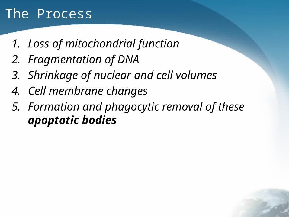
The Process
1. Loss of mitochondrial function
2. Fragmentation of DNA
3. Shrinkage of nuclear and cell volumes
4. Cell membrane changes
5. Formation and phagocytic removal of these apoptotic bodies
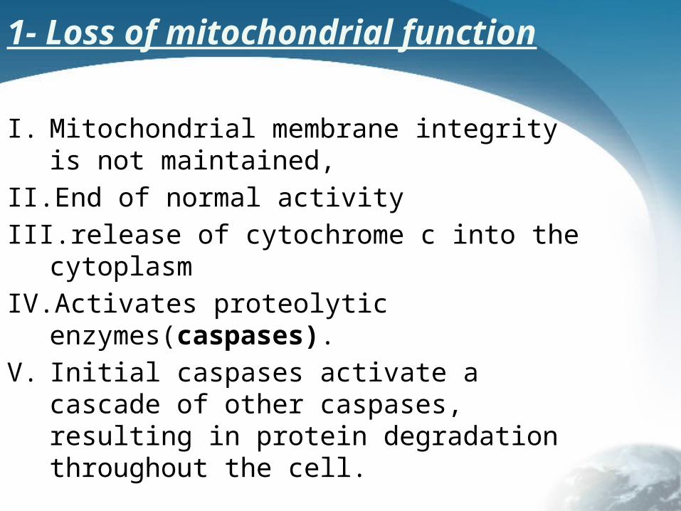
1- Loss of mitochondrial function
I. Mitochondrial membrane integrity is not maintained,
II. End of normal activity
III. release of cytochrome c into the cytoplasm
IV. Activates proteolytic enzymes(caspases).
V. Initial caspases activate a cascade of other caspases, resulting in protein degradation throughout the cell.
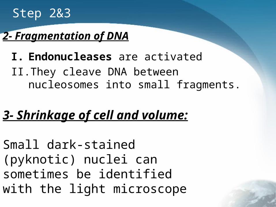
Step 2&3
I. Endonucleases are activated
II. They cleave DNA between nucleosomes into small fragments.
2- Fragmentation of DNA
3- Shrinkage of cell and volume:
Small dark-stained (pyknotic) nuclei can sometimes be identified with the light microscope
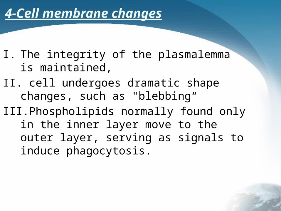
4-Cell membrane changes
I. The integrity of the plasmalemma is maintained,
II. cell undergoes dramatic shape changes, such as "blebbing“
III. Phospholipids normally found only in the inner layer move to the outer layer, serving as signals to induce phagocytosis.
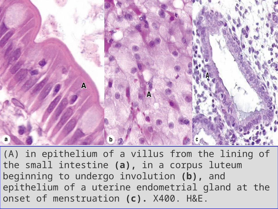
(A) in epithelium of a villus from the lining of the small intestine (a), in a corpus luteum beginning to undergo involution (b), and epithelium of a uterine endometrial gland at the onset of menstruation (c). X400. H&E.
Necrosis
• Caused by microorganisms, viruses, chemicals, and other harmful agents.
• Necrotic cells swell; their organelles increase in volume; and finally they burst, releasing their contents into the extracellular space.
• Macrophages engulf the debris of necrotic cells by phagocytosis and then secrete molecules that activate other immunodefensive cells to promote inflammation.
Reference
Junqueira's Basic Histology 12th ed.