antiproliferative and apoptotic potential of marine ... · cytotoxicity by trypan blue exclusion...
TRANSCRIPT
International Journal of Pharmacy and Biological Sciences
ISSN: 2321-3272 (Print), ISSN: 2230-7605 (Online)
IJPBS | Volume 8 | Issue 2 | APR-JUN | 2018 | 475-485
Research Article | Biological Sciences | Open Access | MCI Approved|
|UGC Approved Journal |
International Journal of Pharmacy and Biological Sciences Nithin T U and Keerthi T Raghavan*
www.ijpbs.com or www.ijpbsonline.com
475
ANTIPROLIFERATIVE AND APOPTOTIC POTENTIAL OF MARINE BACTERIA ALIIDIOMARINA TAIWANENSIS KU31894 ON HUMAN
HEPATOCELLULAR CARCINOMA (HEPG2) CELL LINE
Nithin Thoniparambil Unnikrishnana and Keerthi Thalakattil Raghavana*
aSchool of Biosciences, Mahatma Gandhi University, Kottayam, Kerala, India -686 560
*Corresponding Author, email id:
*Corresponding Author Email: [email protected]
ABSTRACT
Aim: To determine the antiproliferative and apoptotic effects of marine bacterium, Aliidiomarina taiwanensis
KU31894 on human hepatocarcinoma (HepG2) cell lines. Methods: The bacterium was identified by 16S rDNA
analysis and the bioactive metabolites were extracted. The cell viability and proliferation were done by trypan blue
exclusion and MTT assay against HepG2 cells. The bioassay guided fractionation of the promising extract was
performed by silica gel column chromatography. Apoptotic evaluation was done by acridine orange/ethidium
bromide staining and confirmed by DNA fragmentation assay. ROS intermediates generation was determined by
DCF-DA staining. Results: The bacterium was identified as Aliidiomarina taiwanensis and the sequences were
submitted in GenBank under the accession number KU31894. The ethyl acetate extract showed maximum
cytotoxicity by trypan blue exclusion method was selected for MTT assay, the IC50 concentration was obtained as
75.02 ± 1.7µg/ml. After the fractionation, the fraction F7 showed significant cytotoxicity by MTT assay with the
IC50 concentration of 48.75 ± 1.5µg/ml. The acridine orange /ethidium bromide staining of the IC50 concentration
of bio active fraction F7 treated cancer cells exhibited apoptotic characteristics and also showed DNA
fragmentation. The high levels of ROS intermediates as evidenced by DCF-DA staining could have played a
significant role in the apoptotic induction. Conclusion: Our results demonstrated that the proliferation of cancer
cells was significantly reduced with the treatment of bioactive fraction F7 of ethyl acetate extract. Our study
concluded that the marine bacteria Aliidiomarina taiwanensis KU31894 has the ability to produce anticancer
bioactive metabolites.
KEY WORDS
Aliidiomarina taiwanensis KU31894; anticancer activity; bioactive compounds; ethyl acetate extract; Marine
bacteria
1. INTRODUCTION
Hepatocellular carcinoma (HCC) is the most common
malignancy of the liver and is a leading cause of cancer
mortality. It is a complex and heterogeneous tumour
with reduced prognosis and occurs primarily in patients
with chronic liver disease and cirrhosis [1]. The hepatitis
B and C virus infection is the common cause of chronic
liver disease that eventually leads to liver cancer [2].
Several kinds of treatments might be useful for HCC,
such as surgical resection, percutaneous ethanol
injection, liver transplantation, transarterial
chemoembolization, and radioembolization. Moreover,
patients with HCC usually exhibit poor tolerance to
systemic chemotherapy because of their unusual liver
International Journal of Pharmacy and Biological Sciences Nithin T U and Keerthi T Raghavan*
www.ijpbs.com or www.ijpbsonline.com
ISSN: 2230-7605 (Online); ISSN: 2321-3272 (Print)
Int J Pharm Biol Sci.
476
function [3]. The progress of drug resistance in
hepatocellular carcinoma cells after drug therapy points
out the need for the detection of innovative anticancer
agents. Therefore, the use of natural products in this
respect is expansively under study [4]. Evidently, many
scientific societies are now involved in finding out novel
anti cancer drugs from natural products for advance
chemotherapy and to increased survival rates [5].
In recent years, marine microorganisms have attracted
great consideration in the pharmaceutical community
as they produce a broad diversity of structurally
unique and pharmacologically dynamic metabolites [6].
Extreme habitats provide marine microorganisms with
unique physiological and metabolic capabilities, which
may not be attributed by their terrestrial counterparts
[7]. Different kinds of marine microbial secondary
metabolites with remarkable bioactivities such as
antimicrobial, antiviral, antitumor or cytotoxic activity
were isolated [8]. Bioactive diversities of marine
microbial metabolites are probable resource for the
discovery of new anticancer drugs and provide
enormous applications in pharmaceutical and
biomedical research [9]. So far, a significant number of
anticancer compounds have been isolated from marine
microbes but only a few have been applied in clinical
trials and approved for the treatment of different types
of cancers.
The present research work communicates, the
inhibition of the proliferation of human hepatocellular
carcinoma cell line (HepG2) by the bioactive compounds
isolated from the marine bacteria Aliidiomarina
taiwanensis KU31894. The isolate showed promising
cytotoxic and apoptotic activity due to the presence of
various bioactive compounds.
2. MATERIALS AND METHODS
2.1 Identification of marine bacterium
Marine bacteria designated as MBTU_MB2, isolated
from the sea sediments (position Lat 10˚ 21’ 40.64’’N,
Lon 76˚ 6’ 18.30’’ E depth 10 m); collected from the sea
coast area of Thrissur, Kerala, India was used in the
present study. Colony morphology and effect of
physiological parameters such as salt concentration,
temperature, pH on the growth of the bacteria were
recorded [10]. Biochemical characterization of bacteria
was determined by using Analytical Profile Index (API)-
20 E test strips (bioMerieuix). Molecular identification
of the isolate was performed after isolation of the
genomic DNA using the method described for gram
negative bacteria [11]. The primers used for the
amplification of 16s rDNA were 27F
(51AGAGTTTGATCMTGGCTCAG 31) and 1492R (51
AAGGAGGTGWTCCARCC 31) [12]. PCR was carried out
by the thermal cycler under the following conditions- An
initialization step at 94ºC for 4 min followed by 30 cycles
of 94ºC for 1 min, 550C for 1 min, 720C for 2 min followed
by final extension at 72ºC for 1 min and holding
temperature at 4ºC for 1 min. The amplicon sequences
were analyzed using BLAST tool to get the relative
identification of each bacterial species [13]. All the
sequences were aligned using the multiple sequence
alignment program CLUSTAL W [14] and was used for
phylogenetic analysis using the MEGA 6 programs for
calculating the multiple distance matrixes [15]. The
multiple distance matrix obtained was then used to
construct phylogenetic trees using neighbor joining
method [16]. The sequence was deposited in GenBank.
2.2 Microbial production and extraction of
Bioactive Compounds
The isolate was inoculated into 10 ml of Zobell’s marine
broth 2216 (HIMedia) and kept in rotary shaker at 120
rpm for 48 hours at 370C. After incubation the culture
was centrifuged at 10000 rpm for 10 minutes at 40C, the
pellet was collected, washed with PBS (phosphate
buffer saline), resuspended in 1 ml PBS, and transferred
into 500 ml of the same broth. The cell suspension was
kept in rotary shaker at 120 rpm for 7 days at 370C for
the production of secondary metabolites. After
fermentation the culture broth was centrifuged at
10000 rpm for 10 minutes at 40C. The culture
supernatant obtained was used for extraction of
extracellular compounds by liquid-liquid extraction
method using equal amount of organic solvents such as
Hexane, Chloroform and Ethyl acetate. Filtrate and
organic solvent was mixed thoroughly by shaking them
in 1000 mL capacity separating funnel and the mixture
was then allowed to stand for 30 min. Two layers were
formed; the aqueous layer and the organic layer, which
contained the solvent and the bioactive compounds.
The organic layer was further concentrated by
evaporation under vacuum to the least volume [17]. The
concentrated extracts were then subjected to
cytotoxicity study.
2.3 Anticancer study
2.3.1 Cell Lines and Culture Conditions
International Journal of Pharmacy and Biological Sciences Nithin T U and Keerthi T Raghavan*
www.ijpbs.com or www.ijpbsonline.com
ISSN: 2230-7605 (Online); ISSN: 2321-3272 (Print)
Int J Pharm Biol Sci.
477
HepG2 (Liver cancer / Hepatocellular carcinoma) cell
line was initially procured from National Centre for Cell
Sciences (NCCS), Pune, India and maintained in
Dulbecco’s modified Eagles medium (DMEM) (Gibco,
Invitrogen). The cell line was cultured in DMEM
supplemented with 10% FBS, L-glutamine, sodium
bicarbonate and antibiotic solution containing:
Penicillin (100 U/mL), Streptomycin (100 µg/mL), and
Amphotericin B (2.5 µg/mL). Cultured cell lines were
kept at 370C in a humidified 5% CO2 incubator (NBS
Eppendorf, Germany).
2.3.2 Cell viability assay
The trypan blue dye exclusion assay (Sigma-Aldrich
T8154) is a quantitative method to determine the
percentage viability of cells. Briefly, 1×10⁴ HepG2 cells
were seeded into 96-well plates and treated with
different concentration of extracts (6.25, 12.50, 25.00,
50.00, and 100 μg/mL dissolved in 0.1% DMSO) in
triplicates and incubated for 3 hours at 370C. Then, 20
μL of suspension was mixed with equal volume of 0.4%
trypan blue and was counted by Neubauer
hemocytometer by clear-field microscopy. The stained
cells were counted, and percentage of cell death was
calculated [18]. Cell counts were expressed as mean ±
standard deviation (SD). The most active extract was
subjected to MTT assay for long term cytotoxicity
evaluation.
2.3.3 Cell proliferation assay
Cytotoxicity of the active extracts was measured by 3-
(4,5-dimethylthiazol-2-yl)-2,5-diphenyl tetrazolium
bromide (MTT) reduction assay. Cancer cells (HepG2)
were seeded at a density of 5x10⁴ cells per well in 96
well tissue culture plate and incubated for 24 hours. The
cells were treated with different concentrations of
extract (6.25, 12.50, 25.00, 50.00, and 100 μg/mL
dissolved in 0.1 % DMSO) in triplicates for 24 hours at
37oC in 5% CO2 humidifier incubator. After 24 hours, 20
μL of 5mg/ml MTT (pH: 7.4) solution was added to all
the wells and incubated for 3 hours. The medium was
aspirated and then added with DMSO to dissolve the
purple formazan crystals [19]. The absorbance values
were measured by using microplate reader at a
wavelength of 540 nm (ELISA Reader- ERBA, Germany).
Entire plate was observed by an inverted phase contrast
tissue culture microscope (Olympus CKX41 with Optika
Pro5 CCD camera). The IC50 value was calculated by
using GraphPad prism software.
2.4 Separation and bioassay guided fractionation
The separation of the most promising extracts was
performed by High performance thin layer
chromatography (HPTLC) using Solvent systems such as
Chloroform: Methanol (7:3; v/v). 15µL of extracts
dissolved in methanol and spotted on TLC plate (5.0 x
10.0 cm) [20]. The separated bands were visualized
under UV-illuminator and the Retention factor (Rf) value
was calculated by using CAMAG TLC Scanner. The
extract was then subjected to fractionation on a glass
column with silica gel (60– 120 mesh) and the column
was packed as per the wet packing system. The extract
(500 mg) was adsorbed on silica gel and gently layered
on top of the column. Step by step elution was done
with chloroform-methanol as solvent system
standardized by HPTLC. First the column was eluted
with 100% chloroform (Fraction 1). This step was
repeated by reducing the chloroform by 10% in each
fraction and the methanol was increased by 10% in each
fraction until percentage of methanol was 100%. Twelve
fractions were collected (each of 30 mL) and selected for
determining cytotoxicity by trypan blue exclusion
method described earlier. The bioactive fraction
obtained was used for further analysis of cytotoxicity by
MTT assay as described earlier and the apoptotic
potential was also determined by AO/EB staining as
described below.
2.5 Acridine Orange/Ethidium Bromide Staining
DNA-binding dyes Acridine Orange (AO) and Ethidium
Bromide (EB) (Sigma, USA) were used for the
morphological detection of apoptotic cells. After
treating HepG2 Cells with the IC50 concentration of
bioactive fraction for 24 hours, the cells were washed
with cold PBS (pH 7.4) and stained with a mixture of AO
(100 μg/mL) and EB (100 μg/mL) at room temperature
for 10 min [21]. The stained cells were washed twice
with PBS and observed using a fluorescence microscope
in blue filter of fluorescent microscope (Olympus CKX41
with Optika Pro5 camera).
2.6 DNA Fragmentation Analysis
DNA fragmentation assay was performed to confirm the
apoptotic mode of cell death. HepG2 cells were treated
with IC50 concentration of bioactive fraction for 24
hours. After treatment, cells were trypsinized and
collected with PBS. 100 μL of lysis buffer was added to
the pellet and incubated for 30 min on ice. After
incubation, centrifugation was carried out at 10000 rpm
for 30 min at 40C. The supernatant was collected and
International Journal of Pharmacy and Biological Sciences Nithin T U and Keerthi T Raghavan*
www.ijpbs.com or www.ijpbsonline.com
ISSN: 2230-7605 (Online); ISSN: 2321-3272 (Print)
Int J Pharm Biol Sci.
478
mixed with 25:24:1 mixture of
phenol:chloroform:isoamyl alcohol and precipitated
with two equivalents of ice cold ethanol and one-tenth
equivalent of sodium acetate. This was followed by
centrifugation at 12,000g for 20 minutes. The pellet was
resuspended in 30 μL of RNase solution (15 μg/mL) and
6 μL of 6x loading dye for 30 min at 370C which was
electrophoresed [22]. The fragmentation pattern was
imaged in a Syngene Ingenious gel documentation
system (Syngene Bioimaging Pvt. Ltd., Haryana, India).
2.7 Dichlorofluorescein Diacetate (DCF-DA) Staining
HepG2 cells were treated with the IC50 concentration of
bioactive fraction for 24 hours. The cells were washed
with PBS (pH 7.4) and stained with 10 𝜇M of DCF-DA for
30 min at 370C and wrapped in aluminum foil. The
treated cells were then washed twice with cold PBS-
EDTA, collected by trypsinization and centrifugation at
1500 rpm for 5 min, and resuspended in PBS-EDTA [23].
The fluorescence intensity was recorded by using a
fluorimeter at 470 nm excitation and emission at 635
nm. The Quantification of ROS Generation was also
determined using the fluorimeter (Qubit 3.0, Life
technologies, USA).
3. RESULTS AND DISCUSSION
3.1 Identification of marine bacterium
The marine bacterium MBTU_MB2, isolated from the
sea sediments of Kerala, India was used for the present
study was characterized to be halophilic, aerobic, Gram
negative and rod-shaped bacteria. After incubation on
Zobell’s marine agar at 370C, colonies appeared as light
green, circular, convex with entire edges. NaCl was
required for growth and the optimum concentration
was 3%, and optimum temperature for the growth was
observed at 370C, and the optimum pH was 7.
Biochemical characteristics determined using Analytical
Profile Index (API)-20 E test strip (bioMerieuix) revealed
that the bacterium is in the family Idiomarinaceae.
The 16S rDNA of the isolated organism was amplified by
using appropriate primers with the help of thermocycler
and the amplified product was subjected to agarose gel
electrophoresis. The 16S rDNA was sequenced by
dideoxy method and BLAST analysis was performed. The
sequence obtained was deposited in GenBank with the
accession number KU318394. Based on the homology of
16S rDNA sequence, strains with different sequence
similarity from relative species were selected and
multiple sequence alignment comparison was carried
out by BioEdit program. Then, bootstrap values were
calculated by neighbor-joining analysis method using
MEGA 6 software, and the phylogenetic tree was
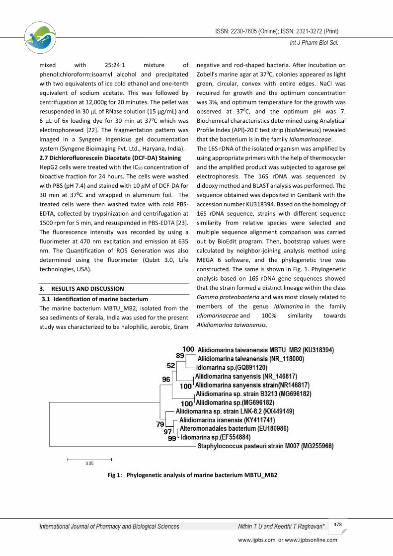
constructed. The same is shown in Fig. 1. Phylogenetic
analysis based on 16S rDNA gene sequences showed
that the strain formed a distinct lineage within the class
Gamma proteobacteria and was most closely related to
members of the genus Idiomarina in the family
Idiomarinaceae and 100% similarity towards
Aliidiomarina taiwanensis.
Fig 1: Phylogenetic analysis of marine bacterium MBTU_MB2
International Journal of Pharmacy and Biological Sciences Nithin T U and Keerthi T Raghavan*
www.ijpbs.com or www.ijpbsonline.com
ISSN: 2230-7605 (Online); ISSN: 2321-3272 (Print)
Int J Pharm Biol Sci.
479
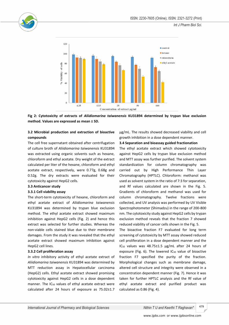
Fig 2: Cytotoxicity of extracts of Aliidiomarina taiwanensis KU31894 determined by trypan blue exclusion
method. Values are expressed as mean ± SD.
3.2 Microbial production and extraction of bioactive
compounds
The cell free supernatant obtained after centrifugation
of culture broth of Aliidiomarina taiwanensis KU31894
was extracted using organic solvents such as hexane,
chloroform and ethyl acetate. Dry weight of the extract
calculated per liter of the hexane, chloroform and ethyl
acetate extract, respectively, were 0.77g, 0.68g and
0.52g. The dry extracts were evaluated for their
cytotoxicity against HepG2 cells.
3.3 Anticancer study
3.3.1 Cell viability assay
The short-term cytotoxicity of hexane, chloroform and
ethyl acetate extract of Aliidiomarina taiwanensis
KU31894 was determined by trypan blue exclusion
method. The ethyl acetate extract showed maximum
inhibition against HepG2 cells (Fig. 2) and hence this
extract was selected for further studies. Whereas the
non-viable cells stained blue due to their membrane
damages. From the study it was revealed that the ethyl
acetate extract showed maximum inhibition against
HepG2 cell lines.
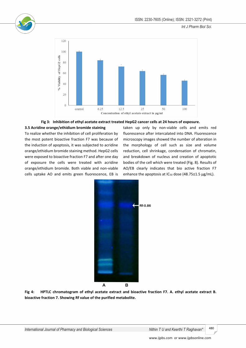
3.3.2 Cell proliferation assay
In vitro inhibitory activity of ethyl acetate extract of
Aliidiomarina taiwanensis KU31894 was determined by
MTT reduction assay in Hepatocellular carcinoma
(HepG2) cells. Ethyl acetate extract showed promising
cytotoxicity against HepG2 cells in a dose dependent
manner. The IC50 values of ethyl acetate extract were
calculated after 24 hours of exposure as 75.02±1.7
μg/mL. The results showed decreased viability and cell
growth inhibition in a dose dependent manner.
3.4 Separation and bioassay guided fractionation
The ethyl acetate extract which showed cytotoxicity
against HepG2 cells by trypan blue exclusion method
and MTT assay was further purified. The solvent system
standardization for column chromatography was
carried out by High Performance Thin Layer
Chromatography (HPTLC). Chloroform: methanol was
used as solvent system in the ratio of 7:3 for separation,
and Rf values calculated are shown in the Fig. 5.
Gradients of chloroform and methanol was used for
column chromatography. Twelve fractions were
collected, and UV analysis was performed by UV Visible
Spectrophotometer (Shimadzu) in the range of 200-800
nm. The cytotoxicity study against HepG2 cells by trypan
exclusion method reveals that the fraction 7 showed
reduced viability of cancer cells shown in the Fig. 5.
The bioactive fraction F7 evaluated for long term
screening of cytotoxicity by MTT assay showed reduced
cell proliferation in a dose dependent manner and the
IC50 values was 48.75±1.5 µg/mL after 24 hours of
exposure (Fig. 6). The lowered IC50 value of bioactive
fraction F7 specified the purity of the fraction.
Morphological changes such as membrane damage,
altered cell structure and integrity were observed in a
concentration dependent manner (Fig. 7). Hence it was
taken for further HPTLC analysis and the Rf value of
ethyl acetate extract and purified product was
calculated as 0.86 (Fig. 4).
International Journal of Pharmacy and Biological Sciences Nithin T U and Keerthi T Raghavan*
www.ijpbs.com or www.ijpbsonline.com
ISSN: 2230-7605 (Online); ISSN: 2321-3272 (Print)
Int J Pharm Biol Sci.
480
Fig 3: Inhibition of ethyl acetate extract treated HepG2 cancer cells at 24 hours of exposure.
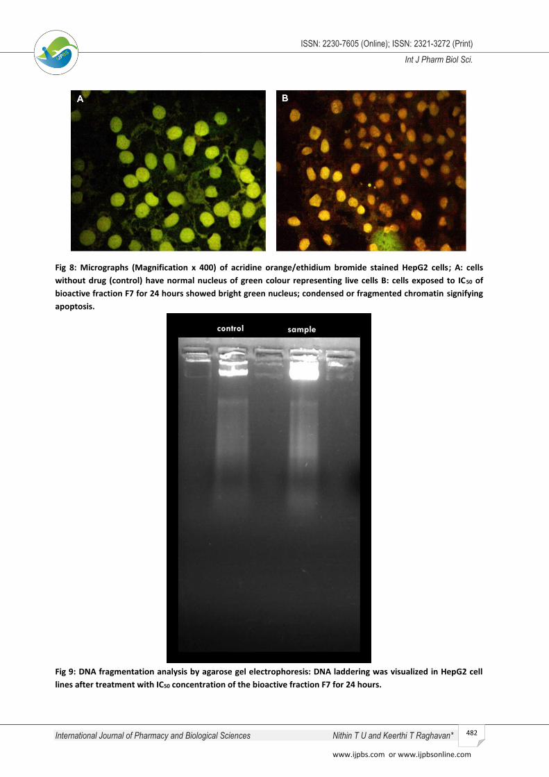
3.5 Acridine orange/ethidium bromide staining
To realize whether the inhibition of cell proliferation by
the most potent bioactive fraction F7 was because of
the induction of apoptosis, it was subjected to acridine
orange/ethidium bromide staining method. HepG2 cells
were exposed to bioactive fraction F7 and after one day
of exposure the cells were treated with acridine
orange/ethidium bromide. Both viable and non-viable
cells uptake AO and emits green fluorescence, EB is
taken up only by non-viable cells and emits red
fluorescence after intercalated into DNA. Fluorescence
microscopy images showed the number of alteration in
the morphology of cell such as size and volume
reduction, cell shrinkage, condensation of chromatin,
and breakdown of nucleus and creation of apoptotic
bodies of the cell which were treated (Fig. 8). Results of
AO/EB clearly indicates that bio active fraction F7
enhance the apoptosis at IC50 dose (48.75±1.5 µg/mL).
Fig 4: HPTLC chromatogram of ethyl acetate extract and bioactive fraction F7. A. ethyl acetate extract B.
bioactive fraction 7. Showing Rf value of the purified metabolite.

International Journal of Pharmacy and Biological Sciences Nithin T U and Keerthi T Raghavan*
www.ijpbs.com or www.ijpbsonline.com
ISSN: 2230-7605 (Online); ISSN: 2321-3272 (Print)
Int J Pharm Biol Sci.
481
Fig 5: Cytotoxicity of fractions against Hep G2 cells by trypan blue exclusion method.
Fig 6: Inhibition of bioactive fraction F7 treated HepG2 cancer cells at 24 hours of exposure
Fig 7: Morphological changes occurred in HepG2 cell lines after treatment with the bioactive fraction F7 for 24
hours.
International Journal of Pharmacy and Biological Sciences Nithin T U and Keerthi T Raghavan*
www.ijpbs.com or www.ijpbsonline.com
ISSN: 2230-7605 (Online); ISSN: 2321-3272 (Print)
Int J Pharm Biol Sci.
482
Fig 8: Micrographs (Magnification x 400) of acridine orange/ethidium bromide stained HepG2 cells; A: cells
without drug (control) have normal nucleus of green colour representing live cells B: cells exposed to IC50 of
bioactive fraction F7 for 24 hours showed bright green nucleus; condensed or fragmented chromatin signifying
apoptosis.
Fig 9: DNA fragmentation analysis by agarose gel electrophoresis: DNA laddering was visualized in HepG2 cell
lines after treatment with IC50 concentration of the bioactive fraction F7 for 24 hours.
International Journal of Pharmacy and Biological Sciences Nithin T U and Keerthi T Raghavan*
www.ijpbs.com or www.ijpbsonline.com
ISSN: 2230-7605 (Online); ISSN: 2321-3272 (Print)
Int J Pharm Biol Sci.
483
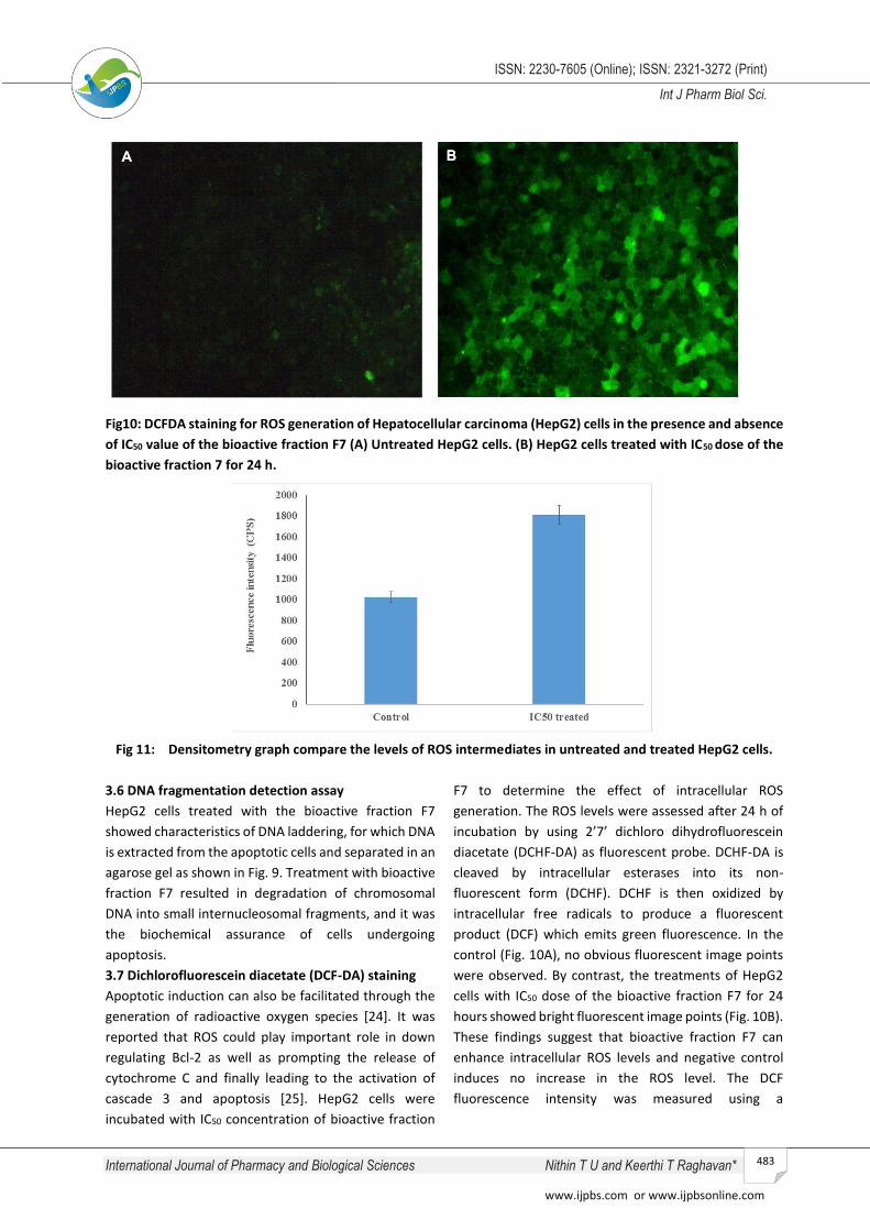
Fig10: DCFDA staining for ROS generation of Hepatocellular carcinoma (HepG2) cells in the presence and absence
of IC50 value of the bioactive fraction F7 (A) Untreated HepG2 cells. (B) HepG2 cells treated with IC50 dose of the
bioactive fraction 7 for 24 h.
Fig 11: Densitometry graph compare the levels of ROS intermediates in untreated and treated HepG2 cells.
3.6 DNA fragmentation detection assay
HepG2 cells treated with the bioactive fraction F7
showed characteristics of DNA laddering, for which DNA
is extracted from the apoptotic cells and separated in an
agarose gel as shown in Fig. 9. Treatment with bioactive
fraction F7 resulted in degradation of chromosomal
DNA into small internucleosomal fragments, and it was
the biochemical assurance of cells undergoing
apoptosis.
3.7 Dichlorofluorescein diacetate (DCF-DA) staining
Apoptotic induction can also be facilitated through the
generation of radioactive oxygen species [24]. It was
reported that ROS could play important role in down
regulating Bcl-2 as well as prompting the release of
cytochrome C and finally leading to the activation of
cascade 3 and apoptosis [25]. HepG2 cells were
incubated with IC50 concentration of bioactive fraction
F7 to determine the effect of intracellular ROS
generation. The ROS levels were assessed after 24 h of
incubation by using 2’7’ dichloro dihydrofluorescein
diacetate (DCHF-DA) as fluorescent probe. DCHF-DA is
cleaved by intracellular esterases into its non-
fluorescent form (DCHF). DCHF is then oxidized by
intracellular free radicals to produce a fluorescent
product (DCF) which emits green fluorescence. In the
control (Fig. 10A), no obvious fluorescent image points
were observed. By contrast, the treatments of HepG2
cells with IC50 dose of the bioactive fraction F7 for 24
hours showed bright fluorescent image points (Fig. 10B).
These findings suggest that bioactive fraction F7 can
enhance intracellular ROS levels and negative control
induces no increase in the ROS level. The DCF
fluorescence intensity was measured using a
International Journal of Pharmacy and Biological Sciences Nithin T U and Keerthi T Raghavan*
www.ijpbs.com or www.ijpbsonline.com
ISSN: 2230-7605 (Online); ISSN: 2321-3272 (Print)
Int J Pharm Biol Sci.
484
fluorimeter at 470 nm excitation and emission at 625
nm (Fig 11).
In the present study, increased levels of ROS in the
treatment group indicate the apoptotic mode of cell
death induced by the bioactive fraction F7. These
observations strongly suggest an outline to the possible
mechanism of action for the anticancer effect of the
isolated bioactive fraction F7 through the contributions
in the generation of ROS intermediates and triggering
the apoptotic cascade leading to the cell death.
4. CONCLUSION
On the basis of these results, it was confirmed that the
proliferation of cancer cells were significantly reduced
with the treatment of bioactive fraction F7 of ethyl
acetate extract. Based on the results of the present
study it was concluded that this Aliidiomarina
taiwanensis KU31894 bacterium isolates from sea
sediment of Kerala, India has the ability to produce
anticancer bioactive metabolites that has potential for
possible applications in future subject to clinical studies.
This is first report about the antiproliferative and
apoptotic potential of the marine bacterium,
Aliidiomarina taiwanensis KU31894. Further it is
suggested that there occurs the possibility for the
production of drug combinations for challenging
different types of cancers mainly liver cancer using
these bioactive compounds from marine bacteria.
ACKNOWLEDGEMENTS
Author Nithin T U, thank Mahatma Gandhi University,
Kottayam, and Kerala, India for financial support
through university JRF and also acknowledges the
Biogenix Research Centre, Trivandrum and DBT MSUB
School of Biosciences, M G University for providing
analytical facility for conducting the experiments.
REFERENCES
[1] Mazzanti, Roberto. “Hepatocellular Carcinoma: Where Are
We?” World Journal of Experimental Medicine 6(1): 21,
(2016)
[2] McGlynn, K. A., W, T. London. “The Global Epidemiology
of Hepatocellular Carcinoma, Present and Future.” Clin
Liver Dis. 15(2): 223–43, (2011)
[4] Abdul Hamid, N. M., Nazmy, M. H., Abdel-Bakey, A. I. Polyol
profile as an early diagnostic and prognostic marker in
natural product chemoprevention of hepatocellular
carcinoma in diabetic rats. Diabetes Res Clin Pract; 92:
228-37, (2011)
[5] Sawadogo, W. R., Schumacher, M., Teiten, M., Carmella, C.,
Dicato, M., Diederich, M. A Survey of Marine Natural
Compounds and Their Derivatives with Anti-Cancer
Activity Reported in 2011. molecules. 2013: 3641-3673,
(2011)
[6] Arif, J. M., Al-Hazzani, A. A., Kunhi, M., Al-Khodairy, F. Novel
marine compounds: Anticancer or genotoxic? Journal of
Biomedicine and Biotechnology, (2), 93–98, (2004)
[7] Wegener, G., Krukenberg, V., Ruff, S. E., Kellermann, M.
Y., & Knittel, K. Metabolic capabilities of microorganisms
involved in and associated with the anaerobic oxidation of
methane. Frontiers in Microbiology, 7: 1–16, (2016)
[8] Javed, F., Qadir, M. I., Janbaz, K. H., Ali, M. Novel drugs
from marine microorganisms. Crit Rev Microbiol. 37(3):
245-249, (2011)
[9] Banerjee, S., Wang, Z., Mohammad, M., Sarkar, F. H.,
Mohammad, R. M. Efficacy of selected natural products as
therapeutic agents against cancer. J Nat Prod; 71(3): 492-
496. (2008)
[10] Christen, R. and Ivanova, E. P. ‘Phylogenetic relationships
among marine Alteromonas -like proteobacteria:
emended description of the family Alteromonadaceae and
proposal of Pseudoalteromonadaceae fam. nov.,
Colwelliaceae fam. nov., Shewanellaceae fam.’,
International Journal of Systematic and Evolutionary
Microbiology, 54: 1773–1788. (2004)
[11] Chen, W., Kuo, T. A simple and rapid method for the
preparation of gram- negative bacterial genomic DNA.
Nucleic Acid Research, 21(9): 11529, (1993)
[12] Chun, J., Goodfellow, M. A Phylogenetic Analysis of the
Genus Nocardia with 16s rRNA Gene Sequences.
International Journal of Systemic Bacteriology, 45(2), 240–
245, (1995)
[13] Zhang. Z., Schwartz. S., Wagner. L., and Miller. W. Journal
of Computational Biology. 7(1-2): 203-214, (2004)
[14] Larkin, M. A., Blackshields, G., Brown, N. P. Clustal W and
Clustal X version 2 . 0. bioinformatics. 23(21): 2947-2948,
(2007)
[15] Tamura K, Peterson D, Peterson N, Stecher G, Nei M,
Kumar S. MEGA5 : Molecular Evolutionary Genetics
Analysis Using Maximum Likelihood , Evolutionary
Distance , and Maximum Parsimony Methods Research
resource. Molecular Biology and Evolution 28(10): 2731-
2739, (2011)
[16] Saitou, N., Nei, M. The Neighbor-joining Method: A New
Method for Reconstructing Phylogenetic Trees. j.
Molecular Biology and Evolution (4) :406–425, (1987)
[17] Atta, H. M., Radwan, H. G. Biochemical studies on the
production of Sparsomycin antibiotic by Pseudomonas
aeurginosa , AZ-SH-B8 using plastic wastes as fermented
substrate. Journal of Saudi Chemical Society, 16(1): 35–44,
(2012)
[18] Patel S, Gheewala N, Suthar A, Shah A. In-vitro
cytotoxicity activity of solanum nigrum extract against
International Journal of Pharmacy and Biological Sciences Nithin T U and Keerthi T Raghavan*
www.ijpbs.com or www.ijpbsonline.com
ISSN: 2230-7605 (Online); ISSN: 2321-3272 (Print)
Int J Pharm Biol Sci.
485
HeLa cell line and Vero cell line. Int J Pharm Pharm Sci;1:
38-47, (2009)
[19] Kennedy, R.K., Ravindra, V.V.P., Pragna, N. Phenazine-1-
carboxamide (PCN) from Pseudomonas sp . strain PUP6
selectively induced apoptosis in lung (A549) and breast
(MDA MB-231 ) cancer cells by inhibition of antiapoptotic
Bcl-2 family proteins. Apoptosis, (20): 858–868, (2015)
[20] Shewiyo, D. H., Kale, E., Risha, P. G., Dejaegher, B.,
Smeyers Verbeke, J., Hayden, Y. Vander. HPTLC methods
to assay active ingredients in pharmaceutical
formulations: A review of the method development and
validation steps. J Pharm Biomed Anal. 66:11-23, (2012)
[21] Ittiyavirah, S. P., Muraly, A., Rajiv, P., Raveendran, R.
Inhibition of growth and induction of apoptosis in PC3 and
A549 cell lines by hydro alcoholic fruit extract of Morinda
tinctoria roxb : in vitro analysis. 3(3):303-307, (2014)
[22] Devika, V., Mohandass, S. Original Research Article
Apoptotic induction of crude extract of Foeniculum
vulgare extracts on cervical cancer cell lines. Int.J.Curr.
Microbiol. App.Sci, 3(3): 657–661, (2014)
[23] Pajaniradje, S., Mohankumar, K., Pamidimukkala, R.,
Subramanian, S., Rajagopalan, R. Antiproliferative and
Apoptotic Effects of Sesbania grandiflora Leaves in Human
Cancer Cells. BioMed Research International, 11: (2014)
[24] Wang, J. Y., Jin, L., Yan, X. G., Sherwin, S., Farrelly, M.,
Zhang, Y. Y., Zhang, X. D. Reactive Oxygen Species Dictate
the Apoptotic Response of Melanoma Cells to TH588.
Journal of Investigative Dermatology, 136 (11): 2277–
2286, (2016)
[25] Passarella S, Atlante A, Valenti D, de Bari L. The role of
mitochondrial transport in energy metabolism.
Mitochondrion;2(5): 319-343, (2003)
*Corresponding Author: Keerthi Thalakattil Raghavan*
Email: [email protected]