antioxidant activity and hepatoprotective effect of ficus asperifolia ... · antioxidant activity...
TRANSCRIPT
Antioxidant activity and hepatoprotective effect of Ficus asperifolia (Miq.) on carbon tetrachloride-
induced hepatic damage in Wistar rats Oluwafemi Adeleke Ojo1,*, Adebola Busola Ojo2, Christopher Akintayo 3, Basiru Olaitan Ajiboye 1, Joseph Awe 1,
Tajudeen Obafemi 1
1Phytomedicine, Biochemical Toxicology and Diabetes Research Laboratory, Department of Biochemistry, Afe Babalola University, Ado-Ekiti, Ekiti State, Nigeria
2 Department of Medical Biochemistry, Afe Babalola University, Ado-Ekiti, Ekiti State, Nigeria. 3Department of Physiology, Afe Babalola University, Ado-Ekiti, Ekiti State, Nigeria.
Abstract Aim: In this study, we investigated the hepatoprotective effect of F. asperifolia (Miq) leaf extract against carbon tetrachloride-induced hepatic damage in male Wistar rats. Methods: Thirty rats (weighing 140 - 180 g) were divided into five groups. In each treatment group, aqueous extract of F. asperifolia (100, 200 and 400 mg/kg body weight) was administered by oral gavage for 21 days before exposure to hepatotoxic-carbon tetrachloride (CCl4) 3 mL/kg. Biochemical parameters such as GSH levels, CAT, GPx, SOD, lipid peroxidation (LPO), ALT, AST, ALP activities as well as Triglycerides (TG), Low Density Lipoprotein- and High Density Lipoprotein-Cholesterol (LDL-C and HDL-C) was examined. Results: Exposure to the CCl4 resulted in significant elevation in the level of LPO with concomitant depletion in the level of GSH, CAT and SOD activities compared with control. In like manner, serum aminotransferase activities were elevated and there were beneficial increases in serum HDL-C levels of rats treated with F. asperifolia. Conclusion: Daily oral administration of F. asperifolia showed beneficial and ameliorative effects in all biochemical parameter evaluated indicating that the studied aqueous leaf extract is a potent natural hepatoprotective agent.
Keywords: Ficus asperifolia (Miq), hepatoprotective effect, antioxidant enzyme activity, Carbon tetrachloride, serum lipid levels.
1. INTRODUCTION
Liver is a vivacious organ that has an extensive range of functions, including detoxification, plasma protein synthesis, and generation of biochemicals necessary for breakdown. Today, liver damage is one of very common aliment in the world leading to series of severe weaknesses ranging from stern metabolic disorders to even mortality[1]. Countless xenobiotics are known to cause hepatotoxicity; one among them is CCl4 that may cause lipid peroxidation [2]. Hence, damage to the liver inflicted by hepatotoxic agents is of grave consequences [3]. Hepatotoxicity is defined as injury to the liver that is associated with impaired liver function as a result of contact to a drug or additional non-infectious agent [4]. Hepatitis is one of the most prevalent diseases in the world [5] and hepatic problems account for a significant numberof liver transplantation documented worldwide. Since theavailable pharmacotherapeutic options for liver diseasesare very limited, there is a great demand for thedevelopment of new effective drugs [6]. In Ayurveda,many indigenous plants have been cited and wellrecognized as hepatoprotective agents [7].Ficus is a genus belonging to family Moraceae, whichcomprises about 850 species of woody trees, shrubs, vines,
epiphytes, and hemiepiphyte. Traditional healers in Africa specify that the leaves and fruits of Ficus asperifolia (Miq.) are used in a wide range of plant preparations as wound healing agents, anthelmintic and purgative [8] as well as to reverse some cases of sterility and infertility [9]. Root decoctions are used in the management of urinary tract ailments, gonorrhoea, asthma and tuberculosis. The root is chewed just in case of cough as well as an ingredient in a prescription to expel worms. Roots bark is also used against eye problems. Decoctions of the root bark are used in the treatment of coughs, worms, haemorrhoids and abnormal enlargement of the spleen. They are also used as ingredients in the curing of heart problems. Though, cold root bark extract is drunk in case of dizziness. Maceration of the root bark, combined with Senna occidentalis and Setaria megaphylla is taken to facilitate childbirth or to heal gonorrhea. Sap from the stem bark is used to stop bleeding, as a treatment of wounds, sores, abscesses, eye ailments, stomach-ache and for the removal of spines, but some traditional healers consider it corrosive to the skin and dangerous to ingest. The stem bark is locally applied on the body for the treatment of malaria. The leaves and young stems bark are abortifacient, analgesic, antidote, diuretic, emetic, oxytocic
Oluwafemi Adeleke Ojo et al /J. Pharm. Sci. & Res. Vol. 9(12), 2017, 2376-2381
2376
and stomachic. A decoction of the stem bark is taken for the treatment of dysentery; diseases of the kidneys and urinary tract; respiratory conditions such as coughs, colds, flu and asthma; hypertension. The young leaf is chewed and swallowed just in case of gastric ulcers. F. asperifolia (Miq.) is widely spread in Nigeria and other African countries such as Senegal, South Africa and Cameroon. F. asperifolia is copious in the grassland regions, especially along river banks and marshy areas at an altitude of up to 1100 m. The leaves are massive and displayed spirally, the limb is largely oval or has a form of ellipse and the roots are most often tough [10]. F. asperifolia has been shown to possess many pharmacological and physiological activities as an antioxidant [11]. Many biologically active secondary metabolites were isolated from Ficus benghalensis stem bark; such as, 20-tetratriaconthene-2-one, 6 heptatriacontene-10-one, pentatriacontan-5-one, β-sitosterol, β -D-glucoside and meso-inositol [12]. This plant is used by the local people for the treatment of various kinds of diseases, such as liver disorder, nausea, sterility and infertility, vomiting, from their traditional knowledge. The leaves are also used as paste for wound dressing. In addition, the fruit extract of F. benghalensis displayed antitumor activity [13], while the methanol extract of F. benghalensis possesses antioxidant activity [14]. F. sycomorus leaf extracts are used in folk medicine in the treatment of infertility and sterility in humans [15, 16]. On this background, the study investigates the hepatoprotective effect of F. asperifolia (Miq.) leaves extract against CCl4 induced hepatotoxicity in male Wistar rats in the search for a new natural agent.
2 MATERIALS AND METHODS 2.1 Plant material Leaves of Ficus asperifolia (Miq) was secured from local suppliers in Ado-Ekiti, Ekiti State. The plant material was authenticated and a voucher specimen (number UHAE/2016/23) was deposited in the herbarium by a senior taxonomist scientist at the Department of Plant Sciences, Ekiti State University, Nigeria. 2.2 Aqueous extraction Fresh leaves of F. asperifolia (Miq) were air dried in the laboratory at room temperature (30 ± 2°C) for 10 days, pulverized using a Kenwood blender and the powders obtained stored until further use. 50 g of the powdered sample was extracted with distilled water of 500 mL for 48 h. The mixture was decanted and filtered using sterile whatman paper No 1. The filtrate measured was evaporated to dryness using a freeze dryer to obtain 12 % yield. 2.3 Experimental animals Male Wistar albino rats (140–180 g) were maintained in the Laboratory Animal House of the College of Sciences, Afe Babalola University. Animals were housed in metallic cages and fed with standard feed diet and water ad libitum. The animals were exposed to alternative cycle of 12 h of dark and light. Male rats were used due to of their constant metabolism as compared to the disparity in the female
physiology. Animals were allowed to adapt to the laboratory environment for one week before investigation. The maintenance and conduct of the animals were in accordance with the internationally accepted standard rules and were approved by Afe Babalola institutional review board with ethical approval number (ABUAD/ACA/121). 2.4 Acute toxicity experiment Male Wistar rats were divided into control and test groups (6 animals each). Control group received the vehicle (normal saline) while the test groups got graded doses (1000–4000 mg/kg) of F. asperifolia (Miq.) aqueous extract orally and were observed for mortality till 48 h and the LD50 was calculated. 2.4.1 Doses Selected The dose selection for the aqueous extract of F. asperifolia (Miq.) was founded on the acute toxicity study, which did not show any adverse effect following oral administration of doses up to 3500 mg/kg. According to modified method of [17], experimental oral doses of 100, 200 and 400 mg/kg of the maximum possible dose of the extract was selected based on the acute toxicity study. 2.5 Experimental induction of hepatic damage CCl4 was dissolved in groundnut oil in the ratio 1:1 v/v. Liver damage was induced in rats following subcutaneous (SC) injection of CCl4 in the lower abdomen at a dose of 3 mL/kg [18]. 2.6 Hepatoprotective activity Thirty male Wistar rats were randomly divided into five groups of six animals each. Rats of the 1st (normal control) and 2nd (CCl4 alone) groups received the vehicle in a dose of 5 mL/kg. The 3th, 4th and 5th groups were treated with the aqueous extract of F. asperifolia (Miq.) in doses of 100, 200 and 400 mg/kg, respectively. All drugs were managed orally by gastric intubation for 7 consecutive days. Two hours after the last dose, normal control rats were administered a single dose of groundnut oil (3 mL/kg, SC), while animals of the 2nd to 5th groups received a single dose of CCl4 (3 mL/kg, SC). After 24 hours following the CCl4 injections, blood sample from each rat (2 mL) was removed by piercing their retro-orbital plexus of veins and collected in previously labeled centrifuging tubes and allowed to clot for 30 min at room temperature. Serum was separated by centrifugation at 10,000 rpm for 5 min. Livers were slice up and was kept in liquid nitrogen for determination of antioxidant status. 2.7 Measurement of liver function markers Liver functions were evaluated by measuring the serum activity of ALT and AST following the method of [19] whereas the activities of ALP were estimated by the methods of [20]. 2.8 Evaluation of CCl4 mediated oxidative stress Liver tissue was homogenized in 10 ml of 100 mM KH2PO4 buffer containing 1 mM EDTA (pH 7.4) and centrifuged at 12,000 rpm for 30 min at 4°C. The activities of the antioxidant enzymes such as SOD, GPx, CAT, were assayed in the hepatic tissue homogenate of the control and experimental rats according to previously described methods [21, 22, 23]. Moreover, the extent of lipid
Oluwafemi Adeleke Ojo et al /J. Pharm. Sci. & Res. Vol. 9(12), 2017, 2376-2381
2377
peroxidation (LPO) was estimated in liver homogenate of all rats as the concentration of thiobarbituric acid reactive product (malondialdhyde – MDA) according to [24], while GSH tissue content was measured according to the method described by [25]. 2.9 Histopathological study Liver from each rat was removed after dissection and preserved in 10% formalin. Liver tissues from each lobe were taken and possessed for paraffin embedding using the standard micro-technique. Sections (5 µm) of livers stained with hemotoxylin and observed microscopically for the evaluation of histopathological changes. 2.10 Data analysis Values are expressed as mean ± standard error of six observations in each group. The significance of difference in means between the control and treated animals’ groups were subjected to one-way analysis of variance (ANOVA), which was followed by the Duncan multiple range test for analysis of biochemical data using SPSS (20.0). Values were considered statistically significant at P < 0.05.
3. RESULTS Subcutaneous injection of CCl4 to rats showed significant elevation of liver marker enzymes (ALT, AST and ALP) in their serum after 24 hours of intoxication. The level of total bilirubin in the serum of CCl4-intoxicated control was also significantly increased when compared to the normal control group. Administration of the aqueous extract of F. asperifolia (Miq.) (100, 200 and 400 mg/kg) once daily for 21 days prior to CCl4, exhibited a significant hepatoprotective activity, resulting in reduction in the elevated serum activities of liver marker enzymes (Table 1) and level of total bilirubin (Table 2) when compared to CCl4- intoxicated rats. The oxidative stress caused by CCl4 in the liver was assessed by measuring the activity of hepatic antioxidant defense enzymes (SOD, GPx and CAT), GSH and the level of malondalydehe (MDA)
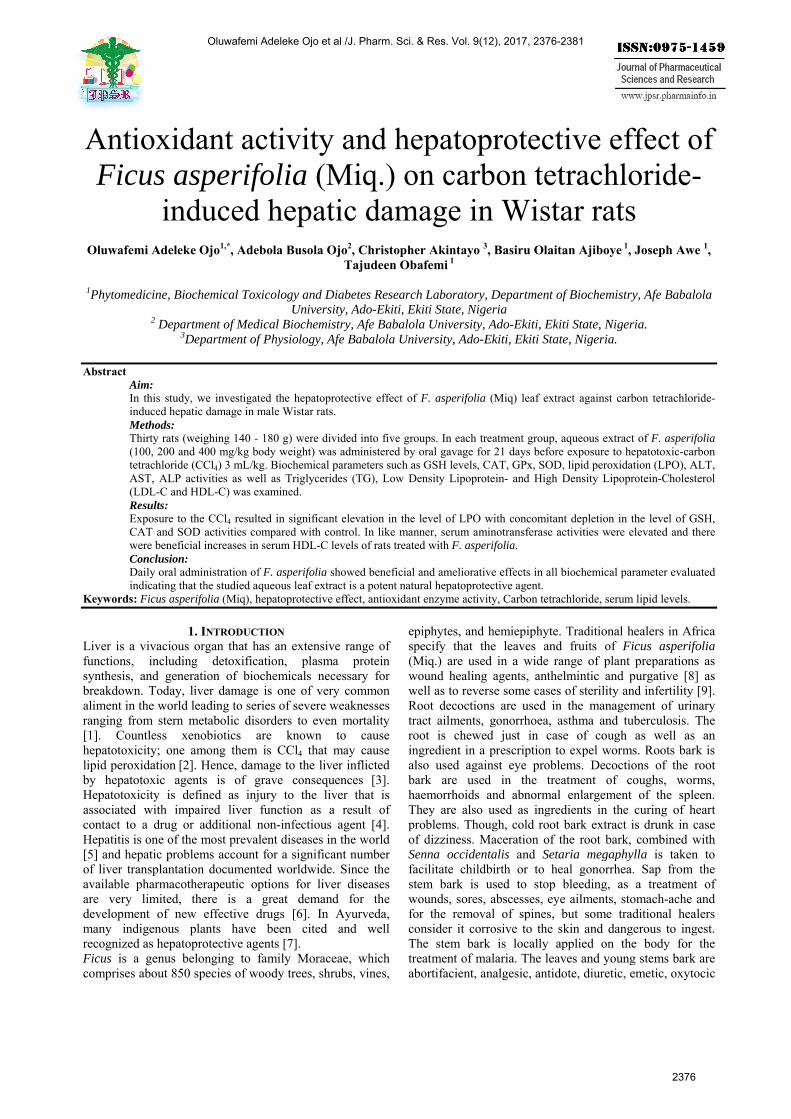
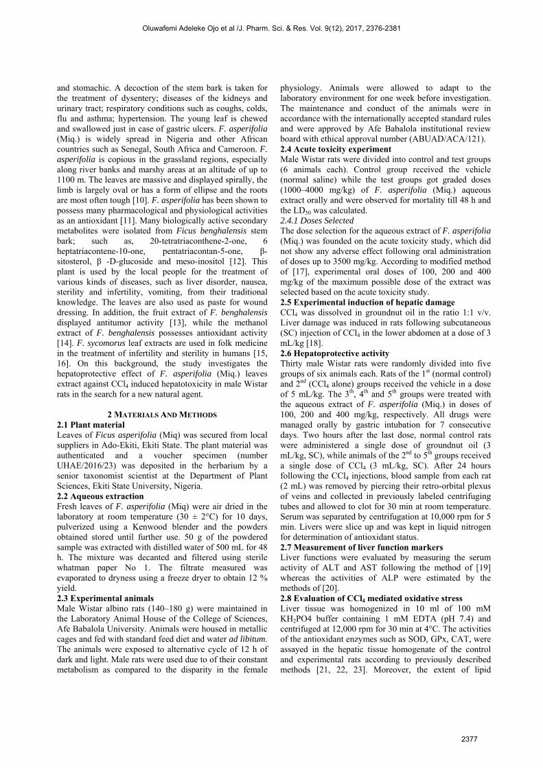
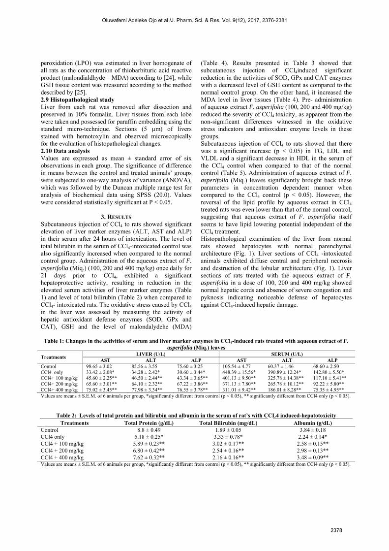
(Table 4). Results presented in Table 3 showed that subcutaneous injection of CCl4induced significant reduction in the activities of SOD, GPx and CAT enzymes with a decreased level of GSH content as compared to the normal control group. On the other hand, it increased the MDA level in liver tissues (Table 4). Pre- administration of aqueous extract F. asperifolia (100, 200 and 400 mg/kg) reduced the severity of CCl4 toxicity, as apparent from the non-significant differences witnessed in the oxidative stress indicators and antioxidant enzyme levels in these groups. Subcutaneous injection of CCl4 to rats showed that there was a significant increase (p < 0.05) in TG, LDL and VLDL and a significant decrease in HDL in the serum of the CCl4 control when compared to that of the normal control (Table 5). Administration of aqueous extract of F. asperifolia (Miq.) leaves significantly brought back these parameters in concentration dependent manner when compared to the CCl4 control (p < 0.05). However, the reversal of the lipid profile by aqueous extract in CCl4 treated rats was even lower than that of the normal control, suggesting that aqueous extract of F. asperifolia itself seems to have lipid lowering potential independent of the CCl4 treatment. Histopathological examination of the liver from normal rats showed hepatocytes with normal parenchymal architecture (Fig. 1). Liver sections of CCl4 -intoxicated animals exhibited diffuse central and peripheral necrosis and destruction of the lobular architecture (Fig. 1). Liver sections of rats treated with the aqueous extract of F. asperifolia in a dose of 100, 200 and 400 mg/kg showed normal hepatic cords and absence of severe congestion and pyknosis indicating noticeable defense of hepatocytes against CCl4-induced hepatic damage.
Table 1: Changes in the activities of serum and liver marker enzymes in CCl4-induced rats treated with aqueous extract of F.
asperifolia (Miq.) leaves
Treatments LIVER (U/L) SERUM (U/L)
AST ALT ALP AST ALT ALP Control 98.65 ± 3.02 85.56 ± 3.55 75.60 ± 3.25 105.54 ± 4.77 60.37 ± 1.46 68.60 ± 2.50 CCl4 only 33.42 ± 2.08* 34.28 ± 2.42* 30.60 ± 3.44* 448.39 ± 15.56* 390.89 ± 12.24* 142.80 ± 5.50* CCl4+ 100 mg/kg 45.60 ± 2.25** 46.50 ± 2.44** 43.34 ± 3.65** 401.13 ± 9.50** 325.78 ± 14.38** 117.10 ± 5.41** CCl4+ 200 mg/kg 65.60 ± 3.01** 64.10 ± 2.32** 67.22 ± 3.86** 371.13 ± 7.80** 265.78 ± 10.12** 92.22 ± 5.80** CCl4+ 400 mg/kg 75.02 ± 3.45** 77.98 ± 3.34** 76.55 ± 3.78** 311.01 ± 9.42** 186.01 ± 8.28** 75.35 ± 4.95** Values are means ± S.E.M. of 6 animals per group, *significantly different from control (p < 0.05), ** significantly different from CCl4 only (p < 0.05).
Table 2: Levels of total protein and bilirubin and albumin in the serum of rat’s with CCL4 induced-hepatotoxicity Treatments Total Protein (g/dL) Total Bilirubin (mg/dL) Albumin (g/dL)
Control 8.8 ± 0.49 1.89 ± 0.05 3.84 ± 0.18 CCl4 only 5.18 ± 0.25* 3.33 ± 0.78* 2.24 ± 0.14* CCl4 + 100 mg/kg 5.89 ± 0.23** 3.02 ± 0.17** 2.58 ± 0.15** CCl4 + 200 mg/kg 6.80 ± 0.42** 2.54 ± 0.16** 2.98 ± 0.13** CCl4 + 400 mg/kg 7.62 ± 0.32** 2.16 ± 0.16** 3.48 ± 0.09** Values are means ± S.E.M. of 6 animals per group, *significantly different from control (p < 0.05), ** significantly different from CCl4 only (p < 0.05).
Oluwafemi Adeleke Ojo et al /J. Pharm. Sci. & Res. Vol. 9(12), 2017, 2376-2381
2378
Table 3: Changes in the levels of liver antioxidant parameters in CCl4-induced rats treated with aqueous extract of F. asperifolia (Miq.) leaves
Treatment Liver
GSH GPx (mg /g tissue)
SOD CAT (U/mg protein)
Control 9.85 ± 0.15 2.95 ± 0.95 42.60 ± 1.01 15.77 ± 0.78 CCl4 only 5.62 ± 0.19* 1.77 ± 0.26* 27.12 ± 1.78* 8.75 ± 0.68* CCl4 + 100 mg/kg 6.42 ± 0.65** 1.48 ± 0.10** 30.38 ± 1.30** 9.29 ± 0.61** CCl4 + 200 mg/kg 7.42 ± 0.25** 1.92 ± 0.35** 36.72 ± 1.44** 10.42 ± 0.47** CCl4 + 400 mg/kg 8.05 ± 0.37** 2.28 ± 0.23** 39.21 ± 1.77** 11.60 ± 0.85** Values are means ± S.E.M. of 6 animals per group, *significantly different from control (p < 0.05), ** significantly different from CCl4 only (p < 0.05).
Table 4: Changes in the levels of lipid peroxidation in CCl4-induced rats treated with aqueous extract of F. asperifolia (Miq.) leaves
Treatments LIVER
(μmol MDA/mg protein) SERUM
(μmol MDA/mg protein) Control 36.05 ± 1.02 46.82 ± 1.98 CCl4 only 77.89 ± 2.05* 128.26 ± 5.61* CCl4 + 100 mg/kg 65.66 ± 1.14** 115.83 ± 5.76** CCl4 + 200 mg/kg 56.42 ± 1.65** 88.42 ± 3.65** CCl4 + 400 mg/kg 45.98 ± 2.42** 62.04 ± 4.40** Values are means ± S.E.M. of 6 animals per group, *significantly different from control (p < 0.05), ** significantly different from CCl4 only (p < 0.05).
Table 5: Effects of aqueous extract of F. asperifolia (Miq.) leaves on lipid profile of CCl4-induced rat Parameters Control CCl4 only CCl4 + 100mg/kg CCl4 + 200mg/kg CCl4 + 400mg/kg
TG (mg/dl) 65.0±1.28 135.6±3.65* 75.0±3.65** 54.50±3.44** 34.80±4.14** HDL (mg/dl) 184.0±1.62 48.60±2.73* 118.2±4.91** 162.40±5.62** 188.4±4.18** VLDL (mg/dl) 15.6±0.25 32.2±0.85* 13.80±0.87** 10.32±0.67** 6.90±0.45** LDL (mg/dl) 193.60±1.14 372.0±2.58* 198.20±4.87** 109.28±1.32** 45.40±7.54** Values are means ± S.E.M. of 6 animals per group, ** significantly different from CCl4 only (p < 0.05). *Key: TG – Triglycerides, HDL- High density lipoprotein, VLDL – Very low density lipoprotein, LDL – Low density lipoprotein.
Figure 1: Histology of liver tissues. (a) Liver section of normal control rat shows central vein surrounded by hepatic cord of cells (normal architecture), (b) liver section of CCl4-treated rats showing massive fatty changes, focal central vein congestion (indicated by arrow), ballooning formation, necrosis with inflammation, and loss of cellular boundaries, (c) liver section of rats treated with CCl4 and 100 mg/kg of F. asperifolia shows lesser fatty infiltration and absence of cell necrosis, rectified hepatocytes degeneration, (d) liver section of rats treated with CCl4 and 200 mg/kg of F. asperifolia showing absence of ballooning, inflammatory cells, and regeneration of hepatocytes around central vein toward near normal liver architecture but slight congestion in central vein, and (e) liver section of rats treated with CCl4 and 400 mg/kg of F. asperifolia showing repairing of hepatocytes.
Oluwafemi Adeleke Ojo et al /J. Pharm. Sci. & Res. Vol. 9(12), 2017, 2376-2381
2379
4. DISCUSSION
Chemicals including environmental toxicants and clinically useful drugs can cause severe cellular injuries in diverse organs of our body through the metabolic activation to highly reactive substances such as free radicals. CCl4 is one such extensively studied environmental toxicant [26]. Toxicity in liver due to CCl4 and other chemicals is attributed to the toxic metabolites formed, responsible for the initiation of CCl4 dependent lipid peroxidation, the dependent monooxygenase systems followed by its conversion to more chemically active form, trichloromethyl radical (CCl3). The enzymes involved in this process are located in the endoplasmic reticulum of the liver and its activities are reliant on many environmental factors. Some herbal extracts are known to prevent the oxidative injuries in different organs by varying the levels of cytochrome P-450 through their antioxidant properties [27]. The present study was conducted to evaluate the protective effect of F. asperifolia (Miq.) against CCl4 induced hepatic damage in rats. The results suggest that the extract possesses protective action against hepatic dysfunctions induced by the potent toxin CCl4. Similarly, rats intoxicated with CCl4 developed significant hepatic damage as manifested by a significant increase in the serum activities of ALT, AST and ALP that are indicators of hepatocyte damage and loss of functional integrity. Pre-treatment of rats with aqueous extract of F. asperifolia in doses of 100, 200 and 400 mg/kg effectively protected rats against CCl4-induced hepatic damage, resulting in reduction in serum activities of liver marker enzymes when compared to the CCl4-intoxicated control rats. Decrease in the level of these enzymes with F. asperifolia is an indication of the stabilization of plasma membrane as well as repair of liver damage caused by CCl4 similar to that reported by [17]. Furthermore, the rise in the level of total bilirubin in serum following CCl4
intoxication is also a measure of hepatotoxicity and could be attributed to impaired hepatic clearance due to hepatic parenchymal damage and biliary obstruction [28]. The ability of the aqueous extract of F. asperifolia (100, 200 and 400 mg/kg) to reduce the level of total bilirubin in the serum of intoxicated rats suggests its potential hepatoprotective effect. These could be due to the presence of phytochemicals such as saponins, alkaloids and phenols [29] The protective effect of F. asperifolia (Miq.) on the liver oxidative damage in rats induced by CCl4, was also evaluated by determining the antioxidant enzymes such as catalase (CAT), superoxide dismutase (SOD), glutathione reduced and peroxidase (GSH and GPx) in liver. Pre-treatment with aqueous extracts of F. asperifolia significantly (p < 0.05) ameliorated the activities of tissue parameters and CAT, SOD and GSH with significant (p < 0.05) increase levels when compared to CCl4 treated group (Table 3). Further it was confirmed by measuring lipid peroxidation (LPO) levels. The levels of MDA were significantly (p<0.05) elevated in CCl4 alone group when
compared with the normal group (Table 4). Treatment with F. asperifolia significantly (p < 0.05) decreased the levels as compared to CCl4 treated group suggesting that F. asperifolia has antioxidant activity as reported by [11]. Abnormal lipid metabolism, resulting in accumulation of LDL-C, VLDL and total cholesterol as well as decreased HDL-cholesterol, is often related to diabetes mellitus [30]. Elevated levels of LDL, VLDL, atherogenic index, coronary artery index and total cholesterol are thought of as major risk factors for cardiovascular disease (CVD). There was a significant (p < 0.05) decrease in HDL and a significant increase in triglycerides (TG), very low density lipoprotein (VLDL) and low density lipoprotein (LDL) in the serum of the CCl4 (Table 5) control when compared to the normal control. Furthermore, the CCl4 treated group administered with aqueous extract of F. asperifolia had these parameters reversed and even lower than that of normal control. These could be due to the presence of bio-constituents in the leaf extract [29].The extent of damage by CCl4 was further supported by histological changes in the liver of animals. Histological investigation of the liver sections showed that the normal liver architecture was disturbed by the hepatotoxin intoxication [31]. There were signs of necrosis and derangement of hepatic cells in the liver section of rats treated with CCl4 alone. Whereas in sections obtained from rats pretreated with F. asperifolia leaf extract and intoxicated with CCl4, the normal cellular architecture was retained as compared to those of the control rats.
CONCLUSION The present study shows that F. asperifolia (Miq.) offered significant protection against the hepatotoxicant CCl4 in rats, which may be attributed to its constituents (which are mainly polyphenolic compounds) with its antioxidant and membrane stabilizing properties. Further studies are required to identify the particular active phytochemicals and the precise mechanism(s) underlying the beneficial effects of this plant.
ACKNOWLEDGMENT Authors acknowledge the laboratory assistant in providing the necessary facilities to carry out this study.
AUTHORS’ CONTRIBUTIONS OAO: designed the experiment; ABO and BOA: conducted analysis and drafting of the manuscript; CA, TO performed the experiments; JA: collection of data; analysis and evaluation of data; statistical analysis; All authors read and approved the final version of the manuscript.
REFERENCES [1] Akilavalli N, Radhika J, Brindha P. (2011). Hepatoprotective
activity of Ocimum sanctum Linn. against lead induced toxicity inalbino rats. Asian J Pharm Clin Res. 1(4):84–87.
Oluwafemi Adeleke Ojo et al /J. Pharm. Sci. & Res. Vol. 9(12), 2017, 2376-2381
2380
[2] Demirdag K, Bakcecioglu I, Ozercan IH, Ozden M, Yilmaz S,Kalkan A. (2004). Role of L-carnitine in the prevention of acuteliver damage induced by carbon tetrachloride in rats. JGastroenterol Hepatotoxi. 19:333–338.
[3] Shahani A, Handa SS. (1995). Hepatoprotective activity of Apium graveolens and Hygrophilla auriculata against paracetamol andthiocetamide intoxication in rats. J Ethnopharmacol. 49:119-126.
[4] Navario VJ, Senior JR. (2006). Drug related hepatotoxicity. J Med.354: 731-739.
[5] Wang N, Li P, Wang Y, Peng Y, Wu I. (2006). Hepatoprotectiveeffect of Hypricum japonicum extract and its function. J Ethnopharmacol. 116:1-6.
[6] Abidemi JA, Kenneth OE, Chidozie CA. (2010). Hepatoprotectiveand in vivo antioxidant effect of Byrsocarpus coccineus scheum andthonn (connaraceae). J Ethnopharmacol. 129:46-52.
[7] Surendra H, Ram A. (2007). Hepatoprotective properties ofBauhinia variegate bark extract. Yakugaku Zasshi. 127:1503-1507.
[8] Sofowora A. (1996). Plantes médicinales et médecine traditionnelled’Afrique. Edition Karthala, 75013 Paris, 375.
[9] Ojo OA, Ojo AB, Ajiboye B, Fadaka A, Imiere OD, Adeyonu O,Olayide, I. (2016). Protective Influence of Ficus asperifolia MiqLeaf Extract on Carbon tetrachloride (CCL4)-Induced TesticularToxicity in rat’s Testes. J App Pharm Sci. 6 (06): 037-041.
[10] Adjanohoun JC, Aboubaker N, Dramane K, Ebot ME, Ekpere JA,EnowOrock EG. (1996). Contribution to Ethnobotanical andFloristic Studies in Cameroon. Lagos, Nigeria: OUA/STRC.Traditional medicine and pharmacopoeia, Pp. 301–325.
[11] Ojo OA, Akintayo CO. (2014). Assessment of Antioxidant activityof Ficus asperifolia Miq aqueous extract- in vitro studies. The Journal of Phytopharmacology. 3(1): 16-21.
[12] Mousa O, Vuorela P, Kiviranta J, Wahab SA, Hiltohen R. (1994).Bioactivity of certain Egyptian Ficus species. J Ethnopharmacol.41:71–76.
[13] Joy PP, Thomas J, Mathew S, Skaria BP. (2001). “Medicinalplants’’, Tropical Hariculture, Naya Prokash, Calcutta, 2:499.
[14] Yadav YC, Srivastava DN, Saini V, Singhal S, Seth AK, Kumar S.(2011). In-vitro antioxidant activity of methanolic extraction ofFicus Benghalensis L. latex. Pharmacologyonline. 1:140–148.
[15] Pakia M, Cooke JA. (2003). The ethnobotany of Midzichenda tribesof the coastal forest areas in Kenya: 2. Medicinal plant uses. S Afr JBot. 69:328–395.
[16] Kone WM, Atindehou KK. (2008). Ethnobotanical inventory ofmedicine in northern Cote d’Ivore (West Africa). S Afr J Bot.74:76–84.
[17] Ojo OA, Ajiboye BO, Oyinloye BE, Akintayo CO. (2014).Prophylactic effects of ethanolic extract of Alstonia boonei stembark against DDVP-induced toxicity in Albino rats. J PharmaBiomed Sci. 4(7): 650-657.
[18] Theophile D, Laure NE, Benoit NT, Anatole A, Emmanual A, PualT, Pierre K. (2006). Antinociceptive and anti-inflammatory effectsof the ethyl acetate stem bark extract of Bridelia scleroneura.Inflamm Pharmacol. 14: 42–47.
[19] Reitman S, Frankel S. (1957). Colorimetric methods for aspartateand alanine monotransferases. Am J Clin Pathol. 28: 55–60
[20] Babson L, Greeley S, Coleman C, Phyllips G. (1996).Phenolphthalein Monophosphate as a substrate for serum alkalinephosphatase,” Clin Chem. 12:482–490.
[21] Sun M, Zigman S. (1978). An improved spectrophotometric assayfor superoxide dismutase based on epinephrine autoxidation. Anal Biochem. 247:81–89.
[22] Mohandas J, Marshall J, Duggin G, Horvath J, Tiller D. (1984).Low activities of glutathione-related enzymes as factors in thegenesis of urinary bladder cancer. Cancer Res. 44: 5086–5091.
[23] Chance B, Maehley A. (1995). Assay of catalases and peroxidases.Methods Enzymol. 2: 764.
[24] Ohkawa H, Ohishi N, Yagi K. (1979). Assay for lipid peroxidationin animal tissue by thiobarbituric acid reaction. Anal Biochem. 95:351.
[25] Moron M, Diperre J, Mannerv K. (1979). Levels of glutathione,glutathione reductase and glutathione-S-transferase activities in ratlungs and liver. Biochem Biophysical Acta 582; 67–71.
[26] Arulkumaran K, Rajasekaran A, Ramasamy R, Jegadeesan M,Kavimani S, Somasundaram A. (2007). Cassia roxburghii seedsprotect Liver against Toxic effects of Ethanol and Carbontetrachloride in rats. Int J PharmTech Res. 1(2): 273-246.
[27] Sil P, Manna P, Sinha M. (2006). Aqueous extract of Terminaliaarjuna prevents carbon tetrachloride induced hepatic and renaldisorders. BMC Comp Alternative Med. 6: 33.
[28] Blanckaert N, Schmid R. In: Zakim, D., Boyer, T.D. (1982).Hepatology: A Textbook of Liver Disease. Eds. W.B. Saunders,Philadelphia, Pp. 246–296.
[29] Ojo OA, Ajiboye BO, Ojo AB, Onikanni SA, Olarewaju OI. (2014). Phytochemical, Proximate Analysis and Mineral Composition ofAqueous Crude Extract of Ficus Asperifolia (Miq). J Adv Med LifeSci. 1(1): 1-4 (VI-I1).
[30] Adeyi AO, Idowu AB, Mafiana CF, Oluwalana SA, Ajayi OL.(2012). Effects of aqueous leave extract of Ficus exasperata onpathophysiology and histopathology of alloxan-induced diabeticalbino rats. J Med Plants Res 6: 5730-5736.
[31] Saha P, Mazumder U, Haldar P, Bala A, Kar B, Naskar, S. (2011).Evaluation of Hepatoprotective activity of Cucurbita maxima aerialparts. J Herbal Med Toxicol. 5: 17-22.
Oluwafemi Adeleke Ojo et al /J. Pharm. Sci. & Res. Vol. 9(12), 2017, 2376-2381
2381