antimicrobial resistance in shigellosis, cholera and ... · antimicrobial resistance in...
TRANSCRIPT
WHO/CDS/CSR/DRS/2001.8ORIGINAL: ENGLISHDISTRIBUTION: GENERAL
World Health Organization
Antimicrobial resistancein shigellosis, choleraand campylobacteriosisDavid A. Sack, Christine Lyke, Carol McLaughlinand Voravit Suwanvanichkij
Copies can be obtained from the CDS Information Resource CentreWorld Health Organization, 1211 Geneva 27, Switzerland
fax: +41 22 791 42 85 • email: [email protected]
WHO/CDS/CSR/DRS/2001.8ORIGINAL: ENGLISHDISTRIBUTION: GENERAL
Antimicrobial resistancein shigellosis, cholera andcampylobacteriosisDavid A. Sack, Christine Lyke, Carol McLaughlinand Voravit Suwanvanichkij Johns Hopkins University School of Hygiene and Public HealthBaltimore, MD, United States of America
World Health Organization
A BACKGROUND DOCUMENT FOR
THE WHO GLOBAL STRATEGY
FOR CONTAINMENT OF
ANTIMICROBIAL
RESISTANCE
Acknowledgement
The World Health Organization wishes to acknowledge the support of the United States Agency for Inter-national Development (USAID) in the production of this document.
© World Health Organization 2001
This document is not a formal publication of the World Health Organization (WHO), and all rights are reserved by the Organiza-tion. The document may, however, be freely reviewed, abstracted, reproduced and translated, in part or in whole, but not for sale orfor use in conjunction with commercial purposes.
The views expressed in documents by named authors are solely the responsibility of those authors.
The designations employed and the presentation of the material in this document, including tables and maps, do not imply theexpression of any opinion whatsoever on the part of the secretariat of the World Health Organization concerning the legal status ofany country, territory, city or area or of its authorities, or concerning the delimitation of its frontiers or boundaries. Dotted lines onmaps represent approximate border lines for which there may not yet be full agreement.
The mention of specific companies or of certain manufacturers’ products does not imply that they are endorsed or recommended byWHO in preference to others of a similar nature that are not mentioned. Errors and omissions excepted, the names of proprietaryproducts are distinguished by initial capital letters.
Designed by minimum graphicsPrinted in Switzerland
Contents
iii
WHO/CDS/CSR/DRS/2001.4 DRUG RESISTANC IN MALARIA
Summary of antimicrobial resistance in bacterial enteric pathogens 1Interventions 2Research 5
Review of Vibrio cholerae 8Introduction 8Microbiology 8Transmission 9Spectrum of illness 9Disease incidence and trends 10Regional resistance trends 11Causes of resistance 14The question of prophylaxis 17Potential vaccines 17Recommendations 18Research priorities 19Conclusion 20
Review of Shigella spp. 21Introduction 21Organisms and syndrome 21Geographical distribution 22Diagnosis and resistance detection 22Pathogenesis 23Therapy 23Drug resistance and trends 25Mechanisms of resistance 26Intervention strategies and research needs 29Conclusion 30
Review of Campylobacter jejuni 31Introduction 31Microbiology 31Transmission 31Spectrum of illness 32Pathogenicity 33Diagnosis and identification 34Therapy 34Antimicrobial resistance 35Mechanisms of resistance development 38Prevention 39Intervention strategies 40Conclusion 40
Conclusion 42
Bibliography 43

1
WHO/CDS/CSR/DRS/2001.8 ANTIMICROBIAL RESISTANCE IN SHIGELLOSIS, CHOLERA AND CAMPYLOBACTERIOSIS
Summary of antimicrobial resistancein bacterial enteric pathogens
view was clearly incorrect, as resistant strains causedlarge epidemics in the United Republic of Tanza-nia and Bangladesh in the late 1970s. Since then,antibiotic resistance patterns have varied widely atdifferent times and in different places, with multi-ply antibiotic-resistant strains commonly foundduring epidemics. Unlike Shigella spp., however,strains of V. cholerae frequently revert to antibioticsensitivity. An example of reversion to resistanceoccurred with the new serotype, O139. Initially allstrains of this new pathogen were resistant totrimethoprim, but now most strains are sensitive.The reversion to sensitivity is probably best ex-plained by the ecology of the vibrio. Being prima-rily an environmental water organism and onlysecondarily a human pathogen, it must adapt tothe conditions of its primary ecological niche, inwhich antibiotic resistance does not provide amajor benefit to the bacteria.
Campylobacter jejuni has yet another ecologicalniche, being primarily adapted to animals, in par-ticular to birds. The industrialization of poultryproduction has provided an environment in whichresistant bacteria flourish, and these strains are theneasily spread to humans. Especially worrisome isthe routine use of fluoroquinolones for growth pro-motion in poultry. Generally, antibiotics used inanimals should be different from those used forhumans, but in this instance an antibiotic class withunique benefits for humans is, for economic rea-sons, being used in animals, with resulting loss ofits effectiveness for treating human disease. Thiswould seem to be a matter for government regula-tion. However, the difficulties are illustrated by theexample of the United States of America, wheredifferent agencies regulate drugs for human andanimal use. The loss of the fluoroquinolones aseffective therapy for C. jejuni infections means thatciprofloxacin will no longer be efficacious in thesyndromic treatment of dysentery. A question thatis not yet answered is the extent to which thegenetic determinants of fluoroquinolone resistancecan be transferred from C. jejuni to other entericand nonenteric bacteria. If this were to occur, the
Important enteric pathogens are becoming in-creasingly resistant to the major antibiotics thatare needed for optimal treatment of patients. Thethree bacterial pathogens chosen for this review(Vibrio cholerae, Shigella spp. and Campylo-bacter jejuni) are very different from one another.They cause quite different clinical syndromes;their ecology, epidemiology and modes of trans-mission are distinct; and they are widely sepa-rated genetically. The fact that three suchdifferent organisms are becoming increasinglyantibiotic-resistant underlines the pervasivenessof the pressures that lead to the emergence andspread of resistance.
Shigella spp. show a pattern of steadily increasingresistance to antibiotics. Among the four species,S. dysenteriae 1 (Shiga’s bacillus) is generally the firstto develop resistance to a new antibiotic, but thenthe other Shigella species follow. Rarely does sus-ceptibility reappear once resistant strains havebecome endemic in a region. In order to ensureappropriate treatment, continual surveillance isrequired to determine which antibiotics are stillactive. This strategy of “trying to keep one stepahead” implicates the continual development andtesting of new antibiotics, which inevitably are moreexpensive. After extensive use of these new antibi-otics, their prices do fall, but not to the level of theolder, previously effective antibiotics. In this racebetween the development of new antibiotics by thepharmaceutical industry and the development ofresistance in Shigella, it seems that the bacteria arewinning, and we face the prospect of having noeffective antibiotics for future epidemics of shigel-losis. Expecting the pharmaceutical industry todevelop a novel and cost-effective antibiotic everyfew years is unrealistic over the long term.
Vibrio cholerae, the agent that causes cholera, hasa much different history of antibiotic resistance.For many years it was thought that cholera epi-demics caused by antibiotic-resistant strains wereunlikely to occur because the bacteria seemed tolack the ability to retain resistance plasmids. This
ANTIMICROBIAL RESISTANCE IN SHIGELLOSIS, CHOLERA AND CAMPYLOBACTERIOSIS WHO/CDS/CSR/DRS/2001.8
2
spread of resistance originating in antibiotic-treatedanimals would represent an even more serious threat.Among the common bacterial infectious agents,Neisseria gonorrheae and C. jejuni, which were oncegenerally sensitive to most antibiotics, are nowbecoming increasingly resistant to ciprofloxacin.
Clinical consequences of resistance
The clinical consequences of antibiotic resistancevary among the three bacterial diarrhoea agents.For shigellosis, antibiotics are the primary treat-ment. Patients treated with an ineffective antibi-otic may have more complications than if they hadnot been treated, because the antibiotic is likely toaffect the normal intestinal flora, thus actuallyfavouring the growth of the resistant shigella.
In treating cholera, antibiotics have been shownto reduce the duration of illness and the fluid loss,but they are not considered to be a “life-saving”treatment. If the hydration status is maintained withadequate rehydration fluids, patients will recoverwithout antibiotics; however, the illness will per-sist about twice as long, lengthening the hospitalstay and increasing the resources used (about dou-ble the amount of rehydration fluids). As long asskilled manpower and adequate supplies are avail-able, case-fatality rates should remain stable, butcholera epidemics generally occur in areas wherethere is a shortage of services. Thus, in the “realworld” where cholera epidemics occur, antibioticresistance means higher costs, a greater need forsupplies, and more deaths.
The consequences of antibiotic resistance inC. jejuni are more difficult to predict. Most casesare self-limited, and antibiotic treatment is neededonly for severe cases. In developing countries,antibiotics are not normally recommended for thetreatment of diarrhoea due to C. jejuni. However,some infections are severe, with high fever andbacteremia, and these cases do require antibiotictreatment.
Antibiotic resistance in vulnerable populations
Both shigellosis and C. jejuni infections are moresevere among vulnerable populations, such as thosewho are malnourished and those with HIV/AIDS.Thus, one would expect a much greater propor-tion of adverse outcomes among these groups thanamong those who are healthy.
Factors involved in the emergenceof antibiotic resistance
With all three organisms, the primary factor is theoveruse of antibiotics; i.e. antibiotic pressure selectsfor resistant strains. Antibiotic pressure may be ex-erted directly (e.g. use of an antibiotic for shigello-sis to which the bacteria are resistant, thus favouringthe very bacteria one is attempting to eliminate).However, antibiotic pressure on an organism mayoccur indirectly, i.e. from using the antibiotics forentirely different reasons. Examples of inappropri-ate uses of antibiotics that exert selective pressurefor resistance in various bacteria are: administeringthem to patients who have viral upper respiratorytract infections; feeding them to farm animals toenhance their growth.
Relationship between duration of treatment anddevelopment of antibiotic resistance
Several studies have found that patients frequentlydo not finish their prescribed course of antibiotictreatment. This is considered a factor in the devel-opment of antibiotic-resistant strains of some bac-terial species, but there is no evidence for this inenteric pathogens. In enteric infections the strat-egy is shorter courses of treatment, providing suffi-cient antibiotic to treat the illness but no more.Giving an excessively long course of treatment (e.g.a ten-day course for shigellosis) simply adds to theantibiotic pressure, and favours the developmentof resistance. Some clinicians favour treatmentschedules that completely eradicate the bacteriafrom the stool, on the theory that this will stopthat patient from transmitting the organism toothers. In fact, once clinically well the patient rep-resents a very small risk to others. There are manymore asymptomatic infected persons (or animals),who represent a much greater risk than a recoveredpatient with some residual bacteria in the stool.
Interventions
The interventions listed below are based on thefollowing assumptions:
Firstly, the only way to reverse antibiotic resist-ance is to relieve the antibiotic pressure in thebacteria’s environment (the human gut in the caseof Shigella spp., the human gut and environmentalwater in the case of V. cholerae, and animal reser-voirs in the case of Campylobacter spp.). While thereare laboratory methods to “cure” bacteria ofplasmids, these are not practical for public health
3
WHO/CDS/CSR/DRS/2001.8 ANTIMICROBIAL RESISTANCE IN SHIGELLOSIS, CHOLERA AND CAMPYLOBACTERIOSIS
use. However, some bacteria seem to rid themselvesof “genetic baggage” that is not useful to their sur-vival and growth. Thus, antibiotic resistance genesare less likely to be maintained in the absence ofantibiotic pressure.
Secondly, interventions that decrease overall ratesof morbidity and mortality (by targeting the over-all burden of disease) will also decrease the spreadof resistant organisms.
Thirdly, there need to be programmes at local,national and regional levels. Resistance develops inspecific habitats and patterns differ among geo-graphical areas.
Finally, because antibiotic resistance genes moveamong bacterial species, programmes must aim atpreventing resistance not just in certain specificpathogens, but in all of the gut flora, especiallyEscherichia coli.
Interventions aimed at antibiotic resistance inenteric bacteria include:
1. Surveillance. Patients must receive the best avail-able treatment for acute illnesses. Determiningthe susceptibility of individual isolates is notcost-effective, nor would the results be availablerapidly enough to be clinically useful. Therefore,a surveillance system is needed to determine thepredominant patterns of resistance in a givenlocality.
1.1 The surveillance system must use a repre-sentative sample. While random samplingis generally best, it is not as practical as sys-tematic or periodic sampling. The patientsincluded in the sample should be screenedfor recent antibiotic use; otherwise, the sam-ple might include an excess of patients whofailed antibiotic treatment, resulting in anoverestimate of the proportion of antibiotic-resistant organisms.
1.2 Susceptibility testing should include onlythose antibiotics that are clinically effectiveagainst the organism. Some antibiotics arenot clinically useful, even though they mayhave in vitro activity. Testing for suscepti-bility to these antibiotics should be avoidedbecause it may lead providers to prescribeinappropriate antibiotics.
1.3 Antibiotic resistance patterns should bereadily available both to policy-makers andproviders. Policy-makers need the data toformulate drug policy for their programmes,to obtain the most appropriate antibiotics,
and to train the staff in their use. Individualproviders need the information in order togive their patients the best possible treat-ment.
2. Regulating and monitoring antibiotic use. Pro-grammes aimed at reducing inappropriate useof antibiotics require cooperation betweengovernment agencies and pharmaceutical com-panies.
2.1 Government agencies should develop meth-ods for monitoring antibiotic use in theircountry in order to measure the level ofantibiotic pressure.
2.2 The monitoring programme should includeall antibiotics in use: for humans and animals;for therapeutic use as well as for animalfeeds, on farms and in aquaculture.
2.3 Government agencies should restrict the useof antibiotics to those that are appropriateand essential. Antibiotics used in animalfeeds should be different from (and notclosely related to) those needed for humantherapeutic use.
2.4 A single government agency should controlall antibiotic use within a country, whetherfor human therapy or for agriculture.
2.5 The choice of antibiotics for use by govern-ment health agencies should be based ondata generated by the surveillance programmein order to ensure that the antibiotics avail-able match the sensitivity patterns of thepathogens being treated.
2.6 Promotional materials for antibiotics shouldbe screened by and approved by the agencyregulating antibiotics. Unwarranted claimsand promotional strategies should be elimi-nated.
3. Training health care providers in proper anti-biotic use.
3.1 In countries where antibiotics are availableonly by prescription, providers need to beinformed about their appropriate use forcommon illnesses, and also need trainingin counselling patients whose illness doesnot require an antibiotic. Frequently, patients“expect” (and providers feel pressured toprescribe) an antibiotic even when they havea viral infection that will not respond toantibiotics.
ANTIMICROBIAL RESISTANCE IN SHIGELLOSIS, CHOLERA AND CAMPYLOBACTERIOSIS WHO/CDS/CSR/DRS/2001.8
4
3.2 Readily available “standards of care” mayhelp to reinforce good prescribing behav-iour on the part of providers.
3.3 In countries where antibiotics are freelyavailable, unqualified providers, as well aspatients themselves, prescribe them. The useof antibiotics is thus difficult to control, andthe choice of a particular drug may be de-termined more by the profit motive of thedrug retailer than by guidelines for appro-priate treatment. Even in these circum-stances it may be possible to train providersin the appropriate use of antibiotics. Thiswill require cooperation among drug com-panies, distributors, detail salespeople, anddrug retailers. It will also require more care-ful preparation of package inserts and pro-motional materials for antibiotics.
4. Standards of care in hospitals.
Resistant bacteria often emerge in the hospitalenvironment. Due to the number and proxim-ity of very ill patients, who receive antibioticsfor extended periods, antibiotic-resistant strainscan emerge and spread to other patients. Whenthe patients return home, they carry the antibi-otic-resistant strains with them.
Training in the prevention of nosocomial infec-tions is needed, as are adequate supplies andfacilities for hand washing, laundry, and otherbasic hygienic measures.
5. Interruption of transmission of infectious agents:hygiene and public education. Interventions thatinterrupt transmission of shigellosis, cholera andC. jejuni infections will effectively stop the spreadof both sensitive and resistant strains. Such inter-ventions, which are not antibiotic-specific, areattractive because they are broad-spectrum,covering all bacterial enteric pathogens.
5.1 Water and sanitation. Most enteric organ-isms are spread by the faecal-oral route;hence, food and water should be from cleansources, or should be purified or cooked.Improvement in management of faecal waste(use of sanitary latrines, central sewers, etc.)is at least as important as pure water in pre-venting transmission of enteric bacteria, butprogrammes to implement this have notbeen adequately funded.
5.2 Poultry and seafood are especially likely tocarry enteric pathogens and should be well
cooked. Shellfish are particularly likely tobe contaminated with vibrios during thewarm season.
5.3 For enteric agents, hand washing is the mosteffective of the low-tech, low-capital inter-ventions. Several studies have shown that aneffective hand-washing programme (soap,education, and motivation) decreases therates of cholera and shigellosis by about30%. In spite of its demonstrated effective-ness in small, community-based studies,hand-washing campaigns have not beenscaled up to the national level.
5.4 Leftover foods should be kept refrigeratedif possible. If this is not possible, they shouldbe recooked before being eaten.
5.5 Dishes or utensils that have been used tohandle raw meat, especially poultry, shouldnot come into contact with food that is readyto eat. It is very easy to contaminate food inthis manner.
6. Interruption of transmission of infectious agentsusing vaccines. Immunity is independent ofantibiotic-sensitivity patterns; thus, vaccinationreduces the incidence of illness due to both sen-sitive and resistant strains.
6.1 An effective vaccine would be expected toreduce cholera rates in endemic areas. Pre-liminary evidence from studies of a killedoral vaccine suggests that it interrupts trans-mission of the vibrio organism and thatcommunities may develop herd immunity.
6.2 Vaccines for Shigella are being developed butare not yet ready for wide-scale use. BecauseShigella spp. are the most resistant of theenteric pathogens, and also the ones forwhich antibiotics are the only effectivetherapy, a vaccine would be of great value.
6.3 Vaccines for Campylobacter are being devel-oped, but their future is not certain, sincethe burden of disease may not warrant large-scale vaccination.
7. Short-course antibiotic regimens.
7.1 When antibiotics are needed, the shortesteffective course of treatment should begiven. Additional studies of the newer anti-biotics are required in order to determineoptimal schedules.
7.2 Antibiotics should be packaged in a man-
5
WHO/CDS/CSR/DRS/2001.8 ANTIMICROBIAL RESISTANCE IN SHIGELLOSIS, CHOLERA AND CAMPYLOBACTERIOSIS
ner that facilitates the correct dosing (i.e.packages should contain the correct numberof pills for the course of treatment).
8. Avoidance of antibiotic use for illnesses that donot require them.
8.1 The only acute diarrhoeal illnesses for whichantibiotics are indicated are cholera, shigel-losis, severe campylobacteriosis, amoebiasis,giardiasis, severe travellers’ diarrhoea, andCyclospora infection. Antibiotics do not ben-efit patients who have viral gastroenteritis(e.g. rotavirus infection), and the use ofantibiotics in these cases increases antibi-otic pressure.
8.2 Upper respiratory tract infections withoutpneumonia do not require antibiotic treat-ment. Antibiotic use in these illnessesincreases antibiotic pressure on entericpathogens.
9. Alternative treatments for enteric infections.
9.1 Not all patients require antibiotics. Manyenteric infections can be successfully treatedwithout them.
9.2 Clear guidelines are needed to identify thosepatients who do not need antibiotic treat-ment.
9.3 Drugs other than antibiotics are being de-veloped and may be useful in the future.For secretory diarrhoea, these may functionby enhancing absorption or decreasingsecretion, thus lessening purging. They in-clude new oral rehydration solution (ORS)formulations (such as rice ORS), newantisecretory drugs, and short-chain fattyacids (which increase fluid absorption in thecolon). These antisecretory drugs need fur-ther study before they can be widely used.
9.4 For shigellosis, drugs that decrease theexcessive inflammatory response are beingevaluated; these may have utility in decreas-ing the severity of the disease. They willlikely have to be used in combination withan antibiotic, but they might make it possi-ble to decrease the amount of antibioticused.
Research
Considerable research is needed if there is to be areversal of the trend towards increasing antibioticresistance in enteric pathogens. In principle, revers-ing antibiotic resistance is relatively simple: pro-grammes can be implemented to reduce antibioticpressure and to reduce overall transmission (by pro-moting personal hygiene, safe community watersupplies, and the development and use of vaccines).In practice, reducing the amount of antibiotics usedis difficult because of the economic interests ofpharmaceutical companies, drug retailers and agri-culture, as well as the desire of providers andpatients for antibiotics. While an effective pro-gramme must aim to decrease the overall use ofantibiotics, patients who require an antibiotic fortreatment of an illness should not be denied thebest available treatment.
1. Surveillance methods.
1.1 Simple and robust standard methods andcomputer databases are needed to monitorthe agents that cause enteric infections andtheir antibiotic sensitivities. The researchmethodology includes the development ofepidemiological and sampling strategies, aswell as laboratory procedures. An unbiasedsample should cover individuals who havesignificant enteric infections, but excludethose who have recently taken antibiotics.Since antibiotic resistance patterns may varyby geographical area, the sampling shoulduse sentinel sites that reflect the various re-gions being studied.
1.2 Because patient history is frequently unre-liable, assay of the patient’s urine is prob-ably the most reliable field test for recentantibiotic use.
1.3 The laboratory methods must be able toreliably detect Shigella spp and Campylo-bacter spp. These are the most difficultorganisms to culture of the enteric bacteria.Thus, the laboratory should be close to thesentinel clinic/hospital.
1.4 Sample sizes should be sufficient to detectpathogens if they occur with a rate of 2 to5% of all cases of diarrhoea. This will gen-erally be about 500 stool specimens perquarter.
1.5 Sampling may be systematic (e.g. every 50patients) or periodic (e.g. all diarrhoea
ANTIMICROBIAL RESISTANCE IN SHIGELLOSIS, CHOLERA AND CAMPYLOBACTERIOSIS WHO/CDS/CSR/DRS/2001.8
6
patients during a two-day period every twoweeks).
1.6 Some patient information (e.g. age, sex,geographical location, evidence of malnu-trition, type and duration of symptoms)must accompany the sample for correlationwith the laboratory results.
2. Methods for monitoring antibiotic pressurenationally and regionally.
2.1 At the national level, pharmaceutical com-panies should be required to report theamount of each antibiotic sold, itemizingits intended use (human therapy, treatmentof animals, nontherapeutic use in agricul-ture).
2.2 Before this can be accomplished, the regu-latory environment needs to be understood.Research is needed into the policies both ofthe companies and the regulatory agencies,in order to understand their motivations andconstraints and encourage mutual coopera-tion.
2.3 Methods are needed to quantitate, inde-pendently of data provided by the manu-facturers, the amount of antibiotic that isbeing used in animal feed, veterinary medi-cine, aquaculture, and human treatment.
2.4 Potential indicators of antibiotic pressureneed to be evaluated. These might includesurveillance of antibiotic resistance in E. colifrom normal stool specimens.
3. Educating providers in order to change theirprescribing patterns.
The aims are understanding how best to: com-municate to providers the information gainedfrom surveillance; monitor prescribing and us-age patterns to determine whether they havechanged; decrease the demand for antibiotics bypatients; decrease pressure from drug retailers.
4. Educating policy-makers to help them implementrational drug policies.
4.1 The aims are understanding how best to:transmit the information from surveillanceso that it is useful in decision-making;determine whether, and to what extent, thesurveillance data were used in the decision-making process.
4.2 Information is needed on antibiotic pro-curement procedures and policies to help
understand why, and by whom, antibioticsare purchased for use in national health pro-grammes.
4.3 Similarly, information is needed on antibi-otic procurement policies of hospitals inorder to determine how to set and enforcestandards of care for antibiotic use.
5. Impact of various water and sanitation improve-ments on antibiotic resistance.
5.1 Latrines (as well as modern sewage systems)are an effective way to limit transmission ofenteric pathogens. To quantify their impor-tance, the antibiotic-sensitivity patterns offaecal bacteria in areas with good and poorsanitation should be compared. This couldbe followed by an intervention study to de-termine whether improvement of latrineslowers the prevalence of antibiotic-resistantorganisms.
5.2 Hand washing is another effective way tolimit transmission of enteric pathogens.Comparative and intervention studies (asabove) are proposed to quantify its impor-tance.
5.3 Pilot programmes are needed to determinehow best to promote hand washing and theproper handling of food and utensils.
5.4 Computer models are needed to determinethe impact of these interventions on theprevalence of resistance.
6. Evaluation of the effect of vaccination on anti-biotic resistance.
6.1 During efficacy studies of cholera and Shig-ella vaccines, efforts should be made toquantitate their impact on the prevalence ofantibiotic-resistant bacteria. The prevalenceshould be measured in terms of persons in-fected with antibiotic-resistant organisms(not the proportion of isolates that areresistant).
6.2 Computer models are needed to estimatethe impact of vaccination on the prevalenceof resistance.
7. Short-course antibiotic regimens for commonillnesses.
7.1 Additional studies are needed to examinethe effectiveness of short-course antibiotictreatment.
7
WHO/CDS/CSR/DRS/2001.8 ANTIMICROBIAL RESISTANCE IN SHIGELLOSIS, CHOLERA AND CAMPYLOBACTERIOSIS
7.2 Pre-packaged standard regimens should beevaluated to determine whether they arecost-effective and if they improve compli-ance.
7.3 The effect of short-course regimens on theemergence of antibiotic-resistant faecal floraneeds to be compared to that of traditionallonger course therapy. Whether treatmentis for enteric or respiratory illness, the emer-gence of antibiotic-resistant E. coli shouldbe the parameter monitored.
7.4 The effect of different antibiotics on theemergence of antibiotic-resistant faecal floraneeds to be determined. For example, thereis little information about the comparativeeffects of sulfamethoxazole (SMZ) versusciprofloxacin on the emergence of antibi-otic resistance.
7.5 Especially important are studies to discernwhich patients with shigellosis requireantibiotics and which will recover with noantibiotic. Simple clinical parameters areneeded to minimize antibiotic use withoutcompromising treatment results.
8. Alternative approaches to prevention and treat-ment of enteric infections.
Alternatives to antibiotics might include probiotics(such as lactobacilli and/or bifidobacteria) andprebiotics (such as inulin), certain amino acidssuch as L-histidine, as well as improved ORSformulations (e.g. rice ORS or ORS formula-tions that utilize short-chain fatty acids to im-prove fluid absorption), anti-secretory drugs andanti-inflammatory drugs. The potential advan-tage of these treatments is that, since they donot directly kill bacteria, their effect will likelybe independent of antibiotic resistance. Addi-tionally, some of these agents may potentiate theeffect of antibiotics, making possible even shortercourses of treatment. Some of these alternativesmight be used as treatment; others might beuseful as prophylaxis during high-risk periods,e.g. for prevention of travellers’ diarrhoea, or infamily contacts of cholera or shigellosis patients.
9. Development of rapid diagnostic tests to detectcertain patterns of antibiotic resistance.
9.1 Rapid and inexpensive tests would helpguide therapy and rationalize drug treat-ment.
9.2 The ability to test frozen or otherwise pre-served faecal samples would be useful insurveillance. The tests might use PCR,with primers for specific resistance genes.
9.3 Such tests would be useful in developingepidemiological methods to monitor thespread of particular resistance genes. Re-sistance determinants may be transmittedin a variety of ways (plasmids, phages, etc.)and among a variety of bacterial species.Detection of specific genes will help usunderstand their individual ecology.
10. Understanding how antibiotic-resistanceplasmids spread among enteric pathogens.
10.1 Antibiotic resistance in normal faecalE. coli needs to be validated as a surrogatemarker for resistance in pathogens.
10.2 The mechanism of transmission of resist-ance among different species needs to beestablished. For example, C. jejuni are nowfrequently resistant to ciprofloxacin, butthe Enterobacteriaceae generally remainsensitive. If the resistance genes were tospread to Shigella a major treatment op-tion will be lost.
10.3 Certain plasmids containing mutatorgenes have been found in shigellae. Whentransferred to other bacteria they enablethe recipient to develop resistance morequickly by means of chromosomal muta-tions. Thus, plasmids can influence thedevelopment of chromosomal resistance.More work is needed to develop methodsfor recognizing mutator genes.
In summary, antibiotic-resistant enteric bacte-ria represent a major problem, which is becomingincreasingly complex. Great effort will be required,both in basic and applied research, but there is muchthat can be done in the meantime. Programmes tocontrol resistance in enteric pathogens shouldfocus on public health approaches that reduce thenumber of infections by targeting transmission.
ANTIMICROBIAL RESISTANCE IN SHIGELLOSIS, CHOLERA AND CAMPYLOBACTERIOSIS WHO/CDS/CSR/DRS/2001.8
8
Review of Vibrio cholerae
Microbiology
Cholera is caused by strains of V. cholerae O1 orO139. V. cholerae has many serotypes, but only toxi-genic strains (which produce cholera toxin, or CT)belonging to these two serotypes have caused epidemicdiarrhoea. V. cholerae belongs to the Vibrionaceae fam-ily of bacteria, which are normal inhabitants of freshand salt water; thus, an understanding of cholerarequires an understanding of the bacteria’s role inthe environment as well as in the human host.Other species of Vibrio cause diarrhoea or systemicillness, and some may even produce cholera toxin,but they do not cause epidemic diarrhoea.
Patients with cholera excrete large numbers ofthe bacteria in their faeces. The bacteria are easilycultured using special media for their isolation(TCBS or TTGA), and their presence can also bedetected using rapid immunoassay methods at thebedside (SMART Test or coagglutination tests).Specimens for culture should be placed in trans-port medium (e.g. Cary Blair) if they cannot becultured immediately; they are then stable for sev-eral days and can be sent to a regional laboratory.After isolation, standard tests, including aggluti-nation with specific O1 or O139 antiserum, areavailable to confirm identity. V. cholerae is a motile,curved, Gram-negative rod. If trained techniciansand the proper microscope are available, motilityis helpful for rapid diagnosis, since the bacteria canbe readily visualized.
Serogroup O1 has been subdivided into 3serotypes (Ogawa, Inaba, and Hikojima) based ondifferences in factors A, B, and C of the O antigen,and also into two biotypes (classical and El Tor).An individual strain of O1 V. cholerae will thus haveboth a serotype and a biotype designation (for ex-ample a strain might be serotype Ogawa, biotypeEl Tor). All recent isolates of serotype O1 belongto the El Tor biotype, but there is frequent switch-ing between Ogawa and Inaba serotypes in variouslocations. Hikojima strains are very rare and arenot important from a public health standpoint. Theclinical illnesses caused by Ogawa and Inaba strainsare indistinguishable.
Introduction
Cholera, caused by infection with the toxigenicbacteria V. cholerae O1 or O139, continues to causesevere outbreaks of dehydrating diarrhoea in muchof the developing world. Historically, cholera hasbeen one of the major pandemic “plague-type” dis-eases, capable of spreading and devastating largepopulations in epidemics, as well as occurring duringregular seasons in endemic areas. Its traditionalhome is the Ganges delta area of India and Bangla-desh, but over the last two centuries cholera hasspread in waves throughout the world. Since itsspread to Latin America in 1991, nearly all devel-oping countries have been threatened by it. Althoughcholera is a reportable disease, its global incidenceis not known because cases are not reported fromthose countries in Asia where it is most common,and because of underreporting in African and LatinAmerican countries where it occurs more sporadi-cally. Based on sample reporting, the United StatesInstitute of Medicine in 1986 estimated the globaldisease burden from cholera at about 6 million casesannually, with over 600 000 hospitalizations and120 000 deaths. This was before cholera spread toLatin America. However, the World Health Organi-zation (WHO) received reports of only 293 121cases in 1998, with 10 586 deaths; most of the re-ports came from Africa (WHO, 1999). In recentyears, global climatic change (e.g. the effects of ElNiño and Hurricane Mitch) is thought to have con-tributed to increasing rates of cholera in someregions.
Case-fatality rates should be close to zero becausetreatment is simple and inexpensive. However,many cases occur in areas lacking adequate treat-ment, so that fatality rates are commonly about 3%and are often greater than 10%. The highest fatal-ity rates occur in refugee or displaced populationsand in remote areas. Global death rates are diffi-cult to estimate, but during peak years (e.g. theGoma epidemic in 1994) have likely exceeded the120 000 estimate of the Institute of Medicine.
9
WHO/CDS/CSR/DRS/2001.8 ANTIMICROBIAL RESISTANCE IN SHIGELLOSIS, CHOLERA AND CAMPYLOBACTERIOSIS
In 1992, a new serogroup (O139) of epidemiccholera emerged in Bangladesh and India. It hasthe potential to become the eighth pandemic strain.It has many similarities to El Tor cholera; it pro-duces the same toxin (CT) and appears to be asvirulent as El Tor strains. However, populationswith immunity to O1 cholera are not immune tothe O139 strains, since immunity is serogroup-spe-cific (WHO, 1996). In the laboratory, the strainsare recognized by agglutination with O139 antise-rum and by antibiotic sensitivity pattern; otherwise,the two serogroups appear to be identical.
Transmission
Transmission of cholera is predominantly throughfaecally contaminated food and water; thus, it isusually a disease of developing countries or areaswhere clean water supply and adequate sanitationare lacking. Person-to-person transmission is ex-tremely rare, probably because the inoculum neededto cause disease is high (>105 in most cases). Inendemic areas such as Bangladesh, water appearsto be the major vehicle, but in other regions foodhas been implicated. In fact, it is very difficult toseparate the two mechanisms, since the water oftencontaminates the food. The bacteria are able tomultiply in food, increasing the number of bacte-ria ingested and the probability of illness.
While contamination of water due to poor sani-tation is largely responsible for transmission, thisdoes not explain the seasonality of cholera. For ex-ample, the sanitation in rural Bangladesh is con-sistently inadequate, yet cholera is highly seasonal.If lack of sanitation were the only factor, the dis-ease should occur year-round, whereas its incidencevaries predictably during the year, suggesting amajor role of the seasons and the environment inits transmission pattern. V. cholerae is known topersist for years in aquatic reservoirs such as shell-fish and plankton, and the ecological changesassociated with these reservoirs may explain theseasonality of the disease, the initiation of outbreaks,and the emergence of apparently new strains. Eco-logical reservoirs of cholera and their contributionto the epidemiology of the disease require furtherstudy.
Spectrum of illness
The disease is characterized by a short incubationperiod (8 to 72 hours) followed by acute waterydiarrhoea, often associated with vomiting, musclecramps, and complications related to severe dehy-
dration and metabolic acidosis. Rehydration is themainstay of cholera treatment, but antibiotics havebeen shown to be important and cost-effective ad-juncts in severe cases and in epidemic situations.Under optimal treatment conditions, antibiotics arenot considered life-saving, since individual patientscan be adequately treated with only appropriateintravenous and oral rehydration fluids. However,antibiotics are part of the standard treatment ofcholera because they reduce by about 50% theduration of illness, the diarrhoea volume, and therehydration requirements. Shortening the durationand moderating the symptoms are particularlyimportant when treating large numbers of cases;antibiotic treatment reduces the cost and effortrequired to deal with an outbreak.
There are few data correlating rates of antimi-crobial resistance with treatment failure, morbid-ity and mortality. Since treatment failures due toantimicrobial resistance occur mainly in remoteareas where data are not collected, the impact ofresistance is difficult to determine. There is evi-dence, however, that resistance to first-line antibi-otics was a contributing factor in the extraordinarilyhigh death rates during the 1994 cholera epidemicin the Rwandan refugee camps in Goma, Zaire(Goma Epidemiology Group, 1995). In a prospec-tive study of epidemiological characteristics ofresistant and sensitive cholera strains in Bangladesh,secondary infection rates were higher and theduration of illness was longer in patients infectedwith resistant strains (Khan et al., 1986). There wereno adverse clinical complications in patients treatedwith inappropriate antibiotics, but among patientstreated with tetracycline, those infected with tetra-cycline-resistant strains had more severe, longer-lasting diarrhoea than those infected with sensitivestrains (WHO, 1980).
In addition to the human health impact of drugresistance, the loss of effective first-line drugscarries a significant economic cost. Before wide-spread resistance developed to tetracycline andtrimethoprim-sulfamethoxazole (TMP-SMZ),these were inexpensive, widely available, effectivedrugs for treating cholera. In some countries of thedeveloping world, V. cholerae isolates are nowsensitive only to expensive drugs such as fluoro-quinolones, which are unavailable to local healthcentres. Without effective antibiotics, the lengthof hospitalization and of rehydration treatment forsevere cases (and the associated cost) is more thandoubled. The cost of the illness, in terms of treat-ing the patient, lost wages for patient and family
ANTIMICROBIAL RESISTANCE IN SHIGELLOSIS, CHOLERA AND CAMPYLOBACTERIOSIS WHO/CDS/CSR/DRS/2001.8
10
members, and salaries of health care personnel canbe substantial. While the concept that antibioticsare not life saving may thus be true for the individualpatient, it is not true in the real world, where sup-plies of rehydration fluid may be limited andhealthcare personnel may not be sufficiently skilled.The severe and prolonged fluid losses that charac-terize cholera challenge the ability of caregivers toprovide the correct rehydration fluids. Addition-ally, the increased resources needed to manage epi-demics may exhaust clinical supplies, resulting inshortages and inadequate treatment for manypatients.
Disease incidence and trends
In areas with endemic cholera, such as the Gangesdelta, cases appear regularly during predictablecholera seasons. The highest attack rates occur inchildren who lack acquired immunity to theorganism. In contrast, epidemic cholera occurs inareas where populations have little or no previousexposure, and infection rates are more evenly dis-tributed across age groups, although adult malesare often preferentially affected.
Historically, cholera is believed to have origi-nated in the Ganges delta region. Although it mayhave occurred in other parts of the world startingin ancient times, from 1817 until recent years chol-era has spread from the Ganges delta to other con-tinents in successive waves termed pandemics(Pollitzer, 1959). However, the current seventhpandemic, involving V. cholerae O1 biotype El Tor,began in 1961 in Sulawesi (Celebes), Indonesia and
has since spread throughout Asia and Africa, andin 1991 to Latin America. The spread to Peru andother Latin American countries was noteworthybecause they had previously been free of cholerafor over 100 years. Additionally, an endemic focusof cholera persists in the Gulf of Mexico area of theUnited States, apparently related to a marine reser-voir of V. cholerae. Transmission to humans fromthis reservoir occurs via contaminated seafood. In1992, a new stain of serotype O139 was recognizedin Bangladesh and India, and it has spread to othercountries in Asia. It is thought that it may eventu-ally spread beyond Asia and become the eighthpandemic strain.
Prior to 1977, there were no reports of wide-spread, clinically significant resistance in cholera,although there were sporadic reports of plasmid-mediated resistance to tetracycline from several partsof the world. Strains with transferable, multipledrug resistance were first isolated in 1964-1965 inthe Philippines (Kobari, Takakura & Nakatomi,1970; Kuwahara et al., 1967). Multiply drug-resistant strains of cholerae O1 were isolated in theUnited Republic of Tanzania (Mhalu, Muari &Ijumba, 1979) and Bangladesh (Glass et al., 1980).Although in both cases resistance was mediated byconjugative plasmids, the resistance patterns dif-fered. As the local epidemic subsided over time inBangladesh, the resistant strains were replaced bysensitive strains. It has been hypothesized that wide-spread prophylactic tetracycline use in Tanzania andits availability over-the-counter in Bangladeshgenerated the selective pressure for these multiplyresistant stains. Since the late 1970s, strains ofV. cholerae O1 isolated from various locales (India,Bangladesh, East Africa, Thailand, Latin America)have shown plasmid-encoded high-level resistanceto tetracycline, ampicillin, kanamycin, streptomy-cin, sulfonamides, TMP and gentamicin. Suchmultiply antibiotic-resistant V. cholerae have beengiven the acronym MARV.
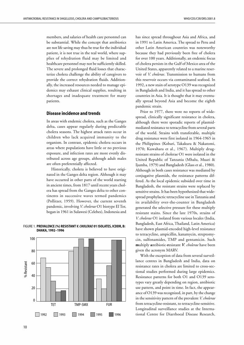
With the exception of data from several surveil-lance centres in Bangladesh and India, data onresistance rates in cholera are limited to cross-sec-tional studies performed during large epidemics.Resistance patterns for both O1 and O139 sero-types vary greatly depending on region, antibioticuse pattern, and point in time. In fact, the appear-ance of O139 was recognized, in part, by the changein the sensitivity pattern of the prevalent V. choleraefrom tetracycline-resistant, to tetracycline-sensitive.Longitudinal surveillance studies at the Interna-tional Centre for Diarrhoeal Disease Research,
TET TMP-SMX FUR
100
80
60
40
20
0
% R
esist
ant
1992 1993 1994 1995 1996
FIGURE 1. PREVALENCE (%) RESISTANT V. CHOLERAE 01 ISOLATES, ICDDR, B:DHAKA, 1992–1996

11
WHO/CDS/CSR/DRS/2001.8 ANTIMICROBIAL RESISTANCE IN SHIGELLOSIS, CHOLERA AND CAMPYLOBACTERIOSIS
Bangladesh (ICDDR,B) reveal that susceptibilitypatterns fluctuate from year to year (see Figures 1and 2). In some regions, resistance has emergedquickly to each new antibiotic used as the drug ofchoice for treating diarrhoeal disease. In Calcutta,resistance to TMP-SMZ quickly emerged over thecourse of a year during which it was heavily used;this was followed by an explosion of resistance tonalidixic acid (NA) when it became the first-linedrug (Jesudason & Saaya, 1997). In several instances,such as in Bangladesh in 1979, rates of resistanceto certain antibiotics rapidly increased concomi-tant with their use, but then declined without anychange in antibiotic use patterns (Glass et al.,1983[1]). Currently, we lack an understanding ofwhy such resistant strains should appear and thenrapidly disappear while the antibiotics are stillbeing intensively used. However, these phenom-ena suggest that there is an extensive pool of resist-ance genes and that strains with various resistanceprofiles will continue to appear, disappear, andreappear. One must also keep in mind the limitedreliability of published data describing such events;they may not accurately reflect resistance rates in aregion since there is a publication bias towardsobservations of high resistance rates. With theselimitations in mind, the available data on resist-ance rates are presented by region.
Regional resistance trends
Bangladesh. Since the mid-1960s, the ICDDR,Bhas maintained surveillance of cholera at its fieldstations in Matlab and Dhaka. Prior to 1979, noresistant strains were found. In 1979, strains ofV. cholerae O1 resistant to tetracycline, ampicillin,kanamycin, streptomycin and TMP-SMZ wereisolated (Glass et al., 1980). The abrupt emergenceof multiple drug resistance suggested that an R-plasmid was involved. Evaluation of 10 isolatesrevealed 3 distinct R-plasmids of the C incompat-ibility group mediating the resistance (Threlfall,Rowe & Huq, 1980). The strains disappeared after5 months, without major changes in antibiotic usepatterns (WHO, 1980). Two years later, in August1981, a different MARV strain (ampicillin, kan-amycin, sulfonamides, tetracycline and gentamicin)caused a small outbreak in Dhaka that quickly sub-sided (Threlfall & Rowe, 1982). Glass et al.,1983[1]) hypothesized that “the appearance andrapid disappearance of the strain in the secondoutbreak confirms the laboratory finding that,without antibiotic pressure, these strains have no
selective advantage and may continue to reappearand disappear on their own time”. Dramaticincreases in resistance to both tetracycline andTMP-SMZ were noted over the course of 1991 and1992, rising from 2% to 90% for tetracycline andfrom 18% to 90% for TMP-SMZ (Khan et al., 1995).In another survey, tetracycline resistance among ElTor strains rapidly increased from 1.9% in 1990 to7.6% in 1991, 61.1% in 1992, and 85.4% in 1993.As of 1994, all isolates in Dhaka were still sensitiveto erythromycin, NA, pivmecillinam and the newerquinolones, although more than 90% of isolateswere resistant to tetracycline, ampicillin, and TMP-SMZ (Bennish, 1994).
O139, a novel variant of cholera that was sensi-tive to tetracycline, erupted in Bangladesh and In-dia in 1993 and has since spread to Thailand,Pakistan, and eight other South-East Asian nations.Fortunately, it has not spread beyond Asia. O139strains from Bangladesh were found to be highlyresistant to streptomycin and TMP-SMZ (althoughsubsequently some isolates have been sensitive),moderately resistant to chloramphenicol and fura-zolidone, and susceptible to azithromycin, cephems,penems, minocycline, and the newer fluoro-quinolones (Yamamoto et al., 1995). In a prospec-tive study in Dhaka and Matlab comparing O1 andO139 strains, researchers found all O139 isolatesto be sensitive to ciprofloxacin, all but one strainsensitive to erythromycin and doxycycline, andmost (95% of O1 and 97% of O139) resistant toTMP-SMZ. However, the resistance patterns of O1isolates seemed to fluctuate from year to year.Researchers attributed this fluctuation to the
TET TMP-SMX FUR
100
80
60
40
20
0
% R
esist
ant
1993 1994 1995 1996
FIGURE 2. PREVALENCE (%) RESISTANT V. CHOLERAE 0139 ISOLATES, ICDDR,B: DHAKA, 1993–1996
ANTIMICROBIAL RESISTANCE IN SHIGELLOSIS, CHOLERA AND CAMPYLOBACTERIOSIS WHO/CDS/CSR/DRS/2001.8
12
instability of plasmids in V. cholerae (Sack et al.,1997).
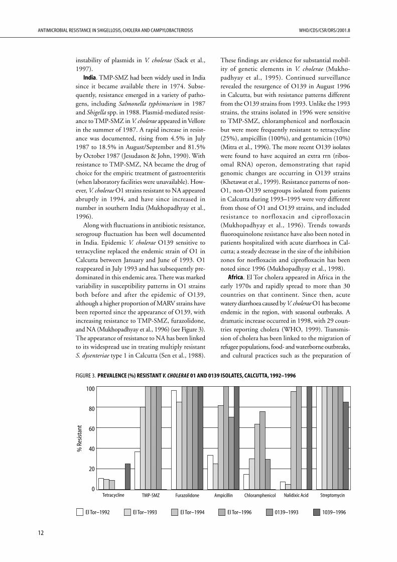
India. TMP-SMZ had been widely used in Indiasince it became available there in 1974. Subse-quently, resistance emerged in a variety of patho-gens, including Salmonella typhimurium in 1987and Shigella spp. in 1988. Plasmid-mediated resist-ance to TMP-SMZ in V. cholerae appeared in Vellorein the summer of 1987. A rapid increase in resist-ance was documented, rising from 4.5% in July1987 to 18.5% in August/September and 81.5%by October 1987 (Jesudason & John, 1990). Withresistance to TMP-SMZ, NA became the drug ofchoice for the empiric treatment of gastroenteritis(when laboratory facilities were unavailable). How-ever, V. cholerae O1 strains resistant to NA appearedabruptly in 1994, and have since increased innumber in southern India (Mukhopadhyay et al.,1996).
Along with fluctuations in antibiotic resistance,serogroup fluctuation has been well documentedin India. Epidemic V. cholerae O139 sensitive totetracycline replaced the endemic strain of O1 inCalcutta between January and June of 1993. O1reappeared in July 1993 and has subsequently pre-dominated in this endemic area. There was markedvariability in susceptibility patterns in O1 strainsboth before and after the epidemic of O139,although a higher proportion of MARV strains havebeen reported since the appearance of O139, withincreasing resistance to TMP-SMZ, furazolidone,and NA (Mukhopadhyay et al., 1996) (see Figure 3).The appearance of resistance to NA has been linkedto its widespread use in treating multiply resistantS. dysenteriae type 1 in Calcutta (Sen et al., 1988).
These findings are evidence for substantial mobil-ity of genetic elements in V. cholerae (Mukho-padhyay et al., 1995). Continued surveillancerevealed the resurgence of O139 in August 1996in Calcutta, but with resistance patterns differentfrom the O139 strains from 1993. Unlike the 1993strains, the strains isolated in 1996 were sensitiveto TMP-SMZ, chloramphenicol and norfloxacinbut were more frequently resistant to tetracycline(25%), ampicillin (100%), and gentamicin (10%)(Mitra et al., 1996). The more recent O139 isolateswere found to have acquired an extra rrn (ribos-omal RNA) operon, demonstrating that rapidgenomic changes are occurring in O139 strains(Khetawat et al., 1999). Resistance patterns of non-O1, non-O139 serogroups isolated from patientsin Calcutta during 1993–1995 were very differentfrom those of O1 and O139 strains, and includedresistance to norfloxacin and ciprofloxacin(Mukhopadhyay et al., 1996). Trends towardsfluoroquinolone resistance have also been noted inpatients hospitalized with acute diarrhoea in Cal-cutta; a steady decrease in the size of the inhibitionzones for norfloxacin and ciprofloxacin has beennoted since 1996 (Mukhopadhyay et al., 1998).
Africa. El Tor cholera appeared in Africa in theearly 1970s and rapidly spread to more than 30countries on that continent. Since then, acutewatery diarrhoea caused by V. cholerae O1 has becomeendemic in the region, with seasonal outbreaks. Adramatic increase occurred in 1998, with 29 coun-tries reporting cholera (WHO, 1999). Transmis-sion of cholera has been linked to the migration ofrefugee populations, food- and waterborne outbreaks,and cultural practices such as the preparation of
Tetracycline TMP-SMZ Furazolidone Ampicillin Chloramphenicol Nalidixic Acid Streptomycin
El Tor–1992 El Tor–1993 El Tor–1994 El Tor–1996 0139–1993 1039–1996
100
80
60
40
20
0
% R
esist
ant
FIGURE 3. PREVALENCE (%) RESISTANT V. CHOLERAE 01 AND 0139 ISOLATES, CALCUTTA, 1992–1996
13
WHO/CDS/CSR/DRS/2001.8 ANTIMICROBIAL RESISTANCE IN SHIGELLOSIS, CHOLERA AND CAMPYLOBACTERIOSIS
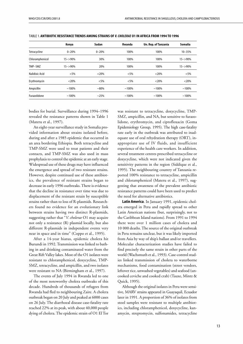
bodies for burial. Surveillance during 1994–1996revealed the resistance patterns shown in Table 1(Materu et al., 1997).
An eight-year surveillance study in Somalia pro-vided information about strains isolated before,during and after a 1985 epidemic that occurred inan area bordering Ethiopia. Both tetracycline andTMP-SMZ were used to treat patients and theircontacts, and TMP-SMZ was also used in massprophylaxis to control the epidemic at an early stage.Widespread use of these drugs may have influencedthe emergence and spread of two resistant strains.However, despite continued use of these antibiot-ics, the prevalence of resistant strains began todecrease in early 1996 outbreaks. There is evidencethat the decline in resistance over time was due todisplacement of the resistant strain by susceptiblestrains rather than to loss of R-plasmids. Research-ers found no evidence for an evolutionary linkbetween strains having two distinct R-plasmids,suggesting rather that “V. cholerae O1 may acquirenot only a resistance (R) plasmid locally, but alsodifferent R-plasmids in independent events verynear in space and in time” (Coppo et al., 1995).
After a 14-year hiatus, epidemic cholera hitBurundi in 1992. Transmission was linked to bath-ing in and drinking contaminated water from theGreat Rift Valley lakes. Most of the O1 isolates wereresistant to chloramphenicol, doxycycline, TMP-SMZ, tetracycline, and ampicillin, and two isolateswere resistant to NA (Birmingham et al., 1997).
The events of July 1994 in Rwanda led to oneof the most noteworthy cholera outbreaks of thisdecade. Hundreds of thousands of refugees fromRwanda had fled to neighbouring Zaire. A choleraoutbreak began on 20 July and peaked at 6000 caseson 26 July. The diarrhoeal disease case-fatality ratereached 22% at its peak, with about 40,000 peopledying of cholera. The epidemic strain of O1 El Tor
was resistant to tetracycline, doxycycline, TMP-SMZ, ampicillin, and NA, but sensitive to furazo-lidone, erythromycin, and ciprofloxacin (GomaEpidemiology Group, 1995). The high case-fatalityrate early in the outbreak was attributed to inad-equate use of oral rehydration therapy (ORT), in-appropriate use of IV fluids, and insufficientexperience of the health care workers. In addition,several treatment centres prescribed tetracycline ordoxycycline, which were not indicated given thesensitivity patterns in the region (Siddique et al.,1995). The neighbouring country of Tanzania re-ported 100% resistance to tetracycline, ampicillinand chloramphenicol (Materu et al., 1997), sug-gesting that awareness of the prevalent antibioticresistance patterns could have been used to predictthe need for alternative antibiotics.
Latin America. In January 1991, epidemic chol-era emerged in Peru and rapidly spread to otherLatin American nations (but, surprisingly, not tothe Caribbean Island nations). From 1991 to 1994there were over 1 million cases of cholera and10 000 deaths. The source of the original outbreakin Peru remains unclear, but it was likely importedfrom Asia by way of ship’s ballast and/or travellers.Molecular characterization studies have failed tofind precisely the same strain in other parts of theworld (Wachsmuth et al., 1993). Case-control stud-ies linked transmission of cholera to waterbornemechanisms, food contamination (street vendors,leftover rice, unwashed vegetables) and seafood (un-cooked ceviche and cooked crab) (Tauxe, Mintz &Quick, 1995).
Although the original isolates in Peru were sensi-tive, MARV strains appeared in Guayaquil, Ecuadorlater in 1991. A proportion of 36% of isolates fromstool samples were resistant to multiple antibiot-ics, including chloramphenicol, doxycycline, kan-amycin, streptomycin, sulfonamides, tetracycline
TABLE 1. ANTIBIOTIC RESISTANCE TRENDS AMONG STRAINS OF V. CHOLERAE O1 IN AFRICA FROM 1994 TO 1996
Kenya Sudan Rwanda Un. Rep. of Tanzania Somalia
Tetracycline 0–20% 0–20% 100% 100% 10–35%
Chloramphenicol 15–>90% 30% 100% 100% 15–>90%
TMP–SMZ 15–>90% 20% 100% 100% 15–>90%
Nalidixic Acid <5% <20% <5% <20% <5%
Erythromycin <20% <5% <5% <20% <20%
Ampicillin ~100% ~80% ~100% ~100% ~100%
Furazolidone ~100% ~25% ~100% ~100% ~100%
ANTIMICROBIAL RESISTANCE IN SHIGELLOSIS, CHOLERA AND CAMPYLOBACTERIOSIS WHO/CDS/CSR/DRS/2001.8
14
and TMP-SMZ (Weber et al., 1994). There wasno clear cause of MARV emergence, but it mayhave been due to prophylactic use of antibiotics inthis region. The recommendation was for adult fam-ily members of patients to receive 500 mg tetracy-cline every 4 hrs for 5 days and pregnant women andchildren to receive erythromycin or TMP-SMZ(Weber et al., 1994). Additional environmentalpressure may have come from the use in the area ofantimicrobials to control other bacteria in hatch-ing shrimp, since fish, shellfish, and conch wereimplicated as vehicles of transmission. MARVstrains of V. cholerae were also reported in Argen-tina and Honduras (Rossi et al., 1993; Dubon etal., 1997).
United States and Europe. Most cholera casesreported in the United States in the last four decadeshave been contracted during foreign travel. While,in 1992, 97% of imported strains were sensitive toall antimicrobial agents tested, resistance increasedin 1993 and 1994, with the majority of isolatesbeing resistant at least to sulfonamides, streptomy-cin and furazolidone. Since 1973, an endemic focusof V. cholerae O1 has emerged and caused sporadiccases in states bordering the Gulf of Mexico. Thesedomestically acquired cases have remained suscep-tible to all antimicrobial agents tested (Mahon etal., 1996). Europe has had outbreaks of MARVcholera, with reports in southern Italy and Albaniain 1994 of strains resistant to TMP-SMZ,chloramphenicol, tetracycline, doxycycline andstreptomycin (Maggi, Carbonara & Santantonio,1996).
Causes of resistance
The emergence and maintenance of drug resistancein cholera is governed by a complex series of bio-logical, environmental, and behavioural factors.Transposons, plasmids, mobile gene cassettes andintegrons mediate the rapid and broad dissemina-tion of genetic information across species lines.Thus, we cannot look simply at resistance withinthe V. cholerae species, but rather consideration mustbe given to the relationship of vibrios with the en-vironment and with other bacterial species in theenvironment.
Microbiological mechanisms of resistance.
Although antimicrobial resistance can result fromthe accumulation of chromosomal point mutations,the vast majority of clinically relevant resistance in
V. cholerae is due to exchange of genetic informa-tion among bacterial strains via plasmids andtransposons. Laboratory experiments have shownthat such exchange of bacterial genetic material cantake place by conjugation, transduction or trans-formation (Ogg, Shrestha & Poudayl, 1978). Inclinical settings, plasmid-mediated transfer hasaccounted for the emergence and dissemination ofresistance genes in cholera.
Most plasmids isolated from V. cholerae O1 arecryptic, but some encode antibiotic resistance de-terminants (R-factors). These R-plasmids are large(110-170 kb), self-transmissible, and usually of theC incompatibility group. In the 1970s it wasreported that, in the laboratory, R-plasmids wereunstable in V. cholerae and were easily eliminatedin drug-free conditions (Yokoto et al., 1972). Asimilar observation had been made earlier byKuwahara et al. (1963) after in vitro transmissionof plasmids from Shigella spp. to V. cholerae. How-ever, stability of certain R-plasmids was reported someyears later (Rahal, Gerbaud & Bouanchaud, 1978).
V. cholerae O1 R-plasmids have been found thatcarry genes encoding resistance to ampicillin, chlo-ramphenicol, gentamicin, kanamycin, spectino-mycin, streptomycin, sulfonamides, tetracyclineand TMP, with up to seven resistance determinantson a single plasmid (Threlfall, Rowe & Huq, 1980).It is thought that most of these genes were acquiredfrom Enterobacteriaceae. Plasmids of the C incom-patibility group are found in a wide variety of bac-terial genera, including Pseudomonas, Proteus,Klebsiella, and Serratia. Bacteria may acquireresistance genes from other species of the normalintestinal flora under the selective pressure of anti-microbial use. V. cholerae R-plasmids have beenshown to carry resistance determinants (e.g. forampicillin and TMP) that are common in entericbacteria (Young & Amyes, 1986). When Rahal,Gerbaud & Bouanchaud (1978) examined thetransferability and maintenance of plasmids, theyfound that although plasmids of most incompat-ibility groups could be transferred from E. coli toV. cholerae, only those of groups C and J were main-tained.
In recent years, light has been shed on the im-portant role that transposons play in resistance. Ithas been found that the resistance of El Tor strainsto TMP, spectinomycin, streptomycin and thevibriostatic agent 0/129 is due to a transposon in-serted into the chromosome (Goldstein, Gerbaud& Courvalin, 1986). This was demonstrated bytransferring the resistance determinants to a trans-
15
WHO/CDS/CSR/DRS/2001.8 ANTIMICROBIAL RESISTANCE IN SHIGELLOSIS, CHOLERA AND CAMPYLOBACTERIOSIS
missible plasmid and then into the chromosome ofE. coli. In V. cholerae O139 strains, resistance de-terminants for SMZ, TMP and streptomycin arecarried on a 62-kb self-transmissible, chromo-somally integrating genetic element that is similarto conjugative transposons found in Bacteroides(Waldor, Tschape & Mekalanos, 1996). In the samestudy, it was found that El Tor O1 strains that re-emerged after the O139 strain had declined hadsimilar self-transmissible transposons carryingresistance genes, suggesting that these elements arewidely disseminated in Vibrio spp. and confer someselective advantage to the bacteria.
Environmental determinants of resistance
Because V. cholerae can readily exchange genetic in-formation among strains and with other bacterialspecies, controlling the emergence of resistance re-quires an understanding of the source of R-plasmids.The gut and other environments (soil, sewage, etc.)contain a variety of organisms that cannot be cul-tured. This makes it impossible to precisely trackthe transfer and dissemination of resistance genesin nature. Although not definitive, most available datasuggest that other enteric bacteria, such as E. coli andnon-O1, non-O139 V. cholerae serogroups in the en-vironment have been the intermediate hosts.
Resistance genes are common and plentiful innormal bacteria in the environment, such as E. coli.Some of these genes may have first appeared mil-lions of years ago. Since E. coli spend time cyclingthrough animals, humans, sewage, water and soil,one can hypothesize that E. coli may have acquiredresistance genes that evolved to provide protectionagainst antibiotics produced naturally by soil bac-teria. In the antibiotic era there is environmentalselective pressure for the development of resistance.A longitudinal community-based survey of childrenfrom urban Mexico found persistent (13 weeks)faecal shedding of ampicillin-, tetracycline-, andTMP-SMZ-resistant E. coli in the majority of thecohort of healthy children, as well as some chil-dren that shed E. coli resistant to chloramphenicol,gentamicin, nitrofurantoin, and norfloxacin. Thefact that the three most commonly used antibiot-ics in this Mexican community are ampicillin,TMP-SMZ and tetracycline suggests that overuseof these drugs for common illnesses exerts a selec-tive pressure on the normal bowel flora, which thenbecome an important reservoir of resistance genesthat can potentially be transferred to pathogenicorganisms such as V. cholerae (Calva, Sifuentes-
Osornio & Ceron, 1996). There is, in fact, evidencefor the local acquisition of R-plasmids by V. cholerae.Most V. cholerae resistance plasmids are transfer-able to E. coli in vitro (Rahal, Gerbaud & Bouan-chaud, 1978) and these plasmids can be transferredback into V. cholerae from laboratory strains of E.coli (Finch et al., 1988). E. coli and V. cholerae withidentical resistance plasmids have been isolated fromthe same patient (Haider & Huq, 1986). Studiesin Bangladesh demonstrated that family contactsof individuals infected with MARV were more likelythan controls to have other multiply antibiotic-resistant bacteria, carrying the identical resistanceplasmid, in their intestinal flora (Glass et al.,1983[1]).
To summarize, there is evidence supporting thehypothesis that resistance genes in V. cholerae canbe acquired locally from enteric flora such as E.coli. If this is the case, antibiotic use for any pur-pose (other diarrhoeal disease, respiratory illness,STD control) will affect the reservoir of resistancegenes in E. coli that are potentially transferable toV. cholerae should an outbreak occur.
There has also been increasing interest in therole that non-O1, non-O139 serogroups may playin the shifting dynamics of V. cholerae and itsresistance patterns. On the basis of gene sequencevariation analysis, Karaolis, Lan & Reeves (1995)suggested that the last two cholera pandemics werelikely caused by independent clones that emergedfrom environmental, nontoxigenic, non-O1V. cholerae. Because there is a high rate of geneticexchange among different Vibrio strains in theenvironment, non-O1, non-O139 strains may beimportant reservoirs of resistance elements. This isespecially important since non-O1, non-O139 strainsare increasingly resistant to ciprofloxacin and otherfluoroquinolones, the only widely used drugs to whichV. cholerae O1 remain universally sensitive.
Behavioural and economic factors in resistance
Because selection for resistance is thought to be afunction of total antibiotic pressure in an area, druguse pattern is an important factor. In most devel-oping countries there is uncontrolled use of inex-pensive broad-spectrum antibiotics. There is ofteninappropriate prescribing by clinicians, or misuseby unskilled health workers or by traditional heal-ers. A majority of the public may purchase antibi-otics, without a prescription, from local pharmacies,as well as from street vendors or drug stalls. Be-cause the unofficial retailers are not guided by any
ANTIMICROBIAL RESISTANCE IN SHIGELLOSIS, CHOLERA AND CAMPYLOBACTERIOSIS WHO/CDS/CSR/DRS/2001.8
16
regulatory criteria for rational antibiotic use, thereis considerable inappropriate self-medication(Okeke, Lamikanra & Edelman, 1999).
As a public health measure, antibiotics are oftenprescribed on a large scale for prophylaxis duringepidemics. This is controversial since, under suchintense selective pressure, the benefit to individu-als is usually offset by the rapid emergence of re-sistance. Experiences with mass prophylaxis in threedifferent epidemic situations are described below.
1. United Republic of Tanzania. When mass tetra-cycline prophylaxis of close contacts was usedfor cholera control in 1977, widespread multi-ple drug resistance appeared within six months.During the first month of the epidemic, all iso-lates were sensitive to tetracycline, but after 5months of extensive tetracycline use for therapyand prophylaxis (1788 kg used by the Ministryof Health [MOH]), 76% of the isolates wereresistant to tetracycline and 52% to chloram-phenicol (Mhalu, Muari & Ijumba, 1979).
2. Cameroon. Mass prophylaxis with sulfadoxine(fanasil) was used during a large outbreak in1983. Multiply drug-resistant strains (sulfa-doxine, tetracycline, chloramphenicol and TMP-SMZ) were isolated in 1984–1985 (Garrigue etal., 1986).
3. Kenya. Mass tetracycline prophylaxis campaignswere carried out from 1981 to 1988. Strains re-sistant to tetracycline, ampicillin and TMP-SMZwere isolated in 1982 (Ichinose et al., 1986).Resistance was found to be mediated by a singleplasmid that differed from the plasmids foundin other regions (United Republic of Tanzania,Nigeria, Bangladesh) (Finch et al., 1988). Stud-ies of isolates from 1982 to1985 have demon-strated the persistence of the resistant strain. Thefact that distinct plasmids persist in differentgeographical areas suggests that resistanceplasmids are acquired locally as a result of localantibiotic pressure (Finch et al., 1988).
Treatment choices and development of resistance
The primary treatment for patients with cholera isrehydration with oral or intravenous fluids. Anti-biotics are given to decrease the volume of purgingand the duration of diarrhoea, and thus to decreasethe cost of treatment (Lindenbaum, Greenough &Islam, 1967) by sparing limited supplies and per-sonnel, shortening hospital stays and returning thepatient to normal function sooner (i.e. shorter
period of lost wages). Inexpensive, effective antibi-otics are very cost-effective as adjunct therapy insevere cases, since they reduce the hospital stay anddecrease the volume of intravenous fluids and ORSneeded for rehydration. While antibiotics rapidlyeradicate organisms from the stool, they probablyhave minimal impact on the dynamics of choleratransmission in the community, as there are envi-ronmental reservoirs and because a large propor-tion of asymptomatic, or only mildly ill, infectedindividuals, who would not normally receive anti-biotics, shed vibrios.
Tetracycline traditionally has been the antibi-otic of choice (adult dose 500 mg every 6 hours for48 to 72 hours; children’s dose 50 mg/kg/day in 4divided doses for 2–3 days), but resistance to this drugis widespread. Many authorities, such as WHO, nowrecommend doxycycline as the first-line drug, sincea single 300 mg dose is effective for adults (Alam etal., 1990; Sack et al., 1978) and guarantees compli-ance without the need for follow-up, a significantlogistical advantage. Strains that are sensitive totetracycline are also sensitive to doxycycline, so thereis no need to test specifically for doxycycline resist-ance. In fact, in vitro susceptibility to doxycycline maynot correlate well with its in vivo activity, owing tothe variable expression of inducible tetracycline re-sistance determinants (Khan et al., 1996).
TMP-SMZ is recommended as the first-linedrug for children and furazolidone (100 mg) forpregnant women. Other effective drugs includeciprofloxacin, erythromycin and chloramphenicol.Sulfadoxine (fanasil), single dose, has also been used,but resistance is increasing in Africa and the drugmay have serious side-effects (Stevens-Johnson syn-drome).
In a study in Bangladesh, erythromycin andciprofloxacin were both shown to be effective al-ternatives for the treatment of MARV strains. Itwas suggested that NA and pivmecillinam shouldbe reserved for the treatment of shigellosis, sincethey did not have significant efficacy in sympto-matic cholera (Khan et al., 1995).
Short- vs. long-course therapy
A three-day course of tetracycline has been theantibiotic regimen of choice for cholera although,as discussed above, doxycycline has some advan-tages. In a randomized double-blind study atICDDR,B, single-dose ciprofloxacin (1 g) was ef-fective in treating both V. cholerae O1 and O139and was more effective than single-dose doxycy-
17
WHO/CDS/CSR/DRS/2001.8 ANTIMICROBIAL RESISTANCE IN SHIGELLOSIS, CHOLERA AND CAMPYLOBACTERIOSIS
cline (Khan et al., 1996). Single-dose regimens areless effective in achieving culture-negativity of thestool (Islam, 1987). However, in cholera, antibiot-ics are used to accelerate the clinical improvementof the individual patient, not to control the spreadof cholera, a purpose for which they are not effec-tive. For this reason, the evaluation of antibioticregimens should be based on clinical improvementof patients (decreased duration and volume of purg-ing) rather than on eradication of vibrios from thestool.
Some clinicians are of the opinion that single-dose or short-course regimens will increase resist-ance. More research is needed to thoroughlyevaluate this. However, data for V. cholerae suggestthat resistance does not develop in an individualpatient but rather that there are shifting populationsof sensitive and resistant strains that are affected byoverall antibiotic use. For these reasons, single-doseor short-course regimens which are clinically effec-tive (e.g. doxycycline or ciprofloxacin) are preferredto longer course (3–5 day) regimens which are morelikely to upset the enteric bacterial ecology of thepatient and to favour resistant over sensitive strains.Additionally, the lower cost and ease of adminis-tration of short-course therapy outweigh the riskof slower eradication of vibrios from the stool.
The question of prophylaxis
During epidemics, there is often pressure to pro-vide prophylactic antibiotics to household contactsof cholera patients, or even to entire communities.Early studies showed that tetracycline or doxycy-cline could prevent secondary cases and reducevibrio excretion in household contacts (Gupta etal., 1978; Joint ICMR-GWB-WHO Cholera StudyGroup, 1971). McCormack et al. (1968) describedthe efficacy of 5 days of tetracycline prophylaxis inreducing secondary infection within the familiesof cholera patients. Khan (1982) later demonstratedthat two 250 mg doses of tetracycline (short-coursetherapy) decreased the number of severe cases ofdiarrhoea and the hospitalization rate among con-tacts during a cholera epidemic in Dhaka. Treat-ment of close contacts has been considered moreappropriate than mass chemoprophylaxis, since 10–25% of household contacts may become vibrioexcretors as compared with less than 1% of com-munity contacts. However, transmission rates arehighly dependent on the local situation. WHO rec-ommends that “selective chemoprophylaxis be con-sidered only when surveillance has shown that, on
average, at least one household member in fivebecomes ill after the first case has appeared” (WHO,1992[2]). However, cholera outbreaks frequentlyoccur quickly and in regions where such epidemi-ologically based decision-making is impractical.
The only situation in which prophylaxis seemswarranted is during an epidemic of tetracycline-sensitive cholera, when a single dose of doxycyclinecan be given to immediate household members (e.g.those who share the same kitchen) within a two-day period after the case is diagnosed. Situations inwhich this can be accomplished are extremely rare,and attempts to follow this procedure frequentlylead to wide-scale antibiotic use and abuse.
Mass antibiotic prophylaxis is not recommendedbecause it has not been shown to be effective andbecause it contributes to the emergence of resist-ant strains. Additionally, there are problems suchas delays in beginning prophylaxis, the fact that itseffects last for a short time, non-compliance, andthe occurrence of side-effects (especially withsulfonamides) (Hernborg, 1985). It is very diffi-cult to selectively treat high-risk contacts withoutdispensing large amounts of antibiotics in the com-munity; this generates selective pressure for resist-ant strains, and thus the possibility of being unableto effectively treat severe cases of disease.
It is important to realize that “prophylactic” an-tibiotic administration does not actually preventinfection. Rather, in the case of cholera, prophy-laxis is intended to kill the bacteria at an early stageof infection; i.e. it is assumed that a large percent-age of the contacts are already infected and are in-cubating the disease. Thus, these are not “secondarycases” infected by contact with the index case, butare rather “co-primary cases” who have not yetexhibited symptoms. Finding and treating themrapidly, appropriately and specifically is not practi-cal in most cases.
Potential vaccines
Injectable vaccines for cholera have been used sincethe late 1800s, but with little lasting benefit. Thecurrently available killed injectable vaccine is notrecommended, since studies in the 1960s showedit to be only 60% effective for a period of 4–6months (WHO, 1996). The recent availability ofimproved oral cholera vaccines, such as the recom-binant oral B subunit killed whole-cell (rBS-WC)vaccine and the live attenuated CVD 103-HgRvaccine has led to renewed interest in the use ofvaccines during cholera epidemics.
ANTIMICROBIAL RESISTANCE IN SHIGELLOSIS, CHOLERA AND CAMPYLOBACTERIOSIS WHO/CDS/CSR/DRS/2001.8
18
A recent analysis of results in sub-Saharan refu-gee camps showed that mass vaccination could becost-effective in controlling cholera if the price ofthe vaccine was sufficiently low (Naficy et al., 1998).This study did not consider the potential role thata vaccine might play in preventing the occurrenceof antibiotic-resistant infections. A vaccine that iseffective in lowering the total number of cases willalso lower the number of resistant infections andcould thus represent an effective intervention meas-ure to control antibiotic resistance in cholera.Additional research is necessary to define the roleof vaccines as preventive measures in endemicregions such as the Ganges delta.
Recommendations
1. Antibiotics that are known to be clinically ef-fective, and to which the bacteria are suscepti-ble in vitro, are appropriate and cost-effectivein the treatment of patients with moderate orsevere cholera. In epidemics, antibiotics arelikely to be life saving, providing better treat-ment to more patients.
2. If the predominant cholera strain is known tobe resistant to an antibiotic, that antibioticshould not be used, as it would not be effica-cious and is potentially harmful. Antibioticsthat are not clinically effective for treating chol-era (e.g. cephalosporins or gentamicin) shouldnot be used even if in vitro tests show the strainto be sensitive.
3. A surveillance system is needed to collect a rep-resentative sample of strains, determine sensi-tivity patterns in a region, and continuallymonitor changes in sensitivity patterns. Sus-ceptibility testing should be carried out in anestablished laboratory with stringent qualitycontrol (QC) standards. Strains with unusualsusceptibility patterns should be sent to refer-ence laboratories for confirmation. The resultsof surveillance should be used to guide antibi-otic treatment of patients with suspected chol-era. It is neither practical nor wise to base thechoice of antibiotic on the sensitivity patternin the individual patient, since antibiotics mustbe given early in treatment if they are to beeffective.
4. If most strains are tetracycline-sensitive, thensingle-dose doxycycline is the preferred treat-ment. If the predominant strains are resistantto tetracycline, then alternative choices need
to be made based on antibiograms of locallyrepresentative strains.
5. If most strains are resistant to all recommendedantibiotics, then treatment must be based onrehydration alone, and plans must be made tomanage the most severe and refractory cases.Occasionally, ciprofloxacin is the only clinicallyeffective antibiotic. An analysis of cost-effec-tiveness should guide the choice of this antibi-otic, but in most areas it will be cost-effective.Ciprofloxacin should be used in a single-doseregimen.
6. Antibiotic pressure leads to the developmentof resistant V. cholerae; thus, inappropriate an-tibiotic use for any illness can lead to resistantcholera. Antibiotics should be carefully targetedto those conditions that truly warrant their use.
7. Antibiotics should not be used to prevent chol-era, except in certain very unusual circum-stances.
8. The use of antibiotics in agriculture should belimited to the treatment of individual animals;they should not be added to feed to promotethe growth of animals and should not be addedto environmental waters for fish or seafoodfarming. In particular, the use of tetracyclineand fluoroquinolones in agriculture should becontrolled.
9. MOHs, nongovernmental organizations(NGOs) and others should base their antibi-otic use and procurement policies on data fromestablished surveillance systems. Policies andprocurement procedures need to be sufficientlyflexible to adapt to changes in antibiotic sus-ceptibility patterns.
10. Information on antibiotic sensitivity generatedby surveillance systems should be readily avail-able in the form of hotlines, web pages, bulle-tins, etc., so that it can be easily consulted bythose responsible for drug policy and procure-ment decisions.
11. Ongoing epidemics, as well as expected chol-era seasons, need to be posted in the same sites,since the number, timing and severity of thecases affect the urgency with which the healthcare system must prepare to treat patients withcholera. A geographical information system(GIS) format may be useful for reportingseasonal occurrences and antibiotic suscepti-bility patterns.

19
WHO/CDS/CSR/DRS/2001.8 ANTIMICROBIAL RESISTANCE IN SHIGELLOSIS, CHOLERA AND CAMPYLOBACTERIOSIS
12. Cost-effectiveness analyses for alternative an-tibiotics must consider the total cost of illnessand not simply the cost of the antibiotic. Over-all costs include the price of the drug perpatient treated (not per tablet), hospital time,nursing care, lost wages, etc.
13. Cholera control programmes that successfullylimit the number of cases will also limit thenumber of antibiotic-resistant infections.Improved water, sanitation, and hygiene pro-grammes, as well as the use of oral vaccines,all help to decrease disease incidence and anti-biotic resistance, especially in high-incidenceareas.
14. Representative strains having multiple anti-biotic resistance should be characterized inmolecular biology laboratories in order tobetter understand the mechanisms.
Research priorities
1. Epidemiological surveillance and communica-tion of findings
1.1 Establishment of a simple and inexpensivesurveillance system for monitoring choleraincidence and antibiotic resistance patterns.One of the systems in use at the ICDDR,Bmay be adapted for other geographicallocations. Any new surveillance systemshould be field-tested in two or three chol-era endemic areas.
1.2 The utility of rapid diagnostic tests (e.g.Smart Test or coagglutination tests) in sur-veillance should be studied. Such tests, ifused appropriately, could help identify thosespecimens that should be sent to the cen-tral laboratory for further testing.
1.3 Centralized or regional resource centres forcholera epidemiology and susceptibility test-ing need to be established so that MOHsand NGOs can have access to current infor-mation about resistance patterns. A resourcecentre should field-test its communicationsmethods in order to optimize their effec-tiveness.
2. Factors affecting antibiotic resistance
2.1 Studies are needed to document the effectof inappropriate antibiotic use on cholerasensitivity patterns, for example comparingthe susceptibility patterns of geographical
areas with high and low antibiotic use. Theutilization of antibiotics in agriculture andaquaculture appears to be another impor-tant issue requiring investigation.
2.2 Studies are needed to document the reap-pearance of sensitive cholera strains, follow-ing epidemics caused by resistant strains, inorder to identify parameters that correlatewith this phenomenon. Similarly, in areaswhere sensitive strains have not reappeared,explanations should be sought.
2.3 Surrogate parameters that correlate with re-sistance in V. cholerae should be sought. Itseems that V. cholerae follow the sensitivitypatterns of Enterobacteriacae, so it is possi-ble that surveillance of resistance patternsin normal E. coli will predict the patterns inV. cholerae.
2.4 Antibiotic susceptibility trends of environ-mental non-O1, non-O139 V. cholerae needto be monitored in order to determine whetherthese will be predictive of patterns in O1 V.cholerae and whether resistance genes are ex-changed among the different serotypes.
3. Studies of constraints on appropriate antibiotic use
3.1 Case reports related to inappropriate antibi-otic policy should be studied to help usunderstand why inappropriate antibioticswere recommended or used.
3.2 The influence of non-medical factors (e.g.procurement methods, pharmaceutical salesmethods, storage and distribution problems)on the choice of antibiotics and on essentialdrug programmes should be determined.
4. Intervention studies
4.1 The impact on resistance trends of control-ling antibiotic use should be quantitated andanalysed in terms of lives saved and costreduction.
4.2 The impact of water and hygiene interven-tions on the incidence of antibiotic-resist-ant cholera should be determined.
4.3 The impact of effective cholera vaccines onthe incidence of antibiotic-resistant cholerashould be studied.
5. Basic studies of antibiotic resistance
5.1 The molecular mechanisms of antibioticresistance need to be determined and the
ANTIMICROBIAL RESISTANCE IN SHIGELLOSIS, CHOLERA AND CAMPYLOBACTERIOSIS WHO/CDS/CSR/DRS/2001.8
20
ConclusionV. cholerae can become multiply antibiotic-resist-ant via the acquisition of plasmids. Multiply resist-ant strains have repeatedly caused epidemics.Antibiotic resistance patterns change from time totime and place to place, requiring continual sur-veillance in order to provide optimal treatment.Susceptible strains sometimes reappear when anti-biotic pressure is lifted, suggesting that cholera maybe one disease in which we might expect relativelyrapid reversion to sensitivity if antibiotic use is con-trolled.
Because epidemics due to antibiotic-resistantstrains remain a threat, effective antibiotics areneeded to reduce costs and treatment time, shortenthe duration of illness, and save lives.
different genes responsible for resistancecatalogued.
5.2 The molecular mechanisms of transmissionof resistance genes (plasmids, transposons,phages, etc.) need to be studied in order tobetter understand how transmission occursand to which other bacterial species.
5.3 A better understanding of receptor mecha-nisms for transmissible genes is required inorder to comprehend the range of bacterialspecies that contribute to resistance inV. cholerae.
21
WHO/CDS/CSR/DRS/2001.8 ANTIMICROBIAL RESISTANCE IN SHIGELLOSIS, CHOLERA AND CAMPYLOBACTERIOSIS
Review of Shigella spp.
term has become synonymous with all clinical pres-entations of shigellosis, although these presentationsrange from asymptomatic carriage to mild, waterydiarrhoea to overt dysentery (Levine et al., 1973;Levine, 1991).
Dysentery is a syndrome characterized by frequent,but small volume, loose stools, consisting largelyof blood and mucus. Fever, pain, and tenesmus(unproductive straining) are frequently present.Unlike secretory diarrhoeas, this syndrome is a re-sult of invasion of the distal small bowel and/orcolon by bacteria. Because dehydration is not assevere as in secretory diarrhoeas, oral rehydrationtherapy does not significantly reduce the case-fatality rates for shigellosis (Keusch & Bennish,1989; Bennish et al., 1990[2]).
Community data on shigellosis are incomplete,but most hospital-based studies suggest that thecase-fatality rate is highest among children less than5 years of age, particularly if there is malnutrition.In epidemic situations a mortality rate as high as3.9% in children under age 1 and 19.3% for infantsunder 4 months of age has been reported. The case-fatality rate declines with increasing age (Bennishet al., 1990[2]; Bennish, 1991). An unusual find-ing from recent studies of epidemic shigellosis inCentral Africa was high mortality rates in youngadults. This region also has high HIV rates, so aninteraction between the two infections is possible,but has not yet been studied. The most commonacute complications, particularly in settings withlimited health care services, include sepsis (causedby Shigella or other enteric organisms), intestinalperforation, toxic megacolon, dehydration, hypo-natraemia, encephalopathy, haemolytic-uraemicsyndrome, and pneumonia. Shigellosis may also befollowed by persistent dysentery, protein-losingenteropathy, and rapidly worsening malnutrition.Because shigellosis often occurs in settings wheretreatment for some of the complications is not avail-able, reducing mortality from this disease hingesupon its prevention (Bennish, 1991).
Introduction
Like Campylobacter spp. and V. cholerae, Shigellaspp. have managed to survive the antibiotic era viaingenious mechanisms of resistance. Unlike diarrhoeadue to C. jejuni, shigellosis may occur in epidemicform, causing considerable morbidity and mortal-ity in developing nations. Shigellosis is primarily adisease of resource-poor, crowded communities thatdo not have adequate sanitation or safe water, andwhere disease rates may be high. Because shigellaehave acquired multiple antimicrobial resistances,the challenge for clinical management is identify-ing which drugs retain their activity and clinicaleffectiveness. In the United States, the Centers forDisease Control and Prevention (CDC) have rec-ommended that sensitivity testing be performed toguide selection of appropriate antimicrobial therapyfor shigellosis (CDC, 1987). However, testingrequires several days to complete, resulting in treat-ment delay, and is generally not feasible in developingcountries. Because antimicrobial susceptibility pat-terns of shigellae may vary greatly in different geo-graphical areas and over time, monitoring resistancepatterns is needed to guide selection of appropri-ate empiric treatment (Jesudason, Lalitha & Koshi,1985; Munshi et al., 1987; Ashkenazi et al., 1995).
Organisms and syndrome
Shigella belongs to the family Enterobacteriacae. Itis a small, unencapsulated, non-motile Gram-negative rod. There are four species of Shigella, clas-sified on the basis of biochemical and serologicaldifferences: S. dysenteriae (Group A), S. flexneri(Group B), S. boydii (Group C), and S. sonnei(Group D). The first three are further divided intomultiple serotypes; thus, the genus contains a largenumber of different pathogens. S. dysenteriae sero-type 1 (also known as the Shiga bacillus) deservesspecial note because it is more likely to producesevere disease, to spread in epidemics, to be par-ticularly antibiotic-resistant, and to produce Shigatoxin. Because organisms of the genus Shigella arethe most common cause of bacillary dysentery, this
ANTIMICROBIAL RESISTANCE IN SHIGELLOSIS, CHOLERA AND CAMPYLOBACTERIOSIS WHO/CDS/CSR/DRS/2001.8
22
Geographical distribution
Shigella is highly adapted to man, with humans andprimates in captivity being the only known naturalhosts. The minimal infective dose is less than 200cells, facilitating transmission in areas where thereare crowding, poor sanitation and poor hygiene(Levine, 1991; DuPont et al., 1989). In endemicareas of the developing world, shigellosis is predomi-nantly a paediatric disease, with the urban poorbeing hardest hit. In developed countries it occursmore commonly where there is overcrowding or poorsanitation (e.g. in institutionalized individuals,children in day-care centres, prisoners, militaryrecruits and residents of Native American reserva-tions) (Keusch & Bennish, 1998; Keusch, 1998;Pickering, Bartlett & Woodward, 1986; Rosenberget al., 1976). Although these groups have higherrates of disease than the population at large,foodborne infection frequently occurs among in-dividuals who are not in high-risk groups.
Shigella transmission is by the faecal-oral route, andmay be via food or water, as well as by person-to-person spread. Contaminated hands often contami-nate food that is then served to others; hand washingdecreases rates of transmission. In some regions,disease rates increase somewhat during the mon-soon season, perhaps due to increased faecal con-tamination of drinking-water (Hossain, Albert &Hasan, 1990). There is also evidence that flies, par-ticularly Musca domestica, the common housefly,may serve as vectors for the transmission of shigel-losis owing to the low inoculum needed to causedisease (Watt & Lindsay, 1948; Levine & Levine,1991).
The geographical distribution and the patho-genicity of the four species of Shigella are different;the reasons for this are still unclear (Keusch &Bennish, 1998; Keusch, 1998). S. flexneri is themost commonly isolated species in the developingworld and the most frequent cause of morbidityand mortality. S. dysenteriae infections also occurin less developed countries, often in epidemics, withperiodic pandemic outbreaks. In industrializedcountries (i.e. those with good water and sanita-tion) the predominant species is S. sonnei. In gen-eral, the illness caused by S. sonnei is less severe,but individual cases of infection with any of theShigella species can be severe (Acheson & Keusch,1995). Infection with Shigella protects against sub-sequent infection with the same serotype; however,because there are multiple serotypes, individualsmay become infected several times.
Diagnosis and resistance detection
The physical signs and symptoms of shigellosis in-clude abdominal cramps, fever and chills, malaise,diarrhoea and/or dysentery, and abdominal tender-ness. Examination of the rectal mucosa shows it tobe inflamed and friable, with ulcers present insevere cases. Blood and mucus in the stool arefrequent manifestations of shigellosis, and abun-dant faecal leukocytes are generally noted on ex-amination due to the inflammatory and invasivecharacteristics of the organism. Diagnosis is con-firmed by culturing a stool specimen, although theorganism may also be cultured from blood in somecases.
Media that inhibit the growth of Gram-positivebacteria, such as MacConkey, Shigella-Salmonella(SS) agar, or xylose-lysine-deoxycholate (XLD), arenecessary for isolation of Shigella from clinical speci-mens. After overnight incubation at 37 °C shigel-lae appear as pale, lactose-non-fermenting colonieson MacConkey agar and as pink colonies on XLDmedium (Shears, 1996). The identity of suspectcolonies can then be confirmed using standardmethods, including agglutination with species-specific antiserum.
Shigellae remain viable for a limited time out-side the human body; therefore, stool specimensshould be processed within a few hours after col-lection (Shears, 1996; Levine, 1991). If this is notpossible, buffered glycerol saline or Cary-Blairmedium can be used as a transport medium.Delayed detection of shigellosis epidemics has oc-curred in the past as a result of poor transport con-ditions or inadequate laboratory facilities. Becauseof this fragility during transport, more rapid andsensitive techniques for detecting Shigella have beendeveloped. These methods utilize gene probes orpolymerase chain reaction (PCR) primers directedtowards virulence genes such as the invasion plas-mid locus (ipl) or that encoding the IpaH antigenvirulence factor. Although more sensitive than theconventional diagnostic methods, these techniquesrequire a sophisticated laboratory and are not widelyused in clinical laboratories (Keusch & Bennish,1998; Keusch, 1998).
Antimicrobial susceptibility testing most oftenuses the disc diffusion (Kirby-Bauer) method dueto its simplicity and to the possibility of testing aseries of antimicrobial agents in one experiment(Bauer et al., 1966; Williams, 1990; Shears, 1996).The agar and broth dilution methods are also widelyused. In these assays the medium contains fixedantibiotic concentrations, and a standard inoculum
23
WHO/CDS/CSR/DRS/2001.8 ANTIMICROBIAL RESISTANCE IN SHIGELLOSIS, CHOLERA AND CAMPYLOBACTERIOSIS
of bacteria is used (Ackerman & Dello Bueno,1996). Several newer methods are also available,including the epsilometer strip method (E-test), inwhich a plastic strip is impregnated with a series ofantibiotic concentrations. Once the strip is placedon agar, diffusion creates a concentration gradient.Lines printed on the plastic strip permit accuratedetermination of the minimum inhibitory concen-tration (MIC) (Ackermann & Dello Buono, 1996;Brown & Brown, 1991; Olsson-Liljequist, 1992).The E-test is comparable to other methods for de-termining the MICs of antibiotics for a variety ofpathogens, especially multi-resistant Gram-negativebacilli, including Shigella species (Huang et al.,1992; Kruse et al., 1992). However, it has severaldrawbacks, the most important of which is its highcost (Huang et al., 1992).
Pathogenesis
Several features of Shigella contribute to its inva-siveness and pathogenicity. The bacteria are able toinvade enterocytes in colonic and rectal epitheliaand to lyse intracellular phagocytic vacuoles, thusescaping into the cytoplasm where they multiplyand then invade adjacent cells (Shears, 1996;Keusch & Bennish, 1998; Keusch, 1998; Bernar-dini et al., 1989). The ability to invade epithelialcells is associated with the presence of large plasmids(120–140 Mdal) that encode outer membrane pro-teins such as invasion plasmid antigens or invasins(Ipa). Strains lacking this plasmid are avirulent(Sansonetti, Kopecko & Formal, 1982). The Ipaare involved in recognition of epithelial cells andinduce a signal transduction cascade. Actin ispolymerized in the cell, resulting in a bacterium-directed endocytosis of the organism (Sansonetti& Phalipon, 1996; Keusch & Bennish, 1998;Keusch, 1998; Shears, 1996; Adam et al., 1995).Intracellular movement is associated with IcsA, aprotein linked to the formation of an actin tailbehind the bacterium. Organisms with mutant IcsAare considerably less virulent (Sansonetti &Phalipon, 1996; Keusch & Bennish, 1998; Keusch,1998). The ability of Shigella to colonize the epi-thelial layer protects the bacteria from exposure tothe extracellular environment. Intracellular growthand multiplication ultimately result in the death ofepithelial cells, with resultant ulceration and mu-cosal inflammation (Keusch & Bennish, 1998;Keusch, 1998).
In addition to the virulence factors discussedabove, endotoxins contribute to the pathogenicity
of Shigella spp. and may be responsible for manyof the systemic symptoms of shigellosis, such asfever, malaise, and body aches. S. dysenteriae type 1produces a potent protein cytotoxin known as Shigatoxin. Studies in primate models and human vol-unteers have demonstrated that Shiga toxin is notneeded to cause disease, although toxin-producingstrains are more pathogenic than toxin-negativetype 1 strains (Fontaine, Arondel & Sansonetti,1988; Levine et al., 1973; Keusch & Bennish, 1998;Keusch, 1998). Shiga toxin has a subunit structureconsisting of functional (A) and binding (B)subunits. The B subunit mediates binding to aspecific cellular receptor known as glycolipid Gb3(globotriaosylceramide) (Jacewicz et al., 1986;Acheson, Donohue-Rolfe & Keusch, 1991). Bind-ing is followed by receptor-mediated endocytosisof the toxin. Once in the cytoplasm, the A subunitcatalyses hydrolysis of the 28S RNA of the 60Sribosomal subunit, resulting in irreversible block-ade of protein synthesis and cell death (Acheson &Keusch, 1995; Keusch & Bennish, 1998; Keusch,1998). Shiga toxin appears to mediate the haemo-lytic-uraemic syndrome and thrombotic thrombo-cytopoenic purpura, which are also associated withinfection by serotypes of E. coli that producehomologous toxins, but not toxin-negative strains(Acheson & Keusch, 1995; Hofmann, 1993;Keusch & Bennish, 1998; Keusch, 1998). Themechanism of these pathogenic effects may involvebinding of the toxin to endothelial cells, leading tomicroangiopathic haemolysis and glomerularlesions (Keusch & Bennish, 1998; Keusch, 1998).
Therapy
As with other infectious diarrhoeal diseases, treat-ment of shigellosis includes both rehydration andantibiotics. Fluid losses in shigellosis are not asdramatic as in the secretory diarrhoeas, and dehy-dration is not usually as significant a problem. Itcan usually be managed with oral rehydrationtherapy. Most controlled clinical trials of antimi-crobial chemotherapy have demonstrated that ef-fective antibiotics shorten the duration of symptomsand eradicate shigellae from the stool more quickly(compared to placebo or to ineffective antibiotics)(Salam & Bennish, 1991). Therefore, adequatetreatment for shigellosis depends upon the avail-ability of effective antimicrobial agents.
A variety of antibiotics are effective for treat-ment of shigellosis, although options are becom-ing limited due to globally emerging drug resistance.
ANTIMICROBIAL RESISTANCE IN SHIGELLOSIS, CHOLERA AND CAMPYLOBACTERIOSIS WHO/CDS/CSR/DRS/2001.8
24
Originally, both sulfonamides and tetracycline wereeffective, but Shigella strains rapidly developed re-sistance to these agents. Ampicillin and TMP-SMZwere then used and continue to be effective in manyindustrialized countries. Unfortunately, in manyparts of the world strains of all species of Shigellahave become resistant to these low-cost agents, andneither can now be confidently used as empirictherapy for shigellosis (Tauxe et al., 1990; Haltalinet al., 1967; Nelson et al., 1976; Chang et al., 1977;DuPont & Steele, 1987; DuPont et al., 1987;Bennish & Salam, 1992; Bennish et al., 1992).
One of the few remaining, relatively inexpen-sive and effective drugs for shigellosis is thequinolone NA. In clinical trials, NA-treated groupsachieved rates of clinical cure (absence of fever and ofunformed stools by day 5 of treatment) and bacte-riological cure (absence of Shigella from the stoolby day 3 of therapy) comparable to the rates inindividuals with ampicillin-sensitive infectionstreated with ampicillin (Salam & Bennish, 1988).Unfortunately, resistance to NA is also common;in regions where it was introduced to treat epidemicshigellosis due to Shiga bacillus, resistance developedwithin six months (Munshi et al., 1987). Althoughthere is concern about the use of fluoroquinolones inchildren, because they cause arthropathy in imma-ture animals, no evidence of arthropathy has beenobserved in children treated with NA (Corrado etal., 1987; Salam & Bennish, 1988). For a life-threat-ening infection like shigellosis, where no other treat-ment may be available, this potential side-effect isconsidered to be a very small risk. Newer fluoro-quinolones, such as norfloxacin and ciprofloxacin,are more active than NA, with more rapid eradica-tion of organisms (including NA-resistant strains)from the stool. (Rogerie et al., 1986). These anti-biotics are more expensive, and emergence of re-sistance even to these drugs is likely if they are widelyused; thus, there are few antibiotic options (Bennish& Salam, 1992; Bennish et al, 1992; Rogerie et al.,1986; Munshi et al., 1987; Hoge et al., 1998).
Clinical trials in Israel demonstrated that third-generation cephalosporins, such as cefixime andceftriaxone, have better rates of bacteriological andclinical cure than ampicillin or TMP-SMZ and thatthey are safe for use in children (Ashkenazi et al.,1993; Varsano et al., 1991). However, in a Bangla-desh trial, Salam et al. (1995) failed to confirm theeffectiveness of cefixime in shigellosis. The reasonfor this disparity is unclear, but may involve thedesign of the clinical trials. Another agent that maybe used in children and adults is pivmecillinam,
an oral pro-drug that is rapidly hydrolyzed tomecillinam (Kabir et al., 1984). Resistance to thisantibiotic will likely occur if it is widely used, sincethe development of resistance during treatment hasbeen reported.
Various antibiotics, as well as the optimal dura-tion of therapy, have been carefully evaluated. Mostcontrolled studies have used 5 days of treatment,although shorter courses of therapy have also beenexplored (Salam & Bennish, 1991). An early studyby Gilman et al. (1981) demonstrated that single-dose ampicillin achieved rates of clinical cure com-parable to the standard 5-day course of treatmentwith ampicillin, although rates of bacteriologicalcure were lower. A subsequent study demonstratedthat a single dose of norfloxacin was as effective as5 days of TMP-SMZ therapy for outpatient treat-ment of mild shigellosis (Gotuzzo et al., 1989). AnIsraeli study comparing two and five days ofceftriaxone for the treatment of severe shigellosisdemonstrated comparable clinical and bacteriologi-cal cure rates (Eidlitz-Marcus et al., 1993). Addi-tional studies are needed to further evaluate variousregimens and their cost-effectiveness (Keusch &Bennish, 1989).
There is not a complete correlation between invitro antibiotic susceptibility and clinical efficacy.Although the infecting organism must be sensitiveto the antibiotic being used, several antibiotics thatwere active in vitro have been ineffective clinically,including the first-generation cephalosporins andgentamicin. Using an ineffective antibiotic, that isone to which the organism is resistant or that isclinically ineffective, may pose a risk. In additionto any potential systemic side-effects of the drug, itmay affect the normal intestinal flora. There is evi-dence that the normal flora compete with theinfecting shigellae; thus, an ineffective antibioticmay actually exacerbate the disease by selectivelypromoting the shigellae. In a rabbit model of shigel-losis, animals must first be treated with tetracyclinein order for orally administered tetracycline-resist-ant bacteria to cause illness. In this model, tetracy-cline apparently reduces the normal flora andencourages the growth of the shigellae, resulting ina lethal infection. It is not known whether admin-istering inappropriate antibiotics exacerbates infec-tion in humans, but case reports suggest that it maydo so. Thus, giving an antibiotic “just in case theorganism is sensitive” may be a dangerous strategy.
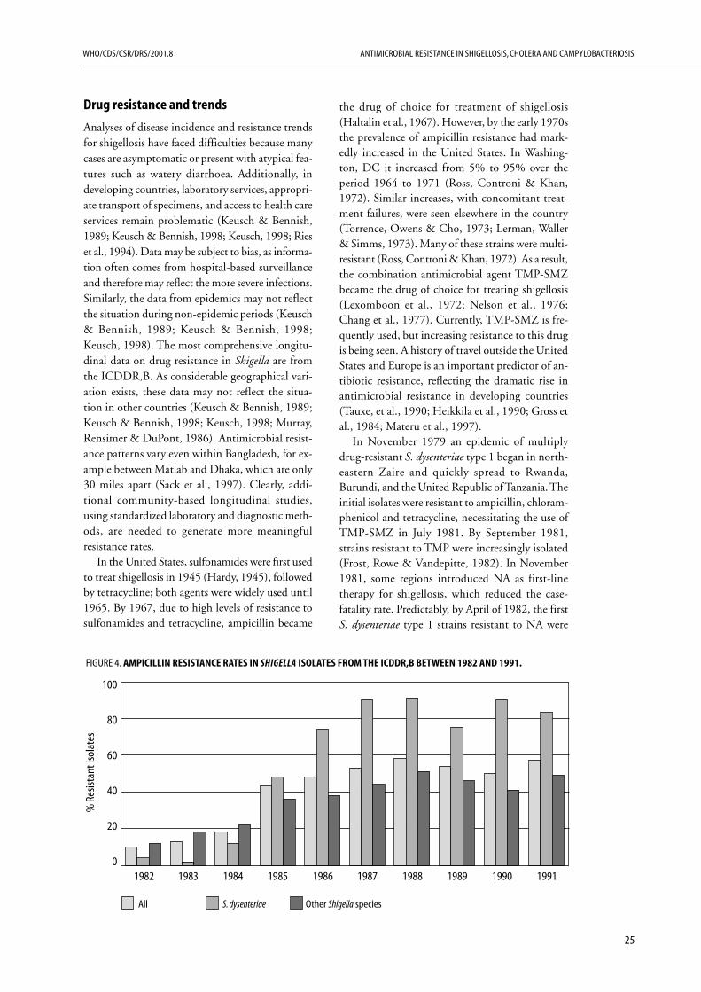
25
WHO/CDS/CSR/DRS/2001.8 ANTIMICROBIAL RESISTANCE IN SHIGELLOSIS, CHOLERA AND CAMPYLOBACTERIOSIS
Drug resistance and trends
Analyses of disease incidence and resistance trendsfor shigellosis have faced difficulties because manycases are asymptomatic or present with atypical fea-tures such as watery diarrhoea. Additionally, indeveloping countries, laboratory services, appropri-ate transport of specimens, and access to health careservices remain problematic (Keusch & Bennish,1989; Keusch & Bennish, 1998; Keusch, 1998; Rieset al., 1994). Data may be subject to bias, as informa-tion often comes from hospital-based surveillanceand therefore may reflect the more severe infections.Similarly, the data from epidemics may not reflectthe situation during non-epidemic periods (Keusch& Bennish, 1989; Keusch & Bennish, 1998;Keusch, 1998). The most comprehensive longitu-dinal data on drug resistance in Shigella are fromthe ICDDR,B. As considerable geographical vari-ation exists, these data may not reflect the situa-tion in other countries (Keusch & Bennish, 1989;Keusch & Bennish, 1998; Keusch, 1998; Murray,Rensimer & DuPont, 1986). Antimicrobial resist-ance patterns vary even within Bangladesh, for ex-ample between Matlab and Dhaka, which are only30 miles apart (Sack et al., 1997). Clearly, addi-tional community-based longitudinal studies,using standardized laboratory and diagnostic meth-ods, are needed to generate more meaningfulresistance rates.
In the United States, sulfonamides were first usedto treat shigellosis in 1945 (Hardy, 1945), followedby tetracycline; both agents were widely used until1965. By 1967, due to high levels of resistance tosulfonamides and tetracycline, ampicillin became
the drug of choice for treatment of shigellosis(Haltalin et al., 1967). However, by the early 1970sthe prevalence of ampicillin resistance had mark-edly increased in the United States. In Washing-ton, DC it increased from 5% to 95% over theperiod 1964 to 1971 (Ross, Controni & Khan,1972). Similar increases, with concomitant treat-ment failures, were seen elsewhere in the country(Torrence, Owens & Cho, 1973; Lerman, Waller& Simms, 1973). Many of these strains were multi-resistant (Ross, Controni & Khan, 1972). As a result,the combination antimicrobial agent TMP-SMZbecame the drug of choice for treating shigellosis(Lexomboon et al., 1972; Nelson et al., 1976;Chang et al., 1977). Currently, TMP-SMZ is fre-quently used, but increasing resistance to this drugis being seen. A history of travel outside the UnitedStates and Europe is an important predictor of an-tibiotic resistance, reflecting the dramatic rise inantimicrobial resistance in developing countries(Tauxe, et al., 1990; Heikkila et al., 1990; Gross etal., 1984; Materu et al., 1997).
In November 1979 an epidemic of multiplydrug-resistant S. dysenteriae type 1 began in north-eastern Zaire and quickly spread to Rwanda,Burundi, and the United Republic of Tanzania. Theinitial isolates were resistant to ampicillin, chloram-phenicol and tetracycline, necessitating the use ofTMP-SMZ in July 1981. By September 1981,strains resistant to TMP were increasingly isolated(Frost, Rowe & Vandepitte, 1982). In November1981, some regions introduced NA as first-linetherapy for shigellosis, which reduced the case-fatality rate. Predictably, by April of 1982, the firstS. dysenteriae type 1 strains resistant to NA were
FIGURE 4. AMPICILLIN RESISTANCE RATES IN SHIGELLA ISOLATES FROM THE ICDDR,B BETWEEN 1982 AND 1991.
1982 1983 1984 1985 1986 1987 1988 1989 1990 1991
All S. dysenteriae Other Shigella species
100
80
60
40
20
0
% R
esist
ant i
sola
tes
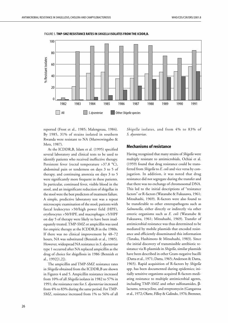
ANTIMICROBIAL RESISTANCE IN SHIGELLOSIS, CHOLERA AND CAMPYLOBACTERIOSIS WHO/CDS/CSR/DRS/2001.8
26
reported (Frost et al., 1985; Malengreau, 1984).By 1985, 31% of strains isolated in southernRwanda were resistant to NA (Mutwewingabo &Mets, 1987).
At the ICDDR,B, Islam et al. (1995) specifiedseveral laboratory and clinical tests to be used toidentify patients who received ineffective therapy.Persistent fever (rectal temperature >37.8 °C),abdominal pain or tenderness on days 3 to 5 oftherapy, and continuing anorexia on days 3 to 5were significantly more frequent in these patients.In particular, continued fever, visible blood in thestool, and an insignificant reduction of shigellae inthe stool were the best predictors of treatment failure.A simple, predictive laboratory test was a repeatmicroscopic examination of the stool; patients withfaecal leukocytes >50/high power field (HPF),erythrocytes >50/HPF, and macrophages >5/HPFon day 5 of therapy were likely to have been inad-equately treated. TMP-SMZ or ampicillin was usedfor empiric therapy at the ICDDR,B in the 1980s.If there was no clinical improvement by 48–72hours, NA was substituted (Bennish et al., 1985).However, widespread NA resistance in S. dysenteriaetype 1 occurred after NA replaced ampicillin as thedrug of choice for shigellosis in 1986 (Bennish etal., 1992[1,2]).
The ampicillin and TMP-SMZ resistance ratesin Shigella obtained from the ICDDR,B are shownin Figures 4 and 5. Ampicillin resistance increasedfrom 10% of all Shigella isolates in 1982 to 57% in1991; the resistance rate for S. dysenteriae increasedfrom 4% to 83% during the same period. For TMP-SMZ, resistance increased from 1% to 56% of all
Shigella isolates, and from 4% to 83% ofS. dysenteriae.
Mechanisms of resistance
Having recognized that many strains of Shigella weremultiply resistant to antimicrobials, Ochiai et al.(1959) found that drug resistance could be trans-ferred from Shigella to E. coli and vice versa by con-jugation. In addition, it was noted that drugresistance did not segregate during the transfer andthat there was no exchange of chromosomal DNA.This led to the initial descriptions of “resistancefactors” or R-factors (Watanabe & Fukusawa, 1961;Mitsuhashi, 1969). R-factors were also found tobe transferable to other enteropathogens such asSalmonella, either directly or indirectly via otherenteric organisms such as E. coli (Watanabe &Fukusawa, 1961; Mitsuhashi, 1969). Transfer ofantimicrobial resistance was thus determined to bemediated by mobile plasmids that encoded resist-ance and efficiently disseminated this information(Tanaka, Hashimoto & Mitsuhashi, 1983). Sincethe initial discovery of transmissible antibiotic re-sistance via R-plasmids in Shigella, similar plasmidshave been described in other Gram-negative bacilli(Datta et al., 1971; Datta, 1965; Anderson & Datta,1965). Rapid acquisition of R-factors by Shigellaspp. has been documented during epidemics; ini-tially sensitive organisms acquired R-factors medi-ating resistance to multiple antimicrobial agents,including TMP-SMZ and other sulfonamides, β-lactams, tetracycline, and streptomycin (Gangarosaet al., 1972; Olarte, Filloy & Galindo, 1976; Bremner,
FIGURE 5. TMP-SMZ RESISTANCE RATES IN SHIGELLA ISOLATES FROM THE ICDDR,B.
100
80
60
40
20
0
% R
esist
ant i
sola
tes
1982 1983 1984 1985 1986 1987 1988 1989 1990 1991
All S. dysenteriae Other Shigella species
27
WHO/CDS/CSR/DRS/2001.8 ANTIMICROBIAL RESISTANCE IN SHIGELLOSIS, CHOLERA AND CAMPYLOBACTERIOSIS
1978; Chun et al., 1984; Kaliyugaperumal, Gupta &Mohapatra, 1978; Adeleye, 1992).
TMP resistance in Enterobacteriaceae was firstfound to be mediated by R-factors in 1972 (Fleming,Datta & Gruneberg, 1972). TMP is a diamino-pyrimidine compound that exerts its antimicrobialeffect by selectively inhibiting bacterial dihydro-folate reductase (DHFR); this enzyme catalyzes thereduction of dihydrofolate to tetrahydrofolate, acofactor required for the synthesis of nucleic acidprecursors. Although TMP resistance can bechromosomally mediated, the most commonmechanism in Enterobacteriaceae, including Shig-ella, is acquisition of an additional, plasmid-encoded, variant DHFR enzyme. The most com-mon of these is known as DHFR I (Heikkila et al.,1990; Huovinen, 1987; Burchall, Elwell & Fling,1982; Haider et al., 1990). Several plasmid-encodedDHFR variants have been characterized, all ofwhich have reduced affinity for TMP (Huovinen,1987; Hickey & Nelson, 1997). These enzymes arealso produced at much higher levels than chromo-somal DHFR, effectively overwhelming the nativeenzyme (Amyes, 1989). DHFR I is often associ-ated with transposons. Transposon 7 (Tn7), the firstto be described, can transpose efficiently and spe-cifically into the E. coli chromosome. The abilityto transpose to this ubiquitous enteric organismmay explain its rapid and widespread dissemina-tion (Lichtenstein & Bremner, 1982; Fling &Richards, 1983). Tn7 also encodes resistance tostreptomycin and spectinomycin, and thereforemulti-drug resistance is common among isolatesresistant to TMP (Heikkila et al., 1990; Amyes &Towner, 1990). Tn7 can jump from plasmid to plas-mid or onto the bacterial chromosome, leavingbehind antimicrobial resistance determinants evenafter the plasmid it was initially carried by is spon-taneously lost (Amyes & Towner, 1990; Barth etal., 1976; Towner, Venning & Pinn, 1982). Further-more, genetic material may also be transferred tospecies outside the Enterobacteriaceae family. In1984, high level TMP-resistant V. cholerae strainsfrom an epidemic in the United Republic of Tan-zania were shown to have acquired a 113-kb resist-ance plasmid that contained Tn7 encoding the sameDHFR-I enzyme found in Shigella (Young &Amyes, 1986).
SMZ blocks another enzyme involved in folicacid metabolism, dihydroopterate synthase(DHPS). Synergy between SMZ and TMP is basedon their blocking sequential steps in this pathway.Similarly to TMP resistance, the most common
mechanism for SMZ resistance in Gram-negativeorganisms is the acquisition of plasmids encodingan altered DHPS with reduced affinity for SMZ.Fortunately, fewer variants of this enzyme have beenidentified than for DHFR (Hickey & Nelson,1997). Resistance to both TMP and SMZ impliesresistance to the combination. Although the chanceof a susceptible strain acquiring multiple resistances issmall, the probability is markedly increased when areservoir of transmissible plasmids exists (Then, 1982).
Once sulfonamide resistance became widespreadin Shigella species, β-lactam antibiotics became thedrugs of choice. β-lactam antibiotics target peni-cillin-binding proteins (PBPs), which carry outenzymatic functions essential for cell-wall structure.The binding of β-lactams to essential PBPs insusceptible species results in bactericidal activity(Georgopapadakou, 1993; Eliopoulos, 1988; Hickey& Nelson, 1997). Although resistance to β-lactamsmay arise in Shigella as a result of mutations thatreduce the affinity of PBPs for the antibiotics, themost common and most clinically significantmechanism of resistance to this class of drugs inGram-negative bacteria is the production of β-lactamases (Ghosh, Kar & Kundu, 1998). Theseenzymes hydrolyze the β-lactam ring, inactivatingthe drug. Since the introduction of ampicillin in1962, β-lactamase genes have increasingly beenfound on bacterial plasmids, facilitating the rapiddissemination of resistance among bacterial species(Anderson & Datta, 1965; Amyes, 1989). β-lactamases are a diverse group of enzymes first rec-ognized in E. coli in 1940 and later evolving inresponse to the development of new β-lactam anti-biotics (Abraham & Chain, 1940; Eliopoulos,1988; Hickey & Nelson, 1997). The genes may belocated on chromosomes, plasmids, or transposonsand may be constitutively expressed or inducible.β-lactamases vary in their spectrum of activityagainst penicillins and cephalosporins, as well as intheir susceptibility to clavulanic acid inhibition(Hickey & Nelson, 1997). In Europe, Asia, andAfrica the most common plasmid-encoded β-lactamase in resistant Gram-negative bacteria, in-cluding Shigella, is TEM-1 (Amyes, 1989; Schumacheret al., 1992). This enzyme can hydrolyze a number ofpenicillins and cephalosporins and is easily transferredto other members of the Enterobacteriaceae family aswell as to V. cholerae (Amyes, 1989; Threlfall, Rowe& Huq, 1980).
Tetracycline resistance is common in manyGram-negative bacteria, including Shigella, the firstorganism in which it was described. Although the
ANTIMICROBIAL RESISTANCE IN SHIGELLOSIS, CHOLERA AND CAMPYLOBACTERIOSIS WHO/CDS/CSR/DRS/2001.8
28
precise mechanism of action of the antibiotic is stillnot well understood, tetracycline binds reversiblyto the 30S subunit of the bacterial ribosome, pre-venting binding of aminoacyl-tRNA and inhibit-ing protein synthesis (Roberts, 1996). Shigella wasalso the first organism in which tetracycline resist-ance determinants were linked to R-plasmids andshown to be transferable to susceptible bacteria byconjugation (Roberts, 1996). Genes carried ontransposons and/or plasmids encode transmem-brane proteins that export the drug by an energy-dependent process (Hickey & Nelson, 1997; Speer,Shoemaker & Salyers, 1992).
Particularly in developing countries, fluoro-quinolone-resistant Shigella is increasingly preva-lent. Initially, low-level resistance to NA was seen.Later, chromosomal mutations were discovered thatconferred greater resistance (Turnidge, 1995). Themutations affect either DNA gyrase (bacterialtopoisomerase II) or the uptake and accumulationof quinolones (Turnidge, 1995; Yamagishi et al.,1981; Sato et al., 1986; Hooper et al., 1986; Hiraiet al., 1986). Although quinolone resistance is notmediated by transferable plasmids, the acquisitionof certain plasmids can contribute to quinoloneresistance by increasing the spontaneous mutationrate (Ambler et al., 1993; Ashraf, Ahmed & Sack,1991). These plasmids encode gene products in-volved in error-prone DNA repair responses, suchas the SOS response in E. coli, which is induced byDNA damage. The organism’s DNA is repaired,but with incorporation of a higher than normalnumber of incorrect bases. Populations of bacteriaharbouring such plasmids may have an evolution-ary advantage.
Many socioeconomic and behavioural factorscontribute to increasing antimicrobial resistance.Shigellosis is more prevalent in individuals of lowsocioeconomic status and education level in devel-oping countries, where there are frequently inad-equate treatment and poor compliance (Haider,Malek & Albert, 1993; Okeke, Lamikanra &Edelman, 1999). In developing countries, healthypeople commonly harbour resistant bacteria as partof their normal flora (Okeke, Lamikanra & Edel-man, 1999; Calva, Sifuentes-Osornio & Ceron,1996) and this resistance can be transferred at lowfrequency, via plasmids and transposons, to Shig-ella (Tauxe, Cavanagh & Cohen, 1989; Platt,Chesham & Kristinsson, 1986; Bratoeva & John,1994; Murray, Rensimer & DuPont, 1982; Levineet al., 1983; Hickey & Nelson, 1997; Petrocheilou,Grinsted & Richmond, 1976). As is the case for V.
cholerae and C. jejuni, selective pressure due toexcessive use of antimicrobial agents, whether mis-use by health care providers (e.g. for childhoodgastroenteritis or viral respiratory infections)(Thamlikitkul, 1988; Okeke, Lamikanra & Edelman,1999; CDC, 1991; Guyon et al., 1994; Bojalil &Calva, 1994; Nizami, Khan & Bhutta, 1996; Paredeset al., 1996; Hui et al., 1997; Reyes et al., 1997), orunrestricted access to them, are responsible for muchof the resistance seen in Shigella (Okeke, Lamikanra& Edelman, 1999; Haider, Malek & Albert, 1993).
In many developing countries antimicrobialagents are readily available without a prescription.In Nagpur, India, despite prohibition by theIndian Pharmaceutical Act, self-prescribing ofantimicrobials was common, primarily for minorrespiratory infections and non-specific complaintssuch as fever, diarrhoea, and abdominal pain (Dua,Kunin & White, 1994). In another study, in ruralBangladesh, 95% of the drugs consumed by thesubjects were obtained from local pharmacies, withphysicians having prescribed only 8% (Hossain,Glass & Khan, 1982). In addition, antimicrobialagents may be of poor quality, due to degradation,counterfeiting, or lack of bioequivalence in the caseof generic drugs. Use of such products results insub-optimal serum concentrations, which promotesselection for resistance (Okeke, Lamikanra &Edelman, 1999).
An increasingly mobile population provides op-portunities for the rapid spread of multi-resistantorganisms in regions where unrestricted antibioticuse is common, particularly in settings where manyindividuals may asymptomatically harbour patho-gens such as Shigella (Bennish et al., 1985; Guerreroet al., 1994; Calva, Sifuentes-Osornio & Ceron,1996). Overcrowding and improper sewage disposalpromote the spread of resistant organisms amongindividuals and provide increased opportunities forgenetic exchange among bacteria, facilitating thedissemination of antibiotic resistance determinants(Okeke, Lamikanra & Edelman, 1999). In manycountries armed conflicts, corruption and misman-agement threaten the public health infrastructure,favouring the spread of infections and of drugresistance (Okeke, Lamikanra & Edelman, 1999).
Lack of facilities and resources for susceptibilitytesting and surveillance, particularly in developingcountries, results in inappropriate clinical decision-making, inappropriate treatment, and thus antibi-otic resistance (Okeke, Lamikanra & Edelman,1999; Hughes & Tenover, 1997). In some cases,even when most strains are known to be resistant,
29
WHO/CDS/CSR/DRS/2001.8 ANTIMICROBIAL RESISTANCE IN SHIGELLOSIS, CHOLERA AND CAMPYLOBACTERIOSIS
inappropriate antibiotics are used under the mis-apprehension that any antibiotic is better thannone. Regular, continuous surveillance is requiredsince resistance rates, particularly in Shigella, varygeographically and also over time within a singlecountry (Sack et al., 1997). When available, theresults of accurate surveillance should be used toguide policies for antibiotic use.
Intervention strategies andresearch needs
1. Programmes aimed at decreasing the totalnumber of infections.
1.1 Resistant organisms thrive where rates of shig-ellosis are highest; thus, programmes target-ing the overall rate of infection will also reducethe number of infections with resistant strainsand the spread of such strains. Shigellosis isprimarily a disease of crowded communitiesthat do not have adequate sanitation or safedrinking-water. Therefore, the long-term strat-egy for control of this disease is still improve-ment of water, sanitation, and socioeconomicstatus (Shears, 1996; Bennish et al., 1990[2]).Theoretically, these public health measures arethe most effective means of disease reduction,but practical considerations have limited theirimplementation. Development projects, suchas the WHO Water and Sanitation Decade,are needed to improve the quality of watersupplies and sanitation globally (Shears, 1996).
1.2 Targeted interventions which have proved ef-fective in reducing Shigella infection include:a) hand-washing programmes which makesoap available and teach people how to use iteffectively; b) encouraging breast-feeding ofinfants and small children (Ahmed et al.,1992); c) latrine programmes to reduce envi-ronmental contamination; and d) pro-grammes to reduce the density of flies, whichcan deposit infectious inocula of Shigella onfood (Ahmed et al., 1992; Levine & Levine,1991; Acheson & Keusch, 1995; Watt &Lindsay, 1948). In a community-based trialin Bangladesh, Ahmed et al. (1992) demon-strated that breast-feeding protects children upto 3 years of age against shigellosis, particu-larly against disease due to multi-resistantorganisms.
1.3 An effective vaccine would prevent Shigellainfections due to either sensitive or resist-
ant strains. Much progress has been madetowards an effective vaccine for shigellosis,but none is yet available for public healthuse. Vaccine development should be encour-aged, as this will likely be the most effectiveway to achieve a significant reduction intransmission. Even if we need five moreyears to develop a successful vaccine for S.flexneri and S. dysenteriae, this is still shorterthan the time needed to achieve significantimprovements in water and sanitation sys-tems. However, vaccine development andwater/sanitation improvements should nothave to compete for attention or resources;both programmes need to proceed simulta-neously and be implemented appropriately.Since current evidence suggests that vaccinesfor shigellosis must be serotype-specific,priority must be given to those organismsthat represent the principal public healthproblems, i.e. S. dysenteriae and S. flexneri.
1.4 Is interrupting transmission by patients fea-sible? Antibiotic treatment of patients eradi-cates the organism from the stool. However,this may not be an important factor since,in most instances, only a small proportionof infected patients actually receive antibi-otics. Individuals with asymptomatic ormild infections are responsible for most ofthe faecal contamination, and treatment ofthese individuals is both unrealistic andunwise. Thus, treatment of patients shouldbe aimed at clinical cure rather than clear-ance of the organism.
2. Limiting treatment to only those patients whoreally need the antibiotic. Because shigellosis isoften a self-limited illness, more research isneeded to help identify those at greatest risk ofdying; newer antimicrobial agents should be re-served for treating those individuals (Ries et al.,1994). There is little information available con-cerning the optimal duration of treatment,appropriate target populations, or the cost-effectiveness of different therapeutic options.Further research is needed in this area (Keusch& Bennish, 1989).
3. Limiting the duration of treatment. In those in-stances when antibiotics are needed, short buteffective courses of treatment may be prudent.This would limit the environmental pressure forselection and dissemination of resistance genesamong bacterial populations (O’Brien, 1997).
ANTIMICROBIAL RESISTANCE IN SHIGELLOSIS, CHOLERA AND CAMPYLOBACTERIOSIS WHO/CDS/CSR/DRS/2001.8
30
Conclusion
Shigellosis is the most difficult of the three entericorganisms being reviewed, in terms of steady trendstowards multiple resistance. Once they have becomeresistant, epidemic and endemic strains of Shigellahave remained resistant; there have only been a fewinstances in which a sensitive strain has reappearedin a region. Additionally, shigellosis is one of thediseases most difficult to prevent because only asmall number of bacteria are required to cause dis-ease. Thus, intensive water and sanitation pro-grammes and vaccine development would seem tobe critical. It is not realistic to continue relying onthe introduction of a new antibiotic every few years.
4. Enforcement of appropriate antibiotic usepolicies. Unfortunately, legislation has not alwaysbeen effective in developing countries. In India,for example, pharmacists continue to sell drugswithout requiring a prescription, despite the factthat they are forbidden to do so under the IndianPharmaceutical Act (Dua, Kunin & White,1994). In addition, there is often overzealouspromotion of antimicrobials by pharmaceuticalcompanies; measures to prevent this unethicalpractice should be implemented (Dua, Kunin& White 1994; Couper, 1997).
5. Education. Continuing education of health careproviders and development of standard treat-ment guidelines for gastroenteritis and viralinfections may reduce the inappropriate use ofantibiotics, thus reducing the pressure that con-tributes to selection for resistance in Shigella(Couper, 1997).
31
WHO/CDS/CSR/DRS/2001.8 ANTIMICROBIAL RESISTANCE IN SHIGELLOSIS, CHOLERA AND CAMPYLOBACTERIOSIS
Review of Campylobacter jejuni
Microbiology
Campylobacters are slim, Gram-negative, curvedrods. Located at one or both ends of the cell is apolar flagellum, making the organism highly mo-tile. C. jejuni and C. coli are microaerophilic andthermophilic, growing best at 42 °C and requiringoxygen concentrations of 3–15% and carbondioxide concentrations of 3–5%. The thermophiliccharacteristic is thought to be an adaptation to theenvironment of animal and bird intestines. Campylo-bacter spp. have a small genome of 1.6–1.7 Mbp,which may partially explain their complex nutri-tional requirements in laboratory culture (Griffiths& Park, 1990). Campylobacters use products ofthe Krebs cycle and amino acids for growth sincethey are incapable of fermenting carbohydrates. C.jejuni can be distinguished from other species byits ability to hydrolyze hippurate, benzoic acid andglycine. Extrachromosomal elements (plasmids)have been found in Campylobacter spp. and encodeantibiotic resistance (Bopp et al., 1985; Taylor etal., 1981; Tenover & Elvrum, 1988; Prasad et al.,1994; Tenover et al., 1985).
Transmission
Campylobacter spp. are part of the normal intesti-nal flora of wild and domesticated animals andbirds. Of particular importance to humans is theircolonization of animals used in food productionincluding poultry, cattle, sheep and swine (Blaser,1997). Poultry comprise the most significant reser-voir worldwide; Campylobacter spp. can be isolatedfrom 30–100% of the birds in many domestic andwild avian species (Blaser & Reller, 1981). House-hold pets such as dogs, cats and birds are additionalanimal reservoirs. Although excretion of Campylo-bacter is not associated with symptoms in poultry,diarrhoeal illnesses have been documented in mam-malian pets and livestock, and this contributes tothe contamination of surface water. Post-slaughterprocessing does not reduce the extent of coloniza-tion of poultry and may, in fact, lead to cross-contamination of previously uncontaminated
Introduction
C. jejuni and related species are increasingly recog-nized as important causative agents of enterocoli-tis. Although relatively recently (1972) recognizedas a significant human pathogen, C. jejuni is nowthe bacterial pathogen most commonly responsi-ble for diarrhoeal illness in the United States (Blaser,1997; Griffiths & Park, 1990). According to theCDC, 46% of culture-proven bacterial gastro-enteritis in the United States in 1996 was due toCampylobacter spp. Salmonella, Shigella and E. coli wereless frequent causes of enteric disease (Altekreuseet al., 1999). Campylobacter is also a common causeof endemic diarrhoeal illness worldwide, althoughthere are regional differences in the characteristicsof the illness and the population affected (Taylor,1992) The extent to which campylobacteriosis isresponsible for travellers’ diarrhoea varies regionally;this appears to be more common in Asia.
The bacteria now known as Campylobacter werefirst described in cattle and sheep, and were namedVibrio foetus because they caused abortion. V. foetuswas isolated from immunocompromised patientsas early as 1947, although its role in acute diar-rhoea was not recognized until 1972 (Dekeyser etal., 1972). Based upon cell wall and DNA differencesfrom other members of the genus Vibrio, V. foetuswas reclassified in 1973 in the genus Campylobacter.This genus comprises an increasing number of spe-cies, but C. jejuni and C. coli are responsible forthe majority of cases of human enterocolitis; C.jejuni accounts for 80–90% of enteric disease.Recent studies by the CDC found that in theUnited States, 99% of cultured Campylobacter spp.were C. jejuni. (Tauxe, 1992). The incidence of in-fection by C. upsaliensis, C. lari, C. foetus, and C.hyointestinalis is unknown, but these organismsseem to be much less common (Ketley, 1997).However, antibiotic-containing agar is used forprimary isolation; this may select for C. jejuni andthus bias the frequency of isolation towards thisspecies.
ANTIMICROBIAL RESISTANCE IN SHIGELLOSIS, CHOLERA AND CAMPYLOBACTERIOSIS WHO/CDS/CSR/DRS/2001.8
32
carcasses. In one study in England, the organismwas isolated from 48% of randomly cultured freshchickens (Hood, Pearson & Shahamat, 1988).Freezing the carcass does not eradicate the bacte-ria. Ingestion of contaminated water, interactionwith colonized pets (especially puppies and kittens),and consumption of unpasteurized milk or under-cooked poultry or meat are all associated with humandisease (Blaser et al., 1978; Blaser & Reller, 1981;Blaser, 1997; Deming et al., 1987; Robinson,1981). Human-to-human transmission, while un-common, has been documented via faecal expo-sure, particularly from young, incontinent children(Blaser, 1997). A history of foreign travel isfrequently documented in citizens of industrializednations who contract Campylobacter enteritis. Al-though enterotoxigenic E. coli (ETEC) is generallythe most common pathogen in travellers’ diarrhoea,Campylobacter species represented 17% of thepathogens isolated from travellers to Thailand(much lower percentages are found in travellers toother countries) (Black, 1990).
Spectrum of illness
Clinical illness ranges from mild, self-limiting, non-inflammatory diarrhoea to severe, inflammatory,bloody diarrhoea with high fever and bacteraemia.In immunosuppressed patients, particularly thosewith HIV, the diarrhoeal illness may be relapsingor unremitting and may be accompanied by bacter-aemia and extraintestinal illness (Tee et al., 1995;Tee & Mijch, 1998; Wang & Blaser, 1986). In ananalysis of 24 isolates from extraintestinal campylo-bacter illness, these appeared to be similar to thosethat cause intestinal disease. However, 52% ofpatients with extraintestinal disease had underly-ing immunosuppression (Blaser et al., 1986).
Although asymptomatic bacterial shedding hasbeen documented, a chronic carrier state has notbeen described in immunocompetent individuals.(Blaser & Reller, 1981) The annual incidence ofC. jejuni diarrhoeal infection was estimated by theCDC to be 5–6 per 100 000 persons in the UnitedStates, although population-based studies suggestthat it may be closer to 1000 per 100 000 (Blaser,1997). Due to the broad range of clinical symptomsand the specific laboratory media and techniquesrequired for culture, Campylobacter infections arethought to be underreported worldwide. The totalnumber of infections in the United States could beclose to 2.4 million/year, with 120–360 deaths(Taylor, 1992).
Curiously, the characteristics of the disease aredifferent in different populations. In industrializedcountries, where infection is relatively rare, its mani-festations are more severe; in developing countries,infection is more common but symptoms aremilder. Patients in industrialized countries usuallyexperience an inflammatory diarrhoea with severeabdominal cramping and fever. Most (60%) stoolsamples from symptomatic, culture-positiveC. jejuni gastroenteritis episodes contain blood and78% of these have polymorphonuclear leukocytes(Blaser et al., 1979). Campylobacter infections areseasonal, peaking in late summer and fall. Sporadicillness and epidemic outbreaks associated with food-or waterborne exposures have been described,suggesting a general lack of immunity in the popula-tion. There is a bimodal age distribution, with onepeak in children under the age of 1 year and the sec-ond peak in young adults between the ages of 15and 34 (Blaser, 1997; Ketley, 1997). At present,rates of infection are rising, apparently linked toincreased poultry consumption (Blaser, 1997;Deming et al., 1987).
In contrast, Campylobacter infection appears tobe hyperendemic and non-seasonal in developingcountries. The illness is characterized by watery,non-inflammatory, relatively mild diarrhoea. Infec-tion rates are typically highest during the first twoyears of life and decrease with age (Taylor et al., 1988;Blaser, 1997; Glass et al., 1983[2]). There is alsoattenuation of the symptoms and reduction inconvalescent-phase bacterial excretion with increas-ing age. Convalescent excretion occurred for 14 ±2 days in Thai children less than 1 year of age andfor 8 ± 2 days in children between the ages of 1 and5 years (Taylor et al., 1988). These children experi-enced reinfection with different Campylobacterserotypes at a rate of 15% per week, demonstrat-ing that the infection rate is high among childrenin developing nations. In Bangladesh, Glass et al.(1983[2]) found that healthy control patients hadnearly the same frequency of Campylobacter infec-tion as did patients presenting with diarrhoea.Asymptomatic infection rates may therefore bequite high in developing countries as compared toindustrialized nations (Glass et al., 1983[2]; Black,1988).
Although efforts have been made to character-ize strains isolated in different regions in order toaccount for the differences in the symptoms, thereis little evidence to suggest that Campylobacterstrains isolated in developing countries are differ-ent from those found in industrialized nations with
33
WHO/CDS/CSR/DRS/2001.8 ANTIMICROBIAL RESISTANCE IN SHIGELLOSIS, CHOLERA AND CAMPYLOBACTERIOSIS
respect to virulence factors (Taylor et al., 1988; Asratet al., 1997). Travellers to developing countriesdevelop an illness typical of their own country. Resi-dents of developing nations average more than fiveinfections with C. jejuni lifetime, as compared withan average of none to one in developed countries(Blaser, 1997). Therefore, the weight of the evidenceis that immunity accounts for the attenuation ofsymptoms with increasing age in developing coun-tries. Consistent with this interpretation are stud-ies in Thailand and Bangladesh demonstrating thatserum levels of C. jejuni cell surface-specific im-munoglobulin A (IgA) rise with increasing age,implying acquisition of protective immunity withspecific mucosal IgA (Blaser et al., 1985; Blaser etal., 1986; Blaser, Taylor & Echeverria, 1986). Ineach age group, and for each immunoglobulin sub-class (IgA, IgG, and IgM), serum antibody to C.jejuni was found in significantly greater amountsthan in Americans (Blaser et al, 1986; Blaser, Taylor& Echeverria, 1986). Thus, geographical differencesin the nature of campylobacteriosis appear to berelated to acquired immunity in developing nations(due to the hyperendemic nature of the disease)rather than to strain differences. However, muchremains to be learned about the molecular biologyof Campylobacter pathogenesis.
There is strong evidence linking Campylobacterinfection to the development of Guillain-BarréSyndrome (GBS). Following intestinal infection,some patients develop an acute peripheral neuropa-thy, starting with ascending weakness or paralysisof the extremities and sometimes progressing toparalysis of the trunk and interference with breath-ing. The mechanism of this complication is thoughtto be related to a ganglioside, found on the surfaceof certain strains of Campylobacter, which mimicsa ganglioside present on nerve cells. This “molecu-lar mimicry” is thought to trigger an autoimmuneresponse, leading to GBS (Moran, Appelmelk &Aspinall, 1996). Certain Penner serotypes (e.g.O19) are more commonly associated with GBS,but other serotypes may also lead to its develop-ment (Aspinall, McDonald & Pang, 1994). The truevirulence marker is not known. Rapid methods fordetecting Gm1 ganglioside on Campylobacter (Sacket al., 1998) may help identify strains more likelyto induce GBS, but more work is needed to deter-mine whether Gm1 is actually the virulence marker.
In the United States about 40% of GBS is asso-ciated with a prior Campylobacter infection(Altekreuse et al., 1999). GBS has not been wellstudied in developing countries; however, with the
eradication of polio, it seems likely that some casesof acute paralysis that would previously have beendiagnosed as polio will turn out to be GBS instead.Thus, Campylobacter-associated GBS may be evenmore important in developing countries, eventhough the diarrhoeal disease tends to be mild. Itseems likely that GBS will be recognized as an“emerging disease,” even though these new caseswill probably represent disease that was previouslymisdiagnosed.
Pathogenicity
The incubation period after ingestion of C. jejuniis thought to average 24–72 hours, but periods of≥1 week have been documented. The precise in-oculum is unknown, but as few as 800 organismshave been shown to cause illness (Black, 1988). Therate of illness increases with increasing numbers oforganisms ingested. Fever, chills, headache, andmyalgia may precede diarrhoea symptoms. Acuteabdominal pain coincides with diarrhoea and maybe severe enough to mimic appendicitis. The diar-rhoea may be severe, with frequent bloody bowelmovements, upwards of eight per day have beenreported. The symptoms generally peak by 24–48hours and subside over a period of a week. Colono-scopy may reveal friable mucosa with oedema andpetechial haemorrhages consistent with a diffusecolitis. Ileal and jejunal involvement have beennoted as well.
The molecular pathogenesis of C. jejuni infec-tions is multifactorial and complex, and seems toinvolve motility, adhesion, invasion, and toxin pro-duction. Campylobacters have the unusual abilityto change from a thin, curved form to a sphericalor coccoid shape. Along with this structural change,they become non-culturable. This viable, non-culturable state (referred to as the VNC state) maybe a dormant state induced by environmental stress(Rollins & Colwell, 1986). It is not known if theVNC form is transmissible to humans (Ketley,1997).
Their cell shape and flagella give campylobactersmotility in the viscous environment of the gut. Theflagellum loci have been well characterized andmapped to adjacent genes, flaA and flab (Guerry etal., 1991). Mutations in these genes affect the abil-ity of C. jejuni to colonize the gut (Nachamkin etal., 1993), indicating that motility plays a role inthe pathogenesis of the organism.
The roles of adhesion and invasion are incom-pletely understood. Fimbriae have recently been
ANTIMICROBIAL RESISTANCE IN SHIGELLOSIS, CHOLERA AND CAMPYLOBACTERIOSIS WHO/CDS/CSR/DRS/2001.8
34
described, and non-fimbriated mutants produceattenuated disease (Doig et al., 1996). The inflam-matory nature of many Campylobacter infectionssuggests that the bacteria invade cells, but there islittle in vivo evidence for this.
The role of toxin production in Campylobacterpathogenesis is highly debated. Cholera-like tox-ins (CLT) have been reported, and they may play arole in the enterotoxigenic, noninflammatorydiarrhoea typical in the developing world.Enterotoxins are secreted proteins that are able topenetrate eukaryotic cells and trigger intracellularcyclic AMP production, leading to ionic changesthat result in watery diarrhoea. There is immuno-logical cross-reactivity among CLT, cholera toxin(CT) and E. coli heat-labile toxin (LT) (Wassenaar,1997). A study in Costa Rica linked the produc-tion of CLT to non-inflammatory diarrhoeal symp-toms, but studies performed elsewhere haveproduced inconsistent and conflicting data (Florin& Antillon, 1992). Multiple cytotoxins, includingcytolethal-distending toxin (CLDT), haemolyticcytotoxins and Shiga-like toxins have also beendescribed in Campylobacter. Cytotoxins are proteinsthat kill eukaryotic cells; E. coli Shiga-like toxin isan example. Although, theoretically, these toxinsmay play a role in the inflammatory type of diar-rhoea commonly seen, this is incompletely under-stood (Ketley, 1997). While there have beensignificant advances in the molecular pathogenesisof C. jejuni, additional research is required tounderstand its ability to cause disease.
Diagnosis and identification
A confirmed diagnosis requires culture of the or-ganism from faeces or blood. Faecal samples shouldarrive in the laboratory within a few hours aftercollection or, if a delay is likely, should be inocu-lated into a transport medium (e.g. Cary-Blairmedium). In the laboratory, rich but selective me-dia (containing antibiotics to reduce the growth ofother species), microaerophilic incubation condi-tions (typically, a GasPak jar conditioned with atablet to lower the oxygen concentration), and anelevated temperature (42 °C) are required. A vari-ety of Campylobacter-specific media are producedcommercially, including Skirrows medium,Butzler’s Medium, and Campy BAP media. A tech-nique for isolating Campylobacter spp. from faecesusing antibiotic-free media involves allowing thebacteria to migrate through a microporous filter.This method appears to be more sensitive, espe-
cially for non-jejuni species that may be inhibitedby the antibiotics used in standard isolation media.Enrichment broth is not normally used for faecalsamples; however, it may be used when isolatingC. jejuni from contaminated liquids such as sur-face water or milk (Griffiths & Park, 1990). Stand-ard blood culture broth will recover Campylobacterfrom blood specimens without the need forspecific selection or enrichment. Techniques for theidentification of species and the typing of strainsof Campylobacter are described in standard micro-biology texts.
Additional experimental methods for character-izing strains may be useful for specialized purposes.The most common technique is serotyping, usingeither the Penner or the Lior system. The Pennersystem identifies a heat-stable somatic (O) antigenand the Lior system a heat-labile flagellar (H) anti-gen. Both systems can type over 90% of humanand animal Campylobacter spp. (Lior et al., 1982;Penner & Hennessy, 1980).
The use of DNA probe analysis is limited bycomplicated protocols, the need for specializedequipment, and the short shelf-life of reagents(Griffiths & Park, 1990). PCR fingerprinting meth-ods using the gyrA and pflA genes hold promise fordetermining the relatedness of C. jejuni isolates andstreamlining the tedious restriction fragment lengthpolymorphism (RFLP) analyses and pulsed field gelelectrophoresis (PFGE) methods that are currentlyin use to characterize Campylobacter strains (Ragim-beau et al., 1998). Thus, while a number of techniquesare available to speciate and type Campylobacter, theirusefulness is limited by the technical requirements,and they must be performed by reference laborato-ries.
If both acute and convalescent sera are available,serodiagnosis of Campylobacter infection is possi-ble using enzyme-linked immunosorbent assay(ELISA) with a glycine extract providing the anti-gen (Blaser et al, 1986; Blaser, Taylor & Echeverria,1986; Griffiths & Park, 1990). Antibody responseto C. jejuni antigens may include the productionof IgA, IgM or IgG. Testing only a convalescentserum sample is not useful because an elevated titremay reflect previous rather than current infection.
Therapy
In immunocompetent individuals, Campylobacterenterocolitis is generally self-limited, with mild tomoderate symptoms, and antibiotic therapy is notrequired for most patients. Supportive care with
35
WHO/CDS/CSR/DRS/2001.8 ANTIMICROBIAL RESISTANCE IN SHIGELLOSIS, CHOLERA AND CAMPYLOBACTERIOSIS
oral rehydration is the preferred treatment. Whenantibiotics are used to treat severe or invasive ill-ness, the definition of cure, by convention, is reliefof diarrhoea. Shedding of organisms may continuefor 2–4 weeks or longer (median <3 weeks) (Blaser,1997). Antibiotics have been shown to reduce theexcretion of Campylobacter; erythromycin reducesits duration from 16.8 to 2 days (Pai et al., 1983)and azithromycin eliminates excretion within 24hours (Kuschner et al., 1995). Whether antibioticsactually reduce the duration of diarrhoeal symp-toms is subject to debate. Pai et al. (1983) foundno change in the duration of diarrhoeal symptomswhen American children received erythromycin forCampylobacter enteritis. Goodman et al. (1990)found that, in patients in Chicago, empirical cipro-floxacin treatment for diarrhoea reduced the dura-tion of symptoms from 3.4 days (with placebo) to2.4 days. Others have found that antibiotic treat-ment reduced the duration of symptoms from anaverage of 90 hours to 30 hours (DuPont et al.,1987). In severe cases, and in immunosuppressedindividuals (particularly those with HIV), relaps-ing or unremitting inflammatory diarrhoea requir-ing antibiotic administration may develop. C. jejunibacteraemia occurs at rates in the order of 0.7% to2% in immunocompetent hosts (Tee, Kaldor &Dwyer, 1986; Wang & Blaser, 1986). In contrast,8.3% of 121 HIV-positive patients with Campylo-bacter enteritis became bacteremic and this wasassociated with 33% mortality (Tee & Mijch,1998). Thus, antibiotic administration should beon a case-by-case basis, with antimicrobials reservedfor serious infections.
C. jejuni is sensitive to several classes of anti-microbials. The drug of choice for Campylobacterenterocolitis is generally a macrolide. Erythromy-cin has traditionally been the first-line therapy,although newer macrolides such as azithromycinare quickly gaining popularity (Tee & Mijch, 1998;Sanchez et al., 1994; Kuschner et al., 1995). Bac-tericidal fluoroquinolones have become the drugsof choice for treating travellers’ diarrhoea and aretherefore used as first-line therapy of Campylobacterinfections contracted abroad. Tetracycline, doxy-cycline and TMP-SMZ have been used as second-line agents, although increasing resistance limitstheir use. Gentamicin, kanamycin, imipenem andampicillin-clavulanic acid are reserved for systemic,refractory illness.
Antimicrobial resistance
In the past, Campylobacter strains were rarely testedfor antibiotic sensitivity because this was technicallydifficult, and because resistance was infrequentlyencountered. Due to increasing antibiotic resist-ance, susceptibility testing is becoming moreimportant. Susceptibility can be determined usingmicrodilution methods, agar dilution methods orthe E-test (Baker, 1992). Agar dilution and theE-test give comparable results; broth microdilutionis inaccurate, particularly for tetracycline (Baker,1992). The tests performed better when incuba-tion was at 42 °C for 16–18 hours. The need formicroaerophilic and thermophilic conditions, andfor including rigorous controls, limits the use ofthese assays to a few laboratories where Campylo-bacter testing is routinely performed.
Worldwide, a complex interplay of factors hascreated an environment fostering the rapid emer-gence of resistance in C. jejuni (see Table 2). Re-sistance to fluoroquinolones and tetracyclines hasrapidly reduced the efficacy of these drugs in manyparts of the world (Sanchez et al., 1994). Majorfactors exerting selective pressure have included theintroduction of fluoroquinolones into veterinarymedicine and the extensive and sometimes indis-criminate use of antimicrobials to treat a variety ofconditions. Antibiotics have long been used as sup-plements in animal feed, to promote growth andto prevent infections associated with the crowdedconditions in which mass-produced livestock areraised. Nearly 45% of the 2.5 million kilograms ofantibiotics consumed in the United States is usedin animal feed (DuPont & Steele, 1987). This heavyselective pressure leads to the carriage of resistantorganisms by livestock, and there is concern thatthis leads to resistant infections in humans (Endtzet al., 1991). After the introduction of tetracycline-supplemented feed, tetracycline-resistant bacteriawere found in the stools of 80% of farm personnel.Much lower rates (7%) were found in neighboursnot exposed to these animals (Levy, FitzGerald &Macone, 1976). More recently (1987), enrofloxacin,a fluoroquinolone, entered the animal husbandrymarket.
Campylobacter infections are especially linked topoultry. Enrofloxacin has been used in broiler chick-ens, reproductive chickens and egg-laying hens toprevent enteritis and respiratory disease. Velazquezet al. (1995) has shown that antibiotic concentra-tions in poultry feed are high enough to select forNA resistance in vitro. Extensive studies in theNetherlands (Endtz et al., 1991) have documented
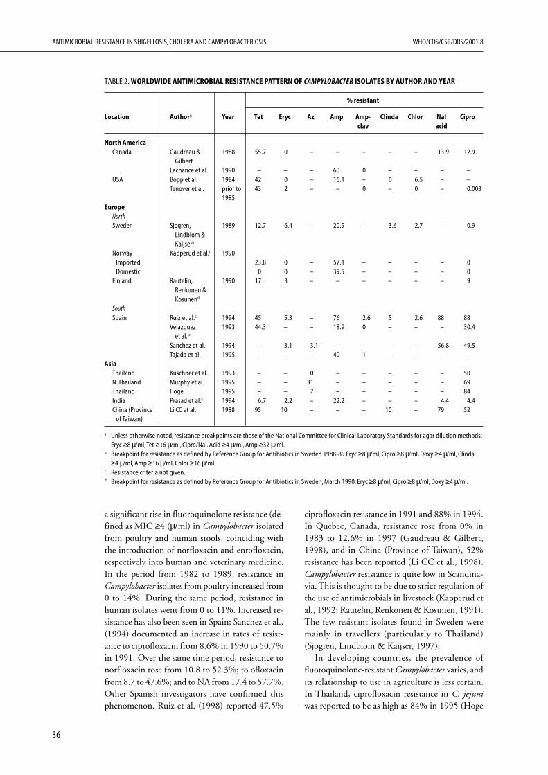
ANTIMICROBIAL RESISTANCE IN SHIGELLOSIS, CHOLERA AND CAMPYLOBACTERIOSIS WHO/CDS/CSR/DRS/2001.8
36
a significant rise in fluoroquinolone resistance (de-fined as MIC ≥4 (µ/ml) in Campylobacter isolatedfrom poultry and human stools, coinciding withthe introduction of norfloxacin and enrofloxacin,respectively into human and veterinary medicine.In the period from 1982 to 1989, resistance inCampylobacter isolates from poultry increased from0 to 14%. During the same period, resistance inhuman isolates went from 0 to 11%. Increased re-sistance has also been seen in Spain; Sanchez et al.,(1994) documented an increase in rates of resist-ance to ciprofloxacin from 8.6% in 1990 to 50.7%in 1991. Over the same time period, resistance tonorfloxacin rose from 10.8 to 52.3%; to ofloxacinfrom 8.7 to 47.6%; and to NA from 17.4 to 57.7%.Other Spanish investigators have confirmed thisphenomenon. Ruiz et al. (1998) reported 47.5%
ciprofloxacin resistance in 1991 and 88% in 1994.In Quebec, Canada, resistance rose from 0% in1983 to 12.6% in 1997 (Gaudreau & Gilbert,1998), and in China (Province of Taiwan), 52%resistance has been reported (Li CC et al., 1998).Campylobacter resistance is quite low in Scandina-via. This is thought to be due to strict regulation ofthe use of antimicrobials in livestock (Kapperud etal., 1992; Rautelin, Renkonen & Kosunen, 1991).The few resistant isolates found in Sweden weremainly in travellers (particularly to Thailand)(Sjogren, Lindblom & Kaijser, 1997).
In developing countries, the prevalence offluoroquinolone-resistant Campylobacter varies, andits relationship to use in agriculture is less certain.In Thailand, ciprofloxacin resistance in C. jejuniwas reported to be as high as 84% in 1995 (Hoge
TABLE 2. WORLDWIDE ANTIMICROBIAL RESISTANCE PATTERN OF CAMPYLOBACTER ISOLATES BY AUTHOR AND YEAR
% resistant
Location Authora Year Tet Eryc Az Amp Amp- Clinda Chlor Nal Ciproclav acid
North AmericaCanada Gaudreau & 1988 55.7 0 – – – – – 13.9 12.9
GilbertLachance et al. 1990 – – – 60 0 – – – –
USA Bopp et al. 1984 42 0 – 16.1 – 0 6.5 – –Tenover et al. prior to 43 2 – – 0 – 0 – 0.003
1985Europe
NorthSweden Sjogren, 1989 12.7 6.4 – 20.9 – 3.6 2.7 – 0.9
Lindblom &Kaijserb
Norway Kapperud et al.c 1990Imported 23.8 0 – 57.1 – – – – 0Domestic 0 0 – 39.5 – – – – 0
Finland Rautelin, 1990 17 3 – – – – – – 9Renkonen &Kosunend
SouthSpain Ruiz et al.c 1994 45 5.3 – 76 2.6 5 2.6 88 88
Velazquez 1993 44.3 – – 18.9 0 – – – 30.4et al. c
Sanchez et al. 1994 – 3.1 3.1 – – – – 56.8 49.5Tajada et al. 1995 – – – 40 1 – – – –
AsiaThailand Kuschner et al. 1993 – – 0 – – – – – 50N. Thailand Murphy et al. 1995 – – 31 – – – – – 69Thailand Hoge 1995 – – 7 – – – – – 84India Prasad et al.c 1994 6.7 2.2 – 22.2 – – – 4.4 4.4China (Province Li CC et al. 1988 95 10 – – – 10 – 79 52
of Taiwan)
a Unless otherwise noted, resistance breakpoints are those of the National Committee for Clinical Laboratory Standards for agar dilution methods:Eryc ≥8 µ/ml, Tet ≥16 µ/ml, Cipro/Nal. Acid ≥4 µ/ml, Amp ≥32 µ/ml.
b Breakpoint for resistance as defined by Reference Group for Antibiotics in Sweden 1988-89 Eryc ≥8 µ/ml, Cipro ≥8 µ/ml, Doxy ≥4 µ/ml, Clinda≥4 µ/ml, Amp ≥16 µ/ml, Chlor ≥16 µ/ml.
c Resistance criteria not given.d Breakpoint for resistance as defined by Reference Group for Antibiotics in Sweden, March 1990: Eryc ≥8 µ/ml, Cipro ≥8 µ/ml, Doxy ≥4 µ/ml.
37
WHO/CDS/CSR/DRS/2001.8 ANTIMICROBIAL RESISTANCE IN SHIGELLOSIS, CHOLERA AND CAMPYLOBACTERIOSIS
et al., 1998). In contrast, as of 1994, a low fre-quency of resistance was reported in India (4.4%of human isolates and 0% of poultry specimens)(Prasad et al., 1994). In humans, ciprofloxacin isfrequently used to shorten episodes of travellers’diarrhoea; however, the contribution of this use tothe overall antibiotic pressure in a given regionwould seem to be marginal. However, this use maybias samples of isolates by selecting for resistantstrains; this may have been the case in the Swedishstudy cited above. Nevertheless, all antibiotic useexerts selective pressure and contributes to the emer-gence of resistance; inappropriate use should beavoided.
In summary, quinolone resistance is emergingglobally in Campylobacter. The heavy antibioticpressure, both from the veterinary arena and fromunregulated antibiotic use in humans, has severelyreduced the effectiveness of this class of anti-microbials for the treatment of Campylobacterenteritis.
Although the quinolones are frequently used,macrolides are the recommended first-line therapyfor Campylobacter enterocolitis. Erythromycin con-tinues to be active, with only low-level resistancereported. Although erythromycin does not provideempirical coverage for travellers’ diarrhoea, newmacrolides such as azithromycin appear to be prom-ising for this use, as they are active against E. coli,Salmonella spp., Shigella, and V. cholerae (Kuschneret al., 1995). Not unexpectedly, azithromycin re-sistance (MIC ≥8 (µ/ml) has now been reported inThailand, with up to 31% of Campylobacter iso-lates resistant in some areas (Murphy et al., 1996).Nevertheless, an evaluation of azithromycin for thetreatment of travellers’ diarrhoea in United Statesmilitary personnel in Thailand noted no treatmentfailures with a three-day course of therapy. Addi-tionally, clearance of Campylobacter occurred within24 hours after a single dose of azithromycin, sug-gesting that short-course therapy might be feasible(Kuschner et al., 1995). Azithromycin is charac-terized by a long half-life and high tissue levels, andit appears to adequately penetrate the intestinalmucosa. In Spain, susceptibility of Campylobacterto macrolides has remained relatively stable, withapproximately 2.6% resistance to erythromycin andazithromycin in l988 and 3.1% in 1992 (Sanchez etal., 1994). Resistance to erythromycin is also low inIndia (1.3% in 1994) (Prasad et al., 1994). No resist-ance has been reported in Quebec, Canada as of 1997(Gaudreau & Gilbert, 1998) and in the UnitedStates resistance is negligible (Tenover et al., 1985).
Tetracyclines were once effective in the treatmentof campylobacteriosis, but resistance has now rel-egated this class of drug to second-line therapy.Taylor et al. (1988) reported that Campylobacterspecies caused 50% of the travellers’ diarrhoea inUnited States military personnel in Thailand, whowere taking doxycycline as prophylaxis for malaria.Tetracycline resistance is also increasing in non-malarial regions of the world, with 55.7% resist-ance (MIC ≥16 (µ/ml) in Quebec, Canada in 1997(Gaudreau & Gilbert, 1998). The resistance ratein Spain is approximately 45% (Ruiz et al., 1998;Velazquez et al., 1995). In 1996, Taiwanese inves-tigators, using the E-test, found 95% resistance inisolates from human stool specimens (Li et al.,1998).
Kanamycin, gentamicin, chloramphenicol andvarious β-lactams are also active against C. jejuni.Recently, multi-drug resistance has been observed,with case reports documenting the development ofresistance over the course of prolonged, unremit-ting, HIV-associated enteritis. Tee et al. (1995) re-ported on three HIV-infected patients in whom thebacteria progressively acquired resistance to theantibiotics used during treatment (macrolides,quinolones, gentamicin and chloramphenicol). Al-though multi-drug resistance is still uncommon,when it does occur it poses problems in managingsevere cases of campylobacteriosis.
In summary, because of regional variations insusceptibility and increasing resistance in Campylo-bacter spp. the choice of antibiotic therapy requirescareful consideration. Empirical antimicrobialtherapy is not recommended; therapy should bereserved for patients with evidence of invasive ill-ness, such as unremitting or severe diarrhoeaaccompanied by fever and the presence ofleukocytes and red blood cells in the faeces.Fluoroquinolones should be avoided as first-linetherapy in much of Asia and Europe. Macrolidesappear to have maintained their activity and there-fore remain the first-line therapy for culture-provencampylobacteriosis throughout most of the world.Life-threatening illness, particularly in HIV-posi-tive patients, may require a different approach; invitro susceptibility testing should be used to guidetherapeutic decisions in these cases. Ampicillin-clavulanic acid, aminoglycosides and imipenemappear to be effective for systemic illness, althoughtreatment failures have been reported (Tee et al.,1995; Molina et al., 1995; Perlman et al., 1988;Tee & Mijch, 1998).
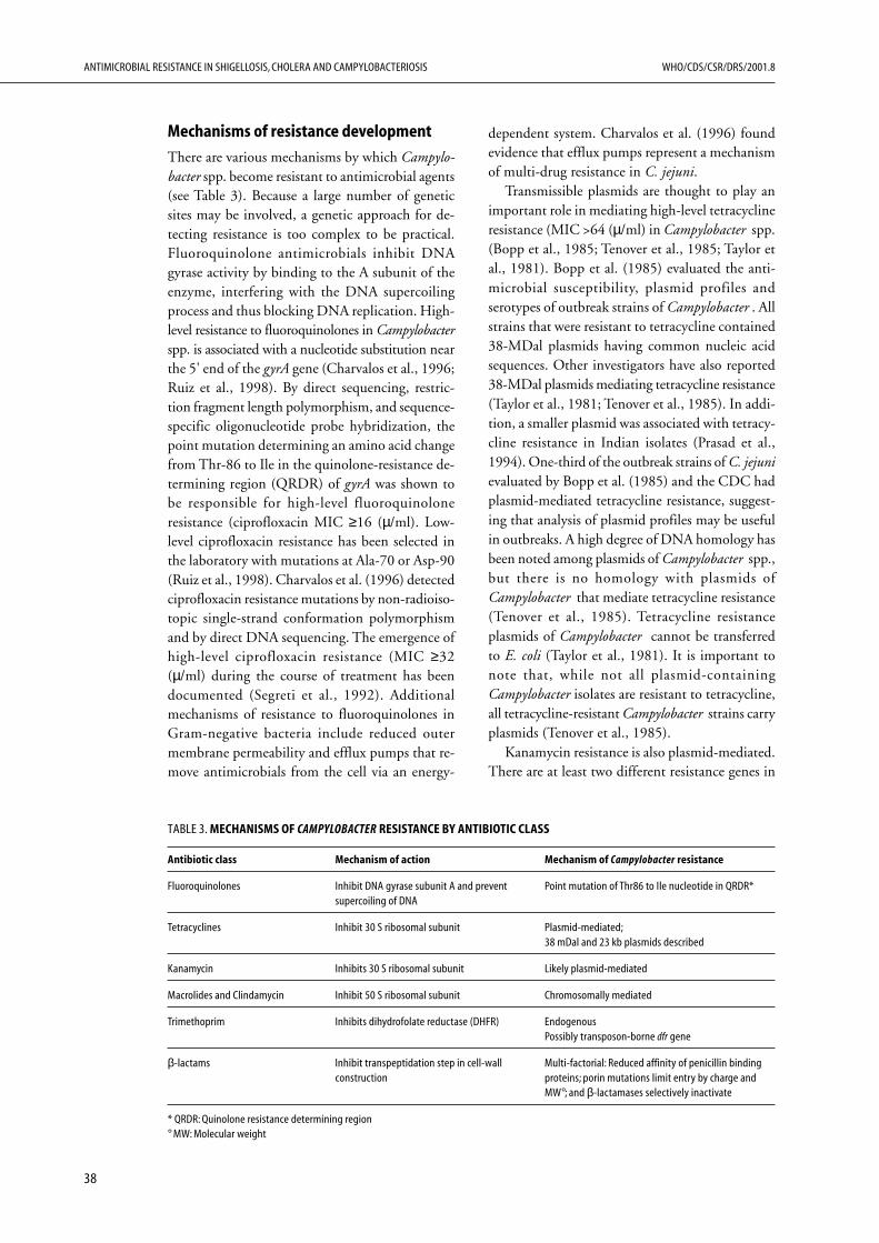
ANTIMICROBIAL RESISTANCE IN SHIGELLOSIS, CHOLERA AND CAMPYLOBACTERIOSIS WHO/CDS/CSR/DRS/2001.8
38
Mechanisms of resistance development
There are various mechanisms by which Campylo-bacter spp. become resistant to antimicrobial agents(see Table 3). Because a large number of geneticsites may be involved, a genetic approach for de-tecting resistance is too complex to be practical.Fluoroquinolone antimicrobials inhibit DNAgyrase activity by binding to the A subunit of theenzyme, interfering with the DNA supercoilingprocess and thus blocking DNA replication. High-level resistance to fluoroquinolones in Campylobacterspp. is associated with a nucleotide substitution nearthe 5' end of the gyrA gene (Charvalos et al., 1996;Ruiz et al., 1998). By direct sequencing, restric-tion fragment length polymorphism, and sequence-specific oligonucleotide probe hybridization, thepoint mutation determining an amino acid changefrom Thr-86 to Ile in the quinolone-resistance de-termining region (QRDR) of gyrA was shown tobe responsible for high-level fluoroquinoloneresistance (ciprofloxacin MIC ≥16 (µ/ml). Low-level ciprofloxacin resistance has been selected inthe laboratory with mutations at Ala-70 or Asp-90(Ruiz et al., 1998). Charvalos et al. (1996) detectedciprofloxacin resistance mutations by non-radioiso-topic single-strand conformation polymorphismand by direct DNA sequencing. The emergence ofhigh-level ciprofloxacin resistance (MIC ≥32(µ/ml) during the course of treatment has beendocumented (Segreti et al., 1992). Additionalmechanisms of resistance to fluoroquinolones inGram-negative bacteria include reduced outermembrane permeability and efflux pumps that re-move antimicrobials from the cell via an energy-
dependent system. Charvalos et al. (1996) foundevidence that efflux pumps represent a mechanismof multi-drug resistance in C. jejuni.
Transmissible plasmids are thought to play animportant role in mediating high-level tetracyclineresistance (MIC >64 (µ/ml) in Campylobacter spp.(Bopp et al., 1985; Tenover et al., 1985; Taylor etal., 1981). Bopp et al. (1985) evaluated the anti-microbial susceptibility, plasmid profiles andserotypes of outbreak strains of Campylobacter . Allstrains that were resistant to tetracycline contained38-MDal plasmids having common nucleic acidsequences. Other investigators have also reported38-MDal plasmids mediating tetracycline resistance(Taylor et al., 1981; Tenover et al., 1985). In addi-tion, a smaller plasmid was associated with tetracy-cline resistance in Indian isolates (Prasad et al.,1994). One-third of the outbreak strains of C. jejunievaluated by Bopp et al. (1985) and the CDC hadplasmid-mediated tetracycline resistance, suggest-ing that analysis of plasmid profiles may be usefulin outbreaks. A high degree of DNA homology hasbeen noted among plasmids of Campylobacter spp.,but there is no homology with plasmids ofCampylobacter that mediate tetracycline resistance(Tenover et al., 1985). Tetracycline resistanceplasmids of Campylobacter cannot be transferredto E. coli (Taylor et al., 1981). It is important tonote that, while not all plasmid-containingCampylobacter isolates are resistant to tetracycline,all tetracycline-resistant Campylobacter strains carryplasmids (Tenover et al., 1985).
Kanamycin resistance is also plasmid-mediated.There are at least two different resistance genes in
TABLE 3. MECHANISMS OF CAMPYLOBACTER RESISTANCE BY ANTIBIOTIC CLASS
Antibiotic class Mechanism of action Mechanism of Campylobacter resistance
Fluoroquinolones Inhibit DNA gyrase subunit A and prevent Point mutation of Thr86 to Ile nucleotide in QRDR*supercoiling of DNA
Tetracyclines Inhibit 30 S ribosomal subunit Plasmid-mediated;38 mDal and 23 kb plasmids described
Kanamycin Inhibits 30 S ribosomal subunit Likely plasmid-mediated
Macrolides and Clindamycin Inhibit 50 S ribosomal subunit Chromosomally mediated
Trimethoprim Inhibits dihydrofolate reductase (DHFR) EndogenousPossibly transposon-borne dfr gene
β-lactams Inhibit transpeptidation step in cell-wall Multi-factorial: Reduced affinity of penicillin bindingconstruction proteins; porin mutations limit entry by charge and
MW°; and β-lactamases selectively inactivate
* QRDR: Quinolone resistance determining region° MW: Molecular weight

39
WHO/CDS/CSR/DRS/2001.8 ANTIMICROBIAL RESISTANCE IN SHIGELLOSIS, CHOLERA AND CAMPYLOBACTERIOSIS
Campylobacter spp. Kanamycin resistance plasmidsshow considerable size variation, ranging from 41to 132 kb, with a high proportion of them alsoencoding the tetO gene (Tenover & Elvrum, 1988).
Campylobacter spp. are resistant to most β-lactams(Tajada et al., 1996). Resistance appears to be mul-tifactorial in nature, including reduced affinity ofpenicillin-binding proteins (PBPs), β-lactamaseproduction, and reduced penetration into the celldue to changes in porins (Lachance et al., 1991;Tajada et al., 1996). According to Tajada et al.(1996), resistance in Campylobacter is primarilymediated by PBPs. Penicillin G and most narrow-spectrum cephalosporins do not bind to the PBPsof Campylobacter but imipenem, amoxicillin (par-ticularly when combined with clavulanic acid) andcefepime (a broader spectrum cephalosporin) areactive against Campylobacter spp. in vitro, whichmay reflect increased binding to PBPs. Reportedly,83–92% of C. jejuni isolates produce a β-lactamase(Lachance et al., 1991). β-lactamase-positive organ-isms are significantly more resistant to ampicillin,amoxicillin and ticarcillin than are β-lactamase-negative bacteria. Among β-lactamase inhibitors,tazobactam was the most potent in inhibiting theenzyme, followed by clavulanic acid and sulbactam(Lachance et al., 1991). Susceptibility testing, incontrast, showed clavulanic acid to perform the bestin combination with amoxicillin, ampicillin orticarcillin (Lachance et al., 1991; Tajada et al., 1996).The activities of piperacillin, most cephalosporins,and imipenem are not affected by the Campylobacterβ-lactamase. Imipenem is highly active against themajority of isolates; where resistance to this agentoccurs in C. jejuni it has been linked to changes inporins (Page et al., 1989).
The outer membrane of Gram-negative bacte-ria is an effective barrier to antibiotics. The mainroute for penetration of many antibiotics is via poresin the outer membrane. C. jejuni pores are thoughtto be smaller than those of E. coli and therefore tolimit passage to solutes with a molecular weight of340 Dal or less. Additionally, the charge of a mol-ecule affects diffusion through the pores; moleculesthat are uncharged or have zero net charge diffusebetter. Molecules smaller than 360 Dal and withneutral charge are the most likely to penetrate theC. jejuni pore (Page et al., 1989). Imipenem, ampi-cillin, amoxicillin, quinolones and nitrofurans allpenetrate the C. jejuni outer membrane well.
Data regarding the mechanism of erythromycinresistance are sparse, but it is believed to be chromo-somally mediated in Campylobacter. There is cross-
resistance with spiramycin, lincomycin,clindamycin and azithromycin (Taylor, 1992).Neither enzymes capable of modifying macrolidesnor efflux mechanisms appear to contribute to re-sistance. Fortunately, erythromycin resistance appearsto be rare in C. jejuni, although resistance rates arehigher in C. coli isolates from livestock.
Trimethoprim, although used as empirical treat-ment of travellers’ diarrhoea, is ineffective for thetreatment of C. jejuni enteritis since all strains areresistant. Studies in Sweden reported a high preva-lence of foreign genetic material encoding a resist-ant dihydrofolate reductase. The genes in questionare known as dfr1 and dfr9. There is evidence thatthey were acquired by transposon-mediated geneticexchange. Heavy trimethoprim use in agriculturewas hypothesized to exert pressure leading to theemergence of resistance (Gibreel & Skold, 1998).
In summary, antibiotic resistance is a globalthreat to the ability to effectively treat C. jejunigastroenteritis. The complex interplay of veterinaryantibiotic use, unregulated human antibiotic use,and the erosion of geographical barriers to micro-bial migration due to the ease of international travel,have contributed to the emergence and spread ofresistance, a process which is unpredictable andcontinually evolving. Vigilant monitoring of resist-ance is required worldwide. WHO, recognizing theimpact of antimicrobial use in animal feed, met inBerlin and formulated recommendations to addressthis global problem (WHO, 1997). Internationalcooperation by a variety of organizations is neededto address it.
Prevention
Campylobacteriosis is a disease that occurs world-wide, costing millions of dollars per year in theUnited States alone (Skirrow & Blaser, 1992).Childhood diarrhoea globally, travellers’ diarrhoea,and epidemic and sporadic diarrhoea in the indus-trialized world could be dramatically reduced byappropriate intervention. Campylobacter-associatedGBS may be the most important factor in terms ofcost, at least in industrialized countries, because ofthe severe and prolonged nature of this complica-tion. The incidence of GBS in developing coun-tries remains to be determined. Research continueson many fronts, but simple hygiene remains themost effective means of preventing Campylobacterenteritis.
The reservoirs of C. jejuni are well described andinclude contaminated surface water, pets and, most
ANTIMICROBIAL RESISTANCE IN SHIGELLOSIS, CHOLERA AND CAMPYLOBACTERIOSIS WHO/CDS/CSR/DRS/2001.8
40
importantly, livestock (including poultry, swine andcattle). Primary prevention of Campylobacterenteritis involves improving personal hygiene, pro-viding clean fresh water supplies, and improvingfood processing and handling. Poultry figuresprominently in human illness, so its production andhandling have been a focus of efforts to reduceCampylobacter colonization and transmission.Attempts to limit contact between farm personneland chicken flocks, to introduce hygienic measuresto limit campylobacter transmission within flocks,or to modify slaughtering methods to reduce cross-contamination have made little impact on thefrequency of colonization. Chlorinating their drink-ing-water did little to reduce the extent of coloni-zation of broiler flocks (Stern et al., 1995).Irradiation of poultry carcasses is effective, but hasnot been widely used due to concerns on the partof the public about its safety. Thus, measures shortof vigilant culture and destruction of infected flockshave done little to curb the ubiquitous coloniza-tion by Campylobacter in large-scale poultry farms.
Colonization of poultry occurs quickly and uni-formly; this may be due to the fact that the birdsare coprophagic. Introduction of Campylobacterspp. into 4 chicks resulted in 100% of the flockbecoming colonized within 2–3 days (Jacobs-Reitsma, 1998). Interventions could, therefore,involve prophylaxis to prevent colonization or treat-ment once colonization occurs. Tsubokura et al.(1997) administered anti-Campylobacter antibod-ies orally to chicken flocks and studied the effecton colonization. Feed was supplemented with bovineand chicken immunoglobulin preparations (derivedfrom the milk or eggs of immunized livestock). Inuncolonized flocks, this treatment produced agreater than 99% reduction in colonization uponsubsequent oral challenge with Campylobacter.However, these preparations did not eliminateCampylobacter from pre-colonized flocks, produc-ing only a transient reduction, by 50–80%, in thenumber of bacteria, as compared to controls. Thisdifference disappeared within 3 days after treat-ment. Reduction in Campylobacter colonization ofpoultry will, presumably, lead to reduction in itstransmission to humans. Passive immunity mayreduce the bacterial load without selecting for re-sistance, in contrast to the use of feed supplementedwith antimicrobials.
There is a significant incidence of locally ac-quired Campylobacter infection, both in industri-alized and developing countries and, additionally,campylobacteriosis represents a large proportion of
travellers’ diarrhoea. In the latter infections, anti-biotic resistance is emerging. For these reasons, ef-forts are under way to develop a vaccine. Progresshas been limited by the antigenic variability ofCampylobacter spp. and the lack of informationabout its pathogenicity. A whole-cell killed vaccineand live attenuated vaccines are being studied(Scott, 1997). The oral route of administration isbeing targeted, with the rationale that stimulationof intestinal immunity should provide adequatedefence against Campylobacter enteritis. Since in-fection with Campylobacter spp. is associated withreactive arthritis and GBS, the vaccines must nottrigger immune responses that would lead to thesecomplications. More information is needed aboutthe specific antigens that trigger GBS in order toeliminate them from vaccines.
Intervention strategies
As is the case for V. cholerae, well-located regionalreference laboratories with standardized methodsof data collection are needed to create a worldwidesurveillance network. The ramifications of antimi-crobial use in animal husbandry are only begin-ning to be understood, although it appears that thelevel of fluoroquinolone resistance in humans can,in part, be directly linked to additives in poultryfeed and water. The ease of acquisition of anti-microbials and the great amount of air travel meanthat regional initiatives would offer little more thanstopgap measures of prevention. A global surveil-lance network with the authority to monitor trendson a worldwide scale and to recommend guidelinesfor human and veterinary antibiotic use worldwidewould be a means of combating the rapidly increas-ing resistance in enteric organisms.
Conclusion
In 25 years, C. jejuni has progressed from a newlyrecognized pathogen to being understood as themost significant cause of diarrhoea in the industri-alized world. It is a common cause of diarrhoea inchildren and travellers internationally, causesendemic diarrhoea and outbreaks of diarrhoea inindustrialized countries, and appears to be the mostcommon cause of Guillain-Barre Syndrome, a dev-astating and costly illness. Despite the large amountof information available, the mechanisms of patho-genicity in Campylobacter are still unclear. Thus,molecular pathogenesis studies represent the nextresearch frontier. Controlling the spread of the or-
41
WHO/CDS/CSR/DRS/2001.8 ANTIMICROBIAL RESISTANCE IN SHIGELLOSIS, CHOLERA AND CAMPYLOBACTERIOSIS
ganism in livestock, ensuring appropriate food han-dling practices, and limiting the use of antibioticsin agriculture are critical to reducing overall dis-ease incidence and resistance. The development ofvaccines, both for humans and animals, will greatlyenhance our ability to contain the spread ofCampylobacter. Emerging drug resistance is a glo-bal problem; therefore, international cooperationwill be required to address it.
ANTIMICROBIAL RESISTANCE IN SHIGELLOSIS, CHOLERA AND CAMPYLOBACTERIOSIS WHO/CDS/CSR/DRS/2001.8
42
Conclusion
developing countries. Without regulation of anti-biotic availability there is little hope of implement-ing rational and effective guidelines for antibioticuse.
Implementation of recommended actions shouldtake place under the umbrella of infectious diseasecontrol and surveillance programmes. There is anurgent need to set up low-technology regional labo-ratories throughout the developing world to dealwith diagnosis and surveillance; these structuresshould also serve other infectious disease controlprogrammes. Local antimicrobial resistance initia-tives could be assisted by the “Surveillance Stand-ards for Antimicrobial Resistance” currently beingprepared (WHO, 2001) and the WHONET soft-ware (WHO, 1999, developed by the WHOCollaborating Centre for Surveillance of Antimi-crobial Resistance). Regional centres could collectsamples, routinely test these for antimicrobial sen-sitivity, and log the data into a regional laboratorysample survey system. Such surveillance systemswould be useful for the early detection of epidem-ics and in choosing antimicrobial therapy based onprevalent resistance patterns. These programmeswould allow laboratories to analyse and share anti-microbial resistance data and facilitate the coordi-nation of control efforts (Institute of Medicine,1998).
In order to develop comprehensive policies, anystrategy for controlling antimicrobial resistancemust consider ecological factors and be based ondata for a wide variety of pathogens. Given the highrate of genetic exchange among species, interven-tions aimed at a single enteric organism will be fruit-less. The United States Agency for InternationalDevelopment (USAID) Infectious Disease Initia-tive intends to use data generated for individualorganisms to create a more general global strategyto combat antimicrobial resistance. V. cholerae,Campylobacter and Shigella are but pieces of a largerpuzzle. In many regions, financial and technicalresources are needed to establish adequate labora-tories and to network them within the areas theyserve as well as with international centres. Supportfrom WHO, CDC, and other donors will beessential in providing resources, training local healthcare workers and laboratory personnel, and edu-cating the public.
The greatest obstacles to implementation willbe at the local level. Changing the antibiotic pre-scribing behaviour of health care providers and theiruse by lay people is a daunting task, requiring com-munication strategies specific to the particular re-gion’s culture, habits and socioeconomic situation.Additionally, there are currently no policies in placeto control the unregulated sale of antibiotics in
43
WHO/CDS/CSR/DRS/2001.8 ANTIMICROBIAL RESISTANCE IN SHIGELLOSIS, CHOLERA AND CAMPYLOBACTERIOSIS
Bibliography
Ashkenazi S et al. Increasing antimicrobial resistance ofShigella isolates in Israel during the period 1984 to 1992.Antimicrob Agents Chemother 1995;39:819–823.
Ashraf MM, Ahmed ZU, Sack DA. Unusual associationof a plasmid with nalidixic acid resistance in an epidemicstrain of Shigella dysenteriae type 1 from Asia. Can JMicrobiol 1991;37:59–63.
Aspinall GO, McDonald AG, Pang H. Lipopoly-saccharides of Campylobacter jejuni serotype O:19: struc-tures of O antigen chains from the serostrain and twobacterial isolates from patients with the Guillain-Barresyndrome. Biochem 1994;33:250–55.
Asrat DA et al. The serotype distribution of Campylo-bacter jejuni and C. coli isolated from patients with diar-rhoea and controls at Tikur Anbassa Hospital, AddisAbaba, Ethiopia. Epidemiol Infect 1997;118:91–95.
Baker CN. The E-test and Campylobacter jejuni. DiagnMicrobiol Infect Dis 1992;15:469–472.
Barth PT et al. Transposition of a deoxyribonucleic acidsequence encoding trimethoprim and streptomycinresistances from R483 to other replicons. J Bacteriol1976;125:800–810.
Bauer AW et al. Antibiotic susceptibility testing by astandardized single disk method. Am J Clin Pathol1966;36:493–496.
Bennish M et al. Multiresistant shigella infections inBangladesh. Lancet 1985;2:441.
Bennish ML et al. Hypoglycemia during diarrhea inchildhood. New Engl J Med 1990[1];322:1357–1363.
Bennish ML et al. Death in shigellosis: Incidence andrisk factors in hospitalized patients. J Infect Dis 1990[2];161:500–506.
Bennish ML. Potentially lethal complications of shigel-losis. Rev Infect Dis 1991;13(suppl 4):S319–S324.
Bennish ML, Salam MA. Rethinking options for thetreatment of shigellosis. J Antimicrob Chemother 1992;30:243–247.
Bennish ML et al. Antimicrobial resistance of Shigellaisolates in Bangladesh, 1983–1990: Increasing frequencyof strains multiply resistant to ampicillin, trimethoprim-sulfamethoxazole, and nalidixic acid. Clin Infect Dis1992;14:1055–1060.
Bennish ML. Cholera: Pathophysiology, clinical features,and treatment. In: Wachsmuth IK, Blake PA, Olsvik O,editors. Vibrio cholerae and Cholera: Molecular to GlobalPerspectives. Washington, ASM Press,1994:229–255.
Abraham EP, Chain E. An enzyme from bacteria able todestroy penicillin. Nature 1940;146:83.
Acheson DW, Donohue-Rolfe A, Keusch GT. The fam-ily of shiga and shiga-like toxins. In: Alouf JE, Freer JH,editors. Sourcebook of Bacterial Protein Toxins. New York,Academic Press, 1991:415–433.
Acheson DW, Keusch GT. Shigella and enteroinvasiveE. coli. In: Blaser MJ et al., editors. Infections of theGastrointestinal Tract. New York, Raven Press, 1995:763–784.
Ackerman BH, Dello Buono FA. In vitro testing of an-tibiotics. Pharmacotherapy 1996; 16:201–221.
Adam T et al. Cytoskeletal rearrangements and the func-tional role of t-plastin during entry of Shigella flexneriinto HeLa cells. J Cell Biol 1995;129:367–381.
Adeleye IA. Conjugal transferability of multiple antibi-otic resistance in three genera of Enterobacteriaceae inNigeria. J Diarrhoeal Dis Res 1992;10:93–96.
Ahmed F et al. Community-based evaluation of the ef-fect of breast-feeding on the risk of microbiologicallyconfirmed or clinically presumptive shigellosis in Bang-ladeshi children. Pediatr 1992;90:406–411.
Alam AN et al. Randomized double blind trial of singledose doxycycline for treating cholera in adults. Brit MedJ 1990;300:1619–1621.
Altekreuse SF et al. Campylobacter jejuni—An emergingfoodborne pathogen. Emerg Infect Dis 1999;5:28–35.
Ambler JE et al. Mutator plasmid in a nalidixic acid-resistant strain of Shigella dysenteriae type 1. J AntimicrobChemother 1993;31:831–839.
Amyes SG. The success of plasmid-encoded resistancegenes in clinical bacteria. An examination of plasmid-mediated ampicillin and trimethoprim resistance genesand their resistance mechanisms. J Med Microbiol1989;28:73–83.
Amyes SGB, Towner KJ. Trimethoprim resistance; epi-demiology and molecular aspects. J Med Microbiol1990;31:1–19.
Anderson ES, Datta N. Resistance to penicillins and itstransfer in Enterobacteriaceae. Lancet 1965;1:407–409.
Ashkenazi S et al. A randomized, double-blind studycomparing cefixime and trimethoprim-sulfamethoxazolein the treatment of childhood shigellosis. J Pediatr1993;123:817–821.
ANTIMICROBIAL RESISTANCE IN SHIGELLOSIS, CHOLERA AND CAMPYLOBACTERIOSIS WHO/CDS/CSR/DRS/2001.8
44
Bernardini ML et al. Identification of icsA, a plasmidlocus of Shigella flexneri that governs bacterial intra- andintercellular spread through interaction with F-actin. ProcNatl Acad Sci USA 1989;86:3867–3871.
Birmingham ME et al. Epidemic cholera in Burundi:patterns of transmission in the Great Rift Valley Lakeregion. Lancet 1997:349:981–985.
Black RE et al. Experimental Campylobacter jejuni in-fection in humans. J Infect Dis 1988;157:472–79.
Black RE. Epidemiology of travelers’ diarrhea and rela-tive importance of various pathogens. Rev Infect Dis1990;12(Suppl 1):S73–S79.
Blaser M et al. Campylobacter enteritis associated withcanine infection. Lancet 1978;2:979–981.
Blaser MJ et al. Campylobacter enteritis: Clinical andepidemiologic features. Ann Intern Med 1979;91:179–185.
Blaser MJ, Reller LB. Campylobacter enteritis. N Engl JMed 1981;305:1444–1451.
Blaser MJ et al. Campylobacter jejuni-specific serum an-tibodies are elevated in healthy Bangladeshi children. JClin Micro 1985;21:164–167.
Blaser MJ et al. Extraintestinal Campylobacter jejuni andCampylobacter coli infections: Host factors and straincharacteristics. J Infect Dis 1986;153: 552–559.
Blaser MJ, Taylor DN, Echeverria P. Immune responseto Campylobacter jejuni in a rural community in Thai-land. J Infect Dis 1986;153:249–254.
Blaser MJ. Epidemiologic and clinical features ofCampylobacter jejuni infections. J Infect Dis 1997;176(Suppl 2):S103–S105.
Bojalil R, Calva JJ. Antibiotic misuse in diarrhea. Ahousehold survey in a Mexican community. J ClinEpidemiol 1994;47:147–156.
Bopp CA et al. In vitro antimicrobial susceptibility, plas-mid analysis, and serotyping of epidemic-associatedCampylobacter jejuni. J Clin Micro1985;21:4–7.
Bratoeva MP, John JF. Dissemination of trimethoprim-resistant clones of Shigella sonnei in Bulgaria. J Infect Dis1989;159:648–653.
Bratoeva MP, John JF. In vivo R-plasmid transfer in apatient with mixed infection of Shigella dysentery.Epidemiol Infect 1994;112:247–252.
Bremner DA. The transfer of antibiotic resistance by R-plasmids in shigellae. NZ Med J 1978;88:9–10.
Brown DFJ, Brown L. Evaluation of the E-test, a novelmethod of quantifying antimicrobial activity. JAntimicrob Chemother 1991;27:185–190.
Burchall JJ, Elwell LP, Fling ME. Molecular mechanismsof resistance to trimethoprim. Rev Infect Dis 1982;4:246–254.
Calva JJ, Sifuentes-Osornio J, Ceron C. Antimicrobialresistance in fecal flora: Longitudinal community-basedsurveillance of children from urban Mexico. AntimicrobAgents Chemother 1996;40:1699–1702.
Centers for Disease Control. Nationwide disseminationof multiply resistant Shigella sonnei following a common-source outbreak. MMWR 1987;36:633–634.
Centers for Disease Control. Shigella dysenteriae type 1-Guatemala, 1991. MMWR 1991;40:421–428.
Chang MJ et al. Trimethoprim-sulfamethoxazole com-pared to ampicillin in the treatment of shigellosis. Pediatr1977;59:726–729.
Charvalos E et al. Detection of ciprofloxacin resistancemutations in Campylobacter jejuni gyrA by nonradio-isotopic single-strand conformation polymorphism anddirect DNA sequencing. J Clin Lab Anal 1996;10:129–133.
Chun D et al. R plasmids conferring multiple drug re-sistance from shigella isolated in Korea. J Hyg (Lond)1984;92:153–160.
Coppo A et al. Vibrio cholerae in the horn of Africa:Epidemiology, plasmids, tetracycline resistance geneamplification, and comparison between O1 and non-O1 strains. Amer J Trop Med Hyg 1995;53:351–359.
Corrado ML et al. Norfloxacin: Review of safety stud-ies. Am J Med 1987;82(Suppl 6B):22–26.
Couper MR. Strategies for the rational use of anti-microbials. Clin Infect Dis 1997;24(Suppl 1):S154–S156.
Datta N. Infectious drug resistance. Br Med Bull1965;21:254–259.
Datta N et al. Properties of an R factor from Pseudomonasaeruginosa. J Bacteriol 1971;108:1244–1249.
Dekeyser P et al. Acute enteritis due to related vibrio:first positive stool cultures. J Infect Dis 1972;125:390–392.
Deming MS et al. Campylobacter enteritis at a univer-sity: Transmission from eating chicken and from cats.Am J of Epid 1987;126:526–534.
Doig P et al. An environmentally regulated pilus-likeappendage involved in Campylobacter pathogenesis. MolMicrobiol 1996;20:885–894.
Dua V, Kunin CM, White LV. The use of antimicrobialdrugs in Nagpur, India. A window on medical care in adeveloping country. Soc Sci Med 1994;38:717–724.
Dubon JM et al. Emergence of multiple drug-resistantVibrio cholerae O1 in San Pedro Sula, Honduras. Lancet1997;349:924.
DuPont HL, Steele JH. Use of antimicrobial agents inanimal feeds: Implications for human health. Rev InfectDis 1987;9:447–460.
DuPont HL et al. Current problems in antimicrobialtherapy for bacterial enteric infection. Am J Med1987;82(suppl 4A):324–328.
45
WHO/CDS/CSR/DRS/2001.8 ANTIMICROBIAL RESISTANCE IN SHIGELLOSIS, CHOLERA AND CAMPYLOBACTERIOSIS
DuPont HL et al. Inoculum size in shigellosis and im-plications for expected mode of transmission. J InfectDis 1989;159:1126–1128.
Eidlitz-Marcus T et al. Comparative efficacy of two- andfive-day courses of ceftriaxone for treatment of severeshigellosis in children. J Pediatr 1993;123:822–824.
Eliopoulos GM. Induction of beta-lactamases.J Antimicrob Chemother 1988;22(Suppl A):37–44.
Endtz HP et al. Quinolone resistance in campylobacterisolated from man and poultry following the introduc-tion of fluoroquinolones in veterinary medicine.J Antimicrob Chemother 1991;27:199–208.
Finch MJ et al. Epidemiology of antimicrobial resistantcholera in Kenya and East Africa. Am J Trop Med Hyg1988;39:484–490.
Fleming MP, Datta N, Gruneberg RN. Trimethoprimresistance determined by R factors. Br Med J 1972;1:726–728.
Fling ME, Richards C. The nucleotide sequence of thetrimethoprim-resistant dihydrofolate reductase geneharbored by Tn7. Nucleic Acids Res 1983;11:5147–5158.
Fontaine A, Arondel J, Sansonetti PJ. Role of Shiga toxinin the pathogenesis of bacillary dysentery, studied byusing a Tox- mutant of Shigella dysenteriae 1. InfectImmun 1988;56:3099–3109.
Florin I, Antillon F. Production of enterotoxin and cy-totoxin in Campylobacter jejuni strains isolated in CostaRica. J Med Microbiol 1992;37:22–29.
Frost JA, Rowe B, Vandepitte J. Acquisition oftrimethoprim resistance in epidemic strain of Shigelladysenteriae type 1 from Zaire. Lancet 1982;1:963.
Frost JA et al. Plasmid characterization of drug-resistantShigella dysenteriae 1 from an epidemic in CentralAfrica. J Hyg Camb 1985;94:163–172.
Gangarosa EJ et al. From the Centers for Disease Con-trol: An epidemic-associated episome? J Infect Dis1972 ;126:215–218.
Gaudreau C, Gilbert H. Antimicrobial resistance of clini-cal strains of Campylobacter jejuni subsp. jejuni isolatedfrom 1985 to 1997 in Quebec, Canada. AntimicrobAgents Chemother 1998;42:2106–2108.
Garrigue GP et al. Résistance aux antibiotiques dessouches de Vibrio cholerae El Tor isolées à Douala(Cameroon). Bull Soc Pathol Exot Filiales 1986;79:305–312.
Georgopapadakou NH. Penicillin-binding proteins andbacterial resistance to beta-lactams. Antimicrob AgentsChemother 1993;37:2045–2053.
Ghosh AS, Kar AK, Kundu M. Alterations in high mo-lecular mass penicillin-binding protein 1 associated withbeta-lactam resistance in Shigella dysenteriae. BiochemBiophys Res Commun 1998;248:669–672.
Gibreel A, Skold, O. High-level resistance to trimetho-prim in clinical isolates of Campylobacter jejuni byacquisition of foreign genes (dfr1 and dfr9) expressingdrug-insensitive dihydrofolate reductases. AntimicrobAgents Chemother 1998,42:3059–3064.
Gilman Rh et al. Single-dose ampicillin therapy forsevere shigellosis in Bangladesh. J Infect Dis 1981;143:164–169.
Glass RI et al. Emergence of multiply antibiotic resist-ant Vibrio cholerae in Bangladesh. J Inf Dis 1980;142:939–942.
Glass RI et al. Plasmid-borne multiple drug resistancein Vibrio cholerae serogroup O1, biotype El Tor:Evidence for a point-source outbreak in Bangladesh.J Infect Dis 1983[1];147:204–209.
Glass RI et al Epidemiologic and clinical features ofendemic Campylobacter jejuni infection in Bangladesh.J Infect Dis 1983[2];148:292–296.
Goldstein F, Gerbaud G, Courvalin P. Transposable re-sistance to trimethoprim and 0/129 in Vibrio cholerae.J Antimicrob Chemother 1986;17:559–569.
Goma Epidemiology Group. Public health impact of theRwandan refugee crisis: What happened in Goma,Zaire in July 1994? Lancet 1995;345:339–344.
Goodman LJ et al. Empiric antimicrobial therapy ofdomestically acquired acute diarrhea in urban adults.Arch Intern Med 1990;150:541–546.
Gotuzzo E et al. Comparison of single-dose treatmentwith norfloxacin and standard 5-day treatment withtrimethoprim-sulfamethoxazole for acute shigellosis inadults. Antimicrob Agents Chemother 1989;33:1101–1104.
Griffiths PL, Park RWA. Campylobacters associated withhuman diarrhoeal disease. J Applied Bacteriol 1990;69:281–301.
Gross RJ et al. Drug resistance in Shigella dysenteriae,S flexneri and S boydii in England and Wales: Increasingincidence of resistance to trimethoprim. Br Med J1984;288:784–786.
Guerrero L et al. Asymptomatic Shigella infections in acohort of Mexican children younger than two years ofage. Pediatr Infect Dis J 1994;13:597–602.
Guerry P et al. Role of two flagellin genes in Campylo-bacter motility. J Bacteriol 1991;173:4757–64.
Gupta PG et al. Effect of doxycycline on transmissionof Vibrio cholerae infection among family contacts ofcholera patients in Calcutta. Bull WHO 1978;56:323–326.
Guyon AB et al. A baseline survey on use of drugs at theprimary health care level in Bangladesh. Bull WHO1994;72:265–271.
Haider K, Huq MI. A transferable resistance plasmid inVibrio cholerae and Escherichia coli isolated from the samepatient. J Diarrheal Dis Res 1986;4:91–93.
ANTIMICROBIAL RESISTANCE IN SHIGELLOSIS, CHOLERA AND CAMPYLOBACTERIOSIS WHO/CDS/CSR/DRS/2001.8
46
Haider K et al. Trimethoprim resistance gene in Shigelladysenteriae 1 isolates obtained from widely scatteredlocations of Asia. Epidemiol Infect 1990;104:219–228.
Haider K, Malek MA, Albert MJ. Occurrence of drugresistance in Shigella species isolated from patients withdiarrhoea in Bangladesh. J Antimicrob Chemother1993;32:509–511.
Haltalin KC et al. Double-blind treatment study of shig-ellosis comparing ampicillin, sulfadiazine, and placebo.J Pediatr 1967;70:970–981.
Hardy AV. Studies of the acute diarrheal diseases: Fur-ther cultural observations on the relative efficacy ofsulfonamides in Shigella infections. Public Health Rep1945;60:1037–1043.
Heikkila E et al. Increase of trimethoprim resistanceamong Shigella species, 1975–1988: Analysis of resist-ance mechanisms. J Infect Dis 1990;161:1242–1248.
Hernborg A. Stevens-Johnson syndrome after massprophylaxis with sulfadoxine for cholera in Mozambique.Lancet 1985;2:1072–1073.
Hickey SM, Nelson JD. Mechanisms of antibacterialresistance. Adv Pediatr 1997;44:1–41.
Hirai K et al. Differences in susceptibility to quinolonesof outer membrane mutants of Salmonella typhimuriumand Escherichia coli. Antimicrob Agents Chemother1986;29:535–538.
Hofmann SL. Southwestern Internal Medicine Confer-ence: Shiga-like toxins in hemolytic-uremic syndromeand thrombotic thrombocytopenic purpura. Am J MedSci 1993;306:398–406.
Hoge CW et al. Trends in antibiotic resistance amongdiarrheal pathogens isolated in Thailand over 15 years.Clin Infect Dis 1998;26:341–345.
Hood AM, Pearson AD, Shahamat M. The extent ofsurface contamination of retailed chickens withCampylobacter jejuni subgroups. Epidemiol Infect 1988;100:17–25.
Hooper DC et al. Genetic and biochemical characteri-zation of norfloxacin resistance in Escherichia coli.Antimicrob Agents Chemother 1986;29:639–644.
Hossain MM, Glass RI, Khan MR. Antibiotic use in arural community in Bangladesh. Int J Epidemiol1982;11:402–405.
Hossain MA, Albert MJ, Hasan KZ. Epidemiology ofshigellosis in Teknaf, a coastal area of Bangladesh: a 10-year survey. Epidemiol Infect 1990;105:41–49.
Huang MB et al. Accuracy of the E-test for determiningantimicrobial susceptibilities of staphylococci, entero-cocci, Campylobacter jejuni, and Gram-negative bacte-ria resistant to antimicrobial agents. J Clin Microbiol1992;30:3243–3248.
Hughes JM, Tenover FC. Approaches to limiting emer-gence of antimicrobial resistance in bacteria in humanpopulations. Clin Infect Dis 1997;24(Suppl 1):S131–S135.
Hui L et al. Patterns and determinants of use of antibi-otics for acute respiratory tract infection in children inChina. Pediatr Infect Dis J 1997;16:560–564.
Huovinen P. Trimethoprim resistance. Antimicrob AgentsChemother 1987;31:1451–1456.
Ichinose Y et al. The characterization of Vibrio choleraeisolated in Kenya in 1983. J Trop Med Hyg 1986;89:269–276.
Institute of Medicine. New Vaccine Development, Estab-lishing Priorities, Vol II. Diseases of Importance in Devel-oping Countries. Washington, DC, Institute of Medicine,1986:378–389.
Institute of Medicine. Antimicrobial Resistance: Issues andOptions. Washington, DC, National Academy Press,1998:92.
Islam MR. Single dose tetracycline in cholera. Gut1987;28:1029–1032.
Islam MR et al. Effect of antimicrobial (nalidixic acid)therapy in shigellosis and predictive values of outcomevariables in patients susceptible or resistant to it. J TropMed Hyg 1995;98:121–125.
Jacewicz M et al. Pathogenesis of shigella diarrhea. XI.Isolation of a shigella toxin-binding glycolipid fromrabbit jejunum and HeLa cells and its identification as aglobotriaosylceramide. J Exp Med 1986;163:1391–1404.
Jacobs-Reitsma W. Experimental horizontal spread ofCampylobacter amongst one-day-old broilers. In:Lastovica AJ, Newell DG, Lastovica EE, editors.Campylobacter, Helicobacter and Related Organisms. CapeTown, SA, Institute of Child Health, 1998: 377–378.
Jesudason MV, Lalitha MK, Koshi G. Changes in inci-dence of shigella subgroups and their antibiotic suscep-tibility pattern in Vellore, South India. J Trop Med Hyg1985;88:355–358.
Jesudason MV, John TJ. Transferable trimethoprim re-sistance of Vibrio cholerae O1 encountered in southernIndia. Trans Roy Soc Trop Med Hyg 1990;84:136–137.
Jesudason MV, Saaya R. Resistance of Vibrio cholerae O1to nalidixic acid. Ind J Med Res 1997;105:153–154.
Joint ICMR-GWB-WHO Cholera Study Group. Effectof tetracycline on cholera carriers in households of chol-era patients. Bull WHO 1971;45:451–455.
Kabir I et al. Comparative efficacies of pivmecillinamand ampicillin in acute shigellosis. Antimicrob AgentsChemother 1984;25:643–645.
Kaliyugaperumal V, Gupta U, Mohapatra LN. Antimi-crobial drug resistance and R factors in Shigella. IndianJ Med Res 1978;68:220–224.
Kapperud G et al. Clinical features of sporadic campylo-bacter infections in Norway. Scand J Infect Dis1992;24:741–749.
Karaolis DK, Lan RR, Reeves PR. The sixth and sev-enth cholera pandemics are due to independent clonesseparately derived from environmental, nontoxigenic,
47
WHO/CDS/CSR/DRS/2001.8 ANTIMICROBIAL RESISTANCE IN SHIGELLOSIS, CHOLERA AND CAMPYLOBACTERIOSIS
non-O1 Vibrio cholerae. J Bacteriol 1995;177:3191–3198.
Ketley JM. Pathogenesis of enteric infection byCampylobacter. Microbiology 1997;143:5–21.
Keusch GT. Shigella. In: Gorbach SL, editor. InfectiousDiarrhea. Boston, Blackwell Scientific Publications,1986:31–50.
Keusch GT, Bennish ML. Shigellosis: Recent progress,persisting problems and research issues. Pediatr Infect DisJ 1989;8:713–719.
Keusch GT, Bennish ML. Shigellosis. In: Evans AS,Philip S, editors. Bacterial Infections of Humans: Epide-miology and Control, 3rd Edition. New York, PlenumPublishing Corp, 1998:631–656.
Keusch GT. Shigellosis. In: Gorbach SL, Bartlett JG,Blacklow NR, editors. Infectious Diseases, 2nd Edition.Philadelphia, WB Saunders Co., 1998:694–698.
Khan MU. Efficacy of short course antibiotic prophy-laxis in controlling cholera in contacts during epidemic.J Trop Med Hyg 1982;85:27–29.
Khan MU et al. Epidemiological differences betweencholera due to multiple antibiotic resistant and multipleantibiotic sensitive Vibrio cholerae infection. Trans RoySoc Trop Med Hyg 1986;80:60–63.
Khan WA et al. Comparative trial of five antimicrobialcompounds in the treatment of cholera in adults. TransRoy Soc Trop Med Hyg 1995;89:103–106.
Khan, WA et al. Randomised controlled trial of single-dose ciprofloxacin and doxycycline for cholera causedby Vibrio cholerae O1 or O139. Lancet 1996;348:296–300.
Khetawat G et al. Resurgent Vibrio cholerae O139:Rearrangement of cholera toxin genetic elements andamplification of rrn operon. Infect Immun 1999;67:148–154.
Kobari KI, Takakura I, Nakatomi M. Antibiotic resist-ant strains of El Tor Vibrio in the Philippines and theuse of furalazine for chemotherapy. Bull WHO1970;43:365–371.
Kruse H et al. Multiresistant Shigella species from Afri-can AIDS patients: Antibacterial resistance patterns andapplication of the E-test for determination of minimuminhibitory concentration. Scand J Infect Dis 1992;24:733–739.
Kuschner RA et al. Use of azithromycin for the treat-ment of Campylobacter enteritis in travelers to Thailand,an area where ciprofloxacin resistance is prevalent. ClinInfect Dis 1995 ;21:536–541.
Kuwahara S et al. Transmission of multiple drug resist-ance from Shigella flexneri to V. cholerae through conju-gation. Jap J Microbiol 1963;7:61–68.
Kuwahara S et al. Drug sensitivity of El Tor Vibrio strainsisolated in the Philippines in 1964 and 1965. Bull WHO1967;37:763–771.
Lachance N et al. Role of the β-lactamase of Campylo-bacter jejuni in resistance to β-lactam agents. AntimicrobAgents Chemother 1991;35:813–818.
Lerman SJ, Waller JM, Simms DH. Resistance of shig-ellae to ampicillin and other antibiotics: South Bronx,New York (1971 and 1972). J Pediatr 1973;83:500–501.
Levine MM et al. Pathogenesis of Shigella dysenteriae 1(Shiga) dysentery. J Infect Dis 1973 ;127:261–270.
Levine MM et al. Recombinant DNA risk assessmentstudies in humans: efficacy of poorly mobilizableplasmids in biologic containment. J Infect Dis 1983;148:699–709.
Levine MM. Shigellosis. In: Strickland GT, Hunter’sTropical Medicine, 7th Edition. Philadelphia, WBSaunders Co., 1991:340–344.
Levine OS, Levine MM. Houseflies (Musca domestica)as mechanical vectors of Shigellosis. Rev Infect Dis1991;13:698–696.
Levy SB, FitzGerald GB, Macone AB. Changes in intes-tinal flora of farm personnel after introduction of tetra-cycline-supplemented feed on a farm. N Engl J Med1976;295:583–588.
Lexomboon U et al. Clinical evaluation of co-trimoxazoleand furazolidone in treatment of shigellosis in children.Br Med J 1972,3:23–26.
Li CC et al. Antimicrobial susceptibilities of Campylo-bacter jejuni and coli by using E-test in Taiwan. Scand JInfect Dis 1998;30:39–42.
Lichtenstein C, Bremner S. Unique insertion site of Tn7in the E. coli chromosome. Nature 1982;297:601–606.
Lindenbaum J, Greenough WB, Islam MR. Antibiotictherapy of cholera. Bull WHO 1967;36:871–883.
Lior H et al. Serotyping of Campylobacter jejuni by slideagglutination based on heat-labile antigenic factors. JClin Microbiol 1982;15:761–768.
McCormack WM et al. Tetracycline prophylaxis in fami-lies of cholera patients. Bull WHO 1968;38:787–792.
Maggi P, Carbonara S, Santantonio T. Ciprofloxacin fortreating cholera. Lancet 1996;348:1446–1447.
Mahon BE et al. Reported cholera in the United States,1992–1994: A reflection of global changes in choleraepidemiology. JAMA 1996;276: 307–312.
Malengreau M. Nalidixic acid in Shigella dysenteriaeoutbreaks. Lancet 1984;2:172.
Materu SF et al. Antibiotic resistance pattern of Vibriocholerae and Shigella causing diarrhea outbreaks in theeastern African region: 1994–1996. E Afr Med J1997;74:193–197.
Mhalu FS, Muari PW, Ijumba J. Rapid emergence of elTor Vibrio cholerae resistant to antimicrobial agents dur-ing the first six months of the fourth cholera epidemicin Tanzania. Lancet 1979;1:345–347.
ANTIMICROBIAL RESISTANCE IN SHIGELLOSIS, CHOLERA AND CAMPYLOBACTERIOSIS WHO/CDS/CSR/DRS/2001.8
48
Mitra R et al. Resurgence of Vibrio cholerae O139 Ben-gal with altered antibiogram in Calcutta, India. Lancet1996;348:1181.
Mitsuhashi S. Review: The R factors. J Infect Dis 1969;119:89–100.
Molina J-M et al. Campylobacter infections in HIV-in-fected patients: Clinical and bacteriological features.AIDS 1995;9:881–885.
Moran AP, Appelmelk BJ, Aspinall GO. Molecular mim-icry of host structures by lipopolysaccharides of Campylo-bacter and Helicobacter spp.: Implications inpathogenesis. J Endotoxin Res 19963:521–531.
Mukhopadhyay AK et al. Biotype traits and antibioticsusceptibility of Vibrio cholerae serogroup O1 before,during, and after the emergence of the O139 serogroup.Epidemiol Infect 1995;115:427–434.
Mukhopadhyay AK et al. Temporal shifts in traits ofVibrio cholerae strains isolated from hospitalized patientsin Calcutta: a 3-year (1993 to 1995) analysis. J ClinMicrobiol 1996;34:2537–2543.
Mukhopadhyay AK et al. Emergence of fluoroquinoloneresistance in strains of Vibrio cholerae isolated from hos-pitalized patients with acute diarrhea in Calcutta, India.Antimicrob Agents Chemother 1998;42:206–207.
Munshi MH et al. Plasmid-mediated resistance to nali-dixic acid in Shigella dysenteriae type 1. Lancet1987;2:419–421.
Murphy GS Jr et al. Ciprofloxacin- and azithromycin-resistant Campylobacter causing traveler’s diarrhea in U.S.troops deployed to Thailand in 1994. Clin Infect Dis1996;22: 868–869.
Murray BE, Rensimer ER, DuPont HL. Emergence ofhigh-level trimethoprim resistance in fecal Escherichiacoli during oral administration of trimethoprim ortrimethoprim-sulfamethoxazole. N Engl J Med1982;306:130–135.
Murray BE. Resistance of Shigella, Salmonella, and otherselected enteric pathogens to antimicrobial agents. RevInfect Dis 1986;8(Suppl 2):S172–S181.
Mutwewingabo A, Mets T. Increase in multiresistanceof Shigella dysenteriae type 1 strain in Rwanda. E AfrMed J 1987;64:812–815.
Naficy A et al. Treatment and vaccination strategies tocontrol cholera in Sub-Saharan refugee settings: A cost-effectiveness analysis. JAMA 1998;279:521–525.
Nachamkin I, Yang X-H, Stern NJ. Role ofCampylobacter jejuni flagella as colonization factors forthree-day-old chicks: Analysis with flagellar mutants.Appl Environ Microbiol 1993;59:1269–1273.
Nelson JD et al. Trimethoprim-sulfamethoxazole therapyfor shigellosis. JAMA 1976;235:1239–1243.
Nizami SQ, Khan IA, Bhutta ZA. Drug prescribingpractices of general practitioners and paediatricians forchildhood diarrhoea in Karachi, Pakistan. Soc Sci Med1996;42:1133–1139.
O’Brien TF. The global epidemic nature of antimicro-bial resistance and the need to monitor and manage itlocally. Clin Infect Dis 1997;24 (Suppl 1):S2–S8.
Ochiai K et al. Studies on the inheritance of drug resist-ance between Shigella strains and Escherichia coli strains.Nihon Iji Shimpo 1959;1861:34–46.
Ogg JE, Shrestha MB, Poudayl L. Phage-inducedchanges in Vibrio cholerae: Serotype and biotype con-versions. Infect Immun 1978;19:231–238.
Okeke IN, Lamikanra A, Edelman R. Socioeconomicand behavioral factors leading to acquired bacterial re-sistance to antibiotics in developing countries. EmergInfect Dis 1999;5:18–27.
Olarte J, Filloy L, Galindo E. Resistance of Shigelladysenteriae type 1 to ampicillin and other antimicrobialagents: Strains isolated during a dysentery outbreak in ahospital in Mexico City. J Infect Dis 1976;133:572–575.
Olsson-Liljequist B. E-test as a routine MIC tool forreference work. Diagn Microbiol Infect Dis 1992;15:479–482.
Page,WJ et al. Characterization of porins of Campylo-bacter jejuni and Campylobacter coli and implications forantibiotic susceptibility. Antimicrob Agents Chemother1989;33:297–303.
Pai CH et al. Erythromycin in treatment of Campylo-bacter enteritis in children. Am J Dis Child 1983;137:286–288.
Paredes P et al. Factors influencing physicians’ prescrib-ing behaviour in the treatment of childhood diarrhoea:Knowledge may not be the clue. Soc Sci Med 1996;42:1141–1153.
Penner JL, Hennessy JN. Passive hemagglutination tech-nique for serotyping Campylobacter foetus spp. jejuni onthe basis of soluble heat-stable antigens. J Clin Microbiol1980;12:732–737.
Perlman DM et al. Persistent Campylobacter jejuniinfections in patients infected with human immuno-deficiency virus (HIV). Ann Intern Med 1988;108:540–546.
Petrocheilou V, Grinsted J, Richmond MH. R-plasmidtransfer in vivo in the absence of antibiotic selection pres-sure. Antimicrob Agents Chemother 1976;10:753–761.
Pickering LK, Bartlett AV, Woodward WE. Acute infec-tious diarrhea among children in day care: Epidemiol-ogy and control. Rev Infect Dis 1986;8:539–547.
Platt DJ, Chesham JS, Kristinsson KG. R-plasmid trans-fer in vivo: A prospective study. J Med Microbiol 1986;21:325–330.
Pollitzer, R. History of the disease. In: WHO Mono-graph Series No 43. Cholera. Geneva, WHO, 1959.
Prasad KN et al. Antimicrobial susceptibility and plas-mid analysis of Campylobacter jejuni isolated fromdiarrheoal patients and healthy chickens in NorthernIndia. J Diarrhoeal Dis Res 1994;12:270–273.
49
WHO/CDS/CSR/DRS/2001.8 ANTIMICROBIAL RESISTANCE IN SHIGELLOSIS, CHOLERA AND CAMPYLOBACTERIOSIS
Ragimbeau C et al. Development of a multiplex PCRgene fingerprinting method using gyrA and pflApolymorphisms to identify genotypic relatedness withinCampylobacter jejuni species. J Applied Microbiol1998;85:829–838.
Rahal K, Gerbaud G, Bouanchaud DH. Stability of theR plasmids belonging to different incompatibility groupsin Vibrio cholerae. Ann Microbiol 1978;129A:409–414.
Rautelin H, Renkonen O-V, Kosunen T. Emergence offluoroquinolone resistance in Campylobacter jejuni andCampylobacter coli in subjects in Finland. AntimicrobAgents Chemother 1991;35:2065–2069.
Reyes H et al. Antibiotic noncompliance and waste inupper respiratory infections and acute diarrhea. J ClinEpidemiol 1997;50:1297–1304.
Ries AA et al. Epidemic Shigella dysenteriae type 1 inBurundi: Panresistance and implications for prevention.J Infect Dis 1994;169:1035–1041.
Roberts MC. Tetracycline resistance determinants:Mechanisms of action, regulation of expression, geneticmobility, and distribution. FEMS Microbiol Rev1996;19:1–24.
Robinson DA. Infective dose of Campylobacter jejuni inmilk. Br Med J 1981;282: 1584.
Rogerie F et al. Comparison of norfloxacin and nalid-ixic acid for treatment of dysentery caused by Shigelladysenteriae type 1 in adults. Antimicrob Agents Chemother1986;29:883–886.
Rollins DM, Colwell, RR. Viable but non-culturablestage of Campylobacter jejuni and its role in the naturalaquatic environment. Appl Environ Microbiol 1986;52:531–538.
Rosenberg ML et al. Shigellosis in the United States:Ten-year review of nationwide surveillance, 1964–1973.Am J Epidemiol 1976;104:543–551.
Ross S, Controni G, Khan W. Resistance of shigellae toampicillin and other antibiotics: Its clinical and epide-miological implications. JAMA 1972;221:45–47.
Rossi A et al. Unusual multiresistant Vibrio cholerae O1El Tor in Argentina. Lancet 1993;342:1172.
Ruiz J et al. Increased resistance to quinolones inCampylobacter jejuni: A genetic analysis of gyrA genemutations in quinolone-resistant clinical isolates.Microbiol Immunol 1998;42:223–226.
Sack DA et al. Single dose doxycycline for cholera.Antimicrob Agents Chemother 1978;14:462–464.
Sack RB et al. Antimicrobial resistance in organisms caus-ing diarrheal disease. Clin Infect Dis 1997;24(Suppl1):S102–S105.
Sack DA et al. A microtiter assay for detecting Campylo-bacter spp and Helicobacter pylori with surfacegangliosides which bind cholera toxin. J Clin Microbiol1998;36: 2043–2045.
Salam MA, Bennish ML. Therapy for shigellosis:Randomized, double-blind trial of nalidixic acid in child-hood shigellosis. J Pediatr 1988;113:901–907.
Salam MA, Bennish ML. Antimicrobial therapy for shig-ellosis. Rev Infect Dis 1991;13(Suppl 4):S332–S341.
Salam MA et al. Treatment of shigellosis: IV cefixime isineffective in shigellosis in adults. Ann Intern Med1995;123:505–508.
Sanchez R et al. Evolution of susceptibilities of Campylo-bacter spp. to quinolones and macrolides. AntimicrobAgents Chemother 1994;38:1879–1882.
Sansonetti PJ, Kopecko DJ, Formal SB. Involvement ofa plasmid in the invasive ability of Shigella flexneri. In-fect Immun 1982;35:852–860.
Sansonetti P, Phalipon A. Shigellosis: From molecularpathogenesis of infection to protective immunity andvaccine development. Res Immunol 1996;147:595–602.
Sato K et al. Purification and properties of DNA gyrasefrom a fluoroquinolone-resistant strain of Escherichia coli.Antimicrob Agents Chemother 1986;30:777–780.
Schumacher H et al. β-lactamases in Shigella. APMIS1992;100:954–956.
Scott DA. Vaccines against Campylobacter jejuni. ClinInfect Dis 1997;176(Suppl 2):S183–S188.
Segreti J et al. High-level quinolone resistance in clini-cal isolates of Campylobacter jejuni. Clin Infect Dis 1992;165:667–670.
Sen D et al. Nalidixic acid-resistant Shigella dysenteriaetype 1 in eastern India. Lancet 1988;2:911.
Shears P. Shigella Infections. Ann Trop Med Parasitol1996,90:105–114.
Siddique AK et al. Why treatment centers failed to pre-vent cholera deaths among Rwandan refugees in Goma,Zaire. Lancet 1995;345:359–361.
Sjogren E, Lindblom G-B, Kaijser B. Norfloxacin re-sistance in Campylobacter jejuni and Campylobacter coliisolates from Swedish patients. J Antimicrob Chemother1997;40:257–261.
Skirrow MB, Blaser MJ. Clinical and epidemiologic con-siderations. In: Nachamkin I, Blase MJ, Tompkins LS,editors. Campylobacter jejuni: Current Status and FutureTrends. Washington, DC, American Society for Micro-biology, 1992:3–8.
Speer BS, Shoemaker NB, Salyers AA. Bacterial resist-ance to tetracycline: Mechanisms, transfer, and clinicalsignificance. Clin Microbiol Rev 1992;5:387–399.
Stern NJ et al. Campylobacter spp. in broilers on thefarm and after transport. Poult Sci 1995;74:937–941.
Tajada P et al. Antimicrobial susceptibilities of Campylo-bacter jejuni and Campylobacter coli to 12 β-lactam agentsand combinations with β-lactamase inhibitors.Antimicrob Agents Chemother 1996;40:1924–1925.
ANTIMICROBIAL RESISTANCE IN SHIGELLOSIS, CHOLERA AND CAMPYLOBACTERIOSIS WHO/CDS/CSR/DRS/2001.8
50
Tanaka T, Hashimoto H, Mitsuhashi S. Conjugal trans-ferability of multiple resistance in Shigella strains.Microbiol Immunol 1983;27:479–484.
Tauxe RV, Cavanagh TR, Cohen ML. Interspecies genetransfer in vivo producing an outbreak of multiply re-sistant shigellosis. J Infect Dis 1989;160:1067–1070.
Tauxe RV et al. Antimicrobial resistance of Shigella iso-lates in the USA: The importance of internationaltravelers. J Infect Dis 1990;162:1107–1111.
Tauxe RV. Epidemiology of Campylobacter jejuni infec-tions in the United States and other industrializednations. In: Nachamkin I, Blase MJ, Tompkins LS, edi-tors. Campylobacter jejuni: Current Status and FutureTrends. Washington, DC, American Society for Micro-biology, 1992:9–19.
Tauxe RV, Mintz ED, Quick RE. Epidemic cholera inthe New World: translating field epidemiology into newprevention strategies. Emerg Infect Dis 1995;1:141–146.
Taylor DE et al. Transmissible plasmids from Campylo-bacter jejuni. Antimicrob Agents Chemother 1981;19:831–835.
Taylor DE. Antimicrobial resistance of Campylobacterjejuni and Campylobacter coli to tetracycline, chloram-phenicol, and erythromycin. In: Nachamkin I, Blase MJ,Tompkins LS, editors. Campylobacter jejuni: Current Sta-tus and Future Trends. Washington, DC,American Soci-ety for Microbiology, 1992:74–86.
Taylor DN et al. Influence of strain characteristics andimmunity on the epidemiology of Campylobacter infec-tions in Thailand. J Clin Microbiol 1988;26:863–868.
Taylor, DN. Campylobacter infections in developingcountries. In: Nachamkin I, Blase MJ, Tompkins LS,editors. Campylobacter jejuni: Current Status and FutureTrends. Washington, DC, American Society for Micro-biology, 1992:20–30.
Tee W, Kaldor J, Dwyer B. Epidemiology ofCampylobacter diarrhoea. Med J Aust 1986;145:499–503.
Tee W et al. Emergence of multidrug resistance inCampylobacter jejuni isolates from three patients infectedwith human immunodeficiency virus. Clin Infect Dis1995;21:634–638.
Tee W, Mijch A. Campylobacter jejuni bacteremia inhuman immunodeficiency virus (HIV)-infected andnon-HIV-infected patients: Comparison of clinical fea-tures and review. Clin Infect Dis 1998;26:91–96.
Tenover FC et al. Survey of plasmids and resistance fac-tors in Campylobacter jejuni and Campylobacter coli.Antimicrob Agents Chemother 1985;27:37–41.
Tenover FC, Elvrum PM. Detection of two differentkanamycin resistance genes in naturally occurring iso-lates of Campylobacter jejuni and Campylobacter coli.Antimicrob Agents Chemother 1988;32:1170–1173.
Thamlikitkul V. Antibiotic dispensing by drug storepersonnel in Bangkok, Thailand. J Antimicrob Chemother1988;21:125–131.
Then RL. Mechanisms of resistance to trimethoprim,the sulfonamides, and trimethoprim-sulfamethoxazole.Rev Infect Dis 1982;4:261–269.
Threlfall EJ, Rowe B, Huq I. Plasmid-encoded multipleantibiotic resistance in Vibrio cholerae El Tor from Bang-ladesh. Lancet 1980;1:1247–1248.
Threlfall EJ, Rowe B. Vibrio cholerae El Tor acquires plas-mid-encoded resistance to gentamicin [letter]. Lancet1982;1:42.
Torrence MB, Owens MT, Cho CT. Ampicillin-resist-ant Shigella. JAMA 1973;226:1359.
Towner KJ, Venning BM, Pinn PA. Occurrence of trans-posable trimethoprim resistance in clinical isolates ofEscherichia coli devoid of self-transmissible resistanceplasmids. Antimicrob Agents Chemother 1982;21:336–338.
Tsubokura K et al. Oral administration of antibodies asprophylaxis and therapy in Campylobacter jejuni-infectedchickens. Clin Exp Immunol 1997;108:451–455.
Turnidge J. Epidemiology of quinolone resistance, east-ern hemisphere. Drugs 1995;49(Suppl 2):43–47.
Varsano I et al. Comparative efficacy of ceftriaxone andampicillin for treatment of severe shigellosis in children.J Pediatr 1991;118:627–632.
Velazquez JB et al. Incidence and transmission of anti-biotic resistance in Campylobacter jejuni and Campylo-bacter coli. J Antimicrob Chemother 1995;35:173–178.
Wachsmuth IK et al. The molecular epidemiology ofcholera in Latin America. J Infect Dis 1993;167:621–626.
Waldor MK, Tschape H, Mekalanos JJ. A new type ofconjugative transposon encodes resistance to sulfa-methoxazole, trimethoprim, and streptomycin in Vibriocholerae O139. J Bacteriol 1996;178:4157–4165.
Wang WL, Blaser MJ. Detection of pathogenic Campylo-bacter species in blood culture systems. J Clin Microbiol1986;23: 709–714.
Wassenaar TM. Toxin production by Campylobacter spp.Clin Microbiol Rev 1997;10:466–476.
Watanabe T, Fukusawa T. Episome-mediated transfer ofdrug resistance in Enterobacteriaceae. J Bacteriol 1961;81:659–678.
Watt J, Lindsay DR. Diarrheal disease control studies:effect of fly control in a high morbidity area. PublicHealth Reports 1948;63:1319–1333.
Weber JT et al. Epidemic cholera in Ecuador: multidrug-resistance and transmission by seafood and water.Epidemiol Infect 1994;112:1–11.
Williams JD. Prospects for standardisation of methodsand guidelines for disc susceptibility testing. Eur J ClinMicrobiol Infect Dis 1990;9:496–501.
World Health Organization. Cholera surveillance: Mul-tiply antibiotic-resistant O-group 1 Vibrio cholerae. WHOWeekly Epidemiological Record 1980;55:161–162.
51
WHO/CDS/CSR/DRS/2001.8 ANTIMICROBIAL RESISTANCE IN SHIGELLOSIS, CHOLERA AND CAMPYLOBACTERIOSIS
World Health Organization. Management of the patientwith cholera. WHO/CDC, June 1992[1] CDC, Chol-era – chapter 4, www.cdc.gov/epo/dih/chp4.
World Health Organization. WHO guidance on formu-lation of national policy on the control of cholera.1992 [2],WHO/CDD/SER/92.16 Rev 1.
World Health Organization. Cholera Fact Sheet N107.March 1996 (www.who.int/inf-fs/en/fact107.html).
World Health Organization. The medical impact of anti-microbial use in food animals. Report and Proceedings ofa WHO Meeting. Berlin, Germany, 13–17 October1997. WHO/EMC/ZOO/97.4.
World Health Organization. Cholera Surveillance, 1998.WHO Weekly Epidemiological Record 1999;74:257.
World Health Organization. WHONET 5. Microbiol-ogy laboratory database software,Geneva 1999. WHO/CDS/CSR/DRS/99.1.
World Health Organization. Surveillance standards forantimicrobial resistance. Geneva, 2001. CDS/CSR/DRS/2001.5 (in preparation).
Yamagishi J et al. New nalidixic acid resistance muta-tions related to deoxyribonucleic acid gyrase activity. JBacteriol 1981;148: 450–458.
Yamamoto T et al. Survey of in vitro susceptibilities ofVibrio cholerae O1 and O139 to antimicrobial agents.Antimicrob Agents Chemother 1995;39:241–244.
Yokoto T et al. Genetic behavior of R-factors in Vibriocholerae. J Bacteriol 1972;109:440–442.
Young HK, Amyes SGB. Plasmid trimethoprim resist-ance in Vibrio cholerae: Migration of the type Idihydrofolate reductase gene out of the Enterobacte-riaceae. J Antimicrob Chemother 1986;17: 697–703.