antimicrobial photodynamic therapy: assessment of genotoxic effects on keratinocytes in vitro
TRANSCRIPT
Cutaneous Biology
Antimicrobial photodynamic therapy: assessment of genotoxiceffects on keratinocytes in vitro
B . Z E I N A , J . G R E E N M A N , * D . C O R R Y * A N D W . M . P U R C E L L *
Dermatology Departments of Teshreen Hospital, Damascus, Syria and Milton Keynes General Hospital, Milton Keynes MK6 5LD,
U.K.
*Centre for Research in Biomedicine, Faculty of Applied Sciences, University of the West of England, Frenchay Campus, Coldharbour
Lane, Bristol BS16 1QY, U.K.
Accepted for publication 26 July 2002
Summary Background Work has shown that cutaneous microbial species associated with skin conditions of
microbial aetiology are susceptible to killing by antimicrobial photodynamic therapy (APDT) using
visible light and methylene blue.
Objectives To evaluate immediate and delayed genotoxicity of APDT on keratinocytes in vitro.
Methods A combination of methylene blue (100 lg mL)1) and visible light (42 mW cm)2), as
used in studies of microbe and keratinocyte cytotoxicity, was employed to test a human
keratinocyte cell line (H103) for genotoxic damage by comet assay.
Results The comet assay was able to detect genotoxic damage in H2O2-treated keratinocytes
(positive control). APDT did not cause either immediate or delayed genotoxic damage in
keratinocytes following APDT of up to 180 min.
Conclusions APDT sufficient to reduce microbes by seven log cycles showed no detectable
genotoxic effects on keratinocytes. APDT applied in vivo may represent a useful low-risk alternative
to conventional antimicrobial treatment in dermatology.
Key words: antimicrobial photodynamic therapy, comet assay, cutaneous microorganisms, DNA
damage, genotoxicity, keratinocytes, methylene blue, safety
Bacterial resistance to antibiotics in humans and
animals is causing concern worldwide.1,2 An alternat-
ive therapeutic approach may be to use antimicrobial
photodynamic therapy (APDT), which involves killing
organisms by light in the presence of a photosensitizer.
Excitation of the sensitizer by absorption of light of
appropriate wavelength in the presence of oxygen
converts the sensitizer to its photoactive triple state,
which reacts either with a local substrate (type I
reaction) to form cytotoxic radicals, or with molecular
oxygen (type II reaction) to produce cytotoxic singlet
oxygen (1O2) and free radicals. The reactive oxygen
species generated lead to ⁄ induce cell death.3
We have shown that APDT sufficient to reduce
microbes by seven log cycles has little cytotoxic effect
on keratinocytes.4 This implies that the method may be
a useful alternative and ⁄ or adjuvant to antibiotics and
antiseptics for microbe-associated skin disease. How-
ever, prior to testing in vivo, it is important to determine
possible genotoxic effects of effective APDT on skin cells
in vitro. To the best of our knowledge, no previous
studies have examined the effects of methylene blue
(MB) and light on potential DNA damage and repair in
keratinocytes.
The present in vitro study investigated APDT against
keratinocytes using the comet assay to determine
genotoxicity. The data may indicate a therapeutic ⁄ dos-
ing regimen whereby microbes could be killed effect-
ively without genotoxic damage to keratinocytes.
Materials and methods
Antimicrobial photodynamic therapy
APDT using defined polychromatic visible light (with
< 1% spectrum content of ultraviolet or infrared) andCorrespondence: Professor John Greenman.
E-mail: [email protected]
British Journal of Dermatology 2003; 148: 229–232.
� 2003 British Association of Dermatologists 229
MB as a photosensitizer, and the methods for kerati-
nocyte culture, have been previously described.4
Keratinocyte treatment
Cell suspensions (� 106 cells mL)1 final concentra-
tion) of the human keratinocyte cell line H103 were
prepared in triplicate by adding 2 mL of cells, 1 mL of
MB and 17 mL of phosphate-buffered saline to each
Petri dish and mixing. Following a 5-min pre-irradi-
ation period with MB, the test was exposed to visible
light (42 mW cm)2) and MB for a total of 180 min
(� 452 J), leading to an approximate 80% kill. The
light alone and MB alone controls were exposed to light
or MB, respectively, for the same periods of time as the
test. Duplicate samples (1 mL) were harvested at times
0, 45, 90 and 180 min of light exposure. One sample
was prepared directly for the comet assay to examine
immediate effects and another prepared after a 4-h
dark incubation period to examine delayed genotoxi-
city. For the latter, the medium containing MB was
removed, cells were harvested by centrifugation (350 g,
5 min; Beckman, Luton, U.K.) and resuspended in cell
culture medium for 4 h at 37 �C in the dark.
Comet assay
For the preparation of sample slides, 1Æ2% (w ⁄ v) low
melting point (LMP) agarose (Sigma-Aldrich, Poole,
U.K.) was prepared and dissolved in 10 mL aliquots, and
molten agarose pipetted on to frosted microscope slides
(1 mL per slide) and left to set at room temperature.
Samples of cell suspensions (1 mL) were mixed with an
equal amount of molten LMP agarose and pipetted on to
the cooled base agarose (one sample per slide).
The slides containing samples were immersed in
lysis mix buffer [2Æ5 mol L)1 NaCl, 100 mmol L)1
disodium ethylenediamine tetraacetic acid (Na2EDTA),
10 mmol L)1 Tris and 10 mmol L)1 NaOH to pH 10,
plus 10% dimethylsulphoxide and 1% (v ⁄ v) Triton
X-100] for 2 h at 4 �C, allowing cell lysis to occur.
Slides were left in alkaline buffer (300 mmol L)1
NaOH, 1 mmol L)1 Na2EDTA, pH 12Æ6) for 1 h. Each
slide was electrophoresed (Bio-Rad Laboratories Ltd,
Hemel Hempstead, U.K.) at 20 V with a current setting
of 275 mA for 24 min.
After two 5-min periods of washing with neutraliza-
tion buffer (400 mmol L)1 Tris–HCl, pH 7Æ4) the slides
were dried and stained with ethidium bromide solution
(80 lg mL)1) (approximately 0Æ2 mL per slide). Slides
were kept for 1 h in the dark to complete the staining.
The numbers of intact and lysed cells and the extent
of the DNA migration from individual lysed cells
(comets) were observed through visualization using a
confocal microscope (Nikon Europe BV, Badhoevdorp,
the Netherlands). The degree of DNA damage from a
sample of 200 lysed cells per slide was determined by
observation, and categorized using a four-stage comet
scoring system,5 where 0 ¼ no observable migration
and 3 ¼ maximum dispersion and migration of the
stained DNA. Intact cells that had survived the slide
preparation lysis were not included in the comet
scoring.
To show that the comet assay was capable of
detecting DNA damage, positive controls were included
consisting of keratinocytes treated overnight at 4 �C
with hydrogen peroxide (600 lmol L)1).
Results
Peroxide-treated keratinocytes (positive control)
showed genotoxic damage (Table 1). Most cells (87%)
were lysed following slide preparation. Of 200 lysed
Table 1. Comparison of keratinocyte genotoxicity testing (the
comet assay) both immediately and following a 4 h recovery period
at 37 �C in the dark, using standard light intensity (25 cm light
source ¼ 42 mW cm)2) and methylene blue photosensitizer at
100 lg mL)1
APDT treatment time (min)
0 45 90 180
Immediate comet assay
% cell lysisa 90 80 80 90
Grade 0 100 100 100 100
Grade 1 0 0 0 0
Grade 2 0 0 0 0
Grade 3 0 0 0 0
The comet assay after 4 h recovery
% cell lysisa 90 90 90 90
Grade 0 100 100 100 100
Grade 1 0 0 0 0
Grade 2 0 0 0 0
Grade 3 0 0 0 0
Positive control (H2O2)b
% cell lysisa 87
Grade 0 0
Grade 1 0
Grade 2 5
Grade 3 95
APDT, antimicrobial photodynamic therapy. aLysis following slide
preparation with lysis buffer. Intact cells (with observationally intact
cytoplasmic membrane) were distinct from lysed cells (with a diffuse
appearance) and were not comet graded. bOvernight incubation with
H2O2 at 4 �C.
2 3 0 B . Z E I N A et al.
� 2003 British Association of Dermatologists, British Journal of Dermatology, 148, 229–232
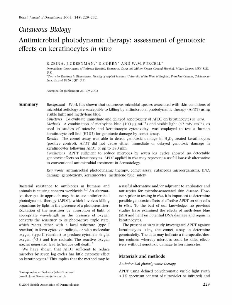
cells, some 95% were comet class 3 and the remainder
were typically class 2; no cells scored as class 1 or 0.
The typical appearance of a class 2 comet is shown in
Figure 1.
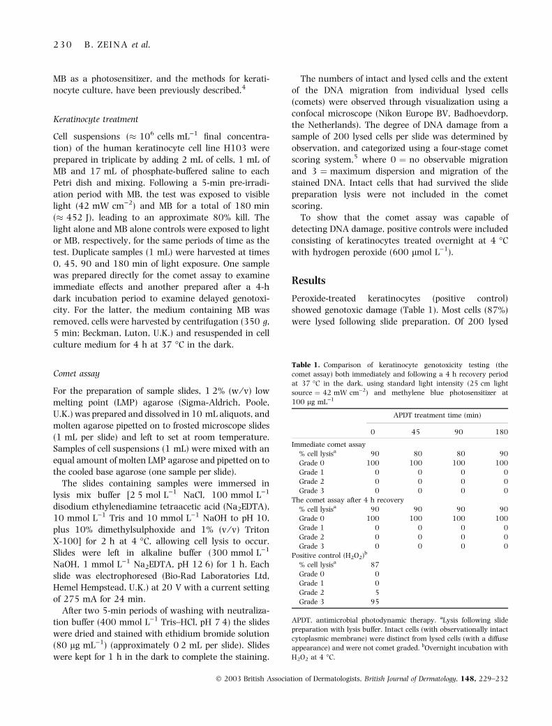
Treatment of keratinocytes by APDT followed by the
comet assay showed cell lysis at 80–90% (Table 1). The
comets were all class 0 for both immediate and 4-h
dark-incubated samples and for all treatment times.
The typical appearance of class 0 comets is shown in
Figure 2.
Discussion
The comet assay used was reported to detect very low
levels of DNA damage (both single and double strand)
in individual cells.6 Furthermore, it has been used
following a cell recovery period to detect delayed
damage (e.g. apoptosis) or repair.7
Keratinocytes treated with peroxide showed a high
proportion of cells with a typical comet appearance
and showed the technique capable of detecting DNA
damage. None of the APDT-treated cells showed
detectable signs of DNA damage. The cumulative
total of scored cells was 600, suggesting that the
DNA damage rate was lower than the detection
limit of 0Æ165%. Although DNA damage is the first
stage of cancer initiation, the relationship between
mutagenicity and DNA strand breakage is not
straightforward,8 and only a small proportion of
damaged cells becomes cancerous as most are repaired
or killed.9
The proposed key toxic species during APDT is
singlet oxygen capable of reacting with subcellular
targets, including DNA.3 MB is lipophilic and localizes
predominantly in the cytoplasm and membranes.10,11
As the diffusion distance of singlet oxygen in a cell is
< 0Æ075 lm,12 it is unlikely that DNA is the primary
target.
The skin contains other cells in addition to keratino-
cytes and it is possible that these types may be more
susceptible to genotoxic damage. APDT using MB
caused DNA damage in a human myeloid leukaemic
cell line in vitro13 and the genotoxic damage was
repaired following dark incubation (4 h at 37 �C).
Those data from microbe14 and keratinocyte killing4
with these on genotoxicity suggest that a wide safety
margin exists for APDT between bacterial eradication
and keratinocyte damage.
References
1 Wise R, Hart T, Cars O et al. Antimicrobial resistance. Br Med J
1998; 317: 609–10.
2 Hart CA, Kariuki S. Antimicrobial resistance in developing
countries. Br Med J 1998; 317: 647–50.
3 Wainwright M. Photodynamic antimicrobial chemotherapy
(PACT). J Antimicrob Chemother 1998; 42: 13–28.
4 Zeina B, Greenman J, Corry D, Purcell WM. Cytotoxicity effects of
antimicrobial photodynamic therapy on keratinocytes in vitro. Br
J Dermatol 2002; 146: 568–73.
5 Collins AR, Ai-guo M, Duthie SJ. The kinetics of repair of oxidative
DNA damage (strand breaks and oxidised pyrimidines) in human
cells. Mutat Res 1995; 336: 69–77.
6 Olive P, Durand RE, Judit P. Analysis of DNA damage in indi-
vidual cells. Methods Cell Biol 2001; 64: 235–49.
7 Olive P, Frazer G, Banath J. Radiation-induced apoptosis meas-
ured in TK6 human B lymphoblast cells using the comet assay.
Radiat Res 1993; 136: 130–6.
8 Noodt BB, Kvam E, Steen HB, Moan J. The primary DNA damage,
HPRT mutation and cell inactivation photoinduced with various
sensitizers in V79 cells. Photochem Photobiol 1993; 58: 541–7.
9 MacKie R. Epidermal skin tumours. In: Textbook of Dermatology
(Champion RH, Burton JL, Burns DA, Breathnach SM, eds), 6th
edn, Vol. 3. Oxford: Blackwell Science, 1998: 1651–93.
10 Ito T, Kobayashi K. A survey of in vivo photodynamic activity of
xanthenes, thiazines, and acridines in yeast cells. Photochem
Photobiol 1977; 26: 581–7.
Figure 1. Comet grade 2 (from cells treated with hydrogen perox-
ide). Figure 2. Comet grade 0 (from cells treated with antimicrobial
photodynamic therapy).
G E N O T O X I C I T Y O F A P D T O N K E R A T I N O C Y T E S I N V I T R O 2 3 1
� 2003 British Association of Dermatologists, British Journal of Dermatology, 148, 229–232
11 Muller T. Supravital uptake of methylene blue by dendritic cells
within stratified squamous epithelia: a light and electron micro-
scope study. Biotech Histochem 1996; 71: 96–101.
12 Moan J. On the diffusion length of singlet oxygen in cells and
tissues. J Photochem Photobiol B Biol 1990; 6: 343–7.
13 McNair FI, Marples B, West CML et al. A comet assay of DNA
damage and repair in K562 cells after photodynamic therapy
using haematoporphyrin derivative, methylene blue and meso-
tetrahydroxyphenylchlorin. Br J Cancer 1997; 75: 1721–9.
14 Zeina B, Greenman J, Purcell WM, Das B. Killing of cutaneous
microbial species by photodynamic therapy. Br J Dermatol 2001;
144: 274–8.
2 3 2 B . Z E I N A et al.
� 2003 British Association of Dermatologists, British Journal of Dermatology, 148, 229–232