antibody-mediated rejection in kidney transplantation: …€¦ · the 1-year graft loss rate...
TRANSCRIPT
1. Introduction
2. Classification of
antibody-mediated rejection
3. Diagnosis of
antibody-mediated rejection
4. Therapeutic strategies in
antibody-mediated rejection
5. Conclusion
6. Expert opinion
Review
Antibody-mediated rejection inkidney transplantation: an updateJessica G Lucas, Jeannie P Co, Uzoamaka T Nwaogwugwu, Imran Dosani &Kalathil K Sureshkumar†
Allegheny General Hospital, Division of Nephrology and Hypertension, Department of Medicine,
Pittsburgh, Pennsylvania, USA
Introduction: Acute antibody-mediated rejection (AMR) in renal-transplant
recipients is generally less responsive to conventional antirejection therapy
and has a worse prognosis than acute cellular rejection.
Areas covered: This review provides a broad understanding of the patho-
genesis of AMR, recent advances in its therapy, and future directions.
Conventional therapeutic approaches to AMR have minimal impact on
mature plasma cells, the major source of antibody production. Emerging
therapies include bortezomib, a proteasome inhibitor, and eculizumab, an
anti-C5 antibody. In several reports, bortezomib therapy resulted in prompt
reversal of rejection, decreased titers of donor-specific antibodies (DSA),
and improved renal allograft function. Eculizumab also reversed AMR and
prevented its development in patients with high post-transplantation
DSA levels.
Expert opinion: Despite the small sample size and lack of controls, these stud-
ies are encouraging, and although larger studies and long-term follow-up
are needed, bortezomib and eculizumab may play a major future role in
AMR therapy.
Keywords: complement inhibition, donor-specific antibody, humoral rejection,
proteasome inhibition
Expert Opin. Pharmacother. (2011) 12(4):579-592
1. Introduction
Kidney transplant rejections are classified into T-cell-mediated (acute cellular rejec-tion; ACR) and antibody-mediated (humoral) rejection (AMR). AMR occurs in upto 20 -- 30% of all acute rejection episodes following kidney transplantation andcan co-exist with cellular rejection. The term ‘AMR’ defines all allograft rejectionscaused by antibodies directed against donor-specific human leukocyte antigen(HLA), blood group antigen (ABO), or endothelial cell antigens [1]. Alloantibodiespreferentially attack the peritubular and glomerular capillaries, in contrast toT cells, which characteristically infiltrate tubules and arterial endothelium. AcuteAMR has a worse prognosis than ACR and is generally less responsive to conventionalantirejection therapy [2]. The 1-year graft loss rate following AMR varies from15 -- 20%, despite intensive conventional immunosuppressive therapy. Approxi-mately 30% of the patients on the transplant wait list are sensitized to HLA. Immu-nologic memory and preformed anti-HLA antibodies pose a powerful barrier towardssuccessful transplantation. Desensitization protocols have improved both the rate andlong-term outcome of transplantation in high immune-risk patients, such as thosewho are highly sensitized and those with ABO blood group incompatibilities. Nearly
10.1517/14656566.2011.525219 © 2011 Informa UK, Ltd. ISSN 1465-6566 579All rights reserved: reproduction in whole or in part not permitted
Exp
ert O
pin.
Pha
rmac
othe
r. D
ownl
oade
d fr
om in
form
ahea
lthca
re.c
om b
y H
INA
RI
on 0
4/17
/11
For
pers
onal
use
onl
y.
30% of such patients can develop AMR. This underscoresthe importance of developing novel strategies for both earlydiagnosis and therapy of AMR. The current review aims tofocus on the treatment of acute AMR following kidneytransplantation with special emphasis on emerging treatments.
2. Classification of antibody-mediatedrejection
The classification of AMR is based on clinical setting,underlying pathophysiology, and temporal relationship totransplantation. The three types of AMR are: i) hyperacute;ii) acute; and iii) chronic.
2.1 Hyperacute antibody-mediated rejectionHyperacute AMR is caused by preformed donor-specificantibodies (DSA). It is rarely seen nowadays due to the rou-tine use of pretransplantation cross-matching. It usually man-ifests shortly after the vascular anastamoses are establishedbut it can be delayed up to 3 days. Clinically, it is character-ized by widespread vascular thrombosis and the kidney turnscyanotic and flaccid, requiring immediate removal of the allo-graft. Histologically, the major findings associated withhyperacute AMR are neutrophil and platelet margination inglomerular and peritubular capillaries, red blood cell stasis,fibrin deposition and thrombosis within the microvasculature,acute tubular injury and widespread hemorrhagic corticalnecrosis. These changes depend on the interval betweentransplantation and biopsy or removal of the graft [3,4]. Immu-nofluorescence (IF) studies demonstrated IgG in glomerularand peritubular capillaries.
2.2 Acute antibody-mediated rejectionThe reported incidence of acute AMR varies in different cen-ters depending on protocols for performing transplantation inhighly sensitized patients and the methods used to detectDSA. Patients with acute AMR present with sudden onsetof graft dysfunction that often arises in the first few weeksafter transplantation. Presensitization is a major risk factorbut most patients with AMR had a negative cross-match.There are three types of acute AMR: type I is acute tubularnecrosis (ATN) like, type II is glomerular type, resemblingthrombotic microangiopathy, and type III is vascular typewith arterial inflammation.
The more frequent glomerular form of acute AMR is char-acterized by diffuse peritubular capillary (PTC) staining forthe complement component C4d. The histological appear-ance may show scattered glomerular, PTC and tubulointersti-tial neutrophils or monocyte--macrophages. The vascular/arterial type is characterized primarily by necrotizing arteritis,with mural fibrinoid necrosis and variable inflammation inthe artery wall, including lymphocytes, monocytes and neu-trophils along with luminal thrombosis. This lesion typicallyresults in cortical infarction with focal interstitial hemorrhage.In the vascular form of antibody-mediated rejection, IgG andoccasional IgM accompanied by C3 can be found in the wallsof arteries. Rafiq et al. observed 17 patients prospectively tolook for clinical outcomes of three different histopathologictypes of acute AMR [5]. None of the patients with types IIand III acute AMR responded to treatment and lost theirallografts earlier, or later due to transplant glomerulopathy.All patients with type I AMR had good responses to thetreatment, indicating a milder form of injury and pathologicprocess susceptible to current therapeutic modalities.
2.3 Chronic antibody-mediated rejectionChronic AMR is a slow, progressive loss of graft function thatusually develops > 1 year after transplantation. Several studieshave shown that circulating anti-HLA class I or II antibodies,either donor reactive/de novo or non-donor reactive, are foundin a substantial fraction of renal allograft recipients, andthese are associated with later graft loss. Transplant glomerul-opathy and arteriopathy are the pathologic features that areusually attributed to alloimmune mechanisms. Despite thesuccessful treatment, more than 40% of patients with AMRwill develop transplant glomerulopathy -- the major chronichistologic lesion associated with chronic antibody-mediateddamage [6]. Transplant glomerulopathy carries one of theworst prognoses of all chronic histological changes with5-year graft survival rates less than 50% from the time ofdiagnosis. The mechanism and the treatment of chronicantibody-mediated damage remain unclear.
3. Diagnosis of antibody-mediated rejection
More detailed pathologic classification of AMR was outlinedat the 2001 Banff meeting. This replaced the original category
Article highlights.
. Acute antibody-mediated rejection (AMR) has a worseprognosis than acute cellular rejection (ACR), whichhighlights the need for developing novel strategies forits early diagnosis and therapy.
. AMR is classified into hyperacute, acute, and chronic.
. C4d deposition in peritubular capillary (PTC) with eithermorphologic evidence of tissue injury or circulatingdonor-specific antibodies (DSA) confirms a diagnosisof AMR.
. Therapeutic options for AMR are evolving.
. PP/immunoadsorption remove circulating antibodies;intravenous immunoglobulin (IVIg)/mycophenolatemofetil (MMF) inhibits them; steroid/rituximab/antithymocyte globulin (ATG)/splenectomy cause B-celldepletion; and MMF/ATG/calcineurin inhibitors suppressT cells.
. Proteasome inhibitors, such as bortezomib, causeapoptosis of mature plasma cells, the major sourceof DSA.
. Eculizumab inhibit terminal complement activation.
. Bortezomib and eculizumab are the emerging therapiesfor AMR.
This box summarizes key points contained in the article.
Antibody-mediated rejection in kidney transplantation
580 Expert Opin. Pharmacother. (2011) 12(4)
Exp
ert O
pin.
Pha
rmac
othe
r. D
ownl
oade
d fr
om in
form
ahea
lthca
re.c
om b
y H
INA
RI
on 0
4/17
/11
For
pers
onal
use
onl
y.
2 of the Banff 97 classification [7]. Criteria for acute AMR inrenal allografts include three cardinal features [7]:
1) Morphologic evidence of acute tissue injury, such as:i) acute tubular injury, ii) neutrophils and/or mononu-clear cells in PTC and/or glomeruli, and/or capillarythrombosis; or iii) intimal arteritis/fibrinoid necrosis/intramural or transmural inflammation in arteries.
2) Immunopathologic evidence for antibody action,such as: i) C4d and/or (rarely) immunoglobulin inPTC or ii) immunoglobulin and complement inarterial fibrinoid necrosis.
3) Serologic evidence of circulating antibodies to donorHLA or other antidonor endothelial antigens (DSA).
C4d deposition in PTC along with one of two remainingcriteria clinches a diagnosis of AMR [7].
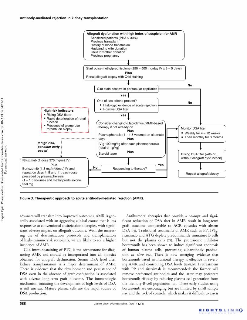
3.1 C4d stainingC4d is a fragment of C4b, an activation product of the classiccomplement pathway. Splitting of C4 into C4a and C4b istriggered by antidonor antibodies. C4b (and C4d) containan occult sulfhydryl group that forms a covalent, thioesterbond with nearby proteins on activation by antibody andC1 [8]. No functional role of C4d per se has been reported.C4d acts as an immunologic foot print of complementactivation and antibody activity. PTC deposition of C4d isstrongly associated with circulating antibody to donor HLAclass I or class II antigens and is currently the best singlemarker of complement-fixing circulating antibodies to theendothelium [9]. Tissue deposition of C4d can be detectedeither by monoclonal antibody and IF in frozen section orby polyclonal antibody and immunohistochemistry onformalin-fixed paraffin tissue section (Figure 1).
PTC staining for C4d -- but not its deposition in glomeru-lar capillaries, arteries or arterioles -- is a marker of AMR. C4dstaining can be diffuse or focal. Feucht et al. reported capillarydeposition of C4d that binds covalently to the capillary walland therefore persists in graft tissue, in 51 of 93 biopsiesfrom allografts with early graft dysfunction [10]. In renal allog-rafts with AMR, C4d deposits are detected on the luminalsurface of PTC endothelial cells or between endothelial cellsand the PTC basement membrane [11]. C4d was found to be95% sensitive and 96% specific for the presence of DSA inone study [12]. Occasionally, C4d staining can be detected asan isolated finding in the absence of DSA and graft dysfunc-tion. This may represent a state of accommodation (growingresistance of endothelial cells against humoral effectors) orpresence of harmless antibodies [13].
3.2 Donor-specific antibodiesAntibodies to donor HLA class I or II antigens (DSA) arepresent in 88 -- 95% of patients who have C4d depositionand acute graft dysfunction versus < 10% in C4d-negativeacute rejection. Antibodies to donor ABO antigens show a
similar association. DSA positivity in patients at transplanta-tion is a significant risk factor for AMR compared to patientswithout DSA [14]. In sensitized patients, pretransplant DSAagainst class 1 HLA predicted subsequent AMR and reducedgraft function in a recent study [15]. Mature plasma cells arethe major source of DSA production. C4d deposition withoutdetectable circulating DSA could result from antibody levelsbelow the detection threshold due to immunoadsorption bythe graft.
The advent of solid-phase antibody testing has greatlyenabled the characterization of the HLA-specific antibodiesand has largely replaced cell-based antibody testing methodsthat require viable cells. Solid-phase antibody testing employseither an enzyme-linked immunosorbent assay (ELISA)-basedsystem or a color-coded bead-based fluorometric assay. Thelatter is more sensitive and employs soluble HLA antigen-coated beads that can be detected by flow cytometer or bythe Luminex technology (LABScreen, One Lambda, Inc.,Canoga Park, CA). Fluorometric-bead-based assay is lessaffected by prior therapy with agents such as antithymocyteglobulin (ATG), rituximab or intravenous immunoglobulin(IVIg). Several studies have documented poor long-termallograft function in patients who developed anti-HLAantibodies [16-19]. Recently, major histocompatibility-complex class I chain-related gene A (MICA) antibodies andnon-HLA antibodies to endothelial targets such as angioten-sin II type 1 receptor (AT1-R) have been found to be associ-ated with AMR [20,21]. Activating IgG antibodies targeting theAT1-R were detected in the serum from patients presentingwith refractory vascular rejection, absent anti-HLA DSA andmalignant hypertension [22]. AT1-R blockade with losartanwas found to be beneficial in such patients. At present, thereis no consensus on when to test for DSA, especially in theabsence of allograft dysfunction; this is a subject of ongoingprospective studies. The clinical relevance of low levels ofDSA detected by newer, highly sensitive assays is unclearbut characteristics such as antigen specificity and bindingstrength may be useful in assessing clinical relevance ofsuch DSA.
4. Therapeutic strategies inantibody-mediated rejection
Knowledge of the mechanism of injury in AMR has providedinsights to therapeutic interventions. AMR involves the pro-duction of high levels of DSA by plasma cells. The plasmacells could be pre-existing (prior to transplant) or newly cre-ated from memory or naı̈ve B cells. The main mechanism ofinjury involves antibody-dependent activation of complementcascade with resultant capillaritis and glomerulitis, althoughevidence of a complement-independent mechanism has beenreported [23]. T cells are vital for the initiation of primaryand memory-B-cell responses that result in generation ofplasma cells. Therapeutic approaches to AMR are based onthe following concepts:
Lucas, Co, Nwaogwugwu, Dosani & Sureshkumar
Expert Opin. Pharmacother. (2011) 12(4) 581
Exp
ert O
pin.
Pha
rmac
othe
r. D
ownl
oade
d fr
om in
form
ahea
lthca
re.c
om b
y H
INA
RI
on 0
4/17
/11
For
pers
onal
use
onl
y.
. Circulating antibody removal: plasmapheresis (PP),immunoadsorption.
. Residual antibody inhibition: IVIg, mycophenolatemofetil (MMF).
. Suppression of antibody production or B-cell depletion:steroids, rituximab, ATG, splenectomy.
. Suppression of T-cell response: MMF, ATG,calcineurin inhibitors.
. Plasma-cell apoptosis/depletion: Proteasome inhibitor,e.g., bortezomib
. Inhibition of terminal complement activation: anti-C5antibody, e.g., eculizumab. Emerging therapies targetthe last two mechanisms. Multiple interventions areusually applied simultaneously in AMR. Details ofindividual therapies are described below.
4.1 PlasmapheresisPlasmapheresis (PP) removes alloantibodies from the cir-culation. It is the fastest and most effective method forthe elimination of DSA. PP modalities include plasmaexchange, double filtration PP and immunoadsorption.Plasma exchange has been the preferred method in the UnitedStates because of cost and ease of the procedure [24]. Becauseplasma exchange is the most commonly used method, theterm PP is synonymous with plasma exchange. The usual pre-scription includes 1.0 -- 1.5 volume exchange using albuminsolution daily or on alternate days, continued until serumcreatinine falls within 30% of previous baseline values [25].Although PP is effective in removing DSA from circula-
tion, it does not suppress antibody synthesis and reboundin circulating DSA after PP has been documented [26]. PPis therefore commonly used with agents that neutralizeantibodies (e.g., IVIg) or suppress antibody production
(e.g., calcineurin inhibitors, MMF or rituximab). Pascualet al. reported the successful treatment of five patients withrefractory AMR using a combination of PP and rescue immu-nosuppression with tacrolimus and MMF [27]. They reported100% graft survival at 19.6 months mean follow-up. Anobservational study of 18 patients with AMR treated withPP and intensification of immunosuppression reported1-year and 5-year graft survival rates to be 86 and 78%,respectively [28].
The adverse effects of PP include volume contraction,bleeding diathesis, allergic reaction, blood-borne pathogentransmission and antigen sensitization. Most of these reac-tions can be minimized by the avoidance of FFP use in favorof 5% albumin.
4.2 Intravenous immunoglobulinIVIg is a commercially prepared product from pooled humanplasma of 50,000 -- 100,000 or more screened, healthydonors. It is composed of more than 90% intact IgG, afew dimers, fragments of Fabs (fragment antigen bindingfragments) and traces of IgM and IgA [29].
The mechanism of action of IVIg is unclear. The proposedmechanisms of action include suppression of immuno-globulin synthesis, anti-idiotypic activity against DSA (withresultant neutralization of DSA), blockade of the Fc recep-tor, inhibition of complement activation, and anticytokineactivity [29,30].
IVIg is usually used in combination with PP but some stud-ies employed IVIg alone (usually in high dose 1 -- 2 g/kg). TheCedars-Sinai transplant program used high-dose IVIg pluspulse steroids in seven renal and three cardiac allograft recip-ients with refractory AMR [31]. IVIg was effective in reversingrejection within 2 -- 5 days of infusion with no recurrence inkidney transplant recipients; however, recurrence occurredin two heart transplant recipients. At a mean follow-up of23 months, all renal allografts were functioning with a meanserum creatinine of 1.4 mg/dl. Rocha et al. found similar1-year graft survival (81 vs 84%, p = ns) in AMR patientstreated with IVIg, PP and pulse steroids combined and inpatients with ACR treated with pulse steroids alone or withantilymphocytic therapy [26].
In a non-randomized control trial, Lefaucheur et al. treated12 patients with AMR with high-dose IVIg alone (control)and 12 with PP + IVIg + rituximab. At 36-month follow-up, graft survival rate was 50% in the control group and91.7% in the treatment group [32]. Beneficial effects of com-bined PP plus IVIg were reported in other retrospectivestudies [33,34].
One potential benefit of IVIg is its ability to replenish gam-maglobulin lost during PP, hence decreasing infection risk.Serious adverse effects from IVIg are rare but include asepticmeningitis (which occurs within 48 -- 72 h of administrationand is self-limiting), acute renal failure (osmotic injury,especially with high-dose IVIg), thrombotic events and severeanaphylactic reactions (associated with IgA sensitization in
Figure 1. Immunoflurescence staining of a renal biopsy
specimen in a patient with AMR. Showing C4d deposition
along the peritubular capillaries �20 (arrow pointing
towards peritubular capillary C4d deposit).
Antibody-mediated rejection in kidney transplantation
582 Expert Opin. Pharmacother. (2011) 12(4)
Exp
ert O
pin.
Pha
rmac
othe
r. D
ownl
oade
d fr
om in
form
ahea
lthca
re.c
om b
y H
INA
RI
on 0
4/17
/11
For
pers
onal
use
onl
y.
patients with IgA deficiency, can be avoided by the use ofIVIg with low IgA content) [24]. The severity of commonfirst-dose reactions such as headache, fever, chills, myalgiasand hypotension/hypertension can be reduced by slowingthe infusion rate. Severe adverse effects can be minimized bythe use of isosmolar preparation, sterile water as diluent, andavoidance of IVIg concentrations > 5% [29]. The usual recom-mended dose is 100 mg/kg of IVIg after each PP session and300 -- 400 mg/kg for 1 -- 2 days after last PP with a cumulativedose of 1000 mg/kg [27]. However, various dosing schedulesare currently in use and the optimal dose is poorly defined.
4.3 ImmunoadsorptionIn immunoadsorption (IA), plasma is processed through anadsorbent column and re-infused into the patient. As thereis no loss of volume, no replacement fluid is needed. Thereare two immunoadsorption columns: a protein A adsorptioncolumn that adsorbs immunoglobulin and an ABO antigencolumn that adsorbs specific anti-A or anti-B antibodiesregardless of immunoglobulin class or subclass [35]. In a ran-domized, controlled trial, Bohmig et al. treated five patients(test group) with IA using protein A and another five patientswithout IA with the option of IA rescue after 3 weeks [36].Both groups received tacrolimus conversion and if indicated,anticellular treatment. All IA treated patients responded totreatment within 2 weeks whereas four of the control patientswere dialysis-dependent despite rescue IA. The same group,in a non-randomized trial has previously reported the benefi-cial effects of IA in AMR [37]. Min et al. treated six patientswith AMR employing IA using staphylococcal protein Aplus tacrolimus/MMF combination therapy [38]. Theyreported 100% patient and graft survival in the IA groupwith mean serum creatinine of 1.2 mg/dl after 18monthsmean follow-up.
Immunoadsorption is an attractive strategy for efficient andhighly specific antibody depletion. However, because of thecost, membrane unavailability and the relative ease of usingPP, IA is not commonly used for AMR treatment.
4.4 Antilymphocyte therapyATG is a polyclonal preparation generated from the immuni-zation of rabbits with human thymus. Mechanisms of ATGaction include abrogation of T-cell help by elimination ofCD4+ T-cell and B-cell interaction, direct B-cell toxicityand modulation of alloantibody production. It has also beenshown to induce apoptosis [39]. Many studies have usedATG as part of AMR treatment especially when both cellularand humoral features are seen in biopsy.
Shah et al. used ATG 0.75 mg/kg/day for 5 -- 10 days incombination with plasma exchange in seven patients withAMR [39]. Reversal of AMR occurred in 85% with meanserum creatinine of 1.4 mg/dl at 1-year follow-up and nodifference in graft survival in the treated patients comparedwith those without AMR. ATG can be administered in threeor four divided doses to a cumulative dose of 6 mg/kg. The
platelets and white blood count should be monitored withdose adjustments as needed.
4.5 SteroidMost patients presenting with clinical acute allograft rejectionreceive pulse methylprednisolone therapy empirically or basedon allograft biopsy findings of ACR, AMR or a combinationof the two. Steroids help to treat the cell-mediated compo-nent. Steroids also work by down-regulating the B-cellresponse through decreased activity of T helper cells, whichindirectly suppresses AMR. A commonly used dosing sched-ule is methyl prednisone 250 -- 500 mg/day intravenouslyfor 3 -- 5 days followed by a prednisone taper.
4.6 Mycophenolate mofetilThe mechanism of action of MMF involves blockade oflymphocyte-specific isoforms of inosine monophosphatedehydrogenase. MMF inhibits in vitro antibody productionand reduces in vivo humoral response in transplant recipients.When used in combination with tacrolimus, it limits B-cellresponse in renal allograft recipients with AMR [40]. Cyclo-sporine appears to interfere with the metabolism of MMF,which may decrease the biological effects of this drug onalloantibody production [41].
4.7 DeoxyspergualinDeoxyspergualin (DSG) is an analogue of spergualin andshows antiproliferative action against interleukin (IL)-2 stimu-lated maturation of T cells. It also blocks B-cell differen-tiation, proliferation as well as inhibit cytotoxic T-celldifferentiation. There are very few reports of its use in AMRtreatment. Nojima et al. reported on the use of DSG andPP in five living donor kidney recipients with AMR. Therewas resolution of AMR in four out of five patients(80%) [42]. The dose of DSG was 3 mg/kg/day for 10 dayswith PP of 1 -- 9 sessions depending on treatment outcome.All the patients received pulse steroids. DSG is not a wellrecognized therapy for AMR, and only a few case reports exist.
4.8 SplenectomyThe spleen is the largest lymphoid organ in the body and itplays an important role in alloantibody generation. Splenec-tomy reduces the B-cell immune response and the numbersof precursor and mature plasma cells. Splenectomy has beenused as part of pretransplantation desensitization protocols,especially in highly sensitized patients and ABO mismatch.However, there are case reports on the use of splenectomyfor treatment of refractory AMR. Kaplan et al. reported fourcases of severe AMR (within 2 weeks of transplantation, inhighly sensitized patients) who failed 10-day treatment withstandard therapy including steroids, plasma exchange, IVIg,ATG and rituximab [43]. With persistent deterioration ofrenal function, laparoscopic splenectomy was done as a rescuetherapy with 100% graft survival after 8 months of follow-upwith a mean serum creatinine of 1.3 mg/dl. Locke et al. had
Lucas, Co, Nwaogwugwu, Dosani & Sureshkumar
Expert Opin. Pharmacother. (2011) 12(4) 583
Exp
ert O
pin.
Pha
rmac
othe
r. D
ownl
oade
d fr
om in
form
ahea
lthca
re.c
om b
y H
INA
RI
on 0
4/17
/11
For
pers
onal
use
onl
y.
similar reports on five patients with severe AMR treated withPP, IVIg and rituximab followed by splenectomy within 48 hof diagnosis [44]. They reported resolution of AMR with100% patient and graft survival after 18 months of follow-up and no significant postsplenectomy infectious complica-tions. All patients received pneumococcal, meningococcaland Haemophilus influenza vaccines. The disadvantages ofsplenectomy include a lifelong risk of sepsis with encapsulatedorganisms, permanent effects on the immune system, a lack ofindependent reduction in DSA titers, and surgical risks. Forthese reasons splenectomy is not favored as a conventionaltreatment for AMR.
4.9 RituximabRituximab is a chimeric anti-CD 20 (anti-B-cell) monoclonalantibody that is approved by the US Food and Drug Admin-istration (FDA) for the treatment of lymphoma. The CD20 antigen is expressed early in B-cell ontogeny but it is absenton mature plasma cells [45]. The variable region of rituximabbinds to CD 20 through three different mechanisms andmarks the cell for destruction, thereby leading to a profoundand sustained depletion in the number of circulating B cells.The three mechanisms of action of rituximab includeantibody-dependent cell cytotoxicity, complement-dependentcell killing and induction of apoptotic cell death [24].Genberg et al. examined the pharmacodynamics after a sin-
gle dose of rituximab given to renal transplant recipients.They demonstrated that B-cell elimination was rapid andoccurred in the peripheral blood over 1 -- 2 days [46]. Theeffect on the B-cell population was also prolonged and B cellsdid not re-emerge for 1 year and remained suppressed for2 years. Rituximab has demonstrated benefits in renal trans-plantation and is being used in some pretransplantationdesensitization protocols, post-transplant lymphoproliferativedisorders, allograft de novo or recurrent glomerulonephritis aswell as in the treatment of AMR.The initial report of using rituximab to effectively treat
AMR came from Becker et al. who evaluated 27 patientswith refractory rejection who received a single dose of rituxi-mab [47]. Three grafts were lost but the 24 surviving graftshad good function at the time of discharge. Kaposztas et al.reported a retrospective study of 54 patients with AMR [48].Patients in group A (n = 26) were treated with PP and ritux-imab and group B patients (n = 28) received PP without rit-uximab. Patients with low serum IgG levels also receivedIVIg. Two-year graft survival was significantly better in therituximab group (90 vs 60%, p = 0.005) with the differenceattributed to rituximab. Mulley et al. reported a case seriesof seven patients with refractory AMR who responded totreatment with a single low dose of rituximab (500 mg) [49].All patients recovered renal function with 100% patient andgraft survival at 21 months mean follow-up. Three patientshad significant viral infections but recovered fully. Severalother recent reports support the utility of rituximab intreating acute AMR [50-53]. Some of the studies are limited
by incomplete criteria for AMR diagnosis (especially olderstudies before the advent of DSA and C4d) and inconsistentpatient variables. All cases in which rituximab has shownefficacy have received IVIg, PP and/or steroids. The beneficialeffects of rituximab in this setting are likely multifactorial. Inaddition to depleting B cells and reducing DSA, rituximab hasbeen shown to disrupt T-cell co-stimulator and antigen-pre-senting-cell activities mediated by B cells, thereby diminishingT-cell effector functions.
The optimal dosing and length of therapy for rituximab isunclear. It is also unclear whether multiple doses of rituximabwould yield better depleting activity and antibody reductionthan a single dose. Some individuals have been reportedwith pre-existing antichimeric antibodies; others rapidlydevelop such antibodies de novo. This leads to decreased effi-cacy of rituximab. Although useful as a part of combinationtherapy, the major limitation of rituximab has been its inabil-ity to deplete CD 20 negative plasma cells that continue toproduce DSA and mediate graft injury.
Common adverse reactions (‡ 25%) reported with the useof rituximab in clinical trials of lymphoid malignanciesincluded infusion reactions, fever, lymphopenia, neutropenia,chills, infection and asthenia [54]. The adverse events reportedat ‡ 10% in clinical trials of rheumatoid arthritis includedupper respiratory tract infections, nasopharyngitis, urinarytract infections and bronchitis. Activation of viral infectionssuch as progressive multifocal leukoencephalopathy andhepatitis B also has been reported with rituximab therapy [54].
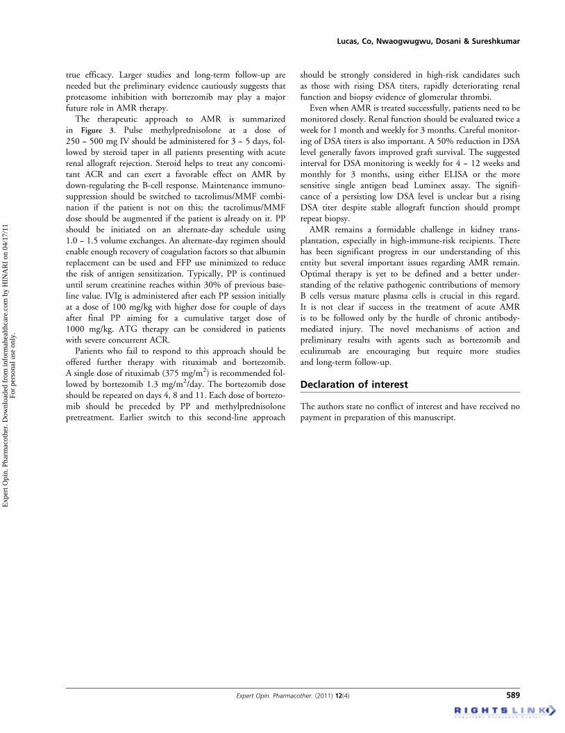
4.10 BortezomibExperts and researchers in the field of AMR have long recog-nized the potential utility of an antihumoral agent with theability to directly target plasma cells. Traditional modalitieshave been able to successfully remove antibodies, inhibit anti-body activity and even suppress antibody production butnone have been shown to affect mature antibody-producingplasma cells. In theory, and now in clinical experience, theproteasome inhibitor bortezomib has been shown to causeplasma-cell apoptosis resulting in the reduction and elimi-nation of circulating DSA levels in patients with acuteAMR [55,56].
Bortezomib (Velcade, Millenium PharmeceuticalsCambridge, MA) is a first-in-class proteasome inhibitor thatis approved by the US FDA for the treatment of multiplemyeloma (a plasma-cell neoplasm) [57]. It is a modified dipep-tidyl boronic acid that is available for intravenous injectionuse only. Bortezomib is a reversible inhibitor of thechymotrypsin-like activity of the 26S proteasome in mamma-lian cells. The 26S proteasome is a large complex thatdegrades ubiquitinated proteins. This ubiquitin--proteasomepathway plays an essential role in regulating the intracellularconcentration of specific proteins, thereby maintaininghomeostasis within cells. Bortezomib inhibition of the 26Sproteasome prevents this targeted proteolysis, which can affectmultiple signaling cascades within the cell. Specifically,
Antibody-mediated rejection in kidney transplantation
584 Expert Opin. Pharmacother. (2011) 12(4)
Exp
ert O
pin.
Pha
rmac
othe
r. D
ownl
oade
d fr
om in
form
ahea
lthca
re.c
om b
y H
INA
RI
on 0
4/17
/11
For
pers
onal
use
onl
y.
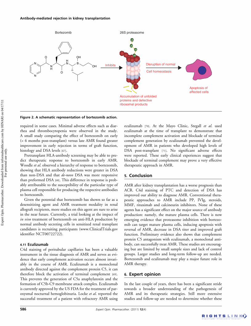
activation of the transcriptional activator nuclear factor kappaB (NF-kB) is prevented, leading to unregulated accumulationof unfolded proteins and defective ribosomal products withinthe endoplasmic reticulum. This can disrupt the normal cellhomeostasis, thus resulting in plasma-cell apoptosis [58].A schematic representation of bortezomib action is shownin Figure 2.
The distinct pathological changes of AMR are caused byhigh levels of DSA, which are produced by plasma cells (eitherfrom those that existed pretransplant or from those newly cre-ated from memory or naı̈ve B cells). By targeting plasma cells,bortezomib may directly destroy the source of this damagingDSA. In some desensitization protocols, it has been observedthat among patients with similar DSA levels at baseline, somedeveloped AMR whereas others did not. This made it difficultto define the exact relationship between the two. However,Burns et al. sought to examine the natural history of AMRin highly sensitized patients undergoing positive cross-match kidney transplantation [59]. In this study, a high DSAlevel after kidney transplantation (particularly within the firstmonth) was the major risk factor for the development ofAMR. There also seemed to be little correlation betweenbaseline DSA levels and post-transplant DSA levels.
Bortezomib is a cytotoxic agent the utility of which wasfirst recognized in the treatment of cancer; it has also beenshown to suppress T-cell function [60]. The recommendeddose of bortezomib is 1.3 mg/m2 given as a 3 -- 5 sec bolusinjection. The mean half-life after first dose ranged from9 to 15 h at doses ranging from 1.45 to 2.00 mg/m2 inpatients with advanced malignancies, but the pharmacokinet-ics of the drug as a single agent have not been fully character-ized at the recommended dose in myeloma patients (the samedose that has been used in the treatment of AMR). The bind-ing of bortezomib to human plasma proteins averaged 83% inthe original study population and the metabolism occurs inthe liver via cytochrome P450 enzymes, 3A4, 2D6, 2C19,2C9, and 1A2 [57]. No pharmacokinetic studies were con-ducted with bortezomib in patients with hepatic or renalimpairment. However, the drug is metabolized by liverenzymes and therefore its clearance may decrease in patientswith hepatic impairment. Also, no clinical information isavailable on the use of bortezomib in patients with creatinineclearance values < 13 ml/min and patients on dialysis.
The safety and efficacy of bortezomib was initially estab-lished in a study by Richardson et al., in which the drug wasgiven to 202 patients with refractory multiple myeloma [61].The most commonly reported adverse events includedasthenic conditions (fatigue, malaise, and weakness: 65%),nausea (64%), diarrhea (51%), anorexia (43%), constipation(43%), thrombocytopenia (43%), neutropenia (11%),peripheral neuropathy (37%), pyrexia (36%), vomiting(36%) and anemia (32%) [54]. More recently, a prospectivestudy looking at the toxicity profile of bortezomib in 50 renaltransplant candidates and recipients found that hematologic(anemia, thrombocytopenia), gastrointestinal (GI: nausea,
diarrhea) problems and mild peripheral neuropathy arecommon but generally mild and transient [62].
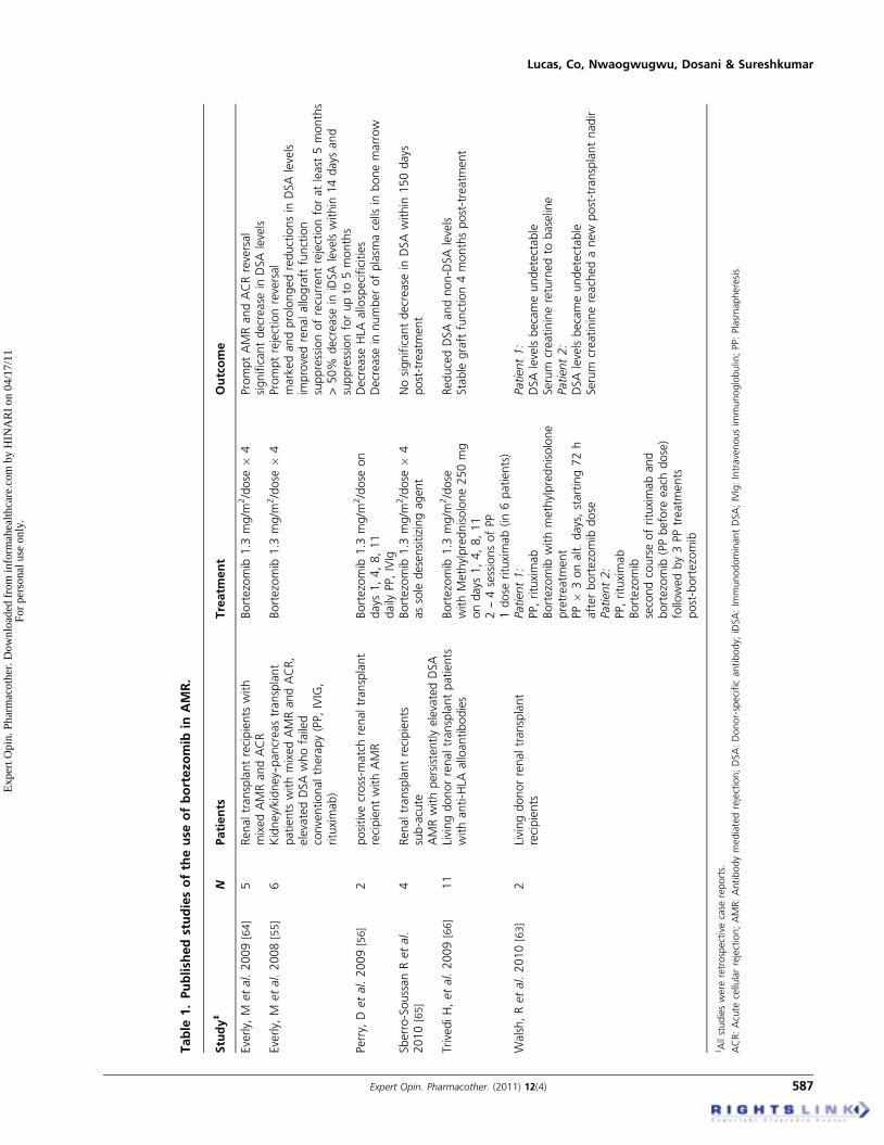
Bortezomib is contraindicated in patients with hyper-sensitivity to bortezomib, boron or mannitol. Currently avail-able studies are summarized in Table 1. The drug is pregnancycategory D and is considered unsafe for nursing mothers.Also, its safety and efficacy in children has not beenestablished [61].
At present, our knowledge of bortezomib as a therapy foracute AMR is based on clinical experience. At the Universityof Cincinnati, Everly et al. treated six kidney transplant recip-ients with refractory AMR and concomitant ACR with borte-zomib at labeled dosing (1.3 mg/m2/dose � 4 doses). Theyfound that in each case this therapy provided: i) promptrejection reversal; ii) marked and prolonged reductionsin DSA levels; iii) improved renal allograft function; andiv) suppression of recurrent rejection for at least 5 months [55].Immunodominant DSA (iDSA) levels were decreased bymore than 50% within 14 days and remained substantiallysuppressed for up to 5 months. In addition, one or more addi-tional DSAs were present at lower concentrations (non-iDSA)in each patient and were also reduced to undetectable levels.Two grafts were lost, one of which was attributed to non-compliance with immunosuppressive medications and theother was lost in the absence of any acute rejection on biopsyfor unknown reasons. The same group subsequently reportedthe outcome of AMR in two patients who received abortezomib-based regimen as primary therapy [63]. Bothpatients experienced prompt AMR reversal and DSA elimina-tion within 14 days. In another study, five patients withmixed ACR and AMR were given four doses of bortezo-mib [64]. There was prompt AMR and ACR reversal in allpatients and significant reduction in DSA in four patients.Commonly recognized bortezomib-related toxicities (GI tox-icity, thrombocytopenia and paresthesias) occurred in someof the patients but all were transient [62].
Sberro-Soussan et al. explored the utility of bortezomib assole desensitization agent in four patients with subacuteAMR and persistent DSA; they did not see any decrease inDSA titers after one cycle of bortezomib [65]. Lack of adjunc-tive steroid use, a critical component for enhancing the pro-apoptotic effect of bortezomib, was described as a possiblereason for the observation. Trivedi et al. reported the firstclinical experience with using bortezomib in patients withstable renal allograft function [66]. Eleven recipients of livingdonor kidney transplantation on clonal stimulation-deletionprotocol with elevated anti-HLA antibodies (> 1000 meanfluorescence intensity) were treated with 1.3 mg/m2 of borte-zomib together with methylprednisolone 250 mg on days 1,4, 8 and 11 followed by two to four sessions of PP. Six ofthe patients were also given a dose of rituximab. Bortezomibeffectively reduced the level of both DSA and non-DSAanti-HLA antibodies although four patients had persistentelevation or reappearance of anti-HLA antibodies, suggestingthat more than one cycle of bortezomib treatment may be
Lucas, Co, Nwaogwugwu, Dosani & Sureshkumar
Expert Opin. Pharmacother. (2011) 12(4) 585
Exp
ert O
pin.
Pha
rmac
othe
r. D
ownl
oade
d fr
om in
form
ahea
lthca
re.c
om b
y H
INA
RI
on 0
4/17
/11
For
pers
onal
use
onl
y.
required in some cases. Minimal adverse effects such as diar-rhea and thrombocytopenia were observed in the study.A small study comparing the effect of bortezomib on early(< 6 months post-transplant) versus late AMR found greaterimprovement in early rejection in terms of graft function,histology and DSA levels [67].Pretransplant HLA-antibody screening may be able to pre-
dict therapeutic response to bortezomib in early AMR.Woodle et al. observed a hierarchy of response to bortezomib,showing that HLA antibody reductions were greater in DSAthan non-DSA and that de-novo DSA was more responsivethan preformed DSA [68]. This difference in response is prob-ably attributable to the susceptibility of the particular type ofplasma cell responsible for producing the respective antibodiesto bortezomib.Given the potential that bortezomib has shown so far as a
desensitizing agent and AMR treatment modality in renalallograft recipients, more studies on this agent are sure to arisein the near future. Currently, a trial looking at the impact ofin vivo treatment of bortezomib on anti-HLA production bynormal antibody secreting cells in sensitized renal transplantcandidates is recruiting participants (www.ClinicalTrials.govidentifier NCT00722722).
4.11 EculizumabC4d staining of peritubular capillaries has been a valuableinstrument in the tissue diagnosis of AMR and serves as evi-dence that early complement activation occurs almost invari-ably in the course of AMR. Eculizumab is a monoclonalantibody directed against the complement protein C5, it cantherefore block the activation of terminal complement [69].This prevents the generation of C5a anaphylatoxin and theformation of C5b-C9 membrane attack complex. Eculizumabis currently approved by the US FDA for the treatment of par-oxysmal nocturnal hemoglobinuria. Locke et al. reported thesuccessful treatment of a patient with refractory AMR using
eculizumab [70]. At the Mayo Clinic, Stegall et al. usedeculizumab at the time of transplant to demonstrate thatincomplete complement activation and blockade of terminalcomplement generation by eculizumab prevented the devel-opment of AMR in patients who developed high levels ofDSA post-transplant [71]. No significant adverse effectswere reported. These early clinical experiences suggest thatblockade of terminal complement may prove a very effectivetherapeutic approach in AMR.
5. Conclusion
AMR after kidney transplantation has a worse prognosis thanACR. C4d staining of PTC and detection of DSA hasimproved our ability to diagnose AMR. Conventional thera-peutic approaches to AMR include PP, IVIg, steroids,MMF, rituximab and calcineurin inhibitors. None of theseagents has a significant effect on the major source of antibodyproduction: namely, the mature plasma cells. There is nowemerging evidence that proteasome inhibition with bortezo-mib can target mature plasma cells, inducing apoptosis withreversal of AMR, decrease in DSA titer and improved graftfunction. Preliminary evidence also shows that complementprotein C5 antagonism with eculizumab, a monoclonal anti-body, can successfully treat AMR. These studies are encourag-ing but are limited by small sample sizes and lack of controlgroups. Larger studies and long-term follow-up are needed.Bortezomib and eculizumab may play a major future role inAMR therapy.
6. Expert opinion
In the last couple of years, there has been a significant stridetowards a broader understanding of the pathogenesis ofAMR and its therapeutic strategies. Still more extensivestudies and follow-up are needed to determine whether these
Inhibits
Bortezomib
Disruption of normal
26S proteasome
Cell homeostasis
Apoptosis ofaffected cells
Accumulation of unfoldedproteins and defectiveribosomal products
Ub
Ub
Ub
Ub
N
NNH
HN B
OH
O
O
OH
Figure 2. A schematic representation of bortezomib action.
Antibody-mediated rejection in kidney transplantation
586 Expert Opin. Pharmacother. (2011) 12(4)
Exp
ert O
pin.
Pha
rmac
othe
r. D
ownl
oade
d fr
om in
form
ahea
lthca
re.c
om b
y H
INA
RI
on 0
4/17
/11
For
pers
onal
use
onl
y.
Table
1.Publish
edstudiesoftheuse
ofbortezo
mib
inAMR.
Study‡
NPatients
Treatm
ent
Outcome
Everly,
Metal.2009
[64]
5Renaltransplantrecipients
with
mixedAMRandACR
Bortezomib
1.3
mg/m
2/dose
�4
PromptAMRandACRreversal
significantdecrease
inDSA
levels
Everly,
Metal.2008
[55]
6Kidney/kidney--pancreastransplant
patients
withmixedAMRandACR,
elevatedDSAwhofailed
conventionaltherapy(PP,IVIG,
rituximab)
Bortezomib
1.3
mg/m
2/dose
�4
Promptrejectionreversal
markedandprolongedreductionsin
DSA
levels
improvedrenalallograft
function
suppressionofrecurrentrejectionforatleast
5months
>50%
decrease
iniDSA
levelswithin
14days
and
suppressionforupto
5months
Perry,
Detal.2009[56]
2positive
cross-m
atchrenaltransplant
recipientwithAMR
Bortezomib
1.3
mg/m
2/dose
on
days
1,4,8,11
daily
PP,IVIg
Decrease
HLA
allospecificities
Decrease
innumberofplasm
acells
inbonemarrow
Sberro-SoussanRetal.
2010
[65]
4Renaltransplantrecipients
sub-acute
AMRwithpersistentlyelevatedDSA
Bortezomib
1.3
mg/m
2/dose
�4
assole
desensitizingagent
Nosignificantdecrease
inDSA
within
150days
post-treatm
ent
TrivediH,etal.2009
[66]
11
Livingdonorrenaltransplantpatients
withanti-HLA
alloantibodies
Bortezomib
1.3
mg/m
2/dose
withMethylprednisolone250mg
ondays
1,4,8,11
2--4sessionsofPP
1dose
rituximab(in6patients)
ReducedDSA
andnon-DSA
levels
Stable
graftfunction4monthspost-treatm
ent
Walsh,Retal.2010[63]
2Livingdonorrenaltransplant
recipients
Patient1:
PP,rituximab
Bortezomib
withmethylprednisolone
pretreatm
ent
PP�
3onalt.days,starting72h
afterbortezomib
dose
Patient2:
PP,rituximab
Bortezomib
secondcourseofrituximaband
bortezomib
(PPbefore
each
dose)
followedby3PPtreatm
ents
post-bortezomib
Patient1:
DSA
levelsbecameundetectable
Serum
creatininereturnedto
baseline
Patient2:
DSA
levelsbecameundetectable
Serum
creatininereachedanew
post-transplantnadir
z Allstudieswere
retrospectivecase
reports.
ACR:Acute
cellularrejection;AMR:Antibodymediatedrejection;DSA:Donor-specificantibody;
iDSA:Im
munodominantDSA;IVIg:Intravenousim
munoglobulin;PP:Plasm
apheresis.
Lucas, Co, Nwaogwugwu, Dosani & Sureshkumar
Expert Opin. Pharmacother. (2011) 12(4) 587
Exp
ert O
pin.
Pha
rmac
othe
r. D
ownl
oade
d fr
om in
form
ahea
lthca
re.c
om b
y H
INA
RI
on 0
4/17
/11
For
pers
onal
use
onl
y.
advances will translate into improved outcomes. AMR is gen-erally associated with an aggressive clinical course that is lessresponsive to conventional antirejection therapies, with signif-icant adverse impact on allograft outcome. With the increas-ing use of desensitization protocols and transplantationof high-immune risk recipients, we are likely to see a higherincidence of AMR.C4d immunostaining of PTC is the cornerstone for diag-
nosing AMR and should be incorporated into all biopsiesobtained for allograft dysfunction. Serum DSA level afterkidney transplantation is a major determinant of AMR.There is evidence that the development and persistence ofDSA even in the absence of graft dysfunction is associatedwith adverse long-term graft outcome. The immunologicmechanism initiating the development of high levels of DSAis still unclear. Mature plasma cells are the major source ofDSA production.
Antihumoral therapies that provide a prompt and signi-ficant reduction of DSA titer in AMR result in long-termgraft outcome comparable to ACR episodes with absentDSA [72]. Traditional treatments of AMR such as PP, IVIg,rituximab and ATG deplete predominantly immature B cellsbut not the plasma cells [73]. The proteasome inhibitorbortezomib has been shown to induce significant apoptosisof human plasma cells, preventing alloantibody produc-tion in vitro [56]. There is now emerging evidence thatbortezomib-based antihumoral therapy is effective in revers-ing AMR and controlling DSA levels [55,63,66]. Pretreatmentwith PP and rituximab is recommended: the former willremove preformed antibodies and the latter may potentatebortezomib efficacy by reducing plasma-cell generation fromthe memory-B-cell population [63]. These early studies usingbortezomib are encouraging but are limited by small samplesize and the lack of controls, which makes it difficult to assess
Yes
Allograft dysfunction with high index of suspicion for AMRSensitized patients (PRA > 30%)Previous transplantHistory of blood transfusionHusband to wife donationChild to mother donationPrevious pregnancy
No
If high risk,consider earlyuse of
High risk indicatorsRising DSA titersRapid deterioration of renal functionPresence of glomerular thrombi on biopsy
No
Yes
Yes
Start pulse methylprednisolone (250 – 500 mg/day IV x 3 – 5 days) Plus
Renal allograft biopsy with C4d staining
C4d stain positive in peritubular capillaries
One of two criteria present? Histologic evidence of acute rejectionPositive DSA titer
Monitor DSA titer
Weekly for 4 – 12 weeksThen monthly for 3 months
Rising DSA titer (with orwithout allograft dysfunction)
Repeat allograft biopsy
Consider changingto tacrolimus /MMF-basedtherapy if not already on
PlusPlasmapheresis (1 – 1.5 volume) on alternatedays Plus
IVIg 100 mg/kg after each plasmapheresis(total of 1g/kg)
PlusSteroid taper
Responding to therapy?
No
Rituximab (1 dose 375 mg/m2 IV)Plus
Bortezomib (1.3 mg/m2/dose) IV andrepeat on days 4, 8 and 11, each dosepreceded by plasmapheresis(1 – 1.5 volume) and methylprednisolone250 mg
Figure 3. Therapeutic approach to acute antibody-mediated rejection (AMR).
Antibody-mediated rejection in kidney transplantation
588 Expert Opin. Pharmacother. (2011) 12(4)
Exp
ert O
pin.
Pha
rmac
othe
r. D
ownl
oade
d fr
om in
form
ahea
lthca
re.c
om b
y H
INA
RI
on 0
4/17
/11
For
pers
onal
use
onl
y.
true efficacy. Larger studies and long-term follow-up areneeded but the preliminary evidence cautiously suggests thatproteasome inhibition with bortezomib may play a majorfuture role in AMR therapy.
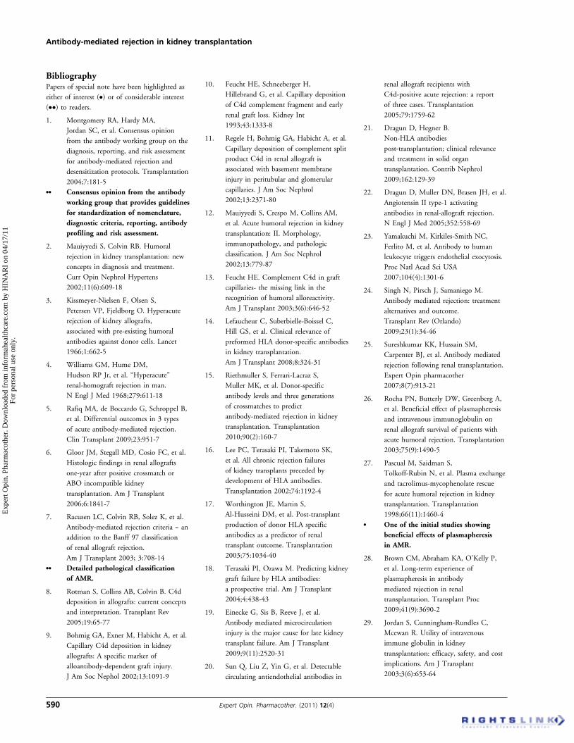
The therapeutic approach to AMR is summarizedin Figure 3. Pulse methylprednisolone at a dose of250 -- 500 mg IV should be administered for 3 -- 5 days, fol-lowed by steroid taper in all patients presenting with acuterenal allograft rejection. Steroid helps to treat any concomi-tant ACR and can exert a favorable effect on AMR bydown-regulating the B-cell response. Maintenance immuno-suppression should be switched to tacrolimus/MMF combi-nation if the patient is not on this; the tacrolimus/MMFdose should be augmented if the patient is already on it. PPshould be initiated on an alternate-day schedule using1.0 -- 1.5 volume exchanges. An alternate-day regimen shouldenable enough recovery of coagulation factors so that albuminreplacement can be used and FFP use minimized to reducethe risk of antigen sensitization. Typically, PP is continueduntil serum creatinine reaches within 30% of previous base-line value. IVIg is administered after each PP session initiallyat a dose of 100 mg/kg with higher dose for couple of daysafter final PP aiming for a cumulative target dose of1000 mg/kg. ATG therapy can be considered in patientswith severe concurrent ACR.
Patients who fail to respond to this approach should beoffered further therapy with rituximab and bortezomib.A single dose of rituximab (375 mg/m2) is recommended fol-lowed by bortezomib 1.3 mg/m2/day. The bortezomib doseshould be repeated on days 4, 8 and 11. Each dose of bortezo-mib should be preceded by PP and methylprednisolonepretreatment. Earlier switch to this second-line approach
should be strongly considered in high-risk candidates suchas those with rising DSA titers, rapidly deteriorating renalfunction and biopsy evidence of glomerular thrombi.
Even when AMR is treated successfully, patients need to bemonitored closely. Renal function should be evaluated twice aweek for 1 month and weekly for 3 months. Careful monitor-ing of DSA titers is also important. A 50% reduction in DSAlevel generally favors improved graft survival. The suggestedinterval for DSA monitoring is weekly for 4 -- 12 weeks andmonthly for 3 months, using either ELISA or the moresensitive single antigen bead Luminex assay. The signifi-cance of a persisting low DSA level is unclear but a risingDSA titer despite stable allograft function should promptrepeat biopsy.
AMR remains a formidable challenge in kidney trans-plantation, especially in high-immune-risk recipients. Therehas been significant progress in our understanding of thisentity but several important issues regarding AMR remain.Optimal therapy is yet to be defined and a better under-standing of the relative pathogenic contributions of memoryB cells versus mature plasma cells is crucial in this regard.It is not clear if success in the treatment of acute AMRis to be followed only by the hurdle of chronic antibody-mediated injury. The novel mechanisms of action andpreliminary results with agents such as bortezomib andeculizumab are encouraging but require more studiesand long-term follow-up.
Declaration of interest
The authors state no conflict of interest and have received nopayment in preparation of this manuscript.
Lucas, Co, Nwaogwugwu, Dosani & Sureshkumar
Expert Opin. Pharmacother. (2011) 12(4) 589
Exp
ert O
pin.
Pha
rmac
othe
r. D
ownl
oade
d fr
om in
form
ahea
lthca
re.c
om b
y H
INA
RI
on 0
4/17
/11
For
pers
onal
use
onl
y.
BibliographyPapers of special note have been highlighted as
either of interest (�) or of considerable interest(��) to readers.
1. Montgomery RA, Hardy MA,
Jordan SC, et al. Consensus opinion
from the antibody working group on the
diagnosis, reporting, and risk assessment
for antibody-mediated rejection and
desensitization protocols. Transplantation
2004;7:181-5.. Consensus opinion from the antibody
working group that provides guidelines
for standardization of nomenclature,
diagnostic criteria, reporting, antibody
profiling and risk assessment.
2. Mauiyyedi S, Colvin RB. Humoral
rejection in kidney transplantation: new
concepts in diagnosis and treatment.
Curr Opin Nephrol Hypertens
2002;11(6):609-18
3. Kissmeyer-Nielsen F, Olsen S,
Petersen VP, Fjeldborg O. Hyperacute
rejection of kidney allografts,
associated with pre-existing humoral
antibodies against donor cells. Lancet
1966;1:662-5
4. Williams GM, Hume DM,
Hudson RP Jr, et al. “Hyperacute”
renal-homograft rejection in man.
N Engl J Med 1968;279:611-18
5. Rafiq MA, de Boccardo G, Schroppel B,
et al. Differential outcomes in 3 types
of acute antibody-mediated rejection.
Clin Transplant 2009;23:951-7
6. Gloor JM, Stegall MD, Cosio FC, et al.
Histologic findings in renal allografts
one-year after positive crossmatch or
ABO incompatible kidney
transplantation. Am J Transplant
2006;6:1841-7
7. Racusen LC, Colvin RB, Solez K, et al.
Antibody-mediated rejection criteria -- an
addition to the Banff 97 classification
of renal allograft rejection.
Am J Transplant 2003; 3:708-14.. Detailed pathological classification
of AMR.
8. Rotman S, Collins AB, Colvin B. C4d
deposition in allografts: current concepts
and interpretation. Transplant Rev
2005;19:65-77
9. Bohmig GA, Exner M, Habicht A, et al.
Capillary C4d deposition in kidney
allografts: A specific marker of
alloantibody-dependent graft injury.
J Am Soc Nephol 2002;13:1091-9
10. Feucht HE, Schneeberger H,
Hillebrand G, et al. Capillary deposition
of C4d complement fragment and early
renal graft loss. Kidney Int
1993;43:1333-8
11. Regele H, Bohmig GA, Habicht A, et al.
Capillary deposition of complement split
product C4d in renal allograft is
associated with basement membrane
injury in peritubular and glomerular
capillaries. J Am Soc Nephrol
2002;13:2371-80
12. Mauiyyedi S, Crespo M, Collins AM,
et al. Acute humoral rejection in kidney
transplantation: II. Morphology,
immunopathology, and pathologic
classification. J Am Soc Nephrol
2002;13:779-87
13. Feucht HE. Complement C4d in graft
capillaries- the missing link in the
recognition of humoral alloreactivity.
Am J Transplant 2003;3(6):646-52
14. Lefaucheur C, Suberbielle-Boissel C,
Hill GS, et al. Clinical relevance of
preformed HLA donor-specific antibodies
in kidney transplantation.
Am J Transplant 2008;8:324-31
15. Riethmuller S, Ferrari-Lacraz S,
Muller MK, et al. Donor-specific
antibody levels and three generations
of crossmatches to predict
antibody-mediated rejection in kidney
transplantation. Transplantation
2010;90(2):160-7
16. Lee PC, Terasaki PI, Takemoto SK,
et al. All chronic rejection failures
of kidney transplants preceded by
development of HLA antibodies.
Transplantation 2002;74:1192-4
17. Worthington JE, Martin S,
Al-Husseini DM, et al. Post-transplant
production of donor HLA specific
antibodies as a predictor of renal
transplant outcome. Transplantation
2003;75:1034-40
18. Terasaki PI, Ozawa M. Predicting kidney
graft failure by HLA antibodies:
a prospective trial. Am J Transplant
2004;4:438-43
19. Einecke G, Sis B, Reeve J, et al.
Antibody mediated microcirculation
injury is the major cause for late kidney
transplant failure. Am J Transplant
2009;9(11):2520-31
20. Sun Q, Liu Z, Yin G, et al. Detectable
circulating antiendothelial antibodies in
renal allograft recipients with
C4d-positive acute rejection: a report
of three cases. Transplantation
2005;79:1759-62
21. Dragun D, Hegner B.
Non-HLA antibodies
post-transplantation; clinical relevance
and treatment in solid organ
transplantation. Contrib Nephrol
2009;162:129-39
22. Dragun D, Muller DN, Brasen JH, et al.
Angiotensin II type-1 activating
antibodies in renal-allograft rejection.
N Engl J Med 2005;352:558-69
23. Yamakuchi M, Kirkiles-Smith NC,
Ferlito M, et al. Antibody to human
leukocyte triggers endothelial exocytosis.
Proc Natl Acad Sci USA
2007;104(4):1301-6
24. Singh N, Pirsch J, Samaniego M.
Antibody mediated rejection: treatment
alternatives and outcome.
Transplant Rev (Orlando)
2009;23(1):34-46
25. Sureshkumar KK, Hussain SM,
Carpenter BJ, et al. Antibody mediated
rejection following renal transplantation.
Expert Opin pharmacother
2007;8(7):913-21
26. Rocha PN, Butterly DW, Greenberg A,
et al. Beneficial effect of plasmapheresis
and intravenous immunoglobulin on
renal allograft survival of patients with
acute humoral rejection. Transplantation
2003;75(9):1490-5
27. Pascual M, Saidman S,
Tolkoff-Rubin N, et al. Plasma exchange
and tacrolimus-mycophenolate rescue
for acute humoral rejection in kidney
transplantation. Transplantation
1998;66(11):1460-4. One of the initial studies showing
beneficial effects of plasmapheresis
in AMR.
28. Brown CM, Abraham KA, O’Kelly P,
et al. Long-term experience of
plasmapheresis in antibody
mediated rejection in renal
transplantation. Transplant Proc
2009;41(9):3690-2
29. Jordan S, Cunningham-Rundles C,
Mcewan R. Utility of intravenous
immune globulin in kidney
transplantation: efficacy, safety, and cost
implications. Am J Transplant
2003;3(6):653-64
Antibody-mediated rejection in kidney transplantation
590 Expert Opin. Pharmacother. (2011) 12(4)
Exp
ert O
pin.
Pha
rmac
othe
r. D
ownl
oade
d fr
om in
form
ahea
lthca
re.c
om b
y H
INA
RI
on 0
4/17
/11
For
pers
onal
use
onl
y.
30. Jordan SC, Vo AA, Peng A, et al.
Intravenous gammaglobulin: a novel
approach to improve transplant rates and
outcomes in highly HLA-sensitized
patients. Am J transplant 2006;6:459-66. Article with detailed discussion of the
beneficial effects of IVIg in highly
sensitized renal transplant recipients.
31. Jordan SC, Quartel AW, Czer LS, et al.
Posttransplant therapy using high dose
human immunoglobulin (intravenous
gammaglobulin) to control acute
humoral rejection in renal and cardiac
allograft recipients and potential
mechanism of action. Transplantation
1998;66:800-5
32. Lefaucheur C, Nochy D, Andrade J,
et al. Comparison of combination
plasmapheresis/ IVIg/ anti CD-20 versus
high dose IVIg in the treatment of
antibody-mediated rejection.
Am J Transplant 2009;9:1099-107
33. Sureshkumar KK, Oti IU, Baroody SC,
et al. Treatment of antibody- mediated
rejection after kidney transplantation.
Transplant Proc 2008;40(5):1373-4
34. White NB, Greenstein SM,
Cantafio AW, et al. Successful rescue
therapy with plasmapheresis and IVIG
for acute humoral renal transplant
rejection. Transplantation
2004;78(5):772-4
35. Tyden G, Kumlien G, Efvergren M.
Present techniques for antibody removal.
Transplantation 2007;84:S27-9
36. Bohmig GA, Wahrmann M, Reagele H,
et al. Immunoadsorption in severe C4d
positive acute kidney allograft rejection.
Am J Transplant 2006;6:1-5
37. Bohmig GA, Regele H, Exner M, et al.
C4d -positive- acute humoral renal
allograft rejection: effective treatment by
immunoadsorption. J Am Soc Nephrol
2001;12:2482-9
38. Min L, Shuming G, Zheng T, et al.
Novel rescue therapy for C4d-positive
acute humoral renal allograft rejection.
Clin Transplant 2005;19(1):51-5
39. Shah A, Nadasdy T, Arend L, et al.
Treatment of C4d positive acute humoral
rejection with plasmapheresis and rabbit
polyclonal antithymocyte globulin.
Transplantation 2004;77(9):1399-405
40. van Gelder T. Mycophenolate Mofetil.
How to further improve using an already
successful drug? Am J Transplant
2005;5(2):199-200
41. Miller J, Mendez R, Pirsch J, Jensik SC.
Safety and efficacy of tacrolimus in
combination with MMF in cadaveric
renal transplant recipients.FK506/MMF
Dose-Ranging Kidney Transplant Study
Group. Transplantation
2000;69(5):875-80
42. Nojima M, Yoshimoto T, Nakao A,
et al. Combined therapy of
deoxyspergualin and plasmapheresis:
a useful treatment for antibody-mediated
acute rejection after kidney
transplantation. Transplant proc
2005;37(2):930-3
43. Kaplan B, Gangemi A, Thielke J, et al.
Successful rescue of refractory, severe
antibody mediated rejection with
splenectomy. Transplantation
2007;83(1):99-100
44. Locke JE, Zachary AA, Haas M, et al.
The utility of splenectomy as rescue
treatment for severe acute antibody
mediated rejection. Am J Transplant
2007;7(4):842-6
45. Reff ME, Carner K, Chambers KS, et al.
Depletion of B cells in vivo by a
chimeric mouse human monoclonal
antibody to CD20. Blood
1994;83:435-45
46. Genberg H, Hansson A, Wernerson A,
et al. Pharmacodynamics of rituximab in
kidney transplantation. Transplantation
2007;84:s33-33
47. Becker YT, Becker BN, Pirsch JD, et al.
Rituximab as treatment for refractory
kidney transplant rejection.
Am J Transplant 2004;4:996-1001. An earlier study showing benefits
of rituximab in AMR.
48. Kaposztas Z, Podder H, Mauiyyedi S,
et al. Impact of rituximab therapy for
treatment of acute humoral rejection.
Clin Transplant 2009;23(1);63-73
49. Mulley WR, Hudson FJ, Tait BD, et al.
A single low fixed dose of rituximab to
salvage renal transplants from refractory
antibody-mediated rejection.
Transplantation 2009;87:286-9
50. Celik A, Saglam F, Cavdar C, et al.
Successful therapy with rituximab of
refractory acute humoral renal transplant
rejection: a case report. Transplant Proc
2008;40:302-4
51. Tanriover B, Wright SE, Foster SV,
et al. High dose intravenous
immunoglobulin and rituximab
treatment for antibody-mediated rejection
after kidney transplant: a cost analysis.
Transplant Proc 2008;40(10):3393-6
52. Faguer S, Kamar N,
Guilbeaud-Frugier C, et al. Rituximab
therapy for acute humoral rejection after
kidney transplantation. Transplantation
2007;83(9):1277-80
53. Wade E, Goral S, Kearns J, et al.
Experience with antibody mediated
rejection in allograft recipients.
Clin Transpl 2006;439-446
54. RITUXAN� (RITUXIMAB): Highlights
of prescription information, Genentech,
Inc., 1DNA way, South San Francisco,
CA: Revised 02/2010
55. Everly MJ, Everly JJ, Susskind B, et al.
Bortezomib provides effective therapy for
antibody- and cell-mediated acute
rejection. Transplantation
2008;88(12):1754-61. Initial study showing beneficial effects
of bortezomib in AMR.
56. Perry DK, Burns JM, Pollinger HS,
et al. Proteasome inhibition causes
apoptosis of normal human plasma cells
preventing alloantibody production.
Am J Transplant 2009;9:201-9. In-vitro study showing plasma cell
apoptosis with bortezomib.
57. Bross PF, Kane R, Farrell AT, et al.
Approval summary for bortezomib for
injection in the treatment of multiple
myeloma. Clin Cancer Res
2004;10:3954-64
58. Meister S, Schubert U, Neubert K, et al.
Extensive immunoglobulin production
sensitizes myeloma cells for proteasome
inhibition. Cancer Res 2007;67:1783-92
59. Burns JM, Connell LD, Perry DK, et al.
Alloantibody levels and acute humoral
rejection early after positive cross-match
kidney transplantation. Am J Transplant
2008;8:2684-94
60. Wang X, Luo H, Chen H, et al. Role
of proteasomes in T cell activation and
proliferation. J Immunol 1998;160:788
61. Richardson PG, Barlogie B, Berenson J,
et al. A Phase 2 Study of bortezomib in
relapsed, refractory myeloma. N Engl
J Med 2003;348(26):2609-17.. Large study demonstrating safety and
efficacy of bortezomib in refractory
multiple myeloma.
62. Walsh RC, Rike Shields A, Safdar S,
et al. Toxicity profile of proteosome
inhibitor-based antihumoral therapy in
renal transplant candidates and recipients
Lucas, Co, Nwaogwugwu, Dosani & Sureshkumar
Expert Opin. Pharmacother. (2011) 12(4) 591
Exp
ert O
pin.
Pha
rmac
othe
r. D
ownl
oade
d fr
om in
form
ahea
lthca
re.c
om b
y H
INA
RI
on 0
4/17
/11
For
pers
onal
use
onl
y.
(abstract 448). American Transplant
Congress, San Diego, CA; 2010
63. Walsh RC, Everly JJ, Brailey P, et al.
Proteasome inhibitor-based primary
therapy for antibody mediated renal
allograft rejection. Transplantation
2010;89:277-84
64. Everly MJ, Everly JJ, Susskind B, et al.
Proteasome inhibition reduces
donor-specific antibody levels.
Transplant Proc 2009;41(1):105-7
65. Sberro-Soussan R, Zuber J,
Suberbielle-Boissel C, et al. Bortezomib
as the sole desensitization agent does not
decrease donor-specific anti-HLA
antibodies. Am J Transplant
2010;10:681-6
66. Trivedi HL, Terasaki PI, Feroz A, et al.
Abrogation of anti-HLA antibodies via
proteasome inhibition. Transplantation
2009;87:1555-61
67. Walsh RC, Brailey P, Girnita A, et al.
Proteasome inhibitor-based antihumoral
therapy: differential response in early
versus late antibody mediated rejection
(abstract 422). American Transplant
Congress, San Diego, CA; 2010
68. Woodle S, Grinita A, Brailey P, et al.
Plasma cell targeted therapy in antibody
mediated rejection: pretransplant
HLA antibody screening predicts
response (abstract 425). American
Transplant Congress, San Diego, CA;
2010
69. Stegall MD, Gloor JM. Deciphering
antibody-mediated rejection: new insights
into mechanisms of treatment.
Curr Opin Organ Transplant
2010;15(1):8-10
70. Locke JE, Margo CM, Singer AL, et al.
The use of antibody to complement
protein C5 for salvage treatment of
severe antibody-mediated rejection.
Am J Transplant 2009;9:231-5. First report of the benefit of
eculizumab in AMR.
71. Stegall MD, Diwan T, Burns P, et al.
Prevention of acute humoral rejection
with C5 inhibition. Am J Transplant
2009;9(s2):241
72. Everly MJ, Everly JJ, Arend LJ, et al.
Reducing de novo donor-specific
antibody levels during acute rejection
diminishes renal allograft loss.
Am J Transplant 2009;9(5):1063-71
73. Ramos EJ, Pollinger HS, Stegall MD,
et al. The effect of desensitization
protocols on human splenic B-cell
populations in vivo. Am J Transplant
2007;7:402-7
AffiliationJessica G Lucas DO, Jeannie P Co MD,
Uzoamaka T Nwaogwugwu MD,
Imran Dosani MD &
Kalathil K Sureshkumar† MD FRCP (Glasg)
FASN†Author for correspondence
Allegheny General Hospital,
Division of Nephrology and Hypertension,
Department of Medicine,
320 East North Avenue,
Pittsburgh, Pennsylvania 15212, USA
Tel: +1 412 359 3319; Fax: +1 412 359 4136;
E-mail: [email protected]
Antibody-mediated rejection in kidney transplantation
592 Expert Opin. Pharmacother. (2011) 12(4)
Exp
ert O
pin.
Pha
rmac
othe
r. D
ownl
oade
d fr
om in
form
ahea
lthca
re.c
om b
y H
INA
RI
on 0
4/17
/11
For
pers
onal
use
onl
y.