antibacterial activity of myrciaria dubia (camu camu...
TRANSCRIPT
Antibacterial activity of Myrciaria dubia (Camu camu) againstStreptococcus mutans and Streptococcus sanguinis
Item type info:eu-repo/semantics/article
Authors Camere Colarossi, Rosella; Ulloa Urizar, Gabriela; MedinaFlores, Dyanne; Caballero García, Stefany; MaytaTovalino, Frank; Del Valle Mendoza, Juana Mercedes
Citation Antibacterial activity of Myrciaria dubia (Camu camu)against Streptococcus mutans and Streptococcus sanguinis2016, 6 (9):740 Asian Pacific Journal of TropicalBiomedicine
DOI 10.1016/j.apjtb.2016.07.008
Publisher Elsevier B.V.
Journal Asian Pacific Journal of Tropical Biomedicine
Rights info:eu-repo/semantics/openAccess
Downloaded 18-Apr-2018 09:52:06
Link to item http://hdl.handle.net/10757/620656
Accepted Manuscript
Antibacterial activity of Myrciaria dubia (Camu camu) against Streptococcus mutansand Streptococcus sanguinis
Rosella Camere-Colarossi, Gabriela Ulloa-Urizar, Dyanne Medina-Flores, StefanyCaballero-García, Frank Mayta-Tovalino, Juana del Valle-Mendoza
PII: S2221-1691(16)30263-5
DOI: 10.1016/j.apjtb.2016.07.008
Reference: APJTB 342
To appear in: Asian Pacific Journal of Tropical Biomedicine
Received Date: 1 April 2016
Accepted Date: 21 July 2016
Please cite this article as: Camere-Colarossi R, Ulloa-Urizar G, Medina-Flores D, Caballero-García S,Mayta-Tovalino F, Valle-Mendoza Jd, Antibacterial activity of Myrciaria dubia (Camu camu) againstStreptococcus mutans and Streptococcus sanguinis, Asian Pacific Journal of Tropical Biomedicine(2016), doi: 10.1016/j.apjtb.2016.07.008.
This is a PDF file of an unedited manuscript that has been accepted for publication. As a service toour customers we are providing this early version of the manuscript. The manuscript will undergocopyediting, typesetting, and review of the resulting proof before it is published in its final form. Pleasenote that during the production process errors may be discovered which could affect the content, and alllegal disclaimers that apply to the journal pertain.
MANUSCRIP
T
ACCEPTED
ACCEPTED MANUSCRIPT
Title: Antibacterial activity of Myrciaria dubia (Camu camu) against Streptococcus
mutans and Streptococcus sanguinis
Authors: Rosella Camere-Colarossi1, Gabriela Ulloa-Urizar2,3, Dyanne Medina-Flores1, Stefany
Caballero-García1, Frank Mayta-Tovalino1, Juana del Valle-Mendoza1,3*
Affiliations:
1School of Dentistry, Faculty of Health Sciences, Peruvian University of Applied Sciences-UPC, Lima, Peru
2Research and Innovation Center of the Faculty of Health Sciences, Peruvian University of Applied Sciences-UPC, Lima, Peru
3Molecular Biology Laboratory, Nutrition Research Institute, Lima, Peru
Keywords:
Antibacterial effect
Medicinal plants
Myrciaria dubia
Cytotoxity
*Corresponding author: Juana del Valle Mendoza, Research and Innovation Center of the Faculty of Health Sciences, Peruvian University of
Applied Sciences-UPC, Av. San Marcos cdra. 2, Cedros de Villa., Lima, Peru.
Tel: +51 13133333, Annex: 2704
Fax: +51 13496025
E-mail: [email protected]
Foundation Project: Supported by Universidad Peruana de Ciencias Aplicadas (UPC) Lima-Peru with Grant No. UPC-501-2015.
The journal implements double-blind peer review practiced by specially invited international editorial board members.
This manuscript included 1 table and 1 figure.
MANUSCRIP
T
ACCEPTED
ACCEPTED MANUSCRIPT
Article history:
Received 4 Apr 2016
Received in revised form 10 May, 2nd revised form 18 May 2016
Accepted 15 Jun 2016
Available online
Abstract
Objective: To evaluate the antibacterial and cytotoxic effect of Myrciaria dubia (Camu camu) (M. dubia) methanol
extract, against Streptococcus mutans (ATCC 25175) (S. mutans) and Streptococcus sanguinis (ATCC 10556) (S.
sanguinis).
Methods: Two methanol extracts of M. dubia were prepared in vitro, from the seeds and pulp. Ten independent tests
were prepared for each type of extract, using 0.12% chlorhexidine solution as positive control. Agar diffusion test was
used by preparing wells with the experimental solutions cultivated in anaerobic conditions for 48 h at 37 °C.
Meanwhile, the minimum inhibitory concentration and the cytotoxic effect over MDCK cell line was found.
Results: A higher antibacterial effect was observed with the methanol seed extract with an inhibitory halo of (21.36 ±
6.35) mm and (19.21 ± 5.18) mm against S. mutans and S. sanguinis, respectively. The methanol extract of the pulp
had an effect of (16.20 ± 2.08) mm and (19.34 ± 2.90) mm, respectively. The minimum inhibitory concentration of
the pulp extract was 62.5 µg/mL for both strains, whereas for the seed antibacterial activity was observed even at low
concentrations. The CC50 of the seeds extract was at a higher concentration than 800 µg/mL and 524.37 µg/mL for the
pulp extract.
Conclusions: The experimental findings demonstrated the antibacterial effect of the methanol extract of M. dubia
against S. mutans and S. sanguinis. These extracts were not cytotoxic at high concentrations.
MANUSCRIP
T
ACCEPTED
ACCEPTED MANUSCRIPT
1. Introduction
The high prevalence of dental caries in the population suggests that a vast majority of patients does not have an
adequate oral hygiene, as consequence of a bad tooth-brushing technique, lack of awareness of oral health or a lack of
motivation[1]. The dental caries is caused by a dental plaque or oral biofilm, which is the principal etiological factor,
containing diverse microorganisms that adhere to the tooth walls creating a microbiota unbalance in the host and
consequent bacterial overgrowth[2]. Therefore, oral antiseptics are used for proliferation control of these microorganisms.
In Peru, dental caries prevalence is around 95% in the general population per the Pan American Health Organization.
The most predominant microorganisms in the oral flora are Streptococcus mutans (S. mutans) and Streptococcus
sanguinis (S. sanguinis)[2,3]. Diverse publications have demonstrated that plant or fruit extracts can improve symptoms or
diminish the prevalence of dental caries, as well as to inhibit the pathogens growth in the dental plaque with less
toxicity[4]. In the last years, the use of natural products is increasing as an alternative treatment for dental disease, due to
their antibacterial, anti-inflammatory and antioxidant effects[4,5].
The World Health Organization has reported that more than 80% of the world population uses some short of
alternative medicine or phytotherapy for therapeutic purposes in dental caries[6,7]. In that sense, Peru is a privilege country
regarding the use of alternative medicine, with more than 80 000 species with the potential in the development of new
therapeutic drugs[5].
The Myrciaria dubia (M. dubia), commonly known as “camu camu”, is a native bush in the amazonic regions. It’s
chemical components have antibacterial, anti-inflammatory and antioxidant properties[8-10]. However, their properties
and clinical relevant use have not been fully studied in the odontological field.
Therefore, the main purpose of this study was to evaluate the in vitro antibacterial and cytotoxic effect of methanol
extracts of both seeds and pulp of M. dubia on S. mutans (ATCC 25175) and S. sanguinis (ATCC 10556) strains.
MANUSCRIP
T
ACCEPTED
ACCEPTED MANUSCRIPT
2. Materials and methods
2.1. Plant material and extracts
The M. dubia fruits were obtained from a naturist house in Lima, Peru. The camu camu pulp and seeds were
separated and immersed independently in absolute methanol (1:1, w/v). The samples were incubated at room temperature
and covered from sunlight by 7 days. The mixtures were filtered through a Whatman No. 4 filter paper and the filtrates
were evaporated at 50 °C[11]. All extracts were stored at 4 °C until used.
2.2. Bacterium strain
Strains of S. mutans and S. sanguinis were used (Genlab del Peru S.A.C., Peru). The cultivation medium was brain
heart infusion (BHI) agar (Oxoid, Hampshire, UK). Cultures were grown anaerobically for 72 h at 37 °C. For
antibacterial activity assay, three or four isolated colonies were inoculated in 3 mL of BHI broth and incubated in
anaerobically conditions for 72 h at 37 °C. The cultures were later diluted with fresh medium to approximate the
density of 0.5 McFarland standard, which represents an estimated concentration of 1.5 × 10⁸ CFU/mL.
The McFarland standard was prepared by inoculating colonies of the bacterial test strain in sterile saline and adjusting
the cell density to the concentration specified before[12].
2.3. Antibacterial screening of the methanol extracts
MANUSCRIP
T
ACCEPTED
ACCEPTED MANUSCRIPT
2.3.1. Determination of antibacterial activity
To determinate the antibacterial activity of the studied extracts, the cup-plate agar diffusion method was used[13]. BHI
agar was autoclaved for 15 min at 121 °C and cooled to 55 °C. The medium was then inoculated with the prepared
bacterial suspension, mixed gently and finally poured into sterile Petri dishes, sugar tubes containing molten agar (10 mL)
were sterilized and cooled to 40–42 °C. The tubes were then inoculated with 0.1 mL of the appropriate culture suspension
of bacterium. These agar plates were incubated under sterile conditions for 8 h at room temperature. Three wells per plate
of 6 mm in diameter and 4 mm in depth were made with a sterile cork borer, preserving a distance of 3 cm between them.
The wells were filled with 100 µL of the corresponding methanol extract. The chlorhexidine at 0.12% was used with
positive controls[14]. The Petri dishes were incubated under the same growth conditions mentioned above. At the end of
the period, the inhibition zones formed were measured in millimeters using a vernier. The inhibition zones with less than
12 mm in diameter were not considered for the antibacterial activity analysis. For each extract, 12 replicates were
assayed.
2.3.2 Determination of minimum inhibitory concentration (MIC)
The MIC was determined using the microdilution method as described by Jayaraman et al.[15] and Clinical and
Laboratory Standards Institute[16]. Serial two-fold dilutions of all the extracts were prepared with sterile saline in
a 96-well microtiter plate, obtaining a concentration range from 500 to 0 µg/mL. Then, 5 µL of S. mutans or S.
sangunis suspension (OD 550 = 0.6) were added to the wells containing the dilutions. Each dose was assayed in
quadruplicate. Uninoculated wells containing sterile saline or saline and extract were used as controls. After
incubation in anaerobic conditions for 72 h at 37 °C, the samples were observed and MIC was recorded as the
lowest concentration of each plant extract that inhibited the bacterial growth as detected by the absence of visual
turbidity.
MANUSCRIP
T
ACCEPTED
ACCEPTED MANUSCRIPT
2.4. Cytotoxicity assay of M. dubia
2.4.1. Cell lines
MDCK cells were obtained from ATCC (American Type Culture Collection, USA). The cells were grown in
minimum essential medium with Earle’s salts (Gibco BRL, Grand Island, NY) supplemented with 10% fetal bovine
serum, 25 µg/L gentamicin and 200 mmol/L L-glutamine (growth medium). Infected cells were maintained in
minimum essential medium with 1% fetal bovine serum, 25 µg/L gentamicin and 200 mmol/L L-glutamine
(maintenance medium). All cells were cultured at 37 °C in a humidified atmosphere with 5% CO2–95% air.
2.4.2 Cytotoxicity assay
Cytotoxicity of M. dubia extract was assessed using an assay based on the color change subsequent to the
reduction of MTT by mitochondrial enzymes[17-20]. The assays were performed using 1 × 10⁴ cell/well in 200 µL of
medium which were cultured in 96-well plates and incubated at 37 °C in a humidified atmosphere with 5% CO2–95%
air. When cell cultures were confluent, the culture medium was removed from the wells, which were replenished with
0.2 mL of the maintenance medium containing M. dubia extract prepared by dilution. M. dubia concentrations had a
range from 0 to 800 µg/mL. Each dose was assayed in quadruplicate. The wells with 0.2 mL maintenance medium but
without M. dubia extract were used as cell controls. All cultures were incubated at 37 °C for 6 days. Later, 20 µL of
MTT stock solution (3 mg/mL in phosphate buffered saline) was added to each well and after 3 h of incubation under
culture conditions, the medium was carefully removed and formazan crystals were solubilized by adding 200 µL of
dimethyl sulfoxide. Finally, cell viability was expressed as the percentage of the absorbance value determined for the
control cultures. Absorbance (A570 nm) was measured in an ELISA reader[17-20]. Cytotoxic concentration 50 (CC50) is
MANUSCRIP
T
ACCEPTED
ACCEPTED MANUSCRIPT
defined as the concentration of compound that reduces the viability of MDCK cells by 50%.
3. Results
3.1. Antibacterial activity of the plant extracts
This study was to determine the in vitro antibacterial effectivity of two methanol extracts of M. dubia against S.
mutans and S. sanguinis strains. The methanol extracts were prepared with the seeds and pulp of M. dubia and 0.12%
chlorhexidine solution was used as control.
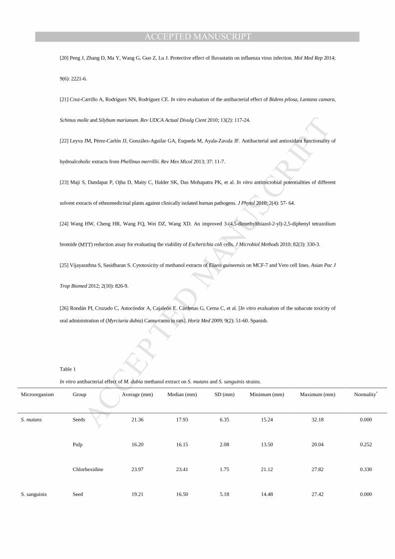
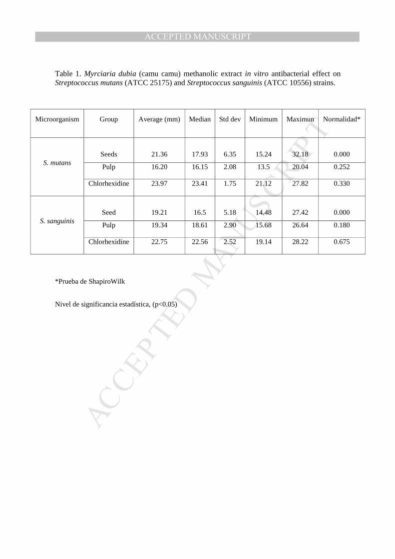
Table 1 shows the median of the methanol extracts inhibition halos. For both S. mutans and S. sanguinis, the seeds
extract of M. dubia had a major antibacterial effect if compared with the pulp extract (Table 1).
The antibacterial effect of seeds and pulps methanol extracts of M. dubia was compared on S. mutans and S.
sanguinis strains. Shapiro Wilk test showed that there is not a normal distribution in the seeds group extract for both
strains.
By comparing the antibacterial effect of seed and pulp extracts against both microorganisms, statistically
significant differences were observed in strains of S. mutans with P = 0.004, whereas for strains of S. sanguinis, no
statistically significant differences were found with P = 0.214.
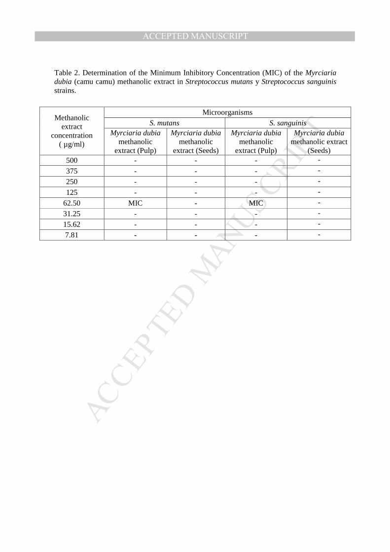
3.2. MIC
Table 2 shows the MIC of the methanol extract of the seeds and pulp M. dubia on strains of S. mutans and S.
sanguinis. The MIC antibacterial effect of the methanol seeds extract against both strains, could not determine due to
MANUSCRIP
T
ACCEPTED
ACCEPTED MANUSCRIPT
an antibacterial activity even at very low concentrations of the extract. However, for the pulp extract, and MIC with a
range of 50 to 75 µg/mL (62.5 µg/mL) was observed for both strains.
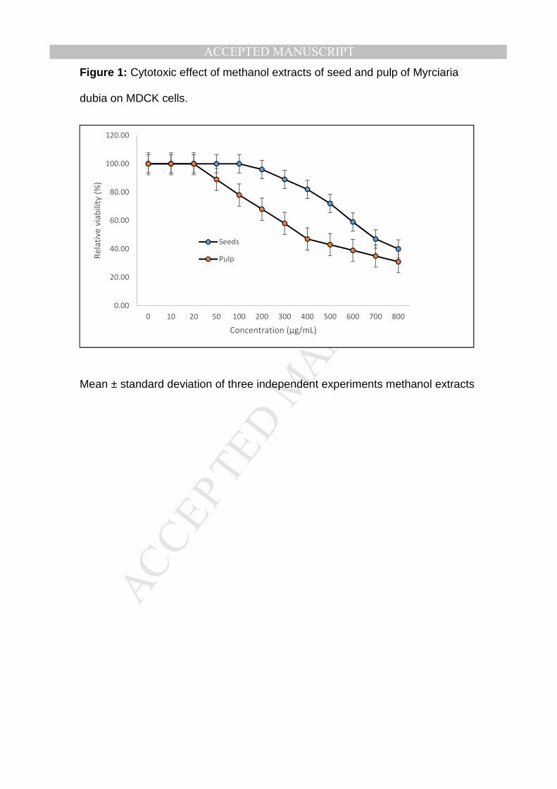
3.3. Toxicity of M. dubia extract
The toxicity of the methanol extracts of M. dubia was determined using MDCK cell. MDCK cell were incubated
with increasing amounts (0 to 800 µg/mL) of M. dubia seeds and pulp extracts and cell viability was determined by
MTT method.
Our results showed that the methanol seeds extract could inhibit 50% of the cellular viability at 725 ug/mL,
whereas the methanol extract from M. dubia pulp can inhibit cellular viability at a 50% with a concentration of 424.37
µg/mL. These values were confirmed by microscopic observation of the cytopathic effect. Thus, the methanol extract
can contain the active compounds of M. dubia to produce biological effects (Figure 1).
4. Discussion
Traditional medicine has been used in several countries over the years, due to its low cost and high effectivity for
certain bacterial diseases[5]. However, their clinical relevance and their impact on dentistry have not been fully
studied.
Several authors have described the methods used to evaluate in vitro susceptibility of bacteria to different agents
or extracts. The diffusion technique (on paper or well) is widely used to assess natural extracts with antimicrobial
activity and has the advanta=ge that its results are highly reproducible. However, the paper technique has some
disadvantages, one of which is the hydrophilic surface, which interferes with some compounds of natural extract and
MANUSCRIP
T
ACCEPTED
ACCEPTED MANUSCRIPT
prevents the diffusion of these in the agar. For the well diffusion technique, the extract has a direct contact with the
agar, so it is considered as a more sensitive technique and can facilitate the assessment of potential antibacterial agents
or any substance interest[13,21]. These features are important and should be considered especially in performing
pioneering studies with natural extracts.
Agar diffusion techniques have showed that the methanol extract of M. dubia has antimicrobial activity against S.
mutans and S. sanguinis, which demonstrate that methanol is an organic and effective solvent, with a high capacity to
extract more phenols and flavonoids as previously described by Leyva et al.[22].
In a recent study by Myoda et al., the antibacterial activity of acetone seed and pulp extracts of M. dubia was
evaluated against strains of Staphylococcus aureus, Escherichia coli and Saccharomyces, showing halos of inhibition
lower than 4-mm diameter for both acetone extracts, besides no antimicrobial activity against strains of Escherichia
coli and Saccharomyces cerevisiae was observed[9].
On the other hand, in our study, the MIC of the seeds methanol extract of M. dubia could not be determined due to
an antibacterial activity even at low concentrations; unlike the study mentioned above where the MIC of acetone
extract was identified less than 6.0 mg/mL[9].
Today, in the pharmacological field and in the phytotherapeutic medicine, it is important to evaluate the toxicity of
natural compounds for their potential impact on public and environmental health[17,18,23]. Therefore, cytotoxicity was
determined on seed and pulp extracts of M. dubia.
There are several tests for the toxicity determination of various substances. But the MTT assay is an effective
method to determine cell viability and has the advantage of being a quantitative method, unlike colorimetric methods,
which are qualitative[11,17,24]. Regarding the sensitivity and efficiency of this method, we found studies such as those
by Fattahi, who used this method to assess cell viability of cells infected with herpes simplex virus, demonstrating the
sensitivity of this method and the simplicity of the assay procedures, therefore allowing the study of a larger number
MANUSCRIP
T
ACCEPTED
ACCEPTED MANUSCRIPT
of compounds[19]. Vijayarathna et al. used the MTT assay showed that the Elaeis guineensis methanol extract had
significant cytotoxic effects on MCF-7 cells[25]. We can conclude that it is important to perform cytotoxic studies on
natural extracts as a requirement for their use as an alternative medicine in patients.
The methanol seed and pulp extracts of M. dubia showed no cytotoxicity against MDCK cell line. These results
agree with the study of Rondán et al., who evaluated the subacute toxicity of camu camu in an in-vivo study, and
found no evidence of subacute toxicity in any of the groups exposed on a histopathologic examination[26].
Currently, no studies evaluating the antibacterial activity of M. dubia against bacteria of the oral cavity are
known. With the results obtained in this study, we observed that methanol extract of seeds and pulp have the
antibacterial effect against high prevalence microorganisms in the oral cavity, like S. mutans and S. sanguinis.
The limitation found in this study was the precarious information regarding the properties and clinical relevance of
the M. dubia fruit in dental use. Our results point out the importance of conducting more studies for the development
of toothpastes containing active compound of M. dubia at low cost and easy access, due the abundance of the fruit in
the Peruvian Amazon. In addition, the use of the extract as a raw material in the manufacture of drugs is an alternative
to the use of synthetic chemicals.
Conflict of interest statement
We declare that we have no conflict of interest.
Acknowledgments
This study was supported by Universidad Peruana de Ciencias Aplicadas (UPC) Lima-Peru with Grant No.
MANUSCRIP
T
ACCEPTED
ACCEPTED MANUSCRIPT
UPC-501-2015.
References
[1] World Health Organization. Oral health. Fact sheet No.°318. Geneva: World Health Organization; 2012. [Online]. Available
from: http://www.who.int/mediacentre/factsheets/fs318/en/ [Accessed on 10th January, 2016]
[2] Shailee F, Girish MS, Kapil RS, Nidhi P. Oral health status and treatment needs among 12- and 15-year-old government and
private school children in Shimla city, Himachal Pradesh, India. J Int Soc Prev Community Dent 2013; 3(1): 44-50.
[3] Dewhirst FE, Chen T, Izard J, Paster BJ, Tanner ACR, Yu WH, et al. The human oral microbiome. J Bacteriol 2010; 192(19):
5002-17.
[4] Francisco KSF. [Fitoterapy: an option in odontological treatment]. Rev Saúde 2010; 4(1): 18-24. Spanish.
[5] Pamo Reyna OG. [Characteristics of original papers about plants properties in Peruvian medical journals]. Rev Peru Med Exp
Salud Publica 2009; 26(3): 314-23. Spanish.
[6] World Health Organization. WHO traditional medicine strategy 2014-2023. Geneva: World Health Organization; 2013.
[Online] Available from: http://www.who.int/iris/bitstream/10665/92455/1/9789241506090_eng.pdf [Accessed on 10th January,
2016]
[7] Marcia Avello L, Isabel Cisternas F. [Origins and situation of phytotherapy in Chile]. Rev Med Chil 2010; 138(10): 1288-93.
Spanish.
[8] Yazawa K, Suga K, Honma A, Shirosaki M, Koyama T. Anti-inflammatory effects of seeds of the tropical fruit camu-camu
(Myrciaria dubia). J Nutr Sci Vitaminol (Tokyo) 2011; 57(1): 104-7.
[9] Myoda T, Fujimura S, Park B, Nagashima T, Nakagawa J, Nishizawa M. Antioxidative and antimicrobial potential of residues
of camu-camu juice production. J Food Agric Environ 2010; 8(2): 304-7.
MANUSCRIP
T
ACCEPTED
ACCEPTED MANUSCRIPT
[10] Souza ALR, Pagani MM, Dornier M, Gomes FS, Tonon RV, Cabral LMC. Concentration of camu–camu juice by the coupling
of reverse osmosis and osmotic evaporation processes. J Food Eng 2013; 119(1): 7-12.
[11] Del Valle Mendoza J, Pumarola T, Gonzales LA, Del Valle LJ. Antiviral activity of maca (Lepidium meyenii) against human
influenza virus. Asian Pac J Trop Med 2014; 7(Suppl 1): S415-20.
[12] Andrews JM, Howe RA; BSAC Working Party on Susceptibility Testing. BSAC standardized disc susceptibility testing method
(version 10). J Antimicrob Chemother 2011; 66(12): 2726-57.
[13] García-Sánchez JE, García-Sánchez E, García-García MI. [Antimicrobial susceptibility testing of anaerobic bacteria]. Enferm
Infecc Microbiol Clin 2014; 32(Suppl 1): 23-9. Spanish.
[14] Medina-Flores D, Ulloa-Urizar G, Camere-Colarossi R, Caballero-García S, Mayta-Tovalino F, del Valle-Mendoza J.
Antibacterial activity of Bixa orellana L. (achiote) against Streptococcus mutans and Streptococcus sanguinis. Asian Pac J Trop
Biomed 2016; 6(5): 400-3.
[15] Jayaraman P, Sakharkar MK, Lim CS, Tang TH, Sakharkar KR. Activity and interactions of antibiotic and phytochemical
combinations against Pseudomonas aeruginosa in vitro. Int J Biol Sci 2010; 6(6): 556-68.
[16] Clinical and Laboratory Standards Institute. Methods for dilution antimicrobial susceptibility tests for bacteria that grow
aerobically; approved standard. Document M07-A9. 9th ed. Wayne: Clinical and Laboratory Standards Institute; 2012.
[17] Sylvester PW. Optimization of the tetrazolium dye (MTT) colorimetric assay for cellular growth and viability. Methods Mol Biol
2011; 716: 157-68.
[18] Taherkhani R, Farshadpour F, Makvandi M. In vitro anti-rotaviral activity of Achillea kellalensis. Jundishapur J Nat Pharm
Prod 2013; 8(3): 138-43.
[19] Fattahi S, Zabihi E, Abedian Z, Pourbagher R, Motevalizadeh Ardekani A, Mostafazadeh A, et al. Total phenolic and flavonoid
contents of aqueous extract of stinging nettle and in vitro antiproliferative effect on hela and BT-474 cell lines. Int J Mol Cell Med
2014; 3(2): 102-7.
MANUSCRIP
T
ACCEPTED
ACCEPTED MANUSCRIPT
[20] Peng J, Zhang D, Ma Y, Wang G, Guo Z, Lu J. Protective effect of fluvastatin on influenza virus infection. Mol Med Rep 2014;
9(6): 2221-6.
[21] Cruz-Carrillo A, Rodríguez NN, Rodríguez CE. In vitro evaluation of the antibacterial effect of Bidens pilosa, Lantana camara,
Schinus molle and Silybum marianum. Rev UDCA Actual Divulg Cient 2010; 13(2): 117-24.
[22] Leyva JM, Pérez-Carlón JJ, González-Aguilar GA, Esqueda M, Ayala-Zavala JF. Antibacterial and antioxidant functionality of
hydroalcoholic extracts from Phellinus merrillii. Rev Mex Micol 2013; 37: 11-7.
[23] Maji S, Dandapat P, Ojha D, Maity C, Halder SK, Das Mohapatra PK, et al. In vitro antimicrobial potentialities of different
solvent extracts of ethnomedicinal plants against clinically isolated human pathogens. J Phytol 2010; 2(4): 57- 64.
[24] Wang HW, Cheng HR, Wang FQ, Wei DZ, Wang XD. An improved 3-(4,5-dimethylthiazol-2-yl)-2,5-diphenyl tetrazolium
bromide (MTT) reduction assay for evaluating the viability of Escherichia coli cells. J Microbiol Methods 2010; 82(3): 330-3.
[25] Vijayarathna S, Sasidharan S. Cytotoxicity of methanol extracts of Elaeis guineensis on MCF-7 and Vero cell lines. Asian Pac J
Trop Biomed 2012; 2(10): 826-9.
[26] Rondán PI, Cruzado C, Astocóndor A, Cajaleón E. Cárdenas G, Cerna C, et al. [In vitro evaluation of the subacute toxicity of
oral administration of (Myrciaria dubia) Camu-camu in rats]. Horiz Med 2009; 9(2): 51-60. Spanish.
Table 1
In vitro antibacterial effect of M. dubia methanol extract on S. mutans and S. sanguinis strains.
Microorganism
Group
Average (mm)
Median (mm)
SD (mm)
Minimum (mm)
Maximum (mm)
Normality*
S. mutans
Seeds
21.36
17.93
6.35
15.24
32.18
0.000
Pulp
16.20
16.15
2.08
13.50
20.04
0.252
Chlorhexidine
23.97
23.41
1.75
21.12
27.82
0.330
S. sanguinis Seed 19.21 16.50 5.18 14.48 27.42 0.000
MANUSCRIP
T
ACCEPTED
ACCEPTED MANUSCRIPT
Pulp
19.34
18.61
2.90
15.68
26.64
0.180
Chlorhexidine
22.75
22.56
2.52
19.14
28.22
0.675
*: Shapiro Wilk test, level of statistical significance, (P < 0.05).
Figure legend Figure 1. Cytotoxic effect of methanol extracts of seed and pulp of M. dubia on MDCK cells.
Values are mean ± SD of three independent experiments.
MANUSCRIP
T
ACCEPTED
ACCEPTED MANUSCRIPT
Table 1. Myrciaria dubia (camu camu) methanolic extract in vitro antibacterial effect on Streptococcus mutans (ATCC 25175) and Streptococcus sanguinis (ATCC 10556) strains.
*Prueba de ShapiroWilk
Nivel de significancia estadística, (p<0.05)
Microorganism Group Average (mm) Median Std dev Minimum Maximun Normalidad*
S. mutans
Seeds
21.36
17.93
6.35
15.24
32.18
0.000
Pulp 16.20 16.15 2.08 13.5 20.04 0.252
Chlorhexidine 23.97 23.41 1.75 21.12 27.82 0.330
S. sanguinis
Seed
19.21
16.5
5.18
14.48
27.42
0.000
Pulp 19.34 18.61 2.90 15.68 26.64 0.180
Chlorhexidine 22.75 22.56 2.52 19.14 28.22 0.675
MANUSCRIP
T
ACCEPTED
ACCEPTED MANUSCRIPT
Table 2. Determination of the Minimum Inhibitory Concentration (MIC) of the Myrciaria dubia (camu camu) methanolic extract in Streptococcus mutans y Streptococcus sanguinis strains.
Methanolic extract
concentration ( µg/ml)
Microorganisms S. mutans S. sanguinis
Myrciaria dubia methanolic
extract (Pulp)
Myrciaria dubia methanolic
extract (Seeds)
Myrciaria dubia methanolic
extract (Pulp)
Myrciaria dubia methanolic extract
(Seeds) 500 - - - -
375 - - - -
250 - - - -
125 - - - -
62.50 MIC - MIC -
31.25 - - - -
15.62 - - - -
7.81 - - - -
MANUSCRIP
T
ACCEPTED
ACCEPTED MANUSCRIPTFigure 1: Cytotoxic effect of methanol extracts of seed and pulp of Myrciaria
dubia on MDCK cells.
Mean ± standard deviation of three independent experiments methanol extracts
0.00
20.00
40.00
60.00
80.00
100.00
120.00
0 10 20 50 100 200 300 400 500 600 700 800
Re
lati
ve v
iab
ilit
y (
%)
Concentration (µg/mL)
Seeds
Pulp