anterior myocardial infarction
TRANSCRIPT
Anterior Myocardial InfarctionClinical Relevance
Anterior STEMI results from occlusion of the left anterior descending artery (LAD).
Anterior myocardial infarction carries the worst prognosis of all infarct locations, mostly due to larger infarct size.
A study comparing outcomes from anterior and inferior infarctions (STEMI + NSTEMI) found that on average, patients with anterior MI had higher incidences of in-hospital mortality (11.9 vs 2.8%), total mortality (27 vs 11%), heart failure (41 vs 15%) and significant ventricular ectopic activity (70 vs 59%) and a lower ejection fraction on admission (38 vs 55%) compared to patients with inferior MI.
In addition to anterior STEMI, other high-risk presentations of anterior ischaemia include left main coronary artery (LMCA) occlusion and Wellens’ syndrome.
How to recognise anterior STEMI
ST segment elevation with Q wave formation in the precordial leads (V1-6) ± the high lateral leads (I and aVL).
Reciprocal ST depression in the inferior leads (mainly III and aVF).
NB. The magnitude of the reciprocal change in the inferior leads is determined by the magnitude of the ST elevation in I and aVL (as these leads are electrically opposite to III and aVF), hence may be minimal or absent in anterior STEMIs that do not involve the high lateral leads.
Patterns of anterior infarction
The nomenclature of anterior infarction can be confusing, with multiple different terms used for the various infarction patterns. The following is a simplified approach to naming the different types of anterior MI.
The precordial leads can be classified as follows:
Septal leads = V1-2 Anterior leads = V3-4 Lateral leads = V5-6
The different infarct patterns are named according to the leads with maximal ST elevation:
Septal = V1-2 Anterior = V2-5 Anteroseptal = V1-4 Anterolateral = V3-6, I + aVL Extensive anterior / anterolateral = V1-6, I + aVL
(NB. While these definitions are intuitive, there is often a poor correlation between ECG features and precise infarct location as determined by imaging or autopsy. For an alternative approach to the naming of myocardial infarctions, take a look at this 2006 article from Circulation)
Three other important ECG patterns to be aware of:
Anterior-inferior STEMI due to occlusion of a “wraparound” LAD: simultaneous ST elevation in the precordial and inferior leads due to occlusion of a variant (“type III”) LAD that wraps around the cardiac apex to supply both the anterior and inferior walls of the left ventricle.
Left main coronary artery occlusion: widespread ST depression with ST elevation in aVR ≥ V1
Wellens’ syndrome: deep precordial T wave inversions or biphasic T waves in V2-3, indicating critical proximal LAD stenosis (a warning sign of imminent anterior infarction)
ECG Examples
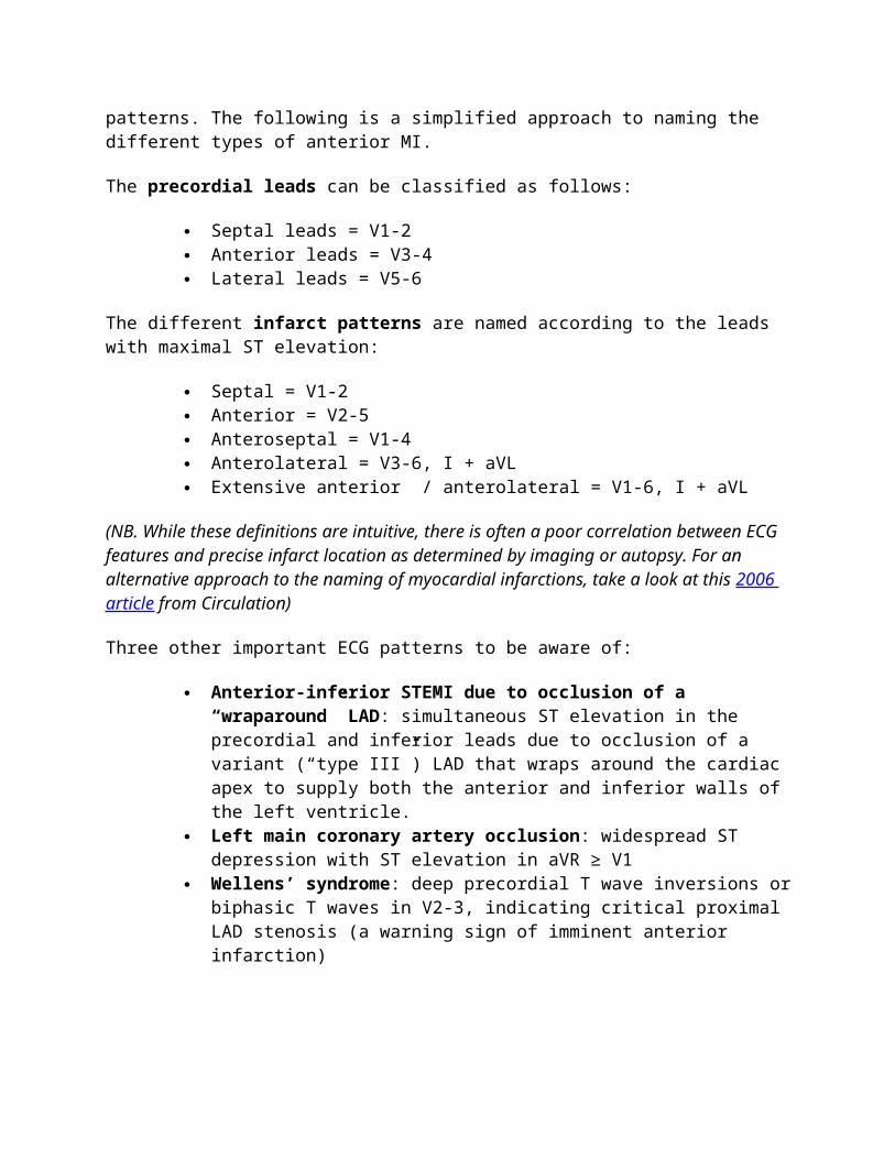
Example 1 - Hyperacute Anteroseptal STEMI
ST elevation is maximal in the anteroseptal leads (V1-4). Q waves are present in the septal leads (V1-2).
There is also some subtle STE in I, aVL and V5, with reciprocal ST depression in lead III.
There are hyperacute (peaked ) T waves in V2-4. These features indicate a hyperacute anteroseptal STEMI
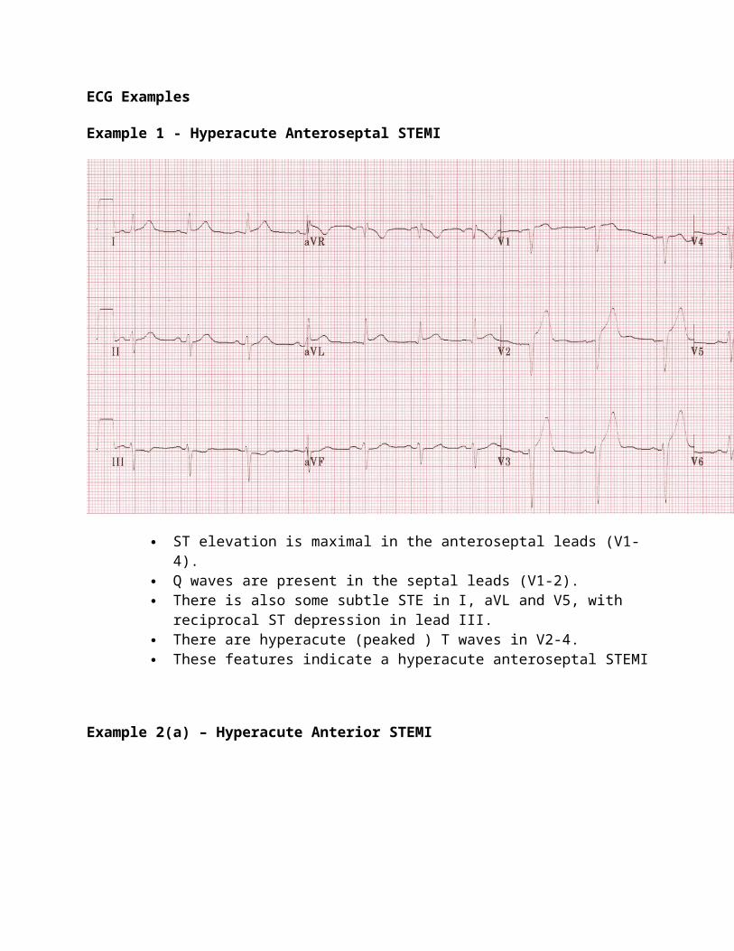
Example 2(a) – Hyperacute Anterior STEMI
Another example of hyperacute STEMI:
There are hyperacute T-waves in V2-6 (most marked in V2 and V3) with loss of R wave height.
The rhythm is sinus with 1st degree AV block. There are premature atrial complexes (beat 4 on the rhythm strip) and multifocal
ventricular ectopy (PVCs of two different types), indicating an “irritable” myocardium at risk of ventricular fibrillation.
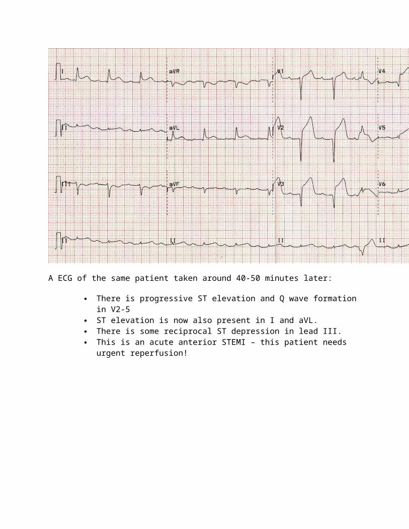
Example 2 (b) – Evolving Anterior STEMI
A ECG of the same patient taken around 40-50 minutes later:
There is progressive ST elevation and Q wave formation in V2-5 ST elevation is now also present in I and aVL. There is some reciprocal ST depression in lead III. This is an acute anterior STEMI – this patient needs urgent reperfusion!
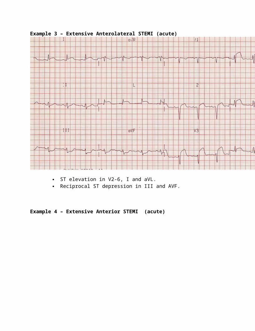
Example 3 – Extensive Anterolateral STEMI (acute)
ST elevation in V2-6, I and aVL. Reciprocal ST depression in III and AVF.
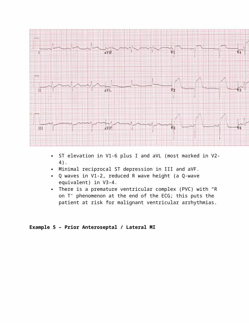
Example 4 – Extensive Anterior STEMI (acute)
ST elevation in V1-6 plus I and aVL (most marked in V2-4). Minimal reciprocal ST depression in III and aVF. Q waves in V1-2, reduced R wave height (a Q-wave equivalent) in V3-4. There is a premature ventricular complex (PVC) with “R on T’ phenomenon at
the end of the ECG; this puts the patient at risk for malignant ventricular arrhythmias.
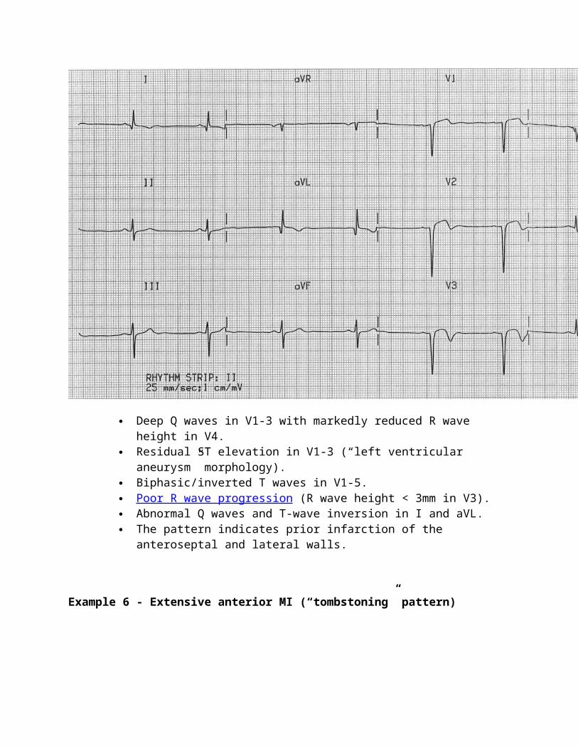
Example 5 – Prior Anteroseptal / Lateral MI
Deep Q waves in V1-3 with markedly reduced R wave height in V4. Residual ST elevation in V1-3 (“left ventricular aneurysm” morphology). Biphasic/inverted T waves in V1-5. Poor R wave progression (R wave height < 3mm in V3). Abnormal Q waves and T-wave inversion in I and aVL. The pattern indicates prior infarction of the anteroseptal and lateral walls.
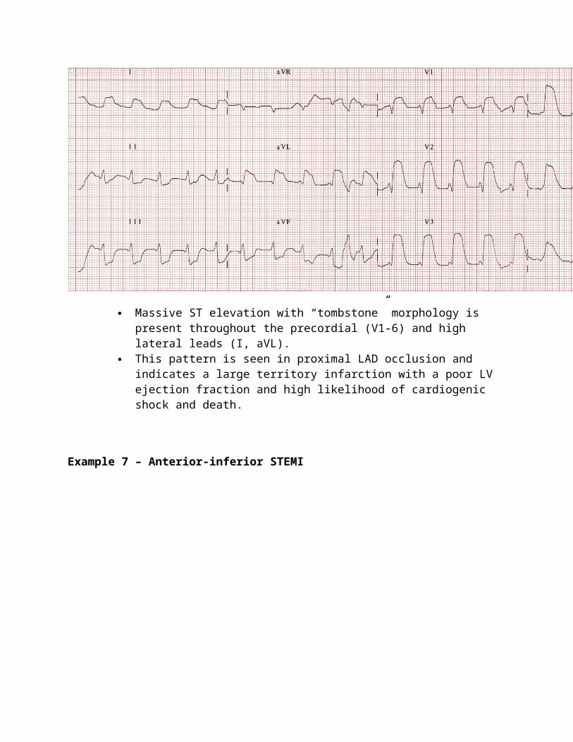
Example 6 - Extensive anterior MI (“tombstoning” pattern)
Massive ST elevation with “tombstone” morphology is present throughout the precordial (V1-6) and high lateral leads (I, aVL).
This pattern is seen in proximal LAD occlusion and indicates a large territory infarction with a poor LV ejection fraction and high likelihood of cardiogenic shock and death.
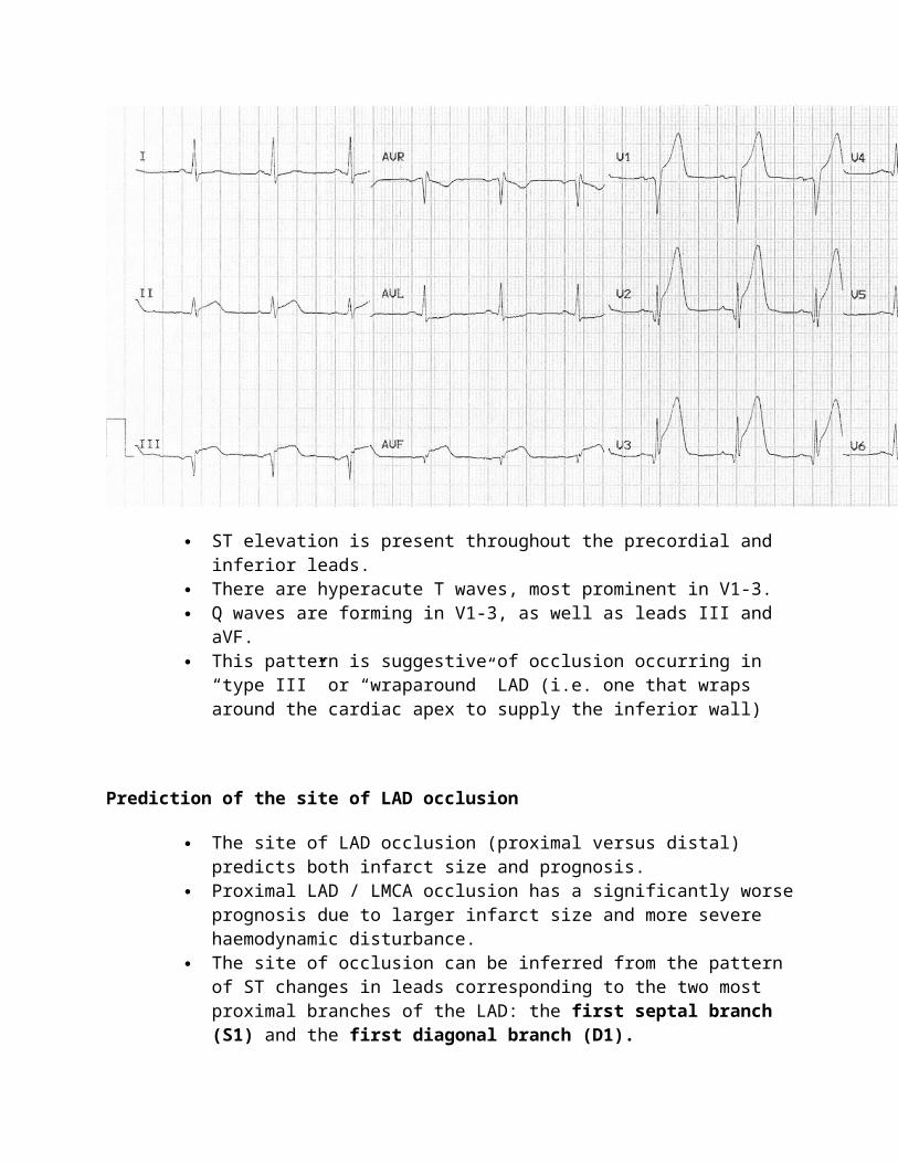
Example 7 – Anterior-inferior STEMI
ST elevation is present throughout the precordial and inferior leads. There are hyperacute T waves, most prominent in V1-3. Q waves are forming in V1-3, as well as leads III and aVF. This pattern is suggestive of occlusion occurring in “type III” or “wraparound”
LAD (i.e. one that wraps around the cardiac apex to supply the inferior wall)
Prediction of the site of LAD occlusion
The site of LAD occlusion (proximal versus distal) predicts both infarct size and prognosis.
Proximal LAD / LMCA occlusion has a significantly worse prognosis due to larger infarct size and more severe haemodynamic disturbance.
The site of occlusion can be inferred from the pattern of ST changes in leads corresponding to the two most proximal branches of the LAD: the first septal branch (S1) and the first diagonal branch (D1).
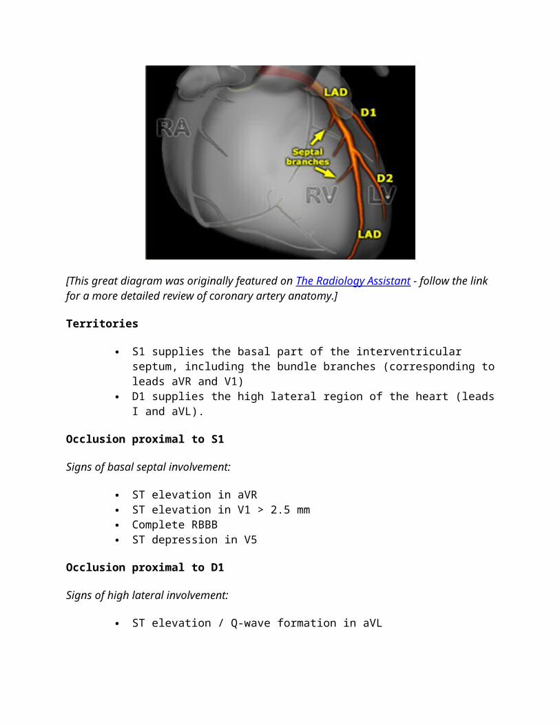
[This great diagram was originally featured on The Radiology Assistant - follow the link for a more detailed review of coronary artery anatomy.]
Territories
S1 supplies the basal part of the interventricular septum, including the bundle branches (corresponding to leads aVR and V1)
D1 supplies the high lateral region of the heart (leads I and aVL).
Occlusion proximal to S1
Signs of basal septal involvement:
ST elevation in aVR ST elevation in V1 > 2.5 mm Complete RBBB ST depression in V5
Occlusion proximal to D1
Signs of high lateral involvement:
ST elevation / Q-wave formation in aVL ST depression ≥ 1 mm in II, III or aVF (reciprocal to STE in aVL)
ST elevation in aVR of any magnitude is 43% sensitive and 95% specific for LAD occlusion proximal to S1. Right bundle branch block in anterior MI is an independent marker of poor prognosis; this is due to the extensive myocardial damage involved rather than the conduction disorder itself.
More Examples
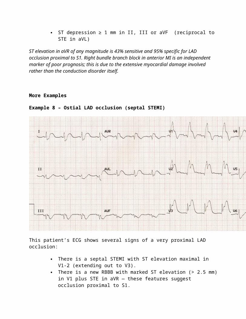
Example 8 – Ostial LAD occlusion (septal STEMI)
This patient’s ECG shows several signs of a very proximal LAD occlusion:
There is a septal STEMI with ST elevation maximal in V1-2 (extending out to V3).
There is a new RBBB with marked ST elevation (> 2.5 mm) in V1 plus STE in aVR — these features suggest occlusion proximal to S1.
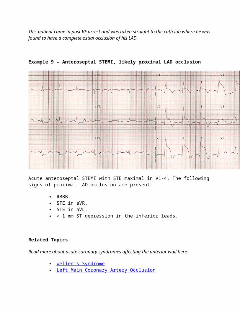
This patient came in post VF arrest and was taken straight to the cath lab where he was found to have a complete ostial occlusion of his LAD.
Example 9 – Anteroseptal STEMI, likely proximal LAD occlusion
Acute anteroseptal STEMI with STE maximal in V1-4. The following signs of proximal LAD occlusion are present:
RBBB. STE in aVR. STE in aVL. > 1 mm ST depression in the inferior leads.
Related Topics
Read more about acute coronary syndromes affecting the anterior wall here:
Wellen’s Syndrome Left Main Coronary Artery Occlusion
Check out these other STEMI patterns:
Inferior STEMI Lateral STEMI Posterior infarction Right ventricular infarction
Further Reading
ECG BASICS — Waves, Intervals, Segments and Clinical Interpretation
ECG CLINICAL CASES — Your favourite ECG’s placed in clinical context with a challenging Q&A approach
ECG and Cardiology Eponymous Syndromes — Cheats guide to eponymous emancipation
ECG Exam Template — a framework for the FACEM part 2 exam. ECG Reference Sites on the WEB — the best of the rest
Author Credits
Words - Ed Burns Pictures - Ed Burns Web Editing - Ed Burns
References
Arbane M, Goy JJ. Prediction of the site of total occlusion in the left anterior descending coronary artery using admission electrocardiogram in anterior wall acute myocardial infarction. Am J Cardiol. 2000 Feb 15;85(4):487-91, A10 [abstract]
Aygul N, Ozdemir K, Tokac M, Aygul MU, Duzenli MA, Abaci A et al. Value of lead aVR in predicting acute occlusion of proximal left anterior descending coronary artery and in-hospital outcome in ST-elevation myocardial infarction: an electrocardiographic predictor of poor prognosis. J Electrocardiol. 2008 Jul-Aug;41(4):335-41 [abstract].
Chan TC, Brady WJ, Harrigan RA, Ornato JP and Rosen PR. ECG in Emergency Medicine and Acute Care. Elsevier 2005.
Bayés de Luna A, Wagner G, Birnbaum Y, Nikus K, Fiol M, Gorgels A et al. International Society for Holter and Noninvasive Electrocardiography. A new terminology for left ventricular walls and location of myocardial infarcts that present Q wave based on the standard of cardiac magnetic resonance imaging: a statement for healthcare professionals from a committee appointed by the International Society for Holter and Noninvasive Electrocardiography. Circulation. 2006 Oct 17;114(16):1755-60 [full text].
Barrabes JA, Figueras J, Moure C, Cortadellas J, Soler-Soler J. Prognostic value of lead aVR in patients with a first non-ST-segment elevation acute myocardial infarction. Circulation 2003; 108: 814 – 819 [full text].
Birnbaum Y, Hasdai D, Sclarovsky S, Herz I, Strasberg B, Rechavia E. Acute myocardial infarction entailing ST-segment elevation in lead aVL: electrocardiographic differentiation among occlusion of the left anterior descending, first diagonal, and first obtuse marginal coronary arteries. Am Heart J. 1996 Jan;131(1):38-42 [abstract].
Engelen DJ, Gorgels AP, Cheriex EC, De Muinck ED, Ophuis AJ, Dassen WR et al. Value of the electrocardiogram in localizing the occlusion site in the left anterior descending coronary artery in acute anterior myocardial infarction. J Am Coll Cardiol. 1999 Aug;34(2):389-95 [full text].
Eskola MJ, Nikus KC, Holmvang L, et al. Value of the 12-lead electrocardiogram to define the level of obstruction in acute anterior wall myocardial infarction: Correlation to coronary angiography and clinical outcome in the DANAMI-2 trial. Int J Cardiol 2009;131:378–383 [abstract].
Hennings JR, Fesmire FM. A new electrocardiographic criteria for emergent reperfusion therapy. Am J Emerg Med. 2011 Jun 22. Epub ahead of print [abstract].
Kosuge M, Kimura K, Ishikawa T, Ebina T, Shimizu T, Hibi K, et al. Predictors of left main or three-vessel disease in patients who have acute coronary syndromes with non-ST-segment elevation. Am J Cardiol 2005; 95: 1366 – 1369 [abstract]
Rostoff P, Piwowarska W, Konduracka E, Libionka A, Bobrowska- Juszczuk M, Stopyra K, et al. Value of lead aVR in the detection of significant left main coronary artery stenosis in acute coronary syndrome. Kardiol Pol 2005;62:128-37 [abstract].
Surawicz B, Knilans T. Chou’s Electrocardiography in Clinical Practice (6th edition), Saunders 2008.
Stone PH, Raabe DS, Jaffe AS, et al. Prognostic significance of location and type of myocardial infarction: independent adverse outcome associated with anterior location. J Am Coll Cardiol 1988; 11:453 [abstract].
Vasudevan K, Manjunath CN, Srinivas KH, Prabhavathi, Davidson D, Kumar S, Yavagal ST. Electrocardiographic localization of the occlusion site in left anterior descending coronary artery in acute anterior myocardial infarction. Indian Heart J. 2004 Jul-Aug;56(4):315-9 [abstract].
Yamaji H, Iwasaki K, Kusachi S, Murakami T, Hirami R, Hamamoto H, et al. Prediction of acute left main coronary artery obstruction by 12-lead electrocardiography. ST segment elevation in lead aVR with less ST segment elevation in lead V(1). J Am Coll Cardiol. 2001 Nov 1;38(5):1348-54 [full text].