anterior and posterior corneal stroma elasticity after corneal collagen crosslinking treatment
TRANSCRIPT
lable at ScienceDirect
Experimental Eye Research 116 (2013) 58e62
Contents lists avai
Experimental Eye Research
journal homepage: www.elsevier .com/locate/yexer
Anterior and posterior corneal stroma elasticity after corneal collagencrosslinking treatmentq
Janice Dias a, Vasilios F. Diakonis b, Vardhaman P. Kankariya b, Sonia H. Yoo b,Noël M. Ziebarth a,*
aBiomedical Atomic Force Microscopy Laboratory, Department of Biomedical Engineering, University of Miami College of Engineering, Miami, FL, USAbBascom Palmer Eye Institute, Miller School of Medicine, University of Miami, Miami, FL, USA
a r t i c l e i n f o
Article history:Received 4 April 2013Accepted in revised form 30 July 2013Available online 9 August 2013
Keywords:corneacrosslinkingkeratoconusectasiamechanical properties
q Grant support: NIH Initiative for MaximizingFellowship (JD); NIH National Research Service AFellowship (1F31EY021714-01, JD).* Corresponding author. Biomedical Atomic Fo
Department of Biomedical Engineering, University of M1251 Memorial Drive MEA 209, McArthur Annex RoomUSA. Tel.: þ1 305 284 4520.
E-mail address: [email protected] (N.M. Zieba
0014-4835/$ e see front matter � 2013 Elsevier Ltd.http://dx.doi.org/10.1016/j.exer.2013.07.028
a b s t r a c t
The purpose of this project was to assess anterior and posterior corneal stromal elasticity after cornealcollagen cross linking (CXL) treatment inhumancadaver eyesusingAtomic ForceMicroscopy (AFM) throughindentation. Twenty four human cadaver eyes (12 pairs) were included in this study and divided into 2groups (6pairs per group). In bothgroups, the left eye (OS) servedas a control (no riboflavinorCXL treatmentwas performed) and the right eye (OD) underwent CXL treatment (30 min of riboflavin pretreatment fol-lowed by 30min of exposure to 3 mW/cm2 of ultraviolet light). In group 1, the anterior stromawas exposedby manual delamination of approximately 50 mm of the corneal stroma including Bowman’s membrane. Ingroup 2, the posterior stroma was exposed by delamination of the anterior 50% of the corneal stromaincluding Bowman’smembrane. Delaminationwas performed after crosslinking treatment in the case of thetreated eyes. In all eyes, the stromal elasticity was quantified using AFM through indentation. Young’smodulus of elasticity for the anterior cornea (group 1)was 245.9� 209.1 kPa (range: 82.3e530.8 kPa) for theuntreated control eyes, and 467.8 � 373.2 kPa (range: 157.4e1126 kPa) for the CXL treated eyes. Young’smodulus for the posterior cornea (group 2) was 100.2� 61.9 kPa (range: 28.1e162.6 kPa) for the untreatedcontrol eyes and 66.0 � 31.8 kPa (range: 31.3e101.7 kPa) for the CXL treated eyes. Young’s modulus of theanterior stromasignificantly increased after CXL treatment (p¼ 0.024),whereas theposterior stromadidnotdemonstrate a significantdifference inYoung’smodulus after CXL treatment (p¼ 0.170). The anterior stromawas stiffer than theposterior stroma for both the control andCXL treatment groups (p¼ 0.077 andp¼ 0.023,respectively). Our findings demonstrate that stiffness of the anterior corneal stroma after CXL treatmentseems to increase significantly, while the posterior stroma does not seem to be affected by CXL.
� 2013 Elsevier Ltd. All rights reserved.
1. Introduction
Corneal collagen cross linking (CXL) treatment is based on aphoto-chemical reaction between ultraviolet light-A (UV-A) andriboflavin (Wollensak et al., 2003a, 2003b; Wollensak, 2006). Thisminimally invasive procedure increases the mechanical strength ofthe cornea, thereby stabilizing corneal ectatic disorders such askeratoconus and iatrogenic keratectasia (after corneal refractive
Student Diversity Graduateward Individual Predoctoral
rce Microscopy Laboratory,iami College of Engineering,209, Coral Gables, FL 33146,
rth).
All rights reserved.
surgery) (Wollensak et al., 2003a, 2003b; Wollensak, 2006). Theintroduction of CXL in routine clinical practice has changed themanagement of the above entities; furthermore, it provides a ‘true’treatment, by inhibiting the progression of keratoconus and cornealectasia. Prior to CXL, all interventions (glasses, contact lenses andintra-corneal ring segment implantation (Kymionis et al., 2007))were used to improve visual function of patients, while they did nottreat the underlying pathophysiology of the corneal tissue.
Theoretically, the CXL treatment’s ability to increase cornealmechanical strength, and therefore inhibit the progression ofcorneal ectatic disorders, stems from the treatment’s formation ofadditional crosslinks within the cornea’s stromal ultrastructure.Comprehensive knowledge of the precise molecular mechanismsassociated with CXL and how it attributes to corneal mechanicalstrengthening remain unknown (Wollensak, 2006; Hayes et al.,2013; Meek and Hayes, 2013). Studies have shown that the CXLprocess is dependent upon the presence of carbonyl groups within
J. Dias et al. / Experimental Eye Research 116 (2013) 58e62 59
cornea (McCall et al., 2010; Brummer et al., 2011). In addition, arecent study conducted by Hayes et al. (2013) demonstrated thatthe creation of crosslinks produced from CXL most likely occurs atfour particular locations, opposing the initial notion of being be-tween collagen fibrils. The four locations are within and betweencollagen molecules located at the surface of the collagen fibrils aswell as within and between proteoglycan core proteins (Hayeset al., 2013). Although such advancements have been made, theexact molecules involved and their reaction pathways have yet tobe identified.
Many studies have been published demonstrating CXLs efficacyindirectly, mostly based on clinical findings, such as steady kera-tometric values and refraction (Kymionis et al., 2012; Vinciguerraet al., 2013). Investigations conducted ex vivo have been able todirectly quantify the efficacy of CXL by the measurement of cornealmechanical strength (Kling et al., 2010; Kohlhaas et al., 2006;Lanchares et al., 2011; Schumacher et al., 2011; Spoerl et al., 1998;Spoerl and Seiler, 1999; Wollensak et al., 2003b; Wollensak andIomdina, 2009a; 2009b). A land mark report by Wollensak et al.(2003a,b) demonstrates significant increase in corneal stiffness(increased by 328.9%) and in Young’s modulus (increased 4.5 times)after CXL assessed using stress-strain measurements (Wollensaket al., 2003b). Ever since then, numerous ex vivo studies havebeen conducted, using various corneal characterization techniques,to confirm such increase in corneal mechanical strength after CXL(Kling et al., 2010; Kohlhaas et al., 2006; Lanchares et al., 2011;Schumacher et al., 2011; Wollensak and Iomdina, 2009a, 2009b;Scarcelli et al., 2013).
Ultraviolet light absorption in the cornea is governed by theLamberteBeer law of absorptione there is an exponential decreasein UV intensity with increasing depth in the cornea. For this reason,it would be expected that the induction of additional crosslinkingbonds within the stroma is not homogeneous, but rather depth-dependent (Kohlhaas et al., 2006). Thus, it is likely that mechani-cal stiffening induced by crosslinking is much more pronounced inthe anterior stromal region (Kohlhaas et al., 2006). In fact, thisphenomenon has been demonstrated in the porcine model usingstrip extensiometry (Kohlhaas et al., 2006) and Brillouin micro-scopy (Scarcelli et al., 2013). These same conclusions were pre-dicted by an inverse computational model that used in vivo datafrom patients as inputs (Roy et al., 2013). The purpose of this studywas to build upon previous studies and quantify the anterior andposterior stromal elasticity in human cadaver eyes after cross-linking using Atomic Force Microscopy (AFM). Through the prin-ciple of nanoindentation and its ability to implement lowindentation depths, AFM can independently characterize thedistinct layers of corneal samples and perform depth-dependentcharacterization studies with proper hydration.
2. Materials and methods
2.1. Tissue acquisition
Experiments were conducted on 12 pairs of human cadaver eyes(age range: 39e88 years) retrieved from the Florida Lions Eye Bank(Miami, Florida). The human eye globes arrived from the eye bankin sealed vials placed in Styrofoam containers filled with ice. Uponarrival in the laboratory, the corneal epitheliumwas removed usinga cotton-tipped applicator and the whole globes were placed,cornea side down, in 20% Dextran solution for 24 h in the refrig-erator at 4 �C to restore corneal thickness to physiological levels(Borja et al., 2004; Duffey et al., 1989; Swinger and Kornmehl,1985).The mean postmortem time of the eyes at the time of receipt was4.58 � 1.31 days (range: 2e7 days), but the actual experimentswere performed 24 h later to enable this initial pretreatment with
20% Dextran. Pachymetry measurements were taken after thepretreatment (DGH 55 Pachmate, DGH Technology Inc., Exton, PA)to ensure the restoration of the corneal thickness within thephysiological range of 400e600 mm. All human eyes were obtainedand used in compliance with the guidelines of the Declaration ofHelsinki for research involving the use of human tissue.
2.2. Corneal collagen cross linking treatment
For this study, the right eyes (OD) were treated using the stan-dard CXL Dresden protocol and the left eyes (OS) served as un-treated controls (with no riboflavin pretreatment). Since theepithelium was removed for swelling restoration purposes, instil-lation of 0.1% riboflavin solution (10 mg riboflavin-5-phosphate in10 mL Dextran 20% solution) was applied on the bare cornealstroma one drop every 5 min for 30 min. The cornea was thenirradiated using UVA light at 378 nm wavelength and with an in-tensity of 3 mW/cm2. The irradiance was performed for 30 min,corresponding to a total surface dose of 5.4 J/cm2. During UVAirradiation, riboflavin solution was applied every 5 min to maintaincorneal saturation with riboflavin.
Six pairs of eyes (n ¼ 12) were prepared for anterior stromameasurements (Group 1) and six pairs (n ¼ 12) were prepared forposterior stroma measurements (Group 2). For anterior stromameasurements, a 4 mm surgical blade (Alcon, Texas, USA) was usedto remove approximately 50 mm of corneal tissue from the intactcornea, thereby removing Bowman’s membrane (17.7 mm thick-ness; Tao et al., 2011) to expose the anterior stromal region, butleaving it connected to the remaining stromal layer and Descemet’smembrane. To expose a lower plane in the cornea, approximately50% of the restored corneal thickness was removed using the samesurgical blade; this plane in the stromawas defined as the posteriorstroma. Pachymetric measurements were taken incrementally tomonitor the amount of stroma removed. The corneas were thenexcised from the whole globes, leaving a generous rim of sclera andplaced in a custom cornea holder with 15% Dextran solution tomaintain corneal hydration during mechanical testing.
2.3. Elasticity assessment
Mechanical property measurements were performed using acustom-built atomic force microscopy (AFM) system. The AFMsystem and experimental procedure have been described in detailpreviously (Ziebarth et al., 2011; Dias and Ziebarth, 2013). Briefly,tip-less AFM cantilevers (nominal spring constant: 4.5 N/m, NSC12series, Mikromasch, San Jose, CA) were modified with glass mi-crospheres (59e74 mm diameter, 15926-100, Polysciences Inc). Themodified tip was then calibrated to determine its spring constantusing a reference force calibration cantilever (nominal springconstant: 10.4 N/m, CLFC-NOBO, Bruker, Camarillo, CA) manufac-tured specifically for the calibration of other probes. The modifiedcantilever tips were lowered onto the corneal samples using apiezoelectric mechanism (60 mm maximal expansion, P-841.40,Physik Instrumente, Germany) with an approach speed of 15 mm/s.When the maximal indentation force of 1000 V (<20 nN, whichcorresponds to<6 mm indentation) was reached, the cantilever wasimmediately retracted at the same speed. The voltage detected atthe photodiode due to deflection of the cantilever was recorded as afunction of piezoelectric displacement. These recordings were usedto derive the sample’s force-indentation curves, after factoring outthe cantilever deflection on a hard surface and incorporating themeasured spring constant. With the use of custom MATLAB pro-grams, the indentation force-indentation depth curves wereanalyzed using the Hertz model for a spherical indenter (Hertz,1881):
J. Dias et al. / Experimental Eye Research 116 (2013) 58e6260
F ¼ 4E R�2�D3=2
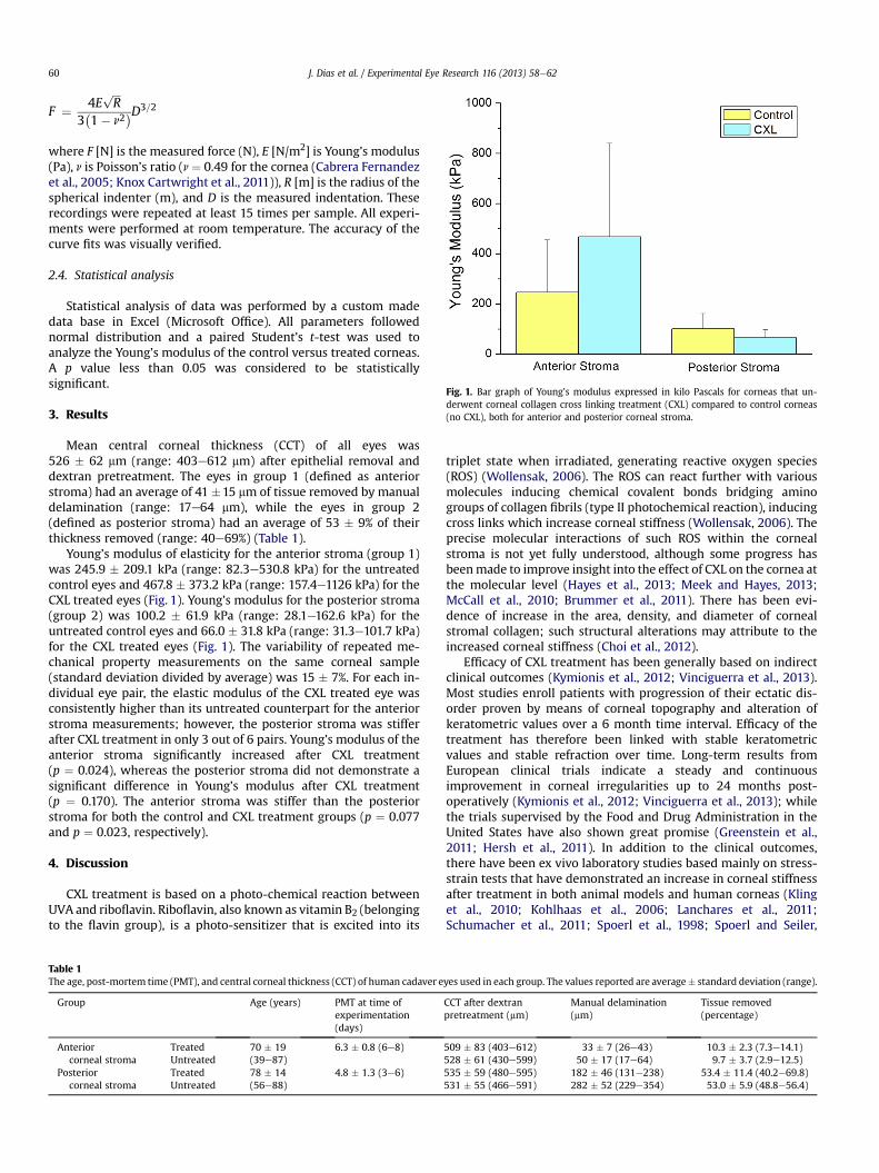
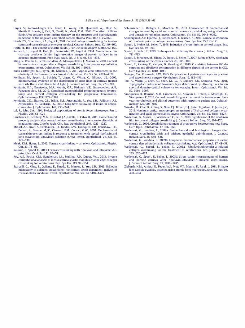
Fig. 1. Bar graph of Young’s modulus expressed in kilo Pascals for corneas that un-derwent corneal collagen cross linking treatment (CXL) compared to control corneas(no CXL), both for anterior and posterior corneal stroma.
ffiffiffip
3 1� n
where F [N] is the measured force (N), E [N/m2] is Young’s modulus(Pa), n is Poisson’s ratio (n ¼ 0.49 for the cornea (Cabrera Fernandezet al., 2005; Knox Cartwright et al., 2011)), R [m] is the radius of thespherical indenter (m), and D is the measured indentation. Theserecordings were repeated at least 15 times per sample. All experi-ments were performed at room temperature. The accuracy of thecurve fits was visually verified.
2.4. Statistical analysis
Statistical analysis of data was performed by a custom madedata base in Excel (Microsoft Office). All parameters followednormal distribution and a paired Student’s t-test was used toanalyze the Young’s modulus of the control versus treated corneas.A p value less than 0.05 was considered to be statisticallysignificant.
3. Results
Mean central corneal thickness (CCT) of all eyes was526 � 62 mm (range: 403e612 mm) after epithelial removal anddextran pretreatment. The eyes in group 1 (defined as anteriorstroma) had an average of 41 �15 mm of tissue removed by manualdelamination (range: 17e64 mm), while the eyes in group 2(defined as posterior stroma) had an average of 53 � 9% of theirthickness removed (range: 40e69%) (Table 1).
Young’s modulus of elasticity for the anterior stroma (group 1)was 245.9 � 209.1 kPa (range: 82.3e530.8 kPa) for the untreatedcontrol eyes and 467.8 � 373.2 kPa (range: 157.4e1126 kPa) for theCXL treated eyes (Fig. 1). Young’s modulus for the posterior stroma(group 2) was 100.2 � 61.9 kPa (range: 28.1e162.6 kPa) for theuntreated control eyes and 66.0 � 31.8 kPa (range: 31.3e101.7 kPa)for the CXL treated eyes (Fig. 1). The variability of repeated me-chanical property measurements on the same corneal sample(standard deviation divided by average) was 15 � 7%. For each in-dividual eye pair, the elastic modulus of the CXL treated eye wasconsistently higher than its untreated counterpart for the anteriorstroma measurements; however, the posterior stroma was stifferafter CXL treatment in only 3 out of 6 pairs. Young’s modulus of theanterior stroma significantly increased after CXL treatment(p ¼ 0.024), whereas the posterior stroma did not demonstrate asignificant difference in Young’s modulus after CXL treatment(p ¼ 0.170). The anterior stroma was stiffer than the posteriorstroma for both the control and CXL treatment groups (p ¼ 0.077and p ¼ 0.023, respectively).
4. Discussion
CXL treatment is based on a photo-chemical reaction betweenUVA and riboflavin. Riboflavin, also known as vitamin B2 (belongingto the flavin group), is a photo-sensitizer that is excited into its
Table 1The age, post-mortem time (PMT), and central corneal thickness (CCT) of human cadaver e
Group Age (years) PMT at time ofexperimentation(days)
Anteriorcorneal stroma
Treated 70 � 19(39e87)
6.3 � 0.8 (6e8)Untreated
Posteriorcorneal stroma
Treated 78 � 14(56e88)
4.8 � 1.3 (3e6)Untreated
triplet state when irradiated, generating reactive oxygen species(ROS) (Wollensak, 2006). The ROS can react further with variousmolecules inducing chemical covalent bonds bridging aminogroups of collagen fibrils (type II photochemical reaction), inducingcross links which increase corneal stiffness (Wollensak, 2006). Theprecise molecular interactions of such ROS within the cornealstroma is not yet fully understood, although some progress hasbeenmade to improve insight into the effect of CXL on the cornea atthe molecular level (Hayes et al., 2013; Meek and Hayes, 2013;McCall et al., 2010; Brummer et al., 2011). There has been evi-dence of increase in the area, density, and diameter of cornealstromal collagen; such structural alterations may attribute to theincreased corneal stiffness (Choi et al., 2012).
Efficacy of CXL treatment has been generally based on indirectclinical outcomes (Kymionis et al., 2012; Vinciguerra et al., 2013).Most studies enroll patients with progression of their ectatic dis-order proven by means of corneal topography and alteration ofkeratometric values over a 6 month time interval. Efficacy of thetreatment has therefore been linked with stable keratometricvalues and stable refraction over time. Long-term results fromEuropean clinical trials indicate a steady and continuousimprovement in corneal irregularities up to 24 months post-operatively (Kymionis et al., 2012; Vinciguerra et al., 2013); whilethe trials supervised by the Food and Drug Administration in theUnited States have also shown great promise (Greenstein et al.,2011; Hersh et al., 2011). In addition to the clinical outcomes,there have been ex vivo laboratory studies based mainly on stress-strain tests that have demonstrated an increase in corneal stiffnessafter treatment in both animal models and human corneas (Klinget al., 2010; Kohlhaas et al., 2006; Lanchares et al., 2011;Schumacher et al., 2011; Spoerl et al., 1998; Spoerl and Seiler,
yes used in each group. The values reported are average� standard deviation (range).
CCT after dextranpretreatment (mm)
Manual delamination(mm)
Tissue removed(percentage)
509 � 83 (403e612) 33 � 7 (26e43) 10.3 � 2.3 (7.3e14.1)528 � 61 (430e599) 50 � 17 (17e64) 9.7 � 3.7 (2.9e12.5)535 � 59 (480e595) 182 � 46 (131e238) 53.4 � 11.4 (40.2e69.8)531 � 55 (466e591) 282 � 52 (229e354) 53.0 � 5.9 (48.8e56.4)
J. Dias et al. / Experimental Eye Research 116 (2013) 58e62 61
1999; Wollensak et al., 2003b; Wollensak and Iomdina, 2009a,2009b).
In our study, we found that corneal collagen crosslinkingtreatment significantly increases corneal stiffness in the anteriorcorneal stromal region only (increased by w1.9 times whencompared to the untreated corneas) while the posterior cornealstroma (as described in our study as approximately 50% of the totalCCT) was not significantly affected by CXL. Since riboflavin serves asthe photosensitizing agent to produce ROS, the ability to produce asignificant biomechanical CXL effect within the stroma is depen-dent upon the concentration of the riboflavin that is able to diffusethrough the stroma (Spoerl et al., 2007; Raiskup and Spoerl, 2013).Studies have investigated the absorption properties of riboflavinand showed that the riboflavin concentration decreases withstromal depth (Wollensak et al., 2010; Spoerl et al., 2010;Sondergaard et al., 2010). Governing the absorption of UV light inthe corneal tissue, the LamberteBeer law states that there is anexponential decrease in UV intensity with increasing depth in thecornea. With the combined effect of riboflavin concentration andthe LamberteBeer law, it would be expected that the induction ofadditional crosslinking bonds within the stroma is not homoge-neous, but rather depth-dependent (Kohlhaas et al., 2006).
Therefore, the results of our study support the theory that themechanical stiffening induced by crosslinking is much more pro-nounced in the anterior stromal region than the posterior. Suchfindings are similar to the qualitative trends found in the studies ofKohlhaas et al. (2006) and Scarcelli et al. (2013), both of whichcharacterized the anterior and posterior stromal regions of cross-linked and normal porcine corneas. In addition, the same trend waspredicted by the finite-element computational model of Roy et al.(2013). All studies agree with this present one that the stiffeningeffect of UV crosslinking is depth-dependent and that the majorityof the stiffening occurs in the anterior stromal region rather than inthe posterior stroma (Kohlhaas et al., 2006; Scarcelli et al., 2013).
Our study assessed the elasticity of normal and crosslinkedhuman cadaver corneas using a custom Atomic Force Microscopy(AFM) system. Conceptually developed by Binnig et al. (1986), AFMhas become an established characterization technique in the arenasof non-biological and biological mechanical testing. AFM technol-ogy enables localized mechanical testing of samples in aqueoussolutions (Karrasch et al., 1994; Lal and John, 1994) and is a suitablecharacterization technique to conduct studies on distinct layerswithin the corneal stroma for depth-dependent analysis. Using thistechnique, standard mechanical properties like Young’s moduluscan be derived by the application of a controlled verticalcompressive force. The force applies stress to the sample, whichwill indent as an indication of the strain experienced. The Young’smodulus obtained from these measurements is therefore definedas the ratio of stress (force) to strain (indentation), along the samevertical axis. Although the versatility of AFM enables the determi-nation of time-dependent properties associatedwith viscoelasticityand poroelasticity, these measurements were not performed in thecurrent study. All experiments were performed with the exactsame experimental parameter settings (i.e., indentation rate andmaximal voltage) and encompassed the application and immediateretraction of maximal force unto the sample. In this particularstudy, we used spherical indenters to probe network layers of thecorneal stroma, as opposed to individual collagen fibers. With thespherical indenter geometry used, the Hertz model was used tocalculate Young’s modulus of elasticity from the force and inden-tation information. However, this model assumes that the sample isisotropic, homogeneous, linearly elastic, and infinitely thick, noneof which accurately describe the cornea because of its heteroge-neous, viscoelastic, and anisotropic nature. Therefore, the use of theHertz model limits our results to only be linear elastic modulus
estimates, as opposed to reflecting the absolute elastic modulusvalues.
It is possible that several experimental constraints may haveaffected the biomechanical measurements obtained. The variationof corneal edema between the samples resulted in a differentinitial total corneal thickness for each sample, despite the 24-hdextran treatment. Although all samples had a thickness withinthe normal physiological range, these slight differences in thick-ness may account for some of the measurement variability be-tween samples. Furthermore, the use of manual delamination toaccess the corneal stromal regions restricted the creation ofreproducible corneal tissue excisions. Consequently, there lied theinability to perform elasticity assessments on the exact sameanterior stromal depths for all the corneas, which can be evidencedby the large range of the amount of stroma removed to expose theanterior stroma (17e64 mm). Previous studies have shown struc-tural differences in the collagen fiber interconnectivity atincreasing depths within the anterior stromal region (Winkleret al., 2011). Although no significant trend was found betweenanterior stromal elastic modulus and stromal depth, such struc-tural differences may have contributed to the variation of elasticmodulus values obtained. Other limitations of this study includethe small sample size of eye pairs used as well as the ex vivoexperimental model, since ex vivo human corneas are affected byfactors such as post mortem, collagen autolysis, stromal swelling,and lack of healing response. The effect that these experimentalconstraints had on the results can be evidenced by the high be-tween sample variability, since the standard deviation of themoduli between samples was on the same order as the mean.Despite the between sample variability, the results for each eyepair consistently showed that the stiffening effect of CXL was sig-nificant in the anterior region but not in the posterior region.
In conclusion, our study validates previous experimental evi-dence of increased corneal stiffness after CXL treatment, using thecharacterization technique of AFM. The results of this studydescribe an anterioreposterior stromal depth dependency con-cerning CXL efficacy.
Financial interest
The authors do not have any proprietary or financial interest inany of the devices presented.
Acknowledgments
Donor human eyes were provided by the Florida Lions Eye Bank.
References
Binnig, G., Quate, C.F., Gerber, C., 1986. Atomic force microscope. Phys. Rev. Lett. 56,930e933.
Borja, D., Manns, F., Lamar, P., Rosen, A., Fernandez, V., Parel, J.M., 2004. Preparationand hydration control of corneal tissue strips for experimental use. Cornea 23,61e66.
Brummer, G., Littlechild, S., McCall, S., Zhang, Y., Conrad, G.W., 2011. The role ofnonenzymatic glycation and carbonyls in collagen cross-linking for the treat-ment of keratoconus. Invest. Ophthalmol. Vis. Sci. 52, 6363e6369.
Cabrera Fernandez, D., Niazy, A.M., Kurtz, R.M., Djotyan, G.P., Juhasz, T., 2005. Finiteelement analysis applied to cornea reshaping. J. Biomed. Opt. 10, 064018.
Choi, S., Lee, S.C., Lee, H.J., Cheong, Y., Jung, G.B., Jin, K.H., Park, H.K., 2012. Structuralresponse of human corneal and scleral tissues to collagen cross-linking treat-ment with riboflavin and ultraviolet A light. Lasers Med. Sci., 1e8.
Dias, J., Ziebarth, N.M., 2013. Anterior and posterior corneal stroma elasticityassessed using nanoindentation. Exp. Eye Res. 115, 41e46.
Duffey, R.J., Tchah, H., Lindstrom, R.L., 1989. Human cadaver corneal thinning forexperimental refractive surgery. Refract. Corneal Surg. 5, 41e42.
Greenstein, S.A., Shah, V.P., Fry, K.L., Hersh, P.S., 2011. Corneal thickness changesafter corneal collagen crosslinking for keratoconus and corneal ectasia: one-year results. J. Cataract Refract. Surg. 37, 691e700.
J. Dias et al. / Experimental Eye Research 116 (2013) 58e6262
Hayes, S., Kamma-Lorger, C.S., Boote, C., Young, R.D., Quantock, A.J., Rost, A.,Khatib, K., Harris, J., Yagi, N., Terrill, N., Meek, K.M., 2013. The effect of Ribo-flavin/UVA collagen cross-linking therapy on the structure and hydrodynamicbehaviour of the ungulate and rabbit corneal stroma. PLoS One 8, e52860.
Hersh, P.S., Greenstein, S.A., Fry, K.L., 2011. Corneal collagen crosslinking for kerato-conus and corneal ectasia: one-year results. J. Cataract Refract. Surg. 37,149e160.
Hertz, H., 1881. The contact of elastic solids. J. Für Die Reine Angew. Mathe. 92, 156.Karrasch, S., Hegerl, R., Hoh, J.H., Baumeister, W., Engel, A., 1994. Atomic force mi-
croscopy produces faithful high-resolution images of protein surfaces in anaqueous environment. Proc. Natl. Acad. Sci. U. S. A. 91, 836e838.
Kling, S., Remon, L., Perez-Escudero, A., Merayo-Lloves, J., Marcos, S., 2010. Cornealbiomechanical changes after collagen cross-linking from porcine eye inflationexperiments. Invest. Ophthalmol. Vis. Sci. 51, 3961e3968.
Knox Cartwright, N.E., Tyrer, J.R., Marshall, J., 2011. Age-related differences in theelasticity of the human cornea. Invest. Ophthalmol. Vis. Sci. 52, 4324e4329.
Kohlhaas, M., Spoerl, E., Schilde, T., Unger, G., Wittig, C., Pillunat, L.E., 2006.Biomechanical evidence of the distribution of cross-links in corneas treatedwith riboflavin and ultraviolet A light. J. Cataract Refract. Surg. 32, 279e283.
Kymionis, G.D., Grentzelos, M.A., Kounis, G.A., Diakonis, V.F., Limnopoulou, A.N.,Panagopoulou, S.I., 2012. Combined transepithelial phototherapeutic keratec-tomy and corneal collagen cross-linking for progressive keratoconus.Ophthalmology 119, 1777e1784.
Kymionis, G.D., Siganos, C.S., Tsiklis, N.S., Anastasakis, A., Yoo, S.H., Pallikaris, A.I.,Astyrakakis, N., Pallikaris, I.G., 2007. Long-term follow-up of intacs in kerato-conus. Am. J. Ophthalmol. 143, 236e244.
Lal, R., John, S.A., 1994. Biological applications of atomic force microscopy. Am. J.Physiol. 266, C1eC21.
Lanchares, E., del Buey, M.A., Cristobal, J.A., Lavilla, L., Calvo, B., 2011. Biomechanicalproperty analysis after corneal collagen cross-linking in relation to ultraviolet Airradiation time. Graefes Arch. Clin. Exp. Ophthalmol. 249, 1223e1227.
McCall, A.S., Kraft, S., Edelhauser, H.f., Kidder, G.W., Lundquist, R.R., Bradshaw, H.E.,Dedeic, Z., Dionne, M.J.C., Clement, E.M., Conrad, G.W., 2010. Mechanisms ofcorneal tissue cross-linking in response to treatment with topical riboflavin andlong wavelength ultraviolet radiation (UVA). Invest. Ophthalmol. Vis. Sci. 51,129e138.
Meek, K.M., Hayes, S., 2013. Corneal cross-linking e a review. Ophthalmic. Physiol.Opt. 33, 78e93.
Raiskup, F., Spoerl, E., 2013. Corneal crosslinking with riboflavin and ultraviolet A. I.principles. Ocul. Surf. 11, 65e74.
Roy, A.S., Rocha, K.M., Randleman, J.B., Stulting, R.D., Dupps, W.J., 2013. Inversecomputational analysis of in vivo corneal elastic modulus change after collagencrosslinking for keratoconus. Exp. Eye Res. 113, 92e104.
Scarcelli, G., Kling, S., Quijano, E., Pineda, R., Marcos, S., Yun, S.H., 2013. Brillouinmicroscopy of collagen crosslinking: noncontact depth-dependent analysis ofcorneal elastic modulus. Invest. Ophthalmol. Vis. Sci. 54, 1418e1425.
Schumacher, S., Oeftiger, L., Mrochen, M., 2011. Equivalence of biomechanicalchanges induced by rapid and standard corneal cross-linking, using riboflavinand ultraviolet radiation. Invest. Ophthalmol. Vis. Sci. 52, 9048e9052.
Sondergaard, A.P., Hjortdal, J., Breitenbach, T., Ivarsen, A., 2010. Corneal distributionof riboflavin prior to collagen cross-linking. Curr. Eye Res. 35, 116e121.
Spoerl, E., Huhle, M., Seiler, T., 1998. Induction of cross-links in corneal tissue. Exp.Eye Res. 66, 97e103.
Spoerl, E., Seiler, T., 1999. Techniques for stiffening the cornea. J. Refract. Surg. 15,711e713.
Spoerl, E., Mrochen, M., Sliney, D., Trokel, S., Seiler, T., 2007. Safety of UVA-riboflavincross-linking of the cornea. Cornea 26, 385e389.
Spoerl, E., Raiskup, F., Kampik, D., Geerling, G., 2010. Correlation between UV ab-sorption and riboflavin concentration in different depths of the cornea in CXL.Curr. Eye Res. 35, 1040e1041.
Swinger, C.A., Kornmehl, E.W., 1985. Dehydration of post-mortem eyes for practiceand experimental surgery. Ophthalmic. Surg. 16, 182e183.
Tao, A., Wang, J., Chen, Q., Shen, M., Lu, F., Dubovy, S.R., Shousha, M.A., 2011.Topographic thickness of Bowman’s layer determined by ultra-high resolutionspectral domaineoptical coherence tomography. Invest. Ophthalmol. Vis. Sci.52, 3901e3907.
Vinciguerra, R., Romano, M.R., Camesasca, F.I., Azzolini, C., Trazza, S., Morenghi, E.,Vinciguerra, P., 2013. Corneal cross-linking as a treatment for keratoconus: four-year morphologic and clinical outcomes with respect to patient age. Ophthal-mology 120, 908e916.
Winkler, M., Chai, D., Kriling, S., Nien, C.J., Brown, D.J., Jester, B., Juhasz, T., Jester, J.V.,2011. Nonlinear optical macroscopic assessment of 3-d corneal collagen orga-nization and axial biomechanics. Invest. Ophthalmol. Vis. Sci. 52, 8818e8827.
Wollensak, G., Aurich, H., Wirbelauer, C., Sel, S., 2010. Significance of the riboflavinfilm in corneal collagen crosslinking. J. Cataract Refract. Surg. 36, 114e120.
Wollensak, G., 2006. Crosslinking treatment of progressive keratoconus: new hope.Curr. Opin. Ophthalmol. 17, 356e360.
Wollensak, G., Iomdina, E., 2009a. Biomechanical and histological changes aftercorneal crosslinking with and without epithelial debridement. J. CataractRefract. Surg. 35, 540e546.
Wollensak, G., Iomdina, E., 2009b. Long-term biomechanical properties of rabbitcornea after photodynamic collagen crosslinking. Acta Ophthalmol. 87, 48e51.
Wollensak, G., Spoerl, E., Seiler, T., 2003a. Riboflavin/ultraviolet-a-inducedcollagen crosslinking for the treatment of keratoconus. Am. J. Ophthalmol.135, 620e627.
Wollensak, G., Spoerl, E., Seiler, T., 2003b. Stress-strain measurements of humanand porcine corneas after riboflavin-ultraviolet-A-induced cross-linking.J. Cataract Refract. Surg. 29, 1780e1785.
Ziebarth, N.M., Arrieta, E., Feuer, W.J., Moy, V.T., Manns, F., Parel, J., 2011. Primatelens capsule elasticity assessed using atomic force microscopy. Exp. Eye Res. 92,490e494.