angiology: open access - omics international · repeat revascularization after percutaneous...
TRANSCRIPT
Recent Perspectives on Left Main Bifurcation InterventionsDebabrata Dash*
Nanavati Superspeciality Hospital, Mumbai, India*Corresponding author: Debabrata Dash, Nanavati Superspeciality Hospital, Mumbai, India, Tel: 912266529666; E-mail: [email protected]
Received date: July 19, 2016; Accepted date: August 10, 2016; Published date: August 17, 2016
Copyright: © 2016 Dash D, This is an open-access article distributed under the terms of the Creative Commons Attribution License, which permits unrestricted use,distribution, and reproduction in any medium, provided the original author and source are credited.
Abstract
For several decades, coronary bypass grafting has been considered as the gold standard treatment ofunprotected left main (LM) disease. However, because of large vessel caliber and anatomic accessibility,percutaneous coronary intervention (PCI) for LM has been attractive option for interventional cardiologists. PCI ofLM bifurcation can be technically demanding that warrants reinforcement of integrated approach combiningadvanced devices, tailored techniques, adjunctive support of physiologic evaluation, and adjunctive pharmacologicagents. The provisional one -stent approach has shown more favourable outcome than two-stent technique, makingthe former the preferred strategy in most types of LM bifurcation lesions. In complex lesions, two-stent techniquemay be required and may yield superior results. Selecting the proper strategy using intravascular ultrasound forostium of the side branch (SB) is critical for reducing the risk for SB occlusion and for improving patient's outcome.Even unnecessary complex interventions can be deferred by measuring fractional flow reserve in angiographicisolated SB. Importantly, final successful procedure is more important than the type of stenting technique.,emphasizing the greater importance of optimizing the selected technique rather than choice of method. Alongsidethe evolution of bifurcation techniques, there has been development of several dedicated bifurcation stents whichare safe and effective in LM bifurcation PCI both at short and mid-term follow up.
Keywords: Left main coronary artery; Percutaneous coronaryinterventions; Bifurcation lesions
IntroductionSignificant unprotected LM disease constitutes approximately
5-10% of patients undergoing coronary angiography [1,2].Randomized clinical trials (RCTs) have demonstrated a higher rate ofrepeat revascularization after percutaneous coronary intervention(PCI) compared with coronary artery bypass grafting (CABG), but alower incidence of cerebrovascular events; no differences werereported in overall major adverse cardiovascular events (MACEs)[3-8]. Although CABG has been the gold standard therapy for LMdisease until recently, significant innovation in stent technology,revascularization techniques and antithrombotic therapies make PCIfeasible [9]. Treatment of ostial and mid-shaft has shown excellentoutcomes with minimal mortality and long-term complicationscompared with distal LM bifurcations [6]. Lack of RCTs addressingLM bifurcation has led to uncertainties regarding optimal stentingstrategy. Although the provisional one-stent technique has been thedefault strategy based on non-randomized studies and extrapolationsfrom results of non- LM bifurcation trials, two-stent techniques areselected more frequently for LM bifurcation than non-LM [10]. Issuesrelated to bifurcation PCI are common in practice and strategies toovercome will continue to evolve. This review therefore discussesvarious stenting techniques to manage LM bifurcation lesions.
Anatomy and Physiology of LMAs the LM supplies approximately two-thirds of the blood to the
heart and 100% to the left ventricle, severe LM disease would reduceflow to significant areas of the myocardium, placing the patient at highrisk for life–threatening LV dysfunction and arrhythmias. 11 It is a
large artery and therefore tends to have a high plaque volume. It also isprone to calcification. Plaque shift and incomplete stent expansion aretherefore important technical considerations in stenting of LM [11].
The distal LM, by definition always ends in a bifurcation, or eventrifurcation, giving rise to the left anterior descending (LAD) and leftcircumflex (LCX) arteries, and probably an intermedius artery. Greaterelastic tissue content of this artery explains elastic recoil and highrestenosis following balloon angioplasty [11,12]. Seventy percent ofsignificant LM lesions involve the bifurcation. Intimal atherosclerosisin this location is accelerated primarily in area of low shear stress alongthe lateral wall extending distally on the myocardial walls of the LADand LCX arteries. Involvement of flow divider (carina) is minimal orabsent. A long LM (≥10 mm) has more pressure drop and lower shearstress contributing to plaque formation [13]. The current trend to treatdistal LM bifurcation by extending the main vessel stent into theproximal LAD is supported by continuous extension of plaque fromLM to proximal LAD artery in 90% of cases [14]. The LM typically hasa diameter ranging between 4.5-6 mm in a majority of cases while theLAD and LCX have diameters ranging from 3.5-4.5 mm and 3.0-4.5mm respectively. The take- off angulation of LCX is greater than 90° inmore than 70% of the patients. The size discrepancy and take-off angleof LCX have great implications for LM bifurcation stenting [15].
Patient selectionTraditionally patients referred for LM PCI are those who have been
turned down for CABG because of excessive surgical risk such as poorLV function, porcelain aorta, advanced age, poor distal targets and thepresence of severe co-morbidities. Patients with ostial or mid-shaftdisease have improved clinical outcomes compared with patients withLM bifurcation and trifurcation after PCI. Similarly, the extent andcomplexity of concomitant disease in other coronary arteries portendsincreased risk. Chronic total occlusion of right coronary artery has
Angiology: Open Access Dash, Angiol 2016, 4:3DOI: 10.4172/2329-9495.1000181
Research Article Open Access
Angiol, an open access journalISSN:2329-9495
Volume 4 • Issue 3 • 1000181
higher risk of adverse outcomes compared to one without. Whenstratified by score, the 5-year incidence of major adverse cardiac orcerebrovascular events (MACCE) in patients with LM disease wassimilar between groups with low (<23) and intermediate (23-33)SYNTAX (Synergy Between PCI with TAXUS and Cardiac Surgery)scores, continuing the trend noted at 12 months within the LM diseasecohort.8 Therefore, the SYNTAX score continues to be an importanttool in the LM disease evaluation and suggests that patients with lowor intermediate scores have similar long-term outcomes with PCI orCABG. In addition, the SYNTAX data demonstrate a significantlylower rate of stroke in the PCI group at 1 year and maintain a trend at5 years [8,16]. Min et al found that a EuroScore ≥ 6 was anindependent predictor of deaths in patients undergoing PCI or CABGfor LM disease in MAIN-COMPARE (Revascularization forUnprotected Left main Coronary Artery Stenosis; Comparison ofPercutaneous Coronary Angioplasty Versus SurgicalRevascularization) registry [17]. Combining the SYNTAX and theEuroSCORE (European system for cardiac operative risk evaluation)into a common risk model (Global Risk Classification) was correlatedwith a significant improvement in predicting cardiac mortality inpatients undergoing LM PCI [18]. Another score, the NERS (New RiskStratification Score) demonstrated a higher sensitivity and specificityto predict clinical outcome [19]. Similarly, fractional flow reserve(FFR) has been integrated in addition to SYNTAX score. Nam et al.[20], demonstrated 'Functional SYNTAX Score' (FSS) to be betterpredictive accuracy for MACE compared with the traditional SYNTAXscore. The author feels that following are the group of patients withunprotected LM disease that are likely to have favourable clinicaloutcomes with PCI as that of CABG [9].
Ostial and/or mid-shaft LM disease
• Isolated LM disease• LM disease plus single-vessel disease• LM bifurcational disease treatable by single stent approach• Low or intermediate Syntax score (Syntax score<33)
Choosing a LM Bifurcation TreatmentDistal LM lesions are mostly treated as true bifurcation. The
exception to this is when one branch is small (usually the LCX), whenone branch is chronically occluded or if protected by a patent graft. AsLCX supplies large myocardial territory in many patients, thepossibility of circulatory collapse after LM-LAD stenting should bekept in mind. Therefore, the presence or absence of significant diseasein LCX ostium is considered as an important factor in selecting astenting strategy. The provisional one- stent approach is preferred forLM bifurcations with insignificant LCX ostial stenosis or a non-dominant left coronary artery (LCA). In contrast, the elective two-stentapproach may have to be employed in patients with significant stenosisof the osium of the LCX with dominant LCA (Table 1) [21,22]. FFRevaluation for the side branch (SB) can be used to make correct choiceof the treatment strategy [23]. If the LCX is either occluded, itsdiameter is less than 2.5 mm, it can be ignored and a stent can beplaced between the LM and the LAD [24]. A guidewire kept in a smallLCX may help to maintain flow after a single stent is placed across theostium. For a non-diseased LCX ostium, if the angle of bifurcation is ofT shape, it is the operator’s choice to place a protective guidewire but itmay not be necessary. However, if the bifurcation angle is of Y shape, aprotective wire is recommended. For a significant and diseased LCXostium, there are several techniques depending on the bifurcationangle. If it is of T shape, the T-stent, mini-crush, double kiss (DK)
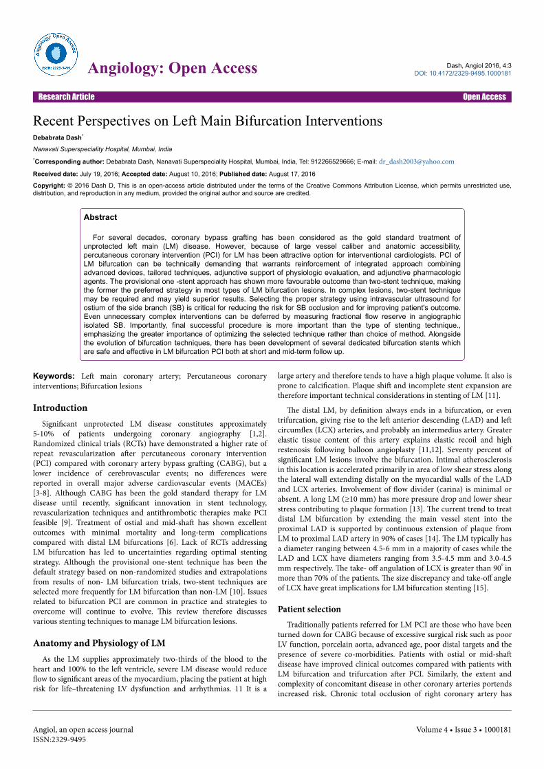
crush or T and protrusion (TAP) stent technique is recommendedwhereas if the angle is of Y shape, the culotte, mini-crush or DK crushtechnique is recommended, while T stenting is not (Figure 1) [9,11].
As LM bifurcation disease is mostly diffuse and not focal,angiography may be inaccurate in assessing the disease severity of bothbranch ostia [25]. Thus angiography-guided PCI may lead to SBocclusion for a "true" bifurcation resulting in unnecessary complexintervention that may be preventable. Preprocedural intravascularultrasound (IVUS) may be quite useful in selecting an appropriate andsafe strategy for LM bifurcation PCI as it throws light on disease statusof distal LM complex including the LCX ostium (Figure 2). Han et aldemonstrated that IVUS reduced the rate of SB occlusion after MBstenting in bifurcation [21]. Moreover IVUS-guided LM PCI has beenassociated with reduced mortality [26,27]. IVUS-derived minimallumen>3.7 mm2 or plaque burden<56% in the LCX ostum couldexclude functional SB compromise ( FFR<0.80) after MB stenting inLM bifurcations [28].
Preference for the provisional one-stent approach
• Small LCX<2.5 mm in diameter
• No or insignificant LCX ostial disease
• Lesion in ostial LCX extending<5 mm
• Diminutive LCX, right dominant coronary system
• Wide angle between LAD and LCX
• No significant ostial LCX disease by IVUS (MLA>4 mm2 and PB<50%)
Preference for two-stent technique
• Significant and long (>5 mm) lesion in ostial LCX
• Complex lesion in LCX ostium
• Large LCX ≥ 2.5 mm in diameter
• Diseased left dominant coronary system
• Narrow angle between LAD and LCX
• Significant LCX ostial disease by IVUS ( MLA<4 mm2 and PB>50%
• Poor result after provisional stenting
Stenosis>75%
Reduced flow (TIMI flow <III)
Dissection
Ostial LCX with MLA<4 mm2 after cross-over stenting.
Table 1: Recommendation for provisional one-stent or two-stentapproach to LM bifurcation PCI.
The provisional one-stent techniqueThis is a single–stent strategy allowing the positioning of a second
stent if required (T, TAP, inverted culotte technique). The LAD andLCX are wired. A stent is deployed from LM to the LAD. It should besized according to distal main branch (MB) reference for drug-elutingstents (DES) or 0.25 mm larger for bioabsorbable vascular scaffolds(BVS) based on expert consensus. Post dilatation of BVS beyond 0.5mm larger than their size may result in strut fractures. The proximaloptimisation technique (POT) using short oversized non-compliantballoon should be employed just before carina to ensure adequate stentapposition in proximal MB (LAD). The POT might be used to avoidabluminal rewiring by a second wire [29]. The guidewires are thenexchanged, the LAD wire can be withdrawn and passed through thestent struts to the LCX, and the “jailed” wire in the LCX is withdrawn
Citation: Dash D (2016) Recent Perspectives on Left Main Bifurcation Interventions. Angiol 4: 181. doi:10.4172/2329-9495.1000181
Page 2 of 9
Angiol, an open access journalISSN:2329-9495
Volume 4 • Issue 3 • 1000181
and advanced to the LAD. Final kissing balloon inflation ( FKBI) maybe performed in significant ostial SB lesions (TIMI flow<3 or
FFR<0.8). Predilatation of LCX in most cases probably not needed, butmay be considered.
Figure 1: Algorithm for left main (LM) bifurcation percutaneous coronary intervention (PCI)* In general minimal lumen area>4 mm2 orplaque burden<50% of LCX ostium is considered in significant stenosis. †Adequate stent apposition and expansion to avoid restenosis (MSA:5 mm2 LCX ostium, 6 mm2 for proximal left anterior descending artery, 7 mm2 for polygon of confluence, and 8 mm2 for distal left main)without procedural complications. LM: Left Main; PCI: Percutaneous coronary Intervention; IVUS: Intravascular Ultrasound; LAD: LeftAnterior Descending artery; LCX: Left Circumflex artery; MB: Main Branch; SB: Side Branch; TAP: T Aand Protrusion; DK crush: DoubleKiss Crush.
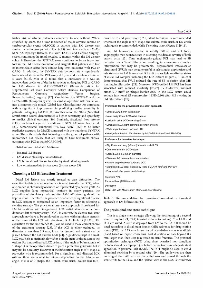
The T and protrusion (TAP) stentingThis modification of T stenting technique can be used in majority of
the bifurcation lesions especially when the bifurcation angle is lessthan 90°. It can provide good reconstruction of distal LM bifurcationwith minimal stent overlap [9,11]. The LM-LAD is stented jailing theLCx guidewire. Kissing balloon inflation is performed after rewiringthe LCX. After positioning the proximal edge of the LCX stent 1-2 mminside the LAD stent, the LCX stent is deployed at high pressure withdeflated balloon kept in the LAD stent. Then, the LCX balloon isslightly retrieved and aligned to the LAD balloon. Afterwards, a FKBIis performed in order to reconstruct the carina (Figure 3).
The culotte stentingThis technique is suitable for lesions where the angulation between
LAD and LCX is <60°and two vessel are of similar diameter. The moreangulated branch usually the LM-LCX is stenetd. The LAD is rewired
through the stent struts and dilated. A second stent is advancedthrough the struts of the first into the LAD. The LM-LAD stent is thendeployed. Each limb of the culotte is dilated at high pressure usingnon-compliant balloon followed by FKBI at medium pressure. Incontemporary culotte stenting, POT is recommended after first andsecond stent deployment, as well as a final POT after kissing ballooninflation. It is advisable to avoid a long overlap of stents in theproximal MB whenever possible (mini-cullote). This technique ensuresnear-perfect coverage of the carina and the LCX ostium. The maindisadvantage of the technique is that rewiring both branches throughstent struts can be technically demanding, and time-consuming. Open-cell stents are preferred for this technique.
The classical T stentingThis technique is performed when the angle between the two vessels
is close to 90°.
Citation: Dash D (2016) Recent Perspectives on Left Main Bifurcation Interventions. Angiol 4: 181. doi:10.4172/2329-9495.1000181
Page 3 of 9
Angiol, an open access journalISSN:2329-9495
Volume 4 • Issue 3 • 1000181

Figure 2: Integrated use of intravascular ultrasound (IVUS) and fractional flow reserve (FFR) in LM bifurcation PCI, A. Coronaryangiography (CAG) showed "true" LM bifurcation lesion ( Medina 1,1,1), B. IVUS revealing significant left anterior descending artery (LAD)disease, C. IVUS showing very minimal disease at left circumflex artery (LCX) ostium, D. Provisional stenting of LM-LAD. E. Proximaloptimization technique (POT) with non compliant short bigger balloon, F. Post stenting FFR of LCX: 0.90, G. IVUS demonstrating minimalstent area (MSA) of 6.5 mm2 at LAD ostium, H. IVUS interrogation of LCX showing MSA of 4.2 mm2 at ostium.
After placing the wires in both the LAD and LCX, a stent isdeployed in LCX, making sure to cover the ostium with minimalprotrusion into the LAD. The LM- LAD lesion is then stented. TheLCX is rewired and dilated followed by FKBI [9,11]. This technique isassociated with the risk of leaving a small gap between the branchesleading to restenosis at the LCX ostium . For this reason, this techniquehas largely been replaced by the modified T stenting technique.
The modified T stentingThe modified T stenting is performed by simultaneously positioning
stents at LCX and LAD with the LCX stent minimally protruding intothe LAD, when the angulations between the branches approach 90°.The LCX stent is deployed first, and then after guidewire and balloonremoval from this branch, the LAD stent is deployed. The procedure iscompleted with FKBI [9,11].
The mini-crush stentingThe mini-crush technique is suitable for LM bifurcation when LAD
diameter exceeds that of LCX and the angle between them is less than
60°. The immediate patency of both branches is assured making thistechnique useful in conditions of instability or complex anatomy. Thistechnique provides excellent coverage of the LCX ostium. The mini-crush technique can be used in almost all true bifurcation lesionsexcept in wide angled bifurcations. The main disadvantage is that inorder to perform FKBI, there is need to re-cross multiple struts withwire and a balloon [9,11]. The SB stent is positioned in the LCX,followed by advancement of the LAD stent. The LCX stent is pulledback into the LAD about 1-2 mm and is deployed. The deployment ofLAD stent crushes the proximal LCX stent against the LM wall. TheLCX is rewired through the stent struts of both LAD and crushed LCXstent to perform FKBI.
The double kiss (DK) crush stentingA stent is placed into the LCX and a balloon placed in LM-LAD. 1-2
mm of the LCX stent is positioned in the LM. The LCx stent isdeployed and then the guidewire and balloon from the LCX areremoved. The prepositioned balloon in LM-LAD is inflated to crushthe protruding segment of the LCX stent against the LM wall.
Citation: Dash D (2016) Recent Perspectives on Left Main Bifurcation Interventions. Angiol 4: 181. doi:10.4172/2329-9495.1000181
Page 4 of 9
Angiol, an open access journalISSN:2329-9495
Volume 4 • Issue 3 • 1000181
Figure 3: T and Protrusion (TAP) technique, A. Baseline CAG revealing significant LM bifurcation lesion involving proximal LAD and LCX( Medina 1, 1, 1), B. Direct stenting of LM- LAD with jailed guidewire in LCX, C. Positioning of LCX stent with minimal protrusion into LM-LAD stent, D. Final kissing balloon inflation ( FKBI) after deployment of LM-LCX stent, E. Final result.
The balloon is removed and a stent is deployed in the LM-LADsegment. The wire is then recrossed into the LCX and FKBI is appliedto finish the procedure (Figure 4). As a result, the DK crush techniqueconsists of five steps: side branch-stenting, balloon-crush, first-kissing,second-crush, and FKBI This technique results in less stent distortion,improved stent apposition, and facilitate FKBI. It may be superior toclassic crushing optimizing acute procedural results and possiblyimproves clinical outcomes by facilitating FKBI [11,30]. DK-CRUSH IIis the only randomized trial to suggest that double stenting may besuperior to provisional stenting and associated with a lower rate ofrestenosis and repeat revascularization [11,31]. DK-CRUSH III studydemonstrated that among patients with bifurcation angle ≥ 70°, NERSscore ≥ 20, and SYNTAX score ≥ 23, the 1-year MACE rate in the DKgroup was significantly less compared to the Culotte group [32].
The V and the simultaneous kissing stent (SKS)The V stenting is performed by placing and deploying two stents
together in narrow angled bifurcation [11,33]. Guidewires are placedin both the LAD and LCX and, with or without predilatation, The twostents are placed into LM and the respective branches and deployed bysimultaneous inflation (Figure 5). The author is not a proponent of SKSthat allows a variable amount of protrusion creating rather long doublebarrel. V stenting is relatively easy and fast and thus an ideal in
emergencies. It is indicated in patients with a short LM free diseaseand critical disease of both the LAD and LCX ostia [11].
Dedicated bifurcation stents (DBS)Several DBS have been adopted recently for LM disease [34]. These
devices offer common advantages over conventional DES to cover LMbifurcation segment. Their design confirms to natural anatomy of thebifurcation and can facilitate more effective SB ostial scaffolding.Furthermore, DBS provides easier and quicker access to MB and SBthereby lowering the risk of SB closure. Stenting of LM with thesedevices is safe and effective both at short and mid-term follow up[35-38]. As the anatomy of LM bifurcation varies considerably, furtherstudies are required to define their role in this subset of patients.
Drug-eluting balloon (DEB)The risk of restenosis is significantly reduced with DEB technology
by delivering cytostatic drug to reduce neo-intimal hyperplasia. Thereis no significant advantage of DEB over DES in bifurcation lesions;thus DEB may be considered in patients not eligible for CABG andwith the need for shorter dual antiplatelet therapy (DAPT). There is acase report of kissing DEB successfully treating LM bifurcation DESstent restenosis [39].
Citation: Dash D (2016) Recent Perspectives on Left Main Bifurcation Interventions. Angiol 4: 181. doi:10.4172/2329-9495.1000181
Page 5 of 9
Angiol, an open access journalISSN:2329-9495
Volume 4 • Issue 3 • 1000181
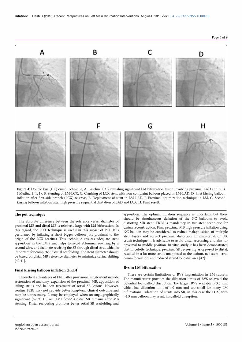
Figure 4: Double kiss (DK) crush technique, A. Baseline CAG revealing significant LM bifurcation lesion involving proximal LAD and LCX( Medina 1, 1, 1), B. Stenting of LM-LCX, C. Crushing of LCX stent with non complaint balloon placed in LM-LAD, D. First kissing ballooninflation after first side branch (LCX) re-cross, E. Deployment of stent in LM-LAD, F. Proximal optimization technique in LM, G. Secondkissing balloon inflation after high pressure sequential dilatation of LAD and LCX, H. Final result.
The pot techniqueThe absolute difference between the reference vessel diameter of
proximal MB and distal MB is relatively large with LM bifurcation. Inthis regard, the POT technique is useful in this subset of PCI. It isperformed by inflating a short bigger balloon just proximal to theorigin of the LCX (carina). This technique ensures adequate stentapposition in the LM stem, helps to avoid abluminal rewiring by asecond wire, and facilitate rewiring the SB through distal strut which isimportant for complete SB ostial scaffolding. The stent diameter shouldbe based on distal MB reference diameter to minimize carina shifting[40,41].
Final kissing balloon inflation (FKBI)Theoretical advantages of FKBI after provisional single-stent include
restoration of anatomy, expansion of the proximal MB, apposition ofjailing struts and balloon treatment of ostial SB lesions. However,routine FKBI may not provide better long-term clinical outcome andmay be unnecessary. It may be employed when an angiographicallysignificant (>75% DS or TIMI flow<3) ostial SB remains after MBstenting. Distal recrossing promotes better ostial SB scaffolding and
apposition. The optimal inflation sequence is uncertain, but thereshould be simultaneous deflation of the NC balloons to avoiddistorting MB stent. FKBI is mandatory in two-stent technique forcarina reconstruction. Final proximal MB high pressure inflation usingNC balloon may be considered to reduce malapposition of multiplestrut layers and correct proximal distortion. In mini-crush or DKcrush technique, it is advisable to avoid distal recrossing and aim forproximal to middle position. In vitro study it has been demonstratedthat in culotte technique, proximal SB recrossing as opposed to distal,resulted in a lot more struts unapposed at the ostium, neo stent -strutcarina formation, and reduced strut-free ostial area [42].
Bvs in LM bifurcationThere are certain limitations of BVS implantation in LM subsets.
The manufacturer provides the dilatation limits of BVS to avoid thepotential for scaffold disruption. The largest BVS available is 3.5 mmwhich has dilatation limit of 4.0 mm and too small for many LMbifurcations. Dilatation of struts into SB, in this case the LCX, with>2.5 mm balloon may result in scaffold disruption.
Citation: Dash D (2016) Recent Perspectives on Left Main Bifurcation Interventions. Angiol 4: 181. doi:10.4172/2329-9495.1000181
Page 6 of 9
Angiol, an open access journalISSN:2329-9495
Volume 4 • Issue 3 • 1000181

Figure 5: V stent technique, A. Baseline CAG showing significant lesion at LM bifurcation, B. Implantation of two stents in LAD and LCXsimultaneously, C. Final result after final kissing balloon inflation.
When the LCX is larger than 2.5 mm and needs treatment at theostium, BVS on the LM may not be ideal. The author feels that apossible compromise is to expand 3.5 mm BVS to 4.0 and avoidmalapposition. One should focus on the maximal lumen diameterrather than vessel diameter to avoid incomplete apposition of scaffolds,and reconsider whether BVS is suitable for the LM. The currentrecommendation is to use provisional stenting in the majority of cases,with sequential noncompliant balloon inflations in the SB and thenMB, reserving mini FKBI (snuggle balloon dilatation) only for cases inwhich it is necessary [43]. POT is recommended with 0.5 mm largerNC balloon. If SB is compromised, an undersized NC balloon (≤2.5mm) is used to open a cell and POT is performed with larger balloonin proximal MB to correct scaffold malapposition (the sequentialstrategy: POT+SB opening+final POT) [44,45]. A final OCT pullbackin the MB should be done to assure adequate strut apposition in thedistal and proximal segment. T or TAP stenting with a metal DES inthe SB is preferable in case of crossover. Unlike the crush or culottetechnique, V-stenting does not deform the BVS struts and is feasible inMedina 0, 1, 1 LM bifurcation [46]. Before a firm recommendation onclinical application can be made, all two-stent techniques wouldrequire further evaluation in clinical trials.
Intracoronary image guided optimizationIVUS is considered to be a useful modality in selecting treatment
strategy, and helpful in optimally expanding the stent, with or withoutpost-stent balloon dilatation, to avoid under- or overstretch of the stentdiameter, and might contribute to better long-term outcomes ascompared with conventional angiography guidance [26]. Stent under-expansion is the most important cause of DES failure. A minimal stentarea (MSA) less than 5.0-5.5 mm2 is the best predictor of firstgeneration DES restenosis and early thrombosis [47,48]. The optimalIVUS-MSA criteria for in-stent restenosis (ISR) were assessed in 403patients undergoing DES implantation for LM PCI. The cut-off valuesfor MSA predicting angiographic restenosis on a segmental basis were5.0 mm2 for LCX ostium, 6.3 mm 2 for LAD ostium, 7.2 mm2 forpolygon of confluence (POC), and 8.2 mm2 for the proximal LMabove POC [49]. Underexpansion was more significantly frequent in
the two-stent technique, the LCX ostium being the most common siteof under-expansion. A smaller IVUS-MSA predicted angiographic ISR9 months after DES implantation to treat LM disease, and post PCIunder-expansion was an independent predictor of 2 year majoradverse cardiac events, especially repeat revascularization [49]. Asubgroup analysis from the MAIN- COMPARE registry demonstratesreduced mortality with IVUS as compared to angiography guidance[26] Another recent IVUS study demonstrated that the incidence ofthe composite of cardiac death, myocardial infarction, and target lesionrevascularization and stent thrombosis are lower in IVUS-guidedgroup [27].
Frequency domain OCT offers superior resolution and can identifystent malapposition, edge dissections, tissue protrusion, and thrombusmore clearly than IVUS [50]. It has been demonstrated that OCT-guided optimization of LM PCI is feasible and safe [51,52]. As bloodmust be adequately replaced by the contrast for clear image,assessment of LM ostium or a relatively large LM is often problematic.Furthermore, since there is no standardized OCT criteria for stentoptimization, this modality may not be useful to guide LM bifurcationPCI. Nevertheless, with accumulating experience and data, it isexpected that OCT will be an important adjunctive tool for LM PCI.
Application of FFRIt may be reasonable to defer LMCA PCI in patients with an
FFR>0.80. In presence of concomitant lesions in both the LAD andLCx without repairing the downstream lesions, the FFR mayunderestimate the true significance of the LM lesion [9]. There may bea discrepancy stenting. One study reports that the need for PCI of theostial LCX after LM-LAD crossover stenting may be reduced, if guidedby FFR [53]. Given the nearly identical one-year MACE rates withboth approaches in DKCRUSH-VI trial, either the angiography-guidedor FFR-guided technique may be recommended for provisional sidebranch stenting of true bifurcation lesions [54]. However, furtherstudies are needed to evaluate the efficacy of this strategy.
Citation: Dash D (2016) Recent Perspectives on Left Main Bifurcation Interventions. Angiol 4: 181. doi:10.4172/2329-9495.1000181
Page 7 of 9
Angiol, an open access journalISSN:2329-9495
Volume 4 • Issue 3 • 1000181
ConclusionCABG remains the optimal treatment for majority of LM lesions.
However, there have been emerging indications and growing trend infavour of PCI in the past few years. This has been supported by currentevidences from clinical trials and large off-label experience updatingcurrent guideline. LM bifurcational lesions continue to poseconsiderable challenges and require expertise and performance ofunique approaches for optimal results. Future randomized studies suchas EXCEL (Evaluation of XIENCE PRIME or XIENCE V Everolimus-Eluting Stent System Versus Coronary Artery Bypass Surgery forEffectiveness of Left Main Revascularization) (NCT01205776) andNOBLE (Nordic-Baltic British Left Main Revascularization)(NCT01496651) are awaited to definitely assess the long- termoutcomes of PCI as compared to CABG. The single LM-LAD stentwith provisional LCX stenting strategy should be the first-linetreatment. Incorporation of FFR-guided PCI strategy may help avoidunnecessary SB interventions. In certain situations or important largeLCX lesions, complex two-stent strategy may be necessary. Meticulousevaluation of LM bifurcations with intracoronary imaging is critical inchoosing the proper stenting technique and in achieving optimizedstent result. There are on-going researches and innovative technologiesthat would further define role of BVS in LM bifurcation PCI.
References1. Stone P, Goldschlager N (1979) Left main coronary artery disease: review
and appraisal. Cardiovasc Med 4: 165-177.2. Demots H, Rosch J, Mc Anulty J (1977) Left main coronary artery
disease. Cardiovasc Clin 8: 201-211.3. Levine GN, Bates ER, Blankenship JC, Bailey SR, Bittl JA, et al. (2011)
2011ACCF/AHA/SCAI Guidelines for Percutaneous CoronaryIntervention. A report of the America College Foundation/AmericanHeart Association Task Force on Practice Guidelines and the Society forCradiovascular Angiography and Interventions. J Am Coll Cardiol 58:e44-e122.
4. Windeckar S, Kolh P, Alfonso F, Collet JP, Cremer J, et al. (2014) ESC/EACTS Guidelines on myocardial revascularization. EuropeanHeartJournal.
5. Chieffo A, Park S, Valgimigli M, Kim HY, Daemen J, et al. (2007)Favourable long-term outcome after drug-eluting stent implantation innonbifurcation lesions that involve unprotected left main coronary artery.Circulation 116: 158-162.
6. Serruys P, Morice M, Kappetein A, Colombo A, Holmes DR, et al. (2009)Percutaneous coronary intervention versus coronary artery bypassgrafting for severe coronary artery disease. N Engl J Med 360: 961-972.
7. Meier B, Gruentzig AR, King SB 3rd, (1984) Risk of side branch occlusionduring coronary angioplasty. Am J Cardiol 53: 10-14.
8. Morice MC, Serruys PW, Kappatein AP, Feldman TE, Ståhle E, et al.(2010) Outcomes in patients with denovo left main disease treated witheither percutaneous coronary interventions or coronary artery bypassgraft tretament in the Synergy Between Percutaneous CoronaryIntervention with TAXUS and Cardiac Surgery (SYNTAX) trial.Circulation 121: 2645-2653.
9. Dash D (2013) Stenting of left main coronary artery stenosis: A to Z.Heart Asia 5: 18-27.
10. Kim WJ, Kim YH, Park DW, Park SJ (2011) Comparison of single-versustwo-stent techniques in treatment of unprotected left main coronarybifurcation disease. Catheter Cardiovasc Interv 77: 775-782.
11. Dash D, Chen SL (2015) Stenting of left main coronary artery stenosis:data to clinical practice. J Cardiovasc Dis Diagn 3: 222.
12. Macaya C, Alfonso F, Iniguez A, Goicolea J, Hernandez R, et al. (1992)Stenting for elastic recoil during coronary angioplasty of the left maincoronary artery. Am J Cardiol 70: 105-107.
13. Maehara M, Mintz GS, Castagna MT, Pichard AD, Satler LF, et al. (2001)Intravscular ultrasound assessment of the stenoses location andmorphology in the left main coronary artery in relation to anatomic leftmain length. Am J Cardiol 88: 1-4.
14. Oviedo C, Maehara A, Mintz GS, Araki H, Choi SY, et al. (2010)Intravascular ultrasound classification of plaque distribution in left maincoronary artery bifurcations: where is the plaque really located? CircCardiovasc Interv 3: 105-112.
15. Alli O Jr, Holmes D (2012) Percutaneous coronary intervention for leftmain and multivessel coronary artery disease-a review of strategies.Interventional Cardiology 7: 86-90.
16. Mohr FW, Morice MC, Kappetein AP, Feldman TE, Ståhle E, et al. (2013)Coronary artery bypass graft surgery versus percutaneous coronaryintervention in patients with three-vessel disease and left main coronarydisease: 5-year follow-up of the randomized, clinical SYNTAX trial.Lancet 381: 629-638.
17. Min SY, Park DW, Yun SC, Park SJ (2010) Major predictors of long-termclinical outcomes after coronary revascularization in patients withunprotected left main coronary disease: analysis from the MAIN-COMPARE study. Circ Cardiovasc Interv 3: 127-133.
18. Capodanno D, Miano M, Cincotta G, Tamburino C (2010) EuroSCORErefines the predictive ability of SYNTAX scoring patients undergoing leftmain percutaneous coronary intervention. Am Heart J 159: 103-109.
19. Chen SL, Chen JP, Mintz G, Xu B, Kan J, et al. (2010) Comparisonbetween the NERS (New Risk Stratification) and the SYNTAX (Synergybetween Percutaneous Intervention with Taxus and Cardiac Surgery)score in outcome prediction for uncomplicated left main stenting. JACCCardiovasc Interv 3: 632-641.
20. Nam CW, Mangiacapra F, Entjes R, Chung IS, Sels JW, et al. (2011)Functional SYNTAX score for risk assessment in multivessel coronaryartery disease. J Am Coll Cardiol 58: 1211-1218.
21. Hahn JY, Chun WJ, Kim JH, Song YB, Oh JH, et al. (2013) Predictors andoutcomes of side branch occlusion after main vessel stenting in coronarybifurcation lesions: results from the COBIS II Registry (COronaryBIfurcation Stenting). J Am Coll Cardiol 62: 1654-1659.
22. Kim YH, Park SJ (2014) TCTAP A-004 Optimal Strategy for BifurcationLesions: CROSS and PERFECT Trials. J Am Coll Cardiol 63: S2.
23. Koo BK, Waseda K, Kang HJ, Kim HS, Nam CW, et al. (2010) Anatomicand functional evaluation of bifurcation lesions undergoing percutaneouscoronary intervention. Circ Cardiovasc Interv 3: 113-119.
24. Fajadet J, Chieffo A (2012) Current management of left main coronaryartery disease. Eur Heart J 33: 36-50.
25. Kang SJ, Mintz GS, Oh JH, Park DW, Lee SW, et al. (2013) Intravascularultrasound assessment of distal left main bifurcation disease: theimportance of the polygon of confluence of the left main, left anteriordescending, and left circumflex arteries. Catheter Cardiovasc Interv 82:737-745.
26. Park SJ, Kim YH, Park DW, Park SW (2009) Impact of intravascularultrasound guidance on long-term mortality in stenting for unprotectedleft main coronary artery stenosis. Circ Cardiovasc Interv 2: 167-177.
27. de la Torre Hernandez JM, Alonso JAB, Hospital JAG, Manterola FA,Camarero TG, et al. (2014) Clinical impact of intravascular ultrasoundguidance in drug-eluting stent implantation for unprotected left maincoronary disease: pooled analysis at the patient-level of 4 registries. J AmColl Cardiol Intv 7: 244-254.
28. Kang SJ, Ahn JM, Kim WJ, Lee JY, Park DW, et al. (2014) Functional andmorphological assessment of side branch after left main coronary arterybifurcation stenting with cross-over technique. Catheter CardiovascInterv 83: 545-552.
29. Lassen JF, Holm NR, Stankovic G, Lefèvre T, Chieffo A, et al. (2014)Percutaneous coronary intervention for coronary bifurcation disease:consensus from the first 10 years of the European Bifurcation Clubmeeting. EuroIntervention 10: 545-560.
30. Chen SL, Zhang JJ, Ye F, Chen YD, Patel T, et al. (2008) Study comparingthe double kissing (DK) crush with classical crush for the treatment of
Citation: Dash D (2016) Recent Perspectives on Left Main Bifurcation Interventions. Angiol 4: 181. doi:10.4172/2329-9495.1000181
Page 8 of 9
Angiol, an open access journalISSN:2329-9495
Volume 4 • Issue 3 • 1000181

coronary bifurcation lesions: the DKCRUSH-1Bifurcation Study withdrug-eluting stents. Eur J Clin Invest 38: 361-371.
31. Chen SL, Santoso T, Zhang J, Ye F, Xu YW, et al. (2011) A RandomizedClinical Study Comparing Double Kissing Crush With ProvisionalStenting for Treatment of Coronary Bifurcation Lesions: Results From theDKCRUSH-II (Double Kissing Crush versus Provisional StentingTechnique for Treatment of Coronary Bifurcation Lesions) Trial. J AmColl Cardiol 57: 914-920.
32. Chen SL, Xu B, Han YL, Sheiban I, Zhang JJ, et al. (2013) Comparison ofdouble kissing crush versus Culotte stenting for unprotected distal leftmain bifurcation lesions: results from a multicenter, randomized,prospective DKCRUSH-III study. J Am Coll Cardiol 61: 1482-1488.
33. Schampaert E, Fort S, Adelman AG, Schwartz L (1996) The V-stent: anovel technique for coronary bifurcation stenting. Cathet CardiovascDiagn 39: 320-326.
34. Grundeken MJ, Magro M, Gil R, Briguori C, Sardella G, et al. (2015)Dedicated stents for distal left main stenting. EuroIntervention 11: V129-V134.
35. Hasegawa T, Ako J, Koo BK, Miyazawa A, Sakurai R, et al. (2009)Analysis of left main coronary artery bifurcation lesions treated withbiolimus-eluting DEVAX AXXESS plus nitinol self-expanding stent:intravascular ultrasound results of the AXXENT trial. CatheterCardiovasc Interv 73: 34-41.
36. Lucisano L, Calcagno S, Pennacchi M, Stio RE, Mancone M, et al. (2014)Results of the self-expandable BA9 stent for treatment of large anglecoronary bifurcation. Minerva Cardioangiol 62: 19-27.
37. Magro M, Girasis C, Bartorelli AL, Tarantini G, Russo F, et al. (2013)Acute procedural and six-month clinical outcome in patients treated witha dedicated bifurcation stent for left main stem disease: the TRYTON LMmulticentre registry. EuroIntervention 8: 1259-1269.
38. Bil J, Gil RJ, Vassilev D, Pawlowski T (2014) Dedicated bifurcationpaclitaxel-eluting stent BiOSS Expert(R) in the treatment of distal leftmain stem stenosis. J Interv Cardiol 27: 242-251.
39. Ielasi A, Anzuini A (2012) Kissing drug-eluting balloons for thetreatment of unprotected distal left main bifurcation drug-eluting stentrestenosis. Cardiovasc Revasc Med 13: 347-349.
40. Kang SJ, Mintz GS, Kim WJ, Lee JY, Oh JH, et al. (2011) Changes in leftmain bifurcation geometry after a single-stent crossover technique: anintravascular ultrasound study using direct imaging of both the leftanterior descending and the left circumflex coronary arteries before andafter intervention. Circ Cardiovasc Interv 4: 355-361.
41. Kang SJ, Kim WJ, Lee JY, Park DW, Lee SW, et al. (2013) Hemodynamicimpact of changes in bifurcation geometry after single-stent cross-overtechnique assessed by intravascular ultrasound and fractional flowreserve. Catheter Cardiovasc Interv 82: 1075-1082.
42. Zhang J, Gao X, Li M, Li B, Ge Z, et al. (2016) Two-stent techniques forcoronary artery bifurcation stenting: insights from imaging of benchdeployments. JCIC 1: 12-19.
43. Seth A, Sengottuvelu G, Ravisekar V (2014) Salvage of side branch byprovisional "TAP technique" using Absorb™ bioresorbable vascularscaffolds for bifurcation lesions: first case reports with technicalconsiderations. Catheter Cardiovasc Interv 84: 55-61.
44. Di Mario C, Foin N, Colombo A (2013) Kissing vanishing stents: are wetrading ephemeral benefit for permanent damage? EuroIntervention 9:777-779.
45. Foin N, Mattesini A, Ghione M, Dall'ara G, Sen S, et al. (2013) Tools &techniques clinical: optimising stenting strategy in bifurcation lesionswith insights from in vitro bifurcation models. EuroIntervention 9:885-887.
46. Sato K, Latib A, Panoulas VF, Colombo A (2014) A case of true left mainbifurcation treated with bioresorbable everolimus-eluting stent V-stenting. J Am Coll Cardiol Intv 7: e103-e104.
47. Hong MK, Mintz GS, Lee CW, Park DW, Choi BR, et al. (2006)Intravascular ultrasound predictors of angiographic restenosis aftersirolimus-eluting stent implantation. Eur Heart J 27: 1305-1310.
48. Sonoda S, Morino Y, Ako J, Terashima M, Hassan AH, et al. (2004)Impact of final stent dimensions on long-term results following sirolimus-eluting stent implantation: serial intravascular ultrasound analysis fromthe sirius trial. J Am Coll Cardiol 43: 1959-1963.
49. Kang SJ, Ahn JM, Song H, Kim WJ, Lee JY, et al. (2011) Comprehensiveintravascular ultrasound assessment of stent area and its impact onrestenosis and adverse cardiac events in 403 patients with unprotected leftmain disease. Circ Cardiovasc Interv 4: 562-569.
50. Waksman R, Kitabata H, Prati F, Albertucci M, Mintz GS (2013)Intravascular ultrasound versus optical coherence tomography guidance.J Am Coll Cardiol 62: S32-40.
51. Burzotta F, Dato I, Trani C, Pirozzolo G, De Maria GL, et al. (2015)Frequency domain optical coherence tomography to assess non-ostial leftmain coronary artery. EuroIntervention 10: e1-8.
52. Fujino Y, Bezerra HG, Attizzani GF, Wang W, Yamamoto H, et al. (2013)Frequency-domain optical coherence tomography assessment ofunprotected left main coronary artery disease-a comparison withintravascular ultrasound. Catheter Cardiovasc Interv 82: E173-83.
53. Nam CW, Hur SH, Koo BK, Doh JH, Cho YK, et al. (2011) Fractionalflow reserve versus angiography in left circumflex ostial intervention afterleft main crossover stenting. Korean Circ J 41: 304-307.
54. Chen SL, Ye F, Zhang JJ, Xu T, Tian NL, et al. (2015) RandomizedComparison of FFR-Guided and Angiography-Guided ProvisionalStenting of True Coronary Bifurcation Lesions: The DKCRUSH-VI Trial(Double Kissing Crush Versus Provisional Stenting Technique forTreatment of Coronary Bifurcation Lesions VI). J Am Coll Cardiol Interv8: 536-546.
Citation: Dash D (2016) Recent Perspectives on Left Main Bifurcation Interventions. Angiol 4: 181. doi:10.4172/2329-9495.1000181
Page 9 of 9
Angiol, an open access journalISSN:2329-9495
Volume 4 • Issue 3 • 1000181