angiology 4 department of anatomy luzhou medical college edited by professor xiao
TRANSCRIPT

AngiologAngiologyy44AngiologAngiologyy44
Department of AnatomyDepartment of AnatomyLuzhou Medical CollegeLuzhou Medical College
Edited by professor Xiao

The veinsThe veins ConceptConcept
Characteristics Characteristics
Specially veinsSpecially veins
Division Division
Larger cavity Larger cavity Thinner wallThinner wallLower pressure Lower pressure Slower flowSlower flowSuperficial veinsSuperficial veinsDeep veinsDeep veinsVenous valvesNumerous anastomoses
Systemic veinsSystemic veins(Coronary veins)(Coronary veins)
Pulmonary veinsPulmonary veins
Diploic veins
Sinus of dura mater

ⅠⅠ. The pulmonary veins. The pulmonary veins They carry oxygenated blood from the lungs to the left atriuThey carry oxygenated blood from the lungs to the left atriu
m. The pulmonary veins are two in number from each lung, sm. The pulmonary veins are two in number from each lung, superior and inferior, that all of them originate from the capilluperior and inferior, that all of them originate from the capillary network in the oveolar wall and devoid of valves. By repeary network in the oveolar wall and devoid of valves. By repeating junctions tributary veins finally form a single trunk in eating junctions tributary veins finally form a single trunk in each lobe (left two and right three). In the pulmonary hilum, tach lobe (left two and right three). In the pulmonary hilum, the tributaries of each lung form the superior and inferior pulhe tributaries of each lung form the superior and inferior pulmonary veins draining into the left atrium. monary veins draining into the left atrium.
Right superior pulmonary vein
Right inferior pulmonary vein
Left superiuorpulmonary vein
Left inferiorpulmonary vein

Pulmonary artery

ⅡⅡ.the systemic veins.the systemic veins
systemisystemic c veinsveins
Superior vena cava
Inferior vena cava
The vein of the heart
The head, neck, upper limb, thorax, upper part of the abdominal wall
Greater cardiac vein middle cardiac vein lesser cardiac vein
Lower limbs pelvis abdomen

1. Superior vena cava1. Superior vena cava
It is a short venous trunk anIt is a short venous trunk and is formed by the junction od is formed by the junction of the two brachiocephalic veif the two brachiocephalic veins. It begins behind the lowens. It begins behind the lower border of the first right costr border of the first right costal cartilage, close to the steral cartilage, close to the sternum, descends on the right num, descends on the right of the ascending aorta and eof the ascending aorta and ends in the upper part of the rnds in the upper part of the right atrium opposite the lowight atrium opposite the lower border of the third sternoer border of the third sternocostal joint. It receives the acostal joint. It receives the azygos vein before superior vzygos vein before superior vena cava joins the right atriuena cava joins the right atriumm

2. 2. The brachiacephalic veinsThe brachiacephalic veins Also called innominate veAlso called innominate ve
in, each is formed behind in, each is formed behind the stenoclavicular joint bthe stenoclavicular joint by the union of the internal y the union of the internal jugular and the subclaviajugular and the subclavian veins of the same side. n veins of the same side. The angle of the union is tThe angle of the union is termed the venous angle. ermed the venous angle. The left brachiacephalic vThe left brachiacephalic vein runs obliquely downwein runs obliquely downwards to the right across thards to the right across the three main branches of e three main branches of the aortic arch, so the left the aortic arch, so the left is much longer than the riis much longer than the right.ght.
Besides the internal jugular and the subclavian veins, its tributaries also include vertebral , internal thoracic and inferior thyroid veins

(1) Internal jugular (1) Internal jugular veinvein
Collects the blood from tCollects the blood from the skull, the brain, the suhe skull, the brain, the superficial parts of face and perficial parts of face and much of the neck. It begimuch of the neck. It begins as a continuation of thns as a continuation of the sigmoid sinus in the juge sigmoid sinus in the jugular foramen at the skull ular foramen at the skull base. Then the vein runs base. Then the vein runs downwards through the downwards through the neck with the internal artneck with the internal artery, common carotid arteery, common carotid artery and vagus nerve withiry and vagus nerve within the carotid sheath in wn the carotid sheath in which the position of the vhich the position of the vein is on lateral, the arterein is on lateral, the artery on medial, and the vagy on medial, and the vagus nerve lies posteriorly us nerve lies posteriorly between them.between them.

The internal jugular vein has thin wall which is The internal jugular vein has thin wall which is unitedunited
with the carotid sheath to provide beneficial with the carotid sheath to provide beneficial condition condition
for blood return. But when the internal jugular for blood return. But when the internal jugular vein is ruptured, it may lead to air embolus.vein is ruptured, it may lead to air embolus.
Clinic note
Tributaries
Facial vein It begins at the medial angle of the eye.
superior ophthalmic vein Cavernous sinusAngular vein
Facial vein
No valves
Dangerous area
Retromandibular vein
Superficial temporal vein
Maxillary vein
Pterygoid venousPlexus is located The temporalis and The lateral pterygoid muscles emissary foramen
Deep facial vein
Facial vein
Internal jugular vein

Pterygoid plexus and its communications

Subclavian vein
Internal jugular vein
Brachiocephalic veinSternoclavicular joint
Outer borderof the first rib
Axillary vein
External jugular veinPosterior division ofretromandibular vein
Clinic note The wall of the subclavian vein is tightly fastened bythe fascia around it . When the vein is hurt in clinic operation, the air can go into the vessel through the aperture, and then could result in the air embolus.
Posterior auricular vein
Superior vena cava

External jugular vein
Posterior auricular vein
Posterior division ofretromandibular vein
Anterior division ofretromandibular vein

The veins of upper limb
Superficial veins
Dorsal venous rete hand
Dorsal digital veins
Cephalic vein
Basilic vein
Median cubital vein
Deep vein

Right ascending lumbar vein
Left ascending lumbar vein
Azygos
Hemiazygos vein
Accessory hemiazygos vein
Subclavian vein
Superior vena cava

Inferior vena cavaAnd its tributaries
Common iliac vein
Right atrium
Fifth lumbar vertebra
Vena cava foramen
Inferior vena cava

The tributaries of the inferior vena cava
Infe
rior v
en
a
cava
Parietal tributaries
Inferior phrenic vein
Lumbar vein
Visceral tributaries
Right testicular vein
Renal vein
Right suprarenal vein
Hepatic vein
(Right ovarian vein)
Pampiniform plexus
Internal vein of the Spermatic cord

Common iliac vein
Internal iliac vein External iliac vein
Parietal tributaries
Visceral tributaries
Superior rectal veinInferior rectal veinInternal pudendal vein
(rectal venous plexus)Internal and external rectal plexuses
Superior rectal veinInferior rectal veinInternal pudendal vein
(rectal venous plexus)Internal and external rectal plexuses
Receive the blood from the regions which the correspondingArteries supply.
Femoral vein
Inferior epigastric vein

The veins of the lower limb
Superficial veinsDeep vein
Dorsal venous arch of foot
Femoral vein
Popliteal vein
Small saphenous vein Great saphenous vein
Superficial iliac circumflex v. Superficial epigastric v. Superficial lateral femoral v. superficial medial femoral v. External pudendal v.
In front of the medial malleolus is superficial and its location is almost no variability, so it often used to transfuse and inject in clinic.

The hepatic portal veinThe hepatic portal vein
Collects the blood from Collects the blood from the abdominal part of the the abdominal part of the digestive canal, except digestive canal, except the anal canal, and from the anal canal, and from the pancreas, the spleen the pancreas, the spleen and the gallbladder and and the gallbladder and the liver.the liver.
Superior mesenteric vein Splenic vein
Inferior mesenteric vein
Hepatoduodenal ligament
form
Between two setsof capillariesNo valvesHigh pressure in portal vein theblood can flow adverselyIt is a functional vessel of liver
Characteristics Tributaries
Superior mesenteric veinSplenic veinInferior mesenteric veinLeft gastric veinRight gastric veinParaumbilical veinCystic vein

The anastomoses between the hepatic portal The anastomoses between the hepatic portal venous system and vena cava systemvenous system and vena cava system
1. anastonoses through the 1. anastonoses through the esophageal venous plexusesophageal venous plexus
Hepatic v. left gastric v. esophagus v. esophageHepatic v. left gastric v. esophagus v. esophageal venous plexus esophageal v. azygos v. supal venous plexus esophageal v. azygos v. superior vena cava.erior vena cava.
2. the anastomoses through the 2. the anastomoses through the rectal venous plexusrectal venous plexus
Hepatic v. Splenic v. Inferior mesenteric v. SHepatic v. Splenic v. Inferior mesenteric v. Superior rectal v. Rectal venous plexus inferior ruperior rectal v. Rectal venous plexus inferior rectal v. and anal v. Internal iliac v. Common iliaectal v. and anal v. Internal iliac v. Common iliac v. Inferior vena cava.c v. Inferior vena cava.

3. 3. The The anastomosesanastomoses through through periumbilicalperiumbilical venous venous reterete
Hepatic portal v. paraumbilical v. periumbilical Hepatic portal v. paraumbilical v. periumbilical venous rete The following routesvenous rete The following routes
①①Superficial epigastric v. Great saphenous v. FemorSuperficial epigastric v. Great saphenous v. Femoral v. External iliac v. Common iliac v. al v. External iliac v. Common iliac v.
Inferior vena cavaInferior vena cava ②②Thracoepigastric v. Lateral thoracic v. Axillary v.Thracoepigastric v. Lateral thoracic v. Axillary v.
Subclavian v. Brachiocephalic v. Subclavian v. Brachiocephalic v. Superior v. Superior v. ③ ③ Superior epigastric v. Internal thoracic v. BrachiSuperior epigastric v. Internal thoracic v. Brachi
ocephalic v. Superior vena cavaocephalic v. Superior vena cava
4. vertebral veins, lumbar veins and anastomose with the veins 4. vertebral veins, lumbar veins and anastomose with the veins of the posterior wall of the abdomen and the posterior wall of thof the posterior wall of the abdomen and the posterior wall of the thorax.e thorax.

5. Anastomoses through the unio5. Anastomoses through the union between small veins posterior n between small veins posterior wall, the small tributaries of the wall, the small tributaries of the superior and inferior mesenteric superior and inferior mesenteric veins anastomose with the small veins anastomose with the small divisions of the posterior intercodivisions of the posterior intercostal vein, the inferior diaphragm stal vein, the inferior diaphragm vein, the renal vein and testiculavein, the renal vein and testicular vein.r vein.
4. vertebral veins, lumbar veins an4. vertebral veins, lumbar veins and anastomose with the veins of the d anastomose with the veins of the posterior wall of the abdomen and tposterior wall of the abdomen and the posterior wall of the thorax.he posterior wall of the thorax.

Paraumbilical veins
Cystic vein
Left gastric vein
Right gastric vein
Splenic vein
Superior mesenteric v.
Inferior mesenteric v.
Hepatic portal vein

Types
The types of the hepatic portal vein


Great saphenous vein
The dorsal venousarch of the foot
Small saphenous v.


Superior rectal vein
Internal rectal plexus
External rectal plexus

The lymphatic systemThe lymphatic system The lymphatic system is an accessory system of The lymphatic system is an accessory system of
the cardiovascular system.the cardiovascular system. The blood— blood capillaries—certain fluid elements filter through The blood— blood capillaries—certain fluid elements filter through
the wall of the capillaries into the tissue spacesthe wall of the capillaries into the tissue spaces and become the and become the tissue fluid.tissue fluid.
The tissue fluid is mostly taken up by the blood capillaries, partly by The tissue fluid is mostly taken up by the blood capillaries, partly by the lymphatic capillaries.the lymphatic capillaries.
CompositionComposition Lymphatic system consists of the lymph conducting channels, Lymphatic system consists of the lymph conducting channels,
lymphoid tissues and lymphoid organs.lymphoid tissues and lymphoid organs. Lymph tissues:Lymph tissues: contain lymphocytes. Diffused lymphoid tissues contain lymphocytes. Diffused lymphoid tissues
and and lymph nodules. Such as aggregated lymphatic lymph nodules. Such as aggregated lymphatic
follicles.follicles. Lymphoid organsLymphoid organs: lymph nodes, tonsils, thymus and spleen.: lymph nodes, tonsils, thymus and spleen. Lymphoid vesselsLymphoid vessels: lymphatic capillaries, lymphatic vessels, : lymphatic capillaries, lymphatic vessels,
lymphatic lymphatic trunks and the lymphatic ductstrunks and the lymphatic ducts

The lymph conducting vessels1. The lymphatic capillaries
Dilated blind ends and form network in the tissue spaces. The wall of lymphatic capillaries consists of a single layer of verlapping endothelial cells attached by anchoring filaments to surrounding connective Tissue. The basal lamina is often lacking and the pericytes are absent.
Epidermis. Hair. Nails. Cornea. Lens. Vitreous body. Articular cartilage. Splenic pulp. Bone marrow. Enament. Central N.S.
The absent of the lymphatic capillaries
Specialized lymphatic capillaries
Intestinal villi
Absorb the fat from the small intestine

2. Lymphatic vessels
3. Lymphatic trunks
4. The lymphatic ducts
Superficial lymphatic vessels
Deep lymphatic vesselsvalves
Left and right jugular trunksLeft and right subclavian trunks
Left and right bronchomediastinal trunksLeft and right lumbar trunksIntestinal trunk
Thoracic duct
Right lymphatic duct
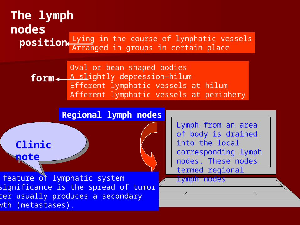
The lymph nodes
position Lying in the course of lymphatic vesselsArranged in groups in certain place
formOval or bean-shaped bodiesA slightly depression—hilum Efferent lymphatic vessels at hilumAfferent lymphatic vessels at periphery
Regional lymph nodesLymph from an area of body is drained into the local corresponding lymph nodes. These nodes termed regional lymph nodes
One feature of lymphatic systemIs significance is the spread of tumorCancer usually produces a secondaryGrowth (metastases).
Clinic noteClinic note

The lymphatic ductsThe lymphatic ducts
The thoracic duct
Posterior mediastinum
Aortic hiatus
Left and right lumbar trunksIntestinal trunk
Cisterna chyli
Left cervical trunkLeft subclavian trunkLeft bronchomediastinal trunk
Left venous angle Right venous angle
Right cervical trunkRight subclavian trunkRight bronchomediastinal trunk
Right lymphatic duct

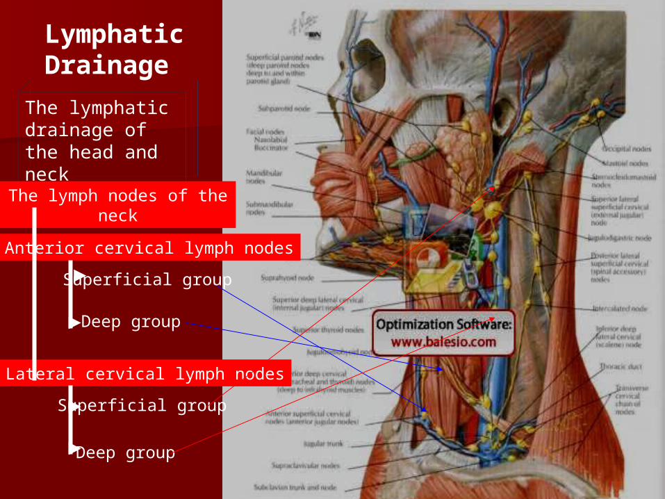
Lymphatic Drainage
The lymphaticdrainage of the head and neck
The lymph nodes of the head lie in
the boundary between the head
and neckthey consist of the occipital, mastoid, parotid, submandibular and submental lymph nodes
Deep lateral cervical lymph nodes
Lymphatic Drainage
The lymphaticdrainage of the head and neck
The lymph nodes of the neck
Anterior cervical lymph nodes
Lateral cervical lymph nodes
Superficial group
Deep group
Superficial group
Deep group

Deep LateralCervical LymphnodesSuperior group
Inferior group
Inferior deep lateral
cervical lymph nodes
Transverse cervical chain
of nodes
Supraclavicular nodes

Inferior group
Inferior deep lateral
cervical lymph nodes
Transverse cervical chain
of nodes
Supraclavicular nodes
Deep LateralCervical Lymphnodes
Virchow’s lymph node
The lymphatic efferents of the head and neck unite to for
m the left and right trunk.

The lymphatic drainage of the upper limbThe lymphatic drainage of the upper limb
All of the vessels of the uppAll of the vessels of the upper limbs drain into the termier limbs drain into the terminal roup of lymph nodes, thnal roup of lymph nodes, the axillary lymph nodes. The e axillary lymph nodes. The superficial tissues are drainsuperficial tissues are drained by vessels which accomped by vessels which accompany the superficial veins to any the superficial veins to pass to the axillary nodes eipass to the axillary nodes either directly, or indirectly. Tther directly, or indirectly. The deep lymph nodes follohe deep lymph nodes follow the principal neurovasculw the principal neurovascular bundle and end in the latar bundle and end in the lateral group of the axillary lyeral group of the axillary lymph nodes.mph nodes.
1. Cubital lymph nodes1. Cubital lymph nodes They are superficial to the dThey are superficial to the d
eep fascia above the medial eep fascia above the medial epicondyle of the humerus epicondyle of the humerus and near the deep blood veand near the deep blood vessels of the cubital fossa.ssels of the cubital fossa.

2. The Axillary Lymph
which are in the loose connective tissue of the axillary fossa and arranged along the blood vessels. They vary from twenty to thirty in number, and are divided into five groups.
①. Anterior group or pectoral lymph nodes
②. Lateral lymph nodes ③. Posterior lymph nodes
or subscapular lymph nodes
④. Central lymph nodes ⑤. apical lymph nodes The efferents of this grou
p unite to form the subclavian trunk.

The intercostal lymph nodes
The superior phrenic lymph nodes
Lymph of anterior wall of thorax
The parasternal lymph nodes
The lymphatic drainage of the thorax
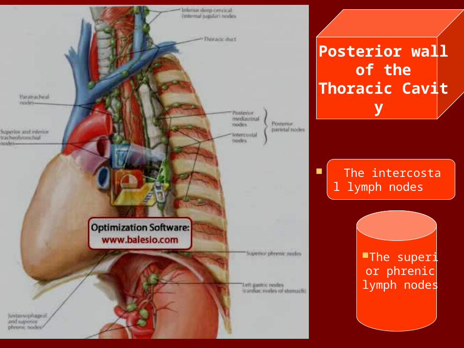
The intercostal lymph nodes
The superior phrenic lymph
nodes
Posterior wall of the
Thoracic Cavity

The lymph nodes of the thoracic Contents
Anterior mediastinal lymph nodes
Posterior mediastinal lymph nodes
The lymph nodes of the trachea, bronchi and
lungs

The pulmonary lymph nodes The broncheopulmonary hilar lymph nod
es The tracheobronchial lymph nodes The paratracheal lymph nodes The efferents of the contents of the thora
x to form left and right bronchomediatinal trunks
The lymph nodes of the trachea, bronchi and lungs

Lymphatic drainage of the abdomenThe lymph nodes of the abdominal wall
The superficial lymphatic vessels
from the anterior abdominal wall above the umbilicus follow the superficial blood vessels to the axillary nodes; the part below umbilicus is drained to the superficial linguin
al lymph nodes. The deep lymphatic vessels from the posterior abdominal wall pass directly, along the course of the lumbar arteries, to lumbar lymph nodes. Those from the upper part of the anterior wall run with the superior epigastric vessels to reach the
parasternal lymph nodes; those of the lower part end in the external i
liac lymph nodes.

The lumbar lympThe lumbar lymph nodes lies alonh nodes lies alongside the abdomigside the abdominal aorta and infenal aorta and inferior vena cava. Trior vena cava. They receive afferehey receive afferents from the postnts from the posterior abdominal erior abdominal wall, the abdomiwall, the abdominal paired viscernal paired viscera and from the coa and from the common iliac nodemmon iliac nodes. Their effrents us. Their effrents unite to form the rinite to form the right and left lumbght and left lumbar trunks.ar trunks.
The lymph nodes of the lower limb
External lymph nodes
Internal lymph nodes
Common iliac nodes
Lumbar lymph nodes
The lymph of the posterior abdominal wall
The lymph of the abdominal paired viscera

The lymph nodes of the abdominal viscera
The lymphatic drainage of the unpaired viscera of the
abdominal cavity
Celiac lymph nodes
Superior mesenteric lymph nodes
Inferior mesenteric lymph nodes

The celiac lymph nodes They lie on the front of the abdominal aorta close to the origin of the celiac artery. They are the terminal group for the stomach, deodenum, liver, gallbladder, pancreas and spleen, and their afferents are derived from the outlying lymph nodes which are placed along branches of the celiac artery.
①. Right and left gastric lymph nodes
②. Right and left gastroepiploic lymph nodes
③. Pyloric lymph nodes
④. Hepatic lymph nodes
⑤. Pancreaticosplenic lymph nodes


The superior mesenteric lymph nodes
Inferior mesenteric lymph nodes

The lymphatic drainage of the pelvis ①. Sacral lymph nodes ②. Internal iliac lymph nodes
③. External iliac lymph nodes ④. Common iliac lymph nodes

The lymphatic drainage of the lower limbs
Most superficial lymphatic vessels follow tMost superficial lymphatic vessels follow the great saphenous vein to end the lower he great saphenous vein to end the lower group of the superficial inguinal lymph nogroup of the superficial inguinal lymph nodes; others run with the small saphenous des; others run with the small saphenous vein to the popliteal lymph nodes. The devein to the popliteal lymph nodes. The deep lymphatic vessels accompany the main ep lymphatic vessels accompany the main blood vessels of the limb. The deep lymphblood vessels of the limb. The deep lymphatic vessels of the foot and leg are interruatic vessels of the foot and leg are interrupted by the popliteal lymph nodes, but thpted by the popliteal lymph nodes, but those from the thigh pass direct to the deep ose from the thigh pass direct to the deep inguinal lymph nodes.inguinal lymph nodes.
The popliteal lymph nodesThe popliteal lymph nodes They are imbedded in the fat if the popliteThey are imbedded in the fat if the poplite
al fossa. The nodes are near the end of the al fossa. The nodes are near the end of the small saphenous vein and arranged along small saphenous vein and arranged along the popliteal blood vessels. They receive athe popliteal blood vessels. They receive afferents from the deep lymphatic vessels fferents from the deep lymphatic vessels which accompany the anterior and posteriwhich accompany the anterior and posterior tibial vessels, and superficial lymphatic or tibial vessels, and superficial lymphatic vessels from the lateral border of the foot, vessels from the lateral border of the foot, and the back lateral side of the calf of the land the back lateral side of the calf of the leg. Their efferents pass to the deep inguineg. Their efferents pass to the deep inguinal lymph nodesal lymph nodes

The inguinal lymph nodesThe inguinal lymph nodes ①①.The superficial inguinal lymph n.The superficial inguinal lymph n
odesodes They are arranged in two groups. TThey are arranged in two groups. T
he upper group forms the a chain ihe upper group forms the a chain immediately below the inguinal ligammediately below the inguinal ligament. The lymphatic vessels from tment. The lymphatic vessels from the lower part the abdominal wall, ehe lower part the abdominal wall, external genitalia, gluteal region and xternal genitalia, gluteal region and perineum pass this group. The loweperineum pass this group. The lower group is disposed vertically along r group is disposed vertically along the terminal part of the great saphethe terminal part of the great saphenous vein. It receive all the superficinous vein. It receive all the superficial lymphatic vessels of the lower lial lymphatic vessels of the lower limb, with the exceptionof those fromb, with the exceptionof those from the lateral border of the foot, anm the lateral border of the foot, and the back and lateral side of the cad the back and lateral side of the calf of the leg. All the superficial inguilf of the leg. All the superficial inguinal lymph nodes send their efferentnal lymph nodes send their efferents to the deep inguinal lymph nodes s to the deep inguinal lymph nodes and the external iliac nodes.and the external iliac nodes.

②. The deep inguinal lymph nodes They are situated medial to the femoral vein and in the femoral canal. They receive lymph from the deep structures of the lower limb and perineum, and the afferents from the superficial inguinal and popliteal lymph nodes. The efferents pass to the external iliac lymph nodes.

The lymphatic drainage of the mammary gland
Efferents vessels of the mammary gland mainly drain to the axillary lymph nodes. There are 3drainage directions:
①. Efferents vessels of the lateral and central part drain to the pectoral lymph nodes.
②. Efferents vessels of the superior part to the apical lymph nodes and subclavicular lymph nodes.
③. Efferents vessels of the medial part to the parasternal lymph nodes. Superficial lymphatic vessels of the medial part of the organ communicate with the vessels of the opposite side.
④. Lymphatic vessels of the medioinferior part communicate with the hepatic lymphatic vessels by the epigastric lymphatic vessels and inferior phrenic lymphatic vessels.

The spleenThe spleenIt is the largest lymphoid organ in The body. and is surrounded by PeritoneumFunctionMain functions are erythrocyteStorage, phagocytosis, cytopoiesisand immune responses.Position
Lies in the left hypochondriac region of the abdomen. It is situated betweenThe fundus of stomach and diaphragm and deep to the 11th left ribs. Its long axis corresponds to the 10th ribs. The normal spleen is not palpable below the left costal margin unless it is enlarged or markedly dislocated.
FormSoft, fragile, dark purplish color.Has two surfaces (diaphragmatic and visceral), two borders (superior and inferior) and two extremities (anterior and posterior).

Splenic notch
Visceral surface
Hilum of spleen
Diaphragmatic surface is convex, smooth and faces upwards. It is in relation with the abdominal surface of the diaphragm.Visceral surface is directed towards the abdominal cavity, and presents gastric, renal, pancreatic and colic impressions.
The hilum of spleenIt has a long fissure nearThe center of its visceral Surface. It is for the exitAnd entrance of blood Vessels and nerves.
Splenic notchSuperior border is sharperAnd marked, near its lateralEnd, by two or three splenic Notches of variable depth,Which can be recognized When the spleen is enlarged


Test for Test for AngiologAngiologyy 1. The entrances of the left and right atria?1. The entrances of the left and right atria? 2. The boundary between the inflow and outflow in the 2. The boundary between the inflow and outflow in the right ventricle?right ventricle? 3. Describe the fibrous skeleton of the heart?3. Describe the fibrous skeleton of the heart? 4. The conduction system of the heart includes?4. The conduction system of the heart includes? 5. Define the systemic circulation.5. Define the systemic circulation. 6. Describe the arterial supply of the thyroid gland.6. Describe the arterial supply of the thyroid gland. 7. Describe the arterial supply of the stomach.7. Describe the arterial supply of the stomach. 8. Describe the hepatic portal vein and its tributaries.8. Describe the hepatic portal vein and its tributaries. 9. Describe the cubital arterial network.9. Describe the cubital arterial network. 10. Describe the dangerous triangle of the face and the 10. Describe the dangerous triangle of the face and the communications of the facial vein.communications of the facial vein.