anesthesia in the ep lab - healing, teaching & discovery · electrophysiology procedures are...
TRANSCRIPT
Procedural Electrophysiology & Anesthesia in the EP Lab
PETER JESSEL, MD
ASSISTANT PROFESSOR

Outline
▪ The Clinical Electrophysiology Lab
▪ Common Arrhythmias and Management
▪ Atrial Fibrillation Ablation
▪ Complications
▪ Device Implantation
▪ Approach to Sedation and Anesthesia

Introduction
▪ Initially EP labs were developed for diagnostic procedures
▪ EP studies were done for diagnosing bradyarrhythmias
▪ Focus is now primarily therapeutic for tachyarrhythmias and device implantation
▪ Electrophysiology procedures are becoming more complex and occurring in a sicker population
▪ Anesthesia is increasingly used as long procedures become more frequent
▪ The EP lab is an isolated environment with distinct challenges for the anesthesiology team
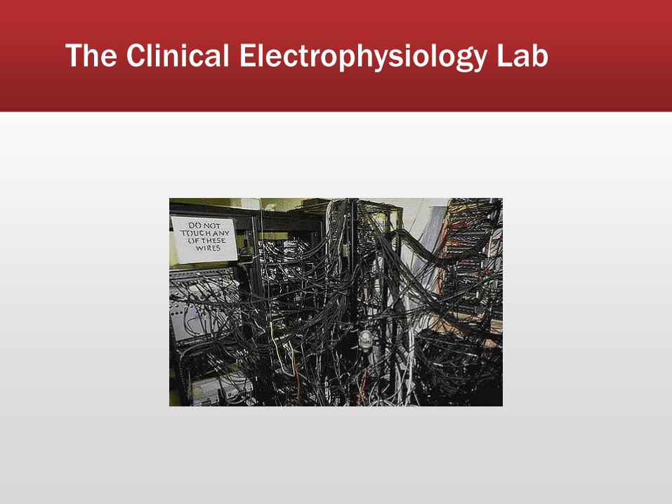
The Clinical Electrophysiology Lab

The Clinical Electrophysiology Lab
▪ Equipment:
▪ Programmed stimulator
▪ Recording system
▪ Biplane Fluoroscopy
▪ 3D mapping system
▪ RF generator
▪ Display boom
▪ Cryoablation system
▪ Intracardiac echo
▪ 2 biphasic defibrillators
▪ Magnetic navigation system (stereotaxis)
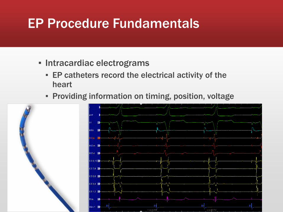
EP Procedure Fundamentals
▪ Intracardiac electrograms
▪ EP catheters record the electrical activity of the heart
▪ Providing information on timing, position, voltage

EP Procedure Fundamentals
▪ Most arrhythmias have a reentrant mechanism
▪ Typically a critical isthmus (slow conduction) that can be ablated to terminate the rhythm and render the local tissue electrically silent

Standard EP Study Catheter Locations
▪ Standard 4 catheter diagnostic study
▪ High right atrium – approximates sinus node location
▪ His bundle – approximates AV nodal conduction
▪ Coronary Sinus – records LA/LV along mitral annulus
▪ RV apex

Standard EP Study Catheter Locations
RAO LAO

SVT EP Study
▪ Usually pace in the ventricle first
▪ Then pace in the atrium
▪ If non-inducible – isoproterenol 1-10ug/min
▪ Prefer lightest sedation possible to increase chance of eliciting SVT
▪ Goal is to induce SVT and perform pacing maneuvers to define mechanism and location
▪ After a diagnosis made, a mapping/ablation catheter will be inserted
Common Arrhythmias
▪ Paroxysmal Supraventricular Tachycardia (PSVT)
1. AV Nodal Reentrant Tachycardia (AVNRT)
2. AV Reciprocating Tachycardia (AVRT)
3. Atrial Tachycardia (AT)
▪ Atrial flutter (AFL)
▪ Atrial fibrillation (AF)
▪ Premature Ventricular Contractions (PVCs)/Ventricular Tachycardia (VT)

PSVT
▪ PSVT - General term applied to intermittent SVT other than AF, AFL, multifocal AT
▪ SVT is a narrow complex tachycardia rate >100 bpm and QRS <120ms
▪ Usually associated with a structurally normal heart
▪ SVT relatively common – 2.25/1,000 normal population
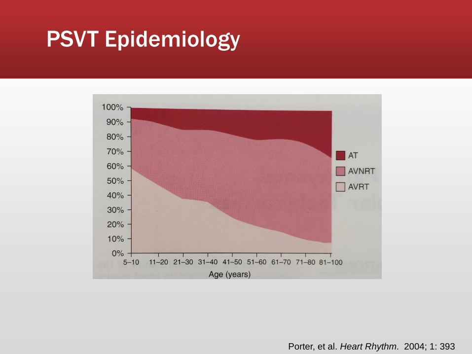
PSVT Epidemiology
Porter, et al. Heart Rhythm. 2004; 1: 393
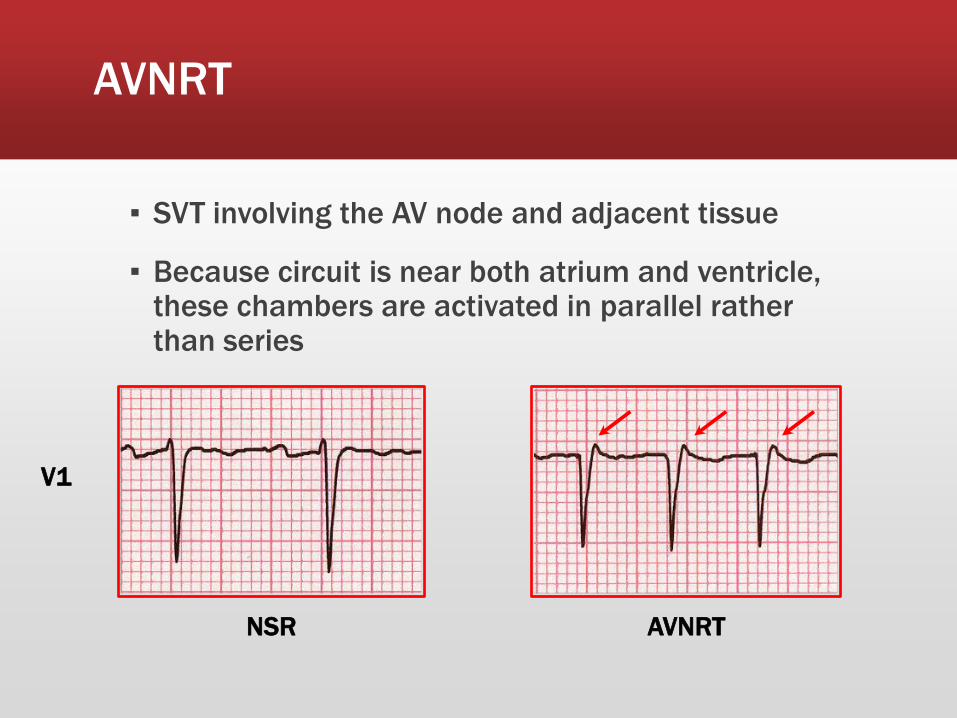
AVNRT
▪ SVT involving the AV node and adjacent tissue
▪ Because circuit is near both atrium and ventricle, these chambers are activated in parallel rather than series
NSR AVNRT
V1
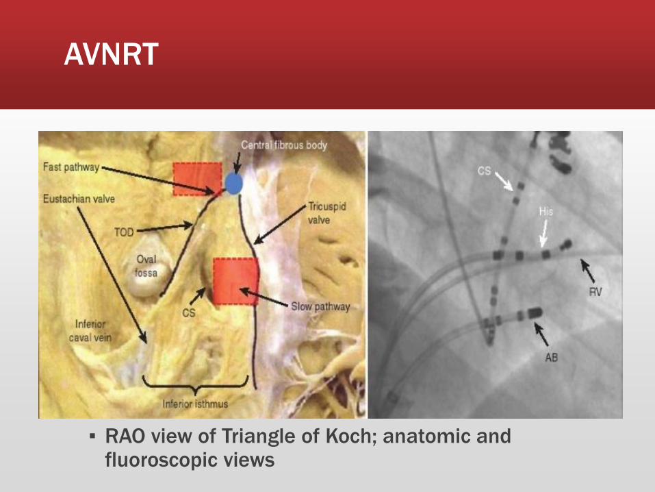
AVNRT
▪ RAO view of Triangle of Koch; anatomic and fluoroscopic views

AVRT
▪ Congenital presence of an accessory pathway between atrium and ventricle
▪ Wolff-Parkinson-White pattern (~0.25% general population) – ventricular preexcitation (delta wave) on ECG, no symptoms or documented SVT
▪ WPW syndrome – ventricular preexcitation + symptoms/PSVT
▪ Many accessory pathways are concealed – no antegrade conduction, but capable of retrograde

AVRT – WPW Pattern
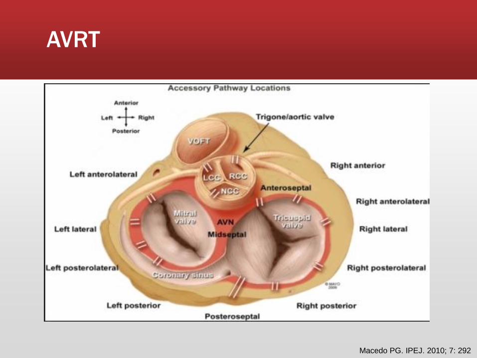
AVRT
Macedo PG. IPEJ. 2010; 7: 292

Atrial Tachycardia
▪ Ectopic atrial focus
▪ Reentrant, triggered or automatic mechanisms
▪ Often has warm up phase
▪ Most right atrial and along the crista terminalis
▪ P wave morphology can be used to localize
▪ Frequent in patients with prior cardiac surgery or congenital heart disease
V1

Atrial Tachycardia
Kistler, et al.JACC. 2006; 48: 1010

PSVT Chronic Management
▪ PSVT is very rarely life threatening
▪ Decision for medical therapy ▪ Frequency of arrhythmia
▪ Severity of symptoms
▪ Side effects
▪ Patient preference
▪ Medications; beta blocker or calcium channel blockers, rarely use antiarrhythmics
▪ Indications for EPS and ablation ▪ Poorly tolerated PSVT (near syncope/syncope, falls)
▪ Failure of medical therapy
▪ Patient preference for ablation
▪ Ablation success rate generally >90%
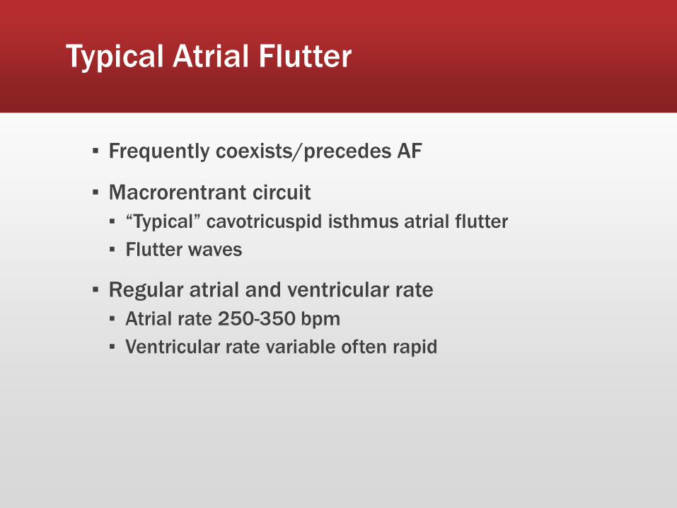
Typical Atrial Flutter
▪ Frequently coexists/precedes AF
▪ Macrorentrant circuit
▪ “Typical” cavotricuspid isthmus atrial flutter
▪ Flutter waves
▪ Regular atrial and ventricular rate
▪ Atrial rate 250-350 bpm
▪ Ventricular rate variable often rapid

Typical Atrial Flutter
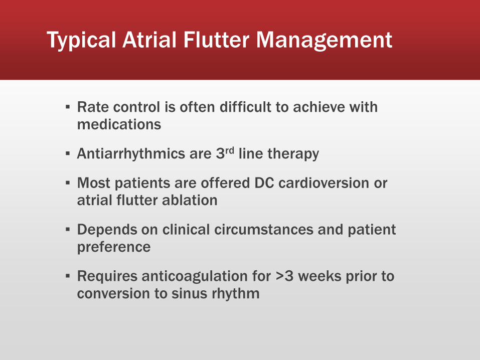
Typical Atrial Flutter Management
▪ Rate control is often difficult to achieve with medications
▪ Antiarrhythmics are 3rd line therapy
▪ Most patients are offered DC cardioversion or atrial flutter ablation
▪ Depends on clinical circumstances and patient preference
▪ Requires anticoagulation for >3 weeks prior to conversion to sinus rhythm
Typical Atrial Flutter – 3D Activation Map

Typical Atrial Flutter
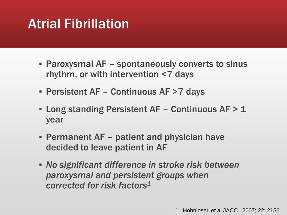
Atrial Fibrillation
▪ Paroxysmal AF – spontaneously converts to sinus rhythm, or with intervention <7 days
▪ Persistent AF – Continuous AF >7 days
▪ Long standing Persistent AF – Continuous AF > 1 year
▪ Permanent AF – patient and physician have decided to leave patient in AF
▪ No significant difference in stroke risk between paroxysmal and persistent groups when corrected for risk factors1
1. Hohnloser, et al.JACC. 2007; 22: 2156

Reasons We Treat AF
▪ Symptoms
▪ wide spectrum from completely asymptomatic to exquisitely sensitive
▪ Risk of stroke
▪ Virchow’s Triad
▪ Endothelial injury – myocyte hypertrophy, fibrotic changes
▪ Stasis - No organized mechanical function, LA dilation
▪ Hypercoaguable state – platelet activation, prothrombin
▪ Rate
▪ Chronically elevated heart rate may cause tachycardia cardiomyopathy
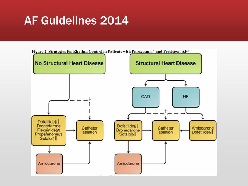
AF Guidelines 2014
AF Ablation Indications
▪ Symptomatic AF refractory or intolerant to at least 1 class I or III antiarrhythmic medication
▪ Ablation may be considered prior to antiarrhythmics, but this situation is rare
▪ Left atrial appendage thrombus or inability to take anticoagulation are contraindications

AF Ablation Patient Selection
More Optimal Patient Less Optimal Patient
Variable
Symptoms Highly symptomatic Minimally symptomatic
Class I and III drugs failed 1 0
AF type Paroxysmal Long-standing persistant
Age Younger (<70 years) Older (70 years)
LA size Smaller (<5.0 cm) Larger (5.0 cm)
Ejection fraction Normal Reduced
Congestive heart failure No Yes
Other cardiac disease No Yes
Pulmonary disease No Yes
Sleep apnea No Yes
Obesity No Yes
Prior stroke/TIA No Yes
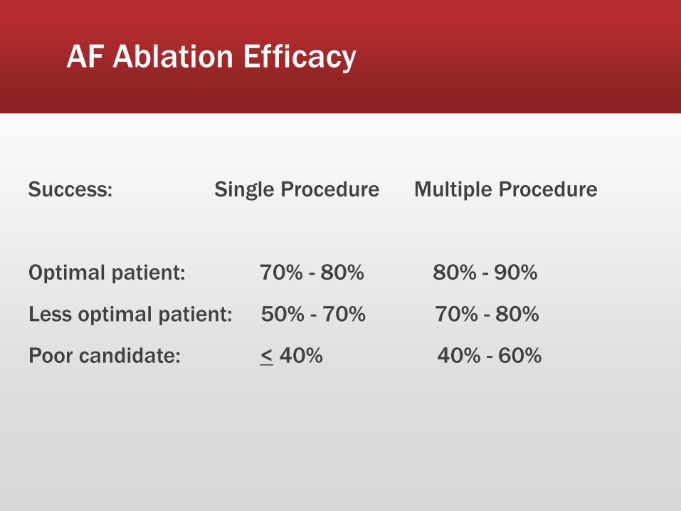
AF Ablation Efficacy
Success: Single Procedure Multiple Procedure
Optimal patient: 70% - 80% 80% - 90%
Less optimal patient: 50% - 70% 70% - 80%
Poor candidate: < 40% 40% - 60%

Pulmonary Vein Isolation (PVI) is the Cornerstone of AF Ablation
“Ablation strategies which target the PVs and/or PV antrum
are the cornerstone for most AF ablation procedures.”
Complete electrical isolation should be the goal for targeted PVs and entrance and/or exit block should be demonstrated
Right Inferior
Pulmonary Vein
Right Superior
Pulmonary Vein
Superior
Vena
Cava
Inferior Vena Cava
2012 HRS Consensus Statement
Left
Superior
Pulmonary
Vein
Left
Inferior
Pulmonary
Vein
Caulkins. Heart Rhythm. 2012 9:632
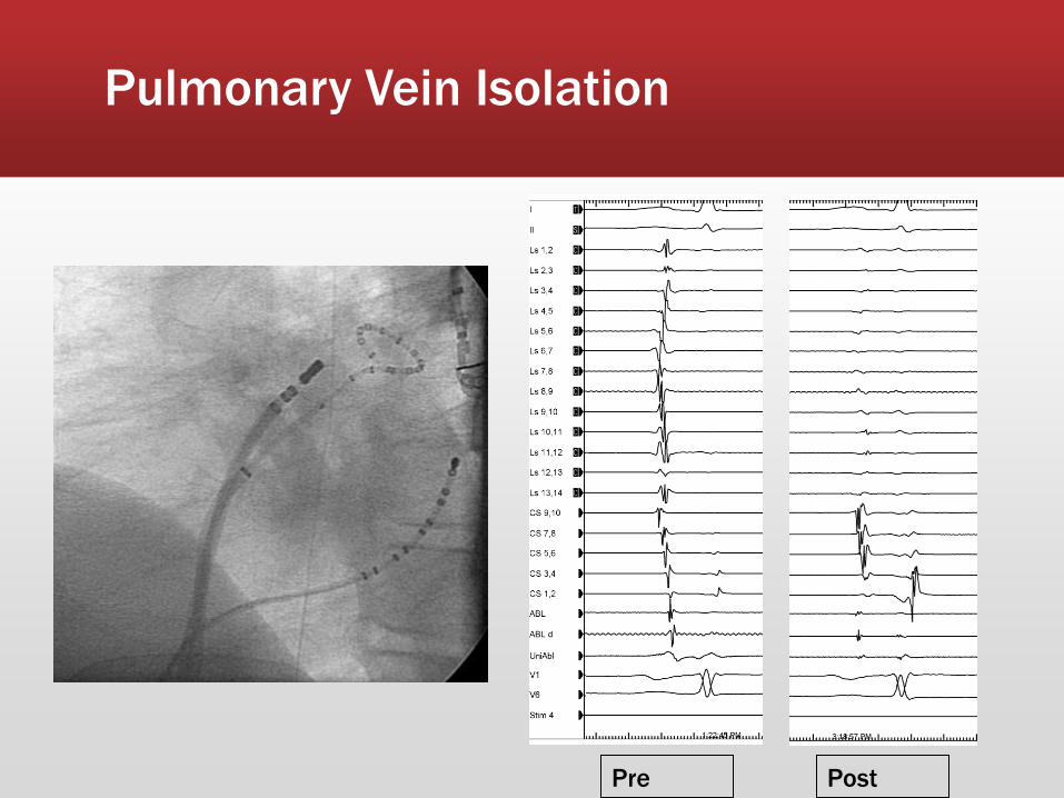
Pulmonary Vein Isolation
Pre Post
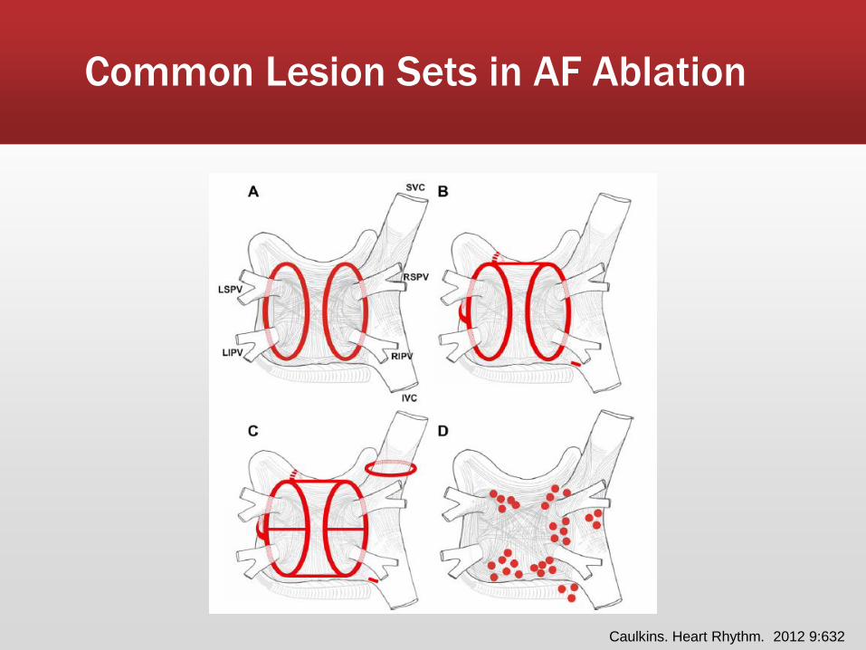
Common Lesion Sets in AF Ablation
Caulkins. Heart Rhythm. 2012 9:632
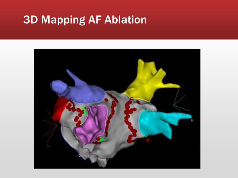
3D Mapping AF Ablation

Radiofrequency AF ablation
▪ Focal RF ablation was not specifically designed for AF ablation
▪ Technically challenging for operator
▪ More versatile than dedicated PVI technologies
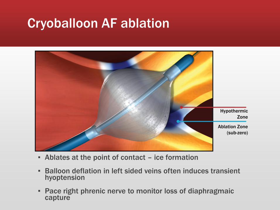
Cryoballoon AF ablation
▪ Ablates at the point of contact – ice formation
▪ Balloon deflation in left sided veins often induces transient hyoptension
▪ Pace right phrenic nerve to monitor loss of diaphragmaic capture
Hypothermic
Zone
Ablation Zone
(sub-zero)
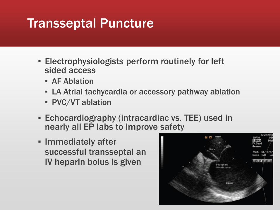
Transseptal Puncture
▪ Electrophysiologists perform routinely for left sided access
▪ AF Ablation
▪ LA Atrial tachycardia or accessory pathway ablation
▪ PVC/VT ablation
▪ Echocardiography (intracardiac vs. TEE) used in nearly all EP labs to improve safety
▪ Immediately after successful transseptal an IV heparin bolus is given

Anticoagulation Strategies
▪ Multiple studies have shown AF ablation is safer on therapeutic warfarin vs. holding warfarin with LMWH bridging1,2
▪ Fewer bleeds and strokes
▪ Device implantation in patients at higher risk for thrombotic events also with fewer bleeding events on therapeutic warfarin3
▪ 81% reduction in pocket hematomas
▪ Management strategy on NOACs still evolving, but generally held prior to procedures
1. Di Biase L, et al. Circulation. 2010; 121:2550–2556
2. Hussein AA, et al. Heart Rhythm. 2009;6: 1425–1429.
3. Birnie DH, et al. NEJM 2013; 30: 368 2084-2093

Periprocedural INR Goal
NEJM.2003; 349:1910-26

AF Ablation Complications
▪ Mortality after AF ablation is 0.05% according to a large world wide survey in 20051
▪ Overall complication rate of 6%
▪ Vascular access complications – 0.5% ▪ Hematoma
▪ Pseudoaneurysm
▪ AV fistula
▪ Tamponade – 1.2%
▪ Stroke/TIA – 1%
▪ Symptomatic PV stenosis – 0.6%
▪ Atrioesophageal fistula – 0.01%
▪ Phrenic nerve injury 0.1% (Cryoballoon ablation 11%2)
1. Cappato R Circulation.2005; 111:1100
2. Packer DL. JACC 2013. 16:1713
Pericardial Tamponade
▪ ↑Pericardial fluid → ↓Ventricular filling → ↓SV
▪ As little as 60-100cc of pericardial fluid can cause tamponade
▪ Diagnosis with LAO fluoroscopy or echocardiography
▪ Pericardiocentesis and drainage usually resolves the effusion
▪ Protamine if heparin, Kcentra (prothrombin complex concentrate) if warfarin
▪ Rare for active novel oral anticoagulant during procedure

Atrioesophageal Fistula
Arana-Rueda. Rev Esp Cardiol. 2009;62(10):1189
▪ Monitoring of esophageal temperature
▪ Limiting power and duration of lesions
▪ PPI is prescribed for 30 days after ablation

Pacemakers and ICDs
John IP. ICD Troubleshooting. 8/2011

Pacemaker/Defibrillator Implantation
▪ Prefer implantation at left shoulder ▪ Easier to place leads
▪ Better vector for defibrillation
▪ Exception is left handed pacemaker patient
▪ Standard EP practice is for IV antibiotics finished before incision ▪ Cefazolin 1-2gm IV
▪ Vancomycin 1gm IV if PCN allergic
▪ Standard access at OHSU axillary stick, but cephalic cut down may be chosen
▪ DFT Testing – VF induction to confirm device ability to terminate ▪ falling out of favor
▪ SIMPLE Trial – DFTs did not improve outcome
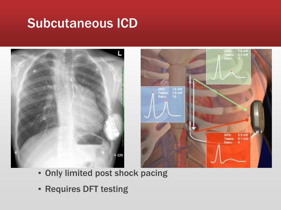
Subcutaneous ICD
▪ Only limited post shock pacing
▪ Requires DFT testing
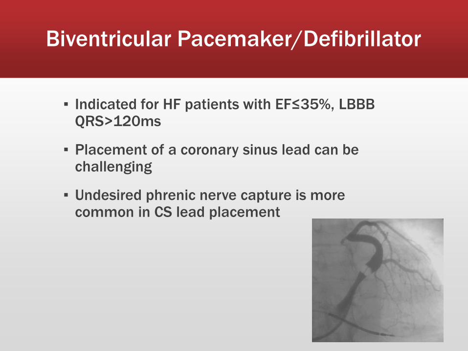
Biventricular Pacemaker/Defibrillator
▪ Indicated for HF patients with EF≤35%, LBBB QRS>120ms
▪ Placement of a coronary sinus lead can be challenging
▪ Undesired phrenic nerve capture is more common in CS lead placement

Device Complications
▪ Complications rise as systems get more complex
▪ VVI pacemaker -> dual chamber ICD -> CRT-D
▪ 2006-2008 NCDR ICD Registry 339, 076
▪ Overall implantation risk 3.36%
▪ Pneumothorax - 0.57%
▪ Usually is managed conservatively
▪ Pocket hematoma – 0.97%
▪ Avoid heparin
▪ Do not aspirate or evacuate pocket
▪ Perforation 0.08%
▪ Lead dislodgement – 1.07%
▪ Death (in lab) – 0.02%
Hammill SC. Heart Rhythm. 2009; 9: 1397
Approach to Sedation and Anesthesia in the EP Lab
▪ EP lab not designed with anesthesia in mind
▪ Patients are not typical elective OR patients
▪ Retrospective analysis of incident reports shows communication and teamwork issues most frequent contributors1
▪ Closed loop verbal orders
▪ Communication with the EP attending or fellow regarding level of sedation
1. Manser, T. Acta Anasthesiol Scan. 2009; 53: 143
Approach to Sedation and Anesthesia in the EP Lab
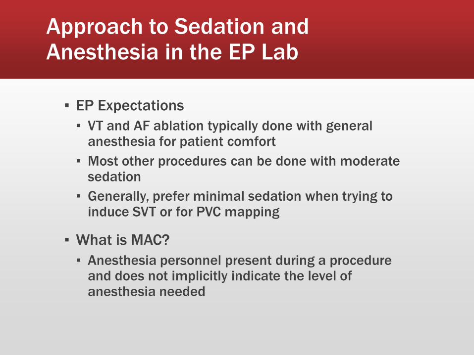
▪ EP Expectations
▪ VT and AF ablation typically done with general anesthesia for patient comfort
▪ Most other procedures can be done with moderate sedation
▪ Generally, prefer minimal sedation when trying to induce SVT or for PVC mapping
▪ What is MAC?
▪ Anesthesia personnel present during a procedure and does not implicitly indicate the level of anesthesia needed
Approach to Sedation and Anesthesia in the EP Lab
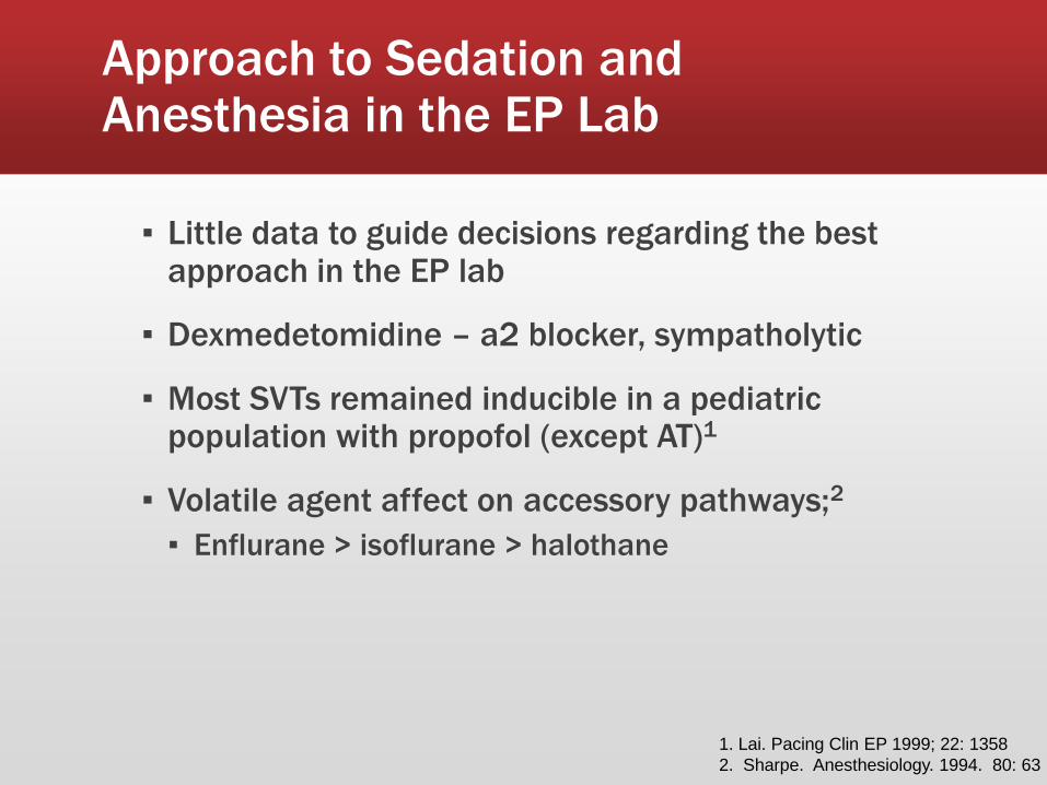
▪ Little data to guide decisions regarding the best approach in the EP lab
▪ Dexmedetomidine – a2 blocker, sympatholytic
▪ Most SVTs remained inducible in a pediatric population with propofol (except AT)1
▪ Volatile agent affect on accessory pathways;2
▪ Enflurane > isoflurane > halothane
1. Lai. Pacing Clin EP 1999; 22: 1358
2. Sharpe. Anesthesiology. 1994. 80: 63
Anesthesia in AF Ablation
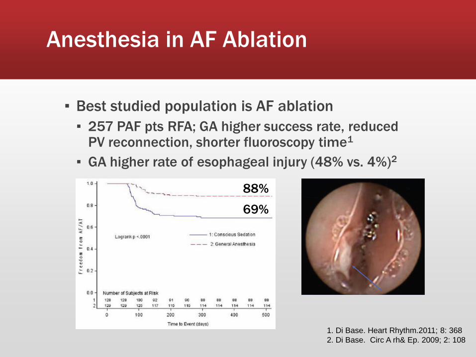
▪ Best studied population is AF ablation
▪ 257 PAF pts RFA; GA higher success rate, reduced PV reconnection, shorter fluoroscopy time1
▪ GA higher rate of esophageal injury (48% vs. 4%)2
1. Di Base. Heart Rhythm.2011; 8: 368
2. Di Base. Circ A rh& Ep. 2009; 2: 108
88%
69%
Deep Sedation Without Assisted Ventilation in AF Ablation
▪ 1000 consecutive patients undergoing AF ablation
▪ 2% Propofol infusion and intermittent fentanyl directed by electrophysiologist
▪ Cessation of propofol occurred in 15.6%
▪ 13.6% due to hypotension (fail maintain >90 SBP)
▪ 1.9% respiratory depression (02 sat <90%)
▪ 0.1% hypersalivation
▪ 1 patient required bag mask ventilation 4min, no intubations required
Salukhe. Europace 2012. 14: 325
Conclusions
▪ Procedural electrophysiology is a continuously evolving field highly dependent on technology
▪ The clinical electrophysiology lab is a unique setting for the anesthesiology team
▪ Optimal sedation and anesthesia strategies in the EP lab are actively being investigated
▪ The EP team very much appreciates your excellent care of our patients


The EP Control Room After Successful Fellow Femoral Access In July