androgen-induced proliferation in the developing larynx of ... · androgen-induced proliferation in...
TRANSCRIPT
DEVELOPMENTAL BIOLOGY 178, 113–123 (1996)ARTICLE NO. 0202
Androgen-Induced Proliferation in the DevelopingLarynx of Xenopus laevis Is Regulated byThyroid Hormone
Michael A. Cohen and Darcy B. Kelley1
Department of Biological Sciences, Columbia University, New York, New York 10027
Exposure to exogenous androgen regulates cell number in the developing larynx of Xenopus laevis and hormone-regulatedlaryngeal development requires secretion of thyroid hormone (TH). We sought to determine whether exposure to TH isboth sufficient and necessary for androgen-evoked cell proliferation (androgen competency) in developing larynx. Androgencompetency was not observed in the premetamorphic larynx (tadpole stage 53, before TH secretion) but was present justprior to metamorphic climax (stage 58, during TH secretion). However, when TH is administered precociously (betweenstages 48 and 50), androgen competency can be observed at stage 53. The stage 52 larynx expresses high levels of themRNA for TH receptor a. The duration of TH exposure required at tadpole stage 48 is greater than 2 days; studies injuveniles indicate that TH exposure need not be maintained in order for androgen competency to persist. The effects ofexposure to TH on androgen competency are long lasting and perhaps permanent. While organotypic cultures obtainedfrom tadpoles during premetamorphosis (stage 52) can proliferate in vitro and proliferation is augmented by TH exposureas it is in vivo, precocious exposure to TH does not induce androgen competency. In contrast, androgen does evokecell proliferation in cultures obtained from metamorphosing (stage 58) tadpoles; proliferation is confined to the cartilagecomponent. Thus, unlike larynges in vivo, muscle will not proliferate in response to androgen, indicating the necessityfor an additional factor not present in vitro. Androgen receptor mRNA expression, believed required for androgen compe-tency, was assessed in vivo and in vitro. The tadpole larynx strongly expresses AR mRNA; expression does not requireexposure to TH nor is expression diminished in culture. q 1996 Academic Press, Inc.
INTRODUCTION ynx seems due to differentiation of androgen sensitive stemcells (Marin et al., 1990). The opening of the sensitive periodfor androgen-evoked cell proliferation is the subject of thisDifferentiation of masculine and feminine phenotypes ininvestigation.vertebrates is directed by steroid hormones, particularly an-
The mature male larynx contains more muscle fibers anddrogens and estrogens. Tissue responsiveness to steroidsa larger and more complex cartilage than that of the female.generally requires expression of functional receptors and isLaryngeal anlage are present at tadpole stage 41; musclespecific to cell type and developmental stage. The develop-begins to differentiate at tadpole stage 41 and cartilage atmental factors that control the opening and closing of sensi-stage 43 (Nieuwkoop and Faber, 1956). The gonads sexuallytive periods for steroid hormone action are largely un-differentiate at tadpole stage 56 (Merchant-Larios and Vil-known. In the sexually differentiated larynx of Xenopuslapando, 1981; Iwasawa and Yamaguchi, 1984; Robertsonlaevis, muscle and cartilage cell numbers and types are con-and Kelley, 1996) and by stage 62 masculinization of laryn-trolled by androgen secretion during early postmetamorphicgeal innervation is apparent, though muscle fiber numbers(PM) stages (reviewed in Kelley, 1996). The closing of theare the same in the sexes (Kelley and Dennison, 1990; Rob-developmental sensitive period for cell addition in the lar-ertson et al., 1994). Muscle fiber number in males exceedsthat of females by PM stage 1 (Ç3 months PM; Marin etal., 1990); from PM1 on, muscle fiber type in the larynx1 To whom correspondence should be addressed at 911 Fairchilddiverges in the sexes (Tobias et al., 1991a; Catz et al., 1992).Science Center for the Life Sciences, Columbia University, New
York, NY 10027. Fax: (212) 531-0425. Sexual differentiation of laryngeal cartilages is first apparent
113
0012-1606/96 $18.00Copyright q 1996 by Academic Press, Inc.All rights of reproduction in any form reserved.
AID DB 8296 / 6x11$$$$81 07-30-96 09:57:19 dbas AP: Dev Bio
114 Cohen and Kelley
Tadpoles were maintained at room temperature (Ç22–247C) inat PM2 (Ç6 months PM; Sassoon and Kelley, 1986; FischerNovaqua (Kordon, Hayward, CA)-treated filtered tap water in 2.5-et al., 1995). During postmetamorphic stages, all laryngealliter polycarbonate tanks (25 tadpoles/tank) under a 12:12 L:D cycletissue types express androgen receptor (AR) and androgenand were fed nettle powder daily. In this population, the gonads ofreceptor mRNA (Kelley et al., 1989; Fischer et al., 1995).males and females do not become morphologically distinguishableThe onset of androgen-induced laryngeal masculiniza-until stage 56 (see Robertson and Kelley, 1996); sex cannot be deter-
tion coincides with the metamorphosis of the tadpole to mined at earlier stages. Postmetamorphic frogs were staged as de-the juvenile frog, a developmental epoch controlled by scribed previously (Tobias et al., 1991a).thyroid hormone (TH). Thyroxine (T4), which is secreted In vivo studies—Tadpoles. Tadpoles were treated with dihy-by the thyroid, is converted to the biologically active form drotestosterone (5a-androstan-17b-ol-3-one; DHT, Sigma) and/ortriiodothyronine (T3 ) within responsive cells. If tadpoles T4 (Sigma) as these are the most fully developmentally character-
ized, circulating forms of androgens and TH, respectively (Kang etare developmentally arrested before the beginning ofal., 1995; Leloup and Buscaglia, 1977). DHT was dissolved in 95%metamorphosis using the T4 synthesis inhibitor propyl-ethanol which was added to tank water to produce a final concen-thiouracil (PTU), androgen-induced laryngeal growth istration of 1.0 1 1007 M (the ethanol concentration was 0.0016%).specifically prevented (Robertson and Kelley, 1996). ThisT4 was solubilized in 0.5 M NaOH, added to tank water, and usedobservation raises the possibility that the sensitive periodat a final concentration of 1 1 1008 M (100 mM NaOH). At stagefor androgen-induced changes in laryngeal cell number is45, tadpoles were randomly assigned to one of four experimental
controlled by TH secretion. groups: untreated, DHT (stage 51–53), T4 (stage 48–50) followedWe wished to determine whether exposure to TH is nec- by DHT (stage 51–53), or T4 (stage 48–50). The treatment para-
essary and sufficient for androgen-evoked cell proliferation digms are summarized in Fig. 1. In pilot studies, tadpoles werein the larynx. The receptors for TH are expressed very early pretreated with T4 for 1, 2, or 7 days, beginning at stage 48. Atin development (Kawahara et al., 1991) but secretion of TH stage 53, DHT-induced cell proliferation was observed only in the
7-day T4 treatment group; thus, this interval was chosen for subse-is restricted to specific stages, beginning at tadpole stagequent studies. Four additional groups were also prepared: in one,54, peaking at 60–62, and falling off by the end of metamor-endogenous TH synthesis was prevented by maintaining tadpolesphosis at tadpole stage 66 (reviewed in Galton, 1983; Doddfrom stage 48 (prior to the onset of TH secretion; Leloup and Bus-and Dodd, 1976). Sex differences in laryngeal cell numberscaglia, 1977) in 0.01% 6-n-propyl-2-thiouracil (PTU, Sigma; thisare not apparent until juvenile stage PM1, 3–4 months afterdose prevents metamorphic-associated laryngeal differentiation inTH secretion is initiated. However, exposure to exogenousX. laevis, Robertson and Kelley, 1996). In another, tadpoles were
androgen can prematurely masculinize cell number and cell treated with T4 from stage 51 to 53. Two groups of stage 58 tadpolestype in juveniles of both sexes (Sassoon et al., 1986; Marin were prepared. One was an untreated control group, and the otheret al., 1990; Tobias et al., 1991b; Fischer et al., 1995; Catz was treated with DHT for 3 days.et al., 1995). In addition, the developing larynx expresses Tadpoles in each group received an injection of 10–20 mCi [3H]-AR mRNA from very early tadpole stages (Cohen and Kel- thymidine in 0.01% fast green into the peritoneal cavity using aley, 1994). We thus employed the induction of cell prolifera- Butterfly Infusion Set (Abbot Hospitals). After 30 min, animals
were anesthetized by immersion in 0.1% MS222 (ethyl m-amino-tion by exogenous androgen (androgen competency) as anbenzoate methanesulfonic acid; Aldrich, Milwaukee, WI) and fixedassay for TH-induced androgen sensitivity before the onsetovernight in 10% neutral buffered Formalin. They were then dehy-of TH secretion, both in tadpoles and in parallel laryngealdrated through an ethanol series, cleared with methyl salicylate,organ cultures.and embedded in Paraplast.
In vivo studies—Juveniles. To determine whether continuedTH exposure is required for androgen-evoked cell proliferation, PM
MATERIALS AND METHODS animals were studied. PM2 animals (Ç6 months PM) were chosenfor study as the larynx evinces a robust proliferative response toandrogen at this stage (Sassoon et al., 1986; Fischer et al., 1995).Animals. X. laevis tadpoles were obtained commerciallyTH synthesis was blocked by maintaining PM2 frogs in 0.01% PTU(Nasco, Fort Atkinson, WI) or from mated pairs injected with 1000for 3 weeks. PTU-treated and control frogs were implanted withunits of human chorionic gonadotropin (Sigma). Embryonic andDHT-filled (0.25 mg/g body weight) or empty silastic tubing in thelarval development of X. laevis has been divided into 66 stagesdorsal lymph sac for 5 days; they then received a [3H]thymidinebased on morphological characteristics (Nieuwkoop and Faber,injection (1 mCi/g body weight), were killed 1 h later (see Fischer1956). Stage 1 represents fertilization of the egg; embryonic devel-et al., 1995 for details), and tissue was processed for autoradiogra-opment then proceeds to hatching at stages 35/36 (Ç50 hr postfer-phy as described above.tilization or PF). Larval or tadpole development (stages 37 through
In vitro studies. Larynges were removed from stage 52 or stage66) has been further classified according to metamorphic status of58 tadpoles and cultured in Opti-MEM I Reduced-Serum mediathe tadpole (Dent, 1968). The period from stage 37 to stage 53 (Ç24(Gibco) supplemented with gentamycin (50 mg/ml), penicillin (50days PF) is termed premetamorphosis; stage 54 marks the start ofIU/ml), streptomycin (50 IU/ml), fungizone (0.125 mg/ml), andprometamorphosis or TH-directed metamorphosis. Forelimbsstripped fetal bovine serum (4%). Fetal serum was stripped of ste-emerge at stage 58 (Ç44 days PF), and metamorphic climax corre-roid and thyroid hormones (see Ishizuya-Oka and Shimozawa,sponds to peak plasma TH levels and occurs at stages 59–62. Circu-1991) using dextran-coated charcoal. Norit-A (1.25 g), Dextran T-lating TH then decreases and metamorphosis from tadpole to frog-
let is complete at stage 66 (Ç58 days PF). 70 (0.125 g), and 12.5 ml FBS were stirred for 2 hr at 47C, an addi-
Copyright q 1996 by Academic Press, Inc. All rights of reproduction in any form reserved.
AID DB 8296 / 6x11$$$$81 07-30-96 09:57:19 dbas AP: Dev Bio
115TH-Induced Androgen Competency in the Tadpole Larynx
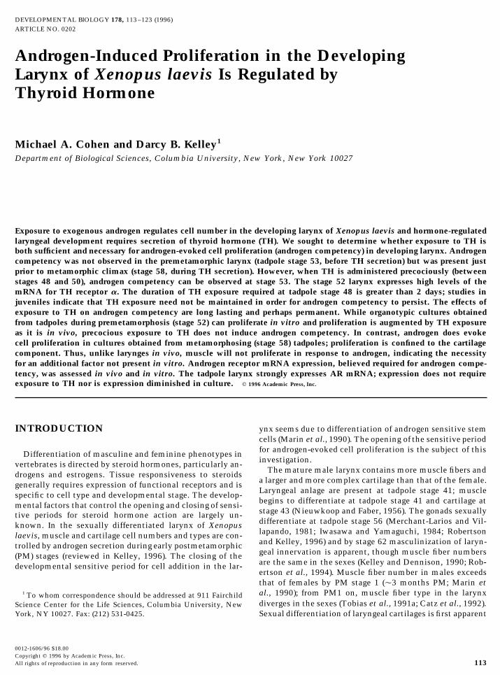
FIG. 1. Diagrammatic representation of the experimental paradigm illustrating the time course of administration of thyroxine (T4) and/or dihydrotestosterone (DHT). All tadpoles received an injection of [3H]thymidine at stage 53 and were killed 30 min later.
tional 100 ml serum was then added, and the mixture stirred over- 4% paraformaldehyde at 47C. Cultured laryngeal explants werefixed in 4% paraformaldehyde for 2 hr at room temperature fol-night and then centrifuged for 30 min at 10,000g, after which the
stripped serum was added to the medium. Because T4 might not lowed by immersion in cresyl violet solution. Tissue was thendehydrated through an ethanol series and cleared in methyl sali-be as efficiently converted to the more active form triiodothyronine
(T3) in vitro as in tadpoles, for laryngeal explants T3 (Sigma) wasused. T3 was dissolved in 0.5 M NaOH and added to the mediumto produce a final concentration of 1 1 1008M.
Hormone treatment paradigms paralleled those used in vivo; pro-liferating cells in all explants were labeled at Day 16. Culture me-dium was supplemented with [3H]thymidine (4 mCi/ml) for 4 hr.The laryngeal explant was then fixed in 10% neutral buffered For-malin for at least 24 hr. Tissue was then cresyl violet stained enbloc and embedded in paraplast as described above.
The development of laryngeal explants was also followed withthe aid of bromodeoxyuridine (BrdU, Boeringer-Mannheim) im-munocytochemistry in order to visualize zones of proliferationin the entire explant in whole mount. The culture medium wassupplemented with 50 mM BrdU at Day 16 for 4 hr. Explants wererinsed with culture medium and fixed with ice-cold absoluteethanol. Tissue was then hydrated in ethanol and incubated in2 N HCl with 0.5% Tween 20 at 377C for 8 min. A monoclonalantibody against BrdU (Boeringer-Mannheim) was used at a dilu-tion of 1:100 overnight at 47C. Tissue was incubated for 1 hrwith Texas red-conjugated goat anti-mouse (Jackson ImmunoRe-search; preadsorbed with laryngeal cartilage at room temperaturefor 1 hr) at room temperature. After rinsing with PBS, the tissuewas mounted in glycerol and then examined using confocal lasermicroscopy.
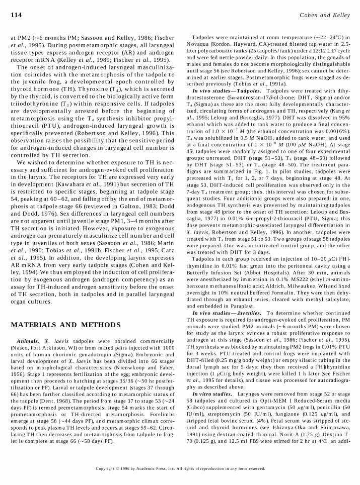
Expression of androgen and thyroid hormone receptor mRNAin larynx. To determine whether larynx expresses the relevantreceptors, expression of AR mRNA (He et al., 1990) and TH re-ceptor a (TRa) mRNA (Yaoita et al., 1990) was examined usingin situ hybridization as in Fischer et al. (1995) with the followingmodifications. The AR mRNA probe was the cDNA fragment FIG. 2. The effects of hormone treatment on number of labeled
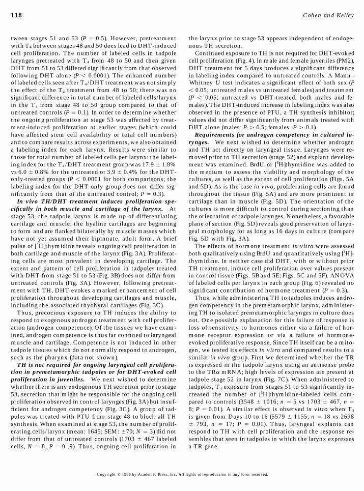
cells per larynx in tadpoles; treatment groups as in Fig. 1. Valuesdescribed previously (He et al., 1990) which contains a portionof the ligand-binding and DNA-binding domains. The TRa probe given are means { standard error of the mean (SEM); the number
of animals in each group is indicated. The mean value for the TH/used was transcribed from pXTRaAA/BS (gift of D. Brown), acDNA from the constituitively expressed TRa mRNA (Yaoita et DHT group differed significantly from the mean value of the un-
treated group; the mean values for the other groups did not differal., 1990). The TRa probe was used only on tadpole stage 52larynges. Tadpole larynges were collected and fixed overnight in significantly from the control value.
Copyright q 1996 by Academic Press, Inc. All rights of reproduction in any form reserved.
AID DB 8296 / 6x11$$$$81 07-30-96 09:57:19 dbas AP: Dev Bio
116 Cohen and Kelley
Autoradiography and data analysis. Tissue from both in vitroand in vivo experiments was serially sectioned at 10 mm. After clear-ing in xylene and rehydrating through an ethanol series, slides weredipped in nuclear track emulsion (NTB-2, Kodak) and exposed for 6weeks (in vitro) or 3 weeks (in vivo) at 47C. Slides were developedin Kodak D-19 developer and counterstained with cresyl violet.
For each tadpole, the total number of labeled cells per larynx wasdetermined by counting labeled cells in every sixth section andthen multiplying by 6. A group mean and standard error of themean was then calculated for each treatment group. A labelingindex was also determined as described previously (Fischer et al.,1995). Counts of labeled cells were determined for three fields (170mm2) per larynx from an area adjacent to the thyohyrals. The ratioof the number of labeled nuclei divided by the total (labeled plusunlabeled) number of nuclei was determined for each field andaveraged to yield a labeling index for each larynx. For laryngealcultures from stage 53 tadpoles, three, equally spaced, transversesections through (25, 50, and 75%) the larynx were chosen, thenumber of labeled cells in the entire section was counted, an aver-age per section obtained, and a total determined by multiplying bythe total number of sections. For laryngeal cultures from stage 58tadpoles, six equally spaced sections were used. Because orientationof the sections was difficult to determine in explants, the abovemethod provided a more representative estimate of proliferationthan that used for in vivo studies.
Statistical analysis. Differences in the number of labeledcells in stage 53 tadpole larynges examined in vivo and in vitrowere assessed using a one-way analysis of variance (ANOVA)with Fisher’s protected least significant difference (PLSD) as apost-hoc test (two-tailed distribution, P £ 0.05; Statview, Aba-cus, Berkeley, CA). For the stage 58 larynges, a Student’s t test(two-tailed distribution, P £ 0.05) was used to compare the num-ber of labeled cells with and without DHT treatment, for eachtissue type (muscle or cartilage) under the two observation condi-tions (in vivo and in vitro). To assess differences in labeling indi-
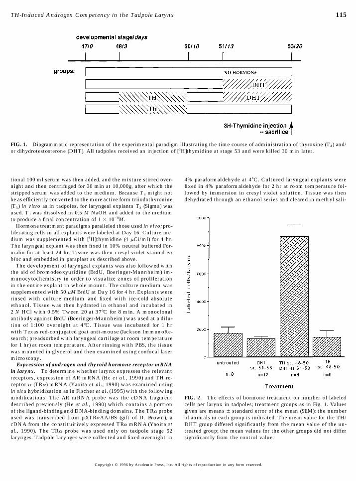
FIG. 3. Representative transverse autoradiographic sectionsthrough the larynx of stage 53 tadpoles prepared following [3H]-thymidine injection. Sections were exposed to nuclear emulsionfor 6 weeks, developed, and counterstained with cresyl violet.(A) Untreated. (B) DHT-treated from stage 51–53. (C) TH-treatedfrom stage 48–50 and then DHT-treated from stage 51–53. Scalebar, 100 mm. Abbreviations: m, muscle; c, cartilage; th, thyo-hyrals.
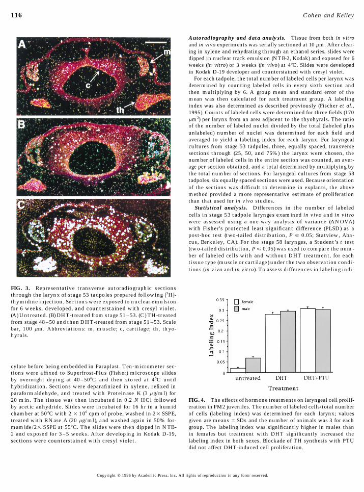
cylate before being embedded in Paraplast. Ten-micrometer sec-tions were affixed to Superfrost-Plus (Fisher) microscope slidesby overnight drying at 40–507C and then stored at 47C untilhybridization. Sections were deparafinized in xylene, refixed inparaformaldehyde, and treated with Proteinase K (3 mg/ml) for20 min. The tissue was then incubated in 0.2 N HCl followed FIG. 4. The effects of hormone treatments on laryngeal cell prolif-
eration in PM2 juveniles. The number of labeled cells/total numberby acetic anhydride. Slides were incubated for 16 hr in a humidchamber at 507C with 2 1 106 cpm of probe, washed in 21 SSPE, of cells (labeling index) was determined for each larynx; values
given are means { SDs and the number of animals was 3 for eachtreated with RNase A (20 mg/ml), and washed again in 50% for-mamide/21 SSPE at 557C. The slides were then dipped in NTB- group. The labeling index was significantly higher in males than
in females but treatment with DHT significantly increased the2 and exposed for 3–5 weeks. After developing in Kodak D-19,sections were counterstained with cresyl violet. labeling index in both sexes. Blockade of TH synthesis with PTU
did not affect DHT-induced cell proliferation.
Copyright q 1996 by Academic Press, Inc. All rights of reproduction in any form reserved.
AID DB 8296 / 6x11$$$$81 07-30-96 09:57:19 dbas AP: Dev Bio
117TH-Induced Androgen Competency in the Tadpole Larynx
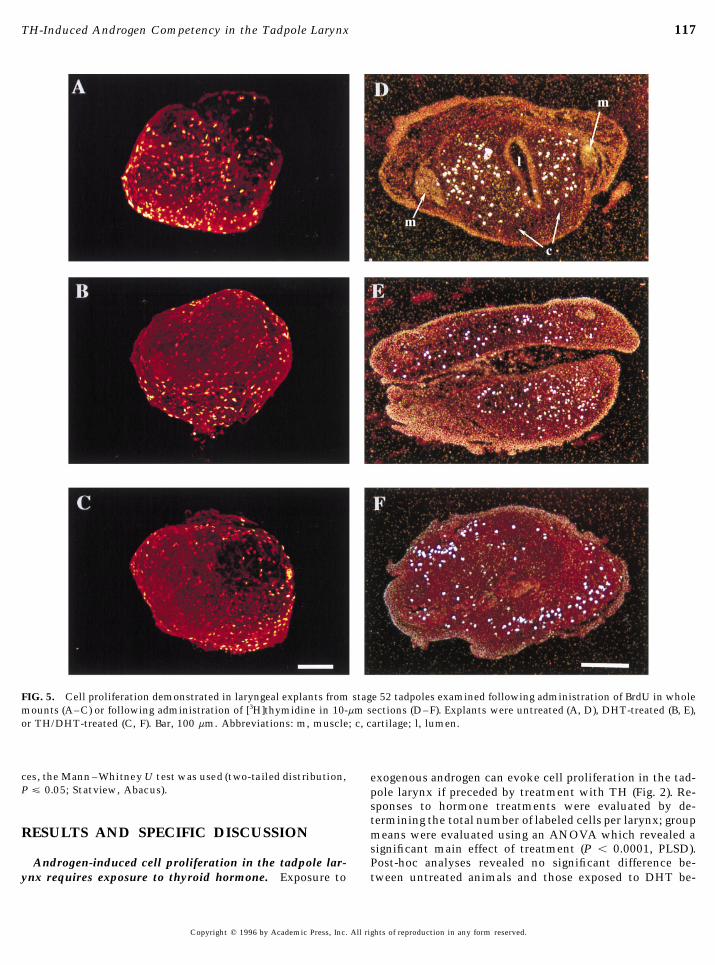
FIG. 5. Cell proliferation demonstrated in laryngeal explants from stage 52 tadpoles examined following administration of BrdU in wholemounts (A–C) or following administration of [3H]thymidine in 10-mm sections (D–F). Explants were untreated (A, D), DHT-treated (B, E),or TH/DHT-treated (C, F). Bar, 100 mm. Abbreviations: m, muscle; c, cartilage; l, lumen.
ces, the Mann –Whitney U test was used (two-tailed distribution, exogenous androgen can evoke cell proliferation in the tad-P £ 0.05; Statview, Abacus). pole larynx if preceded by treatment with TH (Fig. 2). Re-
sponses to hormone treatments were evaluated by de-termining the total number of labeled cells per larynx; group
RESULTS AND SPECIFIC DISCUSSION means were evaluated using an ANOVA which revealed asignificant main effect of treatment (P õ 0.0001, PLSD).
Androgen-induced cell proliferation in the tadpole lar- Post-hoc analyses revealed no significant difference be-tween untreated animals and those exposed to DHT be-ynx requires exposure to thyroid hormone. Exposure to
Copyright q 1996 by Academic Press, Inc. All rights of reproduction in any form reserved.
AID DB 8296 / 6x11$$$$81 07-30-96 09:57:19 dbas AP: Dev Bio
118 Cohen and Kelley
tween stages 51 and 53 (P Å 0.5). However, pretreatment the larynx prior to stage 53 appears independent of endoge-nous TH secretion.with T4 between stages 48 and 50 does lead to DHT-induced
cell proliferation. The number of labeled cells in tadpole Continued exposure to TH is not required for DHT-evokedcell proliferation (Fig. 4). In male and female juveniles (PM2),larynges pretreated with T4 from 48 to 50 and then given
DHT from 51 to 53 differed significantly from that observed DHT treatment for 5 days produces a significant differencein labeling index compared to untreated controls. A Mann–following DHT alone (P õ 0.0001). The enhanced number
of labeled cells seen after T4/DHT treatment was not simply Whitney U test indicates a significant effect of both sex (Põ 0.05; untreated males vs untreated females) and treatmentthe effect of the T4 treatment from 48 to 50; there was no
significant difference in total number of labeled cells/larynx (P õ 0.05; untreated vs DHT-treated, both males and fe-males). The DHT-induced increase in labeling index was alsoin the T4 from stage 48 to 50 group compared to that of
untreated controls (P Å 0.1). In order to determine whether observed in the presence of PTU, a TH synthesis inhibitor;values did not differ significantly from animals treated withthe ongoing proliferation at stage 53 was affected by treat-
ment-induced proliferation at earlier stages (which could DHT alone (males: P ú 0.5; females: P ú 0.1).Requirements for androgen competency in cultured la-have affected stem cell availability or total cell numbers)
and to compare results across experiments, we also obtained rynges. We next wished to determine whether androgenand TH act directly on laryngeal tissue. Larynges were re-a labeling index for each larynx. Results were similar to
those for total number of labeled cells per larynx: the label- moved prior to TH secretion (stage 52) and explant develop-ment was examined. BrdU or [3H]thymidine was added toing index for the T4/DHT treatment group was 17.9 { 1.8%
vs 6.0 { 0.8% for the untreated or 3.9 { 0.4% for the DHT- the medium to assess the viability and morphology of thecultures, as well as the extent of cell proliferation (Figs. 5Aonly-treated groups (P õ 0.0001 for both comparisons; the
labeling index for the DHT-only group does not differ sig- and 5D). As is the case in vivo, proliferating cells are foundthroughout the tissue (Fig. 5A) and are more prominent innificantly from that of the untreated control; P Å 0.3).
In vivo TH/DHT treatment induces proliferation spe- cartilage than in muscle (Fig. 5D). The orientation of thecultures is more difficult to control during sectioning thancifically in both muscle and cartilage of the larynx. At
stage 53, the tadpole larynx is made up of differentiating the orientation of tadpole larynges. Nonetheless, a favorableplane of section (Fig. 5D) reveals good preservation of laryn-cartilage and muscle; the hyaline cartilages are beginning
to form and are flanked bilaterally by muscle masses which geal morphology for as long as 16 days in culture (compareFig. 5D with Fig. 3A).have not yet assumed their bipinnate, adult form. A brief
pulse of [3H]thymidine reveals ongoing cell proliferation in The effects of hormone treatment in vitro were assessedboth qualitatively using BrdU and quantitatively using [3H]-both cartilage and muscle of the larynx (Fig. 3A). Proliferat-
ing cells are most prevalent in developing cartilage. The thymidine. In neither case did DHT, with or without priorTH treatment, induce cell proliferation over values presentextent and pattern of cell proliferation in tadpoles treated
with DHT from stage 51 to 53 (Fig. 3B) does not differ from in control tissue (Figs. 5B and 5E; Figs. 5C and 5F). ANOVAof labeled cells per larynx in each group (Fig. 6) revealed nountreated controls (Fig. 3A). However, following pretreat-
ment with TH, DHT evokes a marked enhancement of cell significant contribution of hormone treatment (P Å 0.3).Thus, while administering TH to tadpoles induces andro-proliferation throughout developing cartilages and muscle,
including the associated thyohyral cartilages (Fig. 3C). gen competency in the premetamorphic larynx, administer-ing TH to isolated premetamorphic larynges in culture doesThus, precocious exposure to TH induces the ability to
respond to exogenous androgen treatment with cell prolifer- not. One possible explanation for this failure of response isloss of sensitivity to hormones either via a failure of hor-ation (androgen competence). Of the tissues we have exam-
ined, androgen competence is thus far confined to laryngeal mone receptor expression or via a failure of hormone-evoked proliferative response. Since TH itself can be a mito-muscle and cartilage. Competence is not induced in other
tadpole tissues which do not normally respond to androgen, gen, we tested its effects in vitro and compared results to asimilar in vivo group. First we determined whether the TRsuch as the pharynx (data not shown).
TH is not required for ongoing laryngeal cell prolifera- is expressed in the tadpole larynx using an antisense probeto the TRa mRNA; high levels of expression are present attion in premetamorphic tadpoles or for DHT-evoked cell
proliferation in juveniles. We next wished to determine tadpole stage 52 in larynx (Fig. 7C). When administered totadpoles, T4 exposure from stages 51 to 53 significantly in-whether there is any endogenous TH secretion prior to stage
53, secretion that might be responsible for the ongoing cell creased the number of [3H]thymidine-labeled cells com-pared to controls (3548 { 1016; n Å 5 vs 1703 { 467, n Åproliferation observed in control larynges (Fig. 3A) but insuf-
ficient for androgen competency (Fig. 3C). A group of tad- 8; P Å 0.01). A similar effect is observed in vitro when T3
is given from Days 10 to 16 (5579 { 1155; n Å 18 vs 2698poles was treated with PTU from stage 48 to block all THsynthesis. When examined at stage 53, the number of prolif- { 793, n Å 17; P Å 0.01). Thus, laryngeal explants can
respond to TH with cell proliferation and the response re-erating cells/larynx (mean: 1645; SEM: {70; N Å 3) did notdiffer from that of untreated controls (1703 { 467 labeled sembles that seen in tadpoles in which the larynx expresses
a TR gene.cells, N Å 8, P Å 0 .9). Thus, ongoing cell proliferation in
Copyright q 1996 by Academic Press, Inc. All rights of reproduction in any form reserved.
AID DB 8296 / 6x11$$$$81 07-30-96 09:57:19 dbas AP: Dev Bio
119TH-Induced Androgen Competency in the Tadpole Larynx
gen-evoked proliferation can be demonstrated by both la-ryngeal cartilage and muscle in tadpoles, androgen compe-tency can only be demonstrated in cartilage in isolatedlarynges, suggesting that an extralaryngeal influence maybe required for muscle.
DISCUSSION
The control of cell proliferation by steroid hormonesplays an important role in the development of target tissues.The ability of these hormones, primarily androgens and es-trogens, to induce mitotic activity is often developmentallyregulated and can be confined to precise sensitive periods(reviewed in Kelley, 1986; 1992). The factors which governthe opening and closing of these periods are by and largeunknown. The larynx of X. laevis is sexually dimorphicwith respect to cell number as a result of an androgen-driven
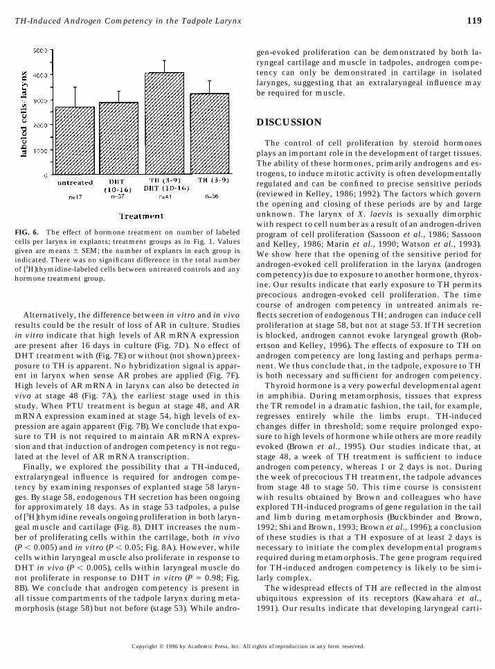
FIG. 6. The effect of hormone treatment on number of labeled program of cell proliferation (Sassoon et al., 1986; Sassooncells per larynx in explants; treatment groups as in Fig. 1. Values and Kelley, 1986; Marin et al., 1990; Watson et al., 1993).given are means { SEM; the number of explants in each group is We show here that the opening of the sensitive period forindicated. There was no significant difference in the total number androgen-evoked cell proliferation in the larynx (androgenof [3H]thymidine-labeled cells between untreated controls and any
competency) is due to exposure to another hormone, thyrox-hormone treatment group.ine. Our results indicate that early exposure to TH permitsprecocious androgen-evoked cell proliferation. The timecourse of androgen competency in untreated animals re-flects secretion of endogenous TH; androgen can induce cellAlternatively, the difference between in vitro and in vivo
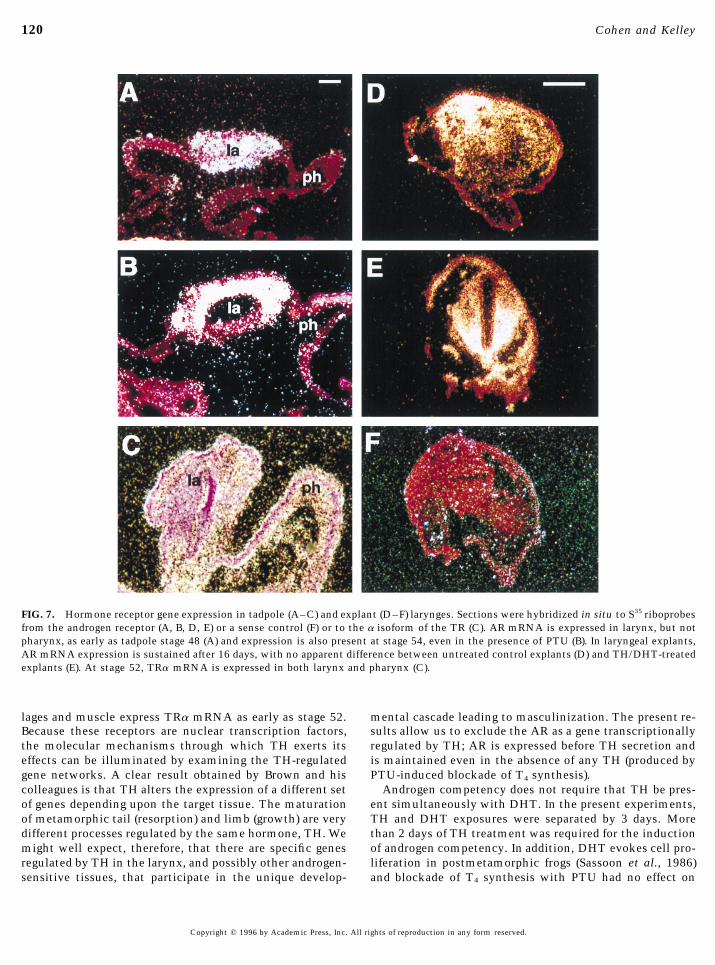
results could be the result of loss of AR in culture. Studies proliferation at stage 58, but not at stage 53. If TH secretionis blocked, androgen cannot evoke laryngeal growth (Rob-in vitro indicate that high levels of AR mRNA expression
are present after 16 days in culture (Fig. 7D). No effect of ertson and Kelley, 1996). The effects of exposure to TH onandrogen competency are long lasting and perhaps perma-DHT treatment with (Fig. 7E) or without (not shown) preex-
posure to TH is apparent. No hybridization signal is appar- nent. We thus conclude that, in the tadpole, exposure to THis both necessary and sufficient for androgen competency.ent in larynx when sense AR probes are applied (Fig. 7F).
High levels of AR mRNA in larynx can also be detected in Thyroid hormone is a very powerful developmental agentin amphibia. During metamorphosis, tissues that expressvivo at stage 48 (Fig. 7A), the earliest stage used in this
study. When PTU treatment is begun at stage 48, and AR the TR remodel in a dramatic fashion, the tail, for example,regresses entirely while the limbs erupt. TH-inducedmRNA expression examined at stage 54, high levels of ex-
pression are again apparent (Fig. 7B). We conclude that expo- changes differ in threshold; some require prolonged expo-sure to high levels of hormone while others are more readilysure to TH is not required to maintain AR mRNA expres-
sion and that induction of androgen competency is not regu- evoked (Brown et al., 1995). Our studies indicate that, atstage 48, a week of TH treatment is sufficient to inducelated at the level of AR mRNA transcription.
Finally, we explored the possibility that a TH-induced, androgen competency, whereas 1 or 2 days is not. Duringthe week of precocious TH treatment, the tadpole advancesextralaryngeal influence is required for androgen compe-
tency by examining responses of explanted stage 58 laryn- from stage 48 to stage 50. This time course is consistentwith results obtained by Brown and colleagues who haveges. By stage 58, endogenous TH secretion has been ongoing
for approximately 18 days. As in stage 53 tadpoles, a pulse explored TH-induced programs of gene regulation in the tailand limb during metamorphosis (Buckbinder and Brown,of [3H]thymidine reveals ongoing proliferation in both laryn-
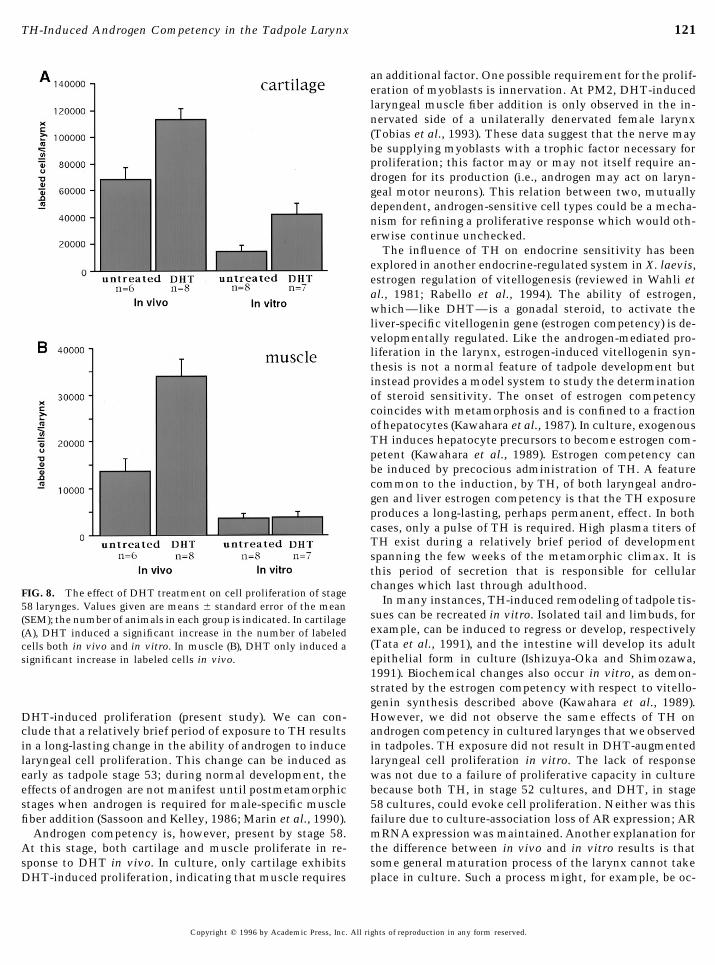
geal muscle and cartilage (Fig. 8). DHT increases the num- 1992; Shi and Brown, 1993; Brown et al., 1996); a conclusionof these studies is that a TH exposure of at least 2 days isber of proliferating cells within the cartilage, both in vivo
(Põ 0.005) and in vitro (Põ 0.05; Fig. 8A). However, while necessary to initiate the complex developmental programsrequired during metamorphosis. The gene program requiredcells within laryngeal muscle also proliferate in response to
DHT in vivo (P õ 0.005), cells within laryngeal muscle do for TH-induced androgen competency is likely to be simi-larly complex.not proliferate in response to DHT in vitro (P Å 0.98; Fig.
8B). We conclude that androgen competency is present in The widespread effects of TH are reflected in the almostubiquitous expression of its receptors (Kawahara et al.,all tissue compartments of the tadpole larynx during meta-
morphosis (stage 58) but not before (stage 53). While andro- 1991). Our results indicate that developing laryngeal carti-
Copyright q 1996 by Academic Press, Inc. All rights of reproduction in any form reserved.
AID DB 8296 / 6x11$$$$81 07-30-96 09:57:19 dbas AP: Dev Bio
120 Cohen and Kelley
FIG. 7. Hormone receptor gene expression in tadpole (A–C) and explant (D–F) larynges. Sections were hybridized in situ to S35 riboprobesfrom the androgen receptor (A, B, D, E) or a sense control (F) or to the a isoform of the TR (C). AR mRNA is expressed in larynx, but notpharynx, as early as tadpole stage 48 (A) and expression is also present at stage 54, even in the presence of PTU (B). In laryngeal explants,AR mRNA expression is sustained after 16 days, with no apparent difference between untreated control explants (D) and TH/DHT-treatedexplants (E). At stage 52, TRa mRNA is expressed in both larynx and pharynx (C).
lages and muscle express TRa mRNA as early as stage 52. mental cascade leading to masculinization. The present re-sults allow us to exclude the AR as a gene transcriptionallyBecause these receptors are nuclear transcription factors,
the molecular mechanisms through which TH exerts its regulated by TH; AR is expressed before TH secretion andis maintained even in the absence of any TH (produced byeffects can be illuminated by examining the TH-regulated
gene networks. A clear result obtained by Brown and his PTU-induced blockade of T4 synthesis).Androgen competency does not require that TH be pres-colleagues is that TH alters the expression of a different set
of genes depending upon the target tissue. The maturation ent simultaneously with DHT. In the present experiments,TH and DHT exposures were separated by 3 days. Moreof metamorphic tail (resorption) and limb (growth) are very
different processes regulated by the same hormone, TH. We than 2 days of TH treatment was required for the inductionof androgen competency. In addition, DHT evokes cell pro-might well expect, therefore, that there are specific genes
regulated by TH in the larynx, and possibly other androgen- liferation in postmetamorphic frogs (Sassoon et al., 1986)and blockade of T4 synthesis with PTU had no effect onsensitive tissues, that participate in the unique develop-
Copyright q 1996 by Academic Press, Inc. All rights of reproduction in any form reserved.
AID DB 8296 / 6x11$$$$81 07-30-96 09:57:19 dbas AP: Dev Bio
121TH-Induced Androgen Competency in the Tadpole Larynx
an additional factor. One possible requirement for the prolif-eration of myoblasts is innervation. At PM2, DHT-inducedlaryngeal muscle fiber addition is only observed in the in-nervated side of a unilaterally denervated female larynx(Tobias et al., 1993). These data suggest that the nerve maybe supplying myoblasts with a trophic factor necessary forproliferation; this factor may or may not itself require an-drogen for its production (i.e., androgen may act on laryn-geal motor neurons). This relation between two, mutuallydependent, androgen-sensitive cell types could be a mecha-nism for refining a proliferative response which would oth-erwise continue unchecked.
The influence of TH on endocrine sensitivity has beenexplored in another endocrine-regulated system in X. laevis,estrogen regulation of vitellogenesis (reviewed in Wahli etal., 1981; Rabello et al., 1994). The ability of estrogen,which—like DHT—is a gonadal steroid, to activate theliver-specific vitellogenin gene (estrogen competency) is de-velopmentally regulated. Like the androgen-mediated pro-liferation in the larynx, estrogen-induced vitellogenin syn-thesis is not a normal feature of tadpole development butinstead provides a model system to study the determinationof steroid sensitivity. The onset of estrogen competencycoincides with metamorphosis and is confined to a fractionof hepatocytes (Kawahara et al., 1987). In culture, exogenousTH induces hepatocyte precursors to become estrogen com-petent (Kawahara et al., 1989). Estrogen competency canbe induced by precocious administration of TH. A featurecommon to the induction, by TH, of both laryngeal andro-gen and liver estrogen competency is that the TH exposureproduces a long-lasting, perhaps permanent, effect. In bothcases, only a pulse of TH is required. High plasma titers ofTH exist during a relatively brief period of developmentspanning the few weeks of the metamorphic climax. It isthis period of secretion that is responsible for cellularchanges which last through adulthood.
FIG. 8. The effect of DHT treatment on cell proliferation of stageIn many instances, TH-induced remodeling of tadpole tis-58 larynges. Values given are means { standard error of the mean
sues can be recreated in vitro. Isolated tail and limbuds, for(SEM); the number of animals in each group is indicated. In cartilageexample, can be induced to regress or develop, respectively(A), DHT induced a significant increase in the number of labeled(Tata et al., 1991), and the intestine will develop its adultcells both in vivo and in vitro. In muscle (B), DHT only induced aepithelial form in culture (Ishizuya-Oka and Shimozawa,significant increase in labeled cells in vivo.1991). Biochemical changes also occur in vitro, as demon-strated by the estrogen competency with respect to vitello-genin synthesis described above (Kawahara et al., 1989).However, we did not observe the same effects of TH onDHT-induced proliferation (present study). We can con-
clude that a relatively brief period of exposure to TH results androgen competency in cultured larynges that we observedin tadpoles. TH exposure did not result in DHT-augmentedin a long-lasting change in the ability of androgen to induce
laryngeal cell proliferation. This change can be induced as laryngeal cell proliferation in vitro. The lack of responsewas not due to a failure of proliferative capacity in cultureearly as tadpole stage 53; during normal development, the
effects of androgen are not manifest until postmetamorphic because both TH, in stage 52 cultures, and DHT, in stage58 cultures, could evoke cell proliferation. Neither was thisstages when androgen is required for male-specific muscle
fiber addition (Sassoon and Kelley, 1986; Marin et al., 1990). failure due to culture-association loss of AR expression; ARmRNA expression was maintained. Another explanation forAndrogen competency is, however, present by stage 58.
At this stage, both cartilage and muscle proliferate in re- the difference between in vivo and in vitro results is thatsome general maturation process of the larynx cannot takesponse to DHT in vivo. In culture, only cartilage exhibits
DHT-induced proliferation, indicating that muscle requires place in culture. Such a process might, for example, be oc-
Copyright q 1996 by Academic Press, Inc. All rights of reproduction in any form reserved.
AID DB 8296 / 6x11$$$$81 07-30-96 09:57:19 dbas AP: Dev Bio
122 Cohen and Kelley
receptor expression and tissue differentiation. Dev. Biol. 170,curring between stages 48 (when we begin TH treatment)115–126.and 58 (when endogenous TH titers are high) and could be
Galton, V. A. (1983). In ‘‘Thyroid Hormone Action in Amphibianconnected with developmental changes in innervation (seeMetamorphosis’’ (J. H. Oppenheimer and H. H. Samuels, Eds.),above) or in circulating factors.pp. 445–483. Academic Press, New York.In summary, we have analyzed the onset of androgen sen-
He, W., Fischer, L., Sun, S., Bilhartz, D., Zhu, X., Young, C., Kelley.sitivity in the larynx of X. laevis and the contribution of D., and Tindall, D. (1990). Molecular cloning of androgen receptorTH to this process. The larynx undergoes remodeling during from divergent species with the PCR technique: Complete cDNAdevelopment in response to androgen, resulting in a sexu- sequence of the mouse androgen receptor and isolation of cDNAally dimorphic vocal system. The onset of sensitivity to probes from dog, guinea pig, and frog. Biochem. Biophys. Res.androgen in the larynx, or androgen competency, is regu- Commun. 171, 697–704.
Iwasawa, H., and Yamaguchi, L. (1984). Ultrastructural study oflated by TH. We can conclude that a major action of TH isgonadal development in Xenopus laevis. Zool. Sci. 1, 591–600.controlling the temporal boundaries for sensitive periods of
Ishizuya-Oka, A., and Shimozawa, A. (1991). Induction of metamor-steroid hormone action. An understanding of how thesephosis by thyroid hormone in anuran small intestine culturedtemporal boundaries are created and impact on embryogen-organotypically in vitro. In Vitro Cell. Dev. Biol. 11, 853 –857.esis now requires analysis of the molecular mechanisms
Kang, L., Marin, M., and Kelley, D. B. (1995). Androgen biosynthesisinvolved.and secretion in developing Xenopus laevis. Gen. Comp. Endocri-nol. 100, 293–307.
Kawahara, A., Kohara, S., and Amano, M. (1989). Thyroid hormonedirectly induces hepatocyte competence for estrogen-dependentACKNOWLEDGMENTSvitellogein synthesis during the metamorphosis of Xenopuslaevis. Dev. Biol. 132, 73–80.We thank Martha Tobias, Joseph Thornton, and Flavio Kamenetz
Kawahara, A., Kohara, S., Sugimoto, Y., and Amano, M. (1987). Afor comments on the manuscript, Don Brown for the gift of thechange of the hepatocyte population is responsible for the pro-TRa probe (pXTRa AA/BS), and Dominique Toran-Allerand for agressive increase of vitellogenin synthetic capacity at and afterprotocol for preparing steroid-free sera. Supported by NS 19949.metamorphosis of Xenopus laevis. Dev. Biol. 122, 139–145.
Kawahara, A., Baker, B. S., and Tata, J. R. (1991). Developmentaland regional expression of thyroid hormone genes during Xeno-pus metamorphosis. Development 112, 933–943.REFERENCES
Kelley, D. B. (1992). Opening and closing a hormone-regulated pe-riod for the development of courtship song. A cellular and molec-Buckbinder, L., and Brown, D. D. (1992). Thyroid hormone-inducedular analysis of vocal effectors. Dev. Psychobiol. 662, 178–188.gene expression changes in the developing frog limb. J. Biol.
Kelley, D. B. (1996). Sexual differentiation in Xenopus laevis. InChem. 267, 25786–25791.‘‘The Biology of Xenopus’’ (R. Tinsley and H. Kobel, Eds.), pp.Brown, D. D., Wang, Z., Furlow, J. D., Kanamori, A., Schartzman,143–176. Oxford Univ. Press, Oxford.R. A., Remo, B. F., and Pinder, A. (1996). The thyroid hormone-
Kelley, D. B., and Dennison, J. (1990). The vocal motor neurons ofinduced tail resorption program during Xenopus laevis metamor-Xenopus laevis: Development of sex differences in axon number.phosis. Proc. Natl. Acad. Sci. 93, 1924–1929.J. Neurobiol. 21, 869–882.Brown, D. D., Wang , Z., Kanamori, A., Eliceiri, B., Furlow, J. D., and
Kelley, D. B., Sassoon, D., Segil, N., and Scudder, M. (1989). Devel-Schwartzman, R. (1995). Amphibian metamorphosis: A complexopment and hormone regulation of androgen receptor levels inprogram of gene expression changes controlled by the thyroidthe sexually dimorphic larynx of Xenopus laevis. Dev. Biol., 131,hormone. [Review] Recent Prog. Hormone Res. 50, 309–315.111–118.Catz, D., Fischer, L., Moschella, T., Tobias, M. L., and Kelley,
Leloup, J., and Buscaglia, M. (1977). La triodothyronine, hormoneD. B. (1992). Sexually dimorphic expression of a laryngeal-spe-de la metamorphose des Amphibiens. C. R. Seances Acad. Sci.cific, androgen-regulated myosin heavy chain gene during Xeno-284, 2261–2263.pus laevis development. Dev. Biol. 154, 65–80.
Marin, M., Tobias, M., and Kelley, D. (1990). Hormone sensitiveCatz, D. S., Fischer, L. M., and Kelley, D. B. (1995). Androgen regula-stages in the sexual differentiation of laryngeal muscle fiber num-tion of a laryngeal-specific myosin heavy chain mRNA whoseber in Xenopus laevis. Development 110, 703–710.expression is sexually differentiated. Dev. Biol. 171, 448–457.
Merchant-Larios, H., and Villalpando, I. (1981). UltrastructuralCohen, M. A., and Kelley, D. B. (1994). Thyroxine exposure governsevents during early gonadal development in Rana pipiens andthe onset of androgen sensitivity in the larynx of Xenopus laevis.Xenopus laevis. Anat. Rec. 199, 349 –360.Dev. Biol. 163, 548a.
Nieuwkoop, P. D., and Faber, J. (1956). ‘‘Normal Table of XenopusDent, J. N. (1968). Survey of amphibian metamorphosis. In ‘‘Meta-laevis (Daudin).’’ North-Holland, Amsterdam.morphosis, Problem in Developmental Biology’’ (W. Etkin and
Rabello, E. M. L., Baker, B. S., and Tata, J. R. (1994). InterplayL. I. Gilbert, Eds.), pp. 271–311. Appleton–Century–Crofts, Newbetween thyroid hormone and estrogen in modulating expressionYork.of their receptors and vitellogenin genes during Xenopus meta-Dodd, M., and Dodd, J. (1976). The biology of metamorphosis. Inmorphosis. Mech. Dev. 45, 49 –57.‘‘Physiology of the Amphibia’’ (B. Lofts, Ed.), Vol III, pp. 467–
Robertson, J. C., and Kelley, D. B. (1996). Thyroid hormone controls529. Academic Press, New York.the onset of androgen sensitivity in Xenopus laevis. Dev. Biol.Fischer, L. M., Catz, D., and Kelley, D. B. (1995). Androgen-directed
development of the Xenopus laevis larynx: Control of androgen 176, 108–123.
Copyright q 1996 by Academic Press, Inc. All rights of reproduction in any form reserved.
AID DB 8296 / 6x11$$$$81 07-30-96 09:57:19 dbas AP: Dev Bio
123TH-Induced Androgen Competency in the Tadpole Larynx
Robertson, J., Watson, J., and Kelley, D. B. (1994). Androgen directs Tobias, M. L., Marin, M., and Kelley, D. B. (1991b). Temporal con-straints on androgen directed laryngeal masculinization in Xeno-sexual differentiation of laryngeal innervation in developing Xen-
opus laevis. J. Neurobiol. 25, 1625–1636. pus laevis. Dev. Biol. 147, 260–270.Tobias, M. L., Marin, M., and Kelley, D. B. (1993). The roles of sex,Sassoon, D., and Kelley, D. (1986). The sexually dimorphic larynx
of Xenopus laevis: Development and androgen regulation. Am. innervation, and androgen in laryngeal muscle fibers of Xenopuslaevis. J. Neurosci. 13, 324–333.J. Anat. 117, 457–472.
Sassoon, D., Segil, N., and Kelley, D. (1986). Androgen-induced Wahli, W., Dawid, I. B., Ryffel, G. U., and Weber, T. (1981). Vitello-genesis and the vitellogenin gene family. Science 212, 298–304.myogenesis and chondrogenesis in the larynx of Xenopus laevis.
Dev. Biol. 113, 135–140. Watson, J., Robertson, J., Sachdev, U., and Kelley, D. (1993). Laryn-Shi, Y. B., and Brown, D. D. (1993). The earliest changes in gene geal muscle and motor neuron plasticity in Xenopus laevis: Anal-
expression in tadpole intestine induced by thyroid hormone. J. ysis of a sensitive period for testicular masculinization of a neuro-Biol. Chem. 268, 20312–20317. muscular system. J. Neurobiol. 24, 1615–1625.
Tata, J. R., Kawahara, A., and Baker, B. S. (1991). Prolactin inhibits Yaoita, Y., Shi, Y. B., and Brown, D. D. (1990). Xenopus laevis alphaboth thyroid hormone-induced morphogenesis and cell death in and beta thyroid hormone receptors Proc. Natl. Acad. Sci. USAcultured amphibian larval tissues. Dev. Biol. 146, 72–80. 87, 7090–7094.
Tobias, M. L., Marin, M., and Kelley, D. B. (1991a). DevelopmentReceived for publication April 4, 1996of functional sex differences in the larynx of Xenopus laevis.
Dev. Biol. 147, 251–259. Accepted June 18, 1996
Copyright q 1996 by Academic Press, Inc. All rights of reproduction in any form reserved.
AID DB 8296 / 6x11$$$$81 07-30-96 09:57:19 dbas AP: Dev Bio