andreas vesalius: celebrating 500 years of dissecting … · open access review article andreas...
TRANSCRIPT
Seediscussions,stats,andauthorprofilesforthispublicationat:https://www.researchgate.net/publication/292672408
AndreasVesalius:Celebrating500yearsofdissectingnature
Article·October2015
DOI:10.5339/gcsp.2015.66
READS
2
4authors,including:
FabioZampieri
UniversityofPaduaMedicalSchool
52PUBLICATIONS74CITATIONS
SEEPROFILE
MohamedElmaghawry
AswanHeartCentre
38PUBLICATIONS82CITATIONS
SEEPROFILE
AlbertoZanatta
UniversityofPadova
36PUBLICATIONS22CITATIONS
SEEPROFILE
Allin-textreferencesunderlinedinbluearelinkedtopublicationsonResearchGate,
lettingyouaccessandreadthemimmediately.
Availablefrom:AlbertoZanatta
Retrievedon:16June2016
OPEN ACCESS Review article
Andreas Vesalius: Celebrating500 years of dissecting natureFabio Zampieri1, Mohamed ElMaghawry2,*, Alberto Zanatta1, Gaetano Thiene1
“ . . . it is better to dissect nature, than to reduce her to abstraction”
Francis Bacon, Novum oragnum scientiarum, 1620
INTRODUCTION
December 31st, 2014 marked the 500-year anniversary of the birth of Andreas Vesalius. Vesalius,
considered as the founder of modern anatomy, had profoundly changed not only human anatomy, but
also the intellectual structure of medicine. The impact of his scientific revolution can be recognized
even today. In this article we review the life, anatomical work, and achievements of Andreas Vesalius.
THE LIFE OF ANDREAS VESALIUS
Andreas van Wesel (Figure 1) was born on December 31st, 1514, in Brussels, which was a city of the
Duchy of Brabant (the southern portion of Belgium) and the Holy Roman Empire. His surname meant
‘weasel’ and his family’s coat of arms, as depicted in Vesalius’ masterpiece (Vesalius 1543a),
represented three weasels (Figure 2). He came from a family of renowned physicians and pharmacists;
both his father (pharmacist) and grandfather (physician) served the Holy Roman Emperor (for a
comprehensive biography of Vesalius, see Cushing 1962). In 1529, he left Brussels to study at the
Catholic University of Leuven (Figure 3), where he embarked on the arts courses. As wealthy young man
of his time, Vesalius studied rhetoric, philosophy and logic in Latin, Classical Greek, and Hebrew at the
Collegium Trilingue. While still in Leuven, Vesalius’ interest focused on medicine. To pursue his medical
education, he moved to France from 1533 to 1536 where he studied at the University of Paris.
Paris had long been the leading medical school north of the Alps. Teaching took the form of lectures
on particular texts in Latin, especially Hippocrates, Galen, Avicenna, and Rhazes. At that time, Paris was
embracing the Humanistic intellectual movement, which was established almost two centuries
previously by Petrarch (1304–1374) in northern Italy and in Padua in particular. As part of Humanism,
many classical books and manuscripts were being retranslated ad fontes, i.e., from the original source.
Trained in classical languages, Vesalius was strongly influenced by the humanist faculty members in
Paris and their retranslations of Galen. However, practical instruction was rare in Paris. Anatomical
dissection was a relatively recent and infrequent exercise. Anatomy was primarily learned from a book,
especially the Introduction to Anatomy of Mondino de’ Liuzzi (1270–1326), a Bolognese professor
who had lived and taught two centuries earlier, and whose anatomy was based on Galen’s work
(first edition: Mondino 1475/1476).
When an anatomy took place, a surgeon or an assistant cut up the body (sector). The professor
(lector) read the words of Mondino, attempting to set his instruction into a broad context of medical
and philosophical knowledge. He was supported by an assistant (ostensor), who indicated on the
cadaver the parts explained from the text (Figure 4).
The outbreak of war between France and the Emperor put an end to Vesalius’ stay in Paris.
In 1536, Vesalius returned to Brabant to spend another year at the Catholic University of Leuven.
Cite this article as: Zampieri F, ElMaghawry M, Zanatta A, Thiene G. Andreas Vesalius: Celebrating500 years of dissecting nature, Global Cardiology Science and Practice 2015:66http://dx.doi.org/10.5339/gcsp.2015.66
http://dx.doi.org/10.5339/gcsp.2015.66
Submitted: 1 August 2015Accepted: 29 September 2015ª 2015 Zampieri, ElMaghawry,Zanatta, Thiene, licenseeBloomsbury Qatar FoundationJournals. This is an open accessarticle distributed under the termsof the Creative CommonsAttribution license CC BY 4.0, whichpermits unrestricted use,distribution and reproduction in anymedium, provided the original workis properly cited.
1Department of Cardiac, Thoracic and
Vascular Sciences, University of Padua
Medical School, Italy2Department of Cardiology, Aswan Heart
Centre, Aswan, Egypt
*Email: mohamed.elmaghawry@
aswanheartcentre.com

During that period, he prepared a paraphrase on the work of the 10th-century Arab physician, Rhazes
(O’Malley 1964).
In 1537, Vesalius, travelled to north of Italy, where there were the best medical schools, among
them Padua, which he referred to as “the most famous gymnasium in the world” (Vesalius 1543a).
He graduated in medicine the same year at Padua, and the day after his graduation, he became
professor of Anatomy and Surgery until 1543 (Cushing 1962; O’Malley 1964).
It was in Padua where Vesalius made his most important contributions as an anatomist, humanist,
and lecturer. The University of Padua enjoyed a long tradition of academic freedom, and anatomical
dissections had been part of the medical education for some time (Premuda & Ongaro 1965–1966).
Vesalius here introduced artistic drawings and detailed printed sheets to support his anatomical
teaching. In 1538, he published the Tabulae anatomicae sex, (Six anatomical plates), which were
six sheets drawn by the artist Jan van Calcar (c.1499–1546) (Figure 5), based largely on Vesalius’ own
drawings (Vesalius 1538). Jan van Calcar was a Brabant born Italian painter, and one of Titian’s
(c.1480–1576) apprentices. At least one of these plates, the Tabula II about liver and vena cava, was
based on an autopsy done by Vesalius on 6th December 1537 in Padua, on a body of an 18-year-old
male. In the National Library of Vienna, in fact, there is a manuscript of Vitus Tritonius Athesinus, friend
of Vesalius and student of medicine in Padua, about this autopsy, in which there is a sketch of the liver
almost identical to the illustration in the second table of Vesalius’ Tabulae (Figure 6) (O’Malley 1958).
Figure 1. Andreas Vesalius portrait in the “Hall of Medicine” at the Bo Palace of the University of Padua.
Page 2 of 20
Zampieri et al. Global Cardiology Science and Practice 2015:66

Figure 2. Coat of arms of the Vesalius family, showing three weasels, as appears at the top of the frontispiece of
his masterpiece: De humani corporis fabrica libri septem (Vesalius 1543a).
Figure 3. Seal of the Catholic University of Leuven.
Page 3 of 20
Zampieri et al. Global Cardiology Science and Practice 2015:66

Taking advantage of the intellectual climate of Padua, Vesalius completed his masterpiece, the
De humani coporis fabrica libri septem in the summer of 1542 (Figure 7). It was based on his knowledge
of Galenic anatomy and physiology, and on the evidence he had gleaned from his many dissections –
principally made in Padua – by which he was able to demonstrate that Galen never dissected a human
corpse. Once the writing was finished, and the blocks for the illustrations were almost ready to be sent
from Venice to his printer, Johannes Oporinus (1507–1568) (Figure 8), in Basle, Vesalius departed to
Basle to supervise the printing of his masterpiece. The Venetian Senate and the Holy Roman Emperor,
Figure 4. Lesson of anatomy from the “Fasciculo de medicina” (Ketham 1494) following the medieval method.
The professor on the chair is thought to be Mondino de’ Liuzzi. The professor was called lector, the barber
dissecting the body was called sector, while the assistant of the professor, in this scene aside the sector, was
called ostensor.
Page 4 of 20
Zampieri et al. Global Cardiology Science and Practice 2015:66
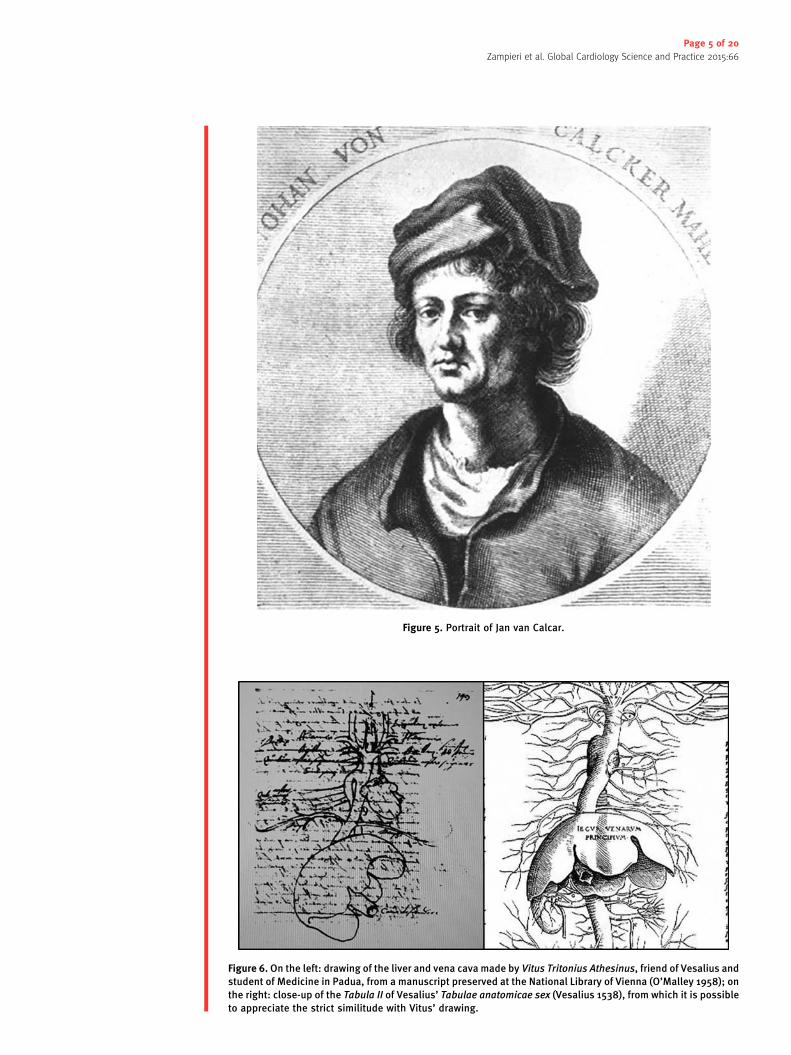
Figure 5. Portrait of Jan van Calcar.
Figure 6. On the left: drawing of the liver and vena cava made by Vitus Tritonius Athesinus, friend of Vesalius and
student of Medicine in Padua, from a manuscript preserved at the National Library of Vienna (O’Malley 1958); on
the right: close-up of the Tabula II of Vesalius’ Tabulae anatomicae sex (Vesalius 1538), from which it is possible
to appreciate the strict similitude with Vitus’ drawing.
Page 5 of 20
Zampieri et al. Global Cardiology Science and Practice 2015:66
Charles V (1500–1558) obtained the copyright, protecting the Fabrica from unauthorized copying
(Vesalius 1543a) and the book is considered a masterpiece of Renaissance printing.
Vesalius arrived in Basle in January 1543, where he continued to dissect. The skeleton he constructed
from the bones of an executed criminal, Jacob Karrar, is still preserved in the Anatomy Museum of
Basle’s University (Olry 1998) (Figure 9). This specimen is estimated to be the oldest anatomical
preparation of a skeleton in the world. During his time in Basle, Vesalius also prepared a digest of the
Fabrica, the Epitome, for students, a mere six chapters long with nine illustrations, printed on poorer
quality paper but of even larger size to make their details clearer (Vesalius 1543b).
Later, Vesalius presented the Fabrica to Charles V and was enrolled at once as one of the Emperor’s
household physicians. In 1544, he married Anne van Hamme, the daughter of a rich counsellor of
Figure 7. Frontispiece of the first edition of Vesalius’ De humani corporis fabrica libri septem (1543), where
Vesalius is making a dissection in a crowded anatomical amphitheater.
Page 6 of 20
Zampieri et al. Global Cardiology Science and Practice 2015:66

Brussels, who bore him a daughter, also named Anne, in 1545. He continued to dissect, but he was
increasingly called in to act as a physician and surgeon, proving how his anatomical knowledge could
be useful also to practice medicine (Suy & Forneau 2015).
By 1555, Vesalius finished a revised edition of the Fabrica (Vesalius 1555) (Figure 10). The revised
edition of the Fabrica can be considered as a novel major contribution, rather than a mere update of
the earlier version. The revised Fabrica was based even more on Vesalius’ own observations and
departed more markedly from traditional Galenism. It gave a strong and uncompromising message that
the human body could only be understood by a clear and careful anatomical investigation into human
corpses. Notable examples to this message are his description of the anatomy of the uterus and the
fetus, and his criticism of Galenic views regarding the permeability of the interventricular septum
(see later) (Zampieri et al. 2014).
Recently, it has been discovered that Vesalius was preparing a third edition of the Fabrica, the great
majority of corrections being stylistic, altering the Latin words but not the overall meaning. This work,
however, never saw the light of day (Nutton 2012).
In 1556, Charles V abdicated his throne and retired in Spain. Vesalius remained in Brussels, and the
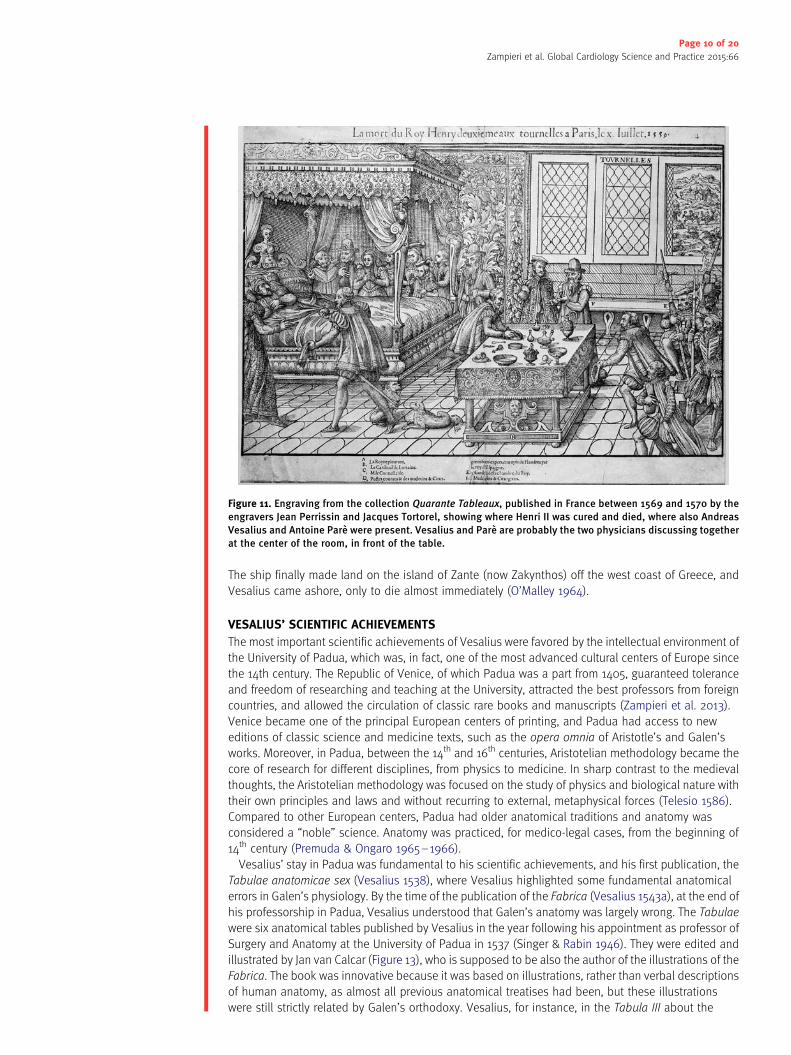
new King Phillip II (1527–1598) recruited him again. In the spring of 1559, the French King, Henri II
(1519–1559), received a wooden lance in his right orbit and temple during a tournament in Paris.
Vesalius was immediately called for, joining Ambroise Pare (1510–1590) and other leading surgeons
from France in an attempt to save the king (Figure 11) (Eftekhari et al. 2015).
Eleven days later, the King finally died after suffering meningismus, fever, left-sided paralysis, and
difficulty in respiration. Vesalius conducted the autopsy and wrote a detailed medical report, which
Figure 8. Engraving depicting Johannes Oporinus, editor of Vesalius’ most important works.
Page 7 of 20
Zampieri et al. Global Cardiology Science and Practice 2015:66

included both clinical history and autopsy findings on the way in which the lance had penetrated the
skull, but without causing a fracture. This again shows the important relationship between anatomy
and clinics, an aspect that has been probably underestimated by historians of medicine, because they
focused too heavily on his anatomical research.
Following a brief return home, Vesalius moved with Philip II to Spain, where he composed his last
anatomical essay, a critique of the Anatomical observations of Gabriele Falloppia (1523–1562)
(Figure 12). In 1564, Vesalius left Spain with his family. His wife and daughter returned to Brussels, while
Vesalius made his way for a pilgrimage to the Holy Land. The reasons of this trip have never been
Figure 9. The skeleton prepared by Vesalius still now preserved at the Anatomy Museum of the Institute of
Anatomy of the University of Basel.
Page 8 of 20
Zampieri et al. Global Cardiology Science and Practice 2015:66

completely clarified. He first stopped at Venice, where it was said that the Venetian senate appointed
him once more to the Chair of Anatomy at Padua, in succession to Falloppia, who had died in 1562.
However, the return journey from the Holy Land was a catastrophe. The boat on which he set sail was
allegedly driven before a storm for forty days without landfall, provisions ran short, and Vesalius fell ill.
Figure 10. Frontispiece of the second edition of Vesalius’ Fabrica (1555).
Page 9 of 20
Zampieri et al. Global Cardiology Science and Practice 2015:66
The ship finally made land on the island of Zante (now Zakynthos) off the west coast of Greece, and
Vesalius came ashore, only to die almost immediately (O’Malley 1964).
VESALIUS’ SCIENTIFIC ACHIEVEMENTS
The most important scientific achievements of Vesalius were favored by the intellectual environment of
the University of Padua, which was, in fact, one of the most advanced cultural centers of Europe since
the 14th century. The Republic of Venice, of which Padua was a part from 1405, guaranteed tolerance
and freedom of researching and teaching at the University, attracted the best professors from foreign
countries, and allowed the circulation of classic rare books and manuscripts (Zampieri et al. 2013).
Venice became one of the principal European centers of printing, and Padua had access to new
editions of classic science and medicine texts, such as the opera omnia of Aristotle’s and Galen’s
works. Moreover, in Padua, between the 14th and 16th centuries, Aristotelian methodology became the
core of research for different disciplines, from physics to medicine. In sharp contrast to the medieval
thoughts, the Aristotelian methodology was focused on the study of physics and biological nature with
their own principles and laws and without recurring to external, metaphysical forces (Telesio 1586).
Compared to other European centers, Padua had older anatomical traditions and anatomy was
considered a “noble” science. Anatomy was practiced, for medico-legal cases, from the beginning of
14th century (Premuda & Ongaro 1965–1966).
Vesalius’ stay in Padua was fundamental to his scientific achievements, and his first publication, the
Tabulae anatomicae sex (Vesalius 1538), where Vesalius highlighted some fundamental anatomical
errors in Galen’s physiology. By the time of the publication of the Fabrica (Vesalius 1543a), at the end of
his professorship in Padua, Vesalius understood that Galen’s anatomy was largely wrong. The Tabulae
were six anatomical tables published by Vesalius in the year following his appointment as professor of
Surgery and Anatomy at the University of Padua in 1537 (Singer & Rabin 1946). They were edited and
illustrated by Jan van Calcar (Figure 13), who is supposed to be also the author of the illustrations of the
Fabrica. The book was innovative because it was based on illustrations, rather than verbal descriptions
of human anatomy, as almost all previous anatomical treatises had been, but these illustrations
were still strictly related by Galen’s orthodoxy. Vesalius, for instance, in the Tabula III about the
Figure 11. Engraving from the collection Quarante Tableaux, published in France between 1569 and 1570 by the
engravers Jean Perrissin and Jacques Tortorel, showing where Henri II was cured and died, where also Andreas
Vesalius and Antoine Pare were present. Vesalius and Pare are probably the two physicians discussing together
at the center of the room, in front of the table.
Page 10 of 20
Zampieri et al. Global Cardiology Science and Practice 2015:66

arteria magna (aorta) and heart, depicted the “rete mirabile”, a network of vessels at the base of
brain that Galen supposed existed in humans, while in fact it exists only in other mammals such as
cow and ox.
In the same table, Vesalius indicated the heart such as the “source of vital spirit and the principle of
arteries” (cor vitalis facultatis fomes et arte princ) (Figure 14). According to Galenian anatomy and
physiology, the liver was the source of venous system, while the heart was the source of the arterial
one. The left ventricle of the heart was the source of the “vital spirit” by the mixture of blood (“natural
spirit”), coming from the right to left ventricle through invisible pores, and air, coming from pulmonary
vein. Vital spirit was the carrier of the “natural heat” through the arteries, without which the body would
have been cold.
Vesalius also followed the part of Galen physiology describing the “pulmonary vein bringing air from
lungs to left atrium” (arteria venalis in sinistrum sinum aerem ex pulmonibus deferens) (Vesalius 1538;
Zampieri et al. 2014) (Figure 14). Finally, another of Galen’s anatomical errors regards the structure of
human carotids (Singer & Rabin 1946; Pagel 1964). Vesalius represented left and right carotids
emerging from the “truncus communis”, while in humans the left carotid emerges separately from
aortic arch. The “truncus communis” structure exists in simians, which proves once again that Galen
did not dissect humans, only primates and other mammals (Figure 15).
The publication of his masterpiece, both in its first and second editions (Vesalius 1543a; 1555), is
considered a turning point not only for human anatomy, but also for medicine in general, because this
wonderful work contained not only seminal discoveries in this discipline, but also a new method in
medical science compared to medieval theory and practice.
Figure 12. Gabriele Falloppia’s portrait in the “Hall of Medicine” at the Bo Palace of the University of Padua.
Page 11 of 20
Zampieri et al. Global Cardiology Science and Practice 2015:66

The Preface to the first edition, dedicated to Charles V, is the most significant text from a
methodological point of view (Premuda 1964). Vesalius complained of the disaggregation of medical
art in what we would now call different specialties, and more particularly the fact that physicians have
opposed “the use of the work of hands” and relegated it to inexperienced and ignorant surgeons and
barbers (Vesalius 1543a).
On the contrary, Vesalius strongly supported that surgery was an ancient and useful part of medicine
itself, not a separate discipline, which was explicitly based on the “investigation of nature”. All the
Preface of the Fabrica can be seen as a defense of the “hand” in its contribution to the knowledge of
the body and medicine (Premuda 1964): “[ . . . ] when the hand is used, medicine flourishes; when it is
neglected, medicine languishes; when it is restored to use, medicine can flourish again” (Vesalius
1543a). This is even more significant because medieval medicine was speculative and adverse toward
practical knowledge (Murdoch 1982).
In contrast to conventional anatomical teaching, Vesalius was a lecturer, a demonstrator and a
dissector all at the same time. The Middle Age model of teaching anatomy required the presence of
three “actors”: Lector, the professor of anatomy who read the textbooks of Mondino de’ Liuzzi without
touching the cadaver; Ostensor, the assistant who indicated the parts of the cadavers described by the
professor at any time; and Sector, who was the vulgar “barber” performing the dissection with his
hands (Figure 4).
Vesalius harshly criticized this model, describing as: “[ . . . ] the hateful method by which one dissects
the body and another describes its parts: the first, perched on a pulpit like a crow, haughtily repeating
ideas that he didn’t learn directly from the cadaver, but that he read in other’s books” (Vesalius 1543a).
In the hardcover of the Fabrica and in Vesalius’ portraits inside the book, his innovation is immediately
Figure 13. Close-up of the sixth table of the Tabulae anatomicae sex, where von Calcar is attributed as editor of
the publication.
Page 12 of 20
Zampieri et al. Global Cardiology Science and Practice 2015:66

appreciable. Vesalius is depicted doing an autopsy on the cadaver of a woman, touching the body with
his right hand and pointing his left hand’s finger (Figure 16). In this way, Vesalius visually demonstrated
to personify all the three actors of the previous method. He was the Professor of Anatomy (Lector),
in fact, he signed the book as Andreas Vesalius “Scholae Medicorum Patavinae Professor” (Figure 17),
he personified the Ostensor with his left hand and the Sector with his right hand.
To confirm the importance of the role of sector personified by Vesalius, the Fabrica deserves a
particular attention to the description of both the anatomical instruments (Figure 18), and the
dissection techniques. Vesalius describes the techniques to prepare bones specimens. Osteology is
probably the most advanced part of his anatomical research and, as proved by the Basle specimen,
he was particularly skilled in skeleton preparation. He described different methods in his Fabrica,
represented also in some wonderfully illustrated initials (Vesalius 1543a). He used, first of all, the
classic method of boiling bodies to extract bones. Another method consisted of intially removing the
majority of soft tissue. Then Vesalius covered the specimen with lime and placed it in a perforated
wooden casket for a time. Then the casket was firmly tied down at the bottom of a river, where the current
gradually removed the remaining soft tissue as it flowed through the casket (Olry 1998) (Figure 19).
As a Humanist, Vesalius recommended his students read the anatomy texts of Galen rather than
Mondino. Vesalius expressed his conviction that mere book learning was not enough and that
demonstrable evidence should take precedence over the written text. This is another fundamental
methodological innovation of Vesalius. Thanks to his direct observation, based on his own dissections
of cadavers, and his reading of Galen’s books, Vesalius was able to scientifically demonstrate that
Galen’s anatomy was largely wrong, because Galen had never dissected human bodies.
We have to bear in mind that, at that time, Galen was considered the most up-to-date knowledge in
medicine, not only because Galen was the most important authority from the Middle Age, but also
because in Vesalius’ time the original works of Galen were being rediscovered. This is thanks in part to
the publication, in Latin, of his original works, free from the interpretations of Arabic translations.
Medical humanists of the 15th and 16th centuries rediscovered a very modern Galen, a physician who
stressed the importance of experience, practice and anatomy for medicine, and the need for a
physician to travel abroad and acquire a universal knowledge. As a consequence, it was even more
significant that Vesalius was able to see beyond Galen’s new orthodoxy. Vesalius, in fact, was well
aware “[ . . . ] how physicians (as well as followers of Aristotle) are upset when they ascertain that Galen,
Figure 14. Close-up of the III table on Arteria magna (the aorta) of Vesalius’ Tabulae anatomicae sex (Vesalius
1538). The heart is defined as “source of vital spirit and the arterial system”; the pulmonary vein is the structure
which “brings air from lungs to the left ventricle” (letter “Q” of the caption on the left). All of these ideas were
strictly Galenic.
Page 13 of 20
Zampieri et al. Global Cardiology Science and Practice 2015:66

in the course of just one anatomical demonstration, gets wrong more than 200 times in the right
description of the parts, the harmony, the use and the function of human body” (Vesalius 1543a).
Vesalius also showed why Galen was wrong: “Indeed, those who are now dedicated to the ancient
study of medicine [ . . . ] are beginning to learn to their satisfaction how little and how feebly men have
laboured in the field of Anatomy to this day from the times of Galen, who [ . . . ] did not dissect the
human body; and the fact is now evident that he described (not to say imposed upon us) the fabric of
the ape’s body, although the latter differs from the former in many respects” (Vesalius 1543a).
That Galen made many mistakes was a foregone conclusion, since human and ape anatomies are
very different. Between Galen’s errors, Vesalius showed that the sternum consisted of three sections,
instead of seven, that the mandible consisted of one bone, instead of two, that the “rete mirabile” did
Figure 16. Close-up of the frontispiece of De humani corporis fabrica (Vesalus 1543a), depicting Vesalius
personifying all the three figures of the previous method for teaching anatomy: he was lector, because professor
of anatomy, ostensor, because he indicated, with his left hand, what he was explaining during the dissection,
and sector, because he made the dissection himself with his right hand.
Figure 15. On the left: close-up from the Tabula III of Vesalius’ Tabulae anatomicae sex (Vesalius 1538), showing
the heart and the structure of the carotids; on the right: structure of the carotids in human and simian,
demonstrating that Vesalius represented in man the structure typical of simian, following Galen’s anatomy
(picture taken from: Singer & Rabin 1946).
Page 14 of 20
Zampieri et al. Global Cardiology Science and Practice 2015:66
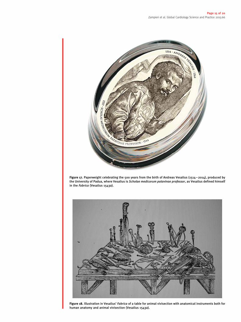
Figure 18. Illustration in Vesalius’ Fabrica of a table for animal vivisection with anatomical instruments both for
human anatomy and animal vivisection (Vesalius 1543a).
Figure 17. Paperweight celebrating the 500 years from the birth of Andreas Vesalius (1514–2014), produced by
the University of Padua, where Vesalius is Scholae medicorum patavinae professor, as Vesalius defined himself
in the Fabrica (Vesalius 1543a).
Page 15 of 20
Zampieri et al. Global Cardiology Science and Practice 2015:66

not exist in man, and that nerves were not hollow. These and many others findings became the starting
point for a new anatomy based on the “book of nature” rather than on classic authorities.
To give a more solid foundation to his discoveries, Vesalius widely used the new instrument of
anatomical illustration. Before Vesalius, Leonardo da Vinci (1452–1519) produced wonderful and
precise anatomical illustrations, but his works have never been published and surely didn’t influence
Vesalius (Thiene & Zampieri, 2013). Berengario da Carpi (1466–1530), professor of anatomy in
Bologna, published the first anatomical illustrations. He edited two enlarged editions of Mondino’s
anatomical work, but his Isagogae breves had a major impact, with many editions in just a few years
(Berengario 1523). However, Berengario’s illustrations can be considered as simply a first attempt and
do not compare to Vesalius’ achievement.
Vesalius considered anatomical illustrations such as a scientific foundation for anatomy in the same
way they were for geometry (Vesalius 1543a). In Vesalius’ Fabrica there are more than 250 illustrations,
some of the whole body, some of specific organs and parts of organs, some of anatomical instruments
and techniques (Saunders & O’Malley 1950). Many of them shows cadavers in allegorical poses and
imaginary, but realistic landscapes (Figure 20). Given that body was a noble object of study, in contrast
to Middle Age attitude in which body and nature were humble objects of study, it deserved to be
represented by the most beautiful and realistic images. As already mentioned, these pieces of art have
Figure 19. Capital letters of the Fabrica showing methods of preparing bones specimens. On top left: capital
letter “O” of the 1543 edition, showing the method of boiling bodies to extract bones. On top right, capital letter
“C” of the 1543 edition, showing three men who carry a casket full of holes about to be immersed in a river.
After having removed the majority of soft tissue, Vesalius covered the specimen with lime and placed it in a
perforated wooden casket for a time. Then the casket was firmly tied down at the bottom of a river, where the
current gradually removed the remaining soft tissue as it flowed through the casket (Olry 1998). On bottom left:
capital letter “P” of 1543 edition, showing cherubs reconstructing the skeleton from the bones obtained with the
methods previously described. On bottom right: capital letter “P” of 1555 edition, showing the same scene.
Page 16 of 20
Zampieri et al. Global Cardiology Science and Practice 2015:66

been attributed to Jan van Calcar, painter who worked at the Titian school in Venice (Hazard 1996).
Probably Vesalius choose van Calcar because he came from a painting school focused on the
vivid representation of nature and human emotions, an approach perfectly combined with his own
(Premuda 1963–1964).
Vesalius’ new anatomy would have brought not only a new morphological knowledge, but also a new
physiology, which fully developed in the 16th and 17th centuries. Vesalius himself stressed the
importance of understanding the function, that is the physiology, of the parts observed by anatomical
research. He believed that, to this end, vivisection of animals could be particularly useful (Vesalius
1543a). One of Vesalius’ discoveries, accomplished in the second edition of the Fabrica, is very
significant in this respect. In the first edition, Vesalius did not deny the patency of the interventricular
Figure 20.One of the Vesalius’ plates on the human skeleton, with the figure in an allegoric pose, thinking with a
skull in his right hand, and a Latin inscription in the base where the skull is posed: “Vivitur ingenio, caetera
mortis erunt”. Which means: genius lives on, all else is mortal.
Page 17 of 20
Zampieri et al. Global Cardiology Science and Practice 2015:66
septum, however he seemed to be, at least, doubtful: “Thus we are compelled to astonishment at the
industry of the Creator who causes the blood to sweat from the right ventricle into the left through
passages which escapes our sight” (Vesalius 1543a).
In the second edition, Vesalius clearly denied this structure: “However much the pits may be
apparent, yet none, as far as can be comprehended by sense, passes through the septum of the heart
from the right ventricle into the left [ . . . ] although they are mentioned by professors of anatomy since
they are convinced that blood is carried from the right ventricle to the left. As a result – as I shall
declare more openly elsewhere – I am in no little doubt regarding the function of the heart in this part”
(Vesalius 1555).
Figure 21. Frontispiece of Realdo Colombo’s De re anatomica (1559) with a portrait of Colombo, at the center,
doing the dissection.
Page 18 of 20
Zampieri et al. Global Cardiology Science and Practice 2015:66
The last part of this quotation is significant as it expresses the idea that a new physiology of the heart
was emerging (Pagel 1964). In another passage of the Fabrica this idea became explicit: “In presenting
reasons for the construction of the heart and the use of the parts I have in large degree fitted my
discourse to the teachings of Galen, not because I believe them to be in entire agreement with the
truth, but because I am yet hesitant to present a completely new use and function for those parts”
(Vesalius 1555).
If the blood could not pass from right to left ventricle through interventricular septum, it was
necessary to think of an alternative pathway, otherwise the fact that arteries filled up with blood would
have been inexplicable. Note that this was necessary even in the absence of a circulatory theory of the
cardiovascular system, such as in Galen’s and Vesalius’ times. For them, blood was produced by the
liver and reached the right side of the heart by the vena cava. The pulmonary artery, which was
conceived to nourish the lungs with blood, originated from the right ventricle. Therefore, it was
necessary that blood passed to the left ventricle, from which the aorta could nourish many other parts
of the body and, in Galen’s physiology, could distribute the vital spirit.
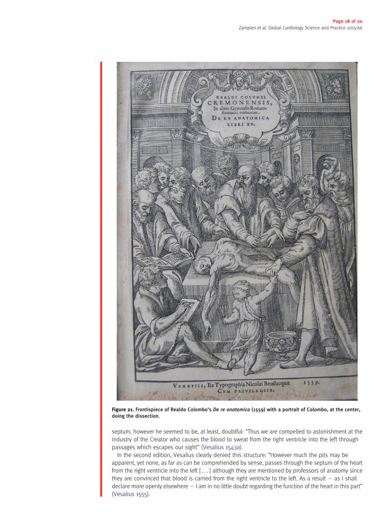
It was therefore not by chance, that pulmonary circulation was later discovered by Realdo Colombo
(1516–1559), one of Vesalius’ students, in his De re anatomica (Colombo 1559; Elmaghawry et al. 2014)
(Figure 21). Colombo, in fact, was the successor of Vesalius at the chair of Surgery and Anatomy in
Padua. It is probable that the doubts and suggestions of his master pushed Colombo to find a solution:
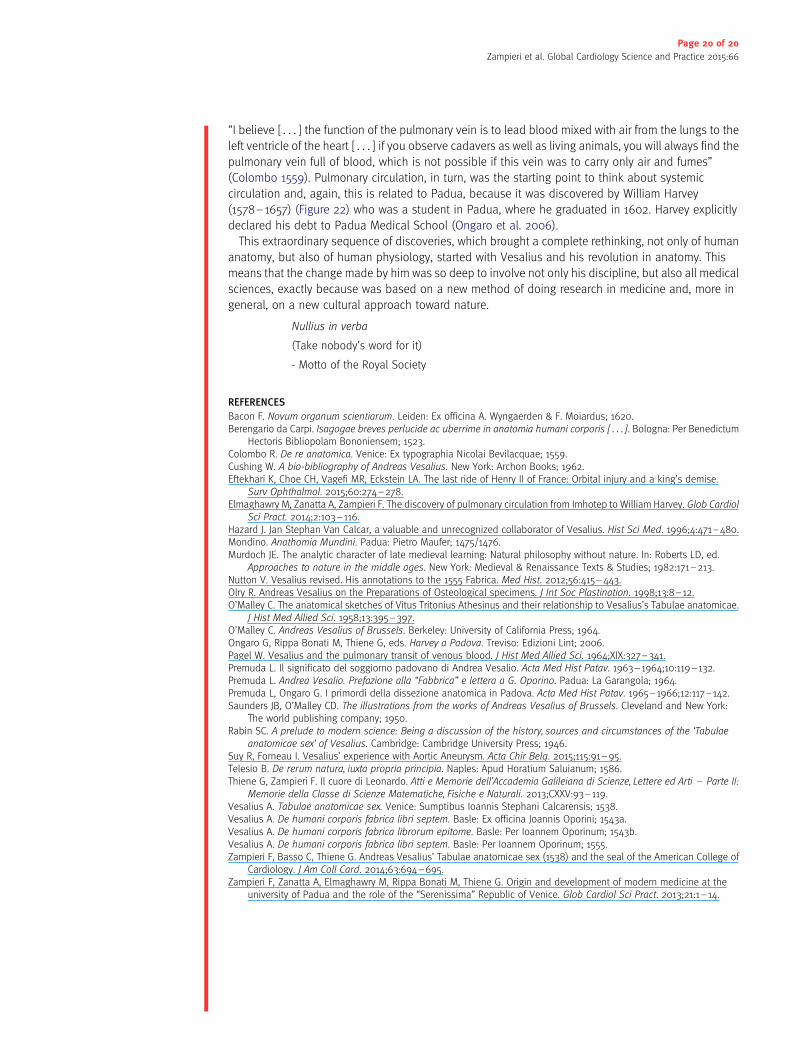
Figure 22. One of the most famous portraits of William Harvey, now displayed at the Royal College of Physicians
in London.
Page 19 of 20
Zampieri et al. Global Cardiology Science and Practice 2015:66
“I believe [ . . . ] the function of the pulmonary vein is to lead blood mixed with air from the lungs to the
left ventricle of the heart [ . . . ] if you observe cadavers as well as living animals, you will always find the
pulmonary vein full of blood, which is not possible if this vein was to carry only air and fumes”
(Colombo 1559). Pulmonary circulation, in turn, was the starting point to think about systemic
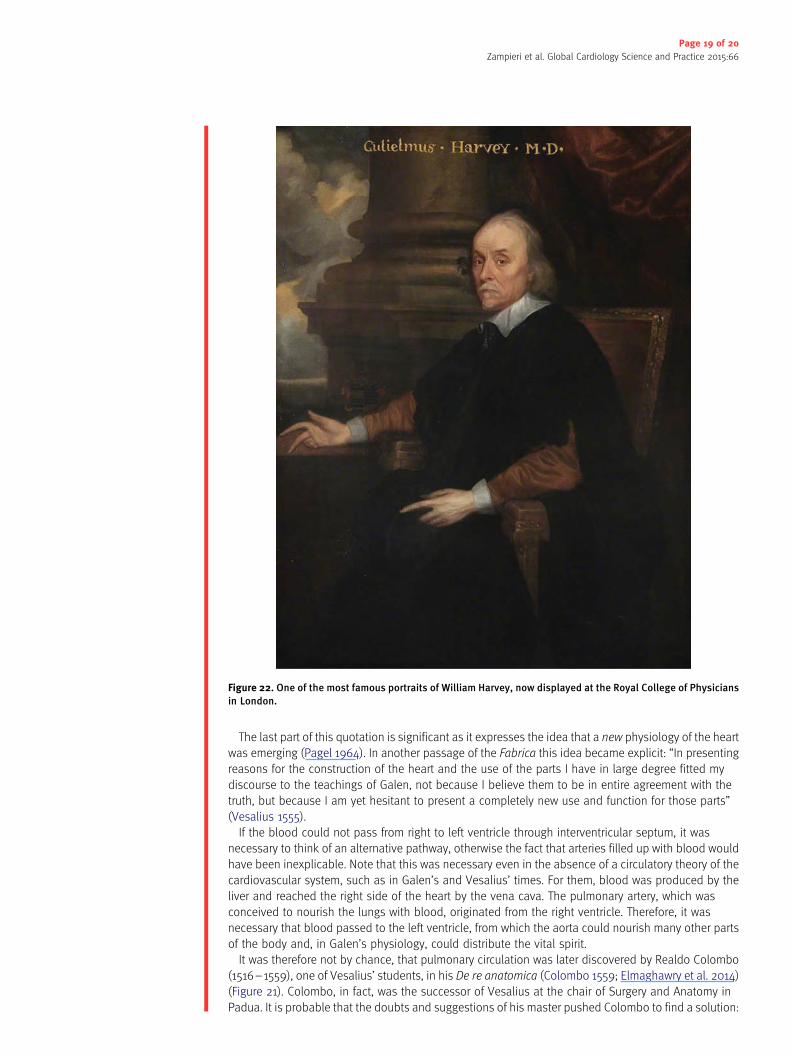
circulation and, again, this is related to Padua, because it was discovered by William Harvey
(1578–1657) (Figure 22) who was a student in Padua, where he graduated in 1602. Harvey explicitly
declared his debt to Padua Medical School (Ongaro et al. 2006).
This extraordinary sequence of discoveries, which brought a complete rethinking, not only of human
anatomy, but also of human physiology, started with Vesalius and his revolution in anatomy. This
means that the change made by him was so deep to involve not only his discipline, but also all medical
sciences, exactly because was based on a new method of doing research in medicine and, more in
general, on a new cultural approach toward nature.
Nullius in verba
(Take nobody’s word for it)
- Motto of the Royal Society
REFERENCES
Bacon F. Novum organum scientiarum. Leiden: Ex officina A. Wyngaerden & F. Moiardus; 1620.Berengario da Carpi. Isagogae breves perlucide ac uberrime in anatomia humani corporis [ . . . ]. Bologna: Per Benedictum
Hectoris Bibliopolam Bononiensem; 1523.Colombo R. De re anatomica. Venice: Ex typographia Nicolai Bevilacquae; 1559.Cushing W. A bio-bibliography of Andreas Vesalius. New York: Archon Books; 1962.Eftekhari K, Choe CH, Vagefi MR, Eckstein LA. The last ride of Henry II of France: Orbital injury and a king’s demise.
Surv Ophthalmol. 2015;60:274–278.Elmaghawry M, Zanatta A, Zampieri F. The discovery of pulmonary circulation from Imhotep to William Harvey. Glob Cardiol
Sci Pract. 2014;2:103–116.Hazard J. Jan Stephan Van Calcar, a valuable and unrecognized collaborator of Vesalius. Hist Sci Med. 1996;4:471–480.Mondino. Anathomia Mundini. Padua: Pietro Maufer; 1475/1476.Murdoch JE. The analytic character of late medieval learning: Natural philosophy without nature. In: Roberts LD, ed.
Approaches to nature in the middle ages. New York: Medieval & Renaissance Texts & Studies; 1982:171–213.Nutton V. Vesalius revised. His annotations to the 1555 Fabrica. Med Hist. 2012;56:415–443.Olry R. Andreas Vesalius on the Preparations of Osteological specimens. J Int Soc Plastination. 1998;13:8–12.O’Malley C. The anatomical sketches of Vitus Tritonius Athesinus and their relationship to Vesalius’s Tabulae anatomicae.
J Hist Med Allied Sci. 1958;13:395–397.O’Malley C. Andreas Vesalius of Brussels. Berkeley: University of California Press; 1964.Ongaro G, Rippa Bonati M, Thiene G, eds. Harvey a Padova. Treviso: Edizioni Lint; 2006.Pagel W. Vesalius and the pulmonary transit of venous blood. J Hist Med Allied Sci. 1964;XIX:327–341.Premuda L. Il significato del soggiorno padovano di Andrea Vesalio. Acta Med Hist Patav. 1963–1964;10:119–132.Premuda L. Andrea Vesalio. Prefazione alla “Fabbrica” e lettera a G. Oporino. Padua: La Garangola; 1964.Premuda L, Ongaro G. I primordi della dissezione anatomica in Padova. Acta Med Hist Patav. 1965–1966;12:117–142.Saunders JB, O’Malley CD. The illustrations from the works of Andreas Vesalius of Brussels. Cleveland and New York:
The world publishing company; 1950.Rabin SC. A prelude to modern science: Being a discussion of the history, sources and circumstances of the ‘Tabulae
anatomicae sex’ of Vesalius. Cambridge: Cambridge University Press; 1946.Suy R, Forneau I. Vesalius’ experience with Aortic Aneurysm. Acta Chir Belg. 2015;115:91–95.Telesio B. De rerum natura, iuxta propria principia. Naples: Apud Horatium Saluianum; 1586.Thiene G, Zampieri F. Il cuore di Leonardo. Atti e Memorie dell’Accademia Galileiana di Scienze, Lettere ed Arti – Parte II:
Memorie della Classe di Scienze Matematiche, Fisiche e Naturali. 2013;CXXV:93–119.Vesalius A. Tabulae anatomicae sex. Venice: Sumptibus Ioannis Stephani Calcarensis; 1538.Vesalius A. De humani corporis fabrica libri septem. Basle: Ex officina Joannis Oporini; 1543a.Vesalius A. De humani corporis fabrica librorum epitome. Basle: Per Ioannem Oporinum; 1543b.Vesalius A. De humani corporis fabrica libri septem. Basle: Per Ioannem Oporinum; 1555.Zampieri F, Basso C, Thiene G. Andreas Vesalius’ Tabulae anatomicae sex (1538) and the seal of the American College of
Cardiology. J Am Coll Card. 2014;63:694–695.Zampieri F, Zanatta A, Elmaghawry M, Rippa Bonati M, Thiene G. Origin and development of modern medicine at the
university of Padua and the role of the “Serenissima” Republic of Venice. Glob Cardiol Sci Pract. 2013;21:1–14.
Page 20 of 20
Zampieri et al. Global Cardiology Science and Practice 2015:66