and john f. lawrence abstract resume
TRANSCRIPT
EVOLUTION OF THE HIND WING IN COLEOPTERA
JARMILA KUKALOVA-PECK Department of Earth Sciences, Carleton University, Ottawa, Ontario, Canada KlS 5B6
and JOHN F. LAWRENCE CSIRO Division of Entomology, GPO Box 1700, Canberra, ACT 2601, Australia
Abstract The Canadian Entomologist 125: 18 1-258 (1 993) A survey is made of the major features of the venation, articulation, and folding in the hind wings of Coleoptera. The documentation is based upon examination of 108 Coleoptera families and 200 specimens, and shown in 101 published figures. Wing veins and articular sclerites are homologized with elements of the neopteran wing groundplan, resulting in wing vein terminology that differs substantially from that gen- erally used by coleopterists. We tabulate the differences between currently used ven- ational nomenclature and the all-pterygote homologous symbols. The use of the neo- pteran groundplan, combined with the knowledge of the way in which veins evolved, provides many strong characters linked to the early evolutionary radiation of Coleop- tera. The order originated with the development of the apical folding of the hind wings under the elytra executed by the radial and medial loop. The loops, which are very complex venational structures, further diversified in four distinctly different ways which mark the highest (suborder) taxa. The remaining venation and the wing articulation have changed with the loops, which formed additional synapomorphies and autapo- morphies at the suborder, superfamily, and sometimes even family and tribe levels. Relationships among the four currently recognized suborders of Coleoptera are re- examined using hind wing characters. The number of wing-related apomorphies are 16 in Coleoptera, seven in Archostemata + Adephaga-Myxophaga, four in Adephaga- Myxophaga, seven in Myxophaga, nine in Archostemata, and five in Polyphaga. The following phylogenetic scheme is suggested: Polyphaga [Archostemata (Adephaga + Myxophaga)]. Venational evidence is given to define two major lineages (the hydro- philoid and the eucinetoid) within the suborder Polyphaga. The unique apical wing folding mechanism of beetles is described. Derived types of wing folding are discussed, based mainly on a survey of recent literature. A sister group relationship between Coleoptera and Strepsiptera is supported by hind wing evidence.
Kukalova-Peck, J., et J.F. Lawrence. 1993. ~volution de l'aile postkrieure chez les Colkopthres. The Canadian Entomologist 125: 181-258.
Resume On trouvera ici les resultats d'une synthkse des principales caract6ristiques relites a la nervation, a l'articulation et au repliement des ailes postCrieures chez les ColCoptkres. Ce travail repose sur l'etude de 200 specimens appartenant 108 familles de ColCop- tkres et sur I'examen de 101 illustrations tirtes de la litterature. Les nervures alaires et les sclCrites articulaires sont homologuts a des eltments du plan de base de l'aile nkoptere, ce qui donne lieu i une terminologie relativement differente de celle qu'uti- lisent gCnCralement les specialistes des ColCoptkres. Nous prksentons ici un tableau qui compare les termes gtntralement employes pour designer les nervures et les symboles homologues de I'aile type d'un pterygote. L'utilisation du plan de base de I'aile ntop- tkre, ajoute a nos connaissances de 1'Cvolution des nervures, jettent de la lumiere sur les caractkres fondamentaux reliCs 2 la radiation Cvolutive primitive des ColCopteres. L'ordre s'est d'abord distingut par le repliement apical de I'aile posttrieure sous l'elytre, le long des boucles radiale et mCdiale. Les boucles, qui sont des structures nervulaires tres complexes, se sont par la suite diversifiees de quatre f a ~ o n differentes qui carac- terisent les taxons les plus evolues (sous-ordres). Les autres nervures et I'articulation de l'aile se sont modifiCs en fonction des boucles, ce qui a donne lieu a d'autres syna- pomorphies et autapomorphies au niveau du sous-ordre et de la super-famille et mCme parfois au niveau de la famille et de la tribu. Les relations entre les quatre sous-ordres
THE CANADIAN ENTOMOLOGIST MarchIApril 1993
actuellement reconnus de ColCoptkres ont kt6 rCCvaluCes en fonction des caractkris- tiques de I'aile postkrieure. Le nombre d'apomorphies reliCes a l'aile sont au nombre de 16 chez les ColCoptkres, de sept chez les ArchostCmates + AdCphages-Myxo- phages, de quatre chez les AdCphages-Myxophages, de sept chez les Myxophages, de neuf chez les Archostkmates et de cinq chez les Polyphages. Le modkle phylogCnCtique suivant est proposC: Polyphages [ArchostCmates (AdCphages + Myxophages)]. Des caractkristiques de la nervation permettent de definir deux lignees principales (les hydrophiloi'des et les eucinCtoi'des) au sein du sous-ordre des Polyphages. Le meca- nisme de repliement apical particulier de I'aile chez les ColCopteres est dCcrit. Les types derivCs de repliement de I'aile sont examines a la lumikre de la litteratwe rCcente. Les caractkristiques de I'aile posterieure nous permettent de croire que les ColCoptkres et les Strepsiptbes representent deux groupes soeurs.
[Traduit par la redaction]
Introduction Evolutionary studies of most pterygote orders draw much information from characters
based on wing venation (Kukalovi-Peck 1991). In Coleoptera, however, use of wing ven- ation and articulation in phylogeny has been minimal, due in part to the complexities of wing folding and the effects of this folding on the venational patterns. The classic work of Forbes (1926) on wing folding patterns generated a number of new phylogenetic hypotheses, some of which (e.g. relationship of Hydroscaphidae, Microsporidae, and Cyathoceridae to Adephaga; affinities of cantharoid and elateroid complexes; placement of Rhipiceridae in Dascilloidea) have been supported by more recent evidence. Most of Forbes' groups, however, are not generally recognized, and the only recent attempt to survey wing venation across the order (Wallace and Fox 1975, 1980) has had little, if any, effect on beetle classification.
Although it is common knowledge that the coleopteran hind wing venation is unique, no attempt has been made to compare the venation in detail with that of other orders or to homologize carefully the major veins and axillary sclerites with those of the neopteran groundplan. It is also common knowledge that the venatidn is different in the four sub- orders, but these differences are usually described very superficially (e.g. number of radial cross-veins, presence of oblong cell) without an attempt to understand their evolutionary implications and functional bases. Finally, no effort has been made to study the structure of the axillary region, which contains a wealth of additional characters for use in phylo- genetic studies.
In the present work, we have attempted to homologize all of the major features of the beetle hind wing (Fig. 12) with the neopteran groundplan (Fig. 4), as interpreted by Kukalovh-Peck (1 983, 199 I), to describe the major patterns of variation in wing venation, articulation, and folding occurring throughout the order, to explain the basic mechanism of folding and unfolding the wing and ways in which this mechanism has been modified in various derived lineages, and to utilize wing characters in defining the suborders of Coleoptera, and the most basal division within the suborder Polyphaga, and in assessing the relationship of Strepsiptera to Coleoptera.
We hope that the reader, after some struggle, will share our excitement over the new prospects for phylogenetic studies that the use of hind wing venation, articulation, and folding may open in the Coleoptera. We also hope that the ultimate benefits to phyloge- netics will be greater than the initial frustration with the unfamiliar symbols and terms which we have found it necessary to use.
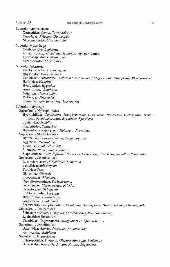
Taxa Studied Hind wings representing the following genera were studied during the course of this
study. Family and superfamily concepts are from Lawrence and Britton (1991).
Volume 125 THE CANADIAN EWOMOI.OGIST
Suborder Archostemata Ommatidae: Omma, Tetraphalerus Cupedidae: Priacma, Distocupes Micromalthidae: Micromalthus
Suborder Myxophaga Cyathoceridae: Lepicerus Tomdincolidae: Claudiella, Hintonia. Ytu, new genus. Hydroscaphidae: Hydroscapha Microsporidae: Microsporus
Suborder Adephaga Trachypachidae: Trachypachus Rhysodidae: Omoglymmius Carabidae: Arthropterus, Calosoma, Catodromus, Megacephala, Omophron, Pheropsophus Haliplidae: Haliplus Hygrobiidae: Hygrobia Amphizoidae: Amphizoa Noteridae: Hydrocanthus Dytiscidae: Hyderodes Gyrinidae: Spanglerogyrus, Macrogyrus
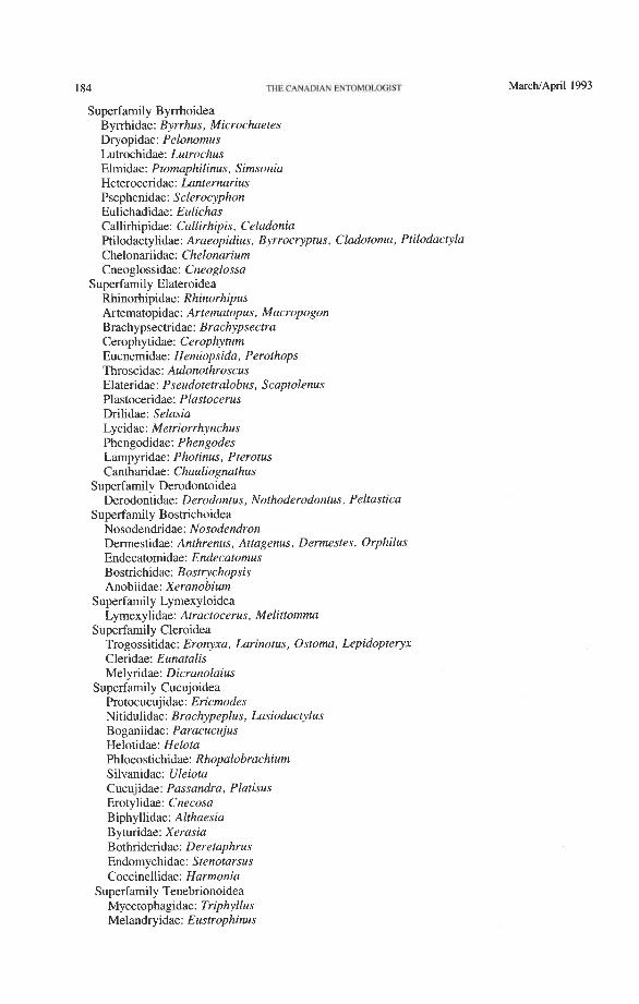
Suborder Polyphaga Superfamily Hydrophiloidea
Hydrophilidae: Coelostoma, Dactylosternum, Helophorus, Hydrochus, Hydrophilus, Limno- xenus, Pseudohydrobius, Rygmodus, Spercheus
Synteliidae: Syntelia Sphaeritidae: Sphaerites Histeridae: Teretriosoma, Hololepta, Pactolinus
Superfamily Staphylinoidea Hydraenidae: Parhydraenida, Tympanogaster Agyrtidae: Necrophilus Leiodidae: Eublackburniella Silphidae: Ptomaphila, Diamesus Staphylinidae: Austrolophrum, Baeocera, Creophilus, Priochirus, Sartallus, Scaphidium
Superfamily Scarabaeoidea Lucanidae: Aesalus, Syndesus, Lamprima Passalidae: Aulacocyclus Trogidae: Trox Glaresidae: Glaresis Pleocomidae: Pleocoma Diphyllostomatidae: Diphyllostoma Geotrupidae: Elephastomus, Frickius Ochodaeidae: Ochodaeus Ceratocanthidae: Cloeotus Hybosoridae: Phaeochrous Glaphyridae: Amphicoma Scarabaeidae: Anoplognathus, Cryptodus, Goniorphnus, Haploscapanes, Phaenognatha
Superfamily Eucinetoidea Scirtidae: Veronatus, Atopida, Macrohelodes, Pseudomicrocara Eucinetidae: Eucinetus Clambidae: Calyptomerus, Acalyptomerus, Sphaerothorav
Superfamily Dascilloidea Dascillidae: Anorus, Dascillus, Notodascillus Rhipiceridae: Rhipicera
Superfamily Buprestoidea Schizopodidae: Dystaxia, Glyptoscelimorpha, Schizopus Buprestidae: Buprestis, Julodis, Nascio, Stigmodera
184 THECANADIAN ENTOMOLOGIST
Superfamily Byrrhoidea Byrrhidae: Byrrhus, Microchaetes Dryopidae: Pelonomus Lutrochidae: Lutrochus Elmidae: Ptomaphilinus, Simsonia Heteroceridae: Lanternarius Psephenidae: Sclerocyphon Eulichadidae: Eulichas Callirhipidae: Callirhipis, Celadonia Ptilodactylidae: Araeopidius, Byrrocryptus, Cladotoma, Ptilodacryla Chelonariidae: Chelonarium Cneoglossidae: Cneoglossa
Superfamily Elateroidea Rhinorhipidae: Rhinorhipus Artematopidae: Arternatopus, Macropogon Brachypsectridae: Brachypsectra Cerophytidae: Cerophytum Eucnemidae: Hemiopsida, Perothops Throscidae: Aulonothroscus Elateridae: Pseudotetralobus, Scaptolenus Plastoceridae: Plastocerus Drilidae: Selasia Lycidae: Metriorrhynchus Phengodidae: Phengodes Lampyridae: Photinus, Pterotus Cantharidae: Chauliognathus
Superfamily Derodontoidea Derodontidae: Derodontus, Nothoderodontus, Peltastica
Superfamily Bostrichoidea Nosodendridae: Nosodendron Dermestidae: Anthrenus, Attagenus, Dermestes, Orphilus Endecatomidae: Endecatomus Bostrichidae: Bostrychopsis Anobiidae: Xeranobium
Superfamily Lymexy loidea Lymexylidae: Atractocerus, Melittomma
Superfamily Cleroidea Trogossitidae: Eronyxa, Larinotus, Ostoma, Lepidopteryx Cleridae: Eunatalis Melyridae: Dicranolaius
Superfamily Cucujoidea Protocucujidae: Ericmodes Nitidulidae: Brachypeplus, Lasiodactylus Boganiidae: Paracucujus Helotidae: Helota Phloeostichidae: Rhopalobrachium Silvanidae: Uleiota Cucujidae: Passandra, Platisus Erotylidae: Cnecosa Biphyllidae: Althaesia Byturidae: Xerasia Bothrideridae: Deretaphrus Endomychidae: Stenotarsus Coccinellidae: Harmonia
Superfamily Tenebrionoidea Mycetophagidae: Triphyllus Melandryidae: Eustrophinus
Volume 125 THE CANADIAN ENTOMOLOGIST
Rhipiphoridae: Trigonodera, Rhipiphorus Tenebrionidae: Cyphaleus, Dysantes Prostomidae: Prostomis Oedemeridae: Calopus Cephaloidae: Stenotrachelus Mycteridae: Genus ? Boridae: Boros Pythidae: Cycloderus Aderidae: Megarenus
Superfamily Chrysomeloidea Cerambycidae: Archetypus, Eurynassa Chrysomelidae: Cucujopsis
Superfamily Curculionoidea Belidae: Rhinotia Attelabidae: Merhynchites Ithyceridae: lthycerus Brentidae: Tracheloschizus
Specimen Preparation and Examination Various types of wing preprations were used in this study, but the most useful for
examining both veins and folds were dry mounts on glass microscope slides. Dried or fluid-preserved beetles were first softened and partly macerated with mild potassium hydroxide, and the hind wings or wings and attached metanotum or metapleura, or both, transferred to alcohol. The preparation was then placed onto a drop of water on a micro- scope slide. The wing was unfolded and the axillary region spread out as much as possible, and then the water was allowed to evaporate until the wing adhered to the glass slide. Normally, these wings remained on the slide without the addition of an adhesive, but for further protection, a square glass cover slip was placed over the wing and attached at its four comers with a water-soluble adhesive. Only slight pressure was exerted on the wing surface, and an air interface usually remained between the wing and the cover slip. In some cases wings were stored in glycerine or alcohol, so that folds could be manipulated. In general, wing slides were of two types: those with detached wings, broken at the axillary region, with one wing attached ventrally and the other dorsally; and those with both wings attached to the metanotum with the entire axillary region intact. In a few preparations, the metapleuron was left attached to the wing, and in others the wing was mounted in the folded position.
Several wing mounts were often made, as the development of some veins is subject to individual variation, and some figures represent a compilation from more than one specimen. Dorsally, veins are sometimes disguised by secondary sclerotizations, but they are clearly visible on the ventral side. In our figures, dorsal and ventral views are sometimes combined, to compile all venational phenomena into one figure. In addition, some scle- rotizations, which might have cluttered the figures and obscured critical features, were omitted or deemphasized. It was often necessary to rotate the specimen, examine it at different angles, and vary the lighting conditions, to observe certain veins and folds. This was especially important in very small wings, such as those of Myxophaga and Clambidae.
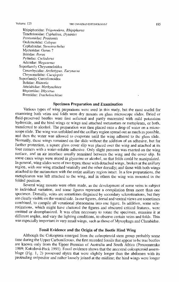
Fossil Evidence and the Origin of the Beetle Hind Wing Although the Coleoptera emerged from the coleopteroid stem group probably some
time during the Upper Carboniferous, the first recorded fossils that appear to be true beetles are known only from the Upper Permian of Australia and South Africa (Ponomarenko 1969; KukalovB-Peck 1991). Fossil evidence shows that the ancestral coleopteroid assem- blage (Fig. 1, 2) possessed elytra that were slightly longer than the abdomen with its protruding ovipositor and rather loosely joined at the midline; the hind wings were longer
186 IHE CANADIAN EWOMOLMilST MarcWApril 1993
than the elytra and the apical region must have extended beyond the elytra when the wings were flexed in resting position. The extinct Protocoleoptera were widely distributed in the Permian of the Northern Hemisphere and appear to represent a side-branch of the coleop- teroid stem assemblage. The best known protocoleopteran family, Tshekardocoleidae, had elytra resembling those of modem Cupedidae and an invaginated meso- and metastemum, as in true Coleoptera, but the antennae had more than 11 annuli, the elytra and hind wings were both extended well beyond the end of the abdomen, and a long, protruding ovipositor was present (Ponomarenko 1969). The hind wing is known from a single specimen (the holotype of Moravocoleus permianus Kukalova, 1969, Fig. 1); the nature of the venation indicates that the wing apex could not be folded beneath the elytra, as it is in Coleoptera. The same lack of specialized coleopteran folding is present in the Upper Permian hind wing illustrated by Ponomarenko (1972) (Fig. 2). In contrast to this, true beetles primi- tively have a more compacted and turtle-like body, with elytra that do not extend beyond the abdomen and genital structures that are telescoped into the abdominal apex. The hind wings have the apical region folded in a highly specialized way to fit beneath the elytra for protection. The Protocoleoptera (and apparently the entire coleopteroid stem assem- blage) lacked the compact body form, which appeared as a basic apomorphy in Coleoptera.
The earliest known beetle fossils (Permosynidae) were characterized by having well sclerotized and convex elytra which were closely coadapted to the abdomen. There were no associated hind wings or genital structures, but it is assumed that the former must have been longer than the elytra and apically folded beneath them and the latter must have been internal (withdrawn into abdominal apex).
Details of hind wing venation in beetle fossils are rarely preserved and often difficult to interpret. The most useful wing fragments have been reported by Ponomarenko (1969; in Arnoldi et al. 1977) and Nikritkin (in Arnoldi et al. 1977) for the following extinct genera: Hadeocoleus (Myxophaga?: Schizophoridae, L. Triassic); Tersus (Myxophaga?: Schizophoridae, U . Jurassic); Necronectes (Adephaga: Coptoclavidae, L. Cretaceous); Cordorabus (Adephaga: Carabidae, U . Jurassic); Notocupes (Archostemata: Ommatidae, L. Triassic); Platycupes and Triadocapes (Cupedidae, L. Triassic); Mesydra (Polyphaga: Hydrophiloidea, L. Cretaceous); and Geotrupoides and Proteroscarabaeus (Polyphaga: Scarabaeoidea, L. Cretaceous).
Wing Venation and Phylogeny Entomologists have been using wing venation in insect systematics for over 100 years.
Through this long experience, the sequences of character changes have become well tested. The use of primary wing venation in phylogeny is based on two major principles: (1) the loss of primary veins and their main branches is irreversible; and (2) the fusions of two primary veins near the wing base (such as the basal stem of M formed by MA + MP, or the fusion of R with MA in blattoids, hemipteroids, and endopterygotes), is irreversible. Also, higher pterygote taxa can be identified by a characteristic basic pattern of braces (cross-veins and veinal fusions) placed in strategic predictable places. However, secondary, usually short, branches (veinal supplements) may be formed in the membrane, usually associated with increased wing size or acting to strengthen strategic parts of the wing under stress, or both. Such secondary branches are best recognized by broadly based comparisons, as their occurrence is limited. Coleoptera often have dense, lightly pig- mented "ghost" branches in the apical region (as in Trogidae, Fig. 50) or near the pos- terior wing margin (as in Creophilus, Fig. 46).
Detailed comparisons of the wings of all Paleozoic and modem insect groups by one of us (JKP), using the two major venational principles mentioned above, have made it possible to reconstruct the groundplan of the pterygote protowing and wing groundplans
Volume 125 THE CANADIAN ENTOMOLOGIST 187
of all higher pterygote taxa: in both the Paleoptera (paleodictyopteroid, ephemeroid- odonatoid) and Neoptera (Fig. 4) (plecopteroid, orthopteroid, blattoid, hemipteroid, and endopterygote) lineages (Kukalova-Peck 1983, 199 1 ; Kukalovh-Peck and Brauckmann 1992). Each of these taxa is characterized by a set of changes in wing venation, which are shared by all members of the group and then further developed and variously transformed within each lineage. The Endopterygota are defined by the following features: (I) a long fusion is present between R + RP and MA in both pairs of wings; (2) an enlarged anal lobe, if (rarely) present in the hind wing, is supported mainly by branches of AP, and the AA area is narrow - the anal fold is an important feature and the claval fold is more-or- less suppressed; (3) a short stem of M is present in the forewing; (4) anal branches often form loops; (5) a brace occurs in the forewing and hind wing between CuA and MP (the arculus: primitively a cross-vein, later a short, direct fusion between the two veins); and (6) MA is + and directed posteriorly, and it separates again either from R or from RP (autapomorphy). The first two conditions are shared with the blattoid and hemipteroid lineages, but (3), (4), and (5) are shared with the hemipteroid lineage only. In addition, endopterygotes have typical neopterous features, such as the special veinal connection between the anals and CUP (anal brace, which links the anal lobe with the remigium), weakly fluted venation (with veins expressed frequently in both dorsal and ventral wing membranes), and axillary sclerites of the neopterous type (lAx, 2Ax, 3Ax, 4Ax) articu- lated with the veins basally in a certain, fixed pattern (Snodgrass 1935; Kukalova-Peck 1991 ; Lawrence et al. 1991). Coleoptera (Fig. 12) share all of these endopterygote attri- butes plus other unique features discussed below.
Predictable specializations in primary venation, used in phylogenetic conclusions, are: (I) loss of branches; (2) changes in the style of branching (from dichotomous to pectinate or unbranched); (3) changes in bracing (from a cross-vein to a direct fusion between two veins); (4) transformation from the basic fluting of sectors (convex "A" sector and concave "P" sector and their respective branches) to a neutral or level position of veins or even to a reversal in fluting; (5) addition of secondary branches (veinal supple- ments) in enlarged wings, especially those in which primary venation has been previously reduced (as in size reduction followed by enlargement); (6) reduction of veinal sections crossed by folds; (7) replacement of a veinal section by sclerotized membrane; and (8) change from an irregular, dense network (archedictyon) to irregular and then regular cross-veins, and finally to clear membrane. The general trend in wing evolution has been from a cockroach-like, thick, almost symmetrical, richly dichotomously branched tegmen without braces to a membranous, highly asymmetrical wing with a few strong branches and several highly specialized braces. In Coleoptera, it is necessary to start with the coleop- teran groundplan, but the succession of venational changes in derived members of various lineages is the same as that given above (with the addition of secondary features discussed below).
Pterygota evolved as their wings diversified; the main Paleozoic lineages of Neoptera primitively have very similar mouthparts and genitalia, but the venational patterns are already fundamentally different (Kukalova-Peck 1991). This suggests that veinal charac- ters are more informative in the definition of related taxa at the highest levels than body characters. After the pterygote groundplan has been reconstructed (Kukalovh-Peck 1983, 1991; Lawrence et al. 1991), the venational and wing articulation characters appear to reflect the relationships among lineages (orders and suborders) which were previously uncertain. It is our hope that proper homologization and linking of the groundplans of the coleopteran wing venation and wing articulation will generate a large number of additional characters at various categoric levels, and thus provide a firmer basis for a cladistic analysis of the order as a whole.
188 THE CANADIAN ENTOMOLOGIST MarchIApril 1993
Homologous Wing Nomenclature The fundamental predicament in using wing venation for defining higher taxa is deter-
mining the complete, and therefore fully homologizable, venational scheme. The follow- ing veinal symbols allow homologization of all primary veins, branches, and braces throughout the Pterygota. The protowing (Fig. 3) contains eight primary veins: precosta (PC), costa (C), subcosta (Sc), radius (R), media (M), cubitus (Cu), anal (A), and jugal (J). Each vein base has its own sclerotized blood sinus, the basivenale (B), which is primi- tively divided. Each vein has two sectors: the convex ( +), anterior (A), and the concave (-), posterior (P); hence, PCA+, PCP-, C A f , CP-, S c A f , S c P , RA+, RP-, M A f , MP- , CuAf , CUP-, AAf , AP- , JAf , and JP- . The two sectors start separately from the divided basivenale and each branches dichotomously about three times. Later, the sectors often fuse together basally into a veinal stem (e.g. RA and RP fuse to form R); stems are assigned general veinal symbols (R, M, Cu). Each sector is dichotomously forked and the branches are given numbers to reflect the dichotomy; for example, RP divides into RP , +, and RP , +, , and then again to form RP,, RP,, RP,, and RP, (Fig. 4). The com- pilation of primitive features in all pterygotes points to an originally largely symmetrical protowing densely filled with dichotomously branched, regularly fluted ( + and -) veinal sectors, interspaced with a fluted archedictyon, and similar to the telson plates of some Crustacea. Paleoptera preserved and further enhanced the original fluted condition, but in Neoptera, the position of RP, MA, MP, and AP often tend to become neutral or uniformly convex (+), and CuA+ and CUP- may occasionally reverse their fluting (the original fluted state is preserved in some fossils and primitive extant forms).
Major Braces. Asymmetrical venation with strategically placed braces between primary veins is crucial for forward flapping flight. A brace can be formed by a cross-vein, or by a portion of a vein being directed to and becoming fused with another vein (veinal brace).
All Pterygota share three veinal braces, indicating that they diversified from a pro- towing that was already involved in some kind of forward locomotion (Fig. 3). These braces are formed as follows:
(1) Fusion of PC and CA, condition of CP. PC originally is close to or adjacent to CA and forms a flat strip (often with minute, serial branchlets creating a serrate margin). CA + and CP- arise separately from the basivenale but usually fuse together near the base. In most pterygotes PC is fully fused with C forming a single tube. This fusion provides a strong anterior margin, which is essential for forward flight. Exceptions occur in some Hemiptera, which have C P running parallel to PC and CA, showing that CP was not part of the anterior margin in the protowing.
(2) Subcostal brace. This veinal brace is formed by the linking of the anterior margin formed by PC + C with the subcostal basivenale (BSc) by a basal portion of ScA. ScA then fuses with the anterior wing margin (PC + C + ScA) for additional strength.
(3) Anal brace. This brace is a sclerotization of membrane or a veinal section that links the anals with the cubitus and prevents the buckling of the anal lobe. All Neoptera share a modified veinal type of anal brace, with AA or AA, +, or AA, either closely adjacent to, fused near the base with, or fully fused with CUP or Cu. The so called "1A" is usually AA,,,, but it may be AA, AA, or AA,,,. In Coleoptera, the anal brace is formed by AA, which forks with the retention of both branches (AA,,,, AA,,,). Thus the sequential numbering of the anal veins is not suitable for homologization. The neop- terous anal brace is often obscured by secondary sclerotization.
All Endopterygota share two braces: (1) Radio-medial brace. MA fuses with R for some length near the base and separates
again distally from R (primitive) or from RP (derived), or it does not separate at all (derived). The medial stem is very short in the forewing and lacking in the hind wing. The radio-medial brace also occurs in hemipteroids and blattoids.

Volume 125 THE CANADIAN ENTOMOLOGIST 189
(2) Medio-cubital brace. Primitively, this is an important cross-vein (arculus) con- necting CuA with MP, but in more derived wings there is a short or long fusion of CuA with MP (as in derived Mecoptera and in Hymenoptera). The medio-cubital brace also occurs in hemipteroids.
All of these pterygote and endopterygote braces occur in at least some Recent Coleop- tera (Fig. 12).
Folds. Wing folds are present in all neopterous wings (Wootton 1981). The medial fold has an erratic path. The claval fold, between the cubital and anal areas, is always concave, and the concave CUP usually follows it closely. The claval fold is especially important in the hind wings of plecopteroids and orthopteroids, but in some endopterygote hind wings CUP is short and runs distally from the claval fold. CUP is short or totally reduced in Coleoptera.
The anal fold in Pterygota is placed between AA and AP. It is especially important in endopterygote hind wings, if these have developed a broad anal lobe, because it replaces the diminished claval fold. In the blattoids, hemipteroids, and endopterygotes, the AA area tends to become narrow and more-or-less adjoined to the remigium, and the anal fold separates the anal lobe from the rest of the wing; consequently the anal lobe (if enlarged) is supported by branches of AP, instead of by all of the anal veins, as in plecopteroids and orthopteroids. This apomorphy is especially marked in the Coleoptera. The endopterygote jugal fold is usually a short, convex ridge between AP and JA (Fig. 4), but it is suppressed in the Coleoptera (Fig. 12).
The only veinal system that lends itself to cladistic analysis is that based on the pterygote groundplan, with homologized venation, folds, fusions, and braces. All these features serve as landmarks for the homologization of coleopteran veinal systems with the groundplan scheme offered below. The advantages of a fully homologizable veinal system are many. Each primary vein in Coleoptera can be seen in a broader context in comparison with other Pterygota, and thus the primitive and derived character states can be identified in all coleopteran taxa.
Hind Wing Articulation and the Groundplan The wing articulation in Coleoptera (Figs. 72-8 1) has excellent potential for reflecting
the basic diversification into suborders, superfamilies, and families, but may also be useful at the subfamilial, tribal, or generic levels. The groundplan concept and the establishment of homologous terms for the various parts of the multi-shaped and variable axillary sclerites are crucial for the assessment of primitive and derived character states in the articular region.
The neopterous wing articulation (Fig. 5) was derived from an ancestral band of small, densely packed, mutually articulated sclerites present in ancestral pterygotes (Fig. 3). The same band of sclerites was also ancestral to the wing articulation in Paleop- tera. In Neoptera, some of the band sclerites became the so called "notal" wing processes (articulated and stiffly hinged or secondarily fused to the tergum), but others formed var- ious clusters, such as the humeral plate (HP), first axillary AX), second axillary A AX), third axillary A AX), and fourth axillary (4Ax) (becoming the posterior "notal" wing process when the sclerite has become fused with the tergum). To understand the cluster sclerites, it is necessary to introduce the band sclerite groundplan.
The band sclerites (Fig. 3) originally (in Paleozoic fossils) received the leg muscles which apparently agitated the protowing. The band was composed of eight transverse rows of sclerites which covered and held open the blood channels and continued as eight primary veinal pairs: PC, C, Sc, R, M, Cu, A, and J. Longitudinally, the band sclerites were arranged into four columns: (1) PR, proxalaria, articulated with the tergum (wing processes or 4Ax); (2) AX, axalaria; (3) F, fulcalaria; and (4) B, basivenalia (sclerotized veinal blood
190 THE CANADIAN ENTOMOLOGIST M a r c W A p r i l 1993
sinuses lacking muscular attachments, veinal bases). Note that the basivenalia in Coleop- tera are primitively divided into the anterior part giving rise to the convex anterior veinal sector and the posterior part giving rise to the concave posterior veinal sector.
The individual sclerites of the ancestral band comprising the neopterous sclerite clus- ters (axillaries) (Fig. 5) are identified by the symbol of the column followed by the veinal symbol denoting the row. For instance, the medial proxalare (PRM) forms part of lAx, the medial axalare (AXM) forms part of 2Ax, the medial fulcalare (FM) forms the median plate, and the medial basivenale (BM) forms the sclerotized blood sinus, subdivided into BMA and BMP and giving rise to the medial veinal sectors MA and MP.
Composition of the Neopterous Articular Sclerites The strongest support for the monophyly of the Neoptera is provided by the wing
articulation. All Neoptera (Fig. 5) have a characteristic set of articular sclerites (humeral plate, HP, and axillary sclerites, 1 Ax, 2Ax, 3Ax), which are composed of identical clusters of smaller, independent, primitive "band" sclerites. In the Coleoptera, the boundaries between the band sclerites within the humeral plate and each of the axillary sclerites are sometimes still visible and appear as dark sutures, grooves, or membranous windows, much as in the large Megaloptera (Kukalova-Peck 1991, fig. 6.16). The composition of the axillary sclerites are as follows:
Tegula. This elevated cluster of sensory setae is located where the precostal and costal axalare (AXPC + AXC) were once placed in the ancestral band. It is not a sclerite. Small remnants of the precostal and costal proxalaria (PRPC and PRC) may persist at the tergum.
HP. The humeral plate consists of four sclerites: the precostal and costal fulcalaria and basivenalia (FPC + FC + BPC + BC). This plate is the only neopterous sclerite that combines elements on the "wing side" and "body side" of the pleural wall, like a bal- ancing lever. It represents the basic apomorphy of Neoptera and does not occur in Paleop- tera. In some Coleoptera (e.g. many scarabaeoids), sutures may be seen on HP indicating the boundaries of the original band sclerites from which it was formed.
Anterior Wing Process. This cluster is composed of the subcostal and radial proxalaria (PRSc + PRR). The original sclerites are primitively separated by a deep groove. The term "notal" wing process should be dropped because it is not a projection of the tergum or notum.
1Ax. The first axillary is composed of the medial proxalare, radial axalare, subcostal axalare, and subcostal fulcalare (PRM + AXR + AXSc + FSc). The original sclerites were primitively separated by shallow grooves or sutures.
2Ax. This cluster is composed of the medial axalare and radial fulcalare (AXM + FR), which were primitively separated by a hinge.
3Ax. This cluster is composed of the cubital, anal, and jugal axalaria and fulcalaria (AXCu + AXA + AXJ + FCu + FA + FJ). 3Ax in most Neoptera (but not in Coleoptera) bears a hinge between AXA + AXJ and FA + FJ, which folds when the wings are flexed backward (a primitive feature for Neoptera). The original sclerites are primitively separated by sutures, open windows, or shallow grooves.
Median Plate. This sclerite corresponds to the medial fulcalare (FM). It is subdivided lengthwise in Coleoptera (derived, probably linked to apical folding).
4AdPosterior Wing Process. This cluster, if present, is composed of anal and jugal proxalaria (PRA + PRJ) primitively separated by a suture. It is primitively articulated to the tergum as 4Ax [e.g. in some Orthoptera and gyrinid Coleoptera (Larsen 1966)], or stiffly attached, or fused to the tergum as the "posterior wing process" (derived). The
Volume 125 THE CANADIAN ENTOMOLCGIST 191
term "notal" wing process is erroneous because the cluster belongs structurally to the wing.
Criteria for Homologizing Wing Veins The first step in the homologization of the coleopteran veinal system is the deter-
mination of all basivenalia of the primary veins. As is evident from the protowing ground- plan (Fig. 3), the basivenalia are closely associated with the axillary sclerites (Figs. 4, 5a, 5b). Basivenalia are sclerotized blood sinuses of the primary veins and as such cannot migrate away from the individual veins (but can be cut off from a vein by a fold). A vein never shifts to another basivenale (Kukalova-Peck 1983). Also, basivenalia articulate always with the same articular sclerites, which have a certain, recognizable pattern shared by all Neoptera (Snodgrass 1935; Kukalova-Peck 1991), as follows: the subcostal basi- venale (BSc) articulates with the first axillary (1Ax); radial basivenale (BR) with the second axillary (2Ax); medial basivenale (BM) with the median plate (divided in Coleoptera); and cubital, anal, and jugal basivenalia (BCu, BA, BJ) with the third axillary (3Ax) (Figs. 4, 5). The characteristic axillary sclerites of Neoptera provide the most reliable guides for the identification of basivenalia, and the basivenalia, in turn, identify unequivocally the primary veins.
A second step in homologization involves the use of the pterostigma as a landmark. The pterostigma is a sclerotized, darkly pigmented blood sinus (Arnold 1963) occurring anterior to RA and near its distal end (but it may overlap RA). Primitively, the pterostigma probably occurs within the first fork of RA (between RA, +, and RA,,,), as in Coleoptera and Mecoptera (Fig. 4). The radial cell of the beetle hind wing is pigmented in Adephaga and Myxophaga and resembles a pterostigma (Figs. 14-16, 18-29). The cell is also placed in a similar position to the pterostigma in Odonata, Zoraptera, some Hemiptera, Raphi- dioptera, Hymenoptera, some Mecoptera, and Strepsiptera. The coleopteran pterostigma was previously recognized by LarsCn (1966).
A third step involves the use of venational fluting, braces, and folds, characteristic of Neoptera and Endopterygota, as landmarks for venation. As members of Neoptera, beetles should have potentially unstable fluting of RP and MP branches [primitively con- cave ( - ) but probably changed to neutral ( ? ) or convex ( + ) in derived forms]. A short, subcostal brace between BSc and the anterior margin and an anal brace between AA and Cu or CUP should be present, as in other Neoptera. As in other endopterygotes, MA should be fused with R close to the wing base and MP branched; CuA should be connected with MP by a cross-vein (mp-cua brace or arculus); and the last posterior branch of MP (MP,) may be fused with the first anterior branch of CuA near the posterior margin to form a small veinal brace (present largely in the mecopteroid complex). The ScA brace may become a sclerotized bulge, as in the neuropteroid orders; and CUP may be suppressed, as in some neuropteroid hind wings and in Hymenoptera.
Rejected Interpretations of Hind Wing Venation The interpretation of beetle hind wing venation most widely used in North America,
England, and Russia is that of Forbes (1922), which was adopted by Crowson (1955) and variously modified by Balfour-Browne (1944), Ponomarenko (1972), Hamilton (1972), and Wallace and Fox (1975, 1980). This system in its original form and with Ponoma- renko's modification is shown for an adephagan (Omma) and polyphagan (Notodascillus) wing in Figures 6 1 1. The most important features of this system are: (1) the designation of the relatively weak vein, lacking a distinct basal connection and forming the anterior part of the oblong cell in Adephaga, and Archostemata as the media; and (2) the designation of the strong vein, originating at the median plate and forming the posterior part of the oblong cell in Adephaga and Archostemata, as the cubitus. The one or two veins located immediately behind the latter and usually lacking basal connections are called anals by
192 THE CANADIAN ENTOMOLOGIST MarchIApril 1993
Forbes and Crowson, but the first of them is considered to be Cu2 by Balfour-Browne and Ponomarenko or PCu by Wallace and Fox, although Hamilton calls the first plical and the second empusal.
Less commonly used, at least in Coleoptera, is the system of Redtenbacher (1886), usually attributed to Comstock and Needham (1898, 1899) and followed, with or without modifications, by Comstock (19 18), Kolbe (1901), Snodgrass (1 909, 1935), d'orchymont (1920, 1921), Graham (1922), and a few recent workers, such as Kaufmann (1960) and Schneider (1978). This scheme agrees generally with the one proposed in the present paper in that the vein called the "cubitus" by Forbes is considered to be the media. In the work by Comstock and Needham, Coleoptera were covered only briefly, and the few illustrations of pupal wing tracheation (based mainly on ~eramb~cidae) were provided only to illustrate that the elytra and hind wings were serially homologous. The first extensive coverage of Coleoptera wings utilizing this scheme was that of d70rchymont (1920, 1921), and it is his terminology that is compared with those of Forbes and the present authors in Figures G11 and in Table 1. The table also includes the systems used by Snodgrass (1909, wing bases only), Ponomarenko (1972), and Hamilton (1972).
The systems of both Comstock and Forbes relied mainly on the evidence provided by the tracheation of the veins in wings of the pupa and imago. The usefulness of venational homologies based solely on tracheal supply has been questioned in more recent works on the subject (see Whitten 1962 and Kukalovi-Peck 1978). Comstock and Needham labelled the main veins in the pupal wing using terms proposed by various 19th century workers, namely C, Sc, R, M, Cu, and A (I, 11,111, V, VII, IX, and XI of Redtenbacher). Forbes (1922), who was influenced by Kiihne's (1914) work on wing tracheation in beetle pupae, correctly pointed out that the trachea labelled "C" by Comstock and Needham was actually SC, and that the true costal trachea followed the anterior edge of the wing and was thus overlooked. The second well-developed trachea was thus labelled R and the third, reduced one, M. As a result, the major vein running through the center of the wing and connected by a bridge [the anterior "arculus" of Forbes (1922); the medial bridge in the present system) to the radial vein was called the cubitus by Forbes and most coleopterists since. In neither of these venational schemes was it suggested that one or more of the primary veins and their tracheae could have divided near the base. Redtenbacher, following a work by Adolph (1879), considered each of the six main veins in the wing to be primitively forked with the formation of a high (convex or + ) and low (concave or - ) sector. Forbes, like Comstock and Needham, dismissed this concept altogether, although Lameere (1922) and others considered it to be the best working hypothesis in studying fossil wing venation.
Little attention was given to the relationship of wing veins to the axillary sclerites by most of these early workers, although Forbes mentioned the connection of his Cu to "the axillary sclerite." Snodgrass (1909), who illustrated the axillary regions of a carabid, hydrophilid, and scarabaeid, illustrated the basal connection of the vein we are calling the medi; to the median plate, not the third axillary. Snodgrass also showed the radialvein in Carabidae dividing into two sections near the base and in the vicinity of the medial bridge (Snodgrass 1909, fig. 197 -the overlying bridge has been cut off in the illustration).
It is evident from a comparison of Figures 6 ,7 and 10, 11 that the Forbes interpretation does not accommodate the characteristic landmarks mentioned above as occurring in Neo- ptera and Endopterygota. In Forbes' scheme the radial cell occurs between "R" and "RS' ' (here between RA,,, and RA,+,), a location that would preclude its being homologous to the pterostigma in other Pterygota. Forbes' "M" has no basivenale (whereas the medial basivenale in Neoptera is always present). Forbes' "Cu' ' articulates basally with the median plate (rather than with the third axillary, as in all other Neoptera). Some of Forbes' anal veins start from what must be the cubital basivenale, whichis impossible, as the cubital basivenale is placed immediately posterior to the median plate and median basivenale in all Neoptera. The anal area in the Forbes scheme is very large and richly branched, as in
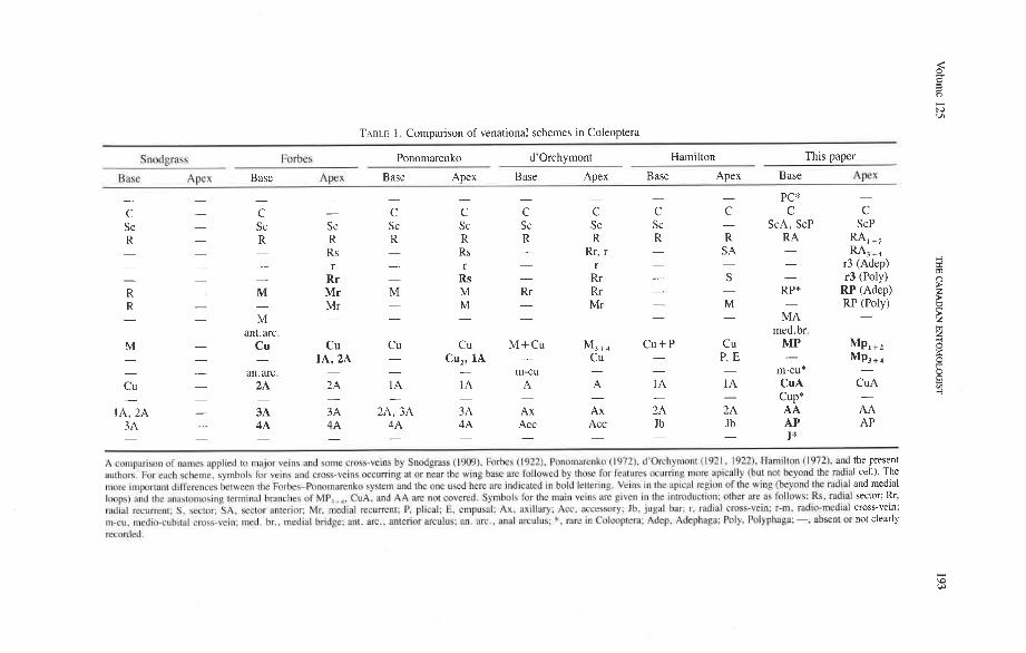
TABLE 1. Comparison of venational schemes in Coleoptera
Snodprass Forbes Ponomarenko d'orchymont Hamilton This paper
Bnsc Apex Base Apex Base Apex Base Apex Base Apex Base AFX
- - - - - - - - - - PC* -
C - C - C C C C C C C C Sc - Sc Sc Sc Sc Sc Sc Sc - ScA, ScP ScP R - R R R R R R R R R A - - - - - - -
RA, +, Rs Rs Rr, r S A
- - - r - r - r - - - RA3 + 4
r3 (Adep) - - Rr - Rs - Rr - S - r3 (Poly) R - M Mr M M Rr Rr - - RP* RP (Adep) R - Mr - M - RP (Poly) - Mr - M -
- - - - - - - - M - MA - ant.arc. med. br.
M - Cu Cu Cu Cu M+Cu M3+, C u + P Cu MP - - -
MP,+* IA, 2A - CU,, IA - CU - P, E -
- MP, + 4
- an.arc. - - - - - - m-cu m-cu* - Cu - 2A 2A 1 A 1A A A 1 A 1 A Cu A Cu A - - - - - - - - - - Cup* -
IA, 2A - 3A 3A 2A, 3A 3A Ax Ax 2A 2A A A A A 3A - 4A 4A 4A 4A Acc Acc Jb Jb AP AP - - - - - - - - - - J* -
A cnmpnrison of names applied to major veins and somc crags-veins by Snodgrass (19091. Forks (1922). Pononutrenko (1972). d'Orchymont (1921. 1922). Hamilton (1972). and the present authors. f ix each scheme. symbols for veins and cmss-veins occurring a! ur near t h ~ wlng hasc are follow-ed by those for fe:~tures ocurring more pic ally (hot not hcyond [he r;~cli;~l cell). The more i rnpon~nl differences hetwcen the Forhes-Ponomnmnko bysrcn) and the one used here are indicated In hold lenering. Veins in the apicdl rrgicln of the wing (beynnd thc md~al and medial Imps) and thc anilktornosing lerminnl branches of MP, ,,. CuA. nnd AA are not covered. Symbols for the main veins am given in the introduction: othcr arc it.; I'ollr?w<: Rs. rad~nl sector; Rr, mclial recurrent: S . ~ c t o c SA. wctclr anlerior; Mr. medial recurrent: P, plical; E. rmptrsal: Ax. axillnry: Acc. accessory: Jb, j i~ga l bar; r. md~al cross-vein: r-m. mtlic*mrdiul cross-vein; m-cu. mcdio-cuhital crt~sc-vcin: nied. br., met1i:il bridge; ant. arc., anterior arcolus; an. arc.. anal an.ulus: *. m e in Culeoplem: Adep. Adephapa: Poly. hilyphag~; -, absent or not clearly wcorded.
1 94 THE CANADIAN ENTOMOLOGIST MarchIApril 1993
orthopteroids, but in Endopterygota the anals are always modestly branched. Also, no venational symbols are available to identify the homologous branches of the anals and other primary veins for phylogenetic considerations.
A comparison of the Comstock-Needham scheme, as modified by d'orchymont (Figs. 8, 9), with that based on the groundplan (Figs. 10, 11) shows that the primary veins agree more-or-less with the pattern of axillary sclerites, but the branches are not identified, homologized, or assigned the numbers that would consistently reflect the dichotomous groundplan branching and allow comparisons with other Pterygota. Also, d'orchymont, like most other workers (except Graham 1922), considered the one or two veins imme- diately following M,,, (our MP, +,) to be branches of the cubitus.
The venational scheme for Coleoptera offered below (Figs. 10, 11, 12, 72-81) is compatible with the pattern of axillary sclerites, shares all apomorphic features charac- teristic of the Neoptera and Endopterygota, and exhibits venational synapomorphies with the neuropteroid complex and with Strepsiptera (Figs. 69-71). All veinal branches are denoted by a numbering system based on the dichotomous branching found in all pterygotes and homologizable with any other pterygote taxon.
Coleopteran Hind Wing Venation In this account we propose to use the veinal symbols derived from the venational
groundplan (the reconstructed protowing shared by all Pterygota; Kukalovi-Peck 1983, 1991). Venational groundplan is a compilation of primitive features assembled over many years by comparing the primitive representatives of all extinct and extant pterygote orders. All character states designated here as primitive are defined by the two well tested assump- tions on which the use of venation in systematics is generally based: (1) primary veins and main branches, once lost, do not reappear; and (2) two primary veins, having fused together near the wing base, do not separate again. The advantage of this veinal system (Figs. 10- 12), over a simplified version tailored to beetles only (Figs. &9), is that veinal characters in all Pterygota (Fig. 3) can be easily compared (all veins and branches are assigned identical, homologous names and symbols); and if a homologized features is compared with the groundplan, the level of specialization becomes readily evident. The use of coleopteran venation in phylogeny is not possible without comparisons with other higher taxa, such as the Protocoleoptera (Figs. 1, 2), Strepsiptera (Figs. 69-71), neuropteroid complex, Endopterygota, and Neoptera; it is also not possible without determining the successions of veinal character states based on the all-pterygote groundplan.
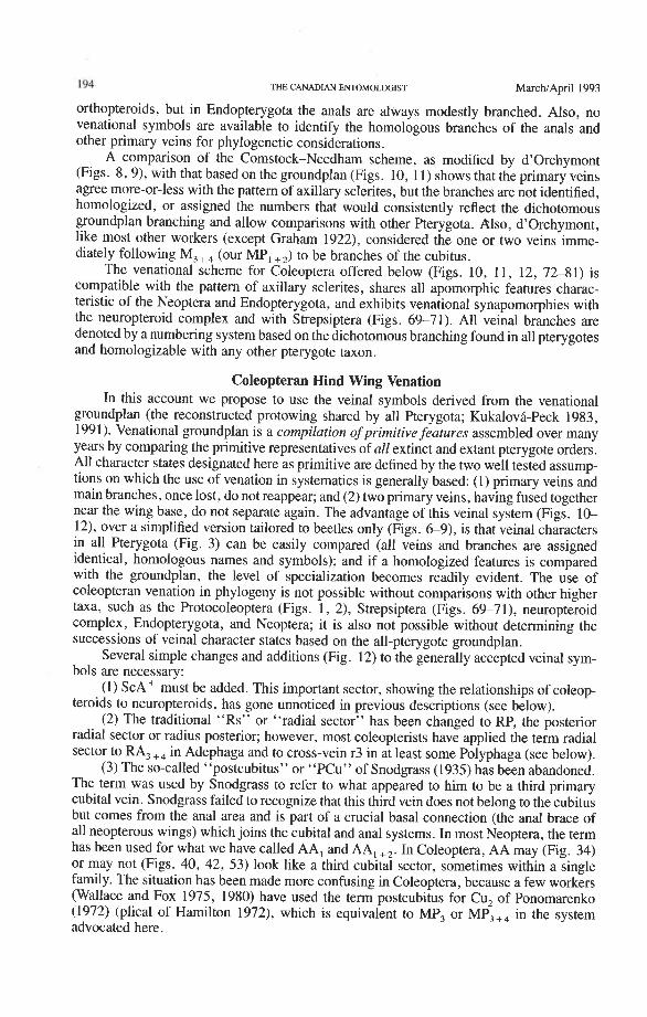
Several simple changes and additions (Fig. 12) to the generally accepted veinal sym- bols are necessary:
(1) ScAf must be added. This important sector, showing the relationships of coleop- teroids to neuropteroids, has gone unnoticed in previous descriptions (see below).
(2) The traditional "Rs" or "radial sector" has been changed to RP, the posterior radial sector or radius posterior; however, most coleopterists have applied the term radial sector to RA,+, in Adephaga and to cross-vein r3 in at least some Polyphaga (see below).
(3) The so-called "postcubitus" or "PCu" of Snodgrass (1935) has been abandoned. The term was used by Snodgrass to refer to what appeared to him to be a third primary cubital vein. Snodgrass failed to recognize that this third vein does not belong to the cubitus but comes from the anal area and is part of a crucial basal connection (the anal brace of all neopterous wings) which joins the cubital and anal systems. In most Neoptera, the term has been used for what we have called AA, and AA, +,. In Coleoptera, AA may (Fig. 34) or may not (Figs. 40, 42, 53) look like a third cubital sector, sometimes within a single family. The situation has been made more confusing in Coleoptera, because a few workers (Wallace and Fox 1975, 1980) have used the term postcubitus for Cu, of Ponomarenko (1972) (plical of Hamilton 1972), which is equivalent to MP, or MP,,, in the system advocated here.
volume 125 THE CANADlAN EmOMOLOGIST 195
(4) Because we consider all primary veins to have two dichotomously branched sec- tors, we have abandoned the sequential numbering of radial, medial, cubital, or anal branches, which is characteristic of most previous venational schemes. We assigned each vein to either an anterior or a posterior sector (e.g. CuA, CUP); the two branches of the first division are labelled 1 + 2 and 3 + 4 (e.g. CuA, +,, CuA,,,); and the four branches of the second division are 1, 2, 3, and 4 (e.g. CuA,, CuA,, CuA,, CuA,).
The following descriptions will emphasize those features important at the ordinal and subordinal levels in Coleoptera. Examples in parentheses are not meant to represent a complete record of the occurrence of an attribute. Homologizations are made with Paleo- zoic Pterygota, to clarify the position of coleopteran venation in a broad evolutionary context. The intention is to clarify venational groundplans of the highest coleopteran taxa, to point out the most primitive character states in primary veins, and to produce a succes- sion of characters useful for cladistic studies.
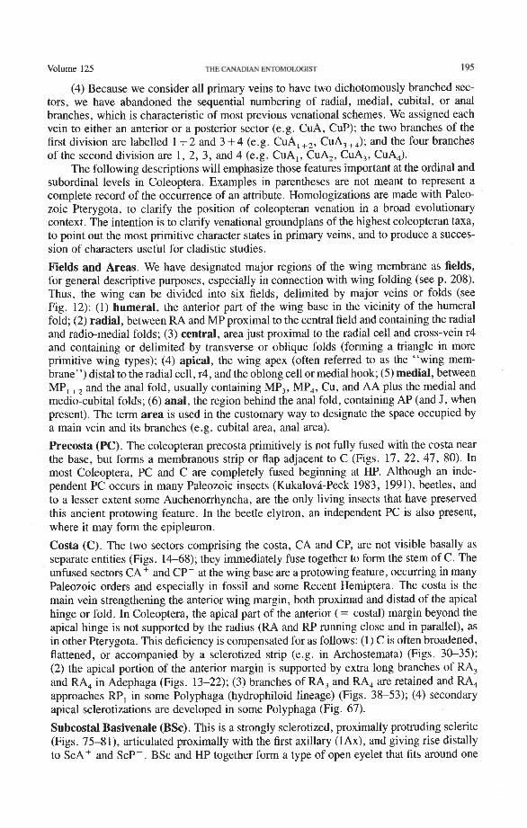
Fields and Areas. We have designated major regions of the wing membrane as fields, for general descriptive purposes, especially in connection with wing folding (see p. 208). Thus, the wing can be divided into six fields, delimited by major veins or folds (see Fig. 12): (1) humeral, the anterior part of the wing base in the vicinity of the humeral fold; (2) radial, between RA and MP proximal to the central field and containing the radial and radio-medial folds; (3) central, area just proximal to the radial cell and cross-vein r4 and containing or delimited by transverse or oblique folds (forming a triangle in more primitive wing types); (4) apical, the wing apex (often referred to as the "wing mem- brane'') distal to the radial cell, r4, and the oblong cell or medial hook; (5) medial, between MP, +, and the anal fold, usually containing MP,, MP,, Cu, and AA plus the medial and medio-cubital folds; (6) anal, the region behind the anal fold, containing AP (and J, when present). The term area is used in the customary way to designate the space occupied by a main vein and its branches (e.g. cubital area, anal area).
Precosta (PC). The coleopteran precosta primitively is not fully fused with the costa near the base, but forms a membranous strip or flap adjacent to C (Figs. 17, 22, 47, 80). In most Coleoptera, PC and C are completely fused beginning at HP. Although an inde- pendent PC occurs in many Paleozoic insects (Kukalovi-Peck 1983, 1991), beetles, and to a lesser extent some Auchenorrhyncha, are the only living insects that have preserved this ancient protowing feature. In the beetle elytron, an independent PC is also present, where it may form the epipleuron.
Costa (C). The two sectors comprising the costa, CA and CP, are not visible basally as separate entities (Figs. 14-68); they immediately fuse together to form the stem of C. The unfused sectors CA+ and CP- at the wing base are a protowing feature, occurring in many Paleozoic orders and especially in fossil and some Recent Hemiptera. The costa is the main vein strengthening the anterior wing margin, both proximad and distad of the apical hinge or fold. In Coleoptera, the apical part of the anterior (= costal) margin beyond the apical hinge is not supported by the radius (RA and RP running close and in parallel), as in other Pterygota. This deficiency is compensated for as follows: (1) C is often broadened, flattened, or accompanied by a sclerotized strip (e.g. in Archostemata) (Figs. 30-35); (2) the apical portion of the anterior margin is supported by extra long branches of RA, and RA, in Adephaga (Figs. 13-22); (3) branches of RA, and RA, are retained and RA, approaches RP, in some Polyphaga (hydrophiloid lineage) (Figs. 38-53); (4) secondary apical sclerotizations are developed in some Polyphaga (Fig. 67).
Subcostal Basivenale (BSc). This is a strongly sclerotized, proximally protruding sclerite (Figs. 75-81), articulated proximally with the first axillary (lAx), and giving rise distally to ScA+ and ScPp. BSc and HP together form a type of open eyelet that fits around one
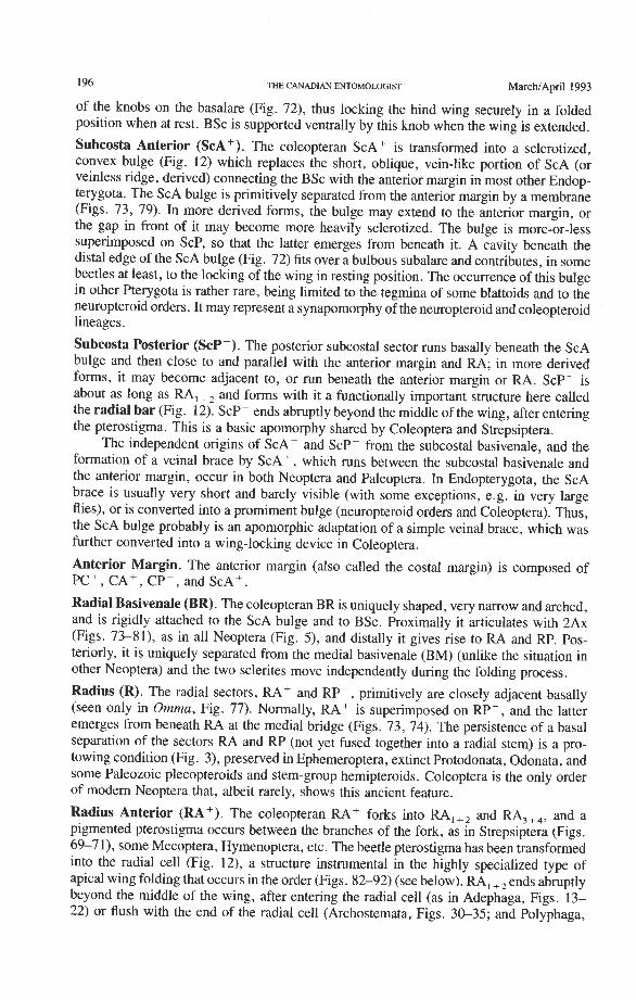
196 THE CANADIAN ENTOMOLOGIST MarchIApril 1993
of the knobs on the basalare (Fig. 72), thus locking the hind wing securely in a folded position when at rest. BSc is supported ventrally by this knob when the wing is extended.
Subcosta Anterior (ScA+). The coleopteran ScA+ is transformed into a sclerotized, convex bulge (Fig. 12) which replaces the short, oblique, vein-like portion of ScA (or veinless ridge, derived) connecting the BSc with the anterior margin in most other Endop- terygota. The ScA bulge is primitively separated from the anterior margin by a membrane (Figs. 73, 79). In more derived forms, the bulge may extend to the anterior margin, or the gap in front of it may become more heavily sclerotized. The bulge is more-or-less superimposed on ScP, so that the latter emerges from beneath it. A cavity beneath the distal edge of the ScA bulge (Fig. 72) fits over a bulbous subalare and contributes, in some beetles at least, to the locking of the wing in resting position. The occurrence of this bulge in other Pterygota is rather rare, being limited to the tegmina of some blattoids and to the neuropteroid orders. It may represent a synapomorphy of the neuropteroid and coleopteroid lineages.
Subcosta Posterior (ScP-). The posterior subcostal sector runs basally beneath the ScA bulge and then close to and parallel with the anterior margin and RA; in more derived forms, it may become adjacent to, or run beneath the anterior margin or RA. ScP- is about as long as RA, +, and forms with it a functionally important structure here called the radial bar (Fig. 12). ScP- ends abruptly beyond the middle of the wing, after entering the pterostigma. This is a basic apomorphy shared by Coleoptera and Strepsiptera.
The independent origins of ScAf and ScPp from the subcostal basivenale, and the formation of a veinal brace by ScA+ , which runs between the subcostal basivenale and the anterior margin, occur in both Neoptera and Paleoptera. In Endopterygota, the ScA brace is usually very short and barely visible (with some exceptions, e.g. in very large flies), or is converted into a promiment bulge (neuropteroid orders and Coleoptera). Thus, the ScA bulge probably is an apomorphic adaptation of a simple veinal brace, which was further converted into a wing-locking device in Coleoptera.
Anterior Margin. The anterior margin (also called the costal margin) is composed of PCf , CA+, CPp, and ScA+.
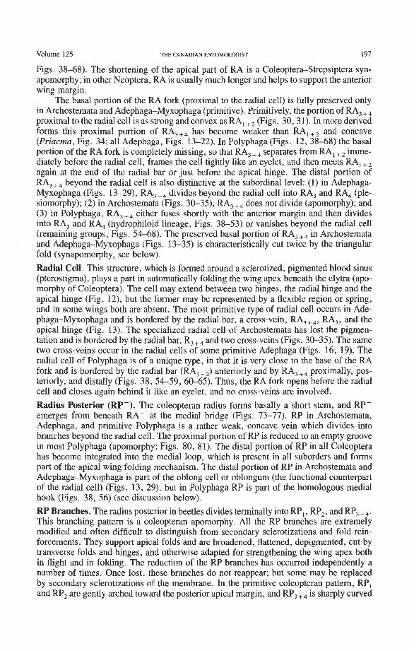
Radial Basivenale (BR). The coleopteran BR is uniquely shaped, very narrow and arched, and is rigidly attached to the ScA bulge and to BSc. Proximally it articulates with 2Ax (Figs. 73-81), as in all Neoptera (Fig. 5) , and distally it gives rise to RA and RP. Pos- teriorly, it is uniquely separated from the medial basivenale (BM) (unlike the situation in other Neoptera) and the two sclerites move independently during the folding process.
Radius (R). The radial sectors, RA' and RPp, primitively are closely adjacent basally (seen only in Omma, Fig. 77). Normally, RAf is superimposed on RP-, and the latter emerges from beneath RA at the medial bridge (Figs. 73, 74). The persistence of a basal separation of the sectors RA and RP (not yet fused together into a radial stem) is a pro- towing condition (Fig. 3), preserved in Ephemeroptera, extinct Protodonata, Odonata, and some Paleozoic plecopteroids and stem-group hemipteroids. Coleoptera is the only order of modern Neoptera that, albeit rarely, shows this ancient feature.
Radius Anterior (RA+). The coleopteran RA+ forks into RA, +, and RA, +,, and a pigmented pterostigma occurs between the branches of the fork, as in Strepsiptera (Figs. 69-71), some Mecoptera, Hymenoptera, etc. The beetle pterostigma has been transformed into the radial cell (Fig. 12), a structure instrumental in the highly specialized type of apical wing folding that occurs in the order (Figs. 82-92) (see below). RA, +,ends abruptly beyond the middle of the wing, after entering the radial cell (as in Adephaga, Figs. 13- 22) or flush with the end of the radial cell (Archostemata, Figs. 30-35; and Polyphaga,
volume 125 THE CANADIAN ENTOMOLOGIST 197
Figs. 38-68). The shortening of the apical part of RA is a Coleoptera-Strepsiptera syn- apomorphy; in other Neoptera, RA is usually much longer and helps to support the anterior wing margin.
The basal portion of the RA fork (proximal to the radial cell) is fully preserved only in Archostemata and Adephaga-Myxophaga (primitive). Primitively, the portion of RA, +, proximal to the radial cell is as strong and convex as RA, +, (Figs. 30,3 1). In more derived forms this proximal portion of RA,,, has become weaker than RA,,, and concave (Priacma, Fig. 34; all Adephaga, Figs. 13-22). In Polyphaga (Figs. 12, 38-68) the basal portion of the RA fork is completely missing, so that RA, +, separates from RA, +, imme- diately before the radial cell, frames the cell tightly like an eyelet, and then meets RA, +, again at the end of the radial bar or just before the apical hinge. The distal portion of RA,+, beyond the radial cell is also distinctive at the subordinal level: (1) in Adephaga- Myxophaga (Figs. 13-29), RA,+, divides beyond the radial cell into RA, and RA, (ple- siomorphy); (2) in Archostemata (Figs. 3&35), RA,,, does not divide (apomorphy); and (3) in Polyphaga, RA,+, either fuses shortly with the anterior margin and then divides into RA, and RA, (hydrophiloid lineage, Figs. 38-53) or vanishes beyond the radial cell (remaining groups, Figs. 54-68). The preserved basal portion of RA,+, in Archostemata and Adephaga-Myxophaga (Figs. 13-35) is characteristically cut twice by the triangular fold (synapomorphy, see below).
Radial Cell. This structure, which is formed around a sclerotized, pigmented blood sinus (pterostigma), plays a part in automatically folding the wing apex beneath the elytra (apo- morphy of Coleoptera). The cell may extend between two hinges, the radial hinge and the apical hinge (Fig. 12), but the former may be represented by a flexible region or spring, and in some wings both are absent. The most primitive type of radial cell occurs in Ade- phaga-Myxophaga and is bordered by the radial bar, a cross-vein, RA,,,, RA,, and the apical hinge (Fig. 13). The specialized radial cell of Archostemata has lost the pigmen- tation and is bordered by the radial bar, R,+, and two cross-veins (Figs. 30-35). The same two cross-veins occur in the radial cells of some primitive Adephaga (Figs. 16, 19). The radial cell of Polyphaga is of a unique type, in that it is very close to the base of the RA fork and is bordered by the radial bar (RA, +,) anteriorly and by RA,+, proximally, pos- teriorly, and distally (Figs. 38, 54-59, 60-65). Thus, the RA fork opens before the radial cell and closes again behind it like an eyelet, and no cross-veins are involved.
Radius Posterior (RP-). The coleopteran radius forms basally a short stem, and RP- emerges from beneath RA+ at the medial bridge (Figs. 73-77). RP in Archostemata, Adephaga, and primitive Polyphaga is a rather weak, concave vein which divides into branches beyond the radial cell. The proximal portion of RP is reduced to an empty groove in most Polyphaga (apomorphy; Figs. 80, 81). The distal portion of RP in all Coleoptera has become integrated into the medial loop, which is present in all suborders and forms part of the apical wing folding mechanism. The distal portion of RP in Archostemata and Adephaga-Myxophaga is part of the oblong cell or oblongum (the functional counterpart of the radial cell) (Figs. 13, 29), but in Polyphaga RP is part of the homologous medial hook (Figs. 38, 56) (see discussion below).
RP Branches. The radius posterior in beetles divides terminally into RP,, RP,, and RP, +,. This branching pattern is a coleopteran apomorphy. All the RP branches are extremely modified and often difficult to distinguish from secondary sclerotizations and fold rein- forcements. They support apical folds and are broadened, flattened, depigmented, cut by transverse folds and hinges, and otherwise adapted for strengthening the wing apex both in flight and in folding. The reduction of the RP branches has occurred independently a number of times. Once lost, these branches do not reappear; but some may be replaced by secondary sclerotizations of the membrane. In the primitive coleopteran pattern, RP, and RP, are gently arched toward the posterior apical margin, and RP, +, is sharply curved
198 THE CANADIAN ENTOMOLOGIST MarcMApril 1993
posteriorly in a distinctive manner (like a saber) (Fig. 13). Deviations from this basic coleopteran condition are frequent, always derived, and of great systematic importance.
Radial Cross-veins (r3 and r4). As many as four cross-veins may have connected RA, +, with RP- in the coleopteroid ancestor; these persist only in Omma (Figs. 30, 31). The first (proximal) cross-vein is preserved in all Archostemata and most Adephaga-Myxo- phaga, but it is completely lost in Polyphaga. The two more permanent, distal cross-veins, r3 and r4, connect the radial cell with the oblong cell or medial hook and are engaged in apical wing folding, as follows: in Archostemata, both r3 and r4 help to connect the radial and medial loops (and thus synchronize them during folding); in Adephaga-Myxophaga, r3 is absent (obliterated by the triangular fold, Fig. 13) and r4 is arched toward the apex forming a "V" (Fig. 15); in Polyphaga, r4 alone connects the radial cell with the medial hook (Figs. 40, 55,60-65), but r3 primitively is directed obliquely (proximo-posteriorly) toward RP (but is always cut off from RP by a longitudinal fold) (Figs. 38, 40). The r3 cross-vein in some narrow hind wings, such as those of many Elateriformia, has been misinterpreted as a "posterior branch of the radius" or "radial sector" or "radial recur- rent," because of its tendency to become longitudinally oriented and secondarily aligned with primary veins, such as RP (Figs. 63, 65, 67). It never reaches the radial basivenale or the R stem, as a true RP sector should, and it may form a more-or-less complete cross- vein between RA,+, and RP, as in some Scirtidae (Fig. 54), Dascillidae (Fig. 60), and schizopodine Buprestidae (Fig. 62). Cross-vein r3 is completely lost in many Polyphaga, but r4 is always present, or has been replaced by toughened membrane following the same course (most Scarabaeoidea, Figs. 5C~53). The cross-vein r4 runs between the radial cell and the point at which RP divides into branches. In Adephaga, it originates from RA, (Figs. 13-16); in Archostemata and Polyphaga, it always arises from RA,+, (Figs. 30- 35, 38, 56, 60). In some Polyphaga, RP, +, or RP, may become adjacent basally to the end of r4 (Figs. 56, 60).
Medial Basivenale (BM). In beetles, BM primitively is divided into two parts, anterior (BMA) and posterior (BMP), which give rise to the separated sectors MA and MP (Figs. 73, 75-81). Proximally, BMA and BMP articulate with the median plate (MED), which is also divided lengthwise into two parts by a suture (coleopteran apomorphy). In some derived forms the suture may be replaced by a gap and one part of MED may become reduced. The medial basivenale in all Coleoptera is distinctly separated from the radial basivenale (BR) because the media and radius tilt away from each other during folding, but in other Endopterygota and in other Neoptera, these two basivenalia are frequently completely fused (derived). In Adephaga and Polyphaga, BMA is shaped like a sickle with the tip arching concavely toward the convexly arched tip of BR (Figs. 79-80, 81) (prim- itive within Coleoptera). In Archostemata, the tip of BMA is completely reduced (Figs. 77, 78) (apomorphy).
Media Anterior (MA+). The convex anterior medial sector (MA+) of all Endopterygota fuses with R near the wing base. In some groups, MA+ may separate again from R (prim- itive; some Neuroptera), or from RP (in Triassic Megaloptera and modem Raphidioptera and Hymenoptera), and still remain convex (Kukalovh-Peck 1991, figs. 6.27E-5). In coleopteran hind wings, MA and MP are not fused together basally into a stem; the short basal portion of MA between the base and the point at which MA fuses with RP is de- sclerotized. The coleopteran MA fuses close to the wing base with R and RP. At the point where MA enters R, a convex brace has been developed, which binds together, like a spring clamp, the otherwise separated bases of the radial and medial loops (apomorphy of Coleoptera); this brace is called the medial bridge (Figs. 74, 7679 , 93-95).
Media Posterior (MP-). The coleopteran MP is primitively concave, as it is in other Neoptera, but a change to the convex condition is frequent, especially in large Polyphaga
Volume 125 THE CANADIAN ENTOMOLOGIST 199
(e.g. some Scarabaeidae). MP forks relatively close to the base (primitive at the Neoptera level), but the fork is obscured by a fold cutting across the very base of MP,,,. MP, +, primitively is strong and mildly, concavely arched, ends in a spike, and does not reach the posterior margin of the wing. This branch functions as the medial bar and combines with rp-mp cross-veins to form part of the medial loop, which is engaged in apical folding. In Archostemata and Adephaga-Myxophaga, the medial bar, just proximal to the oblong cell, has an abrupt hinge (Figs. 19, 24, 33) (synapomorphy), but in Polyphaga, this hinge is lacking (plesiomorphy) (see below). MP, +, primitively forks in all Coleoptera: MP, is simple and MP, fuses with CuA, near the posterior margin (Figs. 34, 39) (apomorphy of Coleoptera). A similar fusion of MP, and CuA, also occurs frequently in the mecopterid complex. In Archostemata and some Adephaga there may be two cross-veins connecting MP, +, and MP,,, (primitive); in Polyphaga there is one or none (derived).
Medio-cubital Cross-vein or Arculus (mp-cua). This cross-vein between MP and CuA, which is located near the base, is an imporant brace occurring in forewings and hind wings and a basic synapomorphy shared by Endopterygota and the hemipteroid complex. The coleopteran mp-cua is crossed by a deep medio-cubital fold and is usually completely obliterated. It is preserved in a few Adephaga (Figs. 14, 18, 21), is very rare in Polyphaga (Fig. 45), and appears to have been eliminated in Archostemata (derived).
Radio-medial Cross-veins (rp-mpl, rp-mp2). Cross-vein rp-mpl extends between RP and MP, +,, delimits the oblong cell proximally, and is retained only in Archostemata and Adephaga-Myxophaga. This cross-vein may actually be formed by MA+ separating from RP. Cross-vein rp-mp2 connects RP,+, and MP, +, (= medial bar) and is present in all suborders. In Archostemata and Adephaga-Myxophaga it delimits the distal portion of the oblong cell; in Polyphaga it becomes part of the medial hook (Fig. 56) (apomorphy).
Radial and Medial Loops. These two structures, each consisting of several veinal parts, together resemble a pair of blunt-tipped scissors with the pivot formed by the medial bridge (Figs. 88-92). The radial loop consists of the radial bar (ScP + RA) and radial cell, as mentioned above. The medial loop comprises the medial bar (MP, +,) and the oblong cell in Archostemata and Adephaga-Myxophaga (symplesiomorphy) or the medial bar and medial hook in Polyphaga (apomorphy). The oblong cell is formed anteriorly by RP and the base of RP,,,, posteriorly by the medial bar, proximally by cross-vein rp-mpl, and distally by rp-mp2. The medial hook is formed anteriorly by RP, distally by RP,,, and cross-vein rp-mp2, and posteriorly by the apex of the medial bar. MP, +, primitively con- tinues beyond the oblong cell or medial hook and forms a spur (medial spur) without reaching the posterior margin of the wing. This spur often gives support to a portion of membrane which folds with the medial loop.
Springs, Crimps, and Hinges. A major feature of the beetle hind wing and one that distinguishes it from that of any other pterygote order is the presence of an intrinsic spring mechanism which maintains the wing in a folded position at rest. This mechanism consists mainly of two localized springs or deflection zones: the radial spring, which is on the radial bar proximal to the end of the radial cell, and the medial spring near the apex of the medial bar. These springs are recognized by the presence of a series of transverse grooves or crimps (Figs. 98-100) which give that section of the bar a combination of strength and flexibility and at the same time prevent the wing from being unfolded while the direct flight muscles are in a relaxed position (see below). Additional crimping may also occur in reinforced membrane adjacent to the radial and medial bars, around cross- vein r4 or elsewhere, and a minor spring (apical spring) may occur beyond the radial cell. These all appear to be involved in the wing folding process, but some of them may have an alternative or supplementary role in adjusting wing shape (and thus camber) during flight (see Wootton 1981).
200 THE CANADIAN ENTOMOLOGIST MarchJApril 1993
Strong and unmodified radial and medial springs probably represent the primitive condition in beetles, but in various lineages one or both of these springs have been mod- ified, weakened, or lost. A common type of spring modification is the development of a hinge, which is an abrupt break in the bar, where it is crossed by a transverse fold (Figs. 88, 96, 100). The radial loop may have a radial hinge and an apical hinge, proximal and distal to the radial cell; and the medial loop may have a medial hinge proximal to the oblong cell. These hinges allow an increase in the degree of wing folding by permitting the bar to be abruptly folded on itself; however they may also weaken the spring mecha- nism, so that extrinsic devices are needed to complete the wing folding process (see below). Radial and apical hinges have developed in both major lineages (Polyphaga and Adephaga- Myxophaga + Archostemata), but the medial hinge is a synapomorphy of the latter group.
Cubital Basivenale (BCu). BCu is divided into two parts (Figs. 75, 77, 79) (primitive within the Pterygota): the anterior portion, BCuA, gives rise to CuA, but CuA is sometimes cut off from BCuA by a short, transverse fold; the posterior BCuP gives rise to a short, free, concave CUP retained in some Polphaga only. CUP- is completely reduced in Archostemata and Adephaga-Myxophaga, as a synapomorphy. BCu articulates proximally with the third axillary sclerite A AX), as in all Neoptera, sometimes by means of a tough- ened strip of membrane. The coleopteran BCu is often connected by a sclerotization with BM (Figs. 79, 81).
Cubitus Anterior (CuA). The CuA in Coleoptera has two braces with the media (the mp- cua cross-vein or arculus, shared by all Endopterygota, and a direct fusion between CuA, and MP,, occurring in many endopterygotes) and one or two braces (fusions) with AA (coleopteran apomorphy). The cubito-anal braces enclose the first and second cubito-anal cells as follows (Figs. 36, 37): (1) CuA forks in all suborders into CuA,,, and CuA,,,; (2) CuA, +, primitively forks again into CuA, and CuA,; (3) CuA, primitively fuses with MP, forming a brace; (4) CuA,,, in all suborders fuses with AA, +, and thus forms the first cubito-anal cell; (5) CuA, in Archostemata (Fig. 30) and primitively in Polyphaga (Fig. 39), reaches the posterior margin, but this never occurs in Adephaga (apomorphy); in many Adephaga and many Polyphaga this CuA, fuses with AA, +,, thus forming the second cubito-anal cell (independently derived in Polyphaga and Adephaga).
Cubitus Posterior (CUP). A free CUP is suppressed in Coleoptera, being either short or ~bsent. A short, free, distinctly concave CUP is present in some Polyphaga (Figs. 38,40, 53,54) (primitive), but when C U P meets AA+ the latter superimposes itself on the former resulting in a more-or-less tubular vein (circular in cross section). A free CUP is absent in Archostemata and Adephaga-Myxophaga (synapomorphy). The claval fold in Coleoptera is reduced to a short, concave groove, which may or may not be accompanied by CUP; in many cases it vanishes entirely.
Anal Basivenale (BA). The coleopteran BA primitively is divided into two parts (primitive at the Neoptera level): a shorter anterior BAA and a longer posterior BAP. BAA is con- nected proximally to 3Ax by a strip of tough membrane, and it gives rise distally to a strong anal anterior (AA+). In beetles, BAA is always crossed by the anal fold and is mostly obliquely oriented or otherwise deformed or completely destroyed by the fold (Figs. 36, 37, 72, 76, 77). BAP is elongate and narrow; AP,,,, if present, is weak and often shortened, and arises from the distal end of BAP; AP,,, is usually strong and arises from the opposite, proximal end.
Anal Fold and Anal Brace. The branches of AA are separated from those of AP by a convex anal fold, along which the AP area is folded under the wing at rest. This type of folding is found elsewhere only in cockroaches and termites and is slightly indicated in some hemipteroids. It results from the fact that the greatly enlarged anal lobe is supported
Volume 125 THE CANADIAN ENTOMOLOGIST 20 1
only by the branches of AP (not by all anal branches, as in orthopteroids and plecopter- oids), but the AA area adjoins the remigium. This trend, occurring in blattoid, hemipteroid, and endopterygote hind wings with an enlarged anal lobe (rare in the last two groups), deemphasizes the claval fold and makes the anal fold more significant (Kukalova-Peck 1991). In the Adephaga, BAA is V-shaped, with the anal fold crossing the tip of the "V" (Fig. 36) (derived). Archostemata have a similar V-shaped BAA, but it is smaller and less conspicuously shaped (Fig. 33) (synapomorphy with Adephaga). In Polyphaga, a small BAA is occasionally present (Fig. 42) (primitive), but in most groups it is completely destroyed by the anal fold.
The anal brace (a connection between the anal branches and the cubitus, preventing the anal region from buckling) occurs in all Neoptera and is usually formed by AA, +,, which becomes partly or fully fused with CUP, but sometimes only AA, is involved in bracing. In the coleopteran anal brace, a portion of AA becomes adjacent near base to the reduced CUP (Fig. 37) (coleopteran apomorphy). Primitively in polyphagans, AA is well distant from the cubital basivenale (Figs. 36,40) but in the derived condition it has shifted toward BCu and sometimes appears to be arising directly from it (Figs. 63-67). This secondary shift occurs sporadically in some other Neoptera as well. When cut off from a reduced, deformed, or destroyed basivenale (BA) by a fold, any anal branch involved in the anal brace may look like a "third" branch of the cubitus [= "post-cubitus" ("PC") of Snodgrass (1935) and numerous followers]. However, this erroneous interpretation becomes obvious when broad comparisons are made. In the Archostemata, Adephaga- Myxophaga, as in primitive Polyphaga, AA is clearly associated with the anal basivenale and does not look like a cubital branch.
Anal Anterior (AA). The anterior sector of the anal vein (AA) forks primitively not far from the base into AA,,, and AA,,,. Neither branch is ever forked and both extend primitively to the posterior wing margin in all suborders (Figs. 14, 19, 34, 39). AA, +, fuses in all Coleoptera with CuA,,,, and thus the first cubito-anal cell is formed (apo- morphy of Coleoptera); AA, +, + CuA,,, may then fuse with CuA, forming the second cubito-anal cell, usually called the anal or wedge cell (Figs. 18, 20, 33-37, 60-63). The wedge cell occurs in all suborders, but has been lost a number of times in connection with the reduction of the medial field. The anterior anal area (AA) is separated from the anal lobe by the anal fold and is braced in two or three places to the cubitus, so that functionally it is an extension of the remigium. This trend also occurs in the hind wings of hemipteroid and blattoid insects (Kukalovi-Peck 1991). Anal Posterior (AP). The posterior anal sector (AP) consists of two separate branches, AP, +, and AP,,,, which arise independently from opposite ends of a long, narrow, often obliquely oriented basivenale (BAP, Figs. 36, 72-75). AP, +,, when present, is weak and runs close to the anal fold (Adephaga, Figs. 14, 16, 19, 20, 36; Polyphaga, Figs. 45, 80) (primitive). AP, +, is absent in Archostemata (apomorphy). AP,,, primitively forks into AP, and AP, in Archostemata (Figs. 30, 33, 34) and Polyphaga (Figs. 45, 54, 55) (prim- itive). AP,,, has lost its branches in Adephaga (apomorphy). Jugal Basivenale (BJ). This is a very small sclerite adjacent to BAP. BJ occurs sporad- ically in all suborders (Figs. 75-79). Jugals (J). In Coleoptera, the jugal sectors, JA and JP, are reduced, short, and usually fused, forming a short, prong-like stem, J, arising from a small BJ. J occurs in Archo- stemata, Adephaga, and in some Polyphaga (Figs. 55, 60-65). In most Polyphaga J is lost.
Coleopteran Hind Wing Articulation The hind wings of Coleoptera have a unique capability within Pterygota: the apical
part of the wing can be spring-folded and drawn in and then stretched out again (a simply
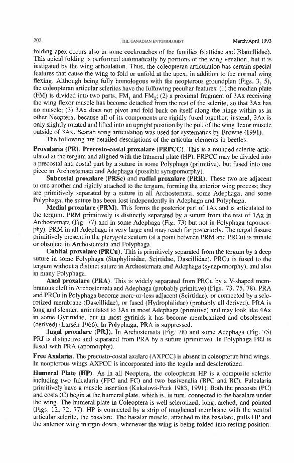
202 THE CANADIAN ENTOMOUXilST MarcMApril 1993
folding apex occurs also in some cockroaches of the families Blattidae and Blattellidae). This apical folding is performed automatically by portions of the wing venation, but it is instigated by the wing articulation. Thus, the coleopteran articulation has certain special features that cause the wing to fold or unfold at the apex, in addition to the normal wing flexing. Although being fully homologous with the neopterous groundplan (Figs. 3, 5), the coleopteran articular sclerites have the following peculiar features: (1) the median plate (FM) is divided into two parts, FM, and FM,; (2) a proximal fragment of 3Ax receiving the wing flexor muscle has become detached from the rest of the sclerite, so that 3Ax has no muscle; (3) 3Ax does not pivot and fold back on itself along the hinge within as in other Neoptera, because all of its components are rigidly fused together; instead, 3Ax is only slightly rotated and lifted into an upright position by the pull of the wing flexor muscle outside of 3Ax. Scarab wing articulation was used for systematics by Browne (1991).
The following are detailed descriptions of the articular elements in beetles.
Proxalaria (PR). Precosto-costal proxalare (PRPCC). This is a rounded sclerite artic- ulated at the tergum and aligned with the humeral plate (HP). PRPCC may be divided into a precostal and costal part by a suture in some Polyphaga (primitive), but fused into one piece in Archostemata and Adephaga (possible synapomorphy).
Subcostal proxalare (PRSc) and radial proxalare (PRR). These two are adjacent to one another and rigidly attached to the tergum, forming the anterior wing process; they are primitively separated by a suture in all Archostemata, some Adephaga, and some Polyphaga; the suture has been lost independently in Adephaga and Polyphaga.
Medial proxalare (PRM). This forms the posterior part of 1Ax and is articulated to the tergum. PRM primitively is distinctly separated by a suture from the rest of 1Ax in Archostemata (Fig. 77) and in some Adephaga (Fig. 73) but not in Polyphaga (apomor- phy). PRM in all Adephaga is very large and may reach far posteriorly. The tergal fissure primitively present in the pterygote scutum (at a point between PRM and PRCu) is minute or obsolete in Archostemata and Polyphaga.
Cubital proxalare (PRCu). This is primitively separated from the tergum by a deep suture in some Polyphaga (Staphylinidae, Scirtidae, Dascillidae). PRCu is fused to the tergum without a distinct suture in Archostemata and Adephaga (synapomorphy), and also in many Polyphaga.
Anal proxalare (PRA). This is widely separated from PRCu by a V-shaped mem- branous cleft in Archostemata and Adephaga (probably primitive) (Figs. 73,75,78). PRA and PRCu in Polyphaga become more-or-less adjacent (Scirtidae), or connected by a scle- rotized membrane (Dascillidae), or fused (Hydrophilidae) (probably all derived). PRA is long and slender, articulated to 3Ax in most Adephaga (primitive) and may look like 4Ax in some Gyrinidae, but in most gyrinids it has become membranized and obsolescent (derived) (LarsCn 1966). In Polyphaga, PRA is suppressed.
Jugal proxalare (PRJ). In Archostemata (Fig. 78) and some Adephaga (Fig. 75) PRJ is distinctive and separated from PRA by a suture (primitive). In Polyphaga PRJ is fused with PRA (apomorphy).
Free Axalaria. The precosto-costal axalare (AXPCC) is absent in coleopteran hind wings. In neopterous wings AXPCC is incorporated into the tegula and desclerotized.
Humeral Plate (HP). As in all Neoptera, the coleopteran HP is a composite sclerite including two fulcalaria (FPC and FC) and two basivenalia (BPC and BC). Fulcalaria primitively have a muscle insertion (Kukalova-Peck 1983, 1991). Both the precosta (PC) and costa (C) begin at the humeral plate, which is, in turn, connected to the basalare under the wing. The humeral plate in Coleoptera is well sclerotized, long, arched, and pointed (Figs. 12, 72, 77). HP is connected by a strip of toughened membrane with the ventral articular sclerite, the basalare. The basalar muscle, attached to the basalare, pulls HP and the anterior wing margin down, whenever the wing is being folded into resting position.
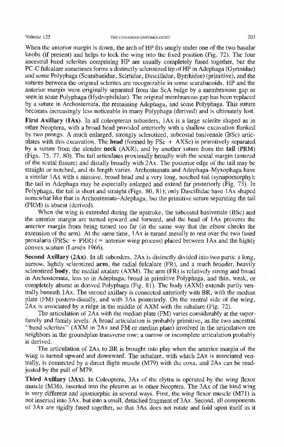
Volume 125 THE CANADIAN ENTOMOLOGIST 203
When the anterior margin is down, the arch of HP fits snugly under one of the two basalar knobs (if present) and helps to lock the wing into the fixed position (Fig. 72). The four ancestral band sclerites comprising HP are usually completely fused together, but the PC-C fulcalare sometimes forms a distinctly sclerotized tip of HP in Adephaga (Gyrinidae) and some Polyphaga (Scarabaeidae, Scirtidae, Dascillidae, Byrrhidae) (primitive), and the sutures between the original sclerites are recognizable in some scarabaeoids. HP and the anterior margin were originally separated from the ScA bulge by a membranous gap as seen in some Polyphaga (Hydrophilidae). The original membranous gap has been replaced by a suture in Archostemata, the remaining Adephaga, and some Polyphaga. This suture becomes increasingly less noticeable in many Polyphaga (derived) and is ultimately lost.
First Axillary (1Ax). In all coleopteran suborders, 1Ax is a large sclerite shaped as in other Neoptera, with a broad head provided anteriorly with a shallow excavation flanked by two prongs. A much enlarged, strongly sclerotized, subcostal basivenale (BSc) artic- ulates with this excavation. The head (formed by FSc + AXSc) is primitively separated by a suture from the slender neck (AXR), and by another suture from the tail (PRM) (Figs. 75, 77, 80). The tail articulates proximally broadly with the scutal margin (anterad of the scutal fissure) and distally broadly with 2Ax. The posterior edge of the tail may be straight or notched, and its length varies. Archostemata and Adephaga-Myxophaga have a similar 1Ax with a massive, broad head and a very long, notched tail (synapomorphy); the tail in Adephaga may be especially enlarged and extend far posteriorly (Fig. 73). In Polyphaga, the tail is short and straight (Figs. 80, 81); only Dascillidae have 1Ax shaped somewhat like that in Archostemata-Adephaga, but the primitive suture separating the tail (PRM) is absent (derived).
When the wing is extended during the upstroke, the subcostal basivenale (BSc) and the anterior margin are turned upward and forward, and the head of 1Ax prevents the anterior margin from being turned too far (in the same way that the elbow checks the extension of the arm). At the same time, 1Ax is turned mesally to rest over the two fused proxalaria (PRSc + PRR) (= anterior wing process) placed between 1Ax and the highly convex scutum (Larstn 1966).
Second Axillary (2Ax). In all suborders, 2Ax is distinctly divided into two parts: a long, narrow, lightly sclerotized arm, the radial fulcalare (FR), and a much broader, heavily sclerotized body, the medial axalare (AXM). The arm (FR) is relatively strong and broad in Archostemata, less so in Adephaga, broad in primitive Polyphaga, and thin, weak, or completely absent in derived Polyphaga (Fig. 81). The body (AXM) extends partly ven- trally beneath 1Ax. The second axillary is connected anteriorly with BR, with the median plate (FM) postero-distally, and with 3Ax posteriorly. On the ventral side of the wing, 2Ax is associated by a ridge in the middle of AXM with the subalare (Fig. 72).
The articulation of 2Ax with the median plate (FM) varies considerably at the super- family and family levels. A broad articulation is probably primitive, as the two ancestral "band sclerites" (AXM in 2Ax and FM or median plate) involved in the articulation are neighbors in the groundplan transverse row; a narrow or incomplete articulation probably is derived.
The articulation of 2Ax to BR is brought into play when the anterior margin of the wing is turned upward and downward. The subalare, with which 2Ax is associated ven- trally, is connected by a direct flight muscle (M79) with the coxa, and 2Ax can be read- justed by the pull of M79.
Third Axillary (3Ax). In Coleoptera, 3Ax of the elytra is operated by the wing flexor muscle (M36), inserted into the pleuron as in other Neoptera. The 3Ax of the hind wing is very different and apomorphic in several ways. First, the wing flexor muscle (M71) is not inserted into 3Ax, but into a small, detached fragment of 3Ax. Second, all components of 3Ax are rigidly fused together, so that 3Ax does not rotate and fold upon itself as it
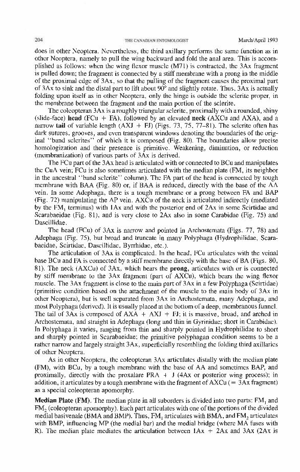
204 CANADIAN ~ M O I ~ O G I S T MarchIApril 1993
does in other Neoptera. Nevertheless, the third axillary performs the same function as in other Neoptera, namely to pull the wing backward and fold the anal area. This is accom- plished as follows: when the wing flexor muscle (M71) is contracted, the 3Ax fragment is pulled down; the fragment is connected by a stiff membrane with a prong in the middle of the proximal edge of 3Ax, so that the pulling of the fragment causes the proximal part of 3Ax to sink and the distal part to lift about 90" and slightly rotate. Thus, 3Ax is actually folding upon itself as in other Neoptera, only the hinge is outside the sclerite proper, in the membrane between the fragment and the main portion of the sclerite.
The coleopteran 3Ax is a roughly triangular sclerite, proximally with a rounded, shiny (slide-face) head (FCu + FA), followed by an elevated neck (AXCu and AXA), and a narrow tail of variable length (AXJ + FJ) (Figs. 73, 75, 77-81). The sclerite often has dark sutures, grooves, and even transparent windows denoting the boundaries of the orig- inal "band sclerites" of which it is composed (Fig. 80). The boundaries allow precise homologization and their presence is primitive. Weakening, diminution, or reduction (membranization) of various parts of 3Ax is derived.
The FCu part of the 3Ax head is articulated with or connected to BCu and manipulates the CuA vein; FCu is also sometimes articulated with the median plate (FM, its neighbor in the ancestral "band sclerite" column). The FA part of the head is connected by tough membrane with BAA (Fig. 80) or, if BAA is reduced, directly with the base of the AA vein. In some Adephaga, there is a tough membrane or a prong between FA and BAP (Fig. 72) manipulating the AP vein. AXCu of the neck is articulated indirectly (mediated by the FM, terminus) with 1Ax and with the posterior end of 2Ax in some Scirtidae and Scarabaeidae (Fig. 81), and is very close to 2Ax also in some Carabidae (Fig. 75) and Dascillidae.
The head (FCu) of 3Ax is narrow and pointed in Archostemata (Figs. 77, 78) and Adephaga (Fig. 75), but broad and truncate in many Polyphaga (Hydrophilidae, Scara- baeidae, Scirtidae, Dascillidae, Byrrhidae, etc.).
The articulation of 3Ax is complicated. In the head, FCu articulates with the veinal base BCu and FA is connected by a stiff membrane directly with the base of BA (Figs. 80, 81). The neck (AXCu) of 3Ax, which bears the prong, articulates with or is connected by stiff membrane to the 3Ax fragment (part of AXCu), which bears the wing flexor muscle. The 3Ax fragment is close to the main part of 3Ax in a few Polyphaga (Scirtidae) (primitive condition based on the attachment of the muscle to the main body of 3Ax in other Neoptera), but is well separated from 3Ax in Archostemata, many Adephaga, and most Polyphaga (derived). It is usually placed at the bottom of a deep, membranous funnel. The tail of 3Ax is composed of AXA + AXJ + FJ; it is massive, broad, and arched in Archostemata, and straight in Adephaga (long and thin in Gyrinidae; short in Carabidae). In Polyphaga it varies, ranging from thin and sharply pointed in Hydrophilidae to short and sharply pointed in Scarabaeidae; the primitive polyphagan condition seems to be a rather narrow and largely straight 3Ax, superficially resembling the folding third axillaries of other Neoptera.
As in other Neoptera, the coleopteran 3Ax articulates distally with the median plate (FM), with BCu, by a tough membrane with the base of AA and sometimes BAP, and proximally, directly with the proxalare PRA + J (4Ax or posterior wing process); in addition, it articulates by a tough membrane with the fragment of AXCu ( = 3Ax fragment) as a special coleopteran apomorphy.
Median Plate (FM). The median plate in all suborders is divided into two parts: FM, and FM, (coleopteran apomorphy). Each part articulates with one of the portions of the divided medial basivenale (BMA and BMP). Thus, FM, articulates with BMA, and FM, articulates with BMP, influencing MP (the medial bar) and the medial bridge (where MA fuses with R). The median plate mediates the articulation between 1Ax + 2Ax and 3Ax (2Ax is
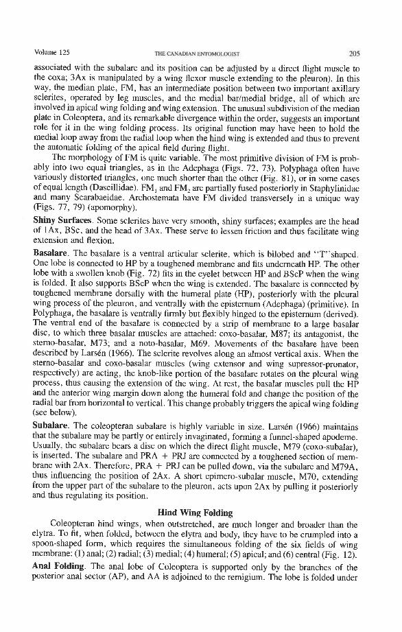
volume 125 THE CANADIAN ENTOMOLOGIST 205
associated with the subalare and its position can be adjusted by a direct flight muscle to the coxa; 3Ax is manipulated by a wing flexor muscle extending to the pleuron). In this way, the median plate, FM, has an intermediate position between two important axillary sclerites, operated by leg muscles, and the medial badmedial bridge, all of which are involved in apical wing folding and wing extension. The unusual subdivision of the median plate in Coleoptera, and its remarkable divergence within the order, suggests an important role for it in the wing folding process. Its original function may have been to hold the medial loop away from the radial loop when the hind wing is extended and thus to prevent the automatic folding of the apical field during flight.
The morphology of FM is quite variable. The most primitive division of FM is prob- ably into two equal triangles, as in the Adephaga (Figs. 72, 73). Polyphaga often have variously distorted triangles, one much shorter than the other (Fig. 81), or in some cases of equal length (Dascillidae). FM, and FM, are partially fused posteriorly in Staphylinidae and many Scarabaeidae. Archostemata have FM divided transversely in a unique way (Figs. 77, 79) (apomorphy).
Shiny Surfaces. Some sclerites have very smooth, shiny surfaces; examples are the head of lAx, BSc, and the head of 3Ax. These serve to lessen friction and thus facilitate wing extension and flexion. Basalare. The basalare is a ventral articular sclerite, which is bilobed and "T"shaped. One lobe is connected to HP by a toughened membrane and fits underneath HP. The other lobe with a swollen knob (Fig. 72) fits in the eyelet between HP and BScP when the wing is folded. It also supports BScP when the wing is extended. The basalare is connected by toughened membrane dorsally with the humeral plate (HP), posteriorly with the pleural wing process of the pleuron, and ventrally with the episternum (Adephaga) (primitive). In Polyphaga, the basalare is ventrally firmly but flexibly hinged to the episternum (derived). The ventral end of the basalare is connected by a strip of membrane to a large basalar disc, to which three basalar muscles are attached: coxo-basalar, M87; its antagonist, the sterno-basalar, M73; and a noto-basalar, M69. Movements of the basalare have been described by LarsCn (1966). The sclerite revolves along an almost vertical axis. When the sterno-basalar and coxo-basalar muscles (wing extensor and wing supressor-pronator, respectively) are acting, the knob-like portion of the basalare rotates on the pleural wing process, thus causing the extension of the wing. At rest, the basalar muscles pull the HP and the anterior wing margin down along the humeral fold and change the position of the radial bar from horizontal to vertical. This change probably triggers the apical wing folding (see below).
Subalare. The coleopteran subalare is highly variable in size. LarsCn (1966) maintains that the subalare may be partly or entirely invaginated, forming a funnel-shaped apodeme. Usually, the subalare bears a disc on which the direct flight muscle, M79 (coxo-subalar), is inserted. The subalare and PRA + PRJ are connected by a toughened section of mem- brane with 2Ax. Therefore, PRA + PRJ can be pulled down, via the subalare and M79A, thus influencing the position of 2Ax. A short epimero-subalar muscle, M70, extending from the upper part of the subalare to the pleuron, acts upon 2Ax by pulling it posteriorly and thus regulating its position.
Hind Wing Folding Coleopteran hind wings, when outstretched, are much longer and broader than the
elytra. To fit, when folded, between the elytra and body, they have to be crumpled into a spoon-shaped form, which requires the simultaneous folding of the six fields of wing membrane: (1) anal; (2) radial; (3) medial; (4) humeral; (5) apical; and (6) central (Fig. 12). Anal Folding. The anal lobe of Coleoptera is supported only by the branches of the posterior anal sector (AP), and AA is adjoined to the remigium. The lobe is folded under
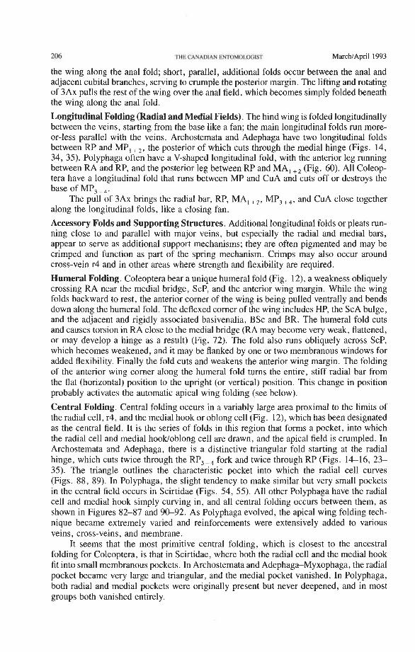
206 THE CANADIAN EWOMOLOFIST MarcWApril 1993
the wing along the anal fold; short, parallel, additional folds occur between the anal and adjacent cubital branches, serving to crumple the posterior margin. The lifting and rotating of 3Ax pulls the rest of the wing over the anal field, which becomes simply folded beneath the wing along the anal fold.
Longitudinal Folding (Radial and Medial Fields). The hind wing is folded longitudinally between the veins, starting from the base like a fan; the main longitudinal folds run more- or-less parallel with the veins. Archostemata and Adephaga have two longitudinal folds between RP and MP, +,, the posterior of which cuts through the medial hinge (Figs. 14, 34, 35). Polyphaga often have a V-shaped longitudinal fold, with the anterior leg running between RA and RP, and the posterior leg between RP and MA, +, (Fig. 60). All Coleop- tera have a longitudinal fold that runs between MP and CuA and cuts off or destroys the base of MP, + ,.
The pull of 3Ax brings the radial bar, RP, MA, +,, MP,+,, and CuA close together along the longitudinal folds, like a closing fan.
Accessory Folds and Supporting Structures. Additional longitudinal folds or pleats run- ning close to and parallel with major veins, but especially the radial and medial bars, appear to serve as additional support mechanisms; they are often pigmented and may be crimped and function as part of the spring mechanism. Crimps may also occur around cross-vein r4 and in other areas where strength and flexibility are required.
Humeral Folding. Coleoptera bear a unique humeral fold (Fig. 12), a weakness obliquely crossing RA near the medial bridge, ScP, and the anterior wing margin. While the wing folds backward to rest, the anterior comer of the wing is being pulled ventrally and bends down along the humeral fold. The deflexed comer of the wing includes HP, the ScA bulge, and the adjacent and rigidly associated basivenalia, BSc and BR. The humeral fold cuts and causes torsion in RA close to the medial bridge (RA may become very weak, flattened, or may develop a hinge as a result) (Fig. 72). The fold also runs obliquely across ScP, which becomes weakened, and it may be flanked by one or two membranous windows for added flexibility. Finally the fold cuts and weakens the anterior wing margin. The folding of the anterior wing comer along the humeral fold tums the entire, stiff radial bar from the flat (horizontal) position to the upright (or vertical) position. This change in position probably activates the automatic apical wing folding (see below).
Central Folding. Central folding occurs in a variably large area proximal to the limits of the radial cell, r4, and the medial hook or oblong cell (Fig. 12), which has been designated as the central field. It is the series of folds in this region that forms a pocket, into which the radial cell and medial hooWoblong cell are drawn, and the apical field is crumpled. In Archostemata and Adephaga, there is a distinctive triangular fold starting at the radial hinge, which cuts twice through the RP,+, fork and twice through RP (Figs. 1 4 1 6 , 23- 35). The triangle outlines the characteristic pocket into which the radial cell curves (Figs. 88, 89). In Polyphaga, the slight tendency to make similar but very small pockets in the central field occurs in Scirtidae (Figs. 54, 55). All other Polyphaga have the radial cell and medial hook simply curving in, and all central folding occurs between them, as shown in Figures 82-87 and 9&92. As Polyphaga evolved, the apical wing folding tech- nique became extremely varied and reinforcements were extensively added to various veins, cross-veins, and membrane.
It seems that the most primitive central folding, which is closest to the ancestral folding for Coleoptera, is that in Scirtidae, where both the radial cell and the medial hook fit into small membranous pockets. In Archostemata and Adephaga-Myxophaga, the radial pocket became very large and triangular, and the medial pocket vanished. In Polyphaga, both radial and medial pockets were originally present but never deepened, and in most groups both vanished entirely.
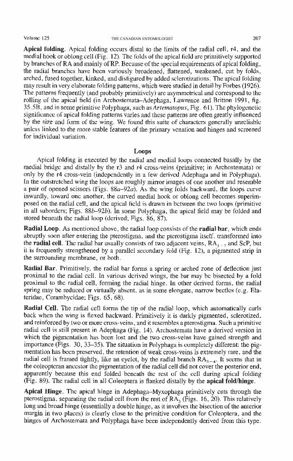
Volume 125 THE CANADIAN ENTOMOLOGIST 207
Apical folding. Apical folding occurs distal to the limits of the radial cell, r4, and the medial hook or oblong cell (Fig. 12). The folds of the apical field are primitively supported by branches of RA and mainly of RP. Because of the special requirements of apical folding, the radial branches have been variously broadened, flattened, weakened, cut by folds, arched, fused together, kinked, and disfigured by added sclerotizations. The apical folding may result in very elaborate folding patterns, which were studied in detail by Forbes (1926). The patterns frequently (and probably primitively) are asymmetrical and correspond to the rolling of the apical field (in Archostemata-Adephaga, Lawrence and Britton 1991, fig. 35.5B, and in some primitive Polyphaga, such as Arternatopus, Fig. 61). The phylogenetic significance of apical folding patterns varies and these patterns are often greatly influenced by the size and form of the wing. We found this suite of characters generally unreliable unless linked to the more stable features of the primary venation and hinges and screened for individual variation.
Loops Apical folding is executed by the radial and medial loops connected basally by the
medial bridge and distally by the r3 and r4 cross-veins (primitive; in Archostemata) or only by the r4 cross-vein (independently in a few derived Adephaga and in Polyphaga). In the outstretched wing the loops are roughly mirror images of one another and resemble a pair of opened scissors (Figs. S8a-92a). As the wing folds backward, the loops curve inwardly, toward one another, the curved medial hook or oblong cell becomes superim- posed on the radial cell, and the apical field is drawn in between the two loops (primitive in all suborders; Figs. 88k92b). In some Polyphaga, the apical field may be folded and stored beneath the radial loop (derived; Figs. 86, 87).
Radial Loop. As mentioned above, the radial loop consists of the radial bar, which ends abruptly soon after entering the pterostigma, and the pterostigma itself, transformed into the radial cell. The radial bar usually consists of two adjacent veins, RA, +, and ScP, but it is frequently strengthened by a parallel secondary fold (Fig. 12), a pigmented strip in the surrounding membrane, or both.
Radial Bar. Primitively, the radial bar forms a spring or arched zone of deflection just proximal to the radial cell. In various derived wings, the bar may be bisected by a fold proximal to the radial cell, forming the radial hinge. In other derived forms, the radial spring may be reduced or virtually absent, as in some elongate, narrow beetles (e.g. Ela- teridae, Cerambycidae; Figs. 65, 68).
Radial Cell. The radial cell forms the tip of the radial loop, which automatically curls back when the wing is flexed backward. Primitively it is darkly pigmented, sclerotized, and reinforced by two or more cross-veins, and it resembles a pterostigma. Such a primitive radial cell is still present in Adephaga (Fig. 14). Archostemata have a derived version in which the pigmentation has been lost and the two cross-veins have gained strength and importance (Figs. 30, 33-35). The situation in Polyphaga is completely different: the pig- mentation has been preserved, the retention of weak cross-veins is extremely rare, and the radial cell is framed tightly, like an eyelet, by the radial branch RA,,,. It seems that in the coleopteran ancestor the pigmentation of the radial cell did not cover the posterior end, apparently because this end folded beneath the rest of the cell during apical folding (Fig. 89). The radial cell in all Coleoptera is flanked distally by the apical foldlhinge.
Apical Hinge. The apical hinge in Adephaga-Myxophaga primitively cuts through the pterostigma, separating the radial cell from the rest of RA, (Figs. 16, 20). This relatively long and broad hinge (essentially a double hinge, as it involves the bisection of the anterior margin in two places) is clearly close to the primitive condition for Coleoptera, and the hinges of Archostemata and Polyphaga have been independently derived from this type.
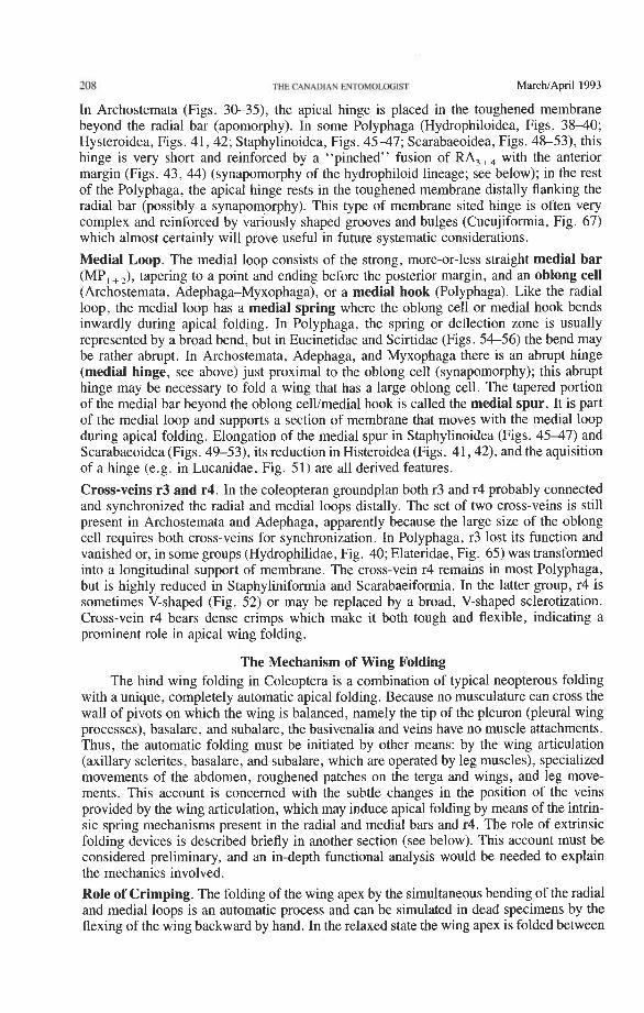
208 THE CANADIAN ENTOMOLDGIST MarchIApril 1993
In Archostemata (Figs. 30-33, the apical hinge is placed in the toughened membrane beyond the radial bar (apomorphy). In some Polyphaga (Hydrophiloidea, Figs. 38-40; Hysteroidea, Figs. 41,42; Staphylinoidea, Figs. 45-47; Scarabaeoidea, Figs. 48-53), this hinge is very short and reinforced by a "pinched" fusion of RA,+, with the anterior margin (Figs. 43, 44) (synapomorphy of the hydrophiloid lineage; see below); in the rest of the Polyphaga, the apical hinge rests in the toughened membrane distally flanking the radial bar (possibly a synapomorphy). This type of membrane sited hinge is often very complex and reinforced by variously shaped grooves and bulges (Cucujiformia, Fig. 67) which almost certainly will prove useful in future systematic considerations.
Medial Loop. The medial loop consists of the strong, more-or-less straight medial bar (MP, +,), tapering to a point and ending before the posterior margin, and an oblong cell (Archostemata, Adephaga-Myxophaga), or a medial hook (Polyphaga). Like the radial loop, the medial loop has a medial spring where the oblong cell or medial hook bends inwardly during apical folding. In Polyphaga, the spring or deflection zone is usually represented by a broad bend, but in Eucinetidae and Scirtidae (Figs. 5&56) the bend may be rather abrupt. In Archostemata, Adephaga, and Myxophaga there is an abrupt hinge (medial hinge, see above) just proximal to the oblong cell (synapomorphy); this abrupt hinge may be necessary to fold a wing that has a large oblong cell. The tapered portion of the medial bar beyond the oblong cell/medial hook is called the medial spur. It is part of the medial loop and supports a section of membrane that moves with the medial loop during apical folding. Elongation of the medial spur in Staphylinoidea (Figs. 45-47) and Scarabaeoidea (Figs. 49-53), its reduction in Histeroidea (Figs. 41,42), and the aquisition of a hinge (e.g. in Lucanidae, Fig. 51) are all derived features.
Cross-veins r3 and r4. In the coleopteran groundplan both r3 and r4 probably connected and synchronized the radial and medial loops distally. The set of two cross-veins is still present in Archostemata and Adephaga, apparently because the large size of the oblong cell requires both cross-veins for synchronization. In Polyphaga, r3 lost its function and vanished or, in some groups (Hydrophilidae, Fig. 40; Elateridae, Fig. 65) was transformed into a longitudinal support of membrane. The cross-vein r4 remains in most Polyphaga, but is highly reduced in Staphyliniformia and Scarabaeiformia. In the latter group, r4 is sometimes V-shaped (Fig. 52) or may be replaced by a broad, V-shaped sclerotization. Cross-vein r4 bears dense crimps which make it both tough and flexible, indicating a prominent role in apical wing folding.
The Mechanism of Wing Folding The hind wing folding in Coleoptera is a combination of typical neopterous folding
with a unique, completely automatic apical folding. Because no musculature can cross the wall of pivots on which the wing is balanced, namely the tip of the pleuron (pleural wing processes), basalare, and subalare, the basivenalia and veins have no muscle attachments. Thus, the automatic folding must be initiated by other means: by the wing articulation (axillary sclerites, basalare, and subalare, which are operated by leg muscles), specialized movements of the abdomen, roughened patches on the terga and wings, and leg move- ments. This account is concerned with the subtle changes in the position of the veins provided by the wing articulation, which may induce apical folding by means of the intrin- sic spring mechanisms present in the radial and medial bars and r4. The role of extrinsic folding devices is described briefly in another section (see below). This account must be considered preliminary, and an in-depth functional analysis would be needed to explain the mechanics involved.
Role of Crimping. The folding of the wing apex by the simultaneous bending of the radial and medial loops is an automatic process and can be simulated in dead specimens by the flexing of the wing backward by hand. In the relaxed state the wing apex is folded between
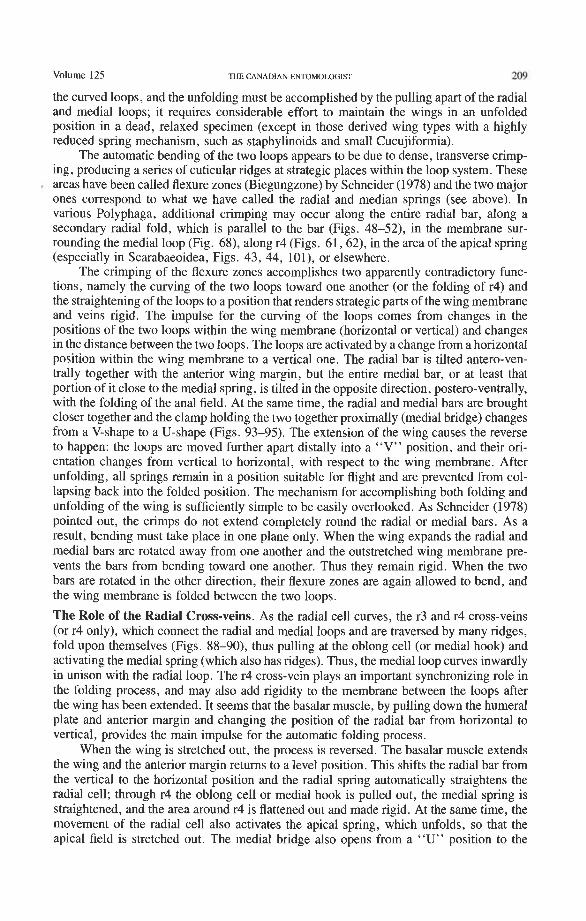
Volume 125 THE CANADIAN ENTOMOLOGIST 209
the curved loops, and the unfolding must be accomplished by the pulling apart of the radial and medial loops; it requires considerable effort to maintain the wings in an unfolded position in a dead, relaxed specimen (except in those derived wing types with a highly reduced spring mechanism, such as staphylinoids and small Cucujiformia).
The automatic bending of the two loops appears to be due to dense, transverse crimp- ing, producing a series of cuticular ridges at strategic places within the loop system. These areas have been called flexure zones (Biegungzone) by Schneider (1978) and the two major ones correspond to what we have called the radial and median springs (see above). In various Polyphaga, additional crimping may occur along the entire radial bar, along a secondary radial fold, which is parallel to the bar (Figs. 48-52), in the membrane sur- rounding the medial loop (Fig. 68), along r4 (Figs. 61,62), in the area of the apical spring (especially in Scarabaeoidea, Figs. 43, 44, 101), or elsewhere.
The crimping of the flexure zones accomplishes two apparently contradictory func- tions, namely the curving of the two loops toward one another (or the folding of r4) and the straightening of the loops to a position that renders strategic parts of the wing membrane and veins rigid. The impulse for the curving of the loops comes from changes in the positions of the two loops within the wing membrane (horizontal or vertical) and changes in the distance between the two loops. The loops are activated by a change from a horizontal position within the wing membrane to a vertical one. The radial bar is tilted antero-ven- trally together with the anterior wing margin, but the entire medial bar, or at least that portion of it close to the medial spring, is tilted in the opposite direction, postero-ventrally, with the folding of the anal field. At the same time, the radial and medial bars are brought closer together and the clamp holding the two together proximally (medial bridge) changes from a V-shape to a U-shape (Figs. 93-95). The extension of the wing causes the reverse to happen: the loops are moved further apart distally into a "V" position, and their ori- entation changes from vertical to horizontal, with respect to the wing membrane. After unfolding, all springs remain in a position suitable for flight and are prevented from col- lapsing back into the folded position. The mechanism for accomplishing both folding and unfolding of the wing is sufficiently simple to be easily overlooked. As Schneider (1978) pointed out, the crimps do not extend completely round the radial or medial bars. As a result, bending must take place in one plane only. When the wing expands the radial and medial bars are rotated away from one another and the outstretched wing membrane pre- vents the bars from bending toward one another. Thus they remain rigid. When the two bars are rotated in the other direction, their flexure zones are again allowed to bend, and the wing membrane is folded between the two loops.
The Role of the Radial Cross-veins. As the radial cell curves, the r3 and r4 cross-veins (or r4 only), which connect the radial and medial loops and are traversed by many ridges, fold upon themselves (Figs. 88-90), thus pulling at the oblong cell (or medial hook) and activating the medial spring (which also has ridges). Thus, the medial loop curves inwardly in unison with the radial loop. The r4 cross-vein plays an important synchronizing role in the folding process, and may also add rigidity to the membrane between the loops after the wing has been extended. It seems that the basalar muscle, by pulling down the humeral plate and anterior margin and changing the position of the radial bar from horizontal to vertical, provides the main impulse for the automatic folding process.
When the wing is stretched out, the process is reversed. The basalar muscle extends the wing and the anterior margin returns to a level position. This shifts the radial bar from the vertical to the horizontal position and the radial spring automatically straightens the radial cell; through r4 the oblong cell or medial hook is pulled out, the medial spring is straightened, and the area around r4 is flattened out and made rigid. At the same time, the movement of the radial cell also activates the apical spring, which unfolds, so that the apical field is stretched out. The medial bridge also opens from a "U" position to the
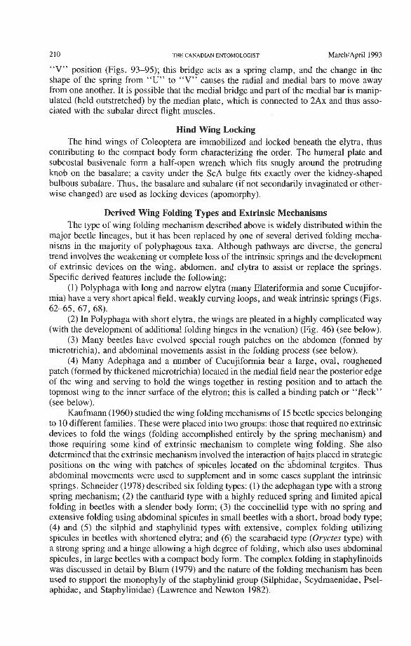
210 THE CANADIAN ENTOMOLOGIST MarchIApril 1993
"V" position (Figs. 93-95); this bridge acts as a spring clamp, and the change in the shape of the spring from "U" to "V" causes the radial and medial bars to move away from one another. It is possible that the medial bridge and part of the medial bar is manip- ulated (held outstretched) by the median plate, which is connected to 2Ax and thus asso- ciated with the subalar direct flight muscles.
Hind Wing Locking The hind wings of Coleoptera are immobilized and locked beneath the elytra, thus
contributing to the compact body form characterizing the order. The humeral plate and subcostal basivenale form a half-open wrench which fits snugly around the protruding knob on the basalare; a cavity under the ScA bulge fits exactly over the kidney-shaped bulbous subalare. Thus, the basalare and subalare (if not secondarily invaginated or other- wise changed) are used as locking devices (apomorphy).
Derived Wing Folding Types and Extrinsic Mechanisms The type of wing folding mechanism described above is widely distributed within the
major beetle lineages, but it has been replaced by one of several derived folding mecha- nisms in the majority of polyphagous taxa. Although pathways are diverse, the general trend involves the weakening or complete loss of the intrinsic springs and the development of extrinsic devices on the wing, abdomen, and elytra to assist or replace the springs. Specific derived features include the following:
(1) Polyphaga with long and narrow elytra (many Elateriformia and some Cucujifor- mia) have a very short apical field, weakly curving loops, and weak intrinsic springs (Figs. 62-65, 67, 68).
(2) In Polyphaga with short elytra, the wings are pleated in a highly complicated way (with the development of additiona! folding hinges in the venation) (Fig. 46) (see below).
(3) Many beetles have evolved special rough patches on the abdomen (formed by microtrichia), and abdominal movements assist in the folding process (see below).
(4) Many Adephaga and a number of Cucujiformia bear a large, oval, roughened patch (formed by thickened microtrichia) located in the medial field near the posterior edge of the wing and serving to hold the wings together in resting position and to attach the topmost wing to the inner surface of the elytron; this is called a binding patch or "fleck" (see below).
Kaufmann (1 960) studied the wing folding mechanisms of 15 beetle species belonging to 10 different families. These were placed into two groups: those that required no extrinsic devices to fold the wings (folding accomplished entirely by the spring mechanism) and those requiring some kind of extrinsic mechanism to complete wing folding. She also determined that the extrinsic mechanism involved the interaction of h@rs placed in strategic positions on the wing with patches of spicules located on the abdominal tergites. Thus abdominal movements were used to supplement and in some cases supplant the intrinsic springs. Schneider (1978) described six folding types: (1) the adephagan type with a strong spring mechanism; (2) the cantharid type with a highly reduced spring and limited apical folding in beetles with a slender body form; (3) the coccinellid type with no spring and extensive folding using abdominal spicules in small beetles with a short, broad body type; (4) and ( 5 ) the silphid and staphylinid types with extensive, complex folding utilizing spicules in beetles with shortened elytra; and (6) the scarabaeid type (Oryctes type) with a strong spring and a hinge allowing a high degree of folding, which also uses abdominal spicules, in large beetles with a compact body form. The complex folding in staphylinoids was discussed in detail by Blum (1979) and the nature of the folding mechanism has been used to support the monophyly of the staphylinid group (Silphidae, Scydmaenidae, Psel- aphidae, and Staphylinidae) (Lawrence and Newton 1982).
Volume 125 THE CANADIAN ENTOMOLOGIST 211
Harnmond (1979) reviewed the subject of wing folding and placed the various folding types into three main grades based on the degree of wing length reduction achieved by the folding process. Hammond's grade 1 included those forms with a simple spring mechanism and little wing length reduction (25%); this is equivalent to the cantharid type in which the spring mechanism is also relatively weak. Grade 2 included those forms with a powerful spring mechanism achieving up to 50% in length reduction, as in many Adephaga. Grades l a and 2a were added for those forms in which the spring is assisted by wing folding spicules, increasing the wing length reduction to 40% and 60%, respectively. Grade 3 included those forms in which wing folding is accomplished by external agents only, with up to 80% reduction being achieved.
It is fairly obvious from our studies that powerful intrinsic springs were primitive in the order and that spring reduction and the reliance on external agents occurred a number of times in different lineages. Abdominal spicules are present in many beetle families, including those with strong springs, and here they may function mainly in the precise alignment of apical folds. In various groups the intrinsic spring has become reduced or lost, so that the wings at rest are not in the folded position. In these cases, extrinsic factors, such as abdominal movements and wing-abdomen spicule interaction have taken over the wing folding function.
Abdominal Tergal Spicules. Abdominal spicule patches are common in all of the major beetle'groups, but their occurrence is often associated with small body size or short, broad body form, or both. They are found in Micromalthidae (1.5-2.5 mm) among the Archo- stemata, in all Myxophaga, in many of the smaller species of both Hydradephaga and Geadephaga, and in most superfamilies of Polyphaga except Dascilloidea, Buprestoidea, and Lymexyloidea. Most Elateroidea (sensu lato, including Cantharoidea) lack abdominal spicules, but they do occur in Drapetes, Anischia, and Throscidae, all of which are of small size. Larger and more elongate members of Cucujoidea, Tenebrionoidea, Chryso- meloidea, and Curculionoidea also lack spicules, which are present in smaller and more compact members of the same groups. The form of these abdominal spicules and their distribution on abdominal tergites may be an important systematic character at the generic and familial levels.
Elytra-Wing Binding Patches. Hammond (1979) also discussed the possible role of high- friction binding patches in the wing folding process. These structures occur near the pos- terior edge of the wing, just behind MP, +, in Polyphaga or at the end of MP, or between MP, and RP, +, in Adephaga. This patch of spicules is usually called the "sub-cubital" fleck or binding patch, although medial fleck is a more appropriate name, if one accepts the revised nomenclature proposed here (Lawrence and Britton 1991). In the folded wing this fleck comes to lie against a similar structure formed by the meeting of apico-sutural binding patches on the two elytra. In Adephaga, these binding patches are restricted to the families Trachypachidae, Noteridae, Amphizoidae, and Dytiscidae, but in the Polyphaga they are widely but unevenly distributed within the Cucujoidea and Tenebrionoidea and occur in scattered members of the Chrysomeloidea. In at least some Polyphaga (e.g. Myce- tophagidae, Leschen and Lawrence 1991), a well-developed binding patch is retained in reduced and aparently non-functional hind wings.
Discussion Basic Adaptations of Coleoptera. The earliest beetles probably occupied very wet envi- ronments. This assumption is based partly on known climatic conditions in the Upper Carboniferous and Lower Permian in Europe and North America, and partly on the habitat preferences and larval structures in modem beetles with the most primitive features (includ- ing the least derived types of hind wing venation). Among the Adephaga, most families are aquatic and the terrestrial Carabidae are often restricted to very wet habitats. Larvae
212 THE CANADIAN ENTOMOU)(jlST MarchIApril 1993
of modem Myxophaga are semi-aquatic. A similar preference for humid conditions occurs in the most primitive polyphagan groups, such as Scirtidae, Hydrophilidae, and Agyrtidae. Only the modem Archostemata, here considered to be the sister group of Adephaga- Myxophaga, are fully terrestrial, but habitats of the numerous extinct genera currently included in the suborder are unknown.
In terrestrial habitats of the Upper Paleozoic, predaceous arthropods, such as arach- nids, scorpions, and myriapods, were abundant, and Paleozoic insects displayed a wide array of protective devices, such as heavily sclerotized cuticle, stiffened forewings (teg- mina), tubercles, spines, and various long sensory projections (Shear and Kukalovi-Peck 1990). This prevalent adaptational trend implies that the elytra very probably functioned as an especially efficient protective device. The adaptive advantage of true Coleoptera over Protocoleoptera and all other insects with strengthened forewings is presumably a result of a great increase in structural integrity associated with body compaction, recession and partial enclosure of leg bases, retraction of genitalia, general reduction of exposed mem- brane, and, most importantly, close coadaption of elytra, pterothorax, and abdomen, com- pletely concealing the wings when not in use. Although improved protection against pred- ators was probably the main driving force behind the evolution of this body form, another important advantage resulted, namely, the sealing off of the area surrounding the meta- thoracic and abdominal spiracles leading to reduced water loss. Such a secondary adap- tation probably made it possible for beetles to spread and diversify in the drier conditions prevailing during the Permian. Coleoptera and early Hemiptera are by far the most impor- tant components of the dry Upper Permian cold temperate zone of Angara (northern Siberia) and Gondwana (Australia).
The retention of full flight capability of the hind wings, combined with the provision for their maximum protection when at rest, was made possible by the evolution of a unique type of automatic, apical folding mechanism, capable of concealing the wings beneath the elytra without sacrificing wing length and thus diminishing flight capability. Our studies indicate that the initial folding mechanism involved two curved struts (radial and medial loops), held in the folded position by intrinsic springs and connected by one or more cross- veins. Rotation of the loops, brought about by the action of direct flight muscles acting through the axillary sclerites, caused the springs to straighten out and the wing membrane to unfold. Such a mechanism is found in basal members of the two main lineages of modem beetles (Archostemata-Adephaga-Myxophaga and Polyphaga), and major variations in this mechanism serve to define these lineages.
Autapomorphies of the Coleopteran Hind Wing. Most of the venational and articular autapomorphies of the Coleoptera are associated with the automatic folding device which allows a well-developed flying wing to be concealed beneath the elytra. The most important are given below:
(1) Presence of the radial bar, consisting of ScP closely associated with RA. (2) Formation of a radial cell enclosing the primitive pterostigma. (3) Formation of a radial loop, consisting of the radial bar, portions of RA branches,
and the radial cell, and capable of coiling automatically postero-basally into the central field.
(4) Basal weakening of RP, which often has no basal connection. (5) Presence of an undivided RP, + ,,
(6) Presence of the medial loop, consisting of MP, one or two cross-veins (rp-mpl, rp-mp2), the tip of RP, and the bases of RP branches (RP,, RP,, RP,,,) and capable of coiling automatically antero-basally into the central field.
(7) Presence of a reinforced bridge (medial bridge), formed partly by MA and joining RA and MP near the base, thus forming a basal connection between the two loops.
(8) Cross-veins r3 and r4 connecting radial and medial loops distally.
Volume 125 THE CANADIAN ENTOMOLOGIST 213
(9) ScA forming a characteristic bulge at base, beneath which lies an acetabulum receiving the apex of the basalare when the wing is flexed.
(10) MP, and CuA, fused distally. (1 1) CuA, +, and AA, +, fused distally. (12) Anal area forming an enlarged lobe (containing AP), which folds beneath the
rest of the wing along the anal fold. (13) Radial and medial basivenalia separated by a wide gap and tilting in opposite
directions when the wing is flexed backward. (14) All original band sclerites comprising 3Ax fused together. (15) Fragment of 3Ax to which the wing flexor muscle is attached separated from the
main body of the sclerite, and the membrane between the fragment and 3Ax serving as a hinge when the wing is pulled into flexed position.
(16) Median plate subdivided lengthwise into two plates, FM, and FM,.
Relationships of the Beetle Suborders. The basic hind wing venation in Coleoptera exhibits several extremely primitive features, which were inherited from the original, largely symmetrical, and not fully flight-adapted wing precursor of ancestral pterygotes: (1) PC adjacent to but not fully fused with C near base; (2) RA and RP adjacent to one another basally (not fully fused into a stem); (3) MA and MP arising individually from the medial basivenale; and (4) CuA and CUP arising individually from the cubital basi- venale. The presence in Coleoptera of features found elsewhere only in very primitive Paleozoic stem assemblages suggests that ancestral Pterygota with protowings developed very rapidly and diverged into all the major lineages before the protowings had time to adapt fully (i.e. in one particular way only) to produce an asymmetrical wing required for flapping flight.
At the same time, coleopteran wings have developed a unique wing folding mecha- nism, consisting of attributes that have no parallels in other pterygote orders. Our differ- ential diagnosis of beetle suborders is based mainly on elements of this folding mechanism, such as the formation of the radial cell and oblong cell (or medial hook), which form the ends of the radial and medial loops, respectively. Because these loops are part of an adap- tational achievement that took place at the time of origin of modem coleopteran lineages, they provide an excellent set of characters for tracing the basic diversifications and radia- tions between and within suborders: the suborders Archostemata, Myxophaga, Adephaga, and Polyphaga each exhibit a unique mixture of primitive and derived venational and articular features, mainly associated with the folding system.
Archostemata + Adephaga-Myxophaga. These sister groups share the following wing synapomorphies:
(1) Central field triangular and forming a large pocket. (2) Medial bar with a distinct, abrupt hinge. (3) Basal portion of RA,+, cut twice by the triangular fold. (4) CUP- completely lost. (5) Anterior anal basivenale (BAA) present, V-shaped. (6) Characteristic 1Ax with a massive, broad head and very long, notched tail. (7) Cubital proxalare (PRCu) fused to the tergum. The radial cell in this group is limited proximally by a radial cross-vein (between
RA, + 2 and RA,,,), instead of by RA, +, as in Polyphaga; two cross-veins sometimes occur and in Archostemata, the second cross-vein limits the radial cell distally. Although the presence of radial cross-veins is almost certainly plesiomorphic, the way in which they support the radial cell might also be considered an apomorphy for this lineage. The pres- ence of an oblong cell and the long RA fork (RA, +,, RA,,,) occurring before the radial cell are symplesiomorphies, lacking in Polyphaga. In the articular area, 1Ax has a large, bilobed tail, which may be a synapomorphy for the group, and 3Ax is relatively narrow
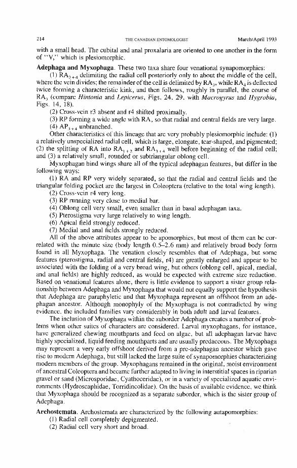
214 THE CANADIAN EE(TOM0LaGIST M~chIApril 1993
with a small head. The cubital and anal proxalaria are oriented to one another in the form of "V," which is plesiomorphic.
Adephaga and Myxophaga. These two taxa share four venational synapomorphies: (1) RA,,, delimiting the radial cell posteriorly only to about the middle of the cell,
where the vein divides; the remainder of the cell is delimited by RA,, while RA, is deflected twice forming a characteristic kink, and then follows, roughly in parallel, the course of RA, (compare Hintonia and Lepicerus, Figs. 24, 29, with Macrogyrus and Hygrobia, Figs. 14, 18).
(2) Cross-vein r3 absent and r4 shifted proximally. (3) RP forming a wide angle with RA, so that radial and central fields are very large. (4) AP, +, unbranched. Other characteristics of this lineage that are very probably plesiomorphic include: (1)
a relatively unspecialized radial cell, which is large, elongate, tear-shaped, and pigmented; (2) the splitting of RA into RA, +, and RA,+, well before beginning of the radial cell; and (3) a relatively small, rounded or subtriangular oblong cell.
iVlyxophagan hind wings share all of the typical adephagan features, but differ in the following ways:
(1) RA and RP very widely separated, so that the radial and central fields and the triangular folding pocket are the largest in Coleoptera (relative to the total wing length).
(2) Cross-vein r4 very long. (3) RP running very close to medial bar. (4) Oblong cell very small, even smaller than in basal adephagan taxa. (5) Pterostigma very large relatively to wing length. (6) Apical field strongly reduced. (7) Medial and anal fields strongly reduced. All of the above attributes appear to be apomorphies, but most of them can be cor-
related with the minute size (body length 0.5-2.6 mm) and relatively broad body form found in all Myxophaga. The venation closely resembles that of Adephaga, but some features (pterostigma, radial and central fields, r4) are greatly enlarged and appear to be associated with the folding of a very broad wing, but others (oblong cell, apical, medial, and anal fields) are highly reduced, as would be expected with extreme size reduction. Based on venational features alone, there is little evidence to support a sister group rela- tionship between Adephaga and Myxophaga that would not equally support the hypothesis that Adephaga are paraphyletic and that Myxophaga represent an offshoot from an ade- phagan ancestor. Although monophyly of the Myxophaga is not contradicted by wing evidence, the included families vary considerably in both adult and larval features.
The inclusion of Myxophaga within the suborder Adephaga creates a number of prob- lems when other suites of characters are considered. Larval myxophagans, for instance, have generalized chewing mouthparts and feed on algae, but all adephagan larvae have highly specialized, liquid feeding mouthparts and are usually predaceous. The Myxophaga may represent a very early offshoot derived from a pre-adephagan ancestor which gave rise to modem Adephaga, but still lacked the large suite of synapomorphies characterizing modem members of the group. Myxophagans remained in the original, moist environment of ancestral Coleoptera and became further adapted to living in interstitial spaces in riparian gravel or sand (Microsporidae, Cyathoceridae), or in a variety of specialized aquatic envi- ronments (Hydroscaphidae, Torridincolidae). On the basis of available evidence, we think that Myxophaga should be recognized as a separate suborder, which is the sister group of Adephaga.
Archostemata. Archostemata are characterized by the following autapomorphies: (1) Radial cell completely depigmented. (2) Radial cell very short and broad.
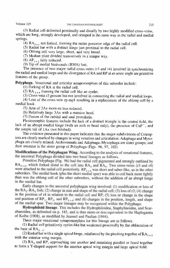
Volume 125 ?WE CANADLAN ENTOMOLOGIfl 215
(3) Radial cell delimited proximally and distally by two highly modified cross-veins, which are long, strongly developed, and crimped in the same way as the radial and medial springs.
(4) RA,,, not forked, forming the entire posterior edge of the radial cell. (5) Radial bar with a distinct hinge just proximal to the radial cell. (6) Oblong cell very large, short, and very broad. (7) Median plate divided transversely in a unique way. (8) AP, +, fully reduced. (9) Tip of medial basivenale (BMA) lost. The presence of two major radial cross-veins (r3 and r4) involved in synchronizing
the radial and medial loops and the divergence of RA and RP at an acute angle are primitive features of the group.
Polyphaga. Venational and articular autapomorphies of this suborder include: (1) Forking of RA at the radial cell. (2) RA ,+, framing the radial cell like an eyelet. (3) Cross-vein r3 present but not involved in connecting the radial and medial loops. (4) Loss of the cross-vein rp-mpl resulting in a replacement of the oblong cell by a
medial hook. (5) Arm of 2Ax more-or-less reduced. (6) Relatively large 3Ax with a massive head. (7) Fusion of the cubital and anal proxalaria. Plesiomorphic features include the lack of a distinct triangle in the central field, the
lack of an abrupt medial hinge (with an arch or bend only), the presence of CUP-, and the simple tail of 1Ax (not bilobed).
The evidence presented in this paper indicates that the major subdivisions of Coleop- tera are clearly marked by changes in wing venation and articulation: Adephaga and Myxo- phaga are closely related; Archostemata and Adephaga-Myxophaga are sister groups; and their ancestor is the sister group of Polyphaga (Figs. 96, 97, 102).
Modifications of the Polyphagan Wing. According to the analysis of venationaI features, the ancestral Polyphaga divided into two basal lineages as follows.
Primitive Polyphaga (Fig. 96) had the radial cell pigmented and strongly outlined by RA,,,, which forked distal to the cell into RA, and RA,. Two cross-veins (r3 and r4) were attached to the radial cell posteriorly. RP,,, was short and saber-like, as in the other suborders. The medial hook (plus the short medial spur) was able to coil back more tightly than was the oblong cell of the other suborders, without the addition of an abrupt hinge in the medial bar.
Early chan~es to the ancestral polyphagan wing involved: (1) modification or Ioss of the RA,-RA, fork; (2) change in size and shape of the radial cell; (3) Ioss of r3; (4) change in the position of r4 in relation to the radial cell and RP: ( 5 ) loss or change in the shape and position of RP,, UP,, and RP,+,; and (6) changes in the position, length, and shape of the medial spur. Two major lineages may be recognized within the Polyphaga:
Hydrophiloid lineage. This includes the Hydrophiloidea, Staphylinoidea. and Scar- abaeaidea, as delimited on p. 183, and is thus more-or-less equivalent to the Haplogastra of Kolbe (1908), as modified by Jeanne1 and Paulian (1944).
Three major venational synapomorphies for this lineage are as follows: (1) Radial cell primitively eyelet-llke but weakened proximally by the obliteration of
the base of RA,,,. (2) Radial bar with a single apical hinge, reinforced by the pinching together of RA, +,
with the anterior wing margin. (3) RA, and RP, approaching one another and remaining parallel or fused together
to form a Y-shaped support for the anterior apical wing margin and large apical field.

216 THE CANADIAN ENTOMOLOGIST MarcNApril 1993
The retention of all primitive radial branches in the apical field (fork RA,-RA,, RPl, RP,, RP, +,) is a symplesiomorphy; however the unique way in which these end branches of the radius are used to support the apical field of the wing might be considered as an additional synapomorphy for this clade. The weakening of the base of the radial cell is an important feature of this group, which is correlated with a fold passing into the cell at this point, allowing the cell to fold on itself. This type of structure also appears to be the precursor of the highly modified "open" radial cells which evolved in Scarabaeoidea and Histeroidea.
Subsequent wing modifications in the hydrophiloid lineage involve marked changes in the radial cell (as the distal, stronger half becomes increasingly more sclerotized, nar- rower, and more obliquely oriented toward the radial bar), in the relative positions RA, and RP,, relative length and shape of RP,,, and its position relative to the medial spur, and in the reduction of rp-mp2. The wing of Hydrophiloidea has a number of primitive features. The radial cell is large, eyelet-like, with two cross-veins (r3 and r4) attached to its posterior margin (plesiomorphy); RA, and RP, meet and form a "Y" (apomorphy); RP, +, is saber-shaped (plesiomorphy); rp-mp2 is not reduced (medial hook is broad) and the medial spur is short (plesiomorphy). In the superfamilies Staphylinoidea and Scara- baeoidea, as well as in the families Sphaeritidae, Synteliidae, and Histeridae (included in Hydrophiloidea on p. 183 but often placed in a separate superfamily, Histeroidea), further modifications have taken place, including the reduction or narrowing of the radial cell, the loss of r3, the shift of r4 proximally, the shift of RP,,, posteriorly to meet the medial spur, which may become atrophied or fused with RP,+,, the loss of rp-mp2 bringing the tips of RP and MP, +, close together, and the reduction of and loss of veins in the medial field (MP,, MP,, CuA, AA). Folding modifications include reduction and loss of the spring mechanisms, development of extrinsic folding devices (abdominal tergal spicules), and formation of a uniquely complex folding pattern in derived staphylinoids.
Eucinetoid lineage. This includes the remainder of the suborder (superfamilies Euci- netoidea, Dascilloidea, Buprestoidea, Byrrhoidea, Elateroidea, Derodontoidea, Bostri- choidea, Lymexyloidea, Cleroidea, Cucujoidea, Tenebrionoidea, Chrysomeloidea, and Curculionoidea, as delimited on pp. 183-185).
Eucinetoid synapomorphies include: (1) Reinforcement of radial cell distally against the apical hinge by a broadening and
sclerotization of the tips of the radial bar and RA, +,. (2) Broadening of the apical hinge. (3) Secondary reinforcement of the membrane flanking the apical hinge distally. (4) Branches of RA, and RA, lost, their function in supporting the anterior margin
is taken over by a sclerotized groove starting at the apical hinge and running in parallel with the anterior apical wing margin.
(5) branches of RP (RP,, RP,, RP,,,), although remaining in the primitive position, become more-or-less reduced (shortened, barely distinguishable, or replaced by sclero- tized membrane).
(6) Apical field short and broad, especially in primitive forms. Some plesiomorphic features of this lineage include: (1) the strongly developed radial
cell, without a weakness at the proximal end; (2) apical hinge located well behind the anterior wing margin; (3) central field with a small, vaguely triangular pocket, reminiscent of the much larger, sharply outlined pockets in the wings of Archostemata, Myxophaga, and Adephaga; and (4) weakness in the medial bar at the point where an abrupt hinge develops in Archostemata, Adephaga, and Myxophaga. The last two features in particular are found only in basal members of the lineage (e.g. Eucinetoidea) and probably represent the primitive condition for Coleoptera.
The primitive radial and medial springs (strongly arched, reinforced by crimping, and unbroken by a hinge) are retained in most of the larger eucinetoids, bostrichoids, and
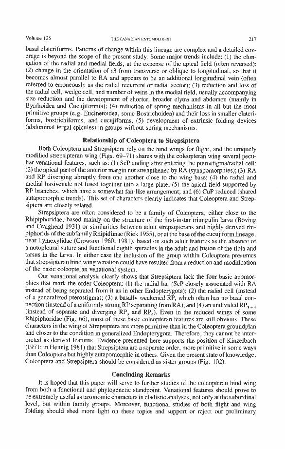
Volume 125 THE CANADIAN ENTOMOLOGIST 217
basal elateriforms. Patterns of change within this lineage are complex and a detailed cov- erage is beyond the scope of the present study. Some major trends include: (1) the elon- gation of the radial and medial fields, at the expense of the apical field (often reversed); (2) change in the orientation of r3 from transverse or oblique to longitudinal, so that it becomes almost parallel to RA and appears to be an additional longitudinal vein (often referred to erroneously as the radial recurrent or radial sector); (3) reduction and loss of' the radial cell, wedge cell, and number of veins in the medial field, usually accompanying size reduction and the development of shorter, broader elytra and abdomen (mainly in Byrrhoidea and Cucujiformia); (4) reduction of spring mechanisms in all but the most primitive groups (e.g. Eucinetoidea, some Bostrichoidea) and their loss in smaller elateri- forms, bostrichiforms, and cucujiforms; (5) development of extrinsic folding devices (abdominal tergal spicules) in groups without spring mechanisms.
Relationship of Coleoptera to Strepsiptera Both Coleoptera and Strepsiptera rely on the hind wings for flight, and the uniquely
modified strepsipteran wing (Figs. 69-71) shares with the coleopteran wing several pecu- liar venational features, such as: (1) ScP ending after entering the pterostigma/radial cell; (2) the apical part of the anterior margin not strengthened by RA (synapomorphies); (3) RA and RP diverging abruptly from one another close to the wing base; (4) the radial and medial basivenale not fused together into a large plate; (5) the apical field supported by RP branches, which have a somewhat fan-like arrangement; and (6) CUP reduced (shared autapomorphic trends). This set of characters clearly indicates that Coleoptera and Strep- siptera are closely related.
Strepsiptera are often considered to be a family of Coleoptera, either close to the Rhipiphoridae, based mainly on the structure of the first-instar triungulin larva (Boving and Craighead 193 1) or similarities between adult strepsipterans and highly derived rhi- piphorids of the subfamily Rhipidiinae (Riek 1955), or at the base of the cucujiform lineage, near Lymexylidae (Crowson 1960, 1981), based on such adult features as the absence of a notopleural suture and functional eighth spiracles in the adult and fusion of the tibia and tarsus in the larva. In either case the inclusion of the group within Coleoptera presumes that strepsipteran hind wing venation could have resulted from a reduction and modification of the basic coleopteran venational system.
Our venational analysis clearly shows that Strepsiptera lack the four basic apomor- phies that mark the order Coleoptera: (1) the radial bar (ScP closely associated with RA instead of being separated from it as in other Endopterygota); (2) the radial cell (instead of a generalized pterostigma); (3) a basally weakened RP, which often has no basal con- nection (instead of a uniformly strong RP separating from RA); and (4) an undivided RP, +, (instead of separate and diverging RP, and RP,). Even in the reduced wings of some Rhipiphoridae (Fig. 66), most of these basic coleopteran features are still obvious. These characters in the wing of Strepsiptera are more primitive than in the Coleoptera groundplan and closer to the condition in generalized Endopterygota. Therefore, they cannot be inter- preted as derived features. Evidence presented here supports the position of Kinzelbach (1971; in Hennig 1981) that Strepsiptera are a separate order, more primitive in some ways than Coleoptera but highly autapomorphic in others. Given the present state of knowledge, Coleoptera and Strepsiptera should be considered as sister groups (Fig. 102).
Concluding Remarks It is hoped that this paper will serve to further studies of the coleopteran hind wing
from both a functional and phylogenetic standpoint. Venational features should prove to be extremely useful as taxonomic characters in cladistic analyses, not only at the subordinal level, but within family groups. Moreover, functional studies of both flight and wing folding should shed more light on these topics and support or reject our preliminary
218 THE CANADIAN ENTOMOLOGIST MarchIApril 1993
hypotheses. The complexities of the articular region have only begun to be understood, and their use in cladistic studies will greatly add to the bank of data available for phylo- genetic inference at various taxonomic levels. Finally, the use of both venational and articular features that can be homologized with those of other insect orders may shed light on major phylogenetic questions, such as the relationship of the coleopteroid lineage to other Endopterygota.
Acknowledgments We are very obliged to the following individuals who offered suggestions and criti-
cism of our ideas or commented on earlier versions of this manuscript: A.A. Calder, K.W. Cooper, T.F. Hlavac, H.F. Howden, A.F. Newton, S .B. Peck, C. A.M. Reid, C.H. Scholtz, M.K. Thayer, and J. Trueman. We are especially grateful to D.J. Browne who pointed out some important details of wing articulation. A.G. Ponomarenko and the Paleontological Institute, Academy of Sciences, Moscow, are thankfully acknowledged for sending us a photograph of the Upper Permian coleopterid wing illustrated in Figure 2. Thanks also to the numerous individuals and institutions who have supplied us with specimens used in wing dissections; C. Costa and P.J. Spangler, in particular, provided us with rare and undescribed groups of Myxophaga. Space and research facilities were provided by Carleton University and C. S . I.R. 0 . Division of Entomology. Operating grants from the Natural Sciences and Engineering Research Council of Canada and financial support from S.B. Peck made it possible for the senior author to spend time in Canberra studying wing preparations housed in the Australian National Insect Collection and preparing a prelim- inary draft of this manuscript. Preparation and labelling of the plates were accomplished by the C.S.I.R.O. Division of Entomology Graphics Unit (S. Smith, C. Hunt, C. Davies). Melbourne University Press is acknowledged for the use of Figures 4 and 5a, 5b (Figs. 1.17C, 6.16A, and 1.18A from The Insects of Australia. 2nd edition).
References Adolph, G.E. 1879. uber Insektenfliigel. Nova Acta Leopoldiana Carolinae 41: 215-291. Arnold, J.W. 1963. A note on the pterostigma in insects. The Canadian Entomologist 95(1): 13-16. Arnoldi, L.V., V.V. Zherikhin, L.M. Nikritkin, and A.G. Ponomarenko. 1977. Mesozoic beetles. Trudy Paleon-
tologicheskogo Instituta 161: 1-204. [In Russian.] Balfour-Browne, W.A.F. 1944. The wing venation of the Adephaga (Coleoptera) with special reference to the
Hydradephaga and some homologies with the Polyphaga. Journal of the Royal Microscopical Society 63: 55-64.
Blum, P. 1979. Zur Phylogenie und okologischen Bedeutung der Elytrenreduktion und Abdomenweglichkeit der Staphyliniden (Coleoptera): vergleichend- und funktionsmorphologische Untersuchungen. Zoolo- gisches Jahrbuch (Anatomie) 102(4): 533-582.
Boving, A.G., and F.C. Craighead. 1931. An illustrated synopsis of the principal larval forms of the Coleoptera. Entomologica Americana (n.s.) 11: 1-35 1.
Browne, D.J. 1991. Wing structure of the genus Eucanthus Westwood; confirmation of the primitive nature of the genus (Scarabaeoidea: Geotrupidae: Bolboceratinae). Journal of the Entomological Society of South Africa 54(2): 221-230.
Comstock, J.H. 1918. The Wings of Insects. Comstock, Ithaca, NY. pp. 1 4 3 0 . Comstock, J.H., and J.G. Needham. 1898. The wings of insects (part). American Naturalist 32: 4 3 4 8 , 81-89,
231-257, 335-340, 413424, 561-565, 769-777, 903-911. 1899. The wings of insects (part). American Naturalist 33: 117-126, 573-582, 845-860.
Crowson, R.A. 1955. The Natural Classification of the Families of Coleoptera. Nathanial Lloyd, London. 214 pp - 1960. The phylogeny of Coleoptera. Annual Review of Entomology 5: 11 1-134.
1981. The Biology of Coleoptera. Academic Press, London. Forbes, W.T.M. 1922. The wing-venation of the Coleoptera. Annals of the Entomological Society of America
15: 328-345, pls. XXIX-XXXV. 1926. The wing-folding patterns of the Coleoptera. Journal of the New York Entomological Society 34:
42-68, 91-139. Graham, S.A. 1922. A study of the wing-venation of the Coleoptera. Annals of the Entomological Society of
America 15: 191-200.
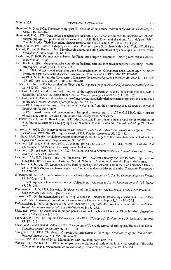
Volume 125 THE CANADIAN ENTOMOLOGIST 219
Hamilton, K.G.A. 1972. The insect wing, part 111. Venation of the orders. Journal ofthe Kansas Entomological Socieo 45: 145-162.
Hammond, P.M. 1979. Wing-folding mechanisms of beetles, with special reference to investigations of Ade- phagan phylogeny. pp. 113-180 in Erwin, T.L., G.E. Ball, D.R. Whitehead and A.L. Halpern (Eds.), Carabid Beetles: Their Evolution, Natural History, and Classification. W. Junk, The Hague.
Hennig, W.W. 1981. Insect Phylogeny (transl. A.C. Pont; rev. notes D. Schlee). Wiley, New York, NY. 514 pp. Jeannel, R., and R. Paulian. 1944. Morphologie abdominale des ColCoptCres et systbmatique de I'ordre. Revue
Francaise d'Enromologie 11: 65-1 10. Kaufmann, T. 1960. Faltungsmechanismen der Flugel bei einigen Coleopteren. Ludwig-Maximilians-Univer-
sitat, Miinich. 73 pp. Kinzelbach, R. 1971. Morphologische Befunde an Facheflugern und ihre phylogenetische Bedeutung (Insecta:
Strepsiptera). Zoologica 41(119): 1-256. Kolbe, H. 1901. Vergleichend morphologische Untersuchungen an Koleopteren nebst Gmndlagen zu einem
System und zur Systematik derselben. Archive der Naturgeschichte 1901: 98-1 12, 128-141. - 1908. Mein System der Coleopteren. Zeitschrifr fur wissenschafilichen Insekten-Biologie 13: 116123,
153-162, 21%226, 2 4 6 2 5 1, 286294, 389-400. Kuhne, 0 . 1914. Der Tracheenverlauf im Fliigel der Koleopterennymphe. Zeitschrift fur wissenschaftliche Zool-
ogie 112: 691-718, pls. 19-20. Kukalovi, J. 1969. On the systematic position of the supposed Permian beetles, Tshekardocoleidae, with a
description of a new collection from Moravia. Sbornik geologicljch VCd. (P)11: 139-162. Kukalovh-Peck, 3. 1978. Origin and evolution of insect wings and their relation to metamorphosis, as documented
by the fossil record. Journal of Morphology 156: 53-126. 1983. Origin of the insect wing and wing articulation from the arthropodan leg. Canadian Journal of
Zoology 61(7): 1618-1669. 1991. Fossil history and the evolution of hexapod structures. pp. 141-179 in C.S.I.R.O. (Ed.), Insects
of Australia, 2nd ed. Volume I. Melbourne University Press, Melbourne. KukalovCPeck, J., and C. Brauckmann. 1992. Most Paleozoic Protorthoptera are ancestral hemipteroids; major
wing braces as clues to a new phylogeny of Neoptera (Insecta). Canadian Journal of Zoology 70: 2452- 2473.
Lameere, A. 1922. Sur la newation alaire des insectes. Bulletin de 1'Academie Royale de Belgique. Classe ScientiJique 1922: 38-149. [English transl., 1923, Psyche, Cambridge 30: 123-132.1
Larskn, 0 . 1966. On the morphology and function of the locomotor organs of the Gyrinidae and other Coleoptera. Opuscula Entomologica Supplementum 30: 1-242.
Lawrence, J.F., and E.B. Britton. 1991. Coleoptera. pp. 543483 in C.S.I.R.O. (Ed.), Insects of Australia, 2nd ed. Volume 2. Melbourne University Press, Melbourne.
Lawrence, J.F., and A.F. Newton, Jr. 1982. Evolution and classification of beetles. Annual Review of Ecology and Systematics 13: 261-290.
Lawrence, J.F., E.S. Nielsen, and I.M. Mackerras. 1991. Skeletal anatomy and key to orders. pp. 3-32 in C.S.I.R.O. (Ed.), Insects of Australia, 2nd ed. Volume 1. Melbourne University Press, Melbourne.
Leschen, R.A.B., and J.F. Lawrence. 1991. Fern sporophagy in Coleoptera from the Juan Fernandez Islands, Chile, with descriptions of two new genera in Cryptophagidae and Mycetophagidae. Systematic Entomology 16: 329-352.
d'orchymont, A. 1920. La newation alaire des Colkopt&res. Annales de la Sociktk Entomologique de France 89: 1-50, pls. 1-3.
1921. Aper~u de la nervation alaire des Colkopteres. Annales de la Sociktk Entomologique de la Belgique 61: 256278.
Ponomarenko, A.G. 1969. Historical development of the Coleoptera-Archostemata. Trudy Paleontologiches- kogo Instituta 125: 1-240. [In Russian.] - 1972. On the nomenclature of the wing-venation in Coleoptera. Entomologicheskoe Obozrenie 51(4):
768-775. [In Russian; translation in Entomological Review, Washington 51(4): 454458.1 Redtenbacher, J. 1886. Vergleichende Studien ijber das Fliigelgeader der Insekten. Annalen des kaiserlichen-
koniglichen natunvissenschaftlichen Hofmuseums 1: 153-232. Riek, E.F. 1955. The Australian rhipidiine parasites of cockroaches (Coleoptera: Rhipiphoridae). Australian
Journal of Zoologie 3: 71-94. Schneider, P. 1978. Die Flug- und Faltungstypen der Kafer (Coleoptera). Zoologisches Jahrbuch der Anatomie
99: 176210. Shear, W.A., and J. Kukalova-Peck. 1990. The ecology of Paleozoic terrestrial arthropods: The fossil evidence.
Canadian Journal of Zoology 68: 1807-1 834. Snodgrass, R.E. 1909. The thorax of insects and articulation of the wings. Proceedings of the United States
National Museum 36: 51 1-595, pls. 40-69. 1935. Principles of Insect Morphology. McGraw-Hill, New York, NY.
Wallace, EL., and R.C. Fox. 1975. A comparative morphological study of the hind wing venation of the order Coleoptera, part I. Proceedings of the Entomological Society of Washington 77: 329-354.
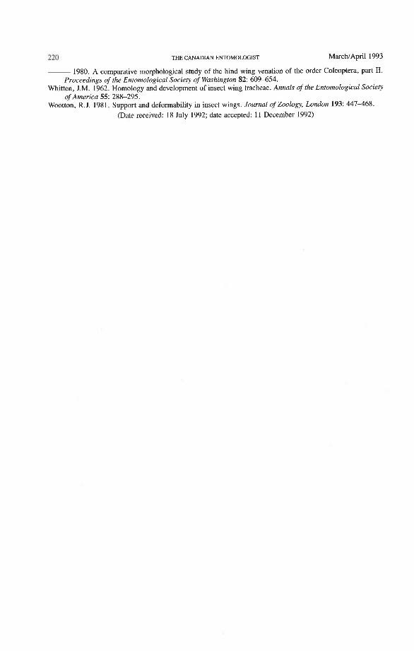
220 THE CANADIAN ENTOMOLOGIST MarcWApril 1993
1980. A comparative morphological study of the hind wing venation of the order Coleoptera, part 11. Proceedings of the Entomological Society of Washington 82: 60%654.
Whitten, J.M. 1962. Homology and development of insect wing tracheae. Annals of the Entomological Society of America 55: 288-295.
Wootton, R.J. 1981. Support and deformability in insect wings. Journal of Zoology, London 193: 447-468. - -
(Date received: 18 July 1992; date accepted: 11 December 1992)
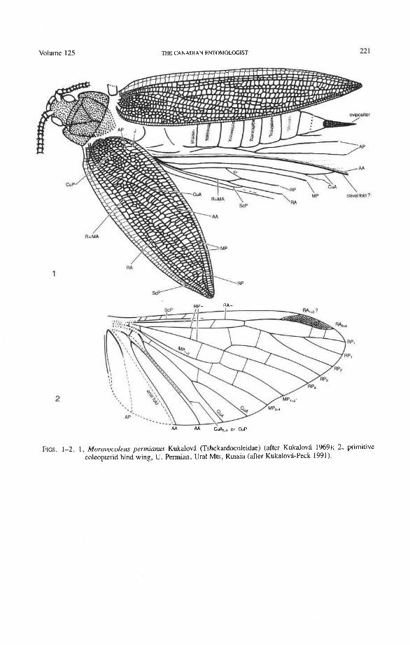
Volume 125 THE CANADIAN ENTOMOLOGIST 22 1
FIGS. 1-2. 1, Moravocoleus permianus Kukalovi (Tshekardocoleidae) (after Kukalova 1969); 2, primitive coleopterid hind wing, U. Permian, Ural Mts, Russia (after KukalovCPeck 1991).
222 THE CANADIAN ENTOMOLOGIST
RA. ..
anal lold \
claval fold
FIGS. 3 4 . 3 , scheme of pterygote wing venation, with articulation, complete veinal system, folds, venational braces, and veinal fusions; the ancestral band of unfused sclerites (P = proxalare, AX = axalare, F = fulcalare, B = basivenale) gave rise to all Recent types of wing articulation (after Kukalova-Peck 1983); 4, generalized
Neoptera wing venation (after Lawrence et al. 1991).
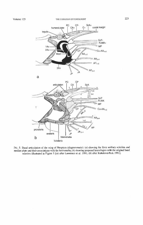
Volume 125 THE CANADIAN FNTOMOLOGIST
PC CP- SCA+ hurneralpl?te 1 I$+ I $+ I aspi a l i n
PC CP articulation I CA I S ~ A
FIG. 5. Basal articulation of the wing of Neoptera (diagrammatic): (a) showing the three axillary sclerites and median plate and their associations with the basivenalia; (b ) showing proposed homologies with the original band
sclerites illustrated in Figure 3 [(a) after Lawrence et al. 1991; (b) after Kukalovh-Peck 19911.
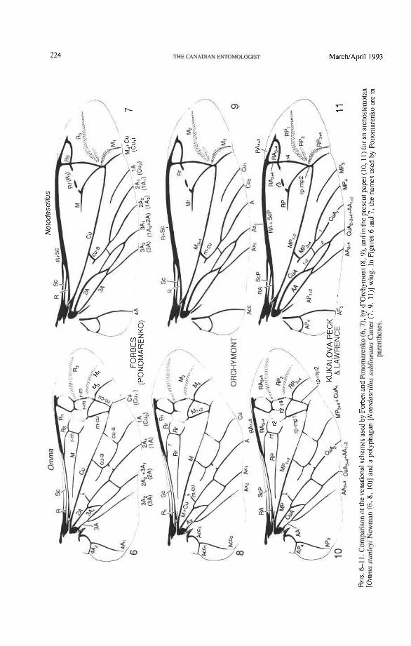
224 THE CANADIAN E~~OMOLI~GIST MarcNA
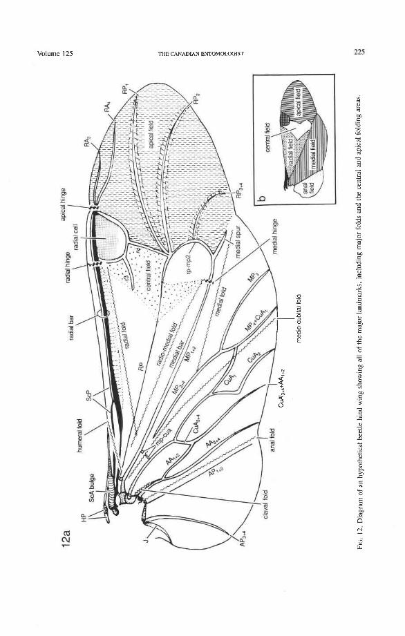
Volume 125 THE CANADIAN ENTOMOLOGIST
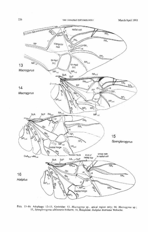
226 THE CANADIAN ENTOMOLOGIST MarchIApril 1993
FIGS. 13-16. Adephaga: 13-1 5, Gyrinidae: 13, Macrogyrus sp., apical region only; 14, Macrogyrus sp.; 15, Spanglerogyrus albiventris Folkerts; 16, Haliplidae: Haliplus bisrriatus Wehncke.
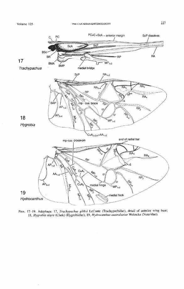
Volume 125 THE CANADIAN ENTOMOLOGIST 227
PC+C+ScA - anterior margin ScP dls?dvas I I
17 Trachypachus medial bridge
18 Hygrobia
FIGS. 17-19. Adephaga: 17, Trachypachus gibbsi LeConte (Trachypachidae), detail of anterior wing base; 18, Hygrobia nigra (Clark) (Hygrobiidae); 19, Hydrocanthus aus~ralasiae Wehncke (Noteridae).

THE CANADIAN ENTOMOLOGET MarcNApril 1993
Trachypachus
FIGS. 20-22. Adephaga: 20, Trachypachus gibbsi LeConte (Trachypachidae); 21, Megacephala sp. (Carabidae); 22, Pheropsophus verticalis (Dejean) (Carabidae).
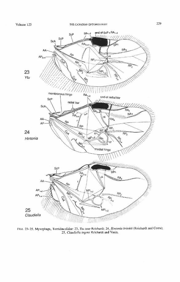
Volume 125 THE CANADIAN ENTOMOLOGIST
FIGS. 23- 25. Myxophaga, Torridincolidae: 2 3 , Ytu zeus Reichardt; 24, Hintonia britskii (Reichardt and Costa); 25, Claudiella ingens Reichardt and Vanin.
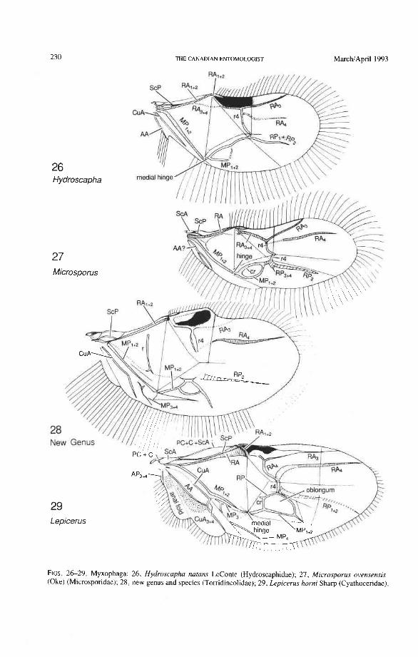
THE CANADIAN ENTOMOLOGIST MarchIApril 1993
FIGS. 2 6 2 9 . Myxophaga: 26, Hydroscapha natans LeConte (Hydroscaphidae); 27, Microsporus ovensensis (Oke) (Microsporidae); 28 , new genus and species (Torridincolidae); 29, Lepicerus horni Sharp (Cyathoceridae).
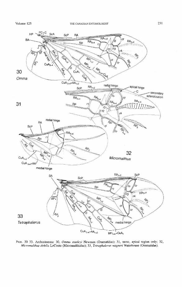
Volume 125 THE CANADIAN ENTOMOLOGIfl
33 Te traphalerus
FIGS. 3C33. Archostemata: 30, Omma stanleyi Newman (Ommatidae); 31, same, apical region only; 32, Micromalthus debilis LeConte (Micromalthidae); 3 3 , Tetraphalerus wagneri Waterhouse (Ommatidae).
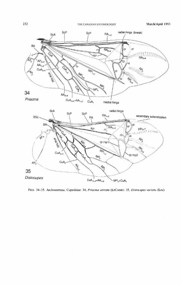
232 ME CANADIAN E~MOI.%IST MarcWApril 1993
Priacma
FIGS. 34-35. Archostemata, Cupedidae: 34, Priacma serrata (LeConte); 35, Distocupes varians (Lea).
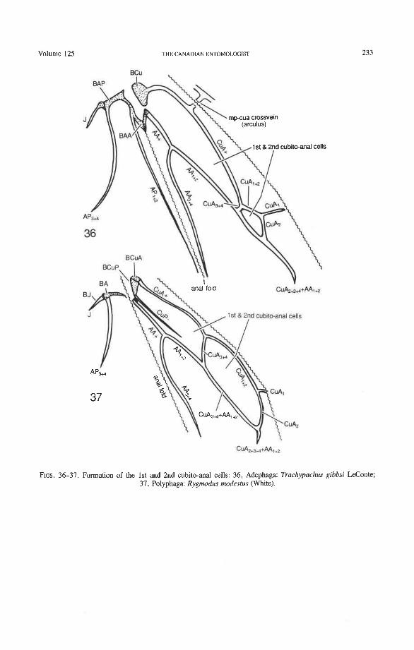
Volume 125 THE CANADIAN ENTOMOLOGIST
d cubiio-anal cells
FIGS. 36-37. Formation of the 1st and 2nd cubito-anal cells: 36, Adephaga: Trachypachus gibbsi LeConte; 37, Polyphaga: Rygmodus modestus (White).
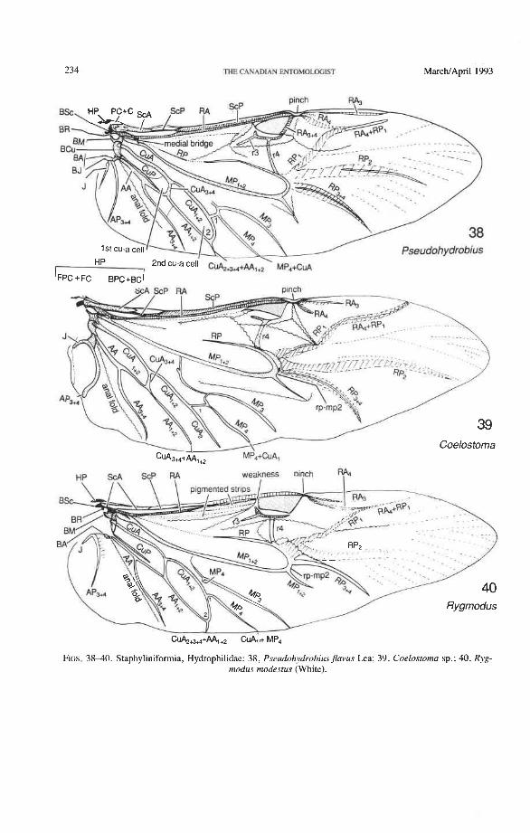
234 THE CANADLAN ENTOMOLOGIST MarcMApril 1993
FIGS. 3 8 4 0 . Staphyliniformia, Hydrophilidae: 38, Pseudohydrobiusflavus Lea; 39, Coelostoma sp.; 40, Ryg- modus modestus (White).
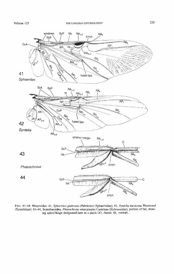
Volume 125 THE CANADIAN ENTOMOLOGIST
windows ScP RA RA. , RA.
Phaeochrous
44
FIGS. 4 1 4 . Histeroidea: 41, Sphaerites glabratus (Fabricius) (Sphaeritidae); 42, Syntelia rnexicana Westwood (Synteliidae); 4 3 4 , Scarabaeoidea, Phaeochrous emarginatus Castelnau (Hybosoridae), portion of bar, show-
ing apical hinge designated here as a pinch (43, dorsal; 44, ventral).
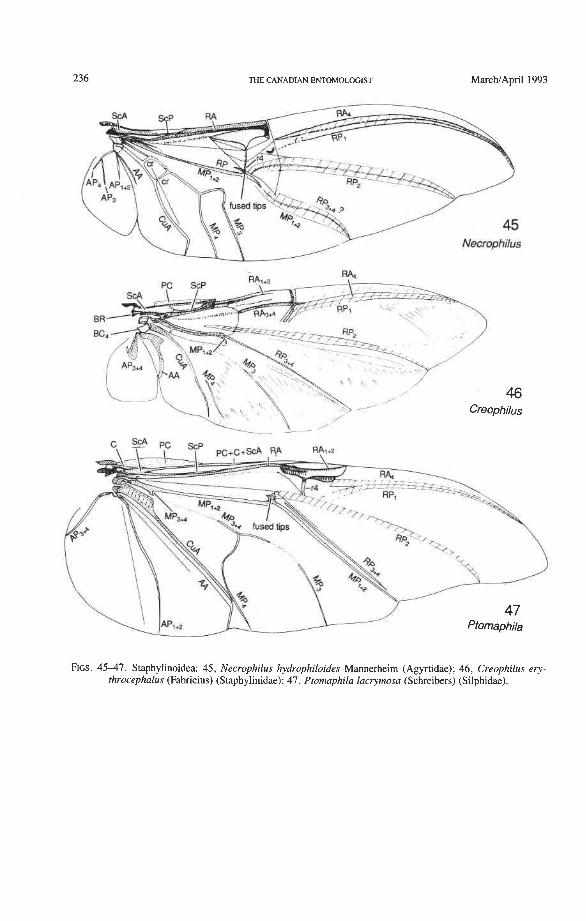
236 THE CANADIAN ENTOMOLOGIST MarchtApril 1993
FIGS. 4 5 4 7 . Staphylinoidea: 45, Necrophilus hydrophiloides Mannerheim (Agyrtidae); 46, Creophilus ery- throcephalus (Fabricius) (Staphylinidae); 47, Ptomaphila lacrymosa (Schreibers) (Silphidae).
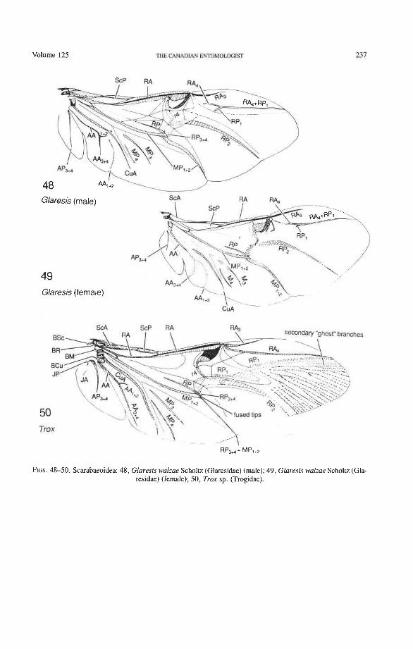
volume 125 THE CANADIAN ENTOMOLGGIST
Glaresis (male) SCA
\
AP, // 49 Glares~s (female)
CUA -
FIGS. 48-50. Scarabaeoidea: 48, Glaresis walzae Scholtz (Glaresidae) (male); 49, Glaresis walzae Scholtz (Gla- residae) (female); 50, Trox sp. (Trogidae).
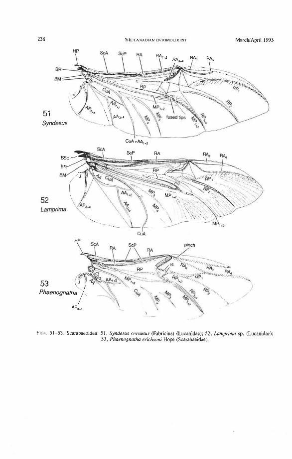
238 THE CANADIAN ENTOMOLOGIST March/April 1993
FIGS. 51-53. Scarabaeoidea: 51, Syndesus cornutus (Fabricius) (Lucanidae); 52, Lamprima sp. (Lucanidae); 53, Phaenognatha erichsoni Hope (Scarabaeidae).
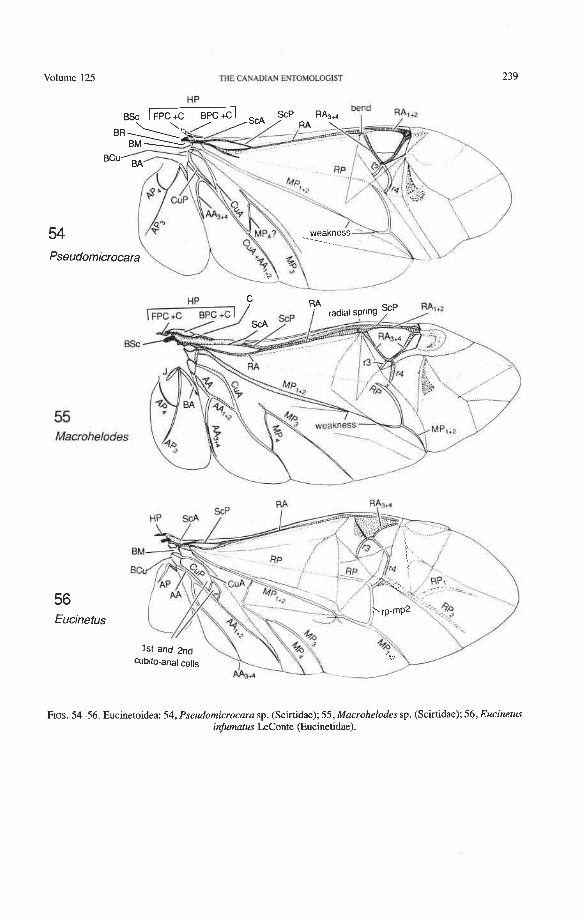
volume 125 THE CANADIAN EhTOMOLOGlST
55 Macrohelodes
56 Eucinetus
FIGS. 54-56. Eucinetoidea: 54, Pseudomicrocara sp. (Scirtidae); 55, Macrohelodes sp. (Scirtidae); 56, Eucinetus infumatus LeConte (Eucinetidae).
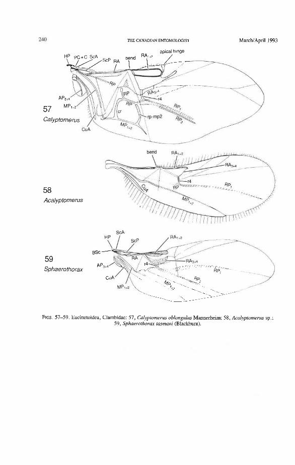
240 THE CANADIAN ENTOMOLOGIST March/April 1993
apical hinge
_.-- -__---
<-..
57 Calyptomerus
CU A --%
l-...
58 Acalyptomerus
59 Sphaerothorax
FIGS. 57-59. Eucinetoidea, Clambidae: 57, Calyptornerus oblongulus Mannerheim, 58, Acalyptomerus sp.; 59, Sphaerothorax tasmani (Blackburn).
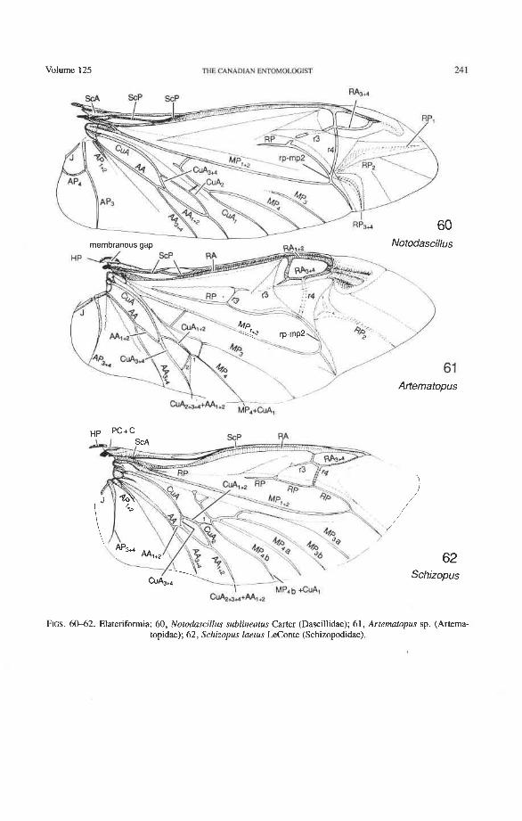
Volume 125 THE CANMIAN ENTDMOLDGIST
62 Schizopus
FIGS. 60-62. Elaterifomia: 60, Notodascillus sublineatus Carter (Dascillidae); 61, Artematopus sp. (Artema- topidae); 62, Schizopus laetus LeConte (Schizopodidae).
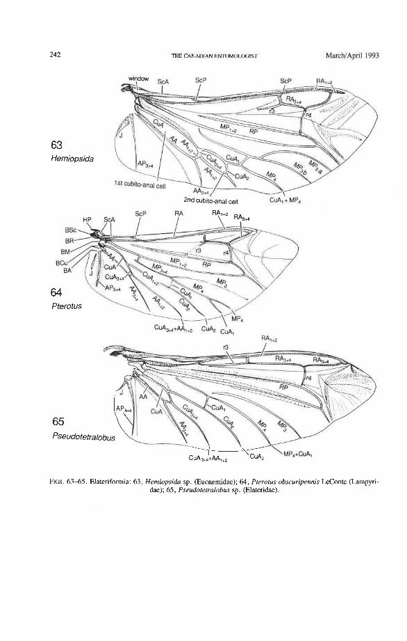
THE CANADIAN ENTOMOLOGIST MarchlApdl 1993
63 Herniopsida
2nd cubito-anal cell CUA, + MP,
FIGS. 6 3 4 5 . Elateriformia: 63, Hemiopsida sp. (Eucnemidae); 64, Pterotus obscuripennis LeConte (Lampyri- dae); 65, Pseudotetralobus sp. (Elateridae).
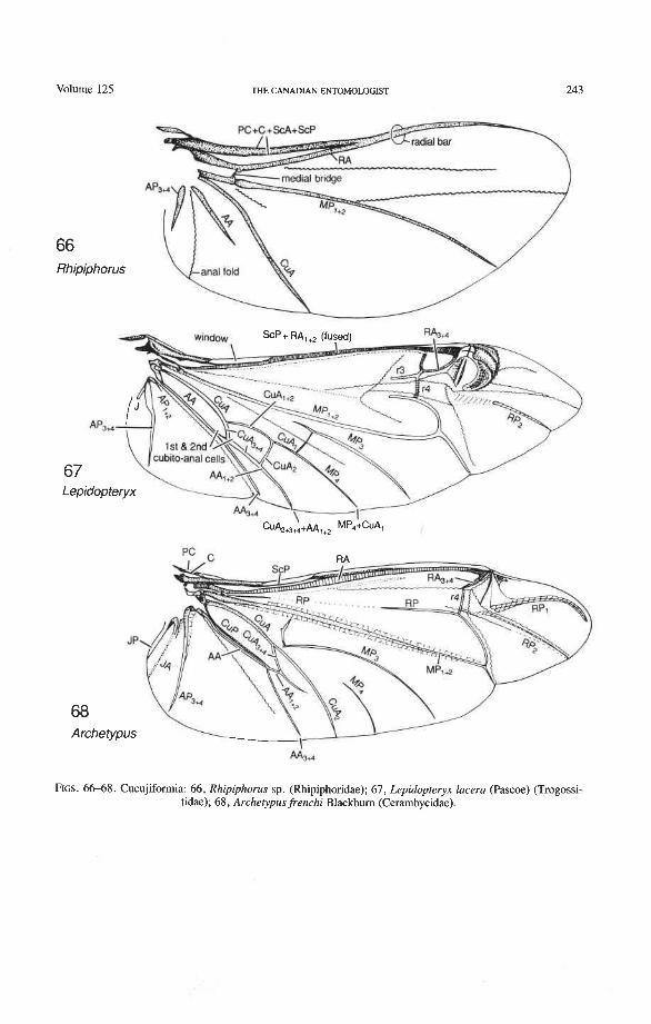
volume 125 THE CANADIAN ENTOMOLOGIST 243
66 Rhipiphorus
JP
68 Archetypus
FIGS. 66-68. Cucujifomia: 66, Rhipiphorus sp. (Rhipiphoridae); 67, Lepidopteryx lacera (Pascoe) (Trogossi-, tidae); 68, Archetypus frenchi Blackbum (Cerambycidae).
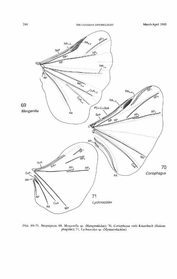
FIGS. 69-71. Strepsiptera: 69, Mengenilla sp. (Mengenillidae); 70, Coriophagus rieki Kinzelbach (Halicto- phagidae); 7 1, Lychnocolax sp. (Myrmecolacidae).
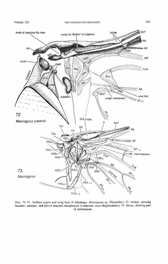
volume 125 THE CANADIAN ENTOMOLOFIfl 245
FIGS. 72-73. Axillary region and wing base of Adephaga, Macrogyrus sp. (Gyrinidae): 72, ventral, showing basalare, subalare, and part of attached metapleuron (composite, semi-diagrammatic); 73, dorsal, showing part
of metatergum.
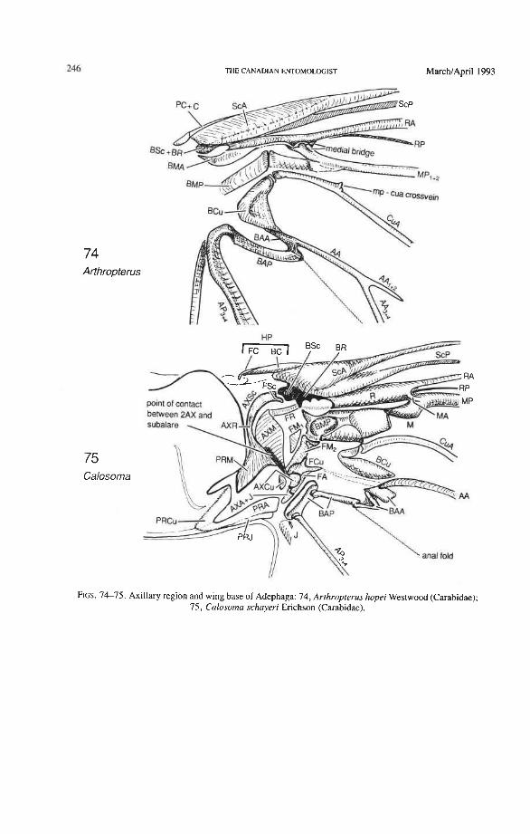
74 Arthropterus
75 Calosoma
THE CANADIAN ENTOMOLOGIST MarchIApril 1993
FIGS. 7 4 7 5 . Axillary region and wing base of Adephaga: 74, Arthropterus hopei Westwood (Carabidae); 75, Calosoma schayeri Erichson (Carabidae).
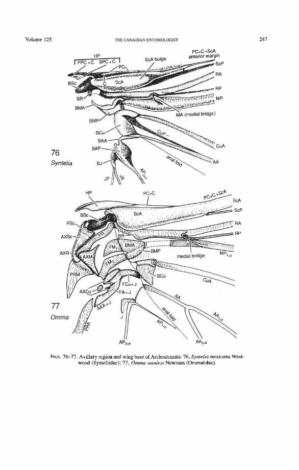
Volume 125 THE CANADIAN ENTOMOLOGIST
FIGS. 7 6 7 7 . Axillary region and wing base of Archostemata: 76, Syntelia mexicana West- wood (Synteliidae); 77, Omma stanleyi Newman (Ommatidae).
THE CANADIAN ENTOMOLQGIST MarchIApril 1993
78 Priacma
membrane HP \ PC
79 Eurynassa claval fold
FIGS. 78-79. Axillary region and wing base: 78, Archostemata, Priacma serrata LeConte (Cupedidae); 79, Polyphaga, Eurynassa sp. (Cerambycidae).
Volume 125 THE CANADIAN ENTOMOLOGIST
80 Elephastomus
bridge R A
MP
CUA
Cryptodus u \\
FIGS. 8681. Axillary region and wing base of Scarabaeoidea: 80, Elephastomus proboscideus (Schreibers) (Geotrupidae); 8 1 , Cryptodus paradoxus Macleay (Scarabaeidae).
THE CANADIAN ENTOMOLOGIST
radial :pring
FIGS. 82-85. Folded hind wings, dorsal and ventral: 82-83, Pseudomicrocara sp. (Scirtidae); 84-85, Notoda- scillus sublineatus Carter (Dascillidae).
Volume 125 THE CANADIAN ENTOMOLOGIST
87 Harmonia . ..
radi? bar radial Finge
88 Distocupes
b -->:-a &.:--..
medial hinge
FIGS. 86-88. 86-87, folded hind wing (dorsal and ventral) of Harmonia oc~omaculata (Fabricius) (Coccinelli- dae); 88, the position of the radial and medial loops in the unfolded (a) and folded (b) wing of Distocupes varians
(Lea) (Cupedidae).
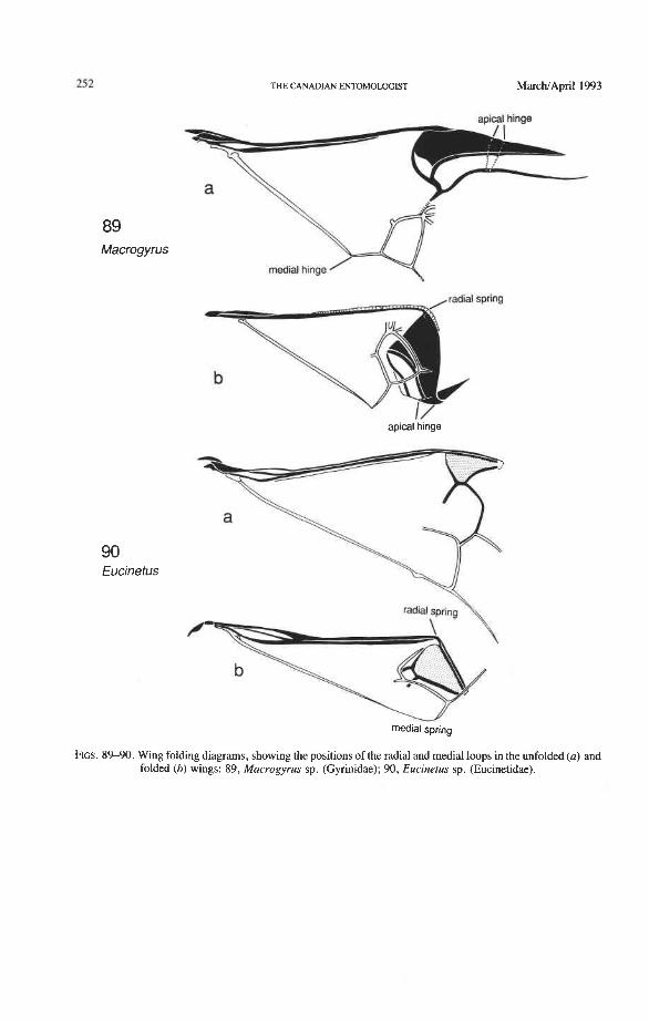
THE CANADIAN ENTOMOLOGIST MarchIApril 1993
89 Macrogyrus
90 Eucinetus
apical hinge
radial spring \
medial spring
FIGS. 8%90. Wing folding diagrams, showing the positions of the radial and medial loops in the unfolded (a) and folded (b) wings: 89, Macrogyrus sp. (Gynnidae); 90, Eucinetus sp. (Eucinetidae).
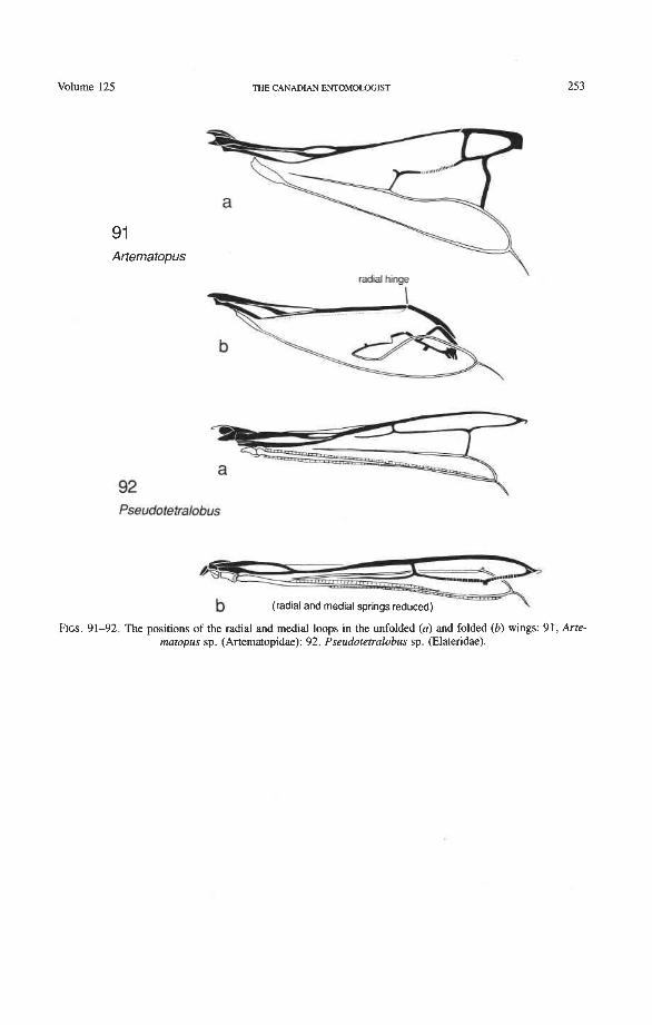
Volume 125
9 1 Artematopus
THE CANADIAN ENTOMOLOGIST
radial hinge \
Pseudotetralobus
FIGS. 91-92. The positions of the radial and medial loops in the unfolded (a) and folded (b) wings: 91, Arte matopus sp. (Artematopidae); 92, Pseudotetralobus sp. (Elateridae).
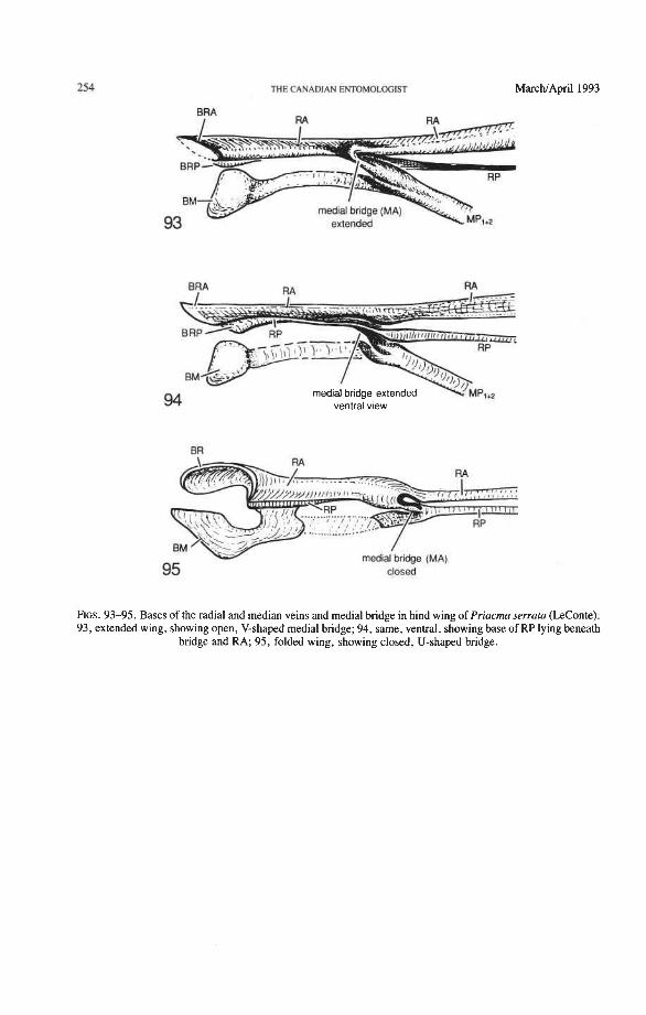
THE CANADIAN ENTOMOtOGIST MarchIApril 1993
BRA
94 medial bridge extended ventral vlew
.- ......... +;...-'
95 closed
FIGS. 93-95. Bases of the radial and median veins and medial bridge in hind wing of Priacma serrata (ZeConte): 93, extended wing, showing open, V-shaped medial bridge; 94, same, ventral, showing base of RP lying beneath
bridge and RA; 95, folded wing, showing closed, U-shaped bridge.
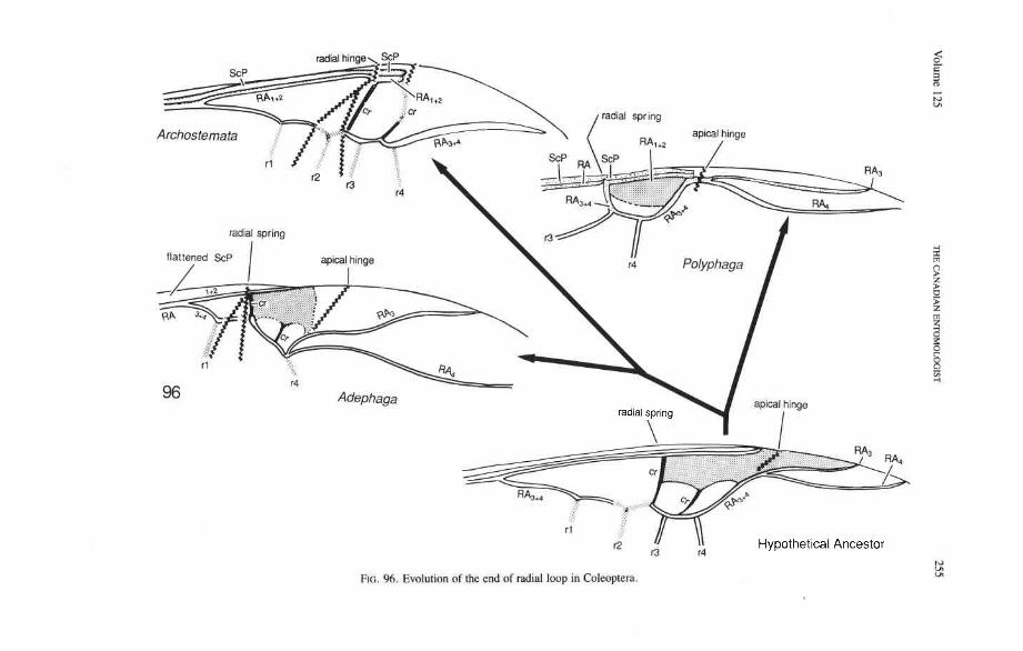
radial hinge ScP
/' nAw
aplcat hinge Archostemata /
Hypothet~cal Ancestor
FIG. 96. Evolution of the end of radial loop in Coleoprem.
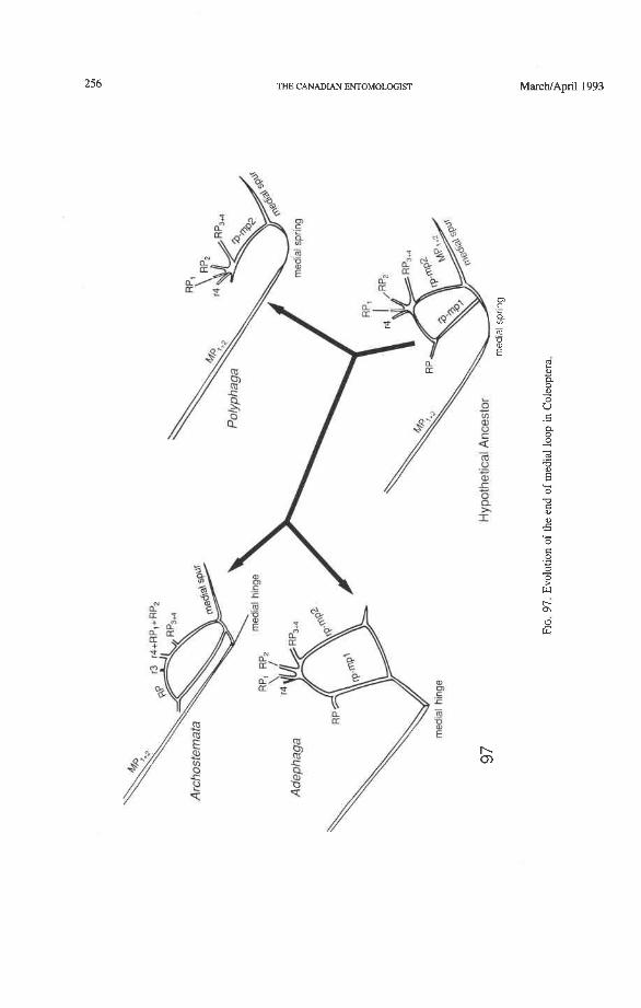
THE CANADIAN ENTOMOLOGlST
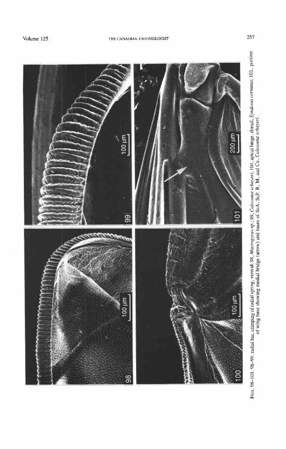
Volume 125 THE CANADIAN ENTOMOLOGIST
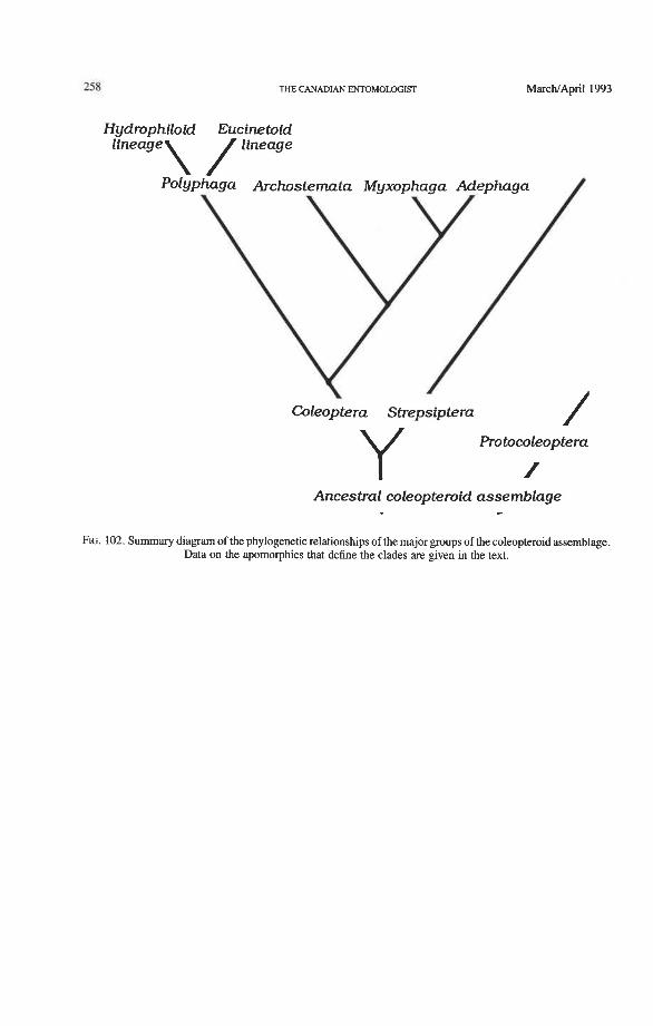
THE CANADIAN ENTOMOLOGIST
Hydrophiloid Eucinetoid
Coleoptera Strepsiptern
Protocoleoptera
1 Ancestral coleopteroid assemblage -
FIG. 102. Summary diagram of the phylogenetic relationships of the major groups of the coleopteroid assemblage. Data on the apomorphies that define the clades are given in the text.