anatomy & physiology lecture notes - heart physiology
TRANSCRIPT
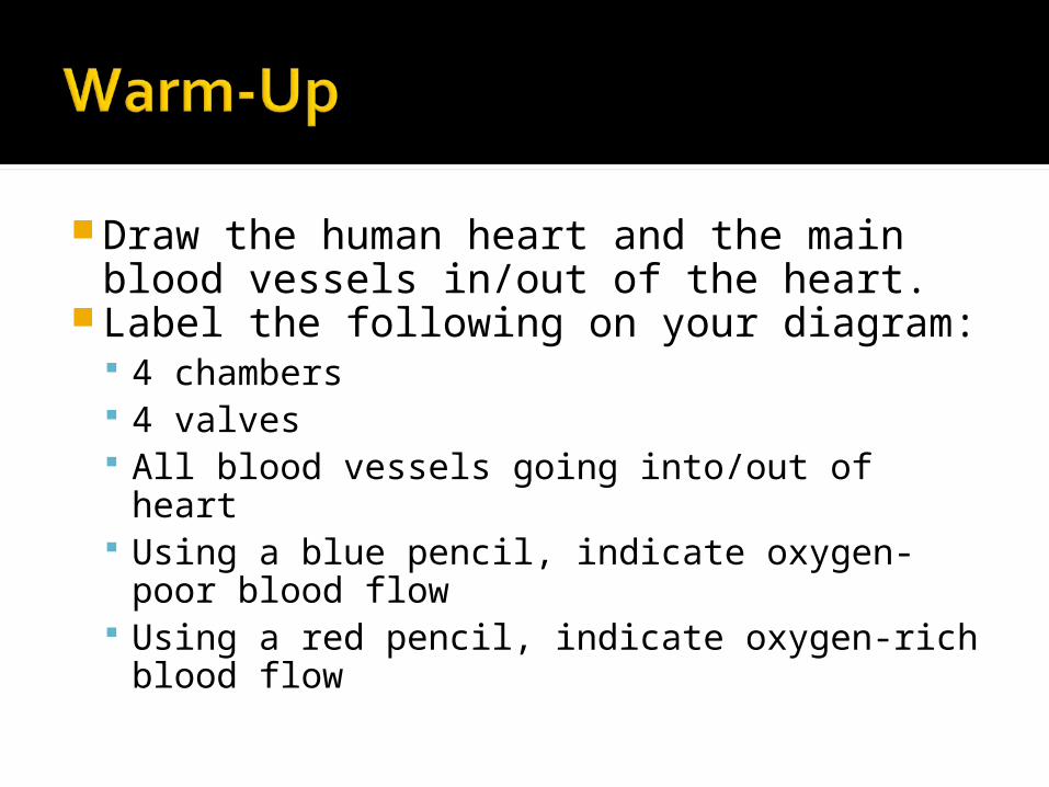
Draw the human heart and the main blood vessels in/out of the heart.
Label the following on your diagram: 4 chambers 4 valves All blood vessels going into/out of heart Using a blue pencil, indicate oxygen-
poor blood flow Using a red pencil, indicate oxygen-rich
blood flow
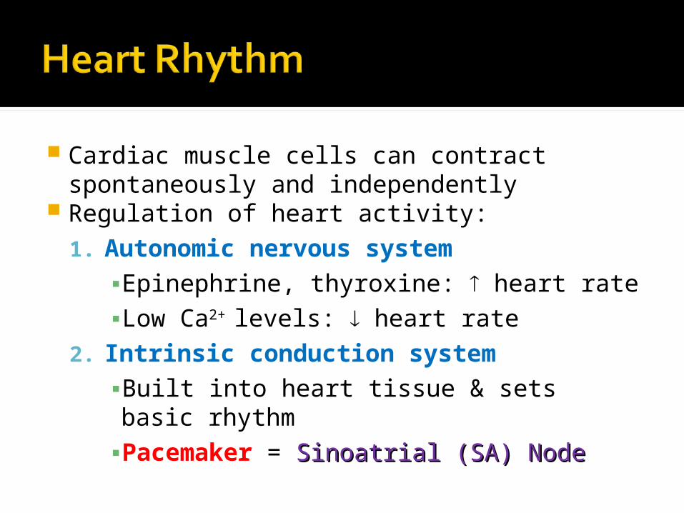
Cardiac muscle cells can contract spontaneously and independently
Regulation of heart activity:1. Autonomic nervous system
▪Epinephrine, thyroxine: heart rate▪Low Ca2+ levels: heart rate
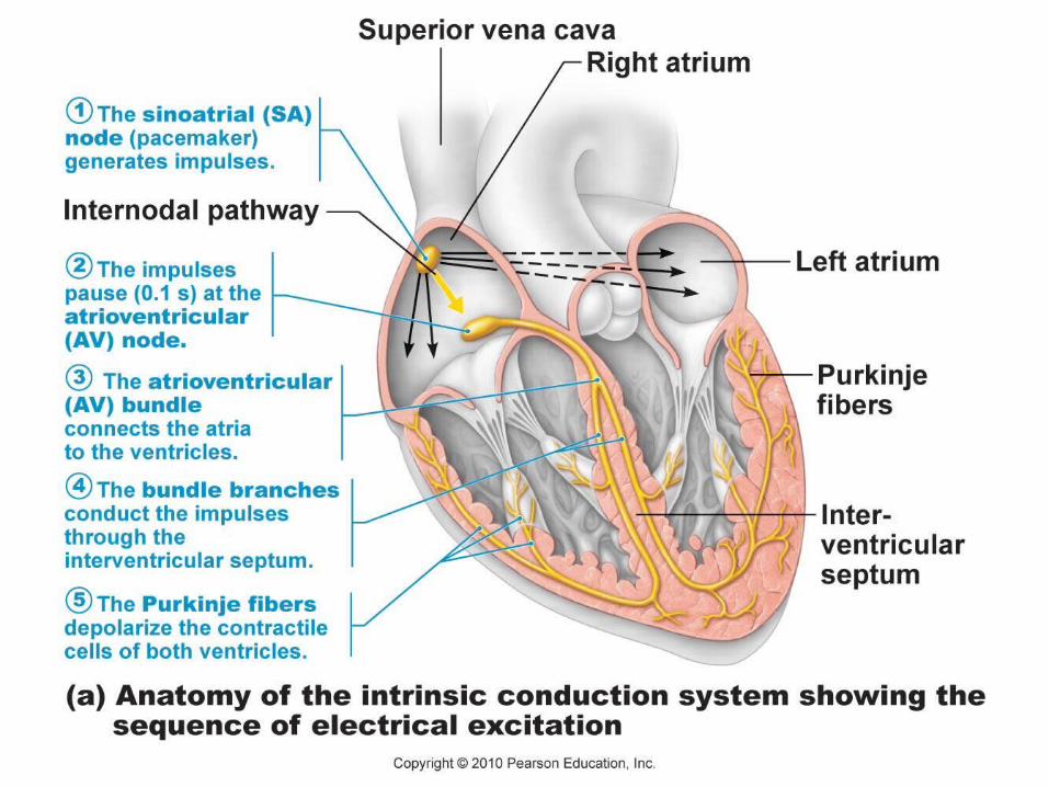
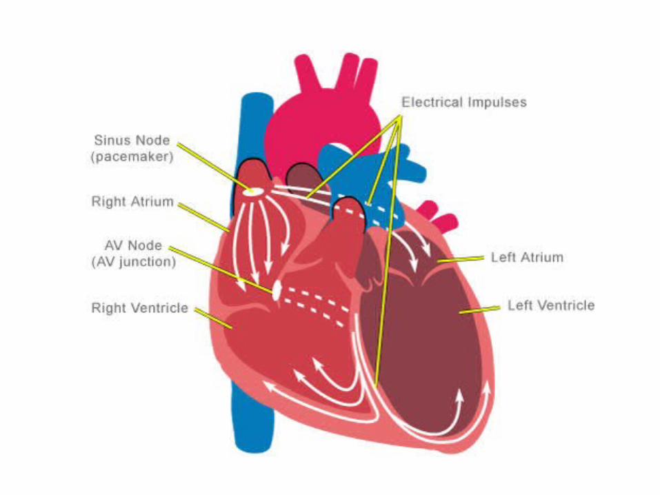
2. Intrinsic conduction system▪Built into heart tissue & sets basic rhythm▪Pacemaker = Sinoatrial (SA) NodeSinoatrial (SA) Node
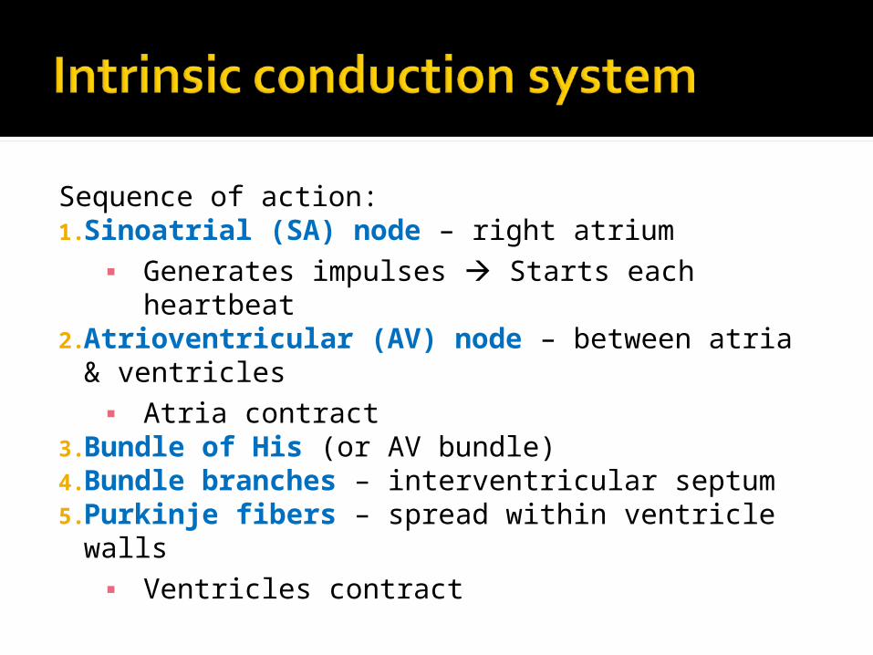
Sequence of action:1.Sinoatrial (SA) node – right atrium
▪ Generates impulses Starts each heartbeat
2.Atrioventricular (AV) node – between atria & ventricles▪ Atria contract
3.Bundle of His (or AV bundle) 4.Bundle branches – interventricular septum5.Purkinje fibers – spread within ventricle walls
▪ Ventricles contract
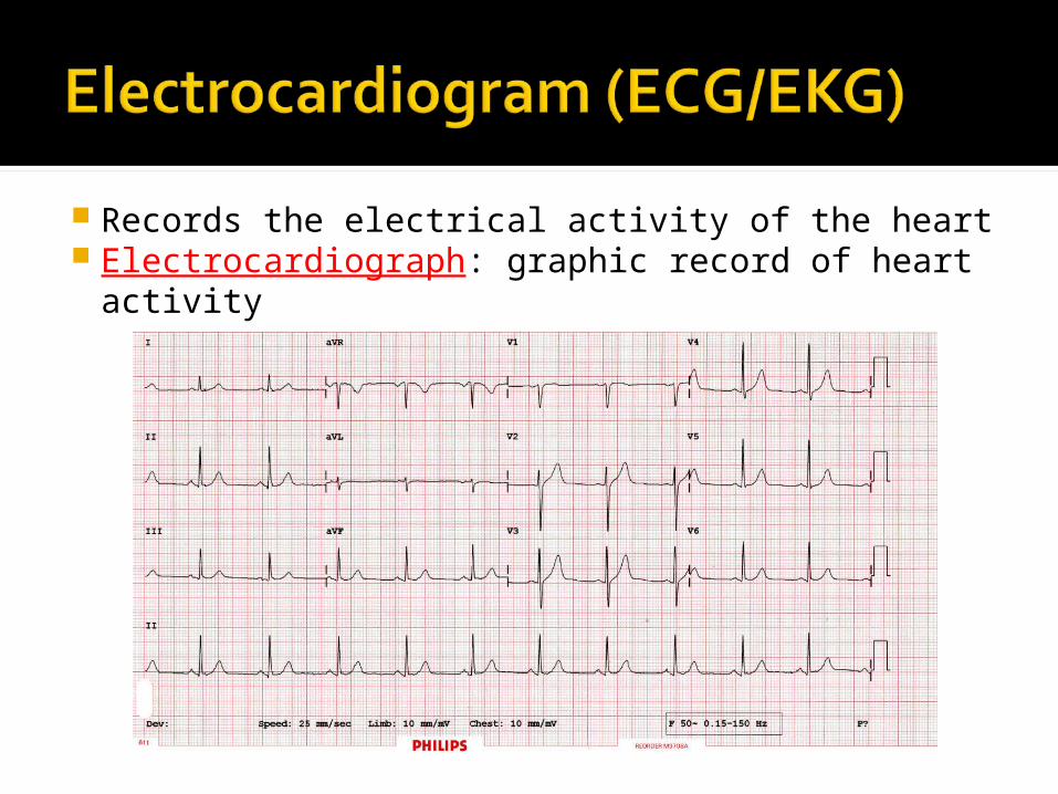
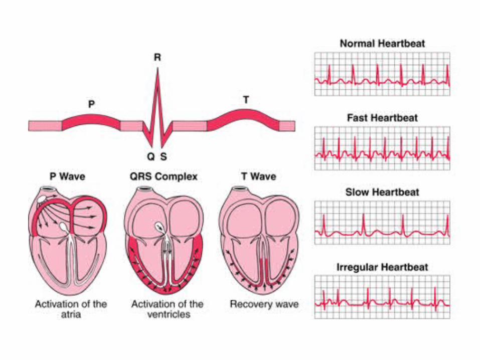
Records the electrical activity of the heart Electrocardiograph: graphic record of heart activity
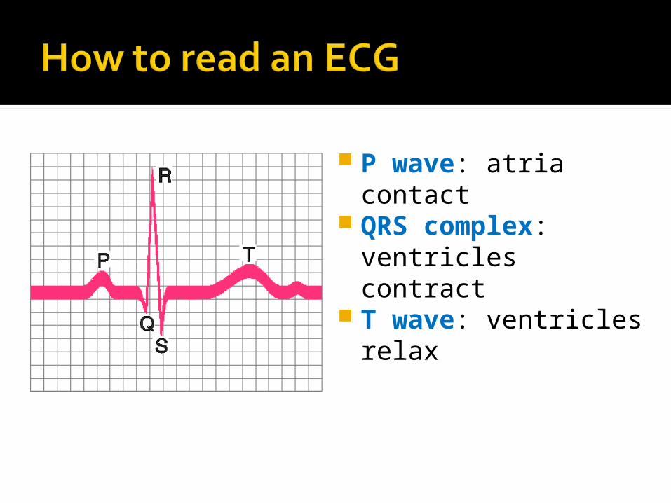
P wave: atria contact
QRS complex: ventricles contract
T wave: ventricles relax

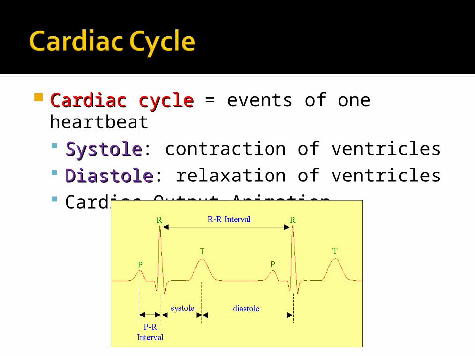
Cardiac cycle Cardiac cycle = events of one heartbeat SystoleSystole: contraction of ventricles DiastoleDiastole: relaxation of ventricles Cardiac Output Animation
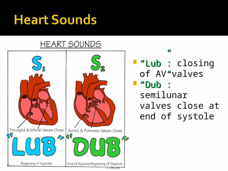
““LubLub””: : closing of AV valves
““DubDub””: : semilunar valves close at end of systole
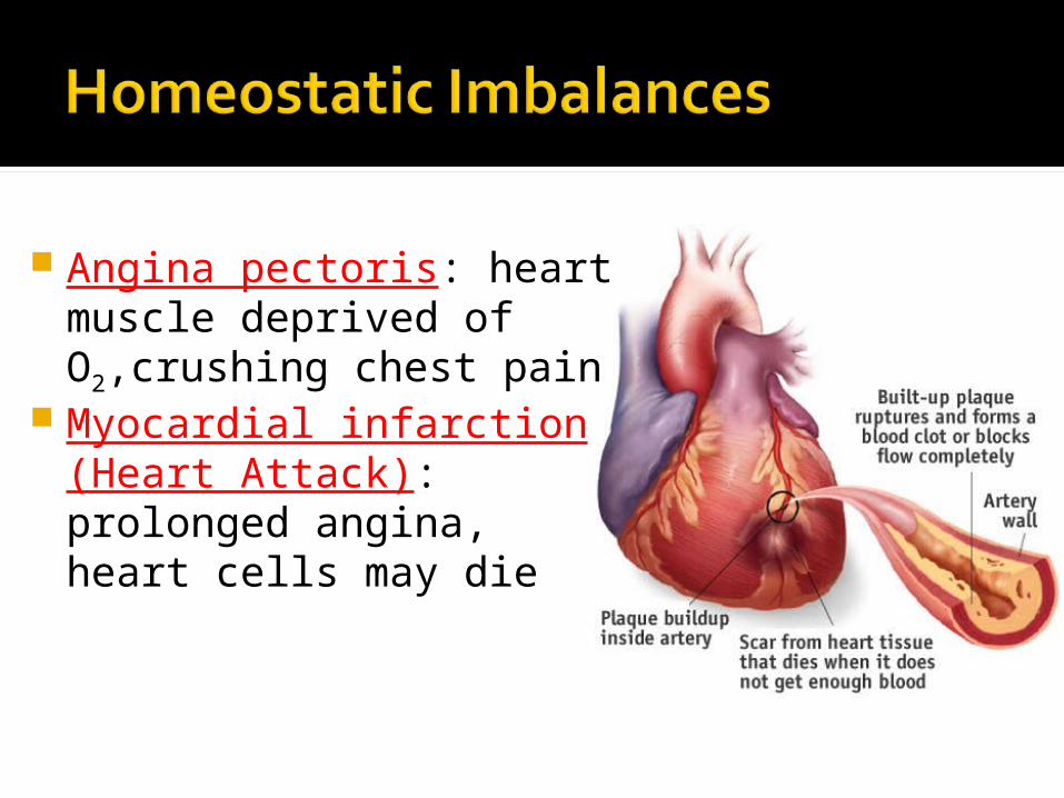
Angina pectoris: heart muscle deprived of O2,crushing chest pain
Myocardial infarction (Heart Attack): prolonged angina, heart cells may die
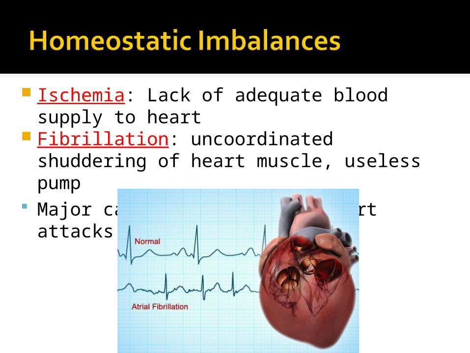
Ischemia: Lack of adequate blood supply to heart
Fibrillation: uncoordinated shuddering of heart muscle, useless pump
Major cause of death from heart attacks
Damage to SA node slower heart rate Install artificial pacemaker
Damage to AV node Heart block: ventricles beat at own rate (slower or not at all)
Tachycardia: rapid heart rate (>100 beats/min)
Bradycardia: very slow heart rate (<60 beats/min)
Heart murmur: abnormal or unusual heart sounds Often valve problems
Cardiac Output (CO) = Heart Rate (HR) x Stroke Volume (SV) Stroke volume: volume of blood pumped
out by one ventricle with each best
Average adult:CO = HR (75 beats/min) x SV (70 ml/beat)
CO = 5250 ml/min
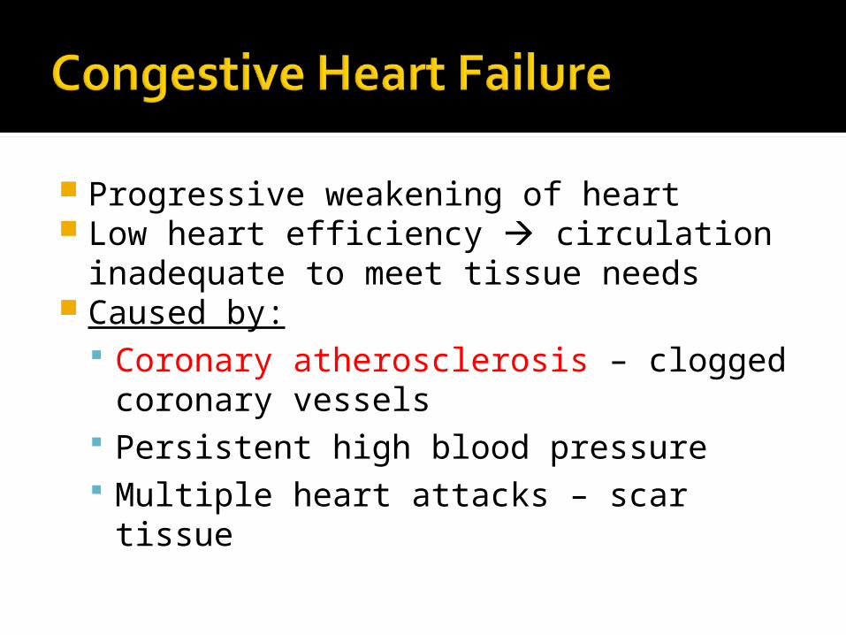
Progressive weakening of heart Low heart efficiency circulation
inadequate to meet tissue needs Caused by:
Coronary atherosclerosis – clogged coronary vessels
Persistent high blood pressure Multiple heart attacks – scar tissue