analysis the expression of anthocyanin pathway genes in developing … · analysis of the...
TRANSCRIPT
Plant Physiol. (1 996) 1 1 1 : 1059-1 066
Analysis of the Expression of Anthocyanin Pathway Genes in Developing Vitis vinifera 1. cv Shiraz Grape Berries and the -
Implications for Pathway Regdation
Paul K. BOSS*, Christopher Davies, and Simon P. Robinson
Cooperative Research Centre for Viticulture, P.O. Box 145, Glen Osmond, South Australia 5064, and Commonwealth Scientific and Industrial Research Organization, Division of Horticulture, G.P.O. Box 350,
Adelaide, South Australia 5001, Australia (P.K.B., C.D., S.P.R.); and Department of Horticulture, Viticulture and Oenology, Waite Agricultura1 Research Institute, University of Adelaide, South Australia 5064, Australia (P.K.B.)
Anthocyanin synthesis in Vitis vinifera L. cv Shiraz grape berries began 1 O weeks postflowering and continued throughout berry ripening. Expression of seven genes of the anthocyanin biosynthetic pathway (phenylalanine ammonia lyase [PAL], chalcone synthase [CHS], chalcone isomerase [CHI], flavanone-3-hydroxylase [F3Hl, dihydroflavonol 4-reductase [DFR], leucoanthocyanidin dioxygen- ase [LDOX], and UDP glucose-flavonoid 3-o-glucosyl transferase [UFCT]) was determined. In flowers and grape berry skins, expres- sion of all of the genes, except UFCT, was detected up to 4 weeks postflowering, followed by a reduction in this expression 6 to 8 weeks postflowering. Expression of CHS, CHI, F3H, DFR, LDOX, and UFCT then increased 1 O weeks postflowering, coinciding with the onset of anthocyanin synthesis. In grape berry flesh, no PAL or UFCT expression was detected at any stage of development, but CHS, CHI, F3H, DFR, and LDOX were expressed up to 4 weeks postflowering. These results indicate that the onset of anthocyanin synthesis in ripening grape berry skins coincides with a coordinated increase in expression of a number of genes in the anthocyanin biosynthetic pathway, suggesting the involvement of regulatory genes. UFCT is regulated independently of the other genes, suggest- ing that in grapes the major control point in this pathway is later than that observed in maize, petunia, and snapdragon.
Anthocyanin biosynthesis has been extensively studied in petunia, snapdragon, and maize, resulting in the eluci- dation of the biosynthetic pathway in which the various anthocyanin pigments are synthesized from Phe. This work has been aided by the availability of a large number of anthocyanin mutants that are nonlethal and have been selected over many years by both plant breeders and ge- neticists (for a review, see Martin and Gerats, 1993). More recently, it has become apparent that the mutants generally fall into two groups. The first group results from mutations in the structural genes coding for enzymes in the anthocy- anin biosynthetic pathway, and many of these genes have been isolated or identified using these mutants. The second group displays altered expression of more than one struc- tural gene, and these are usually the result of mutations in regulatory genes. It has been shown that these regulatory genes are homologous to members of the myc and myb
* Corresponding author; e-mail [email protected]; fax 61-8-3038601.
families of transcription factors (Paz-Ares et al., 1987; Lud- wig et al., 1989; Goodrich et al., 1992). It is interesting that the control of the anthocyanin pathway differs in the three plant species mentioned above. In maize, it appears that the regulation start point is CHS, whereas in snapdragon and petunia the control start points are further on in the pathway, at F3H and DFR, respectively (for a review, see Martin and Gerats, 1993). As yet, the control of the expres- sion of the anthocyanin pathway genes in fruit tissues has not been studied.
The color of red and black grapes results from the accu- mulation of anthocyanins that are usually only located in the skin of the berry. The quantity and quality of color in grape berries at harvest are crucial factors that influence wine making. Each species or variety of grapes has a unique set of anthocyanins, and the anthocyanin profiles of many Vitis spp. and varieties have been described (for a review, see Mazza and Miniati, 1993). Vitis vinifera varieties usually produce 3-monoglucoside, 3-acetylglucoside, and 3-p-coumarylglucoside derivatives of the aglycones del- phinidin, cyanidin, peonidin, petunidin, and malvidin, with malvidin derivatives often being the major forms present. However, there are exceptions. The cultivar Pinot Noir produces only nonacylated anthocyanins (Fong et al., 1971), and many muscat cultivars produce less malvidin derivatives than other anthocyanins (Cravero et al., 1994). There are no reports of any pelargonidin derivatives iso- lated from grape berry skins, and thus the general pathway for anthocyanin synthesis can be modified to account for this fact (Fig. 1).
Grapes are a nonclimacteric fruit, and berry growth fol- lows a double-sigmoid pattern (Coombe and Bishop, 1980). Anthocyanin biosynthesis commences only when ripening of the berry begins (termed véraison by viticulturists) and normally continues throughout the ripening phase of growth. Factors such as variety, growing region, and growth conditions can influence the levels of anthocyanins
Abbreviations: CHI, chalcone isomerase; CHS, chalcone syn- thase; DFR, dihydroflavonol 4-reductase; F3H, flavanone-3- hydroxylase; LDOX, leucoanthocyanidin dioxygenase; PAL, Phe ammonia lyase; UFGT, UDP Glc-flavonoid 3-o-glucosyl transferase.
1059 www.plantphysiol.orgon February 18, 2019 - Published by Downloaded from
Copyright © 1996 American Society of Plant Biologists. All rights reserved.

1060 Boss et al. Plant Physiol. Vol. 11 1, 1996
Phenylalanine
+m
Naringenin chalcone
Eriodictyol
S E I Dihydroquercetin
Leucocyanidin
I
t (DEHyDRATAsE)
F3 'H - F3'H
t---
Cyanidin
OA
OH
+m F3 '5'H Naringenin flavanone -b
Dihydrokaempferol . -+
Pentahydroxyflavanone
+m Dihydromyricetin
Leucodelphinidin
Delphinidin
p q OA
I + mo"
OE
HO AO&: Delphinidin-3- Cyanidin-3- glucoside
glucoside 08
' OGlvc \
OA \ ' 0Gl"C
OE
Peonidin-3- Petunidin-3- Malvidin-3 - glucoside glucoside glucoside
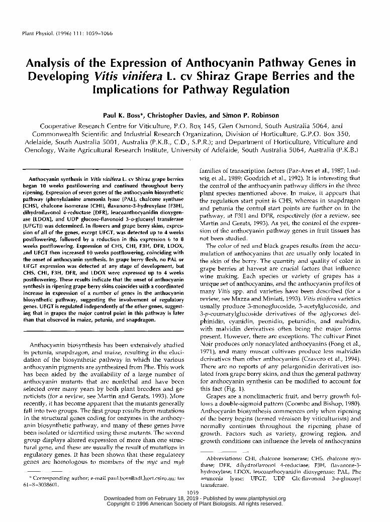
Figure 1. Simplified schematic of the anthocyanin biosynthetic pathway, modified to account for the major products found in grapes. The genes examined in this study are boxed. The dehydratase is putative and is thus written in brackets, and the substrates for flavonoid 3'-hydroxylase (F3'H) and flavonoid 3'5'-hydroxylase (F3'5'H) have not been determined for grapes. C4H, Cinnamate 4-hydroxylase; 4CL, 4-coumarate COA ligase; MT, methyltransferase.
produced and the profile of different pigments (for review, see Mazza and Miniati, 1993). Hrazdina et al. (1984) mea- sured the activity of some anthocyanin biosynthetic en- zymes during the development of De Chaunac berries. Anthocyanin accumulation began when these berries had just begun to accumulate sugar and increased rapidly until the concentration leveled off late in ripening, when the soluble solids were approximately 24 "Brix (a refractive index measure of the total dissolved solids). The increase in activity of the enzymes cinnamate-4-monooxygenase, p - coumarate:CoA-ligase (which catalyzes reactions before CHS in the pathway), and UFGT paralleled the increase in anthocyanin content. However, the activity of both PAL and CHI decreased as accumulation began and then sub- sequently increased. CHS activity was not detected until 3 weeks after anthocyanins were detected, even though it is the first committed step of the anthocyanin biosynthetic pathway. In cv Cardinal grapes, Roubelakis-Angelakis and
Kliewer (1986) found that the increase in PAL activity paralleled anthocyanin accumulation as ripening pro- gressed. Thus, the limited enzymatic studies of anthocya- nin accumulation in grapes have not revealed a great deal about the control of this biosynthetic pathway and what causes anthocyanin biosynthesis to be switched on during ripening.
To further investigate the regulation of anthocyanin pro- duction in grape berries, we utilized grapevine cDNAs encoding enzymes of the anthocyanin biosynthetic path- way isolated by Sparvoli et al. (1994) from grape leaf tissue. Berries from cv Shiraz were sampled throughout develop- ment, and the tissue was used to study anthocyanin pro- files and the expression of seven structural genes from the anthocyanin biosynthetic pathway. Our results suggest that the pattern of control for the anthocyanin pathway in grape berry skin tissue is different from that observed in petunia, snapdragon, and maize.
www.plantphysiol.orgon February 18, 2019 - Published by Downloaded from Copyright © 1996 American Society of Plant Biologists. All rights reserved.
Anthocyanin Biosynthesis in Shiraz Crape Berries 1061
MATERIALS A N D METHODS
Plant Tissue
Berries from Vitis viniferu L. cv Shiraz were sampled at 2-week intervals during the 1994 to 1995 growing season from vines grown at a commercial vineyard in Willunga, South Australia. To define the stage of berry development, a sample of 100 randomly selected berries from 30 bunches was individually labeled and scored each week for deform- ability, length, and width using a Harpenden (British In- dicators, Burgess Hill, West Sussex, UK) skinfold calliper gauge as described by Coombe and Bishop (1980). The volumes of the Shiraz berries were calculated using the formula for an ellipsoid (4/3mbc, where a, b, and c are the semiaxes of the ellipsoid). Another random sample of 50 berries was measured for soluble solids ("Brix) with a refractometer (model 10430; Reichert, Vienna, Austria). Berries for RNA extraction and anthocyanin analysis were randomly sampled every 2 weeks postflowering from ran- domly selected bunches, deseeded, and immediately fro- zen in liquid nitrogen. These samples were stored at -80°C pending further analysis. Separate skin and flesh samples were obtained by peeling frozen berries.
Anthocyanin Extraction
To prepare samples for HPLC analysis, 10 to 20 frozen berries were removed from storage and peeled. The peel tissue was ground in liquid nitrogen using a mortar and pestle. A 0.5-g subsample of the tissue was then added to 1 mL of methanol, and the anthocyanins were extracted for 1 h at -20°C. The grape tissue was pelleted by centrifuga- tion at 10,300g for 15 min at 4"C, and the supernatant was retained for HPLC analysis. A 5-pL aliquot of this sample was diluted to 1 mL in methanol and 1% (v/v) HCI, and total anthocyanins were measured by reading at A,,,.
HPLC Analysis of Anthocyanin Extracts
A 5-pm Gold Pack C28 column (4.6 X 25 mm; Activon, Sydney, Australia) and Varian (Melbourne, Australia) equipment consisting of the Vista 5500 pumps and solvent programer, a Rheodyne (Cotati, CA) injector, and a UV-200 detector (Varian, Melbourne, Australia) operating at 520 nm were used. The signal was received and analyzed using a data acquisition, plotting, and analysis package from DAPA Scientific (Kalamunda, Australia), which measured retention times and peak areas. The weak solvent A was 1.4% (v/v) perchloric acid, the strong solvent B was 100% methanol, and solvent C was water. Solvent A was main- tained at 30% throughout the analyses, and the flow rate was 1.5 mL/min. The initial condition of solvent B was 20%, increased to 35% in 5 min, and then increased to 55% in 35 min. In all cases 100 FL of extract (see above) were injected.
lsolation of Total RNA
Total RNA was isolated from grape berry skin and flesh tissue using the perchlorate method of Rezaian and Krake
(1987) with modifications. A 4-g sample of tissue was re- moved from -80°C storage and ground to a powder using a coffee grinder. The powder was added to 16 mL of extraction buffer (0.3 M Tris-HCI [pH 8.31, 2% [w/v] PEG 4000, 5 M sodium perchlorate, 1% [w/v] SDS, 8.5% [w/v] PVP, and 1% [v/v] p-mercaptoethanol) and stirred rapidly for 30 min at room temperature. This slurry was then centrifuged at 200g for 15 min at 4°C through Centriflo cones (Amicon, Beverly, MA) packed with glass wool, and the "raft" was discarded. The eluate was collected, and nucleic acids were precipitated with 2.5 volumes of etha- nol, incubated at -20°C for 20 min, and then pelleted by centrifugation at 7700g for 15 min at 4°C. This pellet was rinsed with 70% ethanol, dried under vacuum, and resus- pended in 1 mL of 0.1 mM Tris/l mM EDTA (pH 7.6) and 0.2% (v / v) p-mercaptoethanol. The suspension was then extracted three times with an equal volume of phenol: ch1oroform:isoamyl alcohol(25:24:1, v /v ) and once with an equal volume of ch1oroform:isoamyl alcohol (24:1, v / v). The RNA was precipitated by adding 0.1 volume of 3 M
sodium acetate and 2.5 volumes of ethanol to the aqueous phase and incubating at -20°C for at least 20 min. Finally, the RNA was pelleted by centrifugation at 7700g for 15 min at 4"C, washed with 70% ethanol, dried under a vacuum, and resuspended in 300 pL of water.
Northern Blot Analysis
Total RNA was extracted from grape tissues as described above. Aliquots of 4 pg were denatured and fractionated on a 1.2% agarose gel containing 8% formaldehyde. RNA loadings were checked on ethidium bromide-stained gels to confirm that they were equal. The RNA was transferred to a ZetaProbe membrane (Bio-Rad) for at least 15 h and then prehybridized for 2 h at 65°C in 0.25 M sodium phos- phate (pH 7.0), 1 miv EDTA (pH &O), and 7% (w/v) SDS. Membranes were hybridized for 15 h at 65°C in the same buffer with the addition of denatured 32P-labeled probes of the anthocyanin genes. Probes were prepared by random primer labeling to approximately equal specific activities of at least 3 X 10⁶ cpm ng-l DNA. The membrane was then washed twice for 10 min in 2x SSC (150 mM NaCl and 15 mM tri-sodium citrate, pH 7.0) and 0.1% (w/v) SDS (65°C) and then for 15 min in l x SSC and 0.1% (w/v) SDS (65°C). The membranes were exposed to Kodak XAE film with intensifying screens at -80°C.
RESULTS
Crape Berry Development
Data from the measurement of various ripening parameters throughout the development of the cv Shiraz berries sampled are presented in Figure 2 and show that berry growth fol- lowed the typical double-sigmoid curve. The volume of the berries (Fig. 2A) increased during the first 7 weeks of devel- opment to approximately 650 mm3, followed by a cessation in the berry expansion until9 weeks postflowering, after which time the volume began to increase again. Berry volume peaked at week 11 (1183 mm3) and then decreased to a final value of 765 mm3 at harvest. The onset of ripening is indi- cated by an increase in softness, sugar content, berry size,
www.plantphysiol.orgon February 18, 2019 - Published by Downloaded from Copyright © 1996 American Society of Plant Biologists. All rights reserved.
1062 Boss et al. Plant Physiol. Vol. 11 1, 1996
20:D 100
O 2 4 6 8 10 12 14 16
Weeks after flowering
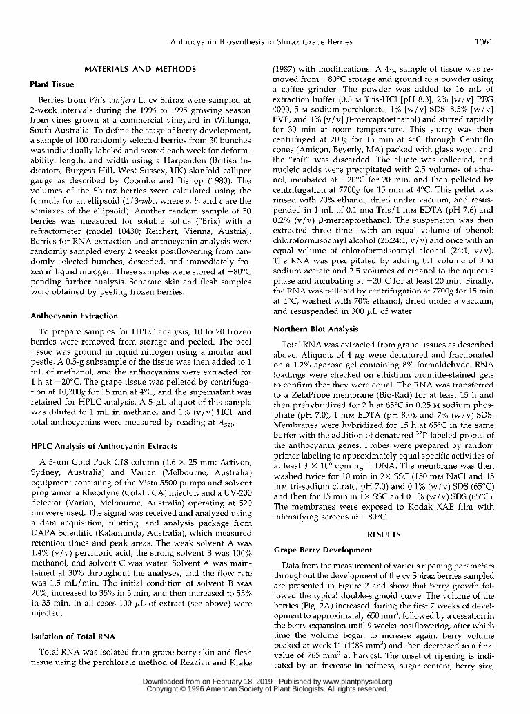
Figure 2. Changes in various parameters measured during the de- velopment and ripening of cv Shiraz grape berries. A, Berry volume; B, berry deformability; C, total soluble solids in the berry juice, measured as OBrix; D, total anthocyanins per gram fresh weight of berry skin. The vertical, dotted line represents véraison.
and, in red and black grapes, the development of skin color. Deformability (a measure of berry softness) began to increase 8 weeks postflowering (Fig. 2B). By 16 weeks postflowering the berries were very soft, deforming by 3.1 mm on average. Soluble solids (measured as "Brix) also began to increase 8 weeks postflowering and con- tinued to increase, reaching a value of 24 "Brix 16 weeks postflowering (Fig. 2C). Anthocyanins were first de- tected in the sample taken 10 weeks postflowering (Fig. 2D), although field observations indicated that some pigmentation was present after 9 weeks (samples for RNA and anthocyanin extraction were only taken on even-numbered weeks). Thus, there seemed to be a slow accumulation of anthocyanins between 9 and 10 weeks postflowering, followed by a substantial increase in an- thocyanin levels up to harvest, 16 weeks postflowering. From the data, véraison is considered to occur between 8 and 9 weeks postflowering, and this is indicated by the vertical, dotted line in Figure 2. Although no anthocya- nins were detected in the flower sample or in the berry skin samples taken up to 8 weeks postflowering, hot acid extraction, which hydrolyzes unpigmented proanthocya- nidins (condensed tannins) into colored anthocyanidin monomers (Harborne, 1989), revealed the presence of
both procyanidin and prodelphinidin (data not shown). Hot acid extraction of the berry flesh samples suggested that the 4- and 8-week postflowering samples possessed proanthocyanidins, whereas the 12- and 16-week post- flowering samples did not (data not shown).
HPLC Analysis of Anthocyanin Accumulation
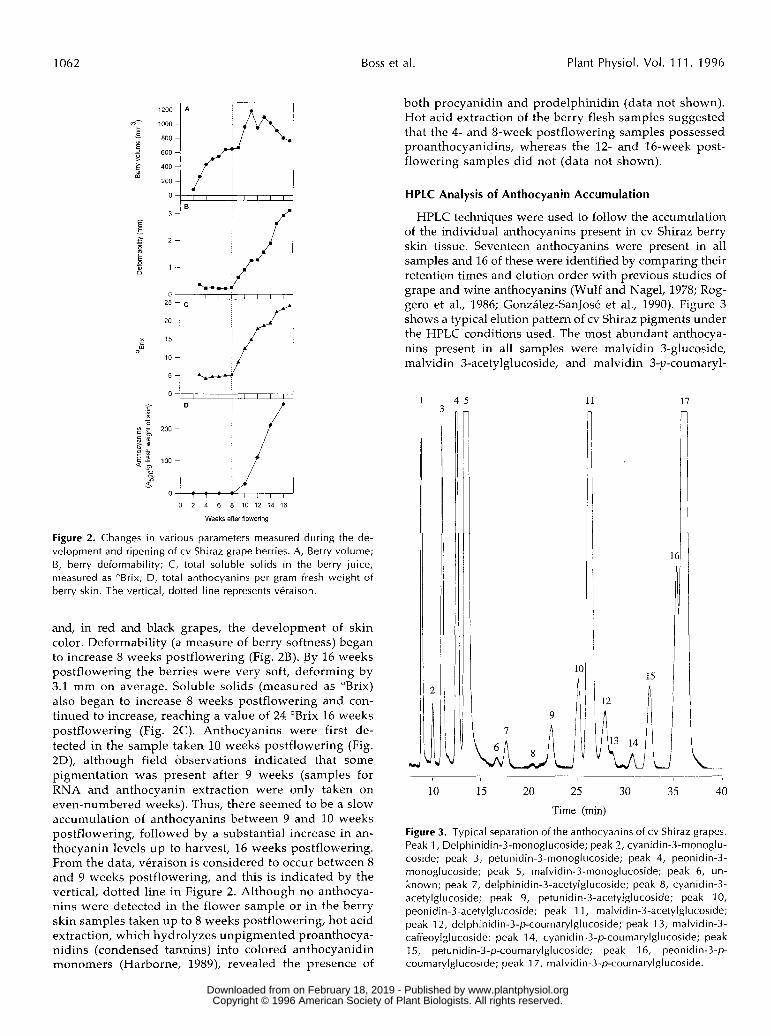
HPLC techniques were used to follow the accumulation of the individual anthocyanins present in cv Shiraz berry skin tissue. Seventeen anthocyanins were present in a11 samples and 16 of these were identified by comparing their retention times and elution order with previous studies of grape and wine anthocyanins (Wulf and Nagel, 1978; Rog- gero et al., 1986; González-SanJosé et al., 1990). Figure 3 shows a typical elution pattern of cv Shiraz pigments under the HPLC conditions used. The most abundant anthocya- nins present in a11 samples were malvidin 3-glucoside, malvidin 3-acetylglucoside, and malvidin 3-p-coumaryl-
1 4 5
n 3
1c
11 17
r
16
15
10 15 20 25 30 35 40
Time (min)
Figure 3. Typical separation of the anthocyanins of cv Shiraz grapes. Peak 1, Delphinidin-3-monoglucoside; peak 2, cyanidin-3-monoglu- coside; peak 3, petunidin-3-monoglucoside; peak 4, peonidin-3- monoglucoside; peak 5, malvidin-3-monoglucoside; peak 6, un- known; peak 7, delphinidin-3-acetylglucoside; peak 8, cyanidin-3- acetylglucoside; peak 9, petunidin-3-acetylglucoside; peak 1 O, peonidin-3-acetylglucoside; peak 1 1, malvidin-3-acetylglucoside; peak 12, delphinidin-3-p-coumarylglucoside; peak 13, malvidin-3- caffeoylglucoside; peak 14, cyanidin-3-p-coumaryIglucoside; peak 15, petunidin-3-p-coumarylglucoside; peak 16, peonidin-3-p- coumarylglucoside; peak 1 7, malvidin-3-p-coumaryIglucoside.
www.plantphysiol.orgon February 18, 2019 - Published by Downloaded from Copyright © 1996 American Society of Plant Biologists. All rights reserved.
Anthocyanin Biosynthesis in Shiraz Grape Berries 1063
cj*:in*o
1 1,ij '<D>, 5
1 J-S1~o
CMin
"E"5en"o
CO ~c ^ro <58- *o -c
< 5>CM
S3
c
'o(O .~c i:»_ 1(D ^=£ w
< t,CMin
o-
100 -
80 -
60 -
40 -
20 -
0 -
40 -
30 -
20 -
10 -
60 -
50 -
40 -
30 -
20 -
10 -
0 -
3-monoglucosides» Mv
*•''
V
— r Pe
•• — ••••*'*"^ ^ H CyT i T T i T i
3-acetylglucosides^ Mv
.*''
..«'Pe
-—JT—~ ~~~* R
3-p-coumarylglucosides .* Mv
*'
.V-J» Pe
--r"'" * R1*•' _ - -— •'''^A- — "̂ r Dpt«=^ f̂crrrtmzl cvT i T i — T i T i
9 10 11 12 13 14 15 16 17
weeks after flowering
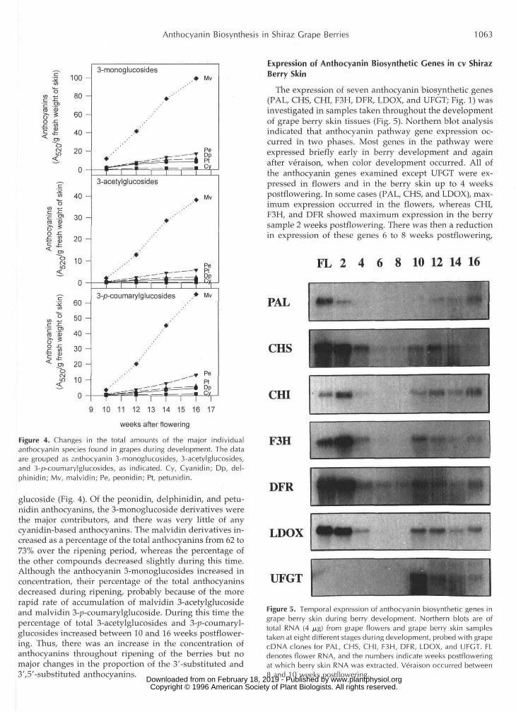
Figure 4. Changes in the total amounts of the major individualanthocyanin species found in grapes during development. The dataare grouped as anthocyanin 3-monoglucosides, 3-acetylglucosides,and 3-p-coumarylglucosides, as indicated. Cy, Cyanidin; Dp, del-phinidin; Mv, malvidin; Pe, peonidin; Pt, petunidin.
glucoside (Fig. 4). Of the peonidin, delphinidin, and petu-nidin anthocyanins, the 3-monoglucoside derivatives werethe major contributors, and there was very little of anycyanidin-based anthocyanins. The malvidin derivatives in-creased as a percentage of the total anthocyanins from 62 to73% over the ripening period, whereas the percentage ofthe other compounds decreased slightly during this time.Although the anthocyanin 3-monoglucosides increased inconcentration, their percentage of the total anthocyaninsdecreased during ripening, probably because of the morerapid rate of accumulation of malvidin 3-acetylglucosideand malvidin 3-p-coumarylgIucoside. During this time thepercentage of total 3-acetylglucosides and 3-p-coumaryl-glucosides increased between 10 and 16 weeks postflower-ing. Thus, there was an increase in the concentration ofanthocyanins throughout ripening of the berries but nomajor changes in the proportion of the 3'-substituted and3',5'-substituted anthocyanins.
Expression of Anthocyanin Biosynthetic Genes in cv ShirazBerry Skin
The expression of seven anthocyanin biosynthetic genes(PAL, CHS, CHI, F3H, DFR, LDOX, and UFGT; Fig. 1) wasinvestigated in samples taken throughout the developmentof grape berry skin tissues (Fig. 5). Northern blot analysisindicated that anthocyanin pathway gene expression oc-curred in two phases. Most genes in the pathway wereexpressed briefly early in berry development and againafter veraison, when color development occurred. All ofthe anthocyanin genes examined except UFGT were ex-pressed in flowers and in the berry skin up to 4 weekspostflowering. In some cases (PAL, CHS, and LDOX), max-imum expression occurred in the flowers, whereas CHI,F3H, and DFR showed maximum expression in the berrysample 2 weeks postflowering. There was then a reductionin expression of these genes 6 to 8 weeks postflowering,
FL 2 4 6 8 10 12 14 16
PAL
CHS
CHI
F3H
DFR
LDOX
UFGT
Figure 5. Temporal expression of anthocyanin biosynthetic genes ingrape berry skin during berry development. Northern blots are oftotal RNA (4 /ng) from grape flowers and grape berry skin samplestaken at eight different stages during development, probed with grapecDNA clones for PAL, CHS, CHI, F3H, DFR, LDOX, and UFGT. FLdenotes flower RNA, and the numbers indicate weeks postfloweringat which berry skin RNA was extracted. Veraison occurred between8 and 10 weeks postflowering.
www.plantphysiol.orgon February 18, 2019 - Published by Downloaded from Copyright © 1996 American Society of Plant Biologists. All rights reserved.
1064 Boss et al. Plant Physiol. Vol. 111, 1996
which coincided with the observed lag phase in berryvolume increase (Fig. 2A). It should be noted that high-level expression of invertase genes was detected in thesame RNA samples used in this study for this specificperiod of berry development (Davies and Robinson, 1996).This demonstrates, first, that the total RNA samples ex-tracted at these times were intact and, second, that thedecline in expression of anthocyanin genes does not reflecta general reduction in mRNA production in grape berryskin at this stage of development. Following this period oflittle or no expression, there was a coordinate increase inexpression of all of the genes except PAL in the 10-weekpostflowering sample at approximately the time of verai-son. Expression of these genes then continued throughoutthe remainder of berry development. The expression ofPAL showed a similar increase following veraison but didnot commence until 12 weeks postflowering. Thus, all ofthe genes of the anthocyanin biosynthetic pathway showeda similar pattern of expression except for the UFGT gene,which was found to be expressed only 10 to 16 weekspostflowering and this expression coincided precisely withthe accumulation of anthocyanin pigments in the berryskin (Figs. 2D and 5).
Expression of Anthocyanin Biosynthetic Genes in ShirazBerry Flesh
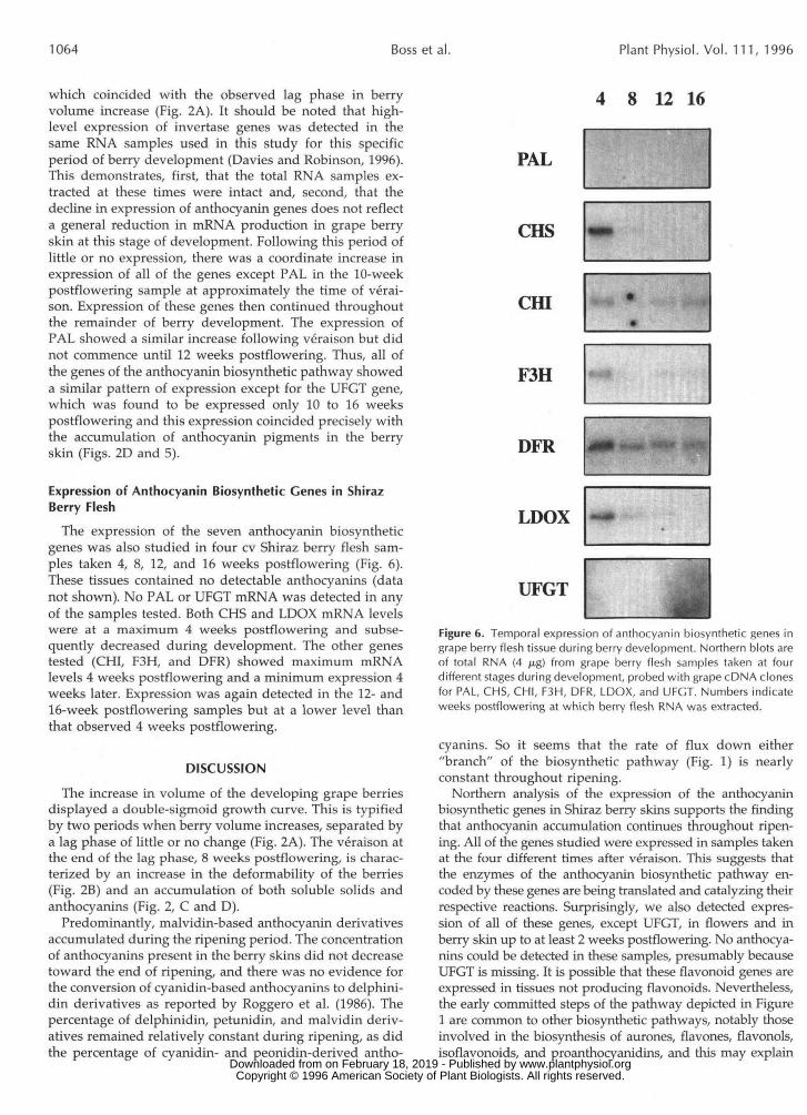
The expression of the seven anthocyanin biosyntheticgenes was also studied in four cv Shiraz berry flesh sam-ples taken 4, 8, 12, and 16 weeks postflowering (Fig. 6).These tissues contained no detectable anthocyanins (datanot shown). No PAL or UFGT mRNA was detected in anyof the samples tested. Both CHS and LDOX mRNA levelswere at a maximum 4 weeks postflowering and subse-quently decreased during development. The other genestested (CHI, F3H, and DFR) showed maximum mRNAlevels 4 weeks postflowering and a minimum expression 4weeks later. Expression was again detected in the 12- and16-week postflowering samples but at a lower level thanthat observed 4 weeks postflowering.
DISCUSSION
The increase in volume of the developing grape berriesdisplayed a double-sigmoid growth curve. This is typifiedby two periods when berry volume increases, separated bya lag phase of little or no change (Fig. 2A). The veraison atthe end of the lag phase, 8 weeks postflowering, is charac-terized by an increase in the deformability of the berries(Fig. 2B) and an accumulation of both soluble solids andanthocyanins (Fig. 2, C and D).
Predominantly, malvidin-based anthocyanin derivativesaccumulated during the ripening period. The concentrationof anthocyanins present in the berry skins did not decreasetoward the end of ripening, and there was no evidence forthe conversion of cyanidin-based anthocyanins to delphini-din derivatives as reported by Roggero et al. (1986). Thepercentage of delphinidin, petunidin, and malvidin deriv-atives remained relatively constant during ripening, as didthe percentage of cyanidin- and peonidin-derived antho-
4 8 12 16
PAL
CHS
Cffl
F3H
DFR
LDOX
UFGT
Figure 6. Temporal expression of anthocyanin biosynthetic genes ingrape berry flesh tissue during berry development. Northern blots areof total RNA (4 .̂g) from grape berry flesh samples taken at fourdifferent stages during development, probed with grape cDNA clonesfor PAL, CHS, CHI, F3H, DFR, LDOX, and UFGT. Numbers indicateweeks postflowering at which berry flesh RNA was extracted.
cyanins. So it seems that the rate of flux down either"branch" of the biosynthetic pathway (Fig. 1) is nearlyconstant throughout ripening.
Northern analysis of the expression of the anthocyaninbiosynthetic genes in Shiraz berry skins supports the findingthat anthocyanin accumulation continues throughout ripen-ing. All of the genes studied were expressed in samples takenat the four different times after veraison. This suggests thatthe enzymes of the anthocyanin biosynthetic pathway en-coded by these genes are being translated and catalyzing theirrespective reactions. Surprisingly, we also detected expres-sion of all of these genes, except UFGT, in flowers and inberry skin up to at least 2 weeks postflowering. No anthocya-nins could be detected in these samples, presumably becauseUFGT is missing. It is possible that these flavonoid genes areexpressed in tissues not producing flavonoids. Nevertheless,the early committed steps of the pathway depicted in Figure1 are common to other biosynthetic pathways, notably thoseinvolved in the biosynthesis of aurones, flavones, flavonols,isoflavonoids, and proanthocyanidins, and this may explain
www.plantphysiol.orgon February 18, 2019 - Published by Downloaded from Copyright © 1996 American Society of Plant Biologists. All rights reserved.
Anthocyanin Biosynthesis in Shiraz Grape Berries 1065
expression of these genes in the absence of anthocyanin syn- thesis in the flowers and young berries. In the floral tissues, it is likely that flavonols are being produced, since they are known to accumulate in the pistils of flowers (Koes et al., 1990) and are essential for pollen viability in maize (Mo et al., 1992). It is also possible that any flavonols produced could be protecting the developing tissues from UV damage (Schmel- zer et al., 1988). The young, developing berries could also be producing a number of flavonoid-derived compounds. For example, isoflavonoids (flavonoid derivatives) may play a role in protecting the young berries from various phytopatho- gens (Lamb et al., 1989) or as a feeding deterrent to insects (Caballero et al., 1986). Young, developing seeds (which may be present in the flower sample) may also accumulate leuco- anthocyanidins, as occurs in petunia (Koes et al., 1990). Ex- pression of the anthocyanin genes up to and including DFR would also be necessary for proanthocyanidin production, and these compounds were detected in young berries and flowers. Nevertheless, the observed expression of LDOX in these early samples is puzzling. LDOX is the putative leuco- anthocyanidin dioxygenase required for the first of two en- zymatic steps between leucoanthocyanidins and anthocyani- dins (Martin and Gerats, 1993), the other step probably being catalyzed by a putative dehydratase (Heller and Forkmann, 1988). Any intermediates between these reactions are pre- sumed to be unstable (Heller and Forkmann, 1988), and no colored anthocyanidins were detected.
In Shiraz grape berries anthocyanins accumulate in the skin but not in the flesh. The pattern of expression seen in the berry flesh samples was similar to that in the berry skin, except that neither PAL nor UFGT expression was de- tected, and CHS was not expressed late in development. Both PAL and CHS are encoded by multigene families in grapes (Sparvoli et al., 1994), and thus, other gene family members, not detected by northern analysis, may be ex- pressed in this tissue. Only one UFGT gene seemed to be present in the grape genome (Sparvoli et al., 1994), and this was not expressed in the berry flesh tissue. The 4- and 8-week postflowering samples possessed proanthocyani- dins, whereas the 12- and 16-week postflowering samples did not, and this may indicate that proanthocyanidin pro- duction in berry flesh is blocked by the lack of PAL and CHS gene expression in the 12- and 16-week postflowering samples.
Anthocyanins begin to accumulate at about véraison, and this coincides with the increase in expression of all seven genes tested from the anthocyanin biosynthetic path- way. This suggests that there is coordinate regulation of all of these genes at this time in the developing grape berry skin. Sparvoli et al. (1994) have also shown that, as antho- cyanins accumulate in dark-grown grape seedlings subse- quently exposed to light, there is a coordinate induction of the genes from the committed steps of the anthocyanin biosynthetic pathway (CHS, CHI, F3H, DFR, LDOX, and UFGT). This is similar to the control of the anthocyanin biosynthetic pathway in maize aleurone, which is regu- lated by the R and C1 gene families (Martin and Gerats, 1993). Nevertheless, the pattern of expression seen in the flower and young berry skin samples prior to véraison
suggests that UFGT expression is under a different regu- latory regime. In these samples, all of which did not pos- sess anthocyanins, UFGT was the only structural gene tested that was not induced.
The start points for the control of the anthocyanin pathway in the species most studied (maize, antirrhinum, and petunia) are different. In maize aleurone, the regulatory genes CZ and R(S) regulate CHS, DFR, and UFGT gene expression (Dooner and Nelson, 1977; Dooner, 1983; Cone et al., 1986; Ludwig et al., 1989). The F3H activity in the aleurone is also influenced by the R gene (Larson, 1989). Thus, it appears that R and CZ may regulate the transcription of all of the anthocyanin bio- synthetic genes in the aleurone (Martin and Gerats, 1993). There are severa1 homologs of R and CZ, and these regulate pigmentation in other maize tissues (Ludwig and Wessler, 1990). In snapdragon flowers, mutations in the regulatory gene delila have little effect on the expression of CHS and CHI (Almeida et al., 1989), but there is repression of F3H, DFR, LDOX (called candi), and UFGT expression in the flower tube (Martin et al., 1991). Studies of mutants of the regulatory genes eluta and of two alleles of the Rosea locus in snapdragon have shown similar results (Martin and Gerats, 1993). This led Martin and Gerats (1993) to suggest that the "key regulatory point" of the production of anthocyanin compounds in this species is the activity of F3H. There also appears to be a link between genes that are induced during anthocyanin biosyn- thesis and those under the control of myc- and myb-like reg- ulatory genes. For example, in snapdragon flowers, F3H, DFR, LDOX, and UFGT are induced in tissue-accumulating anthocyanins, whereas PAL, CHS, and CHI seem to be con- stitutively expressed (Jackson et al., 1992). The genes induced in these flowers are the same genes that are not expressed in snapdragon regulatory gene mutants (Martin et al., 1991). Quattrocchio et al. (1993) showed a similar link between developmental expression and regulatory gene mutants in petunia. The regulatory genes involved in anthocyanin bio- synthesis in petunia flowers have been traced to the loci known as anl, an2, and anl1 (Beld et al., 1989). Another locus, known as an4, controls anthocyanin biosynthesis in petunia anthers (Quattrocchio et al., 1993). The expression of CHS, CHI, and F3H genes in petunia mutated at the an loci is not affected. However, there is a reduction in the expression of DFR and UFGT (Beld et al., 1989; Quattrocduo et al., 1993). Thus, in the case of petunia, the major control point seems to be one step further down the pathway than in snapdragon (compare DFR and F3H).
The structural genes from the grape anthocyanin biosyn- thetic pathway may also be controlled by myc- and myb-like regulatory genes. However, the way in which the structural genes are regulated in grape berry skins appears to be differ- ent from the patterns observed in snapdragon, petunia, and maize. The pattern of gene expression in grape berry skins could be explained in relation to regulatory genes in two ways. First, two types of regulatory genes may be active in the berry skin, one of which is expressed early and that induces expression of a11 of the structural genes except UFGT, and another that is expressed later and results in the induction of expression of a11 of the structural genes. Alternatively, two types of regulatory genes may be present, one that controls
www.plantphysiol.orgon February 18, 2019 - Published by Downloaded from Copyright © 1996 American Society of Plant Biologists. All rights reserved.
1066 Boss et al. Plant Physiol. Vol. 11 1 , 1996
expression of PAL, CHS, CHI, F3H, DFR and LDOX and another that induces UFGT gene expression. In this case the regulatory gene that controls expression of PAL, CHS, CHI, F3H, DFR, and LDOX is expressed early in berry develop- ment, whereas both the regulatory genes are expressed as the grape ripens, resulting in induction of a11 of the genes and thus in anthocyanin biosynthesis. In either case, it appears that the major control point to anthocyanin biosynthesis in grape berry skins is UFGT, and this control is later in the pathway than has been observed i n the studies of maize, petunia, and snapdragon anthocyanin biosynthesis.
In summary, the appearance of anthocyanins in grape berry skins at the onset of ripening coincides with increased expression of each of the genes encoding biosynthetic en- zymes in t h s pathway. This suggests that the induction of anthocyanin synthesis is triggered by regulatory genes. The isolation of these ripening-specific regulatory genes in grape berries is currently being undertaken to understand more fully the nature of this regulation.
ACKNOWLEDCMENTS
The authors would like to thank Dr. C. Tonelli (Universita degli Studi de Milano, Italia) for the grape cDNA clones used in this study. We are also indebted to Di and John Harvey for allowing us to sample material from their vineyard, and we wish to thank Judith Osborne for excellent technical assistance. We are grateful to Brian Loveys and Sue Maffei for their assistance with the HPLC analyses, and we thank Bryan Coombe and Patrick Iland for helpful discussions and advice.
Received November 20, 1995; accepted April 20, 1996. Copyright Clearance Center: 00324889/96/111/1059/08.
LITERATURE ClTED
Almeida J, Carpenter R, Robbins TP, Martin C, Coen ES (1989) Genetic interactions underlying flower colour patterns in Anti- rrhinum mujus. Genes Dev 3: 1758-1767
Beld MGHM, Martin C, Huits H, Stuitje AR, Gerats AGM (1989) Flavonoid synthesis in Petuniu: partia1 characterisation of dihy- droflavonol 4-reductase genes. Plant Mo1 Biol 13: 491-502
Caballero P, Smith CM, Fronczek FR, Fischer NH (1986) Isofla- vones from an insect-resistant variety of soybean and the molecular structure of afrormosin. J Nat Prod 49: 1126-1129
Cone KC, Burr FA, Burr B (1986) Molecular analysis of the maize regulatory locus CZ. Proc Natl Acad Sci USA 83: 9631-9635
Coombe BG, Bishop GR (1980) Development of the grape berry. 11. Changes in the diameter and deformability during véraison. Aust J Agric Res 31: 499-509
Cravero MC, Guidoni S, Schneider A, Di Stefano R (1994) Mor- phological and biochemical characterisation of coloured berry- muscat grapevine cultivars. Vitis 33: 75-80
Davies C, Robinson SP (1996) Sugar accumulation in grape berries: cloning of two putative vacuolar invertase cDNAs and their expression in grapevine tissues. Plant Physiol 111:
Dooner HK (1983) Coordinate genetic regulation of flavonoid biosynthetic enzymes in maize. Mo1 Gen Genet 189: 136-141
Dooner HK, Nelson OE (1977) Genetic control of UDP-glucose: flavonol 3-o-glucosyl transferase in the endosperm of maize. Biochem Genet 15: 501-519
Fong RA, Kepner RE, Webb AD (1971) Acetic-acid-acylated an- thocyanin pigments in the grape skins of a number of varieties of Vitis vinifera. Am J Enol Vitic 22: 150-155
González-SanJosC ML, Santa-Maria G, Díez C (1990) Anthocya- nins as parameters for differentiating wines by grape variety,
275-283
wine-growing region, and wine making methods. J Food Com- pos Ana1 3: 54-66
Goodrich J, Carpenter R, Coen ES (1992) A common gene regu- lates pigmentation pattern in diverse plant species. Cell 68: 955-964
Harborne JB (1989) Phenolic compounds. Methods Plant Biochem 1: 37-99
Heller W, Forkmann G (1988) Biosynthesis. In JB Harborne, ed, The Flavonoids: Advances in Research since 1980. Chapman and Hall, London, pp 399425
Hrazdina G, Parsons GF, Mattick LR (1984) Physiological and biochemical events during development and maturation of grape berries. Am J Enol Vitic 35: 220-227
Jackson D, Roberts K, Martin C (1992) Temporal and spatial control of expression of anthocyanin biosynthetic genes in de- veloping flowers of Antirrhinum mujus. Plant J 2: 425434
Koes RE, van Blokland R, Quattrocchio F, van Tunen AJ, Mo1 JNM (1990) Chalcone synthase promoters in Petuniu are active in pig- mented and unpigmented cell types. Plant Cell 2: 379-392
Lamb CJ, Lawton MA, Dron M, Dixon RA (1989) Signals and transduction mechanisms for activation of plant defenses against microbial attack. Cell 56: 215-224
Larson RL (1989) Genetics, precursors and enzymes in flavonoid biosynthesis in maize. In ED Styles, GA Gavazzi, ML Racchi, eds, The Genetics of Flavonoids. Edizioni Unicopli, Milan, Italy,
Ludwig SR, Habera LF, Dellaporta SL, Wessler SR (1989) Lc, a member of the maize R gene family responsible for tissue spe- cific anthocyanin production, encodes a protein similar to tran- scription activators and contains the myc-homology region. Proc Natl Acad Sci USA 86: 7092-7096
Ludwig SR, Wessler SR (1990) Maize R gene family: tissue-spe- cific helix-loop-helix proteins. Cell 62: 849-851
Martin C, Gerats T (1993) The control of flower coloration. In BR Jordan, ed, The Molecular Biology of Flowering. CAB Interna- tional, Wallingford, CT, pp 219-255
Martin C, Prescott A, Mackay S, Bartlett J, Vrijlandt E (1991) The control of anthocyanin biosynthesis in flowers of Antirrhinum mujus. Plant J 1: 37-49
Mazza G, Miniati E (1993) Anthocyanins in Fruits, Vegetables, and Grains. CRC Press, Boca Raton, FL
Mo Y, Nagel C, Taylor LP (1992) Biochemical complementation of chalcone synthase mutants defines a role for flavonols in func- tional pollen. Proc Natl Acad Sci USA 89: 7213-7217
Paz-Ares J, Ghosal D, Wienand U, Peterson PA, Saedler H (1987) The regulatory CI locus of Zeu muys encodes a protein with homol- ogy to myb proto-oncogene products and with structural similari- ties to transcriptional activators. EMBO J 6 3553-3558
Quattrocchio F, Wing JF, Leppen HTC, Mo1 JNM, Koes RE (1993) Regulatory genes controlling anthocyanin pigmentation are functionally conserved among plant species and have distinct sets of target genes. Plant Cell 5: 1497-1512
Rezaian MA, Krake LR (1987) Nucleic acid extraction and virus detection in grapevine. J Viro1 Methods 17: 277-285
Roggero JP, Coen S, Ragonnet B (1986) High performance liquid chromatography survey on changes in pigment content in rip- ening grapes of Syrah. An approach to anthocyanin metabolism. Am J Enol Vitic 37: 77-83
Roubelakis-Angelakis KA, Kliewer WM (1986) Effects of exoge- nous factors on phenylalanine ammonia-lyase activity and ac- cumulation of anthocyanins and total phenolics in grape berries. Am J Enol Vitic 37: 275-280
Schmelzer E, Jahnen W, Hahlbrock K (1988) In situ localization of light-induced chalcone synthase mRNA, chalcone synthase, and flavonoid end products in epidermal cells of parsley leaves. Proc Natl Acad Sci USA 85: 2989-2993
Sparvoli F, Martin C, Scienza A, Gavazzi G, Tonelli C (1994) Cloning and molecular analysis of structural genes involved in flavonoid and stilbene biosynthesis in grape (Vitis vinifera L.). Plant Mo1 Biol 24: 743-755
Wulf LW, Nagel CW (1978) High-pressure liquid chromato- graphic separation of anthocyanins of Vitis vinifera. Am J Enol Vitic 29: 42-49
pp 71-78
www.plantphysiol.orgon February 18, 2019 - Published by Downloaded from Copyright © 1996 American Society of Plant Biologists. All rights reserved.