an update on acanthamoeba keratitis: diagnosis, pathogenesis and
TRANSCRIPT
An update on Acanthamoeba keratitis: diagnosis, pathogenesisand treatment
Jacob Lorenzo-Morales1,a,*, Naveed A. Khan2,a, and Julia Walochnik3,a
1 University Institute of Tropical Diseases and Public Health of the Canary Islands, University of La Laguna, Avda. Astrofísico Fco.Sánchez, S/N, 38203 La Laguna, Tenerife, Canary Islands, Spain
2 Department of Biological and Biomedical Sciences, Aga Khan University, Karachi, Pakistan3 Institute of Specific Prophylaxis and Tropical Medicine, Medical University of Vienna, Vienna, Austria
Received 30 October 2014, Accepted 9 February 2015, Published online 18 February 2015
Abstract – Free-living amoebae of the genus Acanthamoeba are causal agents of a severe sight-threatening infectionof the cornea known as Acanthamoeba keratitis. Moreover, the number of reported cases worldwide is increasing yearafter year, mostly in contact lens wearers, although cases have also been reported in non-contact lens wearers. Inter-estingly, Acanthamoeba keratitis has remained significant, despite our advances in antimicrobial chemotherapy andsupportive care. In part, this is due to an incomplete understanding of the pathogenesis and pathophysiology of thedisease, diagnostic delays and problems associated with chemotherapeutic interventions. In view of the devastatingnature of this disease, here we present our current understanding of Acanthamoeba keratitis and molecular mecha-nisms associated with the disease, as well as virulence traits of Acanthamoeba that may be potential targets forimproved diagnosis, therapeutic interventions and/or for the development of preventative measures. Novel molecularapproaches such as proteomics, RNAi and a consensus in the diagnostic approaches for a suspected case of Acantha-moeba keratitis are proposed and reviewed based on data which have been compiled after years of working on thisamoebic organism using many different techniques and listening to many experts in this field at conferences,workshops and international meetings. Altogether, this review may serve as the milestone for developing an effectivesolution for the prevention, control and treatment of Acanthamoeba infections.
Key words: Acanthamoeba, keratitis, diagnosis, therapy, pathogenesis.
Résumé – Mise au point sur la kératite à Acanthamoeba : diagnostic, pathogenèse et traitement. Les amibes àvie libre du genre Acanthamoeba sont les agents causant une infection sévère de la cornée, dangereuse pour la vue,appelée kératite à Acanthamoeba. De plus, le nombre de cas signalés à travers le monde est en augmentation annéeaprès année, principalement chez les porteurs de lentilles de contact, bien que des cas de kératite à Acanthamoebaaient également été signalés chez les non-porteurs de lentilles. Fait intéressant, la kératite à Acanthamoeba estrestée significative, en dépit de nos progrès dans la chimiothérapie antimicrobienne et les soins de soutien.En partie, cela est dû à une compréhension incomplète de la pathogenèse et la physiopathologie de la maladie,aux retards du diagnostic et aux problèmes associés aux interventions chimiothérapeutiques. Compte tenu de lanature dévastatrice de cette maladie, nous présentons ici notre compréhension actuelle de la kératite àAcanthamoeba et des mécanismes moléculaires associés à la maladie, ainsi que les traits de virulence deAcanthamoeba qui peuvent être des cibles potentielles pour l’amélioration du diagnostic, les interventionsthérapeutiques et/ou pour l’élaboration de mesures préventives. Des approches moléculaires comme laprotéomique, l’ARNi et des approches consensuelles de diagnostic pour un cas suspecté de kératite àAcanthamoeba sont proposées et examinées sur la base des données qui ont été compilées après des années detravail sur cet organisme amibien, utilisant de nombreuses techniques différentes et l’écoute de nombreux expertssur ce domaine à des conférences, ateliers et réunions internationales. Au total, cette étude peut servir de jalonpour développer une solution efficace pour la prévention, le contrôle et le traitement des infections à Acanthamoeba.
a All authors contributed equally to this manuscript.*Corresponding author: [email protected]
Parasite 2015, 22, 10� J. Lorenzo-Morales et al., published by EDP Sciences, 2015DOI: 10.1051/parasite/2015010
Available online at:www.parasite-journal.org
This is an Open Access article distributed under the terms of the Creative Commons Attribution License (http://creativecommons.org/licenses/by/4.0),which permits unrestricted use, distribution, and reproduction in any medium, provided the original work is properly cited.
OPEN ACCESSREVIEW ARTICLE
1. Introduction – What is Acanthamoebakeratitis?
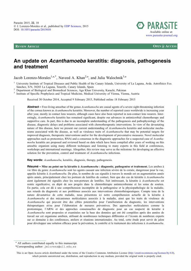
Acanthamoeba species are the causative agents of a sight-threatening infection of the cornea known as Acanthamoebakeratitis (AK) (Fig. 1). Interestingly, AK is increasingly beingrecognized as a severe sight-threatening ocular infection,worldwide. Although contact lens (CL) wear is the leading riskfactor for AK, Acanthamoeba spp. can cause infection in non-contact lens wearers. Patients with AK may experience painwith photophobia, ring-like stromal infiltrate, epithelial defectand lid oedema. If AK is not treated adequately and aggres-sively, it can lead to loss of vision [18, 46, 47, 56, 87, 111,112, 117].
Diagnosis of AK is challenging, and the available treat-ments are lengthy and not fully effective against all strains.Moreover, the pathogenesis of Acanthamoeba keratitis is stillunder study, and the identification of the key factors involvedin this process should be useful for the development of fullyeffective therapies. The current difficulty in effective treatmentis due to the resistant cyst stage of Acanthamoeba. Togetherwith common misdiagnosis of AK in most cases and a lackof a consensus for AK diagnosis, AK has remained significant.Nevertheless, AK is still considered a rare disease and isincluded in the Orphanet database (ORPHA67043) and withan estimated prevalence of 1–9/100,000.
2. Diagnostics of AK
The most important step in AK diagnosis is to think of it.Generally, AK should be considered in all contact lens wearersand in any case of corneal trauma with exposure to soil or con-taminated water [20, 23, 36, 54]. Common symptoms are mas-sive pain, photophobia and tearing. The sooner the disease isdiagnosed, the better the outcome [6, 12, 54, 99, 104]. If diag-nosis is delayed, the amoebae have already penetrated deeplyinto the corneal stroma and successful therapy becomesexceedingly difficult. AK is usually unilateral and progressesslowly, from epithelial to stromal disease. At the beginningof the infection, a diffuse superficial keratopathy is found, latermultifocal infiltrates are almost always observed in the stroma.Acanthamoeba sclerokeratitis is an uncommon complication ofAK and assumedly has an immune-mediated origin. Tu et al.[104] established five levels of AK severity based on slit-lampbiomicroscopy findings: epitheliitis, epitheliitis with radialneuritis, anterior stromal disease, deep stromal keratitis, or ringinfiltrate. The characteristic ring infiltrate is, however, onlyseen in approximately 50% of patients. In the early stage,AK can easily be confused with Herpes simplex keratitis, whilein the advanced stage, the infection resembles the clinical pic-ture of a fungal keratitis or a corneal ulcer (Table 1).
Contact lens wearers typically seek medical help late,because they are used to minor irritations in the eye.
The tentative diagnosis of AK can often be made by in vivoconfocal microscopy (IVCM). The Acanthamoeba cystsappearing as hyper-reflective, spherical structures are usuallywell defined because of their double wall; the trophozoitesare difficult to distinguish from leukocytes and keratocyte
nuclei [110]. However, the direct detection of the causativeagent in a corneal scrape specimen is the only reliable diagnos-tic method for AK. Culture remains the gold standard of Acan-thamoeba laboratory diagnosis, but today several PCR-basedtechniques are also well established and usually increase sensi-tivity significantly [41, 59, 84, 90]. In cases of severe infection,amoeba density is sometimes very high and the amoebae canalready be detected by direct microscopy of the clinical sam-ple, without enrichment. Acanthamoeba trophozoites or cystsare readily recognizable in phase contrast microscopy, but alsostain well in several stains and cysts exhibit auto-fluorescence[46, 54]. However, particularly if patients have already beenpre-treated with antibiotics, amoeba density is usually verylow. Moreover, amoebae exhibit altered morphologies – inthese cases, even culture often remains negative and moleculartechniques are indispensable. Reliable identification below thegenus level requires genotyping. Serological techniques are ofno diagnostic value as specific antibodies are also detected inapparently healthy people due to the ubiquity ofAcanthamoeba.
In contrast to infections with other amoebae, acanthamoe-bae can form cysts within the tissue. As a single cyst surviving
Figure 1. (A) Corneal melting and vascularization in a patient withAcanthamoeba keratitis. (B) Observed corneal damage in AK isshown after sodium fluorescein application. Original.
2 J. Lorenzo-Morales et al.: Parasite 2015, 22, 10
in the cornea can lead to reinfection, the progress of therapyshould be checked regularly. An ongoing infection should bemonitored every 1–2 weeks. After clinical recovery, monthlychecks are sufficient, ideally until 6 months after decline ofsymptoms.
In most countries, the vast majority of AK cases occur inCL wearers and AK can be prevented extensively by strict con-tact lens hygiene. Typically, singular amoebae gain access tothe lens case via tap water or the air, rapidly grow to high den-sities within the lens case if this is not cleaned properly andregularly, and then attach to the lenses and infect the eye[116]. Wearers of soft contact lenses using multipurpose solu-tions are at particular risk, because acanthamoebae adhereespecially well to the hydrophilic plastic of these lenses, andsoft lenses are more difficult to clean than rigid lenses. More-over, soft lenses are often over worn (dailies used for severaldays, monthlies used for several months) and are also the typeof lenses used by people who do not regularly but only occa-sionally (e.g. once a week for sports) wear their contacts,and who are often unaware of proper contact lens hygiene.For prophylaxis of AK, lens cases should be cleaned manuallyand air dried, contact lenses should be cleaned and stored usingan appropriate (best: two-step) contact lens cleaning system,and both, lenses and lens cases have to be exchanged regularly.
2.1. Material
For confirmation of an AK, sampling and investigation ofthe correct material is crucial. Only if amoebae are detectedin corneal scrapings or in corneal biopsies a reliable diagnosiscan be made. Acanthamoebae penetrate the cornea and areusually not found on the corneal surface, thus superficial swabsamples or tear samples often remain negative, particularly inthe advanced stage of the disease and/or if patients havealready been pretreated with antibiotics. On the contrary, con-tact lens containers, even those of entirely healthy CL wearers,are almost always positive for acanthamoebae, at least in PCR.This means that the detection of Acanthamoeba spp. in the CLcase does not necessarily indicate an AK. When the CL case isnegative, however, it is very unlikely that the patient has anAK, unless, of course, the CL case was recently changed.
The optimal material for AK diagnosis is a cornealscraping/biopsy stored in 200 lL of sterile saline (amoebasaline* or PBS or 0.9% NaCl) in order to preventdesiccation.
*See Table 2.
2.2. Sample preparation
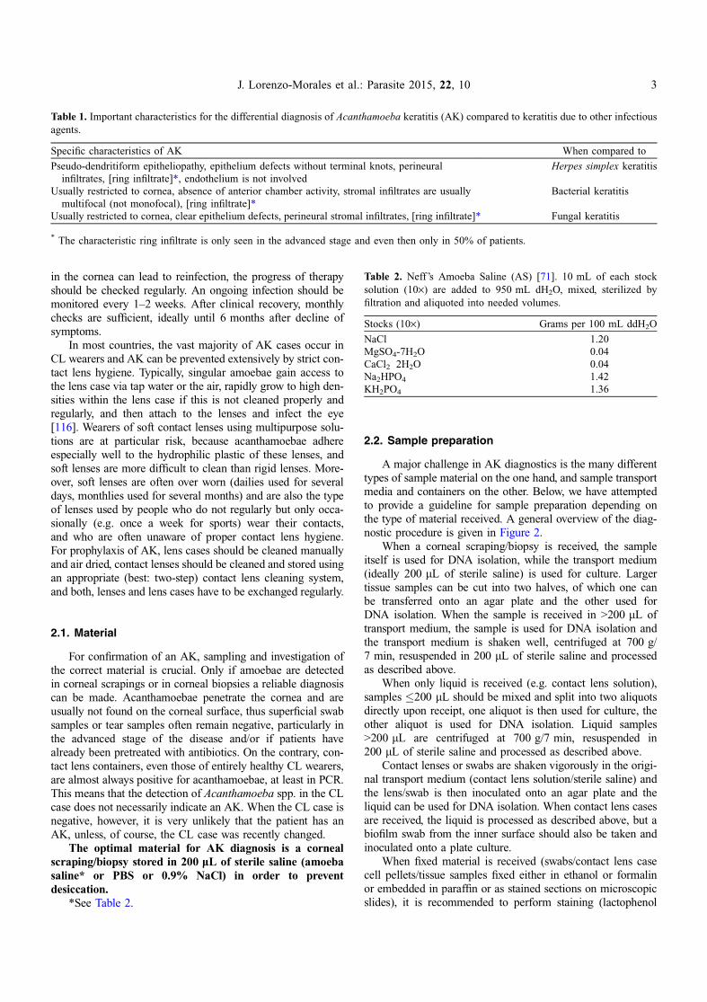
A major challenge in AK diagnostics is the many differenttypes of sample material on the one hand, and sample transportmedia and containers on the other. Below, we have attemptedto provide a guideline for sample preparation depending onthe type of material received. A general overview of the diag-nostic procedure is given in Figure 2.
When a corneal scraping/biopsy is received, the sampleitself is used for DNA isolation, while the transport medium(ideally 200 lL of sterile saline) is used for culture. Largertissue samples can be cut into two halves, of which one canbe transferred onto an agar plate and the other used forDNA isolation. When the sample is received in >200 lL oftransport medium, the sample is used for DNA isolation andthe transport medium is shaken well, centrifuged at 700 g/7 min, resuspended in 200 lL of sterile saline and processedas described above.
When only liquid is received (e.g. contact lens solution),samples �200 lL should be mixed and split into two aliquotsdirectly upon receipt, one aliquot is then used for culture, theother aliquot is used for DNA isolation. Liquid samples>200 lL are centrifuged at 700 g/7 min, resuspended in200 lL of sterile saline and processed as described above.
Contact lenses or swabs are shaken vigorously in the origi-nal transport medium (contact lens solution/sterile saline) andthe lens/swab is then inoculated onto an agar plate and theliquid can be used for DNA isolation. When contact lens casesare received, the liquid is processed as described above, but abiofilm swab from the inner surface should also be taken andinoculated onto a plate culture.
When fixed material is received (swabs/contact lens casecell pellets/tissue samples fixed either in ethanol or formalinor embedded in paraffin or as stained sections on microscopicslides), it is recommended to perform staining (lactophenol
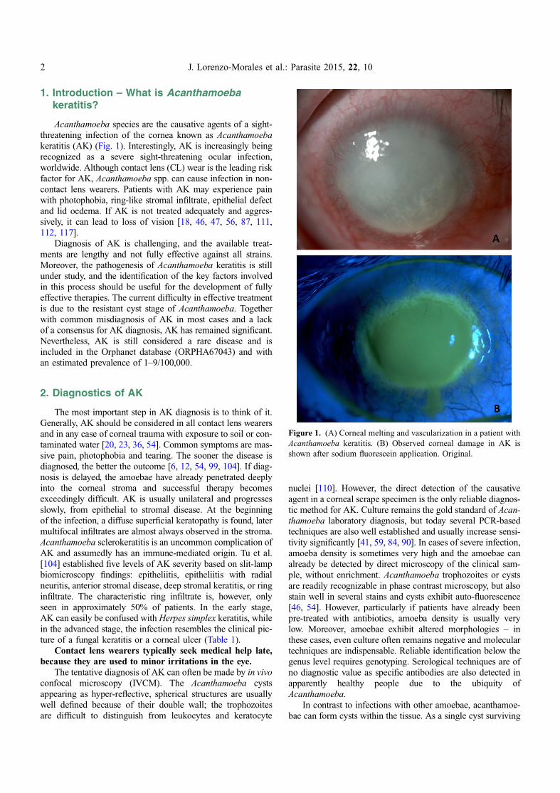
Table 1. Important characteristics for the differential diagnosis of Acanthamoeba keratitis (AK) compared to keratitis due to other infectiousagents.
Specific characteristics of AK When compared to
Pseudo-dendritiform epitheliopathy, epithelium defects without terminal knots, perineuralinfiltrates, [ring infiltrate]*, endothelium is not involved
Herpes simplex keratitis
Usually restricted to cornea, absence of anterior chamber activity, stromal infiltrates are usuallymultifocal (not monofocal), [ring infiltrate]*
Bacterial keratitis
Usually restricted to cornea, clear epithelium defects, perineural stromal infiltrates, [ring infiltrate]* Fungal keratitis
* The characteristic ring infiltrate is only seen in the advanced stage and even then only in 50% of patients.
Table 2. Neff’s Amoeba Saline (AS) [71]. 10 mL of each stocksolution (10·) are added to 950 mL dH2O, mixed, sterilized byfiltration and aliquoted into needed volumes.
Stocks (10·) Grams per 100 mL ddH2O
NaCl 1.20MgSO4-7H2O 0.04CaCl2 Æ 2H2O 0.04Na2HPO4 1.42KH2PO4 1.36
J. Lorenzo-Morales et al.: Parasite 2015, 22, 10 3
cotton blue and/or immunostaining) and/or PCR. However, it isimportant to isolate the DNA using a suitable protocol for therespective material and to adapt the PCR protocol for frag-mented DNA, particularly when the material is formalin-fixed(i.e. amplicons should not exceed 300 bp in length).
2.3. Direct microscopy
In severe infections or when highly contaminated contactlens cases are investigated, the amoebae can usually alreadybe detected by direct microscopy (200·–400· magnification)of the original sample. For microscopic investigation ofamoebae, phase contrast or interference contrast are particu-
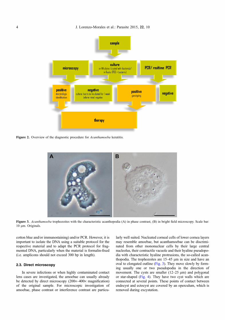
larly well suited. Nucleated corneal cells of lower cornea layersmay resemble amoebae, but acanthamoebae can be discrimi-nated from other mononuclear cells by their large centralnucleolus, their contractile vacuole and their hyaline pseudopo-dia with characteristic hyaline protrusions, the so-called acan-thopodia. The trophozoites are 15–45 lm in size and have anoval to elongated outline (Fig. 3). They move slowly by form-ing usually one or two pseudopodia in the direction ofmovement. The cysts are smaller (12–25 lm) and polygonalor star-shaped (Fig. 4). They have two cyst walls which areconnected at several points. These points of contact betweenendocyst and ectocyst are covered by an operculum, which isremoved during excystation.
Figure 2. Overview of the diagnostic procedure for Acanthamoeba keratitis.
Figure 3. Acanthamoeba trophozoites with the characteristic acanthopodia (A) in phase contrast, (B) in bright field microscopy. Scale bar:10 lm. Originals.
4 J. Lorenzo-Morales et al.: Parasite 2015, 22, 10
2.4. Stains
Stains are practical for the detection of cysts in freshclinical material or in pelleted lens-case solution and for theinvestigation of tissue sections. Fast and easy stains are lac-tophenol-cotton blue or Giemsa, but also calcofluor whiteand acridine orange usually give very good results. If morpho-logical details are to be studied, it is recommended to use asilver stain, which is particularly well-suited for the investiga-tion of the cysts. However, amoebae have to be cultured priorto staining. A general problem is that other cells, particularlyfungi also stain well in these stains. As a specific staining,immunostaining using anti-Acanthamoeba antibodies is rec-ommended which is also the stain of choice for tissue sections.Alternatively, tissue section can be stained with haematoxylin& eosin (HE) [35].
2.4.1. Lactophenol-cotton blue (LPCB)
The material is mixed with an adequate volume of LPCBstain (20 g phenol crystals, 20 mL lactic acid, 40 mL glycerol,0.05 g cotton-blue and 20 mL dH2O; or ready-mixed availablethrough e.g. Sigma-Aldrich) and investigated by light micros-copy [101]. This stain is particularly well suited for Acantha-moeba cysts; the cyst walls and the nucleus appear in anintensive blue, while the cytoplasm stains light-blue.
2.4.2. Acridine orange
Samples are fixed in 95% methanol for 2 min onto a glassslide, air dried, covered with acridine orange staining solution( pH 4) for 2 min, rinsed with H2O and air dried. Cysts appearbright orange and are easily discernible in fluorescencemicroscopy.
2.4.3. Calcofluor white
Samples are transferred to a glass slide, air dried and fixedfor 3 min with methanol. Subsequently, the sample is rinsed inPBS and stained using 2–3 drops of calcofluor white solution(0.1%, e.g. Sigma-Aldrich or Thermo Scientific). After 5 min,
the slide is rinsed with PBS and counter-stained with Evan’sblue (0.05%, e.g. Sigma-Aldrich or Thermo Scientific) for sev-eral seconds. It is important to use an embedding solution with-out auto-fluorescence. Slides are investigated by fluorescencemicroscopy (300–440 nm). Acanthamoeba cysts appear in alight green because the calcofluor white binds to the cellulosein the cyst walls. Evan’s blue diminishes the background fluo-rescence making the trophozoites appear reddish-brown.
2.4.4. Silver
The cysts are harvested from a culture plate/flask, sus-pended in 2 mL amoeba saline (Table 2) and washed threetimes in amoeba saline by centrifugation (500 g/10 min).The sample is fixed for 20 min in 2% formalin and washedin amoeba saline, the supernatant is removed and the pelletis transferred to a glass slide using an inoculating loop andmixed with Mayer’s albumin (glycerine-albumin 1:1, e.g.Hardy Diagnostics). Then, the cysts are fixed onto the slideusing Clarke’s fixative (95% alcohol-acetic acid 9:1) for 2 h.The fixative is removed using dH2O and the slides are incu-bated in 0.5% silver-protein solution in a water bath at60 �C. After 2 h, the slides are transferred to the reducingagent (1% hydroquinone in 5% Na2SO3) and incubated duringgentle shaking for several seconds up to 5 min. The slides arewashed in dH2O, dehydrated in an alcohol series, cleared withxylene, mounted and investigated by bright field microscopy.
2.4.5. Immunostain
To the best of our knowledge, no commercial kit is avail-able, but antisera against the three Acanthamoeba groups (I–III), produced by immunization of a rabbit with Acanthamoebawhole-cell antigen, are available in many laboratories (includ-ing our own) and can be obtained upon request.
2.4.6. Haematoxylin & eosin (HE)
The tissue section is fixed in 10% neutral buffered formalinsolution (e.g. Sigma-Aldrich). Serial sections of 6 lm are pro-
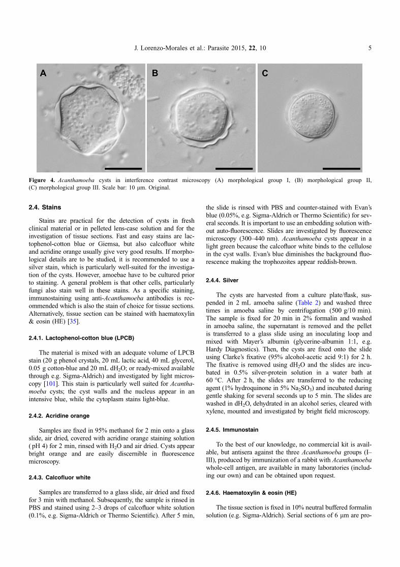
Figure 4. Acanthamoeba cysts in interference contrast microscopy (A) morphological group I, (B) morphological group II,(C) morphological group III. Scale bar: 10 lm. Original.
J. Lorenzo-Morales et al.: Parasite 2015, 22, 10 5
duced, deparaffinized for 1–2 min in xylene, dehydrated inalcohol and washed with dH2O. Subsequently, the sample isstained with haematoxylin and eosin, washed, covered with acover slip and investigated by bright field microscopy.
2.5. Culture
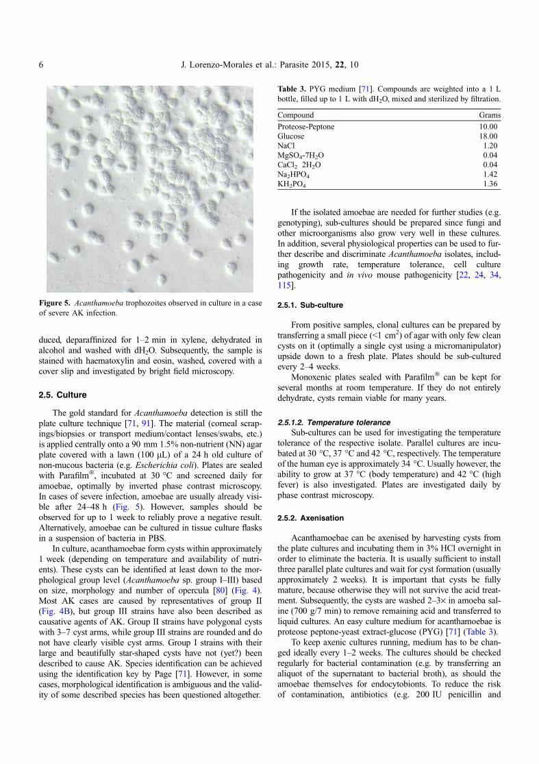
The gold standard for Acanthamoeba detection is still theplate culture technique [71, 91]. The material (corneal scrap-ings/biopsies or transport medium/contact lenses/swabs, etc.)is applied centrally onto a 90 mm 1.5% non-nutrient (NN) agarplate covered with a lawn (100 lL) of a 24 h old culture ofnon-mucous bacteria (e.g. Escherichia coli). Plates are sealedwith Parafilm�, incubated at 30 �C and screened daily foramoebae, optimally by inverted phase contrast microscopy.In cases of severe infection, amoebae are usually already visi-ble after 24–48 h (Fig. 5). However, samples should beobserved for up to 1 week to reliably prove a negative result.Alternatively, amoebae can be cultured in tissue culture flasksin a suspension of bacteria in PBS.
In culture, acanthamoebae form cysts within approximately1 week (depending on temperature and availability of nutri-ents). These cysts can be identified at least down to the mor-phological group level (Acanthamoeba sp. group I–III) basedon size, morphology and number of opercula [80] (Fig. 4).Most AK cases are caused by representatives of group II(Fig. 4B), but group III strains have also been described ascausative agents of AK. Group II strains have polygonal cystswith 3–7 cyst arms, while group III strains are rounded and donot have clearly visible cyst arms. Group I strains with theirlarge and beautifully star-shaped cysts have not (yet?) beendescribed to cause AK. Species identification can be achievedusing the identification key by Page [71]. However, in somecases, morphological identification is ambiguous and the valid-ity of some described species has been questioned altogether.
If the isolated amoebae are needed for further studies (e.g.genotyping), sub-cultures should be prepared since fungi andother microorganisms also grow very well in these cultures.In addition, several physiological properties can be used to fur-ther describe and discriminate Acanthamoeba isolates, includ-ing growth rate, temperature tolerance, cell culturepathogenicity and in vivo mouse pathogenicity [22, 24, 34,115].
2.5.1. Sub-culture
From positive samples, clonal cultures can be prepared bytransferring a small piece (<1 cm2) of agar with only few cleancysts on it (optimally a single cyst using a micromanipulator)upside down to a fresh plate. Plates should be sub-culturedevery 2–4 weeks.
Monoxenic plates sealed with Parafilm� can be kept forseveral months at room temperature. If they do not entirelydehydrate, cysts remain viable for many years.
2.5.1.2. Temperature toleranceSub-cultures can be used for investigating the temperature
tolerance of the respective isolate. Parallel cultures are incu-bated at 30 �C, 37 �C and 42 �C, respectively. The temperatureof the human eye is approximately 34 �C. Usually however, theability to grow at 37 �C (body temperature) and 42 �C (highfever) is also investigated. Plates are investigated daily byphase contrast microscopy.
2.5.2. Axenisation
Acanthamoebae can be axenised by harvesting cysts fromthe plate cultures and incubating them in 3% HCl overnight inorder to eliminate the bacteria. It is usually sufficient to installthree parallel plate cultures and wait for cyst formation (usuallyapproximately 2 weeks). It is important that cysts be fullymature, because otherwise they will not survive the acid treat-ment. Subsequently, the cysts are washed 2–3· in amoeba sal-ine (700 g/7 min) to remove remaining acid and transferred toliquid cultures. An easy culture medium for acanthamoebae isproteose peptone-yeast extract-glucose (PYG) [71] (Table 3).
To keep axenic cultures running, medium has to be chan-ged ideally every 1–2 weeks. The cultures should be checkedregularly for bacterial contamination (e.g. by transferring analiquot of the supernatant to bacterial broth), as should theamoebae themselves for endocytobionts. To reduce the riskof contamination, antibiotics (e.g. 200 IU penicillin and
Figure 5. Acanthamoeba trophozoites observed in culture in a caseof severe AK infection.
Table 3. PYG medium [71]. Compounds are weighted into a 1 Lbottle, filled up to 1 L with dH2O, mixed and sterilized by filtration.
Compound Grams
Proteose-Peptone 10.00Glucose 18.00NaCl 1.20MgSO4-7H2O 0.04CaCl2 Æ 2H2O 0.04Na2HPO4 1.42KH2PO4 1.36
6 J. Lorenzo-Morales et al.: Parasite 2015, 22, 10
200 lg/mL streptomycin) and/or antimycotics (e.g. amphoter-icin B) can be added to the culture medium.
Liquid culture is not suited to initial clinical samples, asbacteria and fungi would overgrow the cultures (a clinicalsample from the eye surface is never sterile).
2.5.3. Cell culture pathogenicity
Trophozoites are harvested from axenic cultures by centri-fugation (700 g/7 min.) and transferred onto a monolayer ofhuman (e.g. HeLA, HEp-2 or keratinocytes) or animal cells(e.g. VERO) in an amoeba/cell ratio of 1:10. The amoebaeare designated as highly cytopathic, when the monolayer iscompletely lysed within 24 h.
2.5.4. Mouse inoculation
Trophozoites are harvested from axenic cultures by centri-fugation (700 g/7 min.), re-suspended in sterile PBS and inoc-ulated into mice intra-nasally or intra-cerebrally. Young miceare generally more susceptible to an Acanthamoeba infection.Pathogenic strains lead to death within a few days up to4 weeks. Importantly, amoebae can lose their pathogenicityduring long-term axenic laboratory culture.
2.6. DNA isolation
For genotyping, actively growing amoebae (~106 cells) areharvested from culture plates and resuspended in 100 lL ofsterile 0.9% NaCl for DNA isolation. Whole-cell DNA canbe isolated from the amoebal suspensions using a commercialDNA isolation kit following the manufacturer’s protocol for therespective type of material. When larger tissue samples arereceived, we recommend homogenization of the material priorto DNA isolation.
2.7. PCR/real-time PCR
The most frequently used PCR for Acanthamoeba diagnos-tics is probably the one established by Schroeder et al. [90, 97]amplifying a fragment of the 18S rRNA gene using theJDP1 (50-GGCCCAGATCGTTTACCGTGAA-30) and JDP2(50-TCTCACAAGCTGCTAGGGAGTCA-30) primers. In thisPCR, the length of the amplicon varies between 423 and551 bp depending on the genotype, and DNA sequencing ofthe amplicon allows for genotyping in most cases. Generally,whichever diagnostic PCR is used, it should be run with at leasttwo different dilutions from each sample (as the proportionamoebal DNA: human DNA can vary greatly) and a genotypeT4 reference strain should be used as a positive and DNA-freewater as a negative control. Amplicons are visualized by aga-rose-gel electrophoresis and, if genotyping is required, therespective bands are extracted from the gel, purified andsubjected to DNA sequencing.
For samples that had been fixed in formaldehyde,we employ a modified PCR using the JDP1 primerfrom the PCR described above and the P2r primer
(50-GACTACGACGGTATCTGATC-30) [113], which amplifiesa shorter (~300 bp) fragment of the 18S rRNA gene.
In the past years, several protocols for real-time PCR havealso been published [43, 61, 81, 84]. A highly sensitive andspecific assay is the multiplex real-time PCR established byQvarnstrom et al. [81], which for AK diagnostics can also berun as a singleplex.
2.8. Genotyping
Sequences of the PCR amplicons can be obtained by directsequencing or by cloning. Generally, it is recommended toobtain sequences from both strands and assemble them to givea consensus sequence. For genotyping, obtained sequences arecompared to sequences of Acanthamoeba reference strains bymultiple alignments with all available genotypes at that time(currently 19) with the model assumption of a <5% sequencedissimilarity within one genotype as established by Gastet al. [33] and Stothard et al. [97]. Worldwide, the vast majorityof AK cases are caused by Acanthamoeba genotype T4, butgenotypes T3 and T11 are also commonly associated withAK, and in fact most genotypes known to date have at leastonce been involved in an AK case [11, 116].
3. Pathogenesis of Acanthamoeba keratitis
The devastating nature of Acanthamoeba keratitis and theproblems associated with its diagnosis and successful therapysuggest a need for complete understanding of the pathogenesisand pathophysiology to find alternative therapeutic interven-tions. Another major concern during the course of therapy isthe ability of Acanthamoeba to transform into dormant cystforms, which may resist recommended levels of antimicrobialchemotherapy. The ability of Acanthamoeba to produce infec-tion requires specific adhesins, production of toxins, and itsability to resist immune/environmental factors and chemother-apeutic agents, which likely enable this pathogen to produceinfection. For simplicity, the information is divided into factorscontributing directly and indirectly to Acanthamoeba pathoge-nicity (Fig. 6).
3.1. Factors contributing directly to thepathogenicity of Acanthamoeba
3.1.1. Adhesion
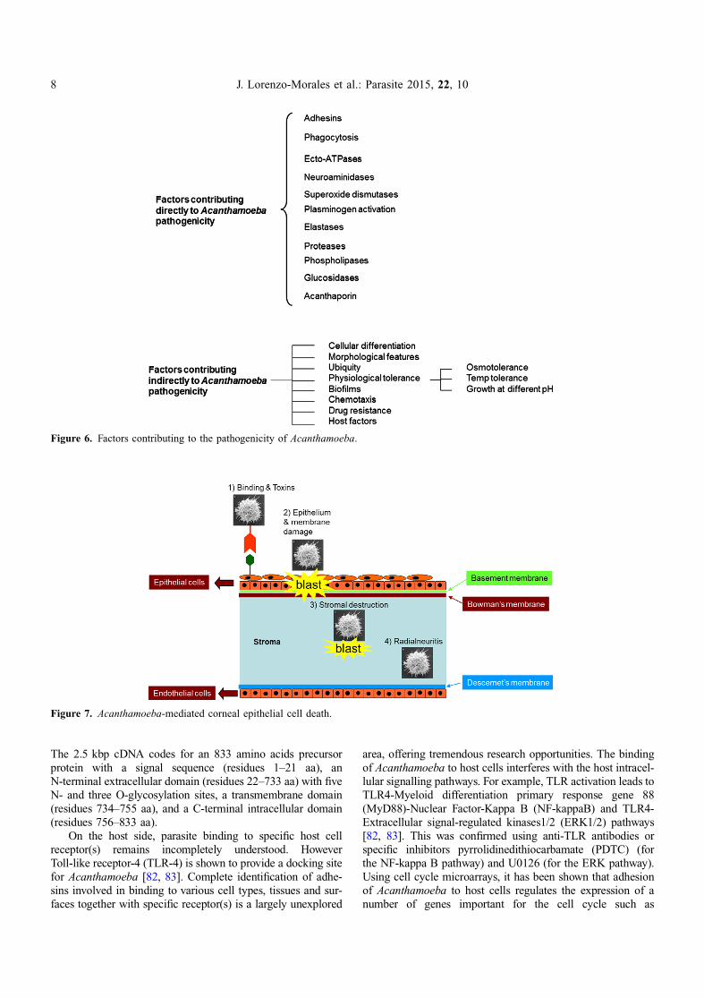
Adhesion is an important step in the pathogenic cascadesof Acanthamoeba keratitis leading to secondary events andamoebae crossing biological barriers (Fig. 7). Several adhesinshave been identified in Acanthamoeba, including a mannose-binding protein [30], a laminin-binding protein with apredicted molecular mass of 28.2 kDa [40] and a 55 kDalaminin-binding protein [87]. Notably, oral immunization withrecombinant mannose-binding protein ameliorates Acantha-moeba keratitis in the Chinese hamster model [30, 31], andhas shown that the mannose-binding protein gene in Acantha-moeba contains six exons and five introns that span 3.6 kbp.
J. Lorenzo-Morales et al.: Parasite 2015, 22, 10 7
The 2.5 kbp cDNA codes for an 833 amino acids precursorprotein with a signal sequence (residues 1–21 aa), anN-terminal extracellular domain (residues 22–733 aa) with fiveN- and three O-glycosylation sites, a transmembrane domain(residues 734–755 aa), and a C-terminal intracellular domain(residues 756–833 aa).
On the host side, parasite binding to specific host cellreceptor(s) remains incompletely understood. HoweverToll-like receptor-4 (TLR-4) is shown to provide a docking sitefor Acanthamoeba [82, 83]. Complete identification of adhe-sins involved in binding to various cell types, tissues and sur-faces together with specific receptor(s) is a largely unexplored
area, offering tremendous research opportunities. The bindingof Acanthamoeba to host cells interferes with the host intracel-lular signalling pathways. For example, TLR activation leads toTLR4-Myeloid differentiation primary response gene 88(MyD88)-Nuclear Factor-Kappa B (NF-kappaB) and TLR4-Extracellular signal-regulated kinases1/2 (ERK1/2) pathways[82, 83]. This was confirmed using anti-TLR antibodies orspecific inhibitors pyrrolidinedithiocarbamate (PDTC) (forthe NF-kappa B pathway) and U0126 (for the ERK pathway).Using cell cycle microarrays, it has been shown that adhesionof Acanthamoeba to host cells regulates the expression of anumber of genes important for the cell cycle such as
Figure 6. Factors contributing to the pathogenicity of Acanthamoeba.
Figure 7. Acanthamoeba-mediated corneal epithelial cell death.
8 J. Lorenzo-Morales et al.: Parasite 2015, 22, 10
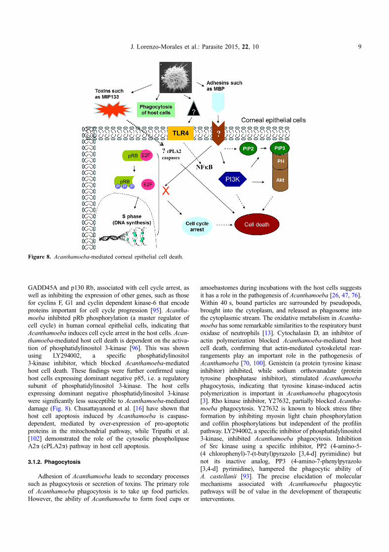
GADD45A and p130 Rb, associated with cell cycle arrest, aswell as inhibiting the expression of other genes, such as thosefor cyclins F, G1 and cyclin dependent kinase-6 that encodeproteins important for cell cycle progression [95]. Acantha-moeba inhibited pRb phosphorylation (a master regulator ofcell cycle) in human corneal epithelial cells, indicating thatAcanthamoeba induces cell cycle arrest in the host cells. Acan-thamoeba-mediated host cell death is dependent on the activa-tion of phosphatidylinositol 3-kinase [96]. This was shownusing LY294002, a specific phosphatidylinositol3-kinase inhibitor, which blocked Acanthamoeba-mediatedhost cell death. These findings were further confirmed usinghost cells expressing dominant negative p85, i.e. a regulatorysubunit of phosphatidylinositol 3-kinase. The host cellsexpressing dominant negative phosphatidylinositol 3-kinasewere significantly less susceptible to Acanthamoeba-mediateddamage (Fig. 8). Chusattayanond et al. [16] have shown thathost cell apoptosis induced by Acanthamoeba is caspase-dependent, mediated by over-expression of pro-apoptoticproteins in the mitochondrial pathway, while Tripathi et al.[102] demonstrated the role of the cytosolic phospholipaseA2a (cPLA2a) pathway in host cell apoptosis.
3.1.2. Phagocytosis
Adhesion of Acanthamoeba leads to secondary processessuch as phagocytosis or secretion of toxins. The primary roleof Acanthamoeba phagocytosis is to take up food particles.However, the ability of Acanthamoeba to form food cups or
amoebastomes during incubations with the host cells suggestsit has a role in the pathogenesis of Acanthamoeba [26, 47, 76].Within 40 s, bound particles are surrounded by pseudopods,brought into the cytoplasm, and released as phagosome intothe cytoplasmic stream. The oxidative metabolism in Acantha-moeba has some remarkable similarities to the respiratory burstoxidase of neutrophils [13]. Cytochalasin D, an inhibitor ofactin polymerization blocked Acanthamoeba-mediated hostcell death, confirming that actin-mediated cytoskeletal rear-rangements play an important role in the pathogenesis ofAcanthamoeba [70, 100]. Genistein (a protein tyrosine kinaseinhibitor) inhibited, while sodium orthovanadate (proteintyrosine phosphatase inhibitor), stimulated Acanthamoebaphagocytosis, indicating that tyrosine kinase-induced actinpolymerization is important in Acanthamoeba phagocytosis[3]. Rho kinase inhibitor, Y27632, partially blocked Acantha-moeba phagocytosis. Y27632 is known to block stress fibreformation by inhibiting myosin light chain phosphorylationand cofilin phosphorylations but independent of the profilinpathway. LY294002, a specific inhibitor of phosphatidylinositol3-kinase, inhibited Acanthamoeba phagocytosis. Inhibitionof Src kinase using a specific inhibitor, PP2 (4-amino-5-(4 chlorophenyl)-7-(t-butyl)pyrazolo [3,4-d] pyrimidine) butnot its inactive analog, PP3 (4-amino-7-phenylpyrazolo[3,4-d] pyrimidine), hampered the phagocytic ability ofA. castellanii [93]. The precise elucidation of molecularmechanisms associated with Acanthamoeba phagocyticpathways will be of value in the development of therapeuticinterventions.
Figure 8. Acanthamoeba-mediated corneal epithelial cell death.
J. Lorenzo-Morales et al.: Parasite 2015, 22, 10 9
3.1.3. Ecto-ATPases
Ecto-ATPases are glycoproteins expressed in the plasmamembranes with their active sites facing the external medium.Ecto-ATPases hydrolyze extracellular ATP and other nucleo-side triphosphates. The resultant ADP can have toxic effectson the host cells. For example, it has been shown that ADPreleased by Acanthamoeba bind to P2y2 purinergic receptorson the host cells, causing an increase in intracellular calcium,inducing caspase-3 activation and finally resulting in apoptosis[59]. A P2 receptor antagonist, suramin, inhibited Acantha-moeba-mediated host cell death [59], suggesting that ecto-ATPases play an important role in Acanthamoeba pathogenesisin a contact-independent mechanism. The clinical isolates ofAcanthamoeba exhibited higher ecto-ATPase activitiescompared with weak pathogenic isolates [94]. Several ecto-ATPases of approximate molecular weights of 62, 100, 218,272 and more than 300 kDa have been described in Acantha-moeba. However, further research is needed to elucidate theirfunction in Acanthamoeba biology, and investigate theirprecise role in Acanthamoeba pathogenesis.
3.1.4. Neuraminidase activity
Acanthamoeba exhibited neuraminidase activity. Theenzyme activity is optimal at pH 5 and at temperatures of25–30 �C. The live amoebae released sialic acid from thehuman cells. Therefore, the neuraminidase of Acanthamoebacould be relevant in the colonization of amoebae, and impor-tant in producing damage of the sialic acid-rich corneal epithe-lium. Interestingly, neuraminidases of Trypanosoma cruzi andAcanthamoeba are immunologically related, as demonstratedby antibodies against neuraminidase of Trypanosoma cruzi,which reacted with Acanthamoeba in immunofluorescence,immunoblotting and enzyme-linked immunosorbent assays[73, 74].
3.1.5. Superoxide dismutase
The enzyme superoxide dismutase catalyzes the dismuta-tion of superoxide into oxygen and hydrogen peroxide. It isan important antioxidant defence exposed to oxygen. Superox-ide is one of the main reactive oxygen species in the cell and assuch, superoxide dismutase plays an important antioxidantrole. Two superoxide dismutases have been identified in Acan-thamoeba: an iron superoxide dismutase (approximate molec-ular weight of 50 kDa) and a copper-zinc superoxidedismutase (approximate molecular weight of 38 kDa). Theseenzymes occur as cytoplasmic and detergent-extractable frac-tions. They may be potential virulence factors of Acantha-moeba by acting both as anti-oxidants and anti-inflammatoryagents. They may also provide additional targets for chemo-therapy and immuno-diagnosis of Acanthamoeba infections[15]. A. castellanii iron superoxide dismutase may play essen-tial roles in the survival of amoebae not only by protectingthemselves from endogenous oxidative stress, but also bydetoxifying oxidative killing of amoebae by host immuneeffector cells [50].
3.1.6. Acanthamoeba-induced plasminogen activation
Acanthamoeba displayed plasminogen activator activity bycatalyzing the cleavage of host plasminogen to form plasmin,which can activate host proteolytic enzymes, such aspro-matrix metalloproteases. Once activated, the matrixmetalloproteases degrade the basement membranes and thecomponents of the extracellular matrix such as type I and typeII collagens, fibronectins and laminin. Thus, the matrixmetalloproteinases are involved in tissue remodelling.The pathogenic Acanthamoeba showed positive chemotacticresponse to the endothelial extracts [109].
3.1.7. Elastase
Acanthamoeba is known to produce elastase with broadspecificity. Moreover, elastases are known to degrade a rangeof connective tissue proteins such as elastin, an elastic fibre,fibrinogen, collagen, and proteoglycans, which together deter-mined the mechanical properties of the connective tissue.Tissues altered by prior elastase treatment are more susceptibleto oxygen radical attack, suggesting their involvement in thepathogenesis and pathophysiology of Acanthamoeba infections[18, 46, 47, 54]. The elastases were in the region of70–130 kDa and serine peptidases were found to be possibleelastase candidates [28].
3.1.8. Proteases
Proteases are degradative enzymes that catalyze the totalhydrolysis of proteins. Acanthamoeba is shown to exhibits pro-teolytic activities. The primary role of Acanthamoeba prote-ases is to degrade food substances for feeding purposes.Pathogenic Acanthamoeba exhibit increased extracellular pro-tease activities. The link between pathogenicity and theincreased levels of extracellular proteases suggests that patho-genic Acanthamoeba utilize proteases to facilitate invasion ofthe host. Acanthamoeba is known to produce serine, cysteineand metalloproteases. Several serine proteases have been iden-tified ranging in molecular weights from >20 kDa to>200 kDa. They are shown to possess collagen degradationactivity, plasminogen activator, and degradation of fibronectin,fibrinogen, IgG, IgA, albumin, haemoglobin, protease inhibi-tors, interleukin-1, chemokines and cytokines [46, 54, 55, 60].A 133 kDa serine protease, called MIP133 has been identifiedas a crucial component of the pathogenic cascade of Acantha-moeba pathogenesis. The MIP133 serine protease is shown toinduce the degradation of keratocytes, iris ciliary body cells,retinal pigment epithelial cells, corneal epithelial cells and cor-neal endothelial cells, and induce apoptosis in macrophage-likecells. The properties of serine proteases facilitate Acantha-moeba invasion of the corneal stroma, leading to secondaryreactions such as oedema, necrosis and inflammatoryresponses. A direct functional role of serine proteases inAcanthamoeba infections is indicated by the observations thatintrastromal injections of Acanthamoeba conditioned mediumproduced corneal lesions in vivo, similar to those observed inAcanthamoeba keratitis patients, and this effect is inhibited
10 J. Lorenzo-Morales et al.: Parasite 2015, 22, 10
by phenylmethylsulfonyl fluoride, a serine protease inhibitor[37, 67, 68]. In addition, the chemically synthesized siRNAagainst the catalytic domain of the extracellular serine prote-ases of Acanthamoeba reduced protease activity and Acantha-moeba-mediated host cell cytotoxicity. These results supportthe idea that extracellular serine proteases are directly involvedin the pathogenesis and virulence of Acanthamoeba [55]. Inaddition, several cysteine proteases have been identified inAcanthamoeba, including 43, 65, 70 and 130 kDa cysteineproteases [46, 47]. In addition to serine and cysteine proteases,there is evidence for metalloprotease activity in Acantha-moeba. An 80 kDa metalloprotease was identified in co-cultures of Acanthamoeba and host cells, but its origin (whetherAcanthamoeba or the host cells) was not established. Later stud-ies identified a 150 kDa extracellular metalloprotease fromAcanthamoeba isolate of the T1 genotype. This metalloproteaseexhibited properties of extracellular matrix degradation, asdemonstrated by its activity against collagen I and III (majorcomponents of the collagenous extracellular matrix), elastin(elastic fibrils of the extracellular matrix), plasminogen(involved in proteolytic degradation of the extracellular matrix),as well as degradation of casein, gelatine and haemoglobin.
Recently, the complete sequence of a type-1 metacaspasefrom Acanthamoeba was reported, comprising 478 amino acids[102]. Later studies revealed that A. castellanii metacaspasesassociate with the contractile vacuole and have an essential rolein cell osmoregulation suggesting its attractiveness as a possi-ble target for treatment therapies against A. castellanii infec-tion [89]. These studies showed that Acanthamoeba exhibiteddiverse proteases and elastases, which could play importantroles in Acanthamoeba infections. The precise mechanismsof protease mode of action at the molecular level are onlybeginning to emerge. Proteases have been shown to be ‘‘drug-gable’’ targets, as evidenced by the widespread use of proteaseinhibitors as effective therapy for hypertension and AIDS, andthe current clinical development of protease inhibitors for dia-betes, cancer, thrombosis, and osteoporosis. As long as issuessuch as the difficulty of achieving selectivity can be addressedthrough targeting allosteric sites, protease-based drug therapyhas tremendous potential in the treatment of many infectiousdiseases. Future studies will further determine the role of pro-teases as vaccine targets, search for novel inhibitors by screen-ing of chemical libraries, or rational development of drugsbased on structural studies should enhance our ability to targetthese important pathogens.
3.1.9. Phospholipases
During phagocytosis, there is a large turnover of the plasmamembrane in Acanthamoeba, indicating that there is controlledlocal degradation of phospholipids leading to instability of themembrane phospholipid bilayer, which would then reform afterthe acylation of the lysophospholipid. In support of this, all ofthe enzymes that are needed for such a cycle are present in theplasma membrane of Acanthamoeba, including phospholipaseA2, acyl CoA:lysolecithin acyltransferase, and acyl CoA syn-thetase. Phospholipase A1 and lysophospholipase are also pres-ent in the plasma membrane of Acanthamoeba. The plasmamembrane lysophospholipase may also serve to protect the cell
from the lytic effect of lysophospholipids either of exogenousor endogenous origin. The plasma membranes have the enzy-matic capability of modulating the fatty acyl composition ofphospholipids by de-acylation and acylation. Our knowledgeof phospholipases in the virulence of Acanthamoeba is frag-mented, however several studies have shown that pathogenicAcanthamoeba that exhibit cytopathic effects on mammaliancells in vitro liberate more phospholipase, suggesting their possi-ble involvement in Acanthamoeba infections. Because phospho-lipases cleave phospholipids, it is reasonable to suspect that theyplay a role in membrane disruptions, penetration of host cells, andcell lysis. However this remains to be determined. Other actionsof phospholipases may involve interference with intracellularsignalling pathways. Phospholipases generate lipids andlipid-derived products that act as second messengers. A. castella-nii lysates and their conditioned medium exhibited phospholi-pase activities [66]. Sphinganine, a PLA2 inhibitor showedrobust amoebistatic properties but had no effect on theviability ofA. castellanii. These studies suggest that Acanthamoebaphospholipases and/or lysophospholipases may play a role inproducing host cell damage or affect other cellular functions suchas induction of inflammatory responses, thus facilitatingAcanthamoeba virulence. More studies are needed to identifyand characterize Acanthamoeba phospholipases and to deter-mine their potential role in the development of therapeuticintervention. This is not a novel concept: earlier studies haveshown that phospholipase C from Clostridium perfringensinduced protection against C. perfringens-mediated gas gan-grene. In addition, targeting of phospholipases using syntheticinhibitor compounds has been shown to prevent Candidainfections. Antibodies produced against Acanthamoebaphospholipases may also be of potential value in the developmentof sensitive and specific diagnostic assays as well as oftherapeutic value [42].
3.1.10. Glycosidases (also called glycoside hydrolases)
Glycoside hydrolases catalyze the hydrolysis of glycosidiclinkage to generate smaller sugars. Glycoside hydrolases areubiquitous in nature and involved in the degradation of bio-mass such as cellulose and in a variety of cellular functions.Together with glycosyltransferases, glycosidases form themajor catalytic machinery for the synthesis and breakage ofglycosidic bonds. Acanthamoeba exhibits glycosidase activitiesincluding beta-glycosidase, alpha-glucosidase, beta-galactosi-dase, beta-N-acetyl-glucosaminidase, beta-N-acetyl-galactosa-minidase and alpha-mannosidase [38, 39]. Acanthamoebaextracts mediate enzymatic lysis of cell walls from severalspecies of bacteria including Micrococcus lysodeikticus,Micrococcus roseus, Streptococcus faecalis, Bacillus megateri-um, Sarcina lutea, Micrococcus radiodurans and limitedactivity against Bacillus subtilis, Bacillus cereus, but has noeffects on Acanthamoeba cyst walls or chitin. Exhaustivedigestion of Micrococcus lysodeikticus cell walls released freeN-acetyl-glucosamine, N-acetyl-muramic acid, glycine, ala-nine, glutamic acid and lysine, suggesting that Acanthamoebapossesses both endo- and exo-hexosaminidases and beta-N-acetyl-hexosaminidases. Since Acanthamoeba is known toutilize maltose, cellobiose, sucrose or lactose, some of the
J. Lorenzo-Morales et al.: Parasite 2015, 22, 10 11
glycosidases indicated above may suggest the utilization ofthese disaccharides [42].
3.1.11. Acanthaporin
Recently, acanthaporin, the first pore-forming toxin wasdescribed from Acanthamoeba [64]. Acanthaporin was isolatedfrom extracts of virulent A. culbertsoni by tracking its pore-forming activity, molecularly cloning the gene of its precursor,and recombinant expression of the mature protein in bacteria.Acanthaporin was cytotoxic for human neuronal cells andexerted antimicrobial activity against a variety of bacterialstrains by permeabilizing their membranes [64]. The tertiarystructures of acanthaporin’s active monomeric form and inac-tive dimeric form, both solved by NMR spectroscopy, revealeda currently unknown protein fold and a pH-dependent triggermechanism of activation.
3.2. Factors contributing indirectly toAcanthamoeba pathogenicity
The ability of Acanthamoeba to produce human diseases isa multifactorial process and is, amongst other factors, depen-dent on its ability to survive outside its host and under diverseconditions (high osmolarity, varying temperatures, food depri-vation and resistance to chemotherapeutic drugs) [24, 48, 106,107, 114]. The ability of Acanthamoeba to overcome such con-ditions can be considered as contributory factors towards dis-ease and are indicated below.
3.2.1. Morphological features
The infective forms of Acanthamoeba or trophozoites donot have a distinct morphology. However, they do possessspine-like structures known as acanthopodia on their surface,which allow them to modulate binding of Acanthamoeba tobiological and inert surfaces. In addition, their amoeboidmotion resembles that of macrophages/neutrophils and it islikely that Acanthamoeba employ similar strategies to traversebiological barriers and invade tissues using the paracellularroute.
3.2.2. Temperature tolerance, osmotolerance and growthat different pH
Being a free-living amoeba, Acanthamoeba is exposed tovarious temperatures, osmolarity and pH. Similarly contactwith tear film exposes Acanthamoeba to high osmolarity(due to salinity in tears), high temperatures as well as altera-tions in pH. For successful transmission, Acanthamoeba mustwithstand such stress and exhibit biological activity. Patho-genic Acanthamoeba showed high levels of heat shock proteins(i.e. HSP60 and HSP70) compared with weak pathogens [75,77]. The higher levels of heat shock proteins in Acanthamoebamay indicate their involvement in (i) tolerance to hosts’ stress-ors and/or (ii) in species’ virulence [75]. The ability of Acan-thamoeba to grow at high temperatures and high osmolaritycorrelates with the pathogenicity of Acanthamoeba isolates,and may provide a good indicator of the pathogenic potential
of a given isolate [46, 47, 54]. The precise mechanisms bywhich pathogenic Acanthamoeba adapt to higher temperaturesand maintain their metabolic activities require further studies.
3.2.3. Cellular differentiation
Cellular differentiation is the ability of Acanthamoeba todifferentiate into a morphologically distinct dormant cyst formor a vegetative trophozoite form. This is a reversible change,dependent on environmental conditions. Cysts are resistant tovarious antimicrobial agents and adverse conditions such asextremes in temperatures, pH, osmolarity, desiccation and cystscan be airborne: all of which presents a major problem in che-motherapy because their persistence may lead to recurrence ofthe disease. Furthermore, Acanthamoeba cysts can survive sev-eral years while maintaining their pathogenicity [62]. Thesecharacteristics suggested that the primary functions of cystslie in withstanding adverse conditions and in the spread ofamoebae throughout the environment. In addition, this mayrepresent the ability of Acanthamoeba to alternate expressionof surface proteins/glycoproteins, in response to changing envi-ronments and/or immune surveillance. Cellular differentiationrepresents a major factor in the transmission of Acanthamoebaand recurrence of its infection. However, the underlyingmolecular mechanisms in these processes remain incompletelyunderstood and warrant further investigation.
3.2.4. Chemotaxis
Chemotaxis directs amoeba movement according to certainchemicals in their environment. This is important asAcanthamoeba moves towards the highest concentration offood molecules, or to flee from poisons. Acanthamoebaexhibits chemosensory responses as observed by their responseto a variety of bacterial products or potential bacterialproducts by moving actively towards the attractant.Acanthamoeba responded to the chemotactic peptides formyl-methionyl-leucyl-phenylalanine, formyl-methionyl-leucyl-phenylalaninebenzylamide, lipopolysaccharide and lipid A.In addition, significant responses to cyclic AMP, lipoteichoicacid and N-acetyl-glucosamine were also found. Interestingly,chemotactic peptide antagonists, mannose, mannosylatedbovine serum albumin and N-acetyl-muramic acid all yieldednon-significant responses. Pretreatment of Acanthamoeba withchemotactic peptides, bacterial products and bacteria reducedthe directional response to attractants. Acanthamoeba grownin the presence of bacteria appeared more responsive to che-motactic peptides. Treatment of Acanthamoeba with trypsinreduced the response of cells to chemotactic peptides, thoughsensitivity was restored within a couple of hours [7, 92]. Thesefindings suggest that Acanthamoeba membranes possess recep-tors sensitive to these bacterial substances, which are differentfrom the mannose-binding protein involved in binding to thehost cells to produce cytotoxicity or involved in binding to bac-teria during phagocytosis. The rate of movement is relativelyconstant (ca. 0.40 lm per sec), indicating that the locomotorresponse to these signals is a taxis, or possibly a klinokinesis,but not an orthokinesis [92].
12 J. Lorenzo-Morales et al.: Parasite 2015, 22, 10
3.2.5. Ubiquity
Acanthamoeba has been found in diverse environments,from drinking water to distilled water wash bottles, deep oceanbottom and Antarctica. It is therefore not surprising that humanbeings encounter and interact regularly with these organisms,as is evidenced by findings that in some regions, up to 100%of the population tested possess Acanthamoeba antibodies,suggesting that these are one of the most ubiquitous protistsand often come into contact with human beings, and giventhe opportunity (e.g. contact lens wear), can cause seriousinfections.
3.2.6. Biofilms
Biofilms are known to play an important role in thepathogenesis of Acanthamoeba keratitis. Biofilms are microbi-ally-derived sessile communities, which can be formed in aque-ous environments as well as on any materials and medicaldevices including intravenous catheters, contact lenses, scleralbuckles, suture material, and intraocular lenses. In the instanceof contact lenses, biofilms are formed through contamination ofthe storage case. Once established, biofilms provide attractiveniches for Acanthamoeba by fulfilling their nutritional require-ments as well as providing resistance to disinfectants. In addi-tion, this allows higher binding of Acanthamoeba to contactlenses. For example, Acanthamoeba exhibits significantlyhigher binding to used and Pseudomonas biofilm-coated hydro-gel lenses compared to unworn contact lenses. The abundantnutrient provided by the biofilm encourages transformation ofAcanthamoeba into the vegetative, infective trophozoite form,and it is important to remember that binding of Acanthamoebato the human corneal epithelial cells most likely occursduring the trophozoite stage as cysts exhibit minimal binding.These findings suggest that biofilms play an important role inAcanthamoeba keratitis in wearers of contact lenses andpreventing biofilm formation is perhaps an important preventa-tive strategy [9, 118].
3.2.7. Host factors
The factors that enable Acanthamoeba to produce diseaseare not limited solely to the pathogen, but most likely involvehost determinants [17, 115]. Evidence for this comes fromrecent studies in the UK, Japan and New Zealand, which sug-gested that the storage cases of contact lenses of 400–800 per10,000 asymptomatic wearers are contaminated with Acantha-moeba. This number is remarkably high compared with theincidence rate of Acanthamoeba keratitis in wearers of contactlenses, which is around 0.01–1.49 per 10,000 [46]. These find-ings suggest that factors such as host susceptibility, tissue spec-ificity, tear factors, sIgA, corneal trauma, as well asenvironmental factors such as osmolarity may be importantin initiating Acanthamoeba infections. However, the extent towhich such host factors contribute to the outcome of Acantha-moeba keratitis is unclear because host factors are more com-plex and difficult to study than those of the pathogen. Overall,it can be concluded that Acanthamoeba traversal of biologicalbarriers and to produce disease is a complex process thatinvolves both pathogen as well as host factors.
4. Acanthamoeba keratitis treatment: aproblem with no simple solution?
The treatment of Acanthamoeba keratitis has evolvedsince the first medical cure was reported in 1985 [46, 54, 86,103–106, 111, 112]. Early diagnosis and aggressive medicaltherapy has improved the management of this difficult infec-tion. Other reported factors that may facilitate effective medi-cal therapy and an improved outcome include early epithelialdebridement (to remove the majority of organisms) and pene-trating keratoplasty in medically-resistant cases.
So far, no chemotherapeutic agent has been described as asingle effective treatment against AK, regardless of the isolateor genotype that causes it. This is because there are many fac-tors, including the wide range of virulence traits that differentisolates possess, which makes it almost impossible to establisha correlation between in vitro and in vivo efficacies. Neverthe-less, to establish the most effective treatment regimen is noteasy for several reasons such as the relatively small numberof reported cases of AK, variable pathogenicity of differentstrains, and the intrinsic fluctuating nature of the diseaseprocess.
4.1. Is there a single effective treatment againstAK?
There are currently no methods or a single drug that caneliminate both cystic and trophozoite forms, while the tropho-zoite form is much more readily eliminated [46, 47, 54, 56].
The available reported treatment regimens in the literaturehave varied widely depending on the manifestation of the dis-ease, the general health status of the infected cornea, and theexperience of the clinician. For example, in the original AKcase reported by Naginton et al. [reviewed in 46, 54], numer-ous topical antimicrobial preparations were tried in conjunctionwith steroids, but both eyes eventually required grafting.Recent years have brought us knowledge of more specific anti-microbials, although unfortunately, surgical grafting of the cor-nea remains the last solution in case of severe infection.
4.2. Current therapeutic approaches
Current AK treatment consists of topical antimicrobialagents, which can achieve high concentrations at the site ofinfection. Moreover, due to the existence of a cyst form inAcanthamoeba that is highly resistant to therapy, a combinationof agents is generally used.
Most of the currently used topical agents are effectiveagainst trophozoites and cysts of Acanthamoeba such as bigu-anides, (i) PHMB [52, 54, 58], which is effective at low con-centrations (0.02%), but is unfortunately toxic to humancorneal cells [52, 54], and (ii) chlorhexidine, which is effectiveagainst both amoebic forms, and at minimal concentrations isnot toxic to corneal epithelial cells [52, 54, 58, 86]. Chlorhex-idine 0.02% is often used in combination with aromatic diami-dines such as 0.1% propamidine isethionate Brolene� (Sanofi,UK), 0.15% dibromopropamidine, hexamidine 0.1% Desome-dine� (Chauvin, France) and neomycin, showing good results
J. Lorenzo-Morales et al.: Parasite 2015, 22, 10 13
if the treatment is applied early in the development of theinfection [54, 86]. Unfortunately, propamidine and hexamidineare not available in all countries.
These topical antimicrobials are administered every hourimmediately after corneal debridement or for the first severaldays of therapy. These agents are then continued hourly for3 days (at least nine times/day is recommended) dependingon clinical response. The frequency is then reduced to every3 h. Two weeks may be required before a response is observed,and the total duration of therapy is a minimum of 3–4 weeks.Some authors also recommend treatment for 6–12 months.Moreover, when therapy is discontinued, close observation ofthe patient is suggested in order to avoid recurrent infection.Some patients have been successfully treated using an antisep-tic as monotherapy; if this is attempted, it should be reservedfor patients with early disease.
4.3. Biguanides as first line treatment against AK
PHMB and chlorhexidine have been reported to be themost effective drugs for treatment of infection and in combina-tion they have been reported to be effective against both cystsand trophozoites [23, 52, 54, 79, 108]. Regarding these twodrugs, it is important to mention that they are active againsta wide spectrum of pathogens by increasing cytoplasmic mem-brane permeability. Chlorhexidine and PHMB both containhighly charged positive molecules capable of binding to themucopolysaccharide plug of the ostiole resulting in penetrationof the amoeba. The drug then binds to the phospholipid bilayerof the cell membrane which is negatively charged resulting indamage, cell lysis and death [52].
Among the observed side effects, toxic keratopathy maydevelop at any time, necessitating significant alteration in thistreatment plan [46, 54, 98]. Elevated intraocular pressure aswell as increased inflammation often requires the use ofantiglaucoma medication and cycloplegics. The role of topicalcorticosteroids as well as surgical intervention with therapeuticpenetrating keratoplasty in the management of this infectionremains controversial and is discussed later.
Brolene� may be accompanied by drug toxicity and resis-tance and topical treatment with miconazole can lead to epithe-lial toxicity [29, 54, 56]. According to Turner et al. [107],resistance is mainly due to the exocyst and endocyst, whichforms a double-walled protective barrier to biocides. Cystsmay also be resistant to biocides because they show little orno metabolic activity and because of selection pressure dueto continuous drug exposure [46, 106, 107]. If resistance todrugs occurs, keratoplasty may be used [29, 46, 51, 54]. Uekiet al. [108] stated that recommended treatment for AK includescorneal scrape with antifungal drugs and antibiotic treatment.However, antifungal, antibacterial, antiviral and even cortico-steroids used can complicate matters because they cause initialimprovement then worsening of the disease [46, 54].
4.4. Steroids controversy in AK treatment
No clear consensus exists about use of steroids. Mostauthorities recommend that steroid use is probably best
avoided. Patients receiving steroids should continue antiamoe-bic therapy for several weeks after the steroids are stopped.In the case of a persistent infection with inflammation, cortico-steroids may be used. However, their use is controversialbecause they cause suppression of the immunological responseof the patient. Moreover, corticosteroids produce inhibition ofthe processes of encystation and excystation of Acanthamoeba,which could be a cause of the appearance of resistance prob-lems [103]. Recent studies have highlighted an association oftopical corticosteroids and a diagnostic delay of AK. Moreover,there is some evidence that suggests that steroid use may resultin increased pathogenicity of the amoebae [85]. McClellanet al. [63] demonstrated in an in vivo model that the additionof topical corticosteroids, even at low doses, promotes anincrease in the number of trophozoites, produced by excyst-ment in the infected corneal stroma. This exposes patients tothe risk of significantly greater corneal destruction throughan increase in amoebic load, which may be greater than theincreased chemotherapy effect on trophozoites compared withthe more resistant cysts [63, 85].
Furthermore, corneal transplantation (keratoplasty) isanother therapeutic option when topical treatment has failed.This intervention is recommended if in the acute phase ofinfection, the cornea becomes too thin or has been damaged,and vision is limited [51, 69]. Nevertheless, there is a risk ofnot eliminating all the trophozoites or cysts that could colonizethe new cornea [98]. A variation of keratoplasty called DALK(Deep Anterior Lamellar Keratoplasty) has been proposed tobe more effective in increasing the survival of transplanted cor-neal cells and to prevent entry of pathogenic organisms at thetime of surgery [72].
4.5. In vitro drug sensitivity testing andpersonalised treatments in AK patients
In vitro drug sensitivity testing, although rarely used, maybe helpful in refractory cases. However, such testing has itslimitations and may not be practical for the clinician. Not onlymay drug sensitivities be variable between strains, but astrain may also become resistant to formerly effective drugs.In addition, testing results may differ between laboratoriesand in some cases may not correlate with the clinical course.Despite these problems, drug sensitivity testing may offer theclinician a small edge in improving chances of therapeutic suc-cess and should be employed when possible. Recently, a patientsuffering from severe AK was healed after a personalized treat-ment with voriconazole, when sensitivity to this drug wasassayed after isolation of the amoebae from the patient’scornea [5].
4.6. Surgical management of AK
Therapeutic penetrating keratoplasty was the mainstay oftreatment for AK before the development of early diagnosisand aggressive medical therapy [10, 21, 36, 72]. The roleand timing of penetrating keratoplasty in AK still remainspoorly defined. Certainly pending or frank corneal perforation
14 J. Lorenzo-Morales et al.: Parasite 2015, 22, 10
is a clear indication for surgical intervention. However, otherindications for surgery are not well defined.
Therapeutic penetrating keratoplasty should be consideredwhen the infectious process spreads to the paracentral cornealstroma despite maximum antiamoebic therapy [21]. Perform-ing this procedure on a more localized infection may allowtotal removal of the organisms by excising the clinicallyinvolved tissue as well as a rim of clear surrounding cornea.This procedure allows the donor tissue to be placed into a rel-atively undamaged and hence non-immunocompromised reci-pient bed. After surgery, medical therapy should becontinued for at least several months to help ensure eliminationof any residual Acanthamoeba organisms in the recipient stro-mal tissue. Once the infection has spread into the peripheralcornea, however, the likelihood of achieving a surgical cureis markedly diminished. Intensive medical management isrequired for several months to eradicate the organism priorto keratoplasty. Unfortunately, the prognosis in these cases ispoor and reinforces the rationale for earlier rather than latersurgical treatment.
Recently, bipedicle conjunctival flap (CF) and cryopre-served amniotic membrane graft (AMG) have been reportedto be effective in AK. They restore ocular surface integrityand provide metabolic and mechanical support for cornealhealing. Nevertheless, in the case of large corneal perforation,penetrating keratoplasty to restore ocular integrity remains asthe only effective surgical option [1].
4.7. Novel therapeutic approaches
Recently, the widespread use of photorefractive surgery hasinspired its use in the setting of AK. Kandori et al. [44]reported four cases in 2010, where early stage AK was treatedwith standard topical therapy, but developed large cornealabscesses in the upper third thickness of the stroma. Thesewere removed using laser phototherapeutic keratectomy(PTK); all eyes experienced no disease recurrence and finalacuities ranged from 20/16 to 20/25.
Cross-linking is another relatively new treatmentoption that has been applied to AK. While in vitro studies byKashiwabuchi et al. [45] and Del Buey et al. [25] have shownno amoebicidal effect of riboflavin combined with UVA expo-sure, clinical case reports have shown a much more promisingpicture. Garduño-Vieyra et al. [32] administered collagencross-linking to a patient in Mexico instead of topical medicaltherapies, which were not commercially available. Significantimprovement was observed after 24 h, with symptoms resolv-ing within 3 months, and 20/20 vision was obtained after5 months. Khan et al. [49] have since reported three similarcases which responded equally well to cross-linking, with allulcers closing within 7 weeks. In subsequent PK surgery forscarring, no organisms were detected in excised tissue. It ispossible that the collagen stabilizing effect prevents furthertissue damage and isolates and prevents reproduction of theamoebae. Although individual case report results seem prom-ising, there are no formal clinical trials thus far to recommendincorporation into standard practice.
4.8. In the search of novel drugs against AK
A new path may be the application of alkylphosphocho-lines. These are phosphocholines esterified to aliphatic alco-hols. They exhibit in vitro and in vivo antineoplastic activityand have been shown to be cytotoxic against Leishmaniaspp., Trypanosoma cruzi and Entamoeba histolytica. A recentstudy has demonstrated that particularly hexadecylphosphoch-oline (miltefosine) is highly effective also against variousstrains of Acanthamoeba. Moreover, it has recently beenapplied in combination with PHMB in AK in Austria withsuccessful outcomes [8, 78, 79].
Recently, the creation of a ‘‘pharmaceutical phylogeny’’has been started for Acanthamoeba in order to elucidate andselect new therapeutic targets [54, 58, 86]. The phylogeny ofAcanthamoeba is closer evolutionarily to human beings thanmany other eukaryotic pathogens [69]. Therefore, part of thehypothesis is that many biochemical processes as well as ther-apeutic targets are evolutionarily conserved and are similar inrelated organisms. This implies that there will be a large num-ber of processes, some still unknown, in Acanthamoeba thatare similar to those of human beings. Therefore, treatmentsthat affect the host could also affect the parasite, for example,phospholipid analogues as mentioned above which have beendemonstrated to be effective against Acanthamoeba [2, 86].However, even though many biological processes are similarin Acanthamoeba to other eukaryotic cells, some proteins suchas tubulins are not sensitive to inhibitors that are normally usedagainst them [86]. Therefore, it is also interesting to find thosetargets that are specific to Acanthamoeba in order to attack theparasite without affecting the host. These targets may be of adifferent nature: specific gene products, biological processesthemselves, transcription or translation mechanisms or physi-cal attributes such as cell membranes. In addition, many anti-biotics active against Acanthamoeba have a mechanism ofaction which is still unknown. In recent years, the possibilityto design and synthesize specific small interfering RNAs(siRNAs) for gene silencing have made RNAi techniques apowerful tool for the study and understanding of new cellularpathways of proteins whose functions are still unknown, aswell as for their use as a therapy against various diseases[14, 88]. In the case of Acanthamoeba, siRNAs have been usedsuccessfully to identify potential therapeutic targets and evenrecently to establish a target and propose statins as a futuretherapy against Acanthamoeba strains [58].
Other drug targets that have been validated using the sameapproach, but without the elucidation of an available chemicalalternative (active principle/drug) include glycogen phosphory-lase and other cellulose synthesis related enzymes, serine pro-teases and myosin IC [4, 27, 53, 55, 57, 65]. The recentlypublished Acanthamoeba castellani genome data will furtherassist in the development of novel therapeutics in the nearfuture [19].
5. Concluding remarks
The number of reported cases of Acanthamoeba keratitis isincreasing worldwide every year, due to increasing contact lens
J. Lorenzo-Morales et al.: Parasite 2015, 22, 10 15
use for vision correction and cosmetic purposes. Increasedawareness combined with early diagnosis of the disease is cur-rently a good pathway towards better outcomes. However, knowl-edge about the pathogenesis and cellular differentiationprocesses in Acanthamoeba are still not fully known and urgentlyrequire further investigation. They hold the key to improved diag-nosis and to development of effective therapeutic approaches.
Acknowledgements. JLM was supported by the Ramón y Cajal Sub-programme from the Spanish Ministry of Economy and Competitiv-ity RYC-2011-08863 and by the grants RICET (Project No. RD12/0018/0012 of the programme of Redes Temáticas de InvestigaciónCooperativa, FIS), Spanish Ministry of Health, Madrid, Spain andthe Project FIS PI13/00490 ‘‘Protozoosis Emergentes por Amebasde Vida Libre: Aislamiento, Caracterización, Nuevas Aproximaci-ones Terapéuticas y Traslación Clínica de los Resultados’’ fromthe Instituto de Salud Carlos III and Project ref. AGUA3 ‘‘Amebasde Vida Libre como Marcadores de Calidad del Agua’’ from Caja-Canarias Fundación. JLM is grateful to the laboratory members ofthe Free-Living Amoebae Laboratory at the University Institute ofTropical Diseases and Public Health of the Canary Islands, Univer-sity of La Laguna, Spain.NAK was supported by the Higher Education Commission, and AgaKhan University, Pakistan, the British Council for the Prevention ofBlindness, UK, and the Royal Society, UK.JW was supported by the Medical University of Vienna, Austria andwould like to thank all members of the Molecular Parasitologylaboratory at the Institute of Specific Prophylaxis and TropicalMedicine, Vienna, Austria.This review was invited by the Editor at the occasion of ICOPA XIII(Mexico, 2014). Its publication is sponsored by the publisher ofParasite, EDP Sciences.Images from Figure 1 were kindly provided by Dr Francisco Arnalich-Montiel, Cornea Unit, Hospital Ramón y Cajal, Madrid, Spain.
References
1. Abdulhalim B, Wagih MM, Gad AA, Boghdadi G, Nagy RR.2015. Amniotic membrane graft to conjunctival flap intreatment of non-viral resistant infectious keratitis: a random-ised clinical study. British Journal of Ophthalmology, 99(1),59–63.
2. Aichelburg AC, Walochnik J, Assadian O, Prosch H, Steuer A,Perneczky G, Visvesvara GS, Aspöck H, Vetter N. 2008.Successful treatment of disseminated Acanthamoeba sp.infection with miltefosine. Emerging Infectious Diseases, 14,1743–1746.
3. Alsam S, Sissons J, Dudley R, Khan NA. 2005. Mechanismsassociated with Acanthamoeba castellanii (T4) phagocytosis.Parasitology Research, 96, 402–409.
4. Aqeel Y, Siddiqui R, Khan NA. 2013. Silencing of xyloseisomerase and cellulose synthase by siRNA inhibits encystationin Acanthamoeba castellanii. Parasitology Research, 112(3),1221–1227.
5. Arnalich-Montiel F, Martín-Navarro CM, Alió JL, López-Vélez R,Martínez-Carretero E, Valladares B, Piñero JE, Lorenzo-Morales J.2012. Successful monitoring and treatment of intraoculardissemination of Acanthamoeba with voriconazole. Archives ofOphthalmology, 130, 1474–1475.
6. Bacon AS, Frazer DG, Dart JK, Matheson M, Ficker LA,Wright P. 1993. A review of 72 consecutive cases ofAcanthamoeba keratitis, 1984–1992. Eye, 7, 719–725.
7. Bagorda A, Parent CA. 2008. Eukaryotic chemotaxis at aglance. Journal of Cell Science, 121, 2621–2624.
8. Barisani-Asenbauer T, Walochnik J, Mejdoubi L, Binder S.2012. Successful management of recurrent Acanthamoebakeratitis using topical and systemic miltefosine. Acta Ophthal-mologica, 900, doi: 10.1111/j.1755-3768.2012.F095.x.
9. Beattie TK, Tomlinson A, McFadyen AK, Seal DV, GrimasonAM. 2003. Enhanced attachment of Acanthamoeba toextended-wear silicone hydrogel contact lenses: a new riskfactor for infection? Ophthalmology, 110, 765–771.
10. Blackman HJ, Rao NA, Lemp MA, Visvesvara GS. 1984.Acanthamoeba keratitis successfully treated with penetratingkeratoplasty: suggested immunogenic mechanisms of action.Cornea, 3, 125.
11. Booton GC, Joslin CE, Shoff M, Tu EY, Kelly DJ, Fuerst PA.2009. Genotypic identification of Acanthamoeba sp. isolatesassociated with an outbreak of Acanthamoeba keratitis. Cornea,28(6), 673–676.
12. Bouheraoua N, Gaujoux T, Goldschmidt P, Chaumeil C,Laroche L, Borderie VM. 2013. Prognostic factors associatedwith the need for surgery in Acanthamoeba keratitis. Cornea,32, 130–136.
13. Brooks SE, Schneider DL. 1985. Oxidative metabolism asso-ciated with phagocytosis in Acanthamoeba castellanii. Journalof Protozoology, 32, 330–333.
14. Campochiaro PA. 2006. Potential applications for RNAi toprobe pathogenesis and develop new treatments for oculardisorders. Gene Therapy, 13, 559–562.
15. Cho JH, Na BK, Kim TS, Song CY. 2000. Purification andcharacterization of an extracellular serine proteinase fromAcanthamoeba castellanii. IUBMB Life, 50, 209–214.
16. Chusattayanond AD, Boonsilp S, Kasisit J, Boonmee A, Warit S.2010. Thai Acanthamoeba isolate (T4) induced apoptoticdeath in neuroblastoma cells via the Bax-mediated pathway.Parasitology International, 59, 512–516.
17. Clarke DW, Niederkorn JY. 2006. The immunobiology of Acan-thamoeba keratitis. Microbes and Infection, 8(5), 1400–1405.
18. Clarke DW, Niederkorn JY. 2006. The pathophysiology ofAcanthamoeba keratitis. Trends in Parasitology, 22(4),175–180.
19. Clarke M, Lohan AJ, Liu B, Lagkouvardos I, Roy S, Zafar N,Bertelli C, Schilde C, Kianianmomeni A, Bürglin TR, Frech C,Turcotte B, Kopec KO, Synnott JM, Choo C, Paponov I, Finkler A,Heng Tan CS, Hutchins AP, Weinmeier T, Rattei T, Chu JS,Gimenez G, Irimia M, Rigden DJ, Fitzpatrick DA, Lorenzo-Morales J, Bateman A, Chiu CH, Tang P, Hegemann P, Fromm H,Raoult D, Greub G, Miranda-Saavedra D, Chen N, Nash P,Ginger ML, Horn M, Schaap P, Caler L, Loftus BJ. 2013. Genomeof Acanthamoeba castellanii highlights extensive lateral genetransfer and early evolution of tyrosine kinase signaling. GenomeBiology, 14(2), R11.
20. Cohen EJ, Buchanan HW, Laughrea PA, Adams CP, GalentinePG, Visvesvara GS, Folberg R, Arentsen JJ, Laibson PR. 1985.Diagnosis and management of Acanthamoeba keratitis. Amer-ican Journal of Ophthalmology, 100(3), 389–395.
21. Cohen EJ, Parlato CJ, Arentsen JJ, Genvert GI, Eagle RC Jr,Wieland MR, Laibson PR. 1987. Medical and surgicaltreatment of Acanthamoeba keratitis. American Journal ofOphthalmology, 103(5), 615–625.
22. Cursons RTM, Brown TJ. 1978. Use of cell cultures as anindicator of pathogenicity of free-living amoebae. Journal ofClinical Pathology, 31, 1–11.
16 J. Lorenzo-Morales et al.: Parasite 2015, 22, 10
23. Dart JK, Saw VP, Kilvington S. 2009. Acanthamoeba keratitis:diagnosis and treatment update 2009. American Journal ofOphthalmology, 148(4), 487–499.
24. De Jonckheere JF. 1980. Growth characteristics, cytopathiceffect in cell culture, and virulence in mice of 36 type strainsbelonging to 19 different Acanthamoeba spp. Applied andEnvironmental Microbiology, 39(4), 681–685.
25. Del Buey MA, Cristóbal JA, Casas P, Goñi P, Clavel A,Mínguez E, Lanchares E, García A, Calvo B. 2012. Evaluationof in vitro efficacy of combined riboflavin and ultraviolet a forAcanthamoeba isolates. American Journal of Ophthalmology,153(3), 399–404.
26. Diaz J, Osuna A, Rosales MJ, Cifuentes J, Mascaro C. 1991.Sucker-like structures in two strains of Acanthamoeba: scan-ning electron microscopy study. International Journal ofParasitology, 21, 365–367.
27. Dudley R, Alsam S, Khan NA. 2007. Cellulose biosynthesispathway is a potential target in the improved treatment ofAcanthamoeba keratitis. Applied Microbiology and Biotech-nology, 75(1), 133–140.
28. Ferreira GA, Magliano AC, Pral EM, Alfieri SC. 2009. Elastasesecretion in Acanthamoeba polyphaga. Acta Tropica, 112,156–163.
29. Ficker L, Seal D, Warhurst D, Wright P. 1990. Acanthamoebakeratitis-resistance to medical therapy. Eye, 4, 835–838.
30. Garate M, Cao Z, Bateman E, Panjwani N. 2004. Cloning andcharacterization of a novel mannose-binding protein of Acan-thamoeba. Journal of Biological Chemistry, 279, 29849–29856.
31. Garate M, Cubillos I, Marchant J, Panjwani N. 2005.Biochemical characterization and functional studies of Acan-thamoeba mannose-binding protein. Infection and Immunity,73, 5775–5781.
32. Garduño-Vieyra L, González-Sánchez CR, Hernández-DaMota SE. 2011. Ultraviolet-a light and riboflavin therapy forAcanthamoeba keratitis: a case report. Case Reports inOphthalmology, 2(2), 291–295.
33. Gast RJ, Ledee DR, Fuerst PA, Byers T. 1996. Subgenussystematics of Acanthamoeba: Four nuclear 18S rDNA sequencetypes. Journal of Eukaryotic Microbiology, 43, 498–504.
34. Griffin JL. 1972. Temperature tolerance of pathogenic andnonpathogenic free-living amoebae. Science, 178, 869–870.
35. Grossniklaus HE, Waring GO 4th, Akor C, Castellano-SanchezAA, Bennett K. 2003. Evaluation of hematoxylin and eosin andspecial stains for the detection of Acanthamoeba keratitis inpenetrating keratoplasties. American Journal of Ophthalmol-ogy, 136(3), 520–526.
36. Hamburg A, De Jonckheere JF. 1980. Amoebic keratitis.Ophthalmologica, 181, 74.
37. He YG, Niederkorn JY, McCulley JP, Stewart GL, Meyer DR,Silvany R, Dougherty J. 1990. In vivo and in vitro collageno-lytic activity of Acanthamoeba castellanii. Investigative Oph-thalmology and Visual Science, 31, 2235–2240.
38. Henrissat B, Callebaut I, Mornon JP, Fabrega S, Lehn P, Davies G.1995. Conserved catalytic machinery and the prediction of acommon fold for several families of glycosyl hydrolases.Proceedings of the National Academy of Science USA, 92(15),7090–7094.
39. Henrissat B, Davies G. 1995. Structures and mechanisms ofglycosyl hydrolases. Structure, 3(9), 853–859.
40. Hong YC, Lee WM, Kong HH, Jeong HJ, Chung DI. 2004.Molecular cloning and characterization of a cDNA encoding a
laminin-binding protein (AhLBP) from Acanthamoeba healyi.Experimental Parasitology, 106, 95–102.
41. Ikeda Y, Miyazaki D, Yakura K, Kawaguchi A, Ishikura R,Inoue Y, Mito T, Shiraishi A, Ohashi Y, Higaki S, Itahashi M,Fukuda M, Shimomura Y, Yagita K. 2012. Assessment of real-time polymerase chain reaction detection of Acanthamoeba andprognosis determinants of Acanthamoeba keratitis. Ophthal-mology, 119, 1111–1119.
42. Im K. 1990. Pathogenic free-living amoebae. Korean Journal ofParasitology, 28, 29–39.
43. Kao PM, Tung MC, Hsu BM, Tsai HL, She CY, Shen SM,Huang WC. 2013. Real-time PCR method for the detection andquantification of Acanthamoeba species in various types ofwater samples. Parasitology Research, 112(3), 1131–1136.
44. Kandori M, Inoue T, Shimabukuro M, Hayashi H, Hori Y,Maeda N, Tano Y. 2010. Four cases of Acanthamoeba keratitistreated with phototherapeutic keratectomy. Cornea, 29(10),1199–1202.
45. Kashiwabuchi RT, Carvalho FR, Khan YA, de Freitas D,Foronda AS, Hirai FE, Campos MS, McDonnell PJ. 2011.Assessing efficacy of combined riboflavin and UV-A light(365 nm) treatment of Acanthamoeba trophozoites. Investiga-tive Ophthalmology and Visual Science, 52(13), 9333–9338.
46. Khan NA. 2009. Acanthamoeba – Biology and Pathogenesis.Caister Academic Press: Norfolk, Great Britain, 290 p.
47. Khan NA. 2006. Acanthamoeba: biology and increasingimportance in human health. FEMS Microbiology Reviews,30, 564–595.
48. Khan NA. 2001. Pathogenicity, morphology and differentiationof Acanthamoeba. Current Microbiology, 43, 391–395.
49. Khan YA, Kashiwabuchi RT, Martins SA, Castro-Combs JM,Kalyani S, Stanley P, Flikier D, Behrens A. 2011. Riboflavinand ultraviolet light a therapy as an adjuvant treatment formedically refractive Acanthamoeba keratitis: report of 3 cases.Ophthalmology, 118(2), 324–331.
50. Kim JY, Na BK, Song KJ, Park MH, Park YK, Kim TS. 2012.Functional expression and characterization of an iron-containing superoxide dismutase of Acanthamoeba castellanii.Parasitology Research, 111, 1673–1682.
51. Kitzmann AS, Goins KM, Sutphin JE, Wagoner MD. 2009.Keratoplasty for treatment of Acanthamoeba keratitis.Ophthalmology, 116, 864–869.
52. Lim N, Goh D, Bunce C, Xing W, Fraenkel G, Poole TR,Ficker L. 2008. Comparison of polyhexamethylene biguanideand chlorhexidine as monotherapy agents in the treatment ofAcanthamoeba keratitis. American Journal of Ophthalmology,145, 130–135.
53. Lorenzo-Morales J, Kliescikova J, Martínez-Carretero E, DePablos LM, Profotova B, Nohynkova E, Osuna A, Valladares B.2008. Glycogen phosphorylase in Acanthamoeba spp.: deter-mining the role of the enzyme during the encystment processusing RNA interference. Eukaryotic Cell, 7(3), 509–517.
54. Lorenzo-Morales J, Martín-Navarro CM, López-Arencibia A,Arnalich-Montiel F, Piñero JE, Valladares B. 2013. Acantha-moeba keratitis: an emerging disease gathering importanceworldwide? Trends in Parasitology, 29(4), 181–187.
55. Lorenzo-Morales J, Ortega-Rivas A, Foronda P, Abreu-Acosta N,Ballart D, Martínez E, Valladares B. 2005. RNA interference(RNAi) for the silencing of extracellular serine proteases genes inAcanthamoeba: Molecular analysis and effect on pathogenecity.Molecular and Biochemical Parasitology, 144, 10–15.
J. Lorenzo-Morales et al.: Parasite 2015, 22, 10 17
56. Marciano-Cabral F, Cabral G. 2003. Acanthamoeba spp. asagents of disease in humans. Clinical Microbiology Reviews,16(2), 273–307.
57. Martín-Navarro CM, Lorenzo-Morales J, López-Arencibia A,Reyes-Batlle M, Piñero JE, Valladares B, Maciver SK. 2014.Evaluation of Acanthamoeba myosin-IC as a potential thera-peutic target. Antimicrobial Agents and Chemotherapy, 58(4),2150–2155.
58. Martín-Navarro CM, Lorenzo-Morales J, Machin RP, López-Arencibia A, García-Castellano JM, de Fuentes I, Loftus B,Maciver SK, Valladares B, Piñero JE. 2013. Inhibition ofHMG-CoA reductase and the application of statins as a noveleffective therapeutic approach against Acanthamoeba infections.Antimicrobial Agents and Chemotherapy, 57(1), 375–381.
59. Mathers WD, Nelson SE, Lane JL, Wilson ME, Allen RC,Folberg R. 2000. Confirmation of confocal microscopy diag-nosis of Acanthamoeba keratitis using polymerase chainreaction analysis. Archives of Ophthalmology, 118, 178–183.
60. Mattana A, Cappai V, Alberti L, Serra C, Fiori PL,Cappuccinelli P. 2002. ADP and other metabolites releasedfrom Acanthamoeba castellanii lead to human monocyticcell death through apoptosis and stimulate the secretion ofproinflammatory cytokines. Infection and Immunity, 70,4424–4432.
61. Maubon D, Dubosson M, Chiquet C, Yera H, Brenier-PinchartMP, Cornet M, Savy O, Renard E, Pelloux H. 2012. A one-stepmultiplex PCR for Acanthamoeba keratitis diagnosis andquality samples control. Investigative Ophthalmology andVisual Science, 53(6), 2866–2872.
62. Mazur T, Hadas E, Iwanicka I. 1995. The duration of the cyststage and the viability and virulence of Acanthamoeba isolates.Tropical Medicine and Parasitology, 46, 106–108.
63. McClellan K, Howard K, Niederkorn JY, Alizadeh H. 2001.Effect of steroids on Acanthamoeba cysts and trophozoites.Investigative Ophthalmology and Visual Science, 42,2885–2893.
64. Michalek M, Sönnichsen FD, Wechselberger R, Dingley AJ,Hung CW, Kopp A, Wienk H, Simanski M, Herbst R, Lorenzen I,Marciano-Cabral F, Gelhaus C, Gutsmann T, Tholey A,Grötzinger J, Leippe M. 2013. Structure and function of aunique pore-forming protein from a pathogenic Acanthamoeba.Nature Chemical Biology, 9, 37–42.
65. Moon EK, Hong Y, Chung DI, Goo YK, Kong HH. 2014.Down-regulation of cellulose synthase inhibits the formation ofendocysts in Acanthamoeba. Korean Journal of Parasitology,52(2), 131–135.
66. Mortazavi PN, Keisary E, Loh LN, Jung SY, Khan NA. 2011.Possible roles of phospholipase A2 in the biological activities ofAcanthamoeba castellanii (T4 genotype). Protist, 162, 168–176.
67. Na BK, Cho JH, Song CY, Kim TS. 2002. Degradation ofimmunoglobulins, protease inhibitors and interleukin-1 by asecretory proteinase of Acanthamoeba castellanii. KoreanJournal of Parasitology, 40, 93–99.
68. Na BK, Kim JC, Song CY. 2001. Characterization andpathogenetic role of proteinase from Acanthamoeba castellanii.Microbial Pathogenesis, 30, 39–48.
69. Nguyen TH, Weisenthal RW, Florakis GJ, Reidy JJ, Gaster RN,Tom D. 2010. Penetrating keratoplasty in active Acanthamoebakeratitis. Cornea, 29, 1000–1004.
70. Niederkorn JY, Alizadeh H, Leher H, McCulley JP. 1999. Thepathogenesis of Acanthamoeba keratitis. Microbes and Infec-tion, 1, 437–443.
71. Page FC, Siemensma FJ. 1991. Nackte Rhizopoda undHeliozoea, in Protozoenfauna, Band 2, Matthes D (Hrsg.).G. Fischer: Stuttgart.
72. Parthasarathy A, Tan DT. 2007. Deep lamellar keratoplasty forAcanthamoeba keratitis. Cornea, 26(8), 1021–1023.
73. Pellegrin JL, Ortega-Barria E, Barza M, Baum J, Pereira ME.1991. Neuraminidase activity in Acanthamoeba species tro-phozoites and cysts. Investigative Ophthalmology and VisualScience, 32, 3061–3066.
74. Pellegrin JL, Ortega-Barria E, Prioli RP, Meijia JS, Pereira ME.1992. The neuraminidases of Trypanosoma cruzi and Acantha-moeba castellanii are immunologically related. TropicalMedicine and Parasitology, 43, 33–37.
75. Pérez-Serrano J, Martínez J, Pérez B, Bernadina WE,Rodríguez-Caabeiro F. 2000. In vitro shock response todifferent stressors in free living and pathogenic Acanthamoeba.International Journal of Parasitology, 30(7), 829–835.
76. Pettit DA, Williamson J, Cabral GA, Marciano-Cabral F. 1996.In vitro destruction of nerve cell cultures by Acanthamoebaspp.: a transmission and scanning electron microscopy study.Journal of Parasitology, 82, 769–777.
77. Podlipaeva IuI, Shmakov LA, Gilichinskiı DA, Gudkov AV.2006. Heat shock protein of HSP70 family revealed in somecontemporary freshwater Amoebae and in Acanthamoeba sp.from cysts isolated from permafrost samples. Tsitologiia, 48(8),691–694.
78. Polat ZA, Obwaller A, Vural A, Walochnik J. 2012. Efficacy ofmiltefosine for topical treatment of Acanthamoeba keratitis inSyrian hamsters. Parasitology Research, 110(2), 515–522.
79. Polat ZA, Walochnik J, Obwaller A, Vural A, Dursun A, Arici MK.2014. Miltefosine and polyhexamethylene biguanide: a newdrug combination for the treatment of Acanthamoeba keratitis.Clinical and Experimental Ophthalmology, 42(2), 151–158.
80. Pussard M, Pons R. 1977. Morphologie de la paroi kystique ettaxonomie du genre Acanthamoeba (Protozoa, Amoebida).Protistologica, 8, 557–598.
81. Qvarnstrom Y, Visvesvara GS, Sriram R, da Silva AJ. 2006.Multiplex real-time PCR assay for simultaneous detection ofAcanthamoeba spp., Balamuthia mandrillaris, and Naegleriafowleri. Journal of Clinical Microbiology, 44(10), 3589–3595.
82. Ren M, Gao L, Wu X. 2010. TLR4: the receptor bridgingAcanthamoeba challenge and intracellular inflammatoryresponses in human corneal cell lines. Immunology and CellBiology, 88, 529–536.
83. Ren MY, Wu XY. 2011. Toll-like receptor 4 signalling pathwayactivation in a rat model of Acanthamoeba keratitis. ParasiteImmunology, 33, 25–33.
84. Rivière D, Szczebara FM, Berjeaud JM, Frère J, Héchard Y.2006. Development of a real-time PCR assay for quantificationof Acanthamoeba trophozoites and cysts. Journal of Microbi-ological Methods, 64(1), 78–83.
85. Robaei D, Carnt N, Minassian DC, Dart JK. 2014. The impactof topical corticosteroid use before diagnosis on the outcome ofAcanthamoeba keratitis. Ophthalmology, 121(7), 1383–1388.
86. Roberts CW, Henriquez FL. 2010. Drug target identification,validation, characterisation and exploitation for treatment ofAcanthamoeba (species) infections. Experimental Parasitology,126, 91–96.
87. Rocha-Azevedo BD, Jamerson M, Cabral GA, Silva-Filho FC,Marciano-Cabral F. 2009. Acanthamoeba interaction withextracellular matrix glycoproteins: biological and biochemical
18 J. Lorenzo-Morales et al.: Parasite 2015, 22, 10
characterization and role in cytotoxicity and invasiveness.Journal of Eukaryotic Microbiology, 56, 270–278.
88. Rondinone CM. 2006. Therapeutic potential of RNAi inmetabolic diseases. Biotechniques, 40, 31–36.
89. Saheb E, Trzyna W, Bush J. 2013. An Acanthamoeba castel-lanii metacaspase associates with the contractile vacuole andfunctions in osmoregulation. Experimental Parasitology, 133,314–326.
90. Schroeder JM, Booton GC, Hay J, Niszl IA, Seal DV, MarkusMB, Fuerst PA, Byers TJ. 2001. Use of subgenic 18S ribosomalDNA PCR and sequencing for genus and genotype identifica-tion of Acanthamoeba from humans with keratitis andfrom sewage sludge. Journal of Clinical Microbiology, 39,1903–1911.
91. Schuster FL. 2002. Cultivation of pathogenic and opportunisticfree-living amebas. Clinical Microbiology Reviews, 15(3),342–354.
92. Schuster FL, Levandowsky M. 1996. Chemosensory responsesof Acanthamoeba castellanii: visual analysis of randommovement and responses to chemical signals. Journal ofEukaryotic Microbiology, 43, 150–158.
93. Siddiqui R, Iqbal J, Maugueret MJ, Khan NA. 2012. The role ofSrc kinase in the biology and pathogenesis of Acanthamoebacastellanii. Parasites and Vectors, 5, 112.
94. Sissons J, Alsam S, Jayasekera S, Khan NA. 2004. Ecto-ATPases of clinical and non-clinical isolates of Acanthamoeba.Microbial Pathogenesis, 37, 231–239.
95. Sissons J, Alsam S, Jayasekera S, Kim KS, Stins M, Khan NA.2004. Acanthamoeba induces cell-cycle arrest in the host cells.Journal of Medical Microbiology, 53, 711–717.
96. Sissons J, Kim KS, Stins M, Jayasekera S, Alsam S, Khan NA.2005. Acanthamoeba castellanii induces host cell death via aphosphatidylinositol 3-kinase-dependent mechanism. Infectionand. Immunity, 73, 2704–2708.
97. Stothard DR, Schroeder-Diedrich JM, Awwad MH, Gast RJ,Ledee DR, Rodriguez-Zaragoza S, Dean CL, Fuerst PA, ByersTJ. 1998. The evolutionary history of the genus Acanthamoebaand the identification of eight new 18S rDNA gene sequencetypes. Journal of Eukaryotic Microbiology, 45, 45–54.
98. Sun X, Zhang Y, Li R, Wang Z, Luo S, Gao M, Deng S, Chen W,Jin X. 2006. Acanthamoeba keratitis: clinical characteristicsand management. Ophthalmology, 113, 412–416.
99. Szentmáry N, Goebels S, Matoula P, Schirra F, Seitz B. 2012.Die Akanthamöbenkeratitis – ein seltenes und oft spätdiagnostiziziertes Chamäleon. Klinische Monatsblätter fürAugenheilkunde, 229(5), 521–528.
100. Taylor WM, Pidherney MS, Alizadeh H, Niederkorn JY. 1995.In vitro characterization of Acanthamoeba castellanii cyto-pathic effect. Journal of Parasitology, 81, 603–609.
101. Thomas PA, Kuriakose T. 1990. Rapid detection of Acantha-moeba cysts in corneal scrapings by lactophenol cotton bluestaining. Archives of Ophthalmology, 108(2), 168.
102. Tripathi T, Abdi M, Alizadeh H. 2013. Role of phospholipaseA2 (PLA2) inhibitors in attenuating apoptosis of the cornealepithelial cells and mitigation of Acanthamoeba keratitis.Experimental Eye Research, 113, 182–191.
103. Trzyna WC, Legras XD, Cordingley JS. 2008. A type-1metacaspase from Acanthamoeba castellanii. MicrobiologyResearch, 163, 414–423.
104. Tu EY, Joslin CE, Sugar J, Shoff ME, Booton GC. 2008.Prognostic factors affecting visual outcome in Acanthamoebakeratitis. Ophthalmology, 115, 1998–2003.
105. Tu EY, Joslin CE, Nijm LM, Feder RS, Jain S, Shoff ME.2009. Polymicrobial keratitis: Acanthamoeba and infectiouscrystalline keratopathy. American Journal of Ophthalmology,148, 13–19.
106. Turner NA, Russell AD, Furr JR, Lloyd D. 2004. Resistance,biguanide sorption and biguanide-induced pentose leakageduring encystment of Acanthamoeba castellanii. Journal ofApplied Microbiology, 96(6), 1287–1295.
107. Turner NA, Russell AD, Furr JR, Lloyd D. 2000. Emergenceof resistance to biocides during differentiation of Acantha-moeba castellanii. Journal of Antimicrobials and Chemother-apy, 46(1), 27–34.
108. Ueki N, Eguchi H, Oogi Y, Shiota H, Yamane S, Umazume H,Mizui K. 2009. Three cases of Acanthamoeba keratitisdiagnosed and treated in the early stage. Journal of MedicalInvestigation, 56(3–4), 166–169.
109. Van Klink F, Alizadeh H, Stewart GL, Pidherney MS, SilvanyRE, He Y, McCulley JP, Niederkorn JY. 1992. Characteriza-tion and pathogenic potential of a soil isolate and an ocularisolate of Acanthamoeba castellanii in relation to Acantha-moeba keratitis. Current Eye Research, 11, 1207–2012.
110. Villani E, Baudouin C, Efron N, Hamrah P, Kojima T, PatelSV, Pflugfelder SC, Zhivov A, Dogru M. 2014. In vivoconfocal microscopy of the ocular surface: from bench tobedside. Current Eye Research, 39(3), 213–231.
111. Visvesvara GS, Moura H, Schuster FL. 2007. Pathogenic andopportunistic free-living amoebae: Acanthamoeba spp.,Balamuthia mandrillaris, Naegleria fowleri, and Sappinia diploi-dea. FEMS Immunology and Medical Microbiology, 50(1), 1–26.
112. Visvesvara GS. 2010. Amebic meningoencephalitides andkeratitis: challenges in diagnosis and treatment. CurrentOpinion in Infectious Diseases, 23(6), 590–594.
113. Walochnik J, Michel R, Aspöck H. 2004. A molecular biologicalapproach to the phylogenetic position of the genus Hyperamoeba.Journal of Eukaryotic Microbiology, 51(4), 433–440.
114. Walochnik J, Obwaller A, Aspöck H. 2000. Correlationsbetween morphological, molecular biological, and physiolog-ical characteristics in clinical and nonclinical isolates ofAcanthamoeba spp. Applied and Environmental Microbiol-ogy, 66(10), 4408–4413.
115. Walochnik J, Obwaller A, Haller-Schober EM, Aspöck H.2001. Anti-Acanthamoeba IgG, IgM, and IgA immunoreac-tivities in correlation to strain pathogenicity. ParasitologyResearch, 87(8), 651–656.
116. Walochnik J, Scheikl U, Haller-Schober EM. 2014. Twentyyears of Acanthamoeba diagnostics in Austria. Journal ofEukaryotic Microbiology, 2014 July 22. doi: 10.1111/jeu.12149. [Epub ahead of print].
117. Wynter-Allison Z, Lorenzo Morales J, Calder D, Radlein K,Ortega-Rivas A, Lindo JF. 2005. Acanthamoeba infection as acause of severe keratitis in a soft contact lens wearer inJamaica. American Journal of Tropical Medicine andHygiene, 73(1), 92–94.
118. Zegans ME, Becker HI, Budzik J, O’Toole G. 2002. The roleof bacterial biofilms in ocular infections. DNA and CellBiology, 21, 415–420.
J. Lorenzo-Morales et al.: Parasite 2015, 22, 10 19
Cite this article as: Lorenzo-Morales J, Khan NA & Walochnik J: An update on Acanthamoeba keratitis: diagnosis, pathogenesis andtreatment. Parasite, 2015, 22, 10.
An international open-access, peer-reviewed, online journal publishing high quality paperson all aspects of human and animal parasitology
Reviews, articles and short notes may be submitted. Fields include, but are not limited to: general, medical and veterinary parasitology;morphology, including ultrastructure; parasite systematics, including entomology, acarology, helminthology and protistology, and molecularanalyses; molecular biology and biochemistry; immunology of parasitic diseases; host-parasite relationships; ecology and life history ofparasites; epidemiology; therapeutics; new diagnostic tools.All papers in Parasite are published in English. Manuscripts should have a broad interest and must not have been published or submittedelsewhere. No limit is imposed on the length of manuscripts.
Parasite (open-access) continues Parasite (print and online editions, 1994-2012) and Annales de Parasitologie Humaine et Comparee(1923-1993) and is the official journal of the Societe Francaise de Parasitologie.
Editor-in-Chief: Submit your manuscript atJean-Lou Justine, Paris http://parasite.edmgr.com/
20 J. Lorenzo-Morales et al.: Parasite 2015, 22, 10