an in vitro study to evaluate the genotoxicity of value added ... akram.pdf · sains malaysiana...
TRANSCRIPT
Sains Malaysiana 40(2)(2011): 163–171
An In Vitro Study to Evaluate the Genotoxicity of Value Added Hydroxyapatite as a Bone Replacement Material(Kajian In Vitro untuk Menilai Genotoksisiti Hidroksiapatit Tambah
Nilai sebagai Bahan Tulang Gantian)
AKRAM HASSAN & DASAN SWAMINATHAN*
ABSTRACT
Hydroxyapatite (HA) used for bone replacement is one of the most active areas of ceramic biomaterials research currently. It has been used clinically for the last 20 years due to its excellent biocompatibility, osseoconduction and osseointegration. Many modifications have been done to develop a stronger, tougher and biocompatible ceramic biomaterial because pure HA is brittle. Researchers in Universiti Sains Malaysia had developed this value added HA that is stronger and less brittle compared to pure HA. The objective of this in vitro study was to evaluate the genotoxic characteristic of the value added HA based material by using Bacterial Reverse Mutation Assay (Ames test). The Bacterial Reverse Mutation Assay of HA was performed on Salmonella typhimurium strains TA98, TA100, TA1535, TA1537 and Escherichia coli strain WP2 uvrA using the preincubation method in the presence and absence of an exogenous metabolic activation system. All the bacterial tester strains treated with and without S9 Mix showed no increase of revertant colonies with increase in concentration of test substance for both the dose finding test and the main test. The number of revertant colonies was less than twice that of the solvent control for all the five bacterial strains and this was reproducible for both the dose finding test and the main test. The numbers of revertant colonies in the negative and positive controls were within the background data of our laboratory. In conclusion the results of the tests showed that the value added HA was considered to have no reverse mutagenic potential under the present test conditions.
Keywords: Ames test; genotoxicity; hydroxyapatite
ABSTRAK
Hidroksiapatit (HA) yang digunakan untuk tulang gantian merupakan salah satu daripada bidang penyelidikan biobahan seramik yang paling aktif masa kini. Ia diguna secara klinikal sejak 20 tahun dahulu disebabkan oleh sifatnya yang sangat baik daripada sudut bioserasi, oseokonduksi dan oseointegrasi. Banyak pengubahsuaian telah dilakukan untuk menghasilkan biobahan seramik yang lebih kuat, lebih keras dan bioserasi disebabkan HA yang asli adalah rapuh. Penyelidik Universiti Sains Malaysia telahpun menghasilkan HA tambah nilai ini yang lebih kuat dan kurang rapuh berbanding dengan HA asli. Tujuan kajian in vitro ini adalah untuk menilai sifat genotoksik bahan berdasarkan HA tambah nilai dengan menggunakan ‘Bacterial Reverse Mutation Assay’ (ujian Ames). ‘Bacterial Reverse Mutation Assay’ untuk HA ini telah dilakukan ke atas Salmonella typhimurium strain TA98, TA100, TA1535, TA1537 dan Escherichia coli strain WP2 uvrA dengan menggunakan cara prainkubasi dengan kehadiran dan ketidakhadiran sistem pengaktifan metabolik eksogenus. Kesemua strain bakteria yang diuji, yang dirawat dengan atau tanpa ‘S9 Mix’ menunjukkan tiada pertambahan koloni ‘revertant’ dengan penambahan kepekatan bahan yang diuji untuk ujian pencarian dos dan ujian utama. Jumlah bilangan koloni ‘revertant’ adalah berkurangan dua kali ganda dengan kawalan pelarut untuk kesemua lima strain bakteria dan ini adalah sama untuk kedua-dua ujian pencarian dos dan ujian utama. Jumlah bilangan koloni ‘revertant’ untuk kawalan negatif dan positif adalah dalam lingkungan data latar makmal. Kesimpulannya, keputusan daripada ujian-ujian menunjukkan HA tambah nilai ini telah dianggap tiada potensi mutagenik berbalik dalam keadaan yang diuji.
Kata kunci: Genotoksisiti; hidroksiapatit; ujian Ames
INTRODUCTION
Ceramic for bone replacement is one of the most active areas of biomaterials research today. At least 40 to 50 different ceramic materials are under evaluation as implant materials. These are divided into three categories: (1) inert, (2) reactive or bonding and (3) resorbable. Like an
autologous cancellous bone graft or a coral template, the resorbable ceramic prosthesis serves as a scaffolding for new bone growth and is eventually replaced by living tissue. The advantage of the resorbable material is that there will be no long term stability or compatibility problems. The disadvantages are strength degradation during the
164
remodeling process, which may cause mechanical implant failure (Bajpai et al. 1976; Ferraro 1979). One of the active resorbable ceramic biomaterials research was on calcium phosphate mainly composed of hydroxyapatite. Virtually all current calcium phosphate biomaterials can be classified as polycrystalline ceramics since their material structure is derived from individual crystals of highly oxidized substance that have been fused together at the crystal grain boundaries by a high temperature process called sintering (Jarcho 1981; Meffert et al. 1985). Hydroxyapatite (HA) forms the principal mineral component of bone and comprises 60% to 70% of the calcified skeleton. Its chemical composition is Ca10( PO4)6 (OH)2 and it has been produced synthetically since the early 1970s and used clinically for the last 20 years (Jarco 1981). All forms of HA have excellent biocompatibility and when placed in contact with viable bone result in osseoconduction and osseointegration. There is no evidence that HA is osteogenic. HA does not cause chronic inflammatory response, toxic reactions or foreign body giant cell reactions (Constantino et al. 1991). The principal limitation of calcium phosphate implant materials is their mechanical properties. Like most ceramics, these materials are quite brittle, have low impact resistance and relatively low tensile strengths (Jarcho 1981). The value added HA base material had been produced by a group of researchers at the School of Engineering, Universiti Sains Malaysia. The test substance was added with zirconia, to make it tougher. This value added HA which was stronger and easier to contour without breaking, was not too brittle. This zirconia can retard the crack propagation by a kind of crack-closing mechanism, which will increase the toughness of the ceramics (Anderson 1990; Smith 1996). An extra element, zirconia has been added to increase the strength of HA structure. Some studies showed that zirconia had no cytotoxic effects when fibroblasts were co-cultured with it or with extracts using different methods (viability of cells and MTT assay) (Piconi and Maccauro 1999 ). On the other hand, Li et al. (1993) evaluated the biocompatibility of various ceramics powder including zirconia with human fibroblast cells in vitro study. In that study, cultured human fibroblast cells were exposed to different types of ceramic powders: zirconia (ZP), alumina (A), tricalcium phosphate (TCP) and hydroxyapatite (HA). The cells’ viability at different exposure times were measured by colony formation. A and HA showed no cytotoxic effects whereas ZP and TCP inhibited cells viability. To develop biocompatibility of the biomaterial which can be used as a bone replacement material, there are several studies that can be undertaken such as animal study, cytotoxicity testing as well as genotoxicity evaluation. The biomaterial used has to maintain its biocompatibility similar to that of the standard biomaterial that is already being used in clinical practice. Biocompatibility has undergone a change of emphasis in the past years and is now generally accepted as involving
two principal areas. The first is the principle of ‘biosafety’, which involves the exclusion of severe deleterious effects of the biomaterial on the organism. This encompasses both cytotoxicity and the complicated field of mutagenesis and carcinogenesis. The second area is concerned with ‘biofunctionality’, an aspect which deals with the ‘ability to perform with an appropriate host response in a specific application (William 1987). Although measures of a medical product’s biocompatibility have largely been reported in terms of irritation, sensitization and systemic toxicity, there is growing concern that devices, their components or material extracts may also exert genotoxic effects. Thus, any attempt to assess the safety of a device intended for intimate body contact or permanent implantation would be incomplete without testing for the presence of toxins that exert an effect on the genetic material of cells. In its set of harmonized standards for the biological evaluation of medical devices, the International Organization for Standardization (ISO) has outlined the need for such genotoxicity testing in ISO 10993-3: “Tests for Genotoxicity, Carcinogenicity and Reproductive Toxicity” (Johnson et al.1998). An international standard (ISO 10993) lays down specific requirements for biocompatibility, including the tests based on the nature of the contact and the duration of implantation of the biomaterial. The standard stipulates that all materials that will be in contact with mucous membrane, bone or dentinal tissue where the contact exceeds 30 days, as well as all implantable devices where the contact exceeds 24 hours, must undergo genotoxicity testing (Chauvel-Labret et al. 2001). Most tests included in this part of the International Standard refer to the Organization for Economic Cooperation and Development (OECD) Guidelines for testing of chemicals. Test methods shall normally be chosen from the OECD Guidelines for testing of chemicals: 471- 485 (Chauvel-Lebret et al. 2001). Genotoxicity test can be defined as a test that applies mammalian or non-mammalian cells, yeasts or fungi to determine whether gene mutations, changes in chromosome structure or other DNA or gene changes are caused by the test materials, devices and/or extracts from these materials (ISO 1992). When the genetic toxicity of a medical device has to be experimentally assessed, a series of in vitro tests can be used. This series shall include at least three assays. At least two of these should preferably use mammalian cells as a target. The tests should preferably cover the three levels of genotoxic effects: DNA effects, gene mutations and chromosomal aberrations (ISO 1992). The aim of this study was to determine this value added hydroxyapatite based material for its genotoxicity properties in vitro study by using Bacterial Reverse Mutation Assay (Ames test).
MATERIALS AND METHODS
TEST SUBSTANCE
The value added HA base material had been produced by a group of researchers at the School of Engineering,
165
Universiti Sains Malaysia and this genotoxicity study was done in SIRIM. This value added HA which is stronger and easier to contour without breaking, is not too brittle. The structural or rational formula is Ca10(PO4)6(OH)2. The test substance was HA with zirconia added to make it tougher. It was a whitish grey fine powder. The test substance was stored at room temperature. Aseptic precautions were taken when handling the test substance.
NEGATIVE AND POSITIVE CONTROLS
The negative control used in this study was sterile distilled water. Specific positive controls were used in order to confirm the reversion properties and the specificity of each tester strain and the efficacy of the metabolic activation system. Toxic positive control application caused genotoxic effect (reverse mutation) to bacterial strains. Four types of positive controls were used and these include: 2-(2-Furyl)-3-(5-nitro-2-furyl)acrylamide(AF-2), Sodium azide (NaN3), 2- d. 2-Aminoanthracene (2AA) and Methoxy-6-chloro-9[3-(2-chloroethyl)-aminopropylamino]acridine-2HCl(ICR-191).
TESTER STRAINS
Salmonella typhimurium strains TA98, TA100, TA1535 and TA 1537 and Escherichia coli strain WP2 uvrA were obtained from the National Institute of Health Science, Tokyo, Japan. S. typhimurium strains TA100, TA1535 and E. coli strain WP2 uvrA were used for detection of base-pair substitution mutations, while S. typhimurium strains TA98 and TA1537 for the detection of frameshift mutations.
MEDIUM AND S9 MIx
Minimal glucose agar plate was supplied by Oriental Yeast Co. Ltd., Japan and consisted of following components: x20 Vogel-Bonner minimum medium E (50 ml/L), 40 w/v % glucose (50ml/L) and Agar ( Bacto-Agar, Difco Laboratories) (15g/L). Soft agar containing 0.5 mM histidine and 0.5 mM biotin for S. typhimurium strains or 0.5 mM tryptophan for E. coli strain was added to soft agar solution containing 0.6 w/v % agar (Bacto-Agar, Difco Laboratories) and 0.5 w/v % NaCl in a ratio of 1:10. S9 Mix was supplied by Kikkoman Co. Ltd., Japan. One ml of S9 Mix contains protein (25.19 mg/ml), cytochrome P-450 (0.97 nmol/mg protein), DMN demethylase activity (4.97 nmol HCHO formed/mg protein/min ), aniline hydroxylase activity (24.02 nmol p-aminophenol formed/mg protein/
hr) and B [a] P hydroxylase activity (16.85 times higher than non induced S9).
PRE-CULTURES
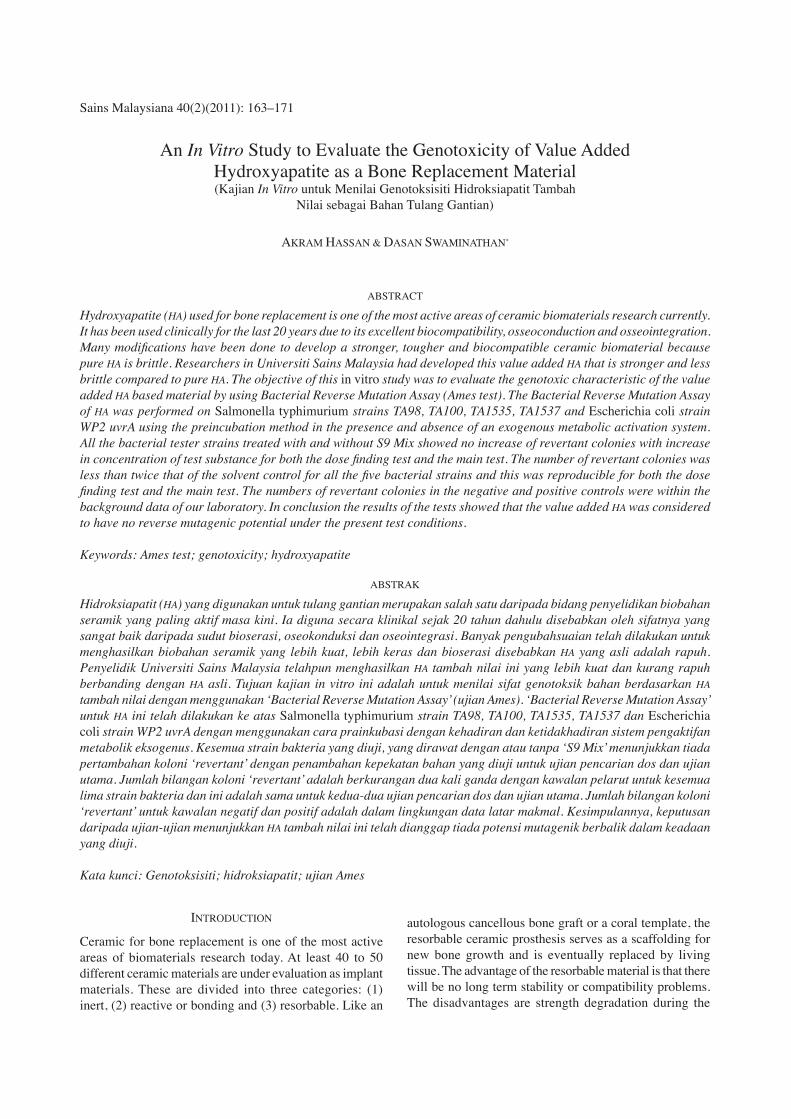
From the stock cultures, 36 µL of bacterial suspension was inoculated in a L-tube containing 18 mL nutrient broth No. 2 (Oxoid Ltd.), the bacterial culture was incubated at 37 ± 0.5°C for 6 to 9 hours with shaking at 57 times/min in a Monod shaker (Figure 1). The viable cells count was calculated from the values which where determined at 660 nanometer by spectrophotometry at the end of incubation.
METHODS
The test was carried out for S. typhimurium TA98, TA100, TA1535, TA1537 and E. coli strain WP2 uvrA using the pre-incubation method both with and without metabolic activation system. Plating was done in triplicate for the negative control and in duplicate for the substance and positive controls.
PROCEDURES
After 0.1 mL of the test substance solution, 0.5 mL of 0.1 M sodium phosphate buffer (pH 7.4) or S9 Mix and 0.1 mL of the bacterial culture were added to a tube, the mixture were incubated 20 min at 37 ± 0.5°C. 2 mL of soft agar was then added to each tube and poured onto a minimal glucose agar plate. After incubation for 48 h at 37 ± 5°C, the number of revertant colonies was counted. As for the sterility test, 0.1 mL of test substance solution, S9 Mix and 0.1 M sodium phosphate buffer (pH 7.4) were poured onto a minimal glucose agar plate and incubated at 37 ± 0.5°C for 48 h to check for the bacterial contamination. Pure water was used as negative control and the following positive controls were used for each bacterial strains.
DOSE FINDING TEST
The test was carried out at the highest dose of 5,000 μg/plate and 4 doses of 2500, 1250, 625 and 313 μg/plate. Growth inhibition was not observed at any of these doses for all the bacterial strains tested both in the presence or absence of S9 Mix. Test plates of TA98, TA100, TA1535, TA1537 and WP2 uvrA with and without S9 Mix showed no increase of revertant colonies for all the doses tested.
TABLE 1. Number of viable cells count
Test TA100(× 10 9/mL)
TA1535(× 10 9/mL)
WP2 uvrA(× 10 9/mL)
TA98(× 10 9/mL)
TA1537(× 10 9/mL)
Dose finding test 1.00 1.12 1.06 1.06 1.02
Main test 1.07 1.00 1.40 1.09 1.03
166
MAIN TEST
Based on the results of the dose finding test, a maximum dose was decided at 5000 µg/plate. The lower 4 doses were obtained by a dilution with a geometric progression of 2, i.e. replicating exactly the procedure carried out in the dose finding test.
MICROSCOPIC OBSERVATION
The state of the revertant colonies (size or number of colonies) and growth inhibition were examined with a stereo microscope.
COLONY COUNTING
The number of colonies were counted with a manual counter or a colony analyzer (Protocol). Each plate was counted three times and the average of the three counts was adopted as the number of revertant colonies on the plate. The average number of revertant colonies for each dose was calculated as the average plate count for a set of duplicate. Decimals of the average figures were rounded off.
INTERPRETATION OF THE RESULTS
Signs of toxicity or growth inhibition for all the bacterial strains under the test condition was described in this report. The test substance was judged to be negative, when the number of revertant colonies was less than twice that of the negative control.
ANALYSIS
Non statistical procedures For this study, a non-statistical procedure was used to evaluate the results of Salmonella experiments (Mortelmans & Zeiger 2000).• Positive: A compound is considered a mutagen if
it produces a reproducible, dose-related increase in the number of revertant colonies in one or more strains. A minimum fold increase, usually 2–3 fold, in revertants (over the solvent control) is the cut-off between a mutagenic and nonmutagenic response. A compound is considered a weak mutagen if it produces a reproducible, dose-related increase in the number of revertant colonies in one or more strains but the number of revertants is not double of the background.
• Negative: A compound is considered a nonmutagen if no dose-related increase in the number of revertant colonies is observed in at least two independent experiments.
• Inconclusive: If a compound cannot be identified clearly as a mutagen or a nonmutagen, the results are classified as inconclusive.
RESULTS
RESULTS OF PLATE COUNTS OF RANGE FINDING TESTS
In the preliminary experiment, the test substance was carried out at the highest dose of 5,000 µg/plate for detection of cytotoxicity. Growth inhibition was not observed and
TABLE 2. Positive controls of bacterial strains
TA100 TA1535 WP2 uvrA TA98 TA1537S9 Mix (-) AF-2
0.1 µg/plateNaN30.5 µg/plate
AF-20.05 µg/plate
AF-20.01 µg/plate
ICR-1911 µg/plate
S9 Mix (+) 2AA0.5 µg/plate
2AA2 µg/plate
2AA10 µg/plate
2AA1 µg/plate
2AA2 µg/plate
FIGURE 1. Overview of bacterial reverse mutation test (Ames Test) Process
167
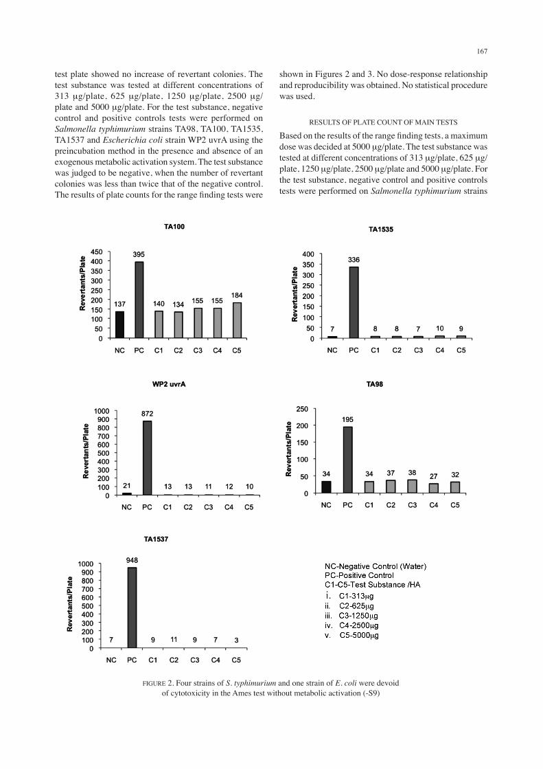
test plate showed no increase of revertant colonies. The test substance was tested at different concentrations of 313 µg/plate, 625 µg/plate, 1250 µg/plate, 2500 µg/plate and 5000 µg/plate. For the test substance, negative control and positive controls tests were performed on Salmonella typhimurium strains TA98, TA100, TA1535, TA1537 and Escherichia coli strain WP2 uvrA using the preincubation method in the presence and absence of an exogenous metabolic activation system. The test substance was judged to be negative, when the number of revertant colonies was less than twice that of the negative control. The results of plate counts for the range finding tests were
shown in Figures 2 and 3. No dose-response relationship and reproducibility was obtained. No statistical procedure was used.
RESULTS OF PLATE COUNT OF MAIN TESTS
Based on the results of the range finding tests, a maximum dose was decided at 5000 µg/plate. The test substance was tested at different concentrations of 313 µg/plate, 625 µg/plate, 1250 µg/plate, 2500 µg/plate and 5000 µg/plate. For the test substance, negative control and positive controls tests were performed on Salmonella typhimurium strains
FIGURE 2. Four strains of S. typhimurium and one strain of E. coli were devoid of cytotoxicity in the Ames test without metabolic activation (-S9)

168
TA98, TA100, TA1535, TA1537 and Escherichia coli strain WP2 uvrA using the preincubation method in the presence and absence of an exogenous metabolic activation system. The test substance was judged to be negative, when the number of revertant colonies was less than twice that of the negative control. The results of plate counts for the main tests were shown in Figures 4 and 5. No dose-response relationship and reproducibility was obtained. No statistical procedure was used.
DISCUSSION
Before new materials are approved for medical use, mutagenesis systems to exclude cytotoxic, mutagenic or carcinogenic properties are applied worldwide. Although indispensable within the framework of in vitro evaluation, these screening procedures are usually very work-intensive and time-consuming. They must be carried out for the raw material as well as the manufactured implant in order to exclude the possibility that the properties of the
FIGURE 3. Four strains of S. typhimurium and one strain of E. coli were devoid of cytotoxicity in the Ames test with metabolic activation (+S9)

169
material are influenced by the industrial manufacturing process (Katzer et al. 2002). To reduce the number of biomechanical studies, evaluation of new biomaterials should nowadays begin with in vitro cytotoxicity and mutagenicity tests. This applies for the development of both temporary and permanent implants and prostheses and for permanent implant particularly under the aspect that carcinogenic potential is often the consequence of chronic exposure to minute concentrations (Katzer et al. 2002). This test is commonly employed as an initial
screen for genotoxic activity and in particular, for point mutation-inducing activity. An extensive data base has demonstrated that many chemicals that are positive in this test also exhibit mutagenic activity in other tests. There are examples of mutagenic agents which are not detected by this test; reason for these shortcomings can be ascribed to the specific nature of the endpoint detected, differences in metabolic activation or differences in bioavailability. On the other hand, factors which enhance the sensitivity of the bacterial reverse mutation test can lead to overestimation
FIGURE 4. The test for mutagenesis in five strains without metabolic activation (-S9) with different concentrations of HA, gave no evidence for mutagenic effects

170
of mutagenic activity (OECD 1997). In this study, all the bacterial tester strains treated with and without S9 Mix showed no increase of revertant colonies with increase in concentration of test substance for both the range finding test and the main test. The number of revertant colonies was less than twice that of the solvent control for all the five bacterial strains and this was reproducible for both the dose finding test and the main test. Positive results from the bacterial reverse mutation test indicate the substance
induces point mutations by base substitutions or frameshifts in the genome of either Salmonella typhimurium and/or Escherichia coli. Negative results indicate that under the test conditions, the test substance is not mutagenic in the tested species. Although all known assays can yield false-positive and false-negative results, experience shows that the combination of two different test methods is a reliable parameter for determining carcinogens which are a risk to human health (Katzer et al. 2002)
FIGURE 5. The test for mutagenesis in five strains with metabolic activation (+S9) with different concentrations of HA, gave no evidence for mutagenic effects
171
CONCLUSION
The present in vitro evaluation indicated that the value added HA was not genotoxic. The genotoxic effect was absent at different concentrations of 313 µg/plate, 625 µg/plate, 1250 µg/plate, 2500 µg/plate and 5000 µg/plate for all the bacterial tester strains treated with and without S9 Mix. No dose-response relationship was obtained in this study for HA. In conclusion, the results of the tests conducted in this research showed that the value added HA was considered to have no reverse mutagenic potential and is a biocompatible biomaterial under the present test conditions. This bone replacement material is now being widely used in Dentistry and Orthopedic surgery in Malaysia and in other countries because of its biocompatibility with human tissue and cost effectiveness.
REFERENCES
Anderson, J.C. 1990. Ceramics and composite. In Material Science, 4th Edition. Chapman & Hall. pp 317-322.
Bajpai, P.K., Wyatt, D.F. & Gilles, N.M. 1976. Use of calcium aluminate phosphorus pentoxide ceramics as bone substitutes. Clin Res. 24: 524A.
Chauvel-Lebret, D.J., Auroy, P., Tricot-Doleux, S. & Bonnaure-Mallet, M. 2001. Evaluation of the capacity of the SCGE assay to assess the genotoxicity of biomaterials. Biomaterials 22: 1795-1801.
Costantino, P.D., Friedman, C.D., Jones, K., Chow, L.C., Pelzer, H.J. & Sisson, G.A. 1991. Hydroxyapatite cement. I. Basic chemistry and histologic properties. Arch Otolaryngol Head Neck Surg. 117: 379-384.
Ferraro, J.W. 1979. Experimental evaluation of ceramic calcium phosphate as a substitute for bone grafts. Plast Reconstr Surg. 63: 634.
ISO (International Organization for Standardization) 10993-3. 1992. Test for genotoxicity, carcinogenecity and reproductive toxicity. Biological Evaluation of Medical Devices - Part 3: 1-7.
Jarcho, M. 1981. Calcium phosphate ceramics as hard tissue prosthetics. Clin Orthop Related Res. 157: 259-278.
Johnson, G.M., Upman, P.J. & Wallin, R.F. 1998. A practical guide to ISO 10993-3: Genotoxicity. An MD & DI October 1998 Column: 1-2.
Katzer, A., Marquardt, H., Westendorf, J., Wening, J.V. & von Foerster, G. 2002. Polyetheretherketone-Cytotoxicity and mutagenicity in vitro. Biomaterials 23: 1749-1759.
Li, J., Hermensson, L. & Soremark, R. 1993. Evaluation of biocompatibility of various ceramic powders with human fibroblast in vitro. Clin Mater. 12: 197-201.
Meffert, R.M., Thomas, J.R., Hamilton, K.M. & Brownstein, C.N. 1985. Hydroxyapatite as an alloplastic graft in the treatment of human periodontal osseous defects. J Periodontol. 56: 63-73.
Mortelmans, K. & Zeiger, E. 2000. The Ames Salmonella/microsome mutagenicity assay. Mutation Res, 455: 29-60
OECD (Organization for Economic Cooperation and Development) TG 471. 1997. Mutagenicity: Reverse mutation test using bacteria. Bacterial Reverse Mutation Test: 1-11.
Piconi, C. & Maccauro, G. 1999. Zirconia as a ceramic biomaterial. Biomaterials 20: 1-25.
Smith, W.F. 1996. Mechanical properties of ceramics. In Principles of Materials Science and Engineering. 3rd Edition. McGraw-Hill Inc: 633.
William, D.F. 1987. Definitions in Biomaterials. In Progress in Biomedical Engineering 4: 54.
Akram Hassan School of Dental Sciences Universiti Sains Malaysia 16150 Kubang Kerian Kelantan, Malaysia
Dasan Swaminathan*
Department of Oral PathologyOral Medicine and Periodontology Faculty of DentistryUniversity of Malaya50603 Kuala LumpurMalaysia
*Corresponding author; email: [email protected]
Received: 3 March 2010Accepted: 9 June 2010