an electrophoretic study of immune sera …kendallasmith.com/pdf/tiselius_kabat.pdf · an...
TRANSCRIPT
AN ELECTROPHORETIC STUDY OF IMMUNE SERA AND PURIFIED ANTIBODY PREPARATIONS*t
BY ARNE TISELIUS, P~r.D., AND ELVIN A. KABAT, ProD.
(From the Institute of Physical Chemistry, University of Upsala, Upsala, Sweden)
(Received for publication, September 20, 1938)
Studies on the eleetrophoresis of serum have shown that besides the albumin normal sera possess three separate globulin components differing in mobility and designated as cz, /3, and ~' (1). The rela- tionship of antibodies to these components is of considerable impor- tance. Differences in the antibody globulin formed in various animal species have already been found by ultracentrifugal studies (2, 3) and in electrophoresis (4) as well as by immunological means (5). It has also been shown (1) that in an antiserum to crystalline egg albumin, the antibody migrated with the slowest (3') component and could be isolated in one of the cells of the apparatus and analyzed for antibody content. The work reported in this communication is an attempt to study and compare the electrochemical properties of the antibody in the original sera with the purified antibody preparations described in (6) and to measure the isoelectric points of these purified preparations. I t is also of importance to make use of and correlate the results obtained using both the mutually independent electro- phoretic and ultracentrifugal methods since molecular weight homo- geneity does not necessarily mean electrochemical homogeneity and vice versa. Since the methods previously described for determining the concentration of the various components are not sufficiently precise, especially in systems of several components, the Lamm scale method (7) has been used with the electrophoresis apparatus (8) and has provided a quantitative method of following changes in the various serum components on removal of antibody.
* The expenses of this investigation were defrayed by grants from the Andersson and Nobel Foundations.
t A preliminary note was published in Science, 1938, 87, 312.
119
on March 29, 2006
ww
w.jem
.orgD
ownloaded from
120 ELECTP.OPHORETIC STUDY O~' TMMUNE SERA
Methods
The new Tiselius electrophoresis apparatus (8), in which sections of the U tube may be moved with respect to one another and cut off the column of solution into four parts to permit isolation of various components, was used. The appara- tus is designed to carry strong currents, which makes possible adequate resolution of the components of serum even in the presence of large amounts of salt. Two methods of observation of the migrating boundaries were applied, Toepler's Schlieren method (9) and the Lamm scale method (7), both depending on refractive index changes in the solution due to the migrating boundary. The former of these in which each component appears as a black band in the U tube image in the focus of the camera, provides a convenient way of following the experiment by direct visual observation. In the latter method an equidistant scale placed behind the U tube is photographed at intervals through the solution. These photographs are compared microscopically with those from a reference scale photographed before the current is started. The displacements of the scale lines, Z, from their positions on the reference scale are plotted as ordinates against the corresponding positions in the U tube cell as abscissae yielding a curve in which each electrochemically distinct component in the solution will have its own peak (Fig. 1) provided the differences in mobility are sufficiently large and the
dc experiment run long enough to produce adequate separation. Since Z = k dx'
where k depends on the refractive index increment of the migrating substance and on known apparatus constants, it is evident that integration of the entire individual curve corresponding to a single peak will give the concentration, c, of the substance having the mobility of that peak. One may thus obtain from the same curve the mobility and concentration of the electrochemically different proteins in the solution. A micro modification of the apparatus in which 2 ml. of solution could be used was found convenient for the isoelectric point measurements on the purified antibody preparations.
In practice, however, since only data on the relative concentrations of the various components were desired and since nitrogen content and the area under each peak were related to protein concentration, it was not necessary to calculate absolute protein concentration in each case. A base line for area measurements was determined by comparing the reference scale and photograph in the adjacent ceils where no boundaries appeared.
I t was also found convenient to compensate the boundary out from under the glass plates by a clockwork mechanism lowering an ebonite rod into the solution (8) before starting the current and then measuring the distance moved by an eyepiece scale fixed to the camera, using the Schlieren method, and in this manner to calculate the mobility.
All experiments were conducted in buffer solutions containing 0.15 x~ NaCI q- 0.02 ~ total phosphates at the desired pH. In the more acid range, the saline was buffered with acetate buffer in which the NaAc concentration was 0.02 x~. This
on March 29, 2006
ww
w.jem
.orgD
ownloaded from
ARNE TISELIUS AND ELVIN A. KABAT 121
high salt concentration was found necessary since some of the horse and pig antibodies precipitated from solution if the usual 0.1 ionic strength buffers were employed. This procedure also enables the direct comparison of mobilities at different pH since the total ionic strength of the solution is but slightly changed by variation of the buffer and has the additional advantage of permitting the direct analysis of the solutions for antibody nitrogen by the absolute quantitative precipitin (10) and agglutinin (11) methods. The high salt concentration, how- ever, does not permit the use of as high voltages as in 0.1 ionic strength buffer. A potential of 120 volts was usually employed giving a current of about 26 to 28 milliamperes and a potential gradient of about 3.4 volts per cm. with the ordinary apparatus. Higher voltages increased the risk of heat convection currents.
For the scale method experiments it was found advisable to use a 1:4 diluted serum to obtain optimum scale line displacements. In the case of unfractionated horse sera, however, it was not possible to determine concentration with the scale method since the curves did not come within a reasonable distance of the base line between components. This seemed also to be the case with the pig serum studied, but in rabbit and monkey antisera determination of concentration was quite satisfactory, These differences between horse and rabbit sera were most marked in higher concentrations and may perhaps indicate partial interaction or com- pound formation between the various globulin components of horse antisera (cf. Kendall, 12).
The usual procedure for studying the distribution of antibody in serum was to dilute the unabsorbed serum 1:4 and dialyze against buffer overnight. Another sample of the same serum was absorbed by addition of the proper amount of antigen and the combined supernatant and washings of the precipitate were diluted to the same volume as the unabsorbed serum and also dialyzed against buffer. In this manner the other components of the serum were present in unchanged concentration.
RESULTS
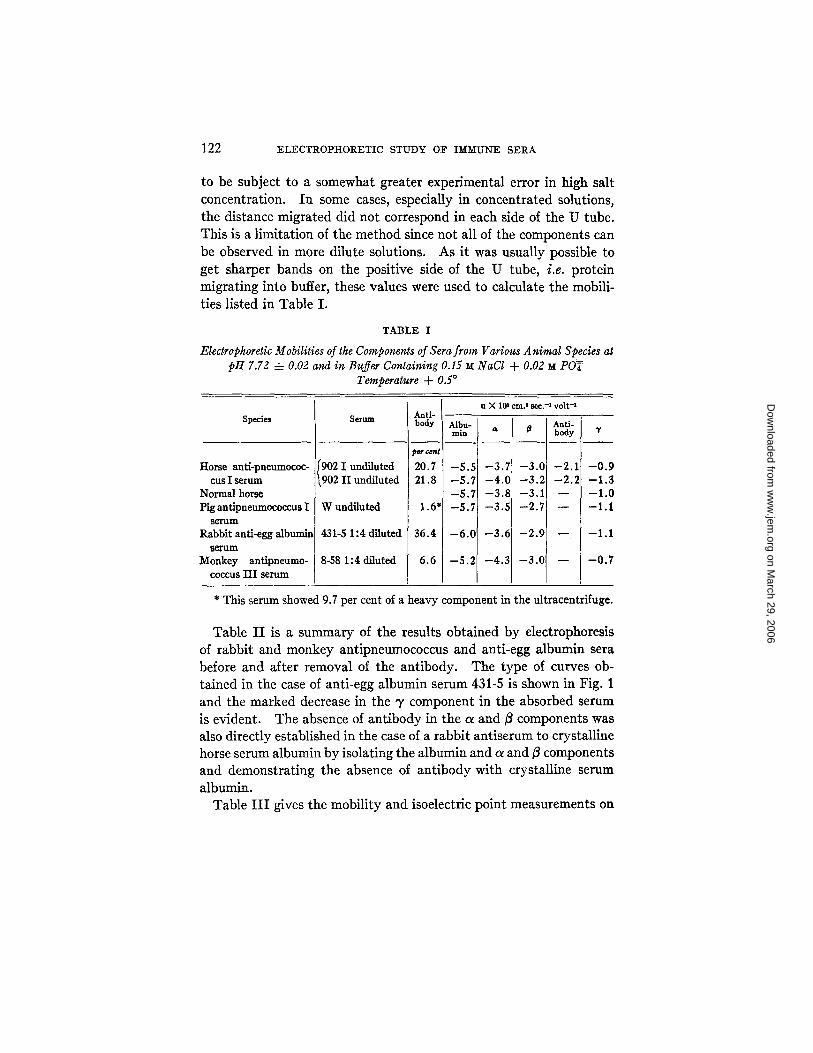
Tab le I is a s u m m a r y of the mobi l i ty d a t a ob ta ined for the an t i se ra f rom different species in the saline phospha te buffer mix ture a t p H
7.72 -4- 0.02. I t will be observed tha t the mobi l i ty of all the compo-
nents is less than in ord inary 0.1 ionic s t rength buffer (c]. 1). I t
can also be no ted t h a t in the horse an t ipneumococcus sera a new componen t migra t ing between j3 and V was present . T h a t this com- ponen t d isappeared on remova l of the an t i body (20.7 per cent of the
to ta l protein) can be seen f rom the Sckliere~ pho tog raph (Fig. 2) of 902, before and af ter absorpt ion of the an t ibody. No new
componen t was observed in the o ther sera a t the same p H . The mobi l i ty values, as measured for the slower components , would seem
on March 29, 2006
ww
w.jem
.orgD
ownloaded from
122 ELECTROPHORETIC STUDY O~P I~M'LTNE SERA
to be subject to a somewhat greater experimental error in high salt concentration. In some cases, especially in concentrated solutions, the distance migrated did not correspond in each side of the U tube. This is a limitation of the method since not all of the components can be observed in more dilute solutions. As it was usually possible to get sharper bands on the positive side of the U tube, i.e. protein migrating into buffer, these values were used to calculate the mobili- ties listed in Table I.
TABLE I
Electrophoretic Mobilities of the Components of Sera from Various Animal Species at ptt 7.72 -4- 0.02 and in Buffer Containing 0.15 ~ NaCl -b 0.02 ~ POT
Temperature + 0.5 °
Species
Horse anfi-pneumococ-
cus I serum
Normal horse
Pig antipneumococcus I s e r u m
Rabb i t anti-egg a lbumin
~ r u m
Monkey ant ipneumo-
coccus I I I serum
Serum
902 1 undiluted 902 II undiluted
W undiluted
431-5 1:4 diluted
8-58 1:4 diluted
Anti- body
20.7
21.8
1.6
36.4
6.6
u X 10 6 cm. 2 sec.-X volt-1
Albu- min I
-5.5 -3.~ -8.7 -4.( -5.7 --3.4 -5.7 -3.~
- 1.7 $3.C - LO - - 3 . 2
- ~.8 - - 3 . 1
- L5 - - 2 . 7
--6.01 - -3 .6
-5.2] -4.3
- .C
- - , . 2
- , . 1
- , . 7
-2.9
- - 3 . 0
~ t i - body
- - 2 . 1
- - 2 . ~
-0.9 -1.3 -1.0 -1.1
-1.1
- 0 . 7
* This serum showed 9.7 per cent of a heavy component in the ultracentrifuge.
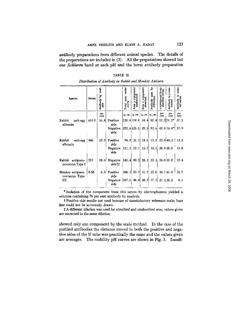
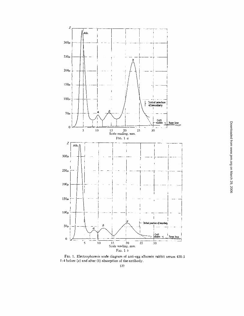
Table I I is a summary of the results obtained by electrophoresis of rabbit and monkey antipneumococcus and anti-egg albumin sera before and after removal of the antibody. The type of curves ob- tained in the case of anti-egg albumin serum 431-5 is shown in Fig. 1 and the marked decrease in the '7 component in the absorbed serum is evident. The absence of antibody in the a and/3 components was also directly established in the case of a rabbit antiserum to crystalline horse serum albumin by isolating the albumin and a and fl components and demonstrating the absence of antibody with crystalline serum albumin.
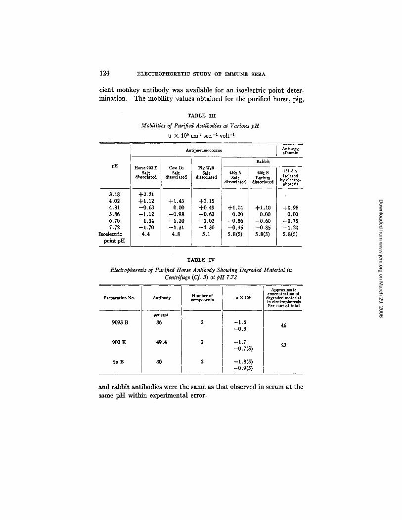
Table I I I gives the mobility and isoelectric point measurements on
on March 29, 2006
ww
w.jem
.orgD
ownloaded from
ARNE TISELIUS AND ELVlN A. KABAT 123
antibody preparations from different animal species. The details of the preparations are included in (3). All the preparations showed but one Schlierer~ band at each pH and the horse antibody preparation
TABLE II
Distribution of Antibody in Rabbit and Monkey Antisera
Species
Rabbit anti-egg albumin
Rabbit anti-egg albumin
Rabbit antipneu- mococcus Type I
Monkey antipneu- mococcus Type HI
Serum
431-5
446
517
8-58
per cent
36.4
12.3
18.6
6.6
Positive side
Negative side
Positive side
Negative side
Negative sidet~
Positive side
Negative side
94.9T I
111.5
143.4J I
206.1
217.6]
I
¢ * o
sq.cm, g.cm.
229.4 19.8
252.4 25.1
31.1
32.1
80.5
53.7
46.4
o#
ta
2,
Sq. I
34
2 c.
2~
t
i °
per Cent
37.2
37.9
13.2
13.8
19.6
10.7
8.1
* Isolation of the component from this serum by electrophoresis yielded a solution containing 76 per cent antibody by analysis.
t Positive side results not used because of unsatisfactory reference scale; base line could not be accurately drawn.
A different dilution was used for absorbed and unabsorbed sera; values given are corrected to the same dilution.
showed only one component by the scale method. In the case of the purified antibodies the distance moved in both the positive and nega- tive sides of the U tube was practically the same and the values given are averages. The mobility pH curves are shown in Fig. 3. Insuiii-
on March 29, 2006
ww
w.jem
.orgD
ownloaded from
124 ELECTROPHORETIC STUDY OF IMMUNE SERA
cicnt monkey antibody was available for an isoelcctric point deter- mination. The mobility values obtained for the purified horse, pig,
TABLE III
Mobilities of Purified Antibodies at Various pH
u × 105 era. 2 sec. -1 volt -1
Antlpneumococcus Anti-egg albumin
Rabbit pH
3.18 4.02 4.81 5.86 6.70 7.72
Isoelectric point pH
Horse 902E Salt
dissociated
+2.21 +1.12 -0 .63 --1.12 --1.34 --1.70
4.4
Cow DI Salt
dissociated
+1.43 0.00
-0 .98 -1 .20 -1.31
4.8
Pig WiB Salt
dissociated
+2.15 +0.49 --0.62 --1.02 --1.30
5.1
45~ A Salt
dissociated
+1.04 0.00
--0.86 --0.95 S.S(S)
4S~ B Barium
dissociated
+1.10 0.00
--0.60 --0.85 s.s(s)
431-5 "r Isolated
by electro- phoresis
+0.95 0.00
-0.75 -1.20 S.8(S)
TABLE IV
Electrophoresis of Purified Horse Antibody Showing Degraded Material in Centrifuge (Cf. 3) at pH 7.72
Approximate concentration of
Preparation No. Antibody Number of components u X 106 degraded material in electrophoresis Per cent of total
9093 B
902 K
Sn B
~ G e n |
86
49.4
30
--1.6 --0.3
-1.7 -0.7(s)
-i.8(s) -o.9(s)
46
22
a n d r a b b i t an t i bod i e s were t he same as t h a t o b s e r v e d in s e r u m a t t he
same p H w i t h i n e x p e r i m e n t a l error .
on March 29, 2006
ww
w.jem
.orgD
ownloaded from
ANN~ TISELIUS AND ELVIN A. KABAT 125
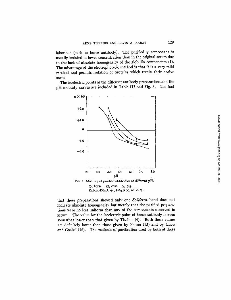
As noted in Table I, pig W serum showed only four components at pH 7.72. From Table I I I and Fig. 3 it will be seen that at pH 7.72, the mobility of pig antibody is practically the same as that of 3" com- ponent and hence they would not be separated at this pH. However, since the isoelectric point of pig antibody is much lower than that of 3' component, a separation was possible by running the pig W serum at pH 5.86 (the isoelectric point of 3" component) and five components were actually observed at this pH.
The production of inhomogeneous degraded components observed in the centrifuge (3) in several horse sera is paralleled in electrophoresis by the production of a new component of lower mobility than the heavy antibody. Table IV gives the mobility values and concentra- tion of the components. 902 K is an antibody solution prepared from a bleeding from horse 902 a year after the bleeding from which 902 E (Table III) was made (cf. 3). This component seems to be present in a concentration about that shown in the ultracentrifuge.
DISCUSSION
The results now obtainable with the new electrophoresis technique (8) make it possible to obtain a more quantitative picture of the rela- tionships of immune substances to the serum globulin and more detaile~d information about the electrochemical properties of immune sera and purified antibodies. Table I indicates that the mobilities of the various normal components in sera from several animal species are approximately the same. The immune horse serum, however, showed the presence of a new component, all of which seemed to be antibody , since it was absent on electrophoresis of the same serum after absorbtion of the antibody as shown in the photograph, Fig. 2. In the ultracentrifuge this serum also showed a heavy component which was the bearer of antibody activity (3). Various species of antibodies showing a heavy component in the ultracentrifuge, how- ever, may not have a mobility sufficiently different from one of the other components at any definite pH to be observed as a distinct component in electrophoresis. Thus pig serum W containing 9.7 per cent of a heavy component in the ultracentrifuge showed only four components at pH 7.72. The purified antibody from this serum had
a mobility so close to that of the ? component as to make effective
on March 29, 2006
ww
w.jem
.orgD
ownloaded from
126 ELECTROPHORETIC STD~DY OF IMMITNE S~.RA
separation at this pH impossible. By selecting a pH at which the difference was more pronounced, the pig serum could be shown to contain five components.
The other group of sera in which the antibody was found to have the same molecular weight as the '7 globulin fraction showed no new component. A comparison of the mobility pH curves (Fig. 3) shows no differences between rabbit antibody solutions prepared by salt dissociation (4562 A), barium dissociation (456~ B), or electrophoretic methods (431-5 3'). The rabbit and monkey antisera, because of the wide separation of the peaks, were easily adapted to quantitative measurements. Table II and Fig. 1 give a comparison of the change in the electrophoresis scale diagram due to the removal of antibody. The values for percentage of antibody obtained by analysis for anti- body nitrogen and total nitrogen (10, 11) (Table II, column 3) and those obtained by integration of the electrophoresis diagrams of the unabsorbed and absorbed sera (last column Table II) are well within experimental error and indicate that all of the antibody is contained in the 3' globulin fraction. I t will be seen that both sides of the U tube are in good agreement for percentage of antibody, but that the total area on the negative side is always greater than that on the positive side. This is perhaps due to the so called ~ boundary (1) observed in most of these sera. The ~ boundary is now thought to be a ~hift in total protein concentration due to a discontinuity in buffer concentra- tion and conductivity formed near the starting position of the protein boundary in concentrated solutions (13). I t has not been included in the curves. The agreement between the two sides in itself indicates that the occurrence of a ~ boundary does not alter the relative con- centration of the other components.
In those antisera in which the antibody occurs in the slowest migrating fraction, the electrophoresis method could be used to isolate fairly pure antibody. I t is of great importance to select antisera in which a very high percentage of the "y component is antibody. On immunization there occurs an increase in total serum protein, as well as in total globulin and antibody (cf. for example 14); therefore determination of the amount of antibody nitrogen would not be a sufficient guide to purification by the electrophoresis method. The agreement between the percentage of antibody in the ~, globulin
on March 29, 2006
ww
w.jem
.orgD
ownloaded from
300u
250u
200~
150~
100u
50~
300,-
250~
200u
/
i ,
{
I : 7
, F . I
•-i/
i
i o f ~
,. / co,1
5 10 15 20 25 30 Scale reading, mm.
FIG. 1 a
i._ i i i ..... I
1 s o . - ' - i - ! . . . . . .
100u : [ ~-____ I .... ~j ............. 17 ........
5 10 15 20 25 30 Scale reading, ram.
F~o. 1 b
F~G. 1. Electrophoresis scale diagram of anti-egg albumin rabbit serum 431-5 1 : 4 before (a) and after (b) absorption of the antibody.
127
on March 29, 2006
ww
w.jem
.orgD
ownloaded from

128 ELECTRO]?I-IORETIC STUDY OF IMMUNE SERA
fraction of 431-5 (Table II) and that found by analysis after elec- trophoretic purification suggests the advisability of a preliminary run by the electrophoresis method as an indication of the suitability of the method for purification of the antibody from a particular antiserum. A test bleeding from the animal could be taken and the four times diluted serum run in the electrophoresis apparatus before and after removal of the antibody. From the scale photograph (Fig. 1), the percentage of antibody in the 3" component could be
FIO. 2. Electrophoretic Schlieren photograph of unabsorbed (a) and ab- sorbed (b) antipneumococcus horse serum.
calculated and the advisability of further immunization or of imme- diate purification of the 3' component decided. For this preliminary test, at most 5 ml. of serum would be required for the ordinary size electrophoresis apparatus (8) but with a new cell of the same length but of smaller cross-section area now being built, the required volume of serum would be reduced to about 2 ml. The method should be most useful for obtaining purified antibodies to protein antigens since the salt dissociation methods are not applicable. In those cases in which the antibody is not in the slowest component the method is too
on March 29, 2006
ww
w.jem
.orgD
ownloaded from
ARNE TISELIUS AND ELVIN A. KABAT 129
laborious (such as horse antibody). The purified "r component is usually isolated in lower concentration than in the original serum due to the lack of absolute homogeneity of the globulin components (1). The advantage of the electrophoretic method is that it is a very mild method and permits isolation of proteins which retain their native state.
The isoelectric points of the different antibody preparations and the pH mobility curves are included in Table I I I and Fig. 3. The fact
u X l O 6
+2.0
-F1.0
0
-1.0
-2 .0
2.0 3.0 4.0 5.0 6.0 7.0 8.0 pH
FIo. 3. Mobility of purified antibodies at different pH.
O, horse. D, cow. A, pig. Rabbit 456~ A + ; 456~ B × ; 431-5 ~.
that these preparations showed only one Schlieren band does not indicate absolute homogeneity but merely that the purified prepara- tions were no less uniform than any of the components observed in serum. The value for the isoelectric point of horse antibody is even somewhat lower than that given by Tiselius (4). Both these values are definitely lower than those given by Felton (15) and by Chow and Goebel (16). The methods of purification used by both of these
on March 29, 2006
ww
w.jem
.orgD
ownloaded from
130 ELECTROPHORETIC STUDY OF IMMUNE SERA
workers were non-specific precipitation methods. As has been noted in the experimental section, the inability to integrate the scale dia- grams in concentrated horse sera because of the poor separation of the globulin peaks may possibly be an indication of loose compound formation in these globulins and that the concentration methods of isoelectric precipitation used by these authors (15, 16) resulted in the precipitation of an addition compound of higher isoelectric point (c.f. also Kendall, 12).
Additional evidence that the washed antigen-antibody precipitates (cf. 10, 11) are free from non-specific protein was obtained by dis- solving a washed specific precipitate of egg albumin-anti-egg albumin in excess antigen and running in the electrophoresis apparatus. Two components were observed, one of excess antigen and another of an antigen-antibody compound. There was no component corresponding to ordinary 5' globulin, indicating the absence of the corresponding non-specific 5" component in the original washed precipitate. A detailed study of the inhibition zone in electrophoresis has been completed.
The data concerning the degradation of horse antibody in the animal on continued immunization (3) indicate that it is also accompanied by a definite change in mobility as noted by the presence of two antibody components in a later bleeding from an animal which had previously shown one component (Table IV). Thus 9093 B showed two components in electrophoresis, both in appreciable concentration and a comparison of the original serum absorbed and unabsorbed showed that some of the slower 5" component was removed on absorp- tion of the antibody. Similarly in horse antibody 902 E (Table III) prepared from an earlier bleeding, only one component was observed in electrophoresis and in the centrifuge (3), while in 902 K (Table IV), prepared in the same way from a bleeding taken after another year of immunization, degraded material was present in the centrifuge and a new component of lower mobility was present in electrophoresis. This is probably a manifestation of a process of degradation and removal of antibody protein in the horse after prolonged immunization.
SUM~&RY
1. Antibody produced in the horse migrates as a new serum com- ponent between the /3 and 5" components, whereas rabbit antibody
on March 29, 2006
ww
w.jem
.orgD
ownloaded from
ARNE TISELIUS AND ELVIN A. KABAT 131
is electrophoretically identical with the 3, globulin component of the serum.
2. In rabbit and monkey antisera the percentage of antibody in the serum and in the 3, globulin fraction can be determined by integration of the electrophoresis diagrams of unabsorbed and absorbed sera. Antibody solutions of high purity can be obtained by electrophoretic isolation of the 3' globulin of rabbit antisera in which the percentage of antibody to total "y globulin is high.
3. The isoelectric points of pig, cow, horse, and rabbit antibodies have been determined.
4. In horse sera prolonged immunization is accompanied by the formation of another antibody component of lower mobility.
We wish to thank Professor The Svedberg for his interest in this work and Dr. Michael Heidelberger for his valuable suggestions and assistance in supplying the various antisera and antibody preparations.
BIBLIOGRAPHY
1. Tiselius, A., Biochem. J., 1937, 31, 1464. 2. Heidelberger, M., and Pedersen, K. O., J. Exp. Med., 1937, 65, 393. 3. Kabat, E. A., and Pedersen, K. O., Science, 1938, 87, 372; Kabat, E. A.,
J. Exp. M~., 1939, 69, 103. 4. Tiselius, A., J. Exp. Med., 1937, 65, 641. 5. Heidelberger, M., and Kendall, F. F.., J. Exp. M~I., 1933, 57, 373. Heidel-
berger, M., Kendall, F. E., and Scherp, H. W., J. Exp. Med., 1936, 64, 559. 6. Heidelberger, M.,.and Kendall, F. E., J. Exp. Med., 1936, 64, 161. Heidel-
berger, M., and Kabat, E. A., J. Exp. M~I., 1938, 67, 181. 7. Lamm, O., Nova Acta Regiae Soc. Scient. Upsaliensis, IV, 1937, 10, 6; Z.
physikal. Chem., Abt. A, 1928, 138, 313; Abt. A, 1929, 143, 177. 8. Tiselius, A., Tr. Faraday Sot., 1937, 33, 524. 9. Tiselius, A., Pedersen, K. O., and Eriksson-Quensel, I.-B., Nature, 1937,
139, 546. 10. Heidelberger, M., Sia, R. H. P., and Kendall, F. E., J. Exp. MaL, 1930, 52,
477. Heidelberger, M., Kendall, F. E., and Soo Hoo, C. M., J. Exp. Med., 1933, 68, 137. Heidelberger, M., and Kendall, F. E., J. Exp. M~., 1935, 61, 359.
11. Heidelberger, M., and Kabat, E. A., Proc. Soc. Exp. Biol. and Med., 1934, 31, 595; J. Exp. Med., 1934, 60, 643.
12. Kendall, F. E., J. Clin. Inv., 1937, 16, 921. 13. Tiselius, A., and Svensson, H., to be published. 14. Boyd,'~W. C., and Bernard, H., J. Immunol., 1937, 33, 111. 15. Felton, L. D., J. Immunol., 1931, 9.1,241, and earlier papers. 16. Chow,!B. F., and Goebel, W. F., J. Exp. Meal., 1935, 69., 179.
on March 29, 2006
ww
w.jem
.orgD
ownloaded from