an efficient genotyping method for genome … efficient genotyping method for genome-modified...
TRANSCRIPT

An Efficient Genotyping Method forGenome-modified Animals and HumanCells Generated with CRISPR/Cas9SystemXiaoxiao Zhu1,2*, Yajie Xu1*, Shanshan Yu1*, Lu Lu1,2, Mingqin Ding1,2, Jing Cheng1, Guoxu Song1,2,Xing Gao1, Liangming Yao1, Dongdong Fan1,2, Shu Meng1, Xuewen Zhang1, Shengdi Hu1 & Yong Tian1
1Laboratory of RNA Biology, Institute of Biophysics, Chinese Academy of Sciences, Beijing 100101, China, 2University of ChineseAcademy of Sciences, Beijing 100080, China.
The rapid generation of various species and strains of laboratory animals using CRISPR/Cas9 technologyhas dramatically accelerated the interrogation of gene function in vivo. So far, the dominant approach forgenotyping of genome-modified animals has been the T7E1 endonuclease cleavage assay. Here, we present apolyacrylamide gel electrophoresis-based (PAGE) method to genotype mice harboring different types ofindel mutations. We developed 6 strains of genome-modified mice using CRISPR/Cas9 system, and utilizedthis approach to genotype mice from F0 to F2 generation, which included single and multiplexedgenome-modified mice. We also determined the maximal detection sensitivity for detecting mosaic DNAusing PAGE-based assay as 0.5%. We further applied PAGE-based genotyping approach to detect CRISPR/Cas9-mediated on- and off-target effect in human 293T and induced pluripotent stem cells (iPSCs). Thus,PAGE-based genotyping approach meets the rapidly increasing demand for genotyping of the fast-growingnumber of genome-modified animals and human cell lines created using CRISPR/Cas9 system or othernuclease systems such as TALEN or ZFN.
Laboratory animal models have significantly contributed to the interrogation of gene function in biologicalsystems. In recent years, novel genome editing technologies have been applied to establish animal and humancell models at an increasing speed1,2. Compared with methods using Zinc Finger Nucleases (ZFN) or
Transcription activator TAL-like Effector Nucleases (TALEN), a more recent technology called ClusteredRegularly Interspaced Short Palindromic Repeats (CRISPR)/CRISPR-associated 9 (Cas9) has played an increas-ing role in generating single and multiplexed genome-modified animal models3–8. This method relies on injectingCas9 mRNA together with single guide RNA (sgRNA) into oocytes that were collected from super-ovulatedanimals. The Cas9 protein forms a complex with the sgRNA, upon which the cognate target is identified by thecomplex by recognition of a short trinucleotide NGG protospacer adjacent motif (PAM) sequence9,10.Subsequently, the catalytic activity of Cas9 causes scission of double-stranded DNA and generates double-strandbreaks (DSBs) in target DNA. Cleaved DSBs will be repaired either by a non-homologous end joining (NHEJ)mechanism, which is error-prone and therefore generates indel mutations in the vicinity of DSBs, or by homo-logous recombination (HR), a process that can be exploited for more precise genomic modification11,12. So far,several reports have described the successful generation of genome-modified animal models from various species,including mouse5,8,13, rat14–16, zebrafish17,18, rabbit19, pig20 and monkey21.
As the number of genome-edited animal models and human cell models increases, genotyping is becoming abottleneck, especially for high-throughput screening22,23. Techniques have been developed for the detection ofindel mutations at the targeted loci, such as Surveyor assays (Cel1), T7 endonuclease 1 (T7E1) assays or HighResolution Melt Analysis (HRMA)24,25. Surveyor nuclease and T7E1 are mismatch-specific DNA endonucleasesthat are used for detecting indel mutations generated by genome engineering nuclease such as ZFN andTALEN26–28. The widely used T7E1 endonuclease targets and digests mismatched heteroduplex double-strandDNA, and as a result produces two or more smaller fragments in an enzymatic reaction. The digested DNAfragments can thus be resolved and visualized by agarose gel electrophoresis8. In addition, the re-hybridizedfragments may also subjected to melting curve analysis (HRMA) that is part of the real-time thermocycler25.
OPEN
SUBJECT AREAS:STEM-CELL
BIOTECHNOLOGY
ANIMAL BREEDING
Received20 May 2014
Accepted20 August 2014
Published19 September 2014
Correspondence andrequests for materials
should be addressed toS.H. (shengdihu@
moon.ibp.ac.cn) orY.T. ([email protected])
* These authorscontributed equally to
this work.
SCIENTIFIC REPORTS | 4 : 6420 | DOI: 10.1038/srep06420 1

Fragments that contain mismatches melt at a lower temperature thanperfect duplexes. To date, genotyping of genome-modified mouseand rat models generated using CRIPSR/Cas9 technology is prim-arily done using the T7E1 assay29,30. However, for genotyping oflarge-scale screening events like cell transfection or mouse embryoinjections, the conventional T7E1 and HRMA assays are both time-and labor-consuming23,31. Therefore, it is highly desirable to designsimple and efficient strategies for the genotyping of indel mutations,especially when required for high-throughput screening methods.
Here, we have applied a one-step polyacrylamide gel electrophor-esis-based (PAGE) approach to successfully genotype 6 differentstrains of genome-modified animals and human induced pluripotentstem cells (iPSCs) harboring indel mutations caused by the CRISPR/Cas9 system. Compared to the traditional T7E1 assay, the PAGE-based approach proves to be a more efficient,time- and labor-savingstrategy, without compromising sensitivity during the genotyping ofCRISPR/Cas9-mediated indel mutations. Therefore, our one-stepPAGE-based approach can replace the T7E1 assay as a routine labor-atory protocol for genotyping laboratory animal models and humancell lines generated with the CRISPR/Cas9 system, but also with othernuclease systems, such as TALEN or ZFN.
ResultsSchematic overview of heteroduplexed DNA detection usingPAGE. PAGE-based assays have been used traditionally for thecharacterization of heteroduplex formation, especially for humanviruses32,33. We first asked whether a PAGE-based approach canidentify genome-modified mice carrying CRISPR/Cas9-mediatedindel mutations with both high sensitivity and accuracy. In orderto generate strains of genetically modified mice, we routinely isolated100–250 zygotes collected from superovulated C57/BL6 mice andmicroinjected simultaneously Cas9 mRNA and sgRNA prior to thetransplantation of the zygotes into the oviducts of pseudo-pregnantICR female mice. After DNA isolation from mouse tails in the F0generation, we amplified the genomic regions spanning the sgRNAbinding site using PCR. After brief denaturation and annealing, PCRfragments from genetically modified animals, which contain a mix-ture of indel mutations and wildtype alleles, formed heteroduplexDNA and homoduplex DNA (Figure 1A). Due to the existence of anopen angle between matched and mismatched DNA strands causedby indel mutations, heteroduplex DNA generally migrated at asignificantly slower pace than homoduplex DNA during nativePAGE, thus making it a useful tool to screen founder colonies thatharbor indel mutations (Figure 1B).
Genotyping genome-modified mice generated by CRIPSR/Cas9system by PAGE. In order to validate the PAGE-based approachfor use in our genotyping assay, we constructed DNA vectorsexpressing sgRNAs that target exon 2 of the Them2 locus, which isdriven by a T7 promoter (Supplementary Table S1). After purifica-tion of transcribed sgRNAs and Cas9 mRNA in vitro, we microin-jected Cas9 mRNA and one sgRNA into the cytoplasm of zygotes asdescribed in Table 1. Genomic DNA was extracted for genotypingfrom mouse tails at postnatal day 10. PCR products were amplifiedusing primer pairs as listed in Supplementary Table S2. After briefdenaturation and annealing, PCR products were subjected to 2.5%agarose gel electrophoresis or 15% PAGE. To compare thegenotyping results obtained from the PAGE-based approach withthat of the T7E1 cleavage assay, we further purified the same frac-tion of PCR products and subjected the DNA to T7E1 cleavage. On a2.5% agarose gel, we detected a single band of similar size for all pupsexcept that for mouse #5, confirming the successful amplification ofPCR reactions (Figure 2A). PCR products from mouse #5 yielded oneextra band in addition to the universal band generated from all othermice, indicating that a larger genomic fragment was modified. Usingthe T7E1 cleavage assay, efficient endonuclease activity was
confirmed by identification of one extra digested band in 7 out of10 pups. In mice #3, #7 and #10, we failed to detect any enzymaticdigestion in comparison to other pups, suggesting wildtype alleleswere maintained in these three mice (Figure 2B). To test the validityand accuracy of the PAGE-based approach, we performed this assayin parallel to the T7E1 cleavage assay. In wildtype control mice, PCRamplification yielded a 165-bp band on a 15% PAGE gel (Figure 2C).However, we detected multiplexed bands containing homoduplexDNA and heteroduplex DNA in 7 out of 10 pups analyzed. Pups#6 and #8 displayed a single pair of heteroduplex bands, indicatingone dominant type of indel mutations exists in these two mice. Pups#3, #7 and #10 displayed band patterns identical to those found inwildtype controls, suggesting that no targeting was achieved in thesethree mice. Interestingly, samples from pups #1, #2 and #5 displayedmultiplexed pairs of heteroduplex bands, suggesting that these threefounders contain biallelic or tri-allelic mutations. To confirm ourfindings, we performed further TA cloning and sequencing(Figure 2D). Five clones from each mouse were randomly selectedfor sequencing. In wildtype control mice as well as in pups #3, #7 and#10, only sequences of wildtype alleles were identified. In addition,we failed to detect any Single Nucleotide Polymorphisms (SNP) nearthe sgRNA binding site. Targeted modifications within a size range of216 bp and 137 bp occurred at the Them2 locus in the remainingmice with variable efficiencies, thus confirming the results shown inFigure 2B and 2C.
In addition, we tested our PAGE-based genotyping approach byexamining genome-modified mice by targeting sgRNA to exon 4 ofthe Agbl3 locus. We analyzed a total of 11 animals from the F0generation for identification of founder mice. On a 2.5% agarosegel, PCR reaction yielded a single band for all mice (Figure 2E). Asubsequent T7E1 assay identified mice #3, #5 and #11 as wildtype,while all other mice exhibited bands cleaved by T7E1 (Figure 2F). Inthe 15% PAGE analysis, all mice except #3, #5 and #11 displayed
Figure 1 | Schematic overview of PAGE-based genotyping protocol foridentification of CRISPR/Cas9-mediated indel mutations.(A) Illustration of heteroduplex DNA formation during denaturation and
annealing. Dark green bars represent four DNA strands (a–d) in cells
harboring monoallelic mutations (orange box). After denaturation and
annealing, two types of homoduplex DNA and two types of heteroduplex
DNA were formed. (B) Identification of heteroduplex DNA fragments by
15% PAGE assay. Since heteroduplex DNA migrates slower due to
formation of an open angle between matched and unmatched genomic
regions, homoduplex DNA and heteroduplex DNA are easily identified
based on their mobility rate.
www.nature.com/scientificreports
SCIENTIFIC REPORTS | 4 : 6420 | DOI: 10.1038/srep06420 2
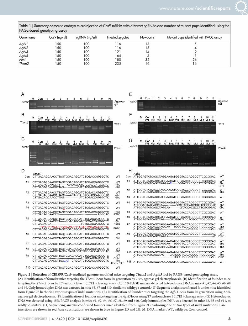
Table 1 | Summary of mouse embryo microinjection of Cas9 mRNA with different sgRNAs and number of mutant pups identified using thePAGE-based genotyping assay
Gene name Cas9 (ng/ml) sgRNA (ng/ml) Injected zygotes Newborns Mutant pups identified with PAGE assay
Agbl1 150 100 116 13 5Agbl2 150 100 116 13 4Agbl3 150 100 121 14 9Agbl5 150 100 64 5 3Nmi 150 100 180 32 26Them2 150 100 235 19 16
Figure 2 | Detection of CRISPR/Cas9-mediated genome-modified mice targeting Them2 and Agbl3 loci by PAGE-based genotyping assay.(A) Identification of founder mice targeting the Them2 locus from F0 generation by 2.5% agarose gel electrophoresis. (B) Identification of founder mice
targeting the Them2 locus by T7 endonuclease 1 (T7E1) cleavage assay. (C) 15% PAGE analysis detected heteroduplex DNA in mice #1, #2, #4, #5, #6, #8
and #9. Only homoduplex DNA was detected in mice #3, #7 and #10, similar to wildtype control. (D) Sequence analysis confirmed founder mice identified
from Figure 2B harboring various types of indel mutations. (E) Identification of founder mice targeting the Agbl3 locus from F0 generation using 2.5%
agarose gel electrophoresis. (F) Identification of founder mice targeting the Agbl3 locus using T7 endonuclease 1 (T7E1) cleavage assay. (G) Heteroduplex
DNA was detected using 15% PAGE analysis in mice #1, #2, #4, #6, #7, #8, #9 and #10. Only homoduplex DNA was detected in mice #3, #5 and #11, as
wildtype control. (H) Sequencing analysis confirmed founder mice identified from Figure 2G harboring one or two types of indel mutations. Base
insertions are shown in red; base substitutions are shown in blue in Figure 2D and 2H. M, DNA marker; WT, wildtype; Con, control.
www.nature.com/scientificreports
SCIENTIFIC REPORTS | 4 : 6420 | DOI: 10.1038/srep06420 3

various migration patterns of heteroduplex DNA (Figure 2G).Sequencing analysis confirmed that 9 out of 13 mice possessed dif-ferent indel mutations (Figure 2H). However, mice #3, #5 and #11yielded only wildtype homoduplex bands, which were confirmed bysequencing. Thus, our data suggest that genotyping based on PAGEachieves results consistent with the T7E1 cleavage assay.
Genotyping multiplexed genome-modified mice generated byCRIPSR/Cas9 system with PAGE. We further examinedmultiplexed genome-modified mice from the F0 generationgenerated using simultaneous targeting of sgRNAs to both Agbl2and Agbl1 loci. The relevant sgRNA sequences and injectionconditions are listed in Table 1 and Supplementary Table S1. Asingle sgRNA was designed to target exon 7 of the Agbl2 locus.PCR with genomic DNA of all 13 pups genotyped yielded oneband of a 137-bp product, as shown by agarose gel assay(Figure 3A). Next, all PCR products were subjected to a 15%PAGE assay, and of those 13 pups, we identified mice #4, #8, #9and #11 as containing a heteroduplex formation (Figure 3B). Toconfirm these results, we subjected all samples to TA cloning andsequencing (Figure 3C). Mice #4, #8 and #9 possessed an identicalmutation, with a deletion of 2 base pairs, whereas mouse # 11possessed a genomic deletion of 6 base pairs length. Within theAgbl1 locus, a sgRNA that target exon 2 of the Agbl1 gene wasdesigned. As expected, all mice displayed similar band patternswhen run on a 2.5% agarose gel (Figure 3D). On the 15% PAGEgel, 5 out of 13 mice displayed a homoduplex DNA band, identicalto that observed for wildtype mice (Figure 3E). Furthermore, weobserved a pair of heteroduplex DNA bands in mice #2, #4, #9,#10 and #12, which was confirmed by subsequent sequencinganalysis (Figure 3F). Notably, both mouse #4 and #9 exhibitedbiallelic indel mutations in the two loci Agbl2 and Agbl1. Together,
these data suggest that a PAGE-based genotyping approach canindeed identify multiplexed indel mutations in mice.
Genotyping F1 and F2 generations of genome-modified mice withPAGE. Given the genotyping results described above, we expectedthat a PAGE-based protocol can also be utilized for genotyping ofgenome-modified mice from F1 and F2 generations. To test this, weproduced F1 generation offsprings by crossing founder #5(Figure 2A) with wildtype C57/BL6 mice (Figure 4A). Genotypingof that generation by 15% PAGE analysis revealed that 5 out of 8 miceharbored the desired indel mutations. For verification, all PCRproducts from mice harboring the desired indel mutations weresubjected to sequencing (Figure 4B). In addition, we bred micecarrying the 3-bp deletion mutation from the F1 generation, andscreened F2 generation offsprings for identification of homozygousmonoallelic animals. We hypothesized that it would not be possibleto identify homozygous indel mutations, unless PCR products fromwildtype alleles were mixed with mutant alleles. We first performedgenotyping analysis by loading PCR samples directly onto 15%PAGE without prior mixing. As shown in Figure 4C, we onlydetected heterozygote mice rather than homozygous monoallelicmice. However, after mixing of the PCR products of mutants withcontrols prior to denaturation and annealing, it was possible toidentify homozygous mice and wildtype mice (Figure 4C). Takentogether, these results suggest that our PAGE-based protocol canbe applied to genotyping of any generation of mice carrying indelmutations.
Detection of various heteroduplexed DNA with PAGE. To betterunderstand the process of heteroduplex formation and its migrationpattern for PAGE analysis, we utilized an array of plasmids carryingindel mutations of the Them2 gene including deletions of 2 bp, 3 bp,
Figure 3 | Identification of CRISPR/Cas9-mediated multiplexed genome-modified mice targeted to Agbl1 and Agbl2 loci by PAGE-based genotypinganalysis. SgRNAs targeting Agbl1 and Agbl2 loci were mixed prior to microinjection into the cytoplasm of oocytes. Pups from F0 generation were
genotyped for targeted modification in Agbl1 and Agbl2 loci. Results from agarose gel electrophoresis of PCR products are shown for the Agbl2 (A) and
Agbl1 (D) loci. (B) Using the PAGE-based genotyping protocol, genomic indel mutations in the Agbl2 locus were detected in mice # 4, #8, #9 and #11,
while all others and wildtype controls exhibited only homoduplex DNA. (C) Sequence analysis confirmed the identification of indel mutations in mice #4,
#8, #9 and #11 from Figure 3B. (E) PAGE analysis identified heteroduplex DNA from the Agbl1 locus in mice #2, #4, #9, #10 and #12. (F) Sequencing
analysis confirmed the identification of indel mutations in mice #2, #4, #9, #10 and #12 from Figure 3E. Note that mice #4 and #9 (shown in red) possessed
indel mutations in both Agbl2 and Agbl1 loci. M, DNA marker; WT, wildtype; Mut, mutant.
www.nature.com/scientificreports
SCIENTIFIC REPORTS | 4 : 6420 | DOI: 10.1038/srep06420 4

4 bp, 7 bp, 8 bp and 16 bp (Figure 5A). As shown in Figure 5B, allplasmids with various indel mutations yielded PCR products ofsimilar size. After mixing PCR products from mutant and wildtypeplasmids followed by denaturation and annealing, all indel muta-tions analyzed were able to produce distinct migration products on a15% PAGE gel (Figure 5C). Noticeably, we were able to detect twotypes of homoduplex DNA in the lanes containing wildtype and -7 bp,-8 bp and -16 bp genomic deletion fragments individually.Since F0 mice may harbor biallelic or more complicated mosaicmutations, we hypothesized that each type of indel mutationshould maintain their specific heteroduplex migration pattern onthe PAGE gel (Figure 5D). We combined PCR products of any twotypes of biallelic mutations from the Them2 locus, and observedsimilar sizes of PCR products among all samples when loaded ontoan agarose gel (Figure 5E). Compared with their corresponding lanesin Figure 5C, different combinations of PCR products carryingbiallelic mutations exhibited specific heteroduplex patterns on the15% PAGE gel (Figure 5C and 5F). Thus, our data suggest that PAGEanalysis can be used to assess the number and even types of indelmutations simply based on their heteroduplex mobility pattern.
Sensitivity assessment of PAGE-based genotyping approach. Toassess the sensitivity of PAGE-based genotyping approach, weperformed PCR reactions with a total of 25 ng DNA templatecontaining various ratios of wildtype and mutant clone DNA fromthe Agbl3 locus, mixed in a 50 ml PCR reaction volume (Figure 6).Primer pairs for amplifying Agbl3 locus were listed in SupplementaryTable S2. PCR products of wildtype 166 bp and mutant 163 bp wereused as loading controls on the agarose gel, and equal amounts ofPCR products of various ratios were loaded into each well(Figure 6A). PCR products were further subjected to T7E1 orPAGE analysis after denaturation and annealing. For the T7E1assay, the minimal detection percentage for mosaic DNA
templates was between 0.5 to 5% (Figure 6B). For the PAGE-basedassay, the maximal detection sensitivity was determined as 0.5%(Figure 6C), which is similar to that of T7E1 assay according to aprevious study34. Because the T7E1 assay allows quantification ofindel frequencies by measuring the intensities of cleaved DNAbands, we further quantified heteroduplexed bands from Figure 6Cand calculated the correlation coefficient (Figure S3). Together, ourdata showed that overall sensitivity for detecting mosaic DNAharboring indel mutations using a PAGE-based approach wassimilar to that of T7E1 assay.
Screening CRISPR/Cas9-mediated on- and off-target mutationsin human 293T and induced pluripotent stem cells with PAGE. Todefine whether our PAGE-based genotyping approach can beemployed in human cell lines harboring indel mutations, plasmidsexpressing Cas9-GFP together with sgRNAs targeting either ATXN1or ATXN2 locus were transfected into human 293T cell lines. Vehicleonly (i.e. lacking sgRNA) and sgRNA targeting a third locus wereused as controls in parallel. Two days post transfection, cellsexpressing GFP were sorted by flow cytometry, and DNA wasextracted for genotyping. Oligos for sgRNA synthesis andgenotyping were designed as listed in Supplementary Table S1 andS3. As expected, PCR products of similar size were detected for allsamples. Using PAGE analysis, we were able to identify one pair ofheteroduplex bands in the sgRNA-ATXN1 lane (marked byasterisks), while no similar band was observed in the controls(Figure 7B). We next electroporated either sgRNA targeting theTBP locus (sgRNA-TBP) or vehicle control (Vehicle), togetherwith a Cas9-GFP plasmid into human iPSCs. Ten days posttransfection, iPSCs were examined for on-target screening. Singlebands of similar sizes were detected on agarose gel (Figure 7C).PAGE-based analysis detected clear heteroduplex DNA in thesgRNA-TBP group (marked by asterisks), not, however, in thevehicle-only group (Figure 7D). Since off-target effects are ofmajor concern when employing the CRISPR/Cas9 system,especially for use in human stem cell research, we wondered if off-target screening could be performed using our PAGE-based assay.To address this issue, we used a novel sgRNA targeting the humanATXN2 locus with a predicted strong off-target effect. All potentialoff-target sites were identified using the CRISPR Design Tool (http://crispr.mit.edu) and BLAST algorithm, as described previously35. Weidentified three off-target sites, namely ADHHC8, SPOCK2 andWNT6, all of which possessed 1 to 3 mismatches to the originalATXN2 sgRNA sequences (Supplementary Table S3). PCRproducts from all three genes revealed a single band of similar sizewhen loaded onto an agarose gel (Figure 7E, Supplementary TableS2). Using PAGE analysis, we identified heteroduplex DNA bands inthe WNT6 locus (marked with an asterisk), which was absent in thecontrols (Figure 7F). We also examined off-target loci in humaniPSCs using our PAGE-based approach. No off-target effects weredetected within any of the predicted loci in iPSCs transfected withsgRNA-TBP (Supplementary Figure S2 and Table S4). All off-targetscreening results were confirmed by sequencing (data not shown).Taken together, our data suggest that PAGE-based genotyping is anefficient method for analyzing indel mutations, as well as forscreening off-target effects in human cells.
DiscussionWe present an efficient one-step method for the genotyping of indelmutations created using the CRIPSR/Cas9 system, in a variety ofmouse strains and human cell lines. Our data show that thisPAGE-based approach can detect different types of indel mutations,with both high sensitivity as well as efficiency. First, the one-stepPAGE-based genotyping approach does not require an enzymaticreaction, which can produce false negative results due to incompletedigestion of mismatched DNA fragments. Instead, the PCR products
Figure 4 | Identification of F2 generation homozygous Them2 mutantmice via PAGE-based genotyping protocol. Mouse #5 (Figure 2D) were
bred with wildtype C57/BL6 mice to obtain F1 mice. We further
intercrossed mice from F1 to obtain homozygous F2 mutant mice.
(A) Identification of F1 offsprings using PAGE-based genotyping
approach. Five out of nine F1 offsprings were identified by PAGE analysis
as carrying one type of indel mutations in the Them2 locus. (B) Sequencing
results from F1 offsprings revealed mice harboring a 3-bp deletion.
(C) Identification of F2 offsprings using PAGE-based genotyping approach.
F2 mice #3, #4, #5 and #7 harboring heterozygous indel mutations in their
Them2 locus were identified by PAGE assay (upper panel). When mixing
PCR products from control mice with PCR products from F2 mice, mice #2
and #8 exhibited migration patterns from heteroduplex DNA (marked by
asterisks), indicating homozygous indel mutation (bottom panel). M, DNA
marker; WT, wildtype; Homo, homozygote; Het, heterozygote.
www.nature.com/scientificreports
SCIENTIFIC REPORTS | 4 : 6420 | DOI: 10.1038/srep06420 5

are directly subjected to PAGE-based electrophoresis for mutantallele detection. Second, our PAGE-based approach can detectmosaic indel mutations with similar sensitivity to that of a conven-tional T7E1 assay. Based on our findings, the PAGE-based assay candetect 0.5% to 5% mutant DNA composition, which is in agreementwith a previous report36. Third, since different heteroduplex com-plexes of indel mutations display specific motility pattern when runon a PAGE gel, the number of different types of mutations can bedirectly assessed from evaluating the stained gel, which providesadditional information not available with other methods25. Fourth,manipulating genome-modified human iPS cells by using theCRISPR/Cas9 system requires analysis of both on- and off-targeteffects. The genome-wide binding of Cas9 protein raises considerableconcern over the off-target issue and has been a significant obstaclefor applying genome-editing tools towards regenerative medi-cine37,38. Off-target sites can tolerate up to five mismatches to thesgRNA sequence and many were mutagenized with frequencies com-parable to the intended on-target site due to the genome-wide bind-ing38,39. Our data suggest that the PAGE-based genotyping approachis efficient for screening on- and off-target effects in human iPS cells,and importantly, can do so in a high-throughput manner. Fifth,unlike the RGEN-RFLP approach that allows more precise quan-tification with a reported R25 0.99 at least in one study36, bothT7E1 and PAGE methods failed to achieve R2 close to 0.99 (FigureS3). This indicates that results of the PAGE and T7E1 assay corre-lated poorly with mutation frequencies. Thus, for quantificationpurposes, RGEN-RFLP is the method of choice and PAGE-based
assay is not suitable for quantification of indel mutations. Finally,although RGEN-RFLP method can be used to genotype mutations, itdoes involve multiple steps, such as purification of Cas9 protein,transcription of sgRNA, as well as in vitro enzymatic incubationsteps36. Evidently, PAGE-based approach provides a cost- andlabor-saving strategy suitable for low-budget laboratories for rel-evant genotyping assay. However, if the target genomic DNA frag-ment contains single nucleotide polymorphisms (SNPs) or allelicmutations, both PAGE-based and T7E1 approach may give rise tofalse positive results. Under such cases, combining our PAGEmethod with a RGEN-RPLP approach can overcome this limitationand faithfully detect indel mutations caused by CRISPR/Cas9system36.
In summary, the use of a one-step PAGE-based approach forgenotyping of CRISPR/Cas9-mediated indel mutations proves tobe a simple and efficient strategy with high sensitivity. This strategycan be applied to any animal model or human cells to detect on- oroff-target indel mutations. It should be the ideal method of choice tomeet the rapidly increasing demand for genotyping of a fast-growingnumber of genome-modified animals and human cell lines, and thuscan be used as routine laboratory protocol for screening indel muta-tions generated by CRISPR/Cas9-system, as well as other nucleases.
MethodsAnimal models. Mice colonies were maintained in standard cages in a SPF animalfacility on a daily 12-hour light/dark cycle. All animal protocols were approved by theInstitutional Animal Care and Use Committee (IACUC) at the Institute ofBiophysics, Chinese Academy of Sciences.
Figure 5 | Illustration of heteroduplex DNA formation from plasmids harboring various indel mutations by PAGE analysis. (A) Sequence analysis
identified DNA plasmids from Them2 locus harboring -2 bp, -3 bp, -4 bp, -6 bp, -7 bp, -8 bp and -16 bp genomic deletion. (B-C) Agarose gel (B) and
PAGE-based approach (C) for detection of various heteroduplex DNA. Agarose gel electrophoresis failed to detect indel mutations when mixing plasmids
from wildtype allele with plasmids harboring various indel mutations; PAGE analysis revealed heteroduplex DNA formation as expected. (D) Schematic
overview of heteroduplex DNA formation in case of samples harboring biallelic indel mutations. Red and blue boxes represent two type of indel
mutations. After denaturation and annealing of PCR products, three types of homoduplex DNA and six types of heteroduplex DNA were formed.
(E–F) Agarose gel (E) and PAGE-based approach (F) for detection of various combinations of heteroduplex DNA. PAGE analysis revealed heteroduplex
DNA formation when mixing plasmids harboring wildtype allele with plasmids harboring two types of indel mutations, whereas agarose gel
electrophoresis failed to detect any indel mutation.
www.nature.com/scientificreports
SCIENTIFIC REPORTS | 4 : 6420 | DOI: 10.1038/srep06420 6

DNA vector preparation. Cas9 expression vector (pST1374-N-NLS-flag-linker-Cas9) for in vitro transcription and Cas9-GFP vector for human 293T celltransfection were obtained from Addgene (Addgene no. 44758 and 44719). ForsgRNA expression in human cells and mouse gene targeting, DNA constructs wereobtained from Addgene (Addgene no. 51132 and 51133). Synthesized oligos forsgRNA expression were denatured at 95uC for 5 minutes and annealed at roomtemperature, before being cloned between two BsaI sites of a linearized PUC57-sgRNA expression vector containing T7 or U6 promoter. The oligo sequences usedfor sgRNA synthesis are listed in Supplementary Table S1.
In vitro transcription. DNA vector expressing Cas9 mRNA was linearized by Agel orXmaI enzyme. Cas9 mRNA was obtained using mMESSAGE mMACHINE T7 kit(Life Technologies, AM1344). Vectors for sgRNA expression were linearized byDraIII and in vitro transcribed using MEGAshortscript T7 kit (Life Technologies,AM1354). The transcribed sgRNA was further purified by pheno-chloroform andprecipitated in cold ethanol followed by elution in RNase-free water. Cas9 mRNA wasalso purified with RNeasy Mini kit (Qiagen, 74104) for embryo microinjection.
Microinjection. Superovulated female C57/BL6 mice were mated to male C57/BL6mice, and fertilized eggs were collected from the oviducts. Cas9 mRNA (150 ng/ml)and transcribed sgRNA (100 ng/ml) were mixed and microinjected into the cytoplasmof fertilized eggs with well-recognized pronuclei in M2 medium (Sigma).Approximately 100–250 zygotes were injected with each corresponding sgRNA andsubsequently transferred to the uterus of pseudo-pregnant ICR females, from whichviable founder mice were obtained. Detailed information are summarized in Table 1.
Mouse genomic DNA preparation. Mouse tail biopsies were digested overnightusing 0.5 mg/ml protein kinase K (Roche, 03508838) in lysis buffer (50 mmol/L Tris-Cl, 100 mmol/L EDTA, 100 mmol/L NaCl, and 1% SDS). On the following day, a5 M NaCl solution was added before pelleting tail debris at 17,000 g for 10 minutes.Then the supernatant containing DNA was precipitated in cold ethanol andresuspended in ddH20. PCR was performed using primer pairs listed inSupplementary Table S2. The typical 50 mL PCR reaction mix contains 1 U Taq DNAPolymerase, 0.4 mM forward and reverse primer pairs, 1.5 mM MgCl2, 200 mMdNTP mix and genomic DNA template (,1 mg). The standard PCR condition was asfollows: 94uC for 5 min; 94uC for 30 s, 58uC for 30 s, 72uC 30 s for 35 cycles; 72uC for5 min followed with denaturation for 5 minutes at 95uC. PCR products were removedfrom the thermocycler and maintained at room temperature for at least 5 minutesallowing for annealing, before loading onto 15% polyacrylamide gel. For controlpurposes, a fraction of PCR products were resolved with ethidium bromide-stained2.5% agarose gel.
PAGE analysis. Direct-load PCR Marker (GenStar, M1201) or DNA Marker 1(Biomed, DM0601) was used for each gel. Washed plates with 1.5 mm spacer wereassembled for casting the acrylamide gel. The annealed PCR products were resolvedby electrophoresis in non-denaturing polyacrylamide gels containing 15%acrylamide-bisacrylamide (2951, w/w), 1X Tris-borate-EDTA (TBE), ammoniumpersulfate, and TEMED. After 2 hours of electrophoresis at 150 V, 33-37 mA,polyacrylamide gel was immersed in 0.5% ethidium bromide solution for 10 minutesbefore visualization using Geldoc XR1 Imaging System (Biorad). Purified PCRsamples with positive heteroduplex bands were subjected to TA cloning andsequencing for confirmation.
T7E1 cleavage assay. PCR amplicons from targeted genomic region were purifiedwith Qiaquick PCR Purification Kit (Qiagen, 28106). For T7E1 cleavage assay,purified PCR products were denatured and annealed in NEBuffer 2 (NEB) using athermocycler. Hybridized PCR products were digested with T7 endonuclease 1 (NEB,M0302L) for 30 minutes at 37uC and subjected to 2.5% agarose gel electrophoresis. AllPCR primer sequences are listed in Supplementary Table S2.
SgRNA design and identification of off-target sites. For mouse targeting, sgRNAtarget sites were selected with the sequence 59-N(19)GG or 59-N(21)GG
Figure 6 | Sensitivity analysis for PAGE-based genotyping approach.(A) A total of 25 ng PCR template per reaction containing various ratios of
wildtype and mutant clone DNA from the Agbl3 locus were tested in a
50 ml volume. Equal volumes of PCR products were loaded in each well of
2.5% agarose gel. (B) PCR products were analyzed using the T7E1 assay.
(C) PCR products were further analyzed using PAGE-based approach. The
minimal detection percentage for mosaic DNA templates in this assay was
similar between T7E1 and PAGE-based approach, as shown in Figure 6B
and 6C.
Figure 7 | Identification of on- and off-target indel mutations fromhuman 293T and iPSCs using PAGE-based genotyping protocol. (A) On-
target screening in 293T cells using agarose gel electrophoresis. 293T cells
were transfected using Cas9-GFP plasmids with either vehicle (no sgRNA),
sgRNA-con (control sgRNA targeting a third locus (data not shown) or
sgRNA-ATXN1 (sgRNA targeting the ATXN1 locus). Two days post
transfections, cells expressing eGFP were sorted by flow cytometry prior to
DNA extraction. (B) On-target screening in 293T cells using the PAGE-
based approach. PAGE-based genotyping protocol detected heteroduplex
DNA formation in cells transfected with sgRNA-ATXN1, but not in
controls. (C) On-target screening in iPSCs using agarose gel
electrophoresis. Human iPSCs were electroporated with either vehicle (no
sgRNA) or sgRNA-TBP (sgRNA targeting the TBP locus). (D) On-target
screening in human induced pluripotent stem cells using PAGE-based
approach. (E) Off-target screening in 293T cells using agarose gel
electrophoresis. 293T cells were transfected using Cas9-GFP plasmids with
either vehicle (no sgRNA), sgRNA-con (control sgRNA targeting a third
locus or sgRNA-ATXN2 (sgRNA targeting the ATXN2 locus) for screening
off-target loci. Three off-target loci including ZDHHC8, SPOCK2 and
WNT6 were identified using CRISPR Design Tool (http://crispr.mit.edu).
(F) Off-target screening in 293T cells using PAGE-based analysis. Note that
off-target effect was only detected in the WNT6 locus, not in controls or
other loci tested. Heteroduplex DNA bands were marked by asterisks in
(B), (D) and (F).
www.nature.com/scientificreports
SCIENTIFIC REPORTS | 4 : 6420 | DOI: 10.1038/srep06420 7

(Supplementary Table S1). The artificial sequence GG were added to the 59 endduring oligo synthesis to provide essential BsaI sites for ligation into sgRNAexpression vectors. For validation of sgRNA site and putative off-target sitesidentification, sgRNA plus PAM sequences were searched using BLAST algorithm(http://www.ensembl.org/Multi/blastview) and CRISPR Design Tool (http://crispr.mit.edu) against mouse genome assembly mm9 and human genome assembly hhg9.All possible off-target sites were screened by ungapped alignment, allowing for up tofour mismatches in the target sgRNA sequence (Supplementary Table S3 and S4).Identified off-target loci were amplified by primers listed in Supplementary Table S2.PCR products were purified with a Qiaquick PCR Purification Kit (Qiagen, 28106)and subjected to 15% PAGE analysis or TA cloning for sequencing. Only indelmutations around the third bases upstream PAM sequence were considered as NHEJ-induced indel mutations.
Human cell culture and gene targeting. Human embryonic kidney (HEK) cell line293T cells were cultured in Dulbecco’s modified Eagle’s Medium (DMEM)-highglucose (Hyclone, SH30022.01) supplemented with 10% FBS. Approximately 5 3 106
cells were co-transfected with 8 mg of Cas9-GFP vector, 2 mg of sgRNA expressionvector using Lipofectamine 2000 according to the manufacturer’s instructions(Invitrogen, 11668-019). Cells were sorted by flow cytometry (BD Biosciences, BDFACS AriaII) 48 hours post transfection and collected for genomic DNA extraction.
Human iPSCs were cultured on mouse embryonic fibroblast feeder cells inKnockout DMEM/F12 supplemented with 20% KOSR, 0.1 mM nonessential aminoacids, 2 mM Glutamax, 1% penicillin/streptomycin, 55 mM b-mercaptoethanol and7 ng/ml bFGF (R&D, 233-FB-01m). Media was changed daily, and cells were pas-saged every 6 to 7 days.
For electroporation, approximately 3 3 106 human iPSCs were digested to singlecells using accutase (Gibco, 17104-019), washed once with PBS and resuspended in500 ml of PBS. 9 mg of Cas9-GFP vectors and 3 mg of sgRNA expression vectors weremixed with the cells before electroporation in a 4-mm cuvette. Electroporationparameters were set to 250 V, 500 mF and infinite resistance. Cells were then platedonto feeders with 10 mM ROCK inhibitor Y-27632 (Sigma, Y0503). Two days posttransfection, cells expressing GFP were sorted with flow cytometry (BD Biosciences,BD FACS AriaII) and cultured on matrigel-coated 6-well plates in mTeSR medium(Stemcell Technologies, 05850) with 10 mM Y-27632 for one week before isolation forgenomic DNA extraction.
1. Mali, P., Esvelt, K. M. & Church, G. M. Cas9 as a versatile tool for engineeringbiology. Nat Methods 10, 957–963 (2013).
2. Li, M., Suzuki, K., Kim, N. Y., Liu, G. H. & Izpisua Belmonte, J. C. A cut above therest: targeted genome editing technologies in human pluripotent stem cells. J BiolChem 289, 4594–4599 (2014).
3. Cong, L. et al. Multiplex genome engineering using CRISPR/Cas systems. Science339, 819–823 (2013).
4. Mali, P. et al. RNA-guided human genome engineering via Cas9. Science 339,823–826 (2013).
5. Wang, H. et al. One-step generation of mice carrying mutations in multiple genesby CRISPR/Cas-mediated genome engineering. Cell 153, 910–918 (2013).
6. Zhou, J. et al. Dual sgRNAs facilitate CRISPR/Cas9-mediated mouse genometargeting. FEBS J, doi:10.1111/febs.12735 (2014).
7. Sung, Y. H. et al. Knockout mice created by TALEN-mediated gene targeting. NatBiotechnol 31, 23–24 (2013).
8. Shen, B. et al. Generation of gene-modified mice via Cas9/RNA-mediated genetargeting. Cell Res 23, 720–723 (2013).
9. Sternberg, S. H., Redding, S., Jinek, M., Greene, E. C. & Doudna, J. A. DNAinterrogation by the CRISPR RNA-guided endonuclease Cas9. Nature 507, 62–67(2014).
10. Jinek, M. et al. Structures of Cas9 endonucleases reveal RNA-mediatedconformational activation. Science 343, 1247997, doi:10.1126/science.1247997(2014).
11. Chapman, J. R., Taylor, M. R. & Boulton, S. J. Playing the end game: DNA double-strand break repair pathway choice. Mol Cell 47, 497–510 (2012).
12. Davis, L. & Maizels, N. Homology-directed repair of DNA nicks via pathwaysdistinct from canonical double-strand break repair. Proc Natl Acad Sci U S A 111,E924–932 (2014).
13. Yang, H. et al. One-step generation of mice carrying reporter and conditionalalleles by CRISPR/Cas-mediated genome engineering. Cell 154, 1370–1379(2013).
14. Li, W., Teng, F., Li, T. & Zhou, Q. Simultaneous generation and germlinetransmission of multiple gene mutations in rat using CRISPR-Cas systems. NatBiotechnol 31, 684–686 (2013).
15. Hu, X. et al. Heritable gene-targeting with gRNA/Cas9 in rats. Cell Res 23,1322–1325 (2013).
16. Ma, Y. et al. Generating rats with conditional alleles using CRISPR/Cas9. Cell Res24, 122–125 (2014).
17. Hwang, W. Y. et al. Efficient genome editing in zebrafish using a CRISPR-Cassystem. Nat Biotechnol 31, 227–229 (2013).
18. Chang, N. et al. Genome editing with RNA-guided Cas9 nuclease in zebrafishembryos. Cell Res 23, 465–472 (2013).
19. Yang, D. et al. Effective gene targeting in rabbits using RNA-guided Cas9nucleases. J Mol Cell Biol 6, 97–99 (2014).
20. Hai, T., Teng, F., Guo, R., Li, W. & Zhou, Q. One-step generation of knockout pigsby zygote injection of CRISPR/Cas system. Cell Res 24, 372–375 (2014).
21. Niu, Y. et al. Generation of Gene-Modified Cynomolgus Monkey via Cas9/RNA-Mediated Gene Targeting in One-Cell Embryos. Cell 156, 836–843 (2014).
22. Zhou, Y. et al. High-throughput screening of a CRISPR/Cas9 library for functionalgenomics in human cells. Nature 509, 487–491 (2014).
23. Shalem, O. et al. Genome-scale CRISPR-Cas9 knockout screening in human cells.Science 343, 84–87 (2014).
24. Sung, Y. H., Jin, Y., Kim, S. & Lee, H. W. Generation of knockout mice usingengineered nucleases. Methods 69, 85–93 (2014).
25. Dahlem, T. J. et al. Simple methods for generating and detecting locus-specificmutations induced with TALENs in the zebrafish genome. PLoS Genet 8,e1002861 (2012).
26. Miller, J. C. et al. An improved zinc-finger nuclease architecture for highly specificgenome editing. Nat Biotechnol 25, 778–785 (2007).
27. Miller, J. C. et al. A TALE nuclease architecture for efficient genome editing. NatBiotechnol 29, 143–148 (2011).
28. Kim, H. J., Lee, H. J., Kim, H., Cho, S. W. & Kim, J. S. Targeted genome editing inhuman cells with zinc finger nucleases constructed via modular assembly.Genome Res 19, 1279–1288 (2009).
29. Zhou, J. et al. One-step generation of different immunodeficient mice withmultiple gene modifications by CRISPR/Cas9 mediated genome engineering. Int JBiochem Cell Biol 46, 49–55 (2014).
30. Li, D. et al. Heritable gene targeting in the mouse and rat using a CRISPR-Cassystem. Nat Biotechnol 31, 681–683 (2013).
31. Wijshake, T., Baker, D. J. & van de Sluis, B. Endonucleases: new tools to edit themouse genome. Biochim Biophys Acta, doi:10.1016/j.bbadis.2014.04.020 (2014).
32. Zou, S. A practical approach to genetic screening for influenza virus variants.J Clin Microbiol 35, 2623–2627 (1997).
33. Zou, S., Stansfield, C. & Bridge, J. Identification of new influenza B virus variantsby multiplex reverse transcription-PCR and the heteroduplex mobility assay.J Clin Microbiol 36, 1544–1548 (1998).
34. Kim, Y. et al. A library of TAL effector nucleases spanning the human genome.Nat Biotechnol 31, 251–258 (2013).
35. Hsu, P. D. et al. DNA targeting specificity of RNA-guided Cas9 nucleases. NatBiotechnol 31, 827–832 (2013).
36. Kim, J. M., Kim, D., Kim, S. & Kim, J. S. Genotyping with CRISPR-Cas-derivedRNA-guided endonucleases. Nat Commun 5, 3157 (2014).
37. Cho, S. W. et al. Analysis of off-target effects of CRISPR/Cas-derived RNA-guidedendonucleases and nickases. Genome Res 24, 132–141 (2014).
38. Wu, X. et al. Genome-wide binding of the CRISPR endonuclease Cas9 inmammalian cells. Nat Biotechnol, doi:10.1038/nbt.2889 (2014).
39. Fu, Y. et al. High-frequency off-target mutagenesis induced by CRISPR-Casnucleases in human cells. Nat Biotechnol 31, 822–826 (2013).
AcknowledgmentsWe thank Ms. Xiang Shi, Ms. Junfeng Hao and Ms. Jianhua Wang for assistance in micemaintenance and DNA preparation. We also thank Mr. Junying Jia for excellent technicalassistance with flow cytometry experiments. This work was supported by the ‘‘StrategicPriority Research Program’’ of the Chinese Academy of Sciences Grant XDA01020203, theNational Key Basic Research Program of China (2014CB964601) and funds from theHundred Talents Program of the Chinese Academy of Sciences (to Y.T.).
Author contributionsY.T. and S.H. designed the study; X.Z., Y.X., L.L., X.G., S.Y. and M.D. performed most of theexperiments; J.C. performed microinjection; G.S., D.F. and X.Z. performed human iPSexperiments; L.Y. and S.M. helped with mice maintenance and breeding; Y.T. and S.H.wrote the manuscript with the help from all authors; Y.T. and S.H. supervised the research.
Additional informationSupplementary information accompanies this paper at http://www.nature.com/scientificreports
Competing financial interests: The authors declare no competing financial interests.
How to cite this article: Zhu, X. et al. An Efficient Genotyping Method forGenome-modified Animals and Human Cells Generated with CRISPR/Cas9 System. Sci.Rep. 4, 6420; DOI:10.1038/srep06420 (2014).
This work is licensed under a Creative Commons Attribution 4.0 InternationalLicense. The images or other third party material in this article are included in thearticle’s Creative Commons license, unless indicated otherwise in the credit line; ifthe material is not included under the Creative Commons license, users will needto obtain permission from the license holder in order to reproduce the material. Toview a copy of this license, visit http://creativecommons.org/licenses/by/4.0/
www.nature.com/scientificreports
SCIENTIFIC REPORTS | 4 : 6420 | DOI: 10.1038/srep06420 8