an effective immuno-pet imaging method to monitor cd8
TRANSCRIPT
Microenvironment and Immunology
An Effective Immuno-PET Imaging Method toMonitor CD8-Dependent Responses toImmunotherapyRichardTavar�e1,2, HelenaEscuin-Ordinas3, StephenMok2,MelissaN.McCracken2,KirstinA.Zettlitz1,2, Felix B. Salazar1,2, Owen N.Witte2,4,5,6, Antoni Ribas2,3,7,8,9, and Anna M.Wu1,2,7
Abstract
The rapidly advancing field of cancer immunotherapy is cur-rently limited by the scarcity of noninvasive and quantitativetechnologies capable of monitoring the presence and abundanceof CD8þ T cells and other immune cell subsets. In this study, wedescribe the generation of 89Zr-desferrioxamine–labeled anti-CD8 cys-diabody (89Zr-malDFO-169 cDb) for noninvasiveimmuno-PET tracking of endogenous CD8þ T cells. We demon-strate that anti-CD8 immuno-PET is a sensitive tool for detectingchanges in systemic and tumor-infiltrating CD8 expression in
preclinical syngeneic tumor immunotherapy models includingantigen-specific adoptive T-cell transfer, agonistic antibody ther-apy (anti-CD137/4-1BB), and checkpoint blockade antibodytherapy (anti–PD-L1). The ability of anti-CD8 immuno-PET toprovide whole body information regarding therapy-inducedalterations of this dynamic T-cell population provides newoppor-tunities to evaluate antitumor immune responses of immu-notherapies currently being evaluated in the clinic. Cancer Res;76(1); 73–82. �2015 AACR.
IntroductionThe rapidly evolving fields of tumor immunology and cancer
immunotherapy have recently led to the FDA approval of severalnew immunotherapies, andmanymore therapies are presently inclinical trials for a variety of cancers. Furthermore, cellular, smallmolecule, antibody-based immunotherapies, and combinationsthereof, are being rigorously tested preclinically for clinical trans-lation. The dynamic tumor microenvironment and tumor het-erogeneity have become important topics in both preclinical and
clinical studies (1–3) but the ability to monitor changes in theimmune status of metastatic cancers is limited. Current methodsto monitor lymphocytes from whole blood or biopsies fromheterogeneous tumors do not reflect the dynamic and spatialinformation required to monitor immune responses to therapeu-tic intervention, many of which elicit whole body changes inimmune cell numbers and localization. Therefore, molecularimaging methods that can noninvasively monitor both systemicand intratumoral alterations in immune cell numbers or locali-zation during experimental therapies have the ability to increasethe understanding of the dynamics of immunotherapeuticmechanism with the potential to provide translatable methodsfor predicting and/or assessing clinical immunotherapeuticresponses.
Analysis of tumor-infiltrating lymphocytes (TIL) has demon-strated the importance of tumor immunemicroenvironment andthat the presence of cytotoxic CD8þ T cells can predict overallsurvival in breast, lung, ovarian, melanoma, and colorectal can-cers (reviewed in refs. 4 and 5). With the recent clinical successesof immunotherapies that alter the tumor immune micro-environment, including adoptive cell transfer (ACT) of T-cellreceptor (TCR)- or chimeric antigen receptor-transduced cytotoxicT cells (6, 7), agonistic antibodies targeting CD137 (4-1BB) andCD40 (8–10), and antibody blockade of the checkpoint inhibi-tors CTLA-4 and PD-1/-L1 (11–13), the ability to noninvasivelymonitor the tumor immune response to therapy has become ofupmost importance.
With this in mind, we have engineered an anti-CD8 antibodyfragment (dimer of scFv or cys-diabody; cDb) from the parentalrat anti-mouse CD8a YTS169.4.2.1 hybridoma (14) for nonin-vasive immuno-PET tracking of cytotoxic T cells inmurinemodelsof cancer immunotherapy. Immuno-PET combines the specificityand affinity of antibodies with the sensitivity of PET for wholebody, quantitative, and noninvasive detection target antigens invivo (15–17). Intact antibodies are engineered into antibody
1Crump Institute for Molecular Imaging, University of California LosAngeles, Los Angeles, California. 2Department of Molecular and Med-ical Pharmacology, University of California Los Angeles, Los Angeles,California. 3Department of Medicine, Division of Hematology-Oncol-ogy, University of California Los Angeles, Los Angeles, California.4Howard Hughes Medical Institute, University of California LosAngeles, LosAngeles,California. 5DepartmentofMicrobiology, Immu-nology and Molecular Genetics, University of California Los Angeles,Los Angeles, California. 6Eli and Edythe BroadCenter of RegenerativeMedicine andStemCell Research,UniversityofCalifornia LosAngeles,Los Angeles, California. 7Jonsson Comprehensive Cancer Center, Uni-versity of California Los Angeles, Los Angeles, California. 8Surgery,Division of Surgical Oncology, University of California Los Angeles,Los Angeles, California. 9Institute for Molecular Medicine, David Gef-fen School of Medicine, University of California Los Angeles, LosAngeles, California.
Note: Supplementary data for this article are available at Cancer ResearchOnline (http://cancerres.aacrjournals.org/).
Corresponding Authors: Richard Tavar�e, California Nanosystems Institute4350B, University of California, Los Angeles, 570Westwood Plaza, Los Angeles,CA 90095-1770. Phone: 310-267-2819; Fax: 310-206-8975; E-mail:[email protected]; and Anna M. Wu, California Nanosystems Institute4335, University of California, Los Angeles, 570 Westwood Plaza, Los Angeles,CA 90095-1770. Phone: 310-794-5088; Fax: 310-206-8975; E-mail:[email protected]
doi: 10.1158/0008-5472.CAN-15-1707
�2015 American Association for Cancer Research.
CancerResearch
www.aacrjournals.org 73
on January 4, 2016. © 2016 American Association for Cancer Research. cancerres.aacrjournals.org Downloaded from
Published OnlineFirst November 16, 2015; DOI: 10.1158/0008-5472.CAN-15-1707
fragments such as the cDb (Fig. 1A) andminibody (dimer of scFv-CH3; Mb) to enhance imaging characteristics, such as rapidclearance for high target-to-background images at short timesafter injection, reduced radiation dose, engineered sites for site-specific conjugation, and the removal of Fc effector functions,among others (17, 18).
The 169 cDb was engineered because it binds to CD8a (Lyt2)expressed on cytotoxic lymphocytes of all mouse strains so it canbe used across murine immunotherapy models, unlike the pre-viously developed 2.43 antibody fragments that bind cytotoxic Tlymphocytes in Lyt2.2þ mice (Balb/c and C57BL/6) but notLyt2.1þ mice (AKR and C3H; refs. 19, 20). Here, we assess theimmuno-PET capabilities of thenewlydeveloped169 cDb tobindto CD8 in vivowhen radiolabeled with 89Zr using the bifunctionalchelator maleimide-DFO (89Zr-malDFO-169 cDb) initially usingwild-type mice and CD8-blocking studies. Subsequently, wetested the targeting capabilities of 89Zr-malDFO-169 cDb totumor-infiltrating CD8þ T cells in three syngeneic murinemodelsof immunotherapy: (i) ACT of antigen-specific T cells (OT-I) tomice bearing antigen-positive and antigen-negative EL4 tumors;(ii) agonistic antibody therapy (anti-CD137/4-1BB) for thetreatment of CT26 colorectal tumors; and (iii) checkpoint block-ade antibody therapy (anti–PD-L1) for the treatment of CT26
colorectal tumors. These models demonstrate not only the capa-bilities of anti-CD8 immuno-PET to target tumor-infiltratingCD8þ T cells, but also provide insight into the systemic alterationsof CD8þ T cells that is characteristic to the immunotherapeuticmechanism of action.
Materials and MethodsC57BL/6, Balb/c, AKR, and OT-I mice were obtained from the
Jackson Laboratories and housed and maintained by the Depart-ment of Laboratory Animal Medicine at the University of Cali-fornia, Los Angeles (UCLA). The UCLA Chancellor's AnimalResearch Committee approved protocols for all animal studies.Information regarding the construction of the anti-CD8 169 cDband routine protein expression and purification, conjugations,89Zr radiolabeling, immunoreactivity, micro-PET acquisition,biodistribution, and data analysis can be found in the Supple-mentary Information.
Dendritic cell generationThe development of dendritic cells (DC) from murine bone
marrow (BM) progenitor cells was performed as previously pub-lished (21). BM cells were cultured overnight in RPMI 1640 (Life
A
50 kDa -40 kDa -
20 kDa -
15 kDa -
10 kDa -
DFO conjugation488 Conjugation
S
VL
VH
CL
CH2
CH1
CH3
S S S
010 15 20
Time (min) Time (min)25 30 35
20
40
60
% T
otal
abs
orba
nce
(mA
U)
% T
otal
abs
orba
nce
(mA
U)
80
100
010 15 20 25 30 35
20
40
60
80
100169 cDb
169 cDbmal488-169cDbmal488-169cDb (488 nm)
malDFO-169 cDb
30 kDa -
60 kDa -
80 kDa -110 kDa -
L 1 2 L 1 2DB
AKRPeripheral
blood
Thymus
Lymph nodes
Spleen
CD
4-P
E A
b
CD8 mal488-169 cDb
BL/6
C
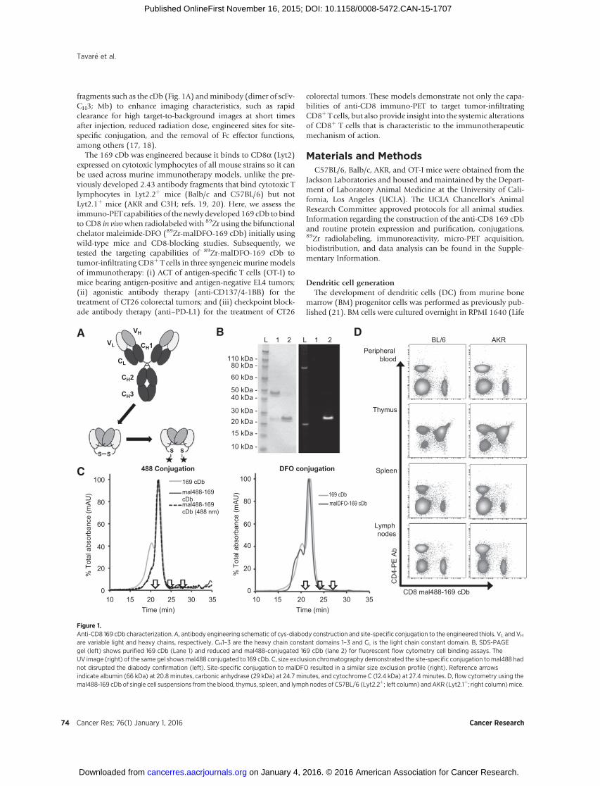
Figure 1.Anti-CD8 169 cDb characterization. A, antibody engineering schematic of cys-diabody construction and site-specific conjugation to the engineered thiols. VL and VH
are variable light and heavy chains, respectively. CH1–3 are the heavy chain constant domains 1–3 and CL is the light chain constant domain. B, SDS-PAGEgel (left) shows purified 169 cDb (Lane 1) and reduced and mal488-conjugated 169 cDb (lane 2) for fluorescent flow cytometry cell binding assays. TheUV image (right) of the same gel showsmal488 conjugated to 169 cDb. C, size exclusion chromatography demonstrated the site-specific conjugation tomal488 hadnot disrupted the diabody confirmation (left). Site-specific conjugation to malDFO resulted in a similar size exclusion profile (right). Reference arrowsindicate albumin (66 kDa) at 20.8 minutes, carbonic anhydrase (29 kDa) at 24.7 minutes, and cytochrome C (12.4 kDa) at 27.4 minutes. D, flow cytometry using themal488-169 cDb of single cell suspensions from the blood, thymus, spleen, and lymph nodes of C57BL/6 (Lyt2.2þ; left column) and AKR (Lyt2.1þ; right column)mice.
Tavar�e et al.
Cancer Res; 76(1) January 1, 2016 Cancer Research74
on January 4, 2016. © 2016 American Association for Cancer Research. cancerres.aacrjournals.org Downloaded from
Published OnlineFirst November 16, 2015; DOI: 10.1158/0008-5472.CAN-15-1707
Technologies) with 10% FCS, 1% penicillin, streptomycin, andamphotericin in a Petri dish. Nonadherent cells were re-plated onday 1 at 1 � 105 cells/well in 6-well plates with murine IL4(500 U/mL; R&D Systems) and murine GM-CSF (100 ng/mL;Amgen) for 7 days. DC were resuspended at 2 to 5� 106 cells/mLin serum-free RPMI and pulsed with OVA257-264 peptide(AnaSpec) at a concentration of 10 mmol/L in serum-free mediafor 90 minutes at room temperature.
OT-I T-cell expansionOT-1 splenocytes are harvested from OT-1 mice followed by 3
days of ex vivo activation with 100 U/mL IL2 and 1 mg/mLOVA257-264 peptide. Then, the activated OT-1 splentocytes wereexpandedwith 100U/mL IL2 for the following 2 days before ACT.
EL4/EL4-Ova tumor modelC57BL/6 mice received total body irradiation of 900 cGy and
then received 6 � 106 freshly isolated BM cells from anotherhealthy C57BL/6 mouse. Two days later, mice were injectedsubcutaneously (s.c.) with either 5 � 105 EL4-Ova or EL4 intothe right or left shoulders, respectively. On day 5 after tumorinoculations when tumors are approximately 5 mm in diameter,mice received 4.5� 106 ex vivo expanded andOVA257-264 peptide-activated OT-I T cells and were vaccinated s.c. with 7.5 � 106
OVA257-264 peptide-pulsed DC. The ACT was followed by threeconsecutive days of intraperitoneal IL2 administration (50,000IU; Novartis). On day 5 after ACT, mice were injected with 89Zr-radiolabeled malDFO-169 cDb for immuno-PET imaging andbiodistribution the following day (22 hours after injection).
Anti-CD137 and anti–PD-L1 CT26 tumor modelBalb/c mice were injected s.c. with 1 � 106 CT26 cells in the
shoulder. Starting on day 7 after inoculation when the tumorshave an average tumor diameter of about 3 to 4 mm, mice wereinjected i.p. with 12.5 mg/kg of either anti-CD137 antibody(clone 3H3; BioXCell) or anti–PD-L1 (clone 10F.9G2; BioXCell)every other day for four treatments. On day 15 after tumorinoculation, mice were injected with 89Zr-radiolabeled mal-DFO-169 cDb for immuno-PET imaging and biodistribution thefollowing day (22 hours after injection). Average tumor diameterwas calculated using calipers on days 7, 11, and 15 after tumorinoculation.
Flow cytometryFlow cytometry was performed on cell suspensions from the
spleen, peripheral blood, thymus, and lymph nodes. Mashingorgans over 75-mm filters (BD Biosciences) in RPMI plus 5% FBSprovided single cell suspensions. Following red blood cell lysisusing ammonium chloride–potassium lysis buffer, the cells werestained for 1 hour on ice, washed with PBS, and analyzed using aBD FACSCanto. The following antibodies were used for staining:Alexa488-conjugated 169 cDb, anti–CD4-PE (clone GK1.5),anti–CD45-APC (clone 30-F11; all fluorescent Abs fromeBioscience).
For tumor digestion to single cell suspensions, tumors wereincubated with Collagenase (type I; Invitrogen) at 1 mg/mL inRPMI plus 5% FBS for 1 hour at 37�C with continual shakingfollowed by straining over a 75-mm filter. The following anti-bodies were used for staining: anti–CD8-FITC (clone 53-6.7),anti–CD4-APC-Cy7 (clone GK1.5), anti–CD3-APC (clone 17A2),
and anti–CD45-PE (clone 30-F11; all fluorescent Abs fromeBioscience).
ResultsThe engineered anti-CD8 169 cys-diabody retained binding toCD8þ T cells
The anti-CD8 169 cys-diabody was engineered from thepreviously described anti-CD8 169 minibody (19) and waspurified to >95% purity (Fig. 1B) with a yield of 9.2 mg/L cellculture supernatant. Size exclusion chromatography (SEC)confirmed the correct molecular weight of approximately 55kDa with a small amount of higher molecular weight multimers(Fig. 1C).
The 169 cDb was conjugated site-specifically to maleimide-Alexa Fluor 488 (mal488) with a dye-to-protein molar ratio of1.5:1 and a recovery of 55% after purification. The mal488 wascovalently coupled to the monomeric 169 cDb as seen by fluo-rescence detection on the SDS-PAGE analysis (Fig. 1B). SECprofiles of both the mal488-169 cDb and native 169 cDb werevery similar (Fig. 1C, left), indicating the conjugate retained itscross-paired dimeric diabody structure. Flow cytometry using themal488-169 cDb on primary cells isolated from the peripheralblood, thymus, spleen, and lymph nodes from both Lyt2.2þ
C57BL/6 and Lyt2.1þAKRmice demonstrated that the engineeredcDb retains the ability to bind CD8a expressed on all mousestrains (Fig. 1D).
89Zr-radiolabeled anti-CD8 169 cDb specifically targets CD8þ
T cells in vivo as detected by immuno-PETSimilar to mal488 conjugation, the site-specific maleimide-
DFO (malDFO) conjugation to the 169 cDb did not disrupt thediabody bivalent conformation as shown by SEC (Fig. 1C, right).The 89Zr radiolabeling efficiency, radiochemical purity, specificactivity, protein dose injected, and immunoreactivity are reportedin Table 1.
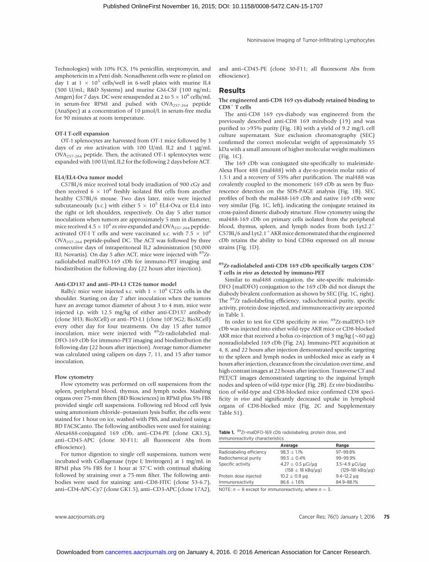
In order to test for CD8 specificity in vivo, 89Zr-malDFO-169cDb was injected into either wild-type AKRmice or CD8-blockedAKR mice that received a bolus co-injection of 3 mg/kg (�60 mg)nonradiolabeled 169 cDb (Fig. 2A). Immuno-PET acquisition at4, 8, and 22 hours after injection demonstrated specific targetingto the spleen and lymph nodes in unblocked mice as early as 4hours after injection, clearance from the circulation over time, andhigh contrast images at 22hours after injection. Transverse CT andPET/CT images demonstrated targeting to the inguinal lymphnodes and spleen of wild-type mice (Fig. 2B). Ex vivo biodistribu-tion of wild-type and CD8-blocked mice confirmed CD8 speci-ficity in vivo and significantly decreased uptake in lymphoidorgans of CD8-blocked mice (Fig. 2C and SupplementaryTable S1).
Table 1. 89Zr-malDFO-169 cDb radiolabeling, protein dose, andimmunoreactivity characteristics
Average Range
Radiolabeling efficiency 98.3 � 1.1% 97–99.8%Radiochemical purity 99.5 � 0.4% 99–99.9%Specific activity 4.27 � 0.5 mCi/mg
(158 � 18 kBq/mg)3.5–4.9 mCi/mg
(129–181 kBq/mg)Protein dose injected 10.2 � 0.9 mg 9.4–12.2 mgImmunoreactivity 86.6 � 1.6% 84.9–88.1%
NOTE: n ¼ 8 except for immunoreactivity, where n ¼ 3.
Noninvasive Imaging of Tumor-Infiltrating Lymphocytes
www.aacrjournals.org Cancer Res; 76(1) January 1, 2016 75
on January 4, 2016. © 2016 American Association for Cancer Research. cancerres.aacrjournals.org Downloaded from
Published OnlineFirst November 16, 2015; DOI: 10.1158/0008-5472.CAN-15-1707
Anti-CD8 immuno-PETdetects antigen-specific tumor targetingof adoptively transferred T cells
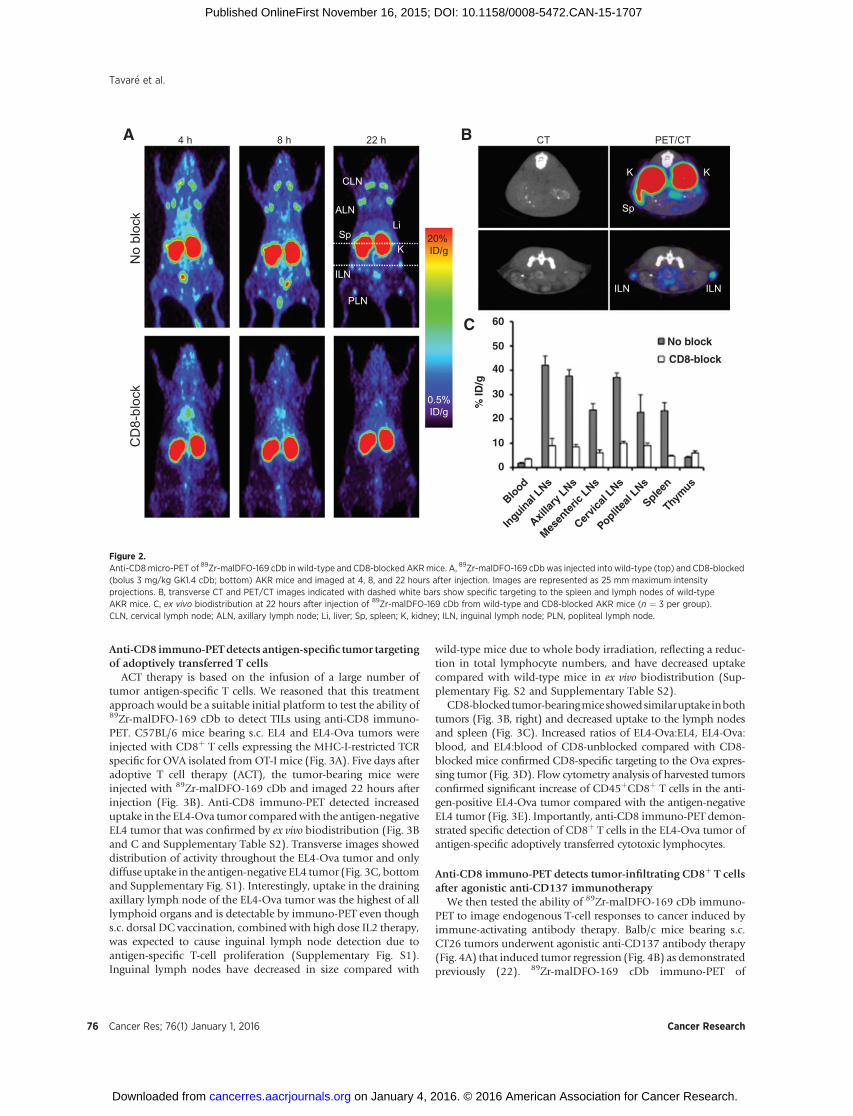
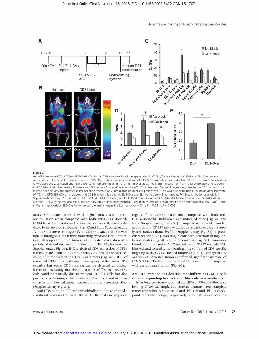
ACT therapy is based on the infusion of a large number oftumor antigen-specific T cells. We reasoned that this treatmentapproach would be a suitable initial platform to test the ability of89Zr-malDFO-169 cDb to detect TILs using anti-CD8 immuno-PET. C57BL/6 mice bearing s.c. EL4 and EL4-Ova tumors wereinjected with CD8þ T cells expressing the MHC-I-restricted TCRspecific for OVA isolated fromOT-I mice (Fig. 3A). Five days afteradoptive T cell therapy (ACT), the tumor-bearing mice wereinjected with 89Zr-malDFO-169 cDb and imaged 22 hours afterinjection (Fig. 3B). Anti-CD8 immuno-PET detected increaseduptake in the EL4-Ova tumor comparedwith the antigen-negativeEL4 tumor that was confirmed by ex vivo biodistribution (Fig. 3Band C and Supplementary Table S2). Transverse images showeddistribution of activity throughout the EL4-Ova tumor and onlydiffuse uptake in the antigen-negative EL4 tumor (Fig. 3C, bottomand Supplementary Fig. S1). Interestingly, uptake in the drainingaxillary lymph node of the EL4-Ova tumor was the highest of alllymphoid organs and is detectable by immuno-PET even thoughs.c. dorsal DC vaccination, combined with high dose IL2 therapy,was expected to cause inguinal lymph node detection due toantigen-specific T-cell proliferation (Supplementary Fig. S1).Inguinal lymph nodes have decreased in size compared with
wild-type mice due to whole body irradiation, reflecting a reduc-tion in total lymphocyte numbers, and have decreased uptakecompared with wild-type mice in ex vivo biodistribution (Sup-plementary Fig. S2 and Supplementary Table S2).
CD8-blocked tumor-bearingmiceshowedsimilaruptake inbothtumors (Fig. 3B, right) and decreased uptake to the lymph nodesand spleen (Fig. 3C). Increased ratios of EL4-Ova:EL4, EL4-Ova:blood, and EL4:blood of CD8-unblocked compared with CD8-blocked mice confirmed CD8-specific targeting to the Ova expres-sing tumor (Fig. 3D). Flow cytometry analysis of harvested tumorsconfirmed significant increase of CD45þCD8þ T cells in the anti-gen-positive EL4-Ova tumor compared with the antigen-negativeEL4 tumor (Fig. 3E). Importantly, anti-CD8 immuno-PET demon-strated specific detection of CD8þ T cells in the EL4-Ova tumor ofantigen-specific adoptively transferred cytotoxic lymphocytes.
Anti-CD8 immuno-PET detects tumor-infiltrating CD8þ T cellsafter agonistic anti-CD137 immunotherapy
We then tested the ability of 89Zr-malDFO-169 cDb immuno-PET to image endogenous T-cell responses to cancer induced byimmune-activating antibody therapy. Balb/c mice bearing s.c.CT26 tumors underwent agonistic anti-CD137 antibody therapy(Fig. 4A) that induced tumor regression (Fig. 4B) as demonstratedpreviously (22). 89Zr-malDFO-169 cDb immuno-PET of
BA
CLN
ALN
ILN
PLN
SpLi
K
No
bloc
kC
D8-
bloc
k
20%ID/g
0.5%ID/g
4 h 8 h 22 h
ILN
K
Sp
K
ILN
TC/TEPTC
C
0
Blood
Inguin
al LNs
Axillar
y LNs
Mesen
teric
LNs
Cervic
al LNs
Poplitea
l LNs
Spleen
Thym
us
10
20
% ID
/g
30
40
50 No block
CD8-block
60
Figure 2.Anti-CD8micro-PET of 89Zr-malDFO-169 cDb in wild-type and CD8-blocked AKRmice. A, 89Zr-malDFO-169 cDbwas injected into wild-type (top) and CD8-blocked(bolus 3 mg/kg GK1.4 cDb; bottom) AKR mice and imaged at 4, 8, and 22 hours after injection. Images are represented as 25 mm maximum intensityprojections. B, transverse CT and PET/CT images indicated with dashed white bars show specific targeting to the spleen and lymph nodes of wild-typeAKR mice. C, ex vivo biodistribution at 22 hours after injection of 89Zr-malDFO-169 cDb from wild-type and CD8-blocked AKR mice (n ¼ 3 per group).CLN, cervical lymph node; ALN, axillary lymph node; Li, liver; Sp, spleen; K, kidney; ILN, inguinal lymph node; PLN, popliteal lymph node.
Tavar�e et al.
Cancer Res; 76(1) January 1, 2016 Cancer Research76
on January 4, 2016. © 2016 American Association for Cancer Research. cancerres.aacrjournals.org Downloaded from
Published OnlineFirst November 16, 2015; DOI: 10.1158/0008-5472.CAN-15-1707
anti-CD137-treated mice showed higher intratumoral probeaccumulation when compared with both anti-CD137-treated/CD8-blocked and untreated tumor-bearing mice that was vali-dated by ex vivobiodistribution (Fig. 4C andE and SupplementaryTable S3). Transverse images of anti-CD137-treated mice showeduptake throughout the tumor, indicating cytotoxic T-cell infiltra-tion, although the CT26 tumors of untreated mice showed aperipheral rim of uptake around the tumor (Fig. 3C, bottom andSupplementary Fig. S3). IHC analysis of CD8 expression of CT26tumors treated with anti-CD137 therapy confirmed the presenceof CD8þ tumor-infiltrating T cells in tumors (Fig. 4D). IHC ofuntreated CT26 tumors showed the majority of the rim is CD8negative but some CD8 staining can be detected at distinctlocations, indicating that the rim uptake of 89Zr-malDFO-169cDb could be partially due to resident CD8þ T cells but alsopossibly due to nonspecific uptake resulting from ruptured vas-culature and the enhanced permeability and retention effect(Supplementary Fig. S4).
Anti-CD8 immuno-PET and ex vivobiodistribution confirmed asignificant increase of 89Zr-malDFO-169 cDb uptake in lymphatic
organs of anti-CD137-treated mice compared with both anti-CD137-treated/CD8-blocked and untreated mice (Fig. 4C andE and Supplementary Table S3). Compared with the ACT model,agonistic anti-CD137 therapy caused a systemic increase in size oflymph nodes (about fivefold; Supplementary Fig. S2) as previ-ously reported (23), resulting in enhanced detection of inguinallymph nodes (Fig. 4C and Supplementary Fig. S3). Tumor-to-blood ratios of anti-CD137-treated, anti-CD137-treated/CD8-blocked, and control tumor-bearingmice confirmedCD8-specifictargeting to the CD137-treated tumors (Fig. 4F). Flow cytometryanalysis of harvested tumors confirmed significant increase ofCD45þCD8þ T cells in the anti-CD137-treated tumor comparedwith the untreated tumor (Fig. 4G).
Anti-CD8 immuno-PET detects tumor-infiltrating CD8þ T cellsin mice responding to checkpoint blockade immunotherapy
It has been previously reported that 25%or 33%of Balb/cmicebearing CT26 s.c. implanted tumors demonstrated completetumor regression in response to anti–PD-1 or anti–PD-L1 check-point blockade therapy, respectively, although nonresponding
A
20%ID/g
0.5%ID/g
B No block
No block
CD8-block
CD8-block
No blockCD8-block
Ova– Ova–Ova+ Ova+
Ova– Ova+Ova– Ova+
EL4/EL4-Ovaimplant
OT-I & DCACT
Immuno-PETbiodistribution
10 11
10
0
0EL4 EL4-Ova
10
20
30
% C
D45
+CD
8+
40
50EL4
-Ova
:EL4
EL4-O
va:b
lood
EL4:b
lood
1
0
2Rat
io 3
4
5
Blood
EL4-Ova
tumor
EL4-Ova
Axil
LNEL4 A
xil LN
Inguinal LNs
Cervica
l LNs
Spleen
Thymus
EL4 Tumor
20
30
% ID
/g
40
50
5 6 70Day -2
IL-2900 cGy
Radiolabelinginjection
C
ED
Figure 3.Anti-CD8 immuno-PET of 89Zr-malDFO-169 cDb in the OT-I adoptive T-cell therapy model. A, C57BL/6 mice bearing s.c. EL4 and EL4-Ova tumorsreceived the full protocol of myelodepletion (900 cGy) with hematopoietic stem cell (HSC)/BM transplantation, adoptive OT-I T cell transfer followed byOVA-pulsed DC vaccination and high dose IL2. B, representative immuno-PET images at 22 hours after injection of 89Zr-malDFO-169 cDb of unblockedand CD8-blocked mice bearing EL4-Ova and EL4 tumors 5 days after adoptive OT-I T cell transfer. Coronal images are presented as 25 mm maximumintensity projections and transverse images are presented as 2 mm maximum intensity projections. C, ex vivo biodistribution at 22 hours after injectionof 89Zr-malDFO-169 cDb of unblocked and CD8-blocked mice bearing EL4-Ova and EL4 tumors (n ¼ 3 per group). Full biodistribution analysis is inSupplementary Table S2. D, ratios of EL4-Ova:EL4, EL4-Ova:blood, and EL4:blood of unblocked and CD8-blocked mice from ex vivo biodistributionanalysis. E, flow cytometry analysis of tumors harvested 5 days after adoptive T-cell therapy was used to determine the percentage of CD45þCD8þ T cellsin the antigen-positive EL4-Ova tumor versus the antigen-negative EL4 tumor (n ¼ 3). � , P < 0.05; †, P < 0.005.
Noninvasive Imaging of Tumor-Infiltrating Lymphocytes
www.aacrjournals.org Cancer Res; 76(1) January 1, 2016 77
on January 4, 2016. © 2016 American Association for Cancer Research. cancerres.aacrjournals.org Downloaded from
Published OnlineFirst November 16, 2015; DOI: 10.1158/0008-5472.CAN-15-1707

A
20%ID/g
0.5%ID/g
CCD137
CD137
CD137 + CD8-block
CD137 +
CD137 treated
CD8-block
Immuno-PETCT26 implantbiodistribution
15 16
0
10
20
30
40
% ID
/g
50
0
10
20
% C
D45
+CD
8+Tu
mor
:blo
od ra
tio
30
40
50
0
0
4
8
ControlCD137
12
Ave
rage
tum
ordi
amet
er (m
m)
16
0 5Day
10 15
2
4
6
8
10
60 CD137
Control
CD137 +CD8-block
7 9 11 13Day 0
Anti–CD137 Ab
Radiolabelinginjection
E
F
G
Control
Control
Control
B
D
‡ ‡
†
Blood
Tumor
D-Axil
lary L
N
ND-Axil
lary L
NIng
uinal
LNs
Cervica
l LNs
Spleen
Thymus
Figure 4.Anti-CD8 immuno-PET of 89Zr-malDFO-169 cDb in the CT26/anti-CD137 immunotherapy model. A, Balb/c mice bearing s.c. CT26 tumors were treatedwith anti-CD137 therapy every other day for four treatments and anti-CD8 immuno-PET was acquired on day 16 after tumor implantation. B, tumor growthcurves of CD137 treated and untreatedmice (average tumor diameter). C, on day 8 after immunotherapy initiation, CD137-treatedmice, CD137-treated/CD8-blockedmice, and control mice (no anti-CD137 therapy) were injected with 89Zr-malDFO-169 cDb and immuno-PET images were acquired at 22 hours after injection.D, CD8 IHC of untreated CT26 tumors or anti-CD137-treated CT26 tumors indicate the presence of increased CD8þ tumor-infiltrating lymphocytes. E, ex vivobiodistribution at 22 hours after injection of 89Zr-malDFO-169 cDb of CD137-treated mice, CD137-treated/CD8-blocked mice, and control mice (n ¼ 3 per group).Full biodistribution analysis is in Supplementary Table S3. F, tumor-to-blood ratios of CD137-treated mice, CD137-treated/CD8-blocked mice, and controlmice. G, flow cytometry analysis of tumors harvested on day 15 was used to determine the percentage of CD45þCD8þ T cells in the CT26 tumors (n ¼ 3).†, P < 0.005; z, P < 0.0005.
Tavar�e et al.
Cancer Res; 76(1) January 1, 2016 Cancer Research78
on January 4, 2016. © 2016 American Association for Cancer Research. cancerres.aacrjournals.org Downloaded from
Published OnlineFirst November 16, 2015; DOI: 10.1158/0008-5472.CAN-15-1707
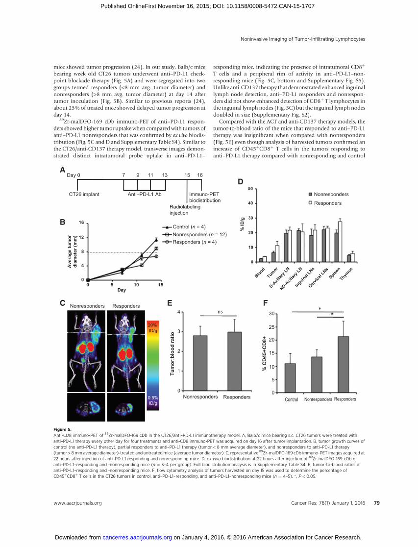
mice showed tumor progression (24). In our study, Balb/c micebearing week old CT26 tumors underwent anti–PD-L1 check-point blockade therapy (Fig. 5A) and were segregated into twogroups termed responders (<8 mm avg. tumor diameter) andnonresponders (>8 mm avg. tumor diameter) at day 14 aftertumor inoculation (Fig. 5B). Similar to previous reports (24),about 25% of treated mice showed delayed tumor progression atday 14.
89Zr-malDFO-169 cDb immuno-PET of anti–PD-L1 respon-ders showed higher tumor uptakewhen comparedwith tumors ofanti–PD-L1 nonresponders that was confirmed by ex vivo biodis-tribution (Fig. 5C andD and Supplementary Table S4). Similar tothe CT26/anti-CD137 therapy model, transverse images demon-strated distinct intratumoral probe uptake in anti–PD-L1–
responding mice, indicating the presence of intratumoral CD8þ
T cells and a peripheral rim of activity in anti–PD-L1–non-responding mice (Fig. 5C, bottom and Supplementary Fig. S5).Unlike anti-CD137 therapy that demonstrated enhanced inguinallymph node detection, anti–PD-L1 responders and nonrespon-ders did not show enhanced detection of CD8þ T lymphocytes inthe inguinal lymph nodes (Fig. 5C) but the inguinal lymph nodesdoubled in size (Supplementary Fig. S2).
Compared with the ACT and anti-CD137 therapy models, thetumor-to-blood ratio of the mice that responded to anti–PD-L1therapy was insignificant when compared with nonresponders(Fig. 5E) even though analysis of harvested tumors confirmed anincrease of CD45þCD8þ T cells in the tumors responding toanti–PD-L1 therapy compared with nonresponding and control
20%ID/g
0.5%ID/g
C Responders
Responders
0 0
5
10
15
20
25
30
1
2
Tum
or:b
lood
ratio
% C
D45
+CD
8+
3
4
Responders Responders
Nonresponders
Nonresponders
Nonresponders NonrespondersControl
Responders (n = 4)Nonresponders (n = 12)Control (n = 4)
D
FE
A
Immuno-PETCT26 implantbiodistribution
15 167 9 11 13Day 0
Anti–PD-L1 Ab
Radiolabelinginjection
B
**ns
Blood0
10
20
30
% ID
/g
40
50
TumorD-A
xillar
y LN
ND-Axil
lary L
NInguinal
LNsCerv
ical L
NsSplee
nThym
us
0
4
8
12
Ave
rage
tum
ordi
amet
er (m
m)
16
0 5Day
10 15
Figure 5.Anti-CD8 immuno-PET of 89Zr-malDFO-169 cDb in the CT26/anti–PD-L1 immunotherapy model. A, Balb/c mice bearing s.c. CT26 tumors were treated withanti–PD-L1 therapy every other day for four treatments and anti-CD8 immuno-PET was acquired on day 16 after tumor implantation. B, tumor growth curves ofcontrol (no anti–PD-L1 therapy), partial responders to anti–PD-L1 therapy (tumor < 8 mm average diameter), and nonresponders to anti–PD-L1 therapy(tumor > 8mm average diameter)-treated and untreated mice (average tumor diameter). C, representative 89Zr-malDFO-169 cDb immuno-PET images acquired at22 hours after injection of anti–PD-L1 responding and nonresponding mice. D, ex vivo biodistribution at 22 hours after injection of 89Zr-malDFO-169 cDb ofanti–PD-L1–responding and -nonresponding mice (n ¼ 3–4 per group). Full biodistribution analysis is in Supplementary Table S4. E, tumor-to-blood ratios ofanti–PD-L1–responding and -nonresponding mice. F, flow cytometry analysis of tumors harvested on day 15 was used to determine the percentage ofCD45þCD8þ T cells in the CT26 tumors in control, anti–PD-L1–responding, and anti–PD-L1–nonresponding mice (n ¼ 4–5). � , P < 0.05.
Noninvasive Imaging of Tumor-Infiltrating Lymphocytes
www.aacrjournals.org Cancer Res; 76(1) January 1, 2016 79
on January 4, 2016. © 2016 American Association for Cancer Research. cancerres.aacrjournals.org Downloaded from
Published OnlineFirst November 16, 2015; DOI: 10.1158/0008-5472.CAN-15-1707
untreated tumors (Fig. 5F). Interestingly, probe uptake in thetumors of anti–PD-L1 nonresponders was similar to tumoruptake to control untreated CT26 tumors and the percentage ofCD45þCD8þ T cells was similar (Fig. 4D and F). Importantly,anti-CD8 immuno-PET was able to detect increased intratumoralCD8þ T lymphocytes in mice responding to checkpoint blockadetherapy.
DiscussionIn the present study, we demonstrate that anti-CD8 immuno-
PET provides an integrated readout that is reflective of bothsystemic and intratumoral alterations in CD8þ T cell numbersdue to threemechanistically differentmodels of immunotherapy.For example, anti-CD8 immuno-PET detection of nontumordraining lymph nodes and the spleen in the ACT model isdecreased, resulting from whole body irradiation, it is enhancedin the anti-CD137 model due to the systemic agonistic activity ofthe therapy on immune cells throughout the body, and does notchange greatly for anti–PD-L1 therapy due to the restrictedexpression of PD-L1 on tumor cells and myeloid-derived sup-pressor cells within the tumor. The successful noninvasive imag-ing of CD8þ T cell responses to cancer was achieved by targeting aphysiologically expressed surface molecule using a clinicallycompatible approach.
The 89Zr-radiolabeled 169 cDb demonstrated specific targetingto CD8 in vivo as detected by immuno-PET. The engineered C-terminal cysteine allowed for site-specific conjugation of thebifunctional chelator maleimide-DFO away from the bindingsite of the cDb to avoid decreased immuno-reactivity uponconjugation. Importantly, the cDb retains its cross-paired, biva-lent structure and does not form monovalent scFvs upon mildreduction and thiol-specific conjugation (25). Comparedwith thepreviously engineered 169 minibody fragment used for 64Cuimmuno-PET (19), the cDb showed less aggregation, slowerblood clearance, and enhanced lymph node and spleen targetingat 22 hours after injection. The cDb exhibited high renal accu-mulation due to the lower molecular weight of the cys-diabody(�55 kDa) compared with the minibody (�80 kDa), the renalfiltration cutoff of approximately 60 kDa, and the use of theresidualizing radiometal 89Zr.
Detecting small regions of interest using PET is inherentlydifficult due to the partial volume effect whereby activity in smallregions of interest near or below the resolution of the scanner,such as lymph nodes, is underestimated (26). This underestima-tion varies due to scanner resolution and the positron range of theradionuclide used, but it can be compensated for using partialvolume correction (26, 27). Even with this limitation, we haveshown anti-CD8 immuno-PET can detect lymph nodes in wild-type mice and in a model of T-cell repopulation after hemato-poietic stem cell transplant (19, 20).
Due to immunotherapy-induced alterations of CD8 expressedin the antigen sink, i.e., spleen and lymph nodes, the optimalprotein dose will be dependent on the immunotherapeuticmechanism of action and the ability to consistently target intra-tumoral CD8þ T cells will rely on a fine balance between blockingthe CD8 antigen sink and displacing tumor uptake. The impor-tance of protein dose for tumor epitope targeting has beendemonstrated previously where a natural antigen sink exists inimaging studies targeting HER2 (28), neuropilin-1 (29), andEGFR (30), and antibody-drug conjugate studies targeting TENB2
(31). A recent publication byMuylle and colleagues studying 89Zr-rituximab immuno-PET in patients with CD20þ B-cell lympho-mas demonstrates the importance of antigen sink and proteindose to obtain consistent lymphoma detection that will berelevant for reproducible anti-CD8 immuno-PET in the clinic(32). In the future, enhanced tumor-infiltrating CD8þ T celltargeting might be achieved in the models presented hereby optimizing the anti-CD8 cys-diabody dose for eachimmunotherapy.
Anti-CD8 immuno-PET is a powerful method used to specif-ically monitor endogenous CD8þ T cells noninvasively withoutthe need for ex vivo manipulation of lymphocytes. Direct radi-olabeling of lymphocytes ex vivo allows for monitoring initial cellmigration of adoptively transferred cells, but suffers from radio-nuclide half-life, probe dilution due to cell division and potentialtoxic effects of the radionuclide on radiosensitive lymphocytes(33–35). Reporter gene transduction of cells ex vivo benefits fromsignal amplification due to cell division, repeat monitoring, andlongitudinal tracking of genetically engineered cells (36–39).However, reporter probes demonstrate high background in clear-ance organs and reporter genes require development of nonim-munogenic reporters for translation (37, 40). Small moleculemetabolic probes that do not require ex vivo cellular manipula-tion, such as 2-deoxy-2-(18F)-fluoro-D-glucose ([18F]-FDG) and1-(20-deoxy-20(18F)fluoroarabinofuranosyl) cysteine ([18F]-FAC),are either not specific for cytotoxic lymphocytes alone ([18F]-FDG) or they target proliferating lymphocytes in secondary lym-phoid organs and fail to detect TILs ([18F]-FAC; ref. 41).
Anti-CD8 immuno-PET was able to detect tumor-infiltratingCD8þ T cell alterations in three immunotherapeutic models thathave shown great promise in the clinic. The development ofanalogous imaging agents for human use would be of great utility,particularly in light of the recent advances in clinical immuno-oncology, including FDA approvals of checkpoint inhibitors suchas ipilimumab,nivolumab, andpembrolizumab, and thebispecificblinatumomab for T-cell recruitment. A fully human imaging agentspecific for humanCD8 can be arrived at through humanization ofexisting anti-CD8 monoclonal antibodies or de novo isolation offully human antibodies by phage display (42). Alternative scaf-folds, such as single domain camelid antibodies labeledwith 18F or64Cu for PET, have demonstrated utility in the detection of themacrophage mannose receptors, MHC Class II, and CD11bexpressedonmyeloid cells inpreclinicalmodels (43,44).However,suchagentsmight requirehumanization to reducepotential immu-nogenicity.With any imaging agent specific for humanCD8T cells,preclinical testing would require the use of transgenic expressinghuman CD8 or humanized mouse models, such as NSG micereconstituted with human peripheral blood mononuclear cells orCD34þ hematopoietic stem cells, which can be used to establishmodels of tumor immunotherapy. Furthermore, it will be essentialto evaluate potential effects of administration on T cell viability,proliferation, and function in vivo. Finally, radiation dose estimatesneed tobecalculatedbasedonthebiodistributionand time-activitycurves of potential CD8 PET tracers in preclinical models, in ordertodetermineappropriate levelsofprotein and radioactivity that canbe administered to patients.
The potential utility of immuno-PET for imaging immune cellsubsets in humans is supported bymany previous clinical studiesusing radiolabeled intact antibodies targeting T and B lympho-cytes for detection of inflammation in vivo using planar gammaimaging and single photon emission CT (45). In oncology,
Tavar�e et al.
Cancer Res; 76(1) January 1, 2016 Cancer Research80
on January 4, 2016. © 2016 American Association for Cancer Research. cancerres.aacrjournals.org Downloaded from
Published OnlineFirst November 16, 2015; DOI: 10.1158/0008-5472.CAN-15-1707
gamma camera imaging of 131I-tositumomab or 111In-ibritumo-mab tiuxetan can be used to confirmCD20 targeting prior to or inconjugation with radioimmunotherapy (46). Success of theseapproaches for imaging immune cell subsets using intact anti-bodies suggests that the transition to bespoke engineered anti-body fragments for immuno-PET should be feasible and favor-able. Importantly, we believe immuno-PET monitoring of lym-phocytes and other immune cell subsets could transform theability to profile the tumor immune microenvironment andantitumor immune responses in the context of cancer immuno-therapy in the clinic.
Disclosure of Potential Conflicts of InterestR. Tavar�e is a consultant for ImaginAb, Inc. A.M. Wu is a board member at
ImaginAb, Inc.; has ownership interest (including patents) in ImaginAb, Inc.;and is a consultant/advisory board member for ImaginAb, Inc. Part of thetechnology described in this manuscript is licensed by the Regents of theUniversity of California to ImaginAb, Inc. and the Regents have taken equityin ImaginAb, Inc. as part of the licensing transaction. No potential conflicts ofinterest were disclosed by the other authors.
Authors' ContributionsConception and design: R. Tavar�e, S. Mok, M.N. McCracken, O.N. Witte,A. Ribas, A.M. WuDevelopment of methodology: R. Tavar�e, S. MokAcquisition of data (provided animals, acquired and managed patients,provided facilities, etc.): R. Tavar�e, H. Escuin-Ordinas, S. Mok, M.N.McCracken, K.A. Zettlitz, F.B. Salazar, A. RibasAnalysis and interpretation of data (e.g., statistical analysis, biostatistics,computational analysis): R. Tavar�e, H. Escuin-Ordinas, S. Mok, M.N.McCracken, K.A. Zettlitz, A.M. Wu
Writing, review, and/or revision of the manuscript: R. Tavar�e, S. Mok, K.A.Zettlitz, O.N. Witte, A. Ribas, A.M. WuAdministrative, technical, or material support (i.e., reporting or organizingdata, constructing databases): K.A. Zettlitz, A. RibasStudy supervision: R. Tavar�e, A.M. Wu
AcknowledgmentsThe authors thank the members of the Crump Institute for Molecular
Imaging, Michael Phelps for his continued support, and Ralph and MarjorieCrump for a donationmade to theCrump Institute forMolecular Imaging at theUCLA.
Grant SupportThis work was supported by NIH grants R21 AI114255 and R21 CA190044,
P50 CA086306, and by the California Institute for Regenerative Medicine(CIRM; RT1-01126-1). R. Tavar�e is supported by the UCLA Scholars in Onco-logic Molecular Imaging training program (NIH R25T CA098010). M.N.McCracken is supported by the CIRM Training Grant TG2-01169 and the UCLAIn vivo Cellular and Molecular Imaging Center Career Development Award P50CA086306. O.N. Witte is an investigator of the Howard Hughes MedicalInstitute and is partially supported by the Eli and Edythe Broad Center ofRegenerative Medicine and Stem Cell Research. A. Ribas is supported by P01CA168585, the Dr. Robert Vigen Memorial Fund, and the Ressler FamilyFoundation. Small animal imaging studies, flow cytometry, and pathologyservices were supported by the UCLA Jonsson Comprehensive Cancer Center(NIH CA016042).
The costs of publication of this articlewere defrayed inpart by the payment ofpage charges. This article must therefore be hereby marked advertisement inaccordance with 18 U.S.C. Section 1734 solely to indicate this fact.
Received June 25, 2015; revised September 25, 2015; accepted October 16,2015; published OnlineFirst November 16, 2015.
References1. Hanahan D, Weinberg RA. Hallmarks of cancer: the next generation. Cell
2011;144:646–74.2. Mantovani A, Allavena P, Sica A, Balkwill F. Cancer-related inflammation.
Nature 2008;454:436–44.3. Schreiber RD, Old LJ, Smyth MJ. Cancer immunoediting: integrating
immunity's roles in cancer suppression and promotion. Science 2011;331:1565–70.
4. Pages F, Galon J, Dieu-NosjeanMC, Tartour E, Sautes-Fridman C, FridmanWH. Immune infiltration in human tumors: a prognostic factor that shouldnot be ignored. Oncogene 2010;29:1093–102.
5. GoodenMJ, de BockGH, LeffersN,Daemen T,NijmanHW. The prognosticinfluence of tumour-infiltrating lymphocytes in cancer: a systematic reviewwith meta-analysis. Br J Cancer 2011;105:93–103.
6. Johnson LA, Morgan RA, Dudley ME, Cassard L, Yang JC, Hughes MS, et al.Gene therapy with human and mouse T-cell receptors mediates cancerregression and targets normal tissues expressing cognate antigen. Blood2009;114:535–46.
7. Rosenberg SA. Raising the bar: the curative potential of human cancerimmunotherapy. Sci Transl Med 2012;4:127ps8.
8. Melero I, Grimaldi AM, Perez-Gracia JL, Ascierto PA. Clinical developmentof immunostimulatory monoclonal antibodies and opportunities forcombination. Clin Cancer Res 2013;19:997–1008.
9. Melero I, Hervas-Stubbs S, Glennie M, Pardoll DM, Chen L. Immunosti-mulatory monoclonal antibodies for cancer therapy. Nat Rev Cancer2007;7:95–106.
10. Vinay DS, Kwon BS. Immunotherapy of cancer with 4-1BB. Mol CancerTher 2012;11:1062–70.
11. Callahan MK, Wolchok JD. At the bedside: CTLA-4- and PD-1-blockingantibodies in cancer immunotherapy. J Leukoc Biol 2013;94:41–53.
12. ShinDS, Ribas A. The evolution of checkpoint blockade as a cancer therapy:what's here, what's next? Curr Opin Immunol 2015;33C:23–35.
13. Topalian SL, Drake CG, Pardoll DM. Immune checkpoint blockade: acommon denominator approach to cancer therapy. Cancer Cell 2015;27:450–61.
14. Cobbold SP, Jayasuriya A, Nash A, Prospero TD, Waldmann H. Therapywith monoclonal antibodies by elimination of T-cell subsets in vivo.Nature 1984;312:548–51.
15. Wu AM. Antibodies and antimatter: the resurgence of immuno-PET. J NuclMed 2009;50:2–5.
16. Knowles SM, Wu AM. Advances in immuno-positron emission tomogra-phy: antibodies for molecular imaging in oncology. J Clin Oncol2012;30:3884–92.
17. Wu AM. Engineered antibodies for molecular imaging of cancer. Methods2014;65:139–47.
18. Olafsen T, Wu AM. Antibody vectors for imaging. Semin Nucl Med2010;40:167–81.
19. Tavar�e R, McCracken MN, Zettlitz KA, Knowles SM, Salazar FB, OlafsenT, et al. Engineered antibody fragments for immuno-PET imaging ofendogenous CD8þ T cells in vivo. Proc Natl Acad Sci U S A2014;111:1108–13.
20. Tavar�e R,McCrackenMN, Zettlitz KA, Salazar FB,Olafsen T,WitteON, et al.ImmunoPET of murine T cell reconstitution post-adoptive stem celltransplant using anti-CD4 and anti-CD8 cys-diabodies. J Nucl Med2015;56:1258–64.
21. Prins RM, Shu CJ, Radu CG, Vo DD, Khan-Farooqi H, Soto H, et al. Anti-tumor activity and trafficking of self, tumor-specific T cells against tumorslocated in the brain. Cancer Immunol Immunother 2008;57:1279–89.
22. Escuin-Ordinas H, Elliott MW, Atefi M, Lee M, Ng C, Wei L, et al. PETimaging to non-invasively study immune activation leading to antitumorresponses with a 4-1BB agonistic antibody. J Immunother Cancer2013;1:14.
23. Schrand B, Berezhnoy A, Brenneman R,Williams A, Levay A, Kong LY, et al.Targeting 4-1BB costimulation to the tumor stromawith bispecific aptamerconjugates enhances the therapeutic index of tumor immunotherapy.Cancer Immunol Res 2014;2:867–77.
24. Duraiswamy J, Kaluza KM, Freeman GJ, Coukos G. Dual blockade of PD-1and CTLA-4 combined with tumor vaccine effectively restores T-cell rejec-tion function in tumors. Cancer Res 2013;73:3591–603.
Noninvasive Imaging of Tumor-Infiltrating Lymphocytes
www.aacrjournals.org Cancer Res; 76(1) January 1, 2016 81
on January 4, 2016. © 2016 American Association for Cancer Research. cancerres.aacrjournals.org Downloaded from
Published OnlineFirst November 16, 2015; DOI: 10.1158/0008-5472.CAN-15-1707
25. Tavar�e R, Wu WH, Zettlitz KA, Salazar FB, McCabe KE, Marks JD, et al.Enhanced immunoPET of ALCAM-positive colorectal carcinoma usingsite-specific 64Cu-DOTA conjugation. Protein Eng Des Sel 2014;27:317–24.
26. SoretM, Bacharach SL, Buvat I. Partial-volume effect in PET tumor imaging.J Nucl Med 2007;48:932–45.
27. Knowles SM, Zettlitz KA, Tavar�e R, Rochefort MM, Salazar FB, Stout DB,et al. Quantitative immunoPET of prostate cancer xenografts with 89Zr-and 124I-labeled anti-PSCA A11 minibody. J Nucl Med 2014;55:452–9.
28. Dijkers EC, Oude Munnink TH, Kosterink JG, Brouwers AH, Jager PL, deJong JR, et al. Biodistribution of 89Zr-trastuzumab and PET imaging ofHER2-positive lesions in patients with metastatic breast cancer. ClinPharmacol Ther 2010;87:586–92.
29. Bumbaca D, Xiang H, Boswell CA, Port RE, Stainton SL, Mundo EE, et al.Maximizing tumour exposure to anti-neuropilin-1 antibody requires sat-uration of non-tumour tissue antigenic sinks in mice. Br J Pharmacol2012;166:368–77.
30. Divgi CR, Welt S, Kris M, Real FX, Yeh SD, Gralla R, et al. Phase I andimaging trial of indium 111-labeled anti-epidermal growth factor receptormonoclonal antibody 225 in patients with squamous cell lung carcinoma.J Natl Cancer Inst 1991;83:97–104.
31. Boswell CA, Mundo EE, Zhang C, Stainton SL, Yu SF, Lacap JA, et al.Differential effects of predosing on tumor and tissue uptake of an 111In-labeled anti-TENB2 antibody-drug conjugate. J NuclMed 2012;53:1454–61.
32. Muylle K, Flamen P, Vugts DJ, Guiot T, Ghanem G, Meuleman N, et al.Tumour targeting and radiation dose of radioimmunotherapy with Y-rituximab in CD20þ B-cell lymphoma as predicted by Zr-rituximabimmuno-PET: impact of preloading with unlabelled rituximab. Eur J NuclMed Mol Imaging 2015;42:1304–14.
33. Pittet MJ, Grimm J, Berger CR, Tamura T, Wojtkiewicz G, Nahrendorf M,et al. In vivo imaging of T cell delivery to tumors after adoptive transfertherapy. Proc Natl Acad Sci U S A 2007;104:12457–61.
34. Griessinger CM, Kehlbach R, Bukala D,Wiehr S, Bantleon R, Cay F, et al. Invivo tracking of Th1 cells by PET reveals quantitative and temporaldistribution and specific homing in lymphatic tissue. J Nucl Med2014;55:301–7.
35. Griessinger CM, Maurer A, Kesenheimer C, Kehlbach R, Reischl G, Ehr-lichmann W, et al. 64Cu antibody-targeting of the T-cell receptor and
subsequent internalization enables in vivo tracking of lymphocytes by PET.Proc Natl Acad Sci U S A 2015;112:1161–6.
36. Koya RC, Mok S, Comin-Anduix B, Chodon T, Radu CG, Nishimura MI,et al. Kinetic phases of distribution and tumor targeting by T cell receptorengineered lymphocytes inducing robust antitumor responses. Proc NatlAcad Sci U S A 2010;107:14286–91.
37. McCracken MN, Gschweng EH, Nair-Gill E, McLaughlin J, Cooper AR,Riedinger M, et al. Long-term in vivo monitoring of mouse and humanhematopoietic stem cell engraftment with a human positron emissiontomography reporter gene. Proc Natl Acad Sci U S A 2013;110:1857–62.
38. Gschweng EH, McCracken MN, Kaufman ML, Ho M, Hollis RP, Wang X,et al. HSV-sr39TK positron emission tomography and suicide gene elim-ination of human hematopoietic stem cells and their progeny in human-ized mice. Cancer Res 2014;74:5173–83.
39. McCracken MN, Vatakis DN, Dixit D, McLaughlin J, Zack JA, Witte ON.Noninvasive detection of tumor-infiltrating T cells by PET reporter imag-ing. J Clin Invest 2015;125:1815–26.
40. Yaghoubi SS, Campbell DO, Radu CG, Czernin J. Positron emissiontomography reporter genes and reporter probes: gene and cell therapyapplications. Theranostics 2012;2:374–91.
41. Nair-Gill E, Wiltzius SM, Wei XX, Cheng D, Riedinger M, Radu CG, et al.PET probes for distinct metabolic pathways have different cell specificitiesduring immune responses in mice. J Clin Invest 2010;120:2005–15.
42. Li K, Zettlitz KA, Lipianskaya J, Zhou Y, Marks JD, Mallick P, et al. A fullyhuman scFv phage display library for rapid antibody fragment reformat-ting. Protein Eng Des Sel 2015;28:307–16.
43. Blykers A, Schoonooghe S, Xavier C, D'Hoe K, Laoui D, D'Huyvetter M,et al. PET imaging ofMMR-expressingmacrophages in tumor stroma using18F-radiolabeled camelid single-domain antibody fragments. J Nucl Med2015;56:1265–71.
44. Rashidian M, Keliher EJ, Bilate AM, Duarte JN, Wojtkiewicz GR, JacobsenJT, et al. Noninvasive imaging of immune responses. Proc Natl Acad Sci U SA 2015;112:6146–51.
45. Malviya G, Galli F, Sonni I, Pacilio M, Signore A. Targeting T and Blymphocytes with radiolabelled antibodies for diagnostic and therapeuticapplications. Q J Nucl Med Mol Imaging 2010;54:654–76.
46. Tomblyn M. Radioimmunotherapy for B-cell non-hodgkin lymphomas.Cancer Control 2012;19:196–203.
Cancer Res; 76(1) January 1, 2016 Cancer Research82
Tavar�e et al.
on January 4, 2016. © 2016 American Association for Cancer Research. cancerres.aacrjournals.org Downloaded from
Published OnlineFirst November 16, 2015; DOI: 10.1158/0008-5472.CAN-15-1707

2016;76:73-82. Published OnlineFirst November 16, 2015.Cancer Res Richard Tavaré, Helena Escuin-Ordinas, Stephen Mok, et al. CD8-Dependent Responses to ImmunotherapyAn Effective Immuno-PET Imaging Method to Monitor
Updated version
10.1158/0008-5472.CAN-15-1707doi:
Access the most recent version of this article at:
Cited articles
http://cancerres.aacrjournals.org/content/76/1/73.full.html#ref-list-1
This article cites 46 articles, 26 of which you can access for free at:
E-mail alerts related to this article or journal.Sign up to receive free email-alerts
Subscriptions
Reprints and
To order reprints of this article or to subscribe to the journal, contact the AACR Publications Department at
Permissions
To request permission to re-use all or part of this article, contact the AACR Publications Department at
on January 4, 2016. © 2016 American Association for Cancer Research. cancerres.aacrjournals.org Downloaded from
Published OnlineFirst November 16, 2015; DOI: 10.1158/0008-5472.CAN-15-1707