amyloidosis: diagnostic investigations, clinical...
TRANSCRIPT
Amyloidosis: Diagnostic investigations, clinical
categories, prognosis and management
National Amyloidosis Centre
Department of Medicine
Royal Free Hospital
Rowland Hill Street
London NW3 2PF, UK
Ayesha Shameem Mahmood
Doctor of Medicine
Division of Medicine
University College London
For completion of MD (Res)
2016

- 2 -
Declaration
I, Ayesha Shameem Mahmood, confirm that the work presented in this thesis
is my own work. I have acknowledged or declared work if derived from other
sources.
- 3 -
Abstract
Background
Amyloidosis is a very rare disorder of protein misfolding characterised by the deposition of
certain proteins in an abnormal fibrillary form within the extracellular space, which disrupts
the normal structure and function of organs throughout the body. Amyloid deposition may
be systemic or localised, though there have been few systematic clinical studies of the latter.
Treatment depends on the respective amyloid fibril type, and comprises chemotherapy
regimens derived from myeloma for the most prevalent systemic monoclonal
immunoglobulin light chain (AL) type. The clinical features of systemic AL amyloidosis are
protean, commonly including a variety of poorly understood coagulation abnormalities and
fatigue symptoms of uncertain cause. Measurement of serum free light chains (FLC) has
been a very important advance in guiding treatment of systemic AL amyloidosis. Novel
treatment approaches include the serum amyloid P component (SAP) depleting drug ((R)-
1-[6-[(R)-2-carboxy-pyrrolidin-1-yl]-6-oxo-hexanoyl] pyrrolidine-2 carboxylic acid which has
shown promise in a pilot study in patients with hereditary fibrinogen amyloidosis.
Aims
The hypothesis of this thesis was to explore the diagnostic investigations, categories
including localised amyloidosis and prognosis and management of certain types of
amyloidosis. To compare the performance of two commercially available serum free light
chain assays and study the prognostic utility of each in systemic AL amyloidosis. To
investigate the underlying bleeding and coagulation abnormalities, associated prognostic
implications, endothelial dysfunction and implications for the possibility of light chain toxicity.
To explore the sleep disordered breathing morbidity in amyloidosis. To investigate the
incidence, patient characteristics and survival outcomes in patients with localised AL
- 4 -
amyloidosis. To explore a subgroup of localised amyloidosis: tracheobronchial and laryngeal
amyloidosis from a clinical and proteomic perspective. To examine two types of treatment
in systemic amyloidosis: the use of lenalidomide based chemotherapy with prior use of
Thalidomide/Bortezomib treatment in systemic AL amyloidosis and CPHPC treatment.
Results and conclusions
Both FreeliteTM and N Latex assays have high sensitivity for detecting abnormal FLC in
patients with systemic light chain amyloidosis, showing an excellent correlation between the
assays for identifying the abnormal light chain subtype but with discordance in the absolute
values.
Coagulation abnormalities in systemic AL amyloidosis were frequent and included the
following abnormalities: elevated concentration of fibrinogen in 42 (56.8%), elevated FVIII
67 (90.5%) and vWF Ag 67 (90.5%). Kaplan Meier estimates showed that vWF Ag (p=0.039)
and FVIII (p=0.01) thresholds greater than 280IU associated with a significant survival
disadvantage. A fall in the vWF Ag levels following chemotherapy in those achieving a clonal
response suggests potential light chain toxicity implications. Albumin concentration lower
than 25g/L correlated with coagulation factors which are prothrombotic, implying that
anticoagulation may be an important consideration in newly diagnosed systemic AL. Thus
these findings suggest the potential prognostic utility of vWF Ag levels and thrombotic risks
associated with newly diagnosed systemic AL patients.
Recurrent overnight oxygen desaturations proved to be frequent in patients with cardiac
and/or soft tissue amyloidosis, although the occurrence of sleep disordered breathing (SDB)
needs confirmation with formal polysomnography. Patients with poor right heart ventricular
systolic function score high with SDB questionnaires, which was associated with adverse
outcome in newly diagnosed cardiac AL amyloidosis.
- 5 -
Localised AL amyloidosis is a very different disease from systemic AL amyloidosis, with a
far superior prognosis. Local surgical resection is adequate in most patients with localised
amyloidosis in whom treatment is needed, and radiotherapy can have a useful role in some
patients whose disease cannot be controlled by local measures. Progression to systemic
AL amyloidosis is extremely rare except among patients with lymph node involvement.
Patients with lymph node involvement and those with isotypic specific circulating free light
chains warrant closer follow up for development of systemic amyloidosis. Most patients with
localised AL have excellent long term outcomes.
Laryngeal and tracheobronchial amyloidosis is a subtype of localised amyloidosis, in which
hoarseness and dyspnoea are the predominant symptoms, the 2 year OS 93% and 90%
respectively. Proteomic analysis of amyloid dissected from biopsies showed the presence
of the amyloid signature proteins, apolipoprotein A1 (in greater amounts protein) and insulin-
like growth factor binding protein complex in all samples compared with patients with
systemic AL or transthyretin amyloidosis. Of interest, apolipoprotein A1 has been described
within the respiratory tract and insulin growth factor has been postulated to play a role in
inflammation, which may be relevant with respect to the pathogenesis and effects of airways
amyloidosis.
Lenalidomide and dexamethasone combination treatment following prior proteasome
inhibitor based therapy produced an overall haematologic response rate of 61%, including
20% complete responses. Renal responses among patients who received prolonged
treatment were surprisingly frequent; twenty one out of 38 (55%) evaluable patients
achieved a renal response (40% on an ITT basis) – 7 (18%) at 6 months, 7 (18%) at 12
months and an additional 7 (18%) patients at 18 months by long term follow up. This raises
the possibility that immunomodulatory effects of lenalidomide therapy might enhance the
otherwise slow natural regression of amyloid deposits.
- 6 -
CPHPC depletes circulating Serum amyloid P (SAP) component as a treatment for systemic
amyloidosis.1 Our study of 10 patients suggested a significant reduction in the natural
progression of renal decline and renal survival along with an excellent safety profile; this
was supported by our QoL assessments using SFv36 questionnaires.
The work in this thesis has thus contributed to improved characterisation and clinical
management of various types of amyloidosis, and has identified several avenues of therapy
that merit further investigation in larger populations and randomised clinical trials.
- 7 -
Ethical Approval
All patients with data used in these clinical research studies described in this thesis gave
explicit informed consent, by signing a consent form whilst visiting the National Amyloidosis
Centre. The consent form was approved by the Royal Free Hospital Ethics Committee (REC
Ref 06/Q0501/42). The dosage and administration of radioactive isotopes were approved
by the Administration of Radioactive Substances Advisory Committee of the Department of
Health.
- 8 -
Acknowledgements
I would like to thank my supervisor Dr Ashutosh Wechalekar for his support in compiling this
thesis. It would not have been possible without his ongoing assistance. I am grateful to
Professor Philip Hawkins for providing the opportunity to work at the National Amyloidosis
Centre and for his continued guidance academically and clinically, and again support in
compiling this thesis. I am also very grateful for the support I have received from Dr Helen
Lachmann, Dr Julian Gillmore, Dr Carol Whelan and the staff at the National Amyloidosis
Centre. I would like to thank my Father, husband and Mother for their much needed help
with continued support, perseverance and patience. I would also like to thank the many
patients and referring physicians for making this clinical research possible.
- 9 -
Publications – from this thesis
Update on treatment of light chain amyloidosis. Shameem Mahmood, Giovanni
Palladini, Vaishali Sanchorawala, Ashutosh Wechalekar. Haematologica. 2014, 99: 209-221
(Original article)
Comparison of free light chain assays: FreeliteTM and N Latex in diagnosis,
monitoring and predicting survival in light chain amyloidosis. Shameem Mahmood,
Nancy L Wassef, Simon J Salter, Sajitha Sachchithanantham, T Lane, D Foard, Carol J
Whelan, Helen J Lachmann, Julian D Gilmore, Philip N Hawkins, Ashutosh D Wechalekar,
American Journal of Clinical Pathology, 2016;146(1):78-85. (Original article).
Utility of factor X concentrate for the treatment of acquired factor X deficiency in systemic
light-chain amyloidosis. Shameem Mahmood, Julie Blundell, Anja Drebes, Philip N.
Hawkins and Ashutosh D. Wechalekar. Blood. 2014; 123(18):2899-900 (Original article)
Natural history and outcomes in localised immunoglobulin light-chain amyloidosis:
a long-term observational study. Shameem Mahmood, Frank Bridoux, Christopher P
Venner, Sajitha Sachchithanantham, Janet A Gilbertson, Dorota Rowczenio, Thomas
Wagner, Rabya Sayed, Ketna Patel, Marianna Fontana, Carol J Whelan, Helen J
Lachmann, Philip N Hawkins, Julian D Gillmore, Ashutosh D Wechalekar. Lancet
Haematology. 2015; 2(6):e241-50. (Original article)
Two types of amyloid in a single heart. Shameem Mahmood, Janet A. Gilbertson, Nigel
Rendell, Carol J. Whelan, Helen J. Lachmann, Ashutosh D. Wechalekar, Philip N. Hawkins,
Julian D. Gillmore. Blood. 2014; 124 (19):3025-3027. (Original article)
- 10 -
Lenalidomide and dexamethasone for systemic AL amyloidosis following prior
treatment with Thalidomide or Bortezomib regimens. Shameem Mahmood, Christopher
P. Venner, Sajitha Sachchithanantham, Thirusha Lane, Lisa Rannigan, Darren Foard, Jenny
H. Pinney, Simon D. J. Gibbs, Carol J. Whelan, Helen J. Lachmann, Julian D. Gillmore,
Philip N. Hawkins and Ashutosh D. Wechalekar. British Journal of Haematology. 2014;
166(6):842-8. (Original article)
High prevalence of recurrent nocturnal desaturations in systemic AL amyloidosis: a
cross-sectional study. Shameem Mahmood, M Sovani, P Smith, L George, C Quarta, S
Sachchithanantham, M Fontana, CJ Whelan, HJ Lachmann, JD Gillmore, PN Hawkins, AD
Wechalekar. Sleep Medicine. 2016; published online 21st December. (Original article)
Publications in process
High von Willebrand factor and factor VIII levels as a novel marker of prognosis and
light chain induced endothelial dysfunction in systemic AL amyloidosis. Shameem
Mahmood, Anne Riddell, Sajitha Sachchithanantham, Carol J Whelan, Helen J Lachmann,
Julian D Gillmore, Philip N Hawkins, Pratima Chowdary, Keith Gomez, Ashutosh D
Wechalekar. Haematologica, submitted August 2016 (Original article)
- 11 -
Oral presentations
Comparison of FreeliteTM and N Latex serum free light chain assays and predicting
survival. European Haematology Association. Poster presentation. 2013.
Localised amyloidosis. 6th UK Amyloidosis Network Workshop. London. 2014.
Sleep Apnoea – a newly identified problem in AL. 7th UK Amyloidosis Network Workshop.
London. March 2015.
Bleeding diathesis and prothrombotic tendencies in newly diagnosed systemic light
chain amyloidosis: important clinical implications in management. Bursary winner for
15th IMW 2015. Presentation at UK Myeloma Autumn Day Nov 2015.
- 12 -
Contents
Abstract ....................................................................................................................... 3
Background .............................................................................................................. 3
Aims.......................................................................................................................... 3
Results and conclusions ........................................................................................... 4
Ethical approval .......................................................................................................... 7
Acknowledgements .................................................................................................... 8
Publications - from this thesis ................................................................................... 9
Oral presentations ...................................................................................................... 11
Contents ...................................................................................................................... 12
Abbreviations .............................................................................................................. 18
List of figures .............................................................................................................. 23
List of tables ................................................................................................................ 31
Chapter One: Introduction ........................................................................................ 33
Amyloid proteins and fibrillogenesis ......................................................................... 34
Epidemiology ............................................................................................................ 35
Types of amyloidosis ................................................................................................ 36
Localised amyloidosis .......................................................................................... 36
Systemic amyloidosis ........................................................................................... 36
Systemic AA amyloidosis ..................................................................................... 38
Dialysis related amyloidosis ................................................................................. 40
Wild type transthyretin amyloidosis, senile systemic amyloidosis ........................ 40
Hereditary transthyretin amyloidosis .................................................................... 41
Hereditary Aα-chain fibrinogen amyloidosis (AFib) ............................................... 42
Hereditary apolipoprotein A1 amyloidosis (AApoA1) ............................................ 42
Apolipoprotein AII amyloidosis (AApoAII) ............................................................. 43
Hereditary Gelsolin amyloidosis (AGel) ................................................................ 43
Lysozyme amyloidosis (ALys) .............................................................................. 44
- 13 -
Systemic light chain (AL) amyloidosis .................................................................. 45
Treatment of AL amyloidosis ............................................................................ 52
Goals of therapy ........................................................................................... 52
Supportive care ............................................................................................ 53
Autologous stem cell transplantation ........................................................... 54
Combination chemotherapy ......................................................................... 58
Alkylators and steroid based regimens ........................................................ 58
Immunomodulatory agents........................................................................... 59
Proteasome inhibitor based regimens .......................................................... 64
Allogeneic stem cell transplantation ............................................................. 66
An approach to treatment ............................................................................ 66
IgM associated AL amyloidosis .................................................................... 69
Organ transplantation .................................................................................. 69
Novel therapies for amyloidosis ................................................................................ 72
Aims and scopes of this thesis ................................................................................. 74
Chapter Two: Materials and methods ...................................................................... 78
Declaration ............................................................................................................... 78
Patient selection ....................................................................................................... 79
Immunoassays ......................................................................................................... 80
Serum free light chain assays .............................................................................. 80
Histology ................................................................................................................... 80
Congo red staining ............................................................................................... 80
Immunohistochemistry.......................................................................................... 81
Laser capture micro dissection and mass spectrometry ........................................... 82
Genetic sequencing .................................................................................................. 85
Overnight pulse oximetry .......................................................................................... 87
Sleep questionnaires ................................................................................................ 87
SAP scintigraphy ...................................................................................................... 87
PET/CT imaging ....................................................................................................... 88
Quality of life (QoL) questionnaire assessments ...................................................... 88
Measuring quality of life (QoL) .............................................................................. 89
Cardiac assessment ................................................................................................. 92
Functional assessment ............................................................................................. 92
- 14 -
Electrocardiogram .................................................................................................... 93
Echocardiography ..................................................................................................... 93
Assessment criteria for diagnosis of amyloid and assessment of organ response ... 94
Statistical analysis .................................................................................................... 96
Results Section One: Diagnostic investigations and prognostic
implications of amyloidosis ....................................................................................... 97
Chapter Three: Comparison of free light chain assays: FreeliteTM and N latex
in diagnosis, monitoring and predicting survival in light chain amyloidosis ....... 98
Introduction ............................................................................................................... 98
Methods .................................................................................................................... 100
Results ...................................................................................................................... 102
Discussion ................................................................................................................ 113
Chapter Four: Bleeding diathesis, thrombotic tendencies and
endothelial dysfunction in newly diagnosed systemic AL ...................................... 118
Introduction ............................................................................................................... 118
Materials and methods ............................................................................................. 121
Patients ................................................................................................................ 121
Haemostasis investigations .................................................................................. 122
Statistical analysis ................................................................................................ 123
Results ...................................................................................................................... 123
Patient characteristics .......................................................................................... 123
Clinical bleeding questionnaire ............................................................................. 125
Coagulation abnormalities .................................................................................... 125
Factor deficiencies ............................................................................................... 125
Von Willebrand factor levels, protein S, C and anti-thrombin results…… ............. 126
ADAMTS13 assays .............................................................................................. 127
Anti-thrombin levels and nephrotic syndrome ....................................................... 127
Prognostic utility of haemostasis investigations .................................................... 129
Bleeding symptoms and association with other variables .................................... 133
vWF:Ag and FVIII levels post chemotherapy ........................................................ 134
Discussion ................................................................................................................ 137
- 15 -
Chapter Five: Frequent occurrence of recurrent nocturnal desaturations in
systemic AL amyloidosis ........................................................................................... 151
Introduction ............................................................................................................... 151
Methods .................................................................................................................... 152
Study population ................................................................................................... 152
Analytical plan ...................................................................................................... 154
Results ...................................................................................................................... 154
Baseline characteristics ........................................................................................ 154
Overnight oximetry ............................................................................................... 157
Relationship between overnight oximetry and cardiac amyloidosis ...................... 164
Discussion ................................................................................................................ 169
Results Section Two: Localised amyloidosis and subtypes ................................. 175
Chapter Six: Natural history and outcomes in localised immunoglobulin
light chain (AL) amyloidosis: a long-term observational study .............................. 176
Introduction ............................................................................................................... 176
Methods .................................................................................................................... 177
Study design ......................................................................................................... 177
Results ...................................................................................................................... 180
Study population ................................................................................................... 180
Treatment ............................................................................................................. 191
Progression and survival ...................................................................................... 192
Discussion ................................................................................................................ 197
Chapter Seven: Laryngeal and tracheobronchial amyloidosis: clinical and
proteomic analysis showing an association with Apo A1 and insulin-like
growth factor binding protein complex .................................................................... 204
Introduction ............................................................................................................... 204
Patients and methods ............................................................................................... 205
Patients ................................................................................................................ 205
Patient questionnaire with quality of life assessment ........................................... 206
Protocol for laser capture ..................................................................................... 206
Statistical analysis and overall survival ................................................................ 210
- 16 -
Results ...................................................................................................................... 210
Patient population ................................................................................................. 210
Treatment strategies ................................................................................................. 214
Surgery ................................................................................................................. 214
Radiotherapy ........................................................................................................ 214
Chemotherapy ...................................................................................................... 214
Quality of life assessment ......................................................................................... 214
Overall survival ......................................................................................................... 215
Proteomic analysis ................................................................................................... 216
Genetic sequencing .................................................................................................. 218
Discussion ................................................................................................................ 218
Strengths and limitations of this study .................................................................. 219
Clinical presenting features .................................................................................. 219
Treatment strategies ............................................................................................ 220
Overall survival ......................................................................................................... 221
Laser Capture and proteomic analysis ..................................................................... 221
Future directions ....................................................................................................... 223
Results Section Three: Management therapeutic options for
systemic amyloidosis ................................................................................................. 224
Chapter Eight: Lenalidomide based regimens following previous
Thalidomide or Bortezomib based regimens ........................................................... 225
Introduction ............................................................................................................... 225
Patients and methods ............................................................................................... 226
Results ...................................................................................................................... 229
Haematologic response and survival ........................................................................ 229
Organ responses ...................................................................................................... 235
Toxicity ..................................................................................................................... 236
Discussion ................................................................................................................ 236
Chapter Nine: Use of targeted therapy for reducing circulating SAP in patients
with hereditary fibrinogen amyloidosis and dialysis related amyloidosis ............ 243
Introduction ............................................................................................................... 243
- 17 -
Patients and methods ............................................................................................... 244
Patients ................................................................................................................ 244
Study design and dosing ...................................................................................... 245
Quality of life (QoL) questionnaire assessments .................................................. 246
Results ...................................................................................................................... 247
Depletion of circulating SAP ................................................................................. 247
Clinical results ...................................................................................................... 247
VAS scores in hereditary fibrinogen and dialysis related amyloidosis patients ..... 252
18F FDG PET-CTs in DRA patients ..................................................................... 255
Safety and adverse side effects ........................................................................... 257
Quality of life ............................................................................................................. 257
Discussion ................................................................................................................ 264
Chapter Ten: General conclusions........................................................................... 268
Future studies planned .............................................................................................. 274
References .................................................................................................................. 276
Appendices ................................................................................................................. 303
Appendix 1 ................................................................................................................ 304
Appendix 2 ................................................................................................................ 305
Appendix 3 ................................................................................................................ 306
Appendix 4 ................................................................................................................ 307
Appendix 5 ................................................................................................................ 308
Supplemental table 1 ............................................................................................ 308
Supplemental table 2 ............................................................................................ 309
Supplemental table 3 ............................................................................................ 310
- 18 -
Abbreviations
Systemic amyloid A amyloidosis AA
Hereditary apolipoprotein AI amyloidosis AApoAI
Hereditary apolipoprotein AII amyloidosis AApoAII
Angiotensin converting enzyme ACE
A disintegrin-like and metalloprotease with Thrombospondin type 1 repeats ADAMTS13
Atrial fibrillation AF
Hereditary fibrinogen A α-chain amyloidosis AFib
Antigen Ag
Gelsolin amyloidosis AGel
Light chain amyloidosis AL
Hereditary lysozyme amyloidosis ALys
Alkaline phosphatase ALP
Activated prothrombin complex concentrates aPCC
Activated partial thromboplastin time APTT
Assisted servo ventilation ASV
Autologous stem cell transplantation ASCT
Hereditary systemic transthyretin amyloidosis ATTRm
Senile systemic amyloidosis ATTRwt
Bence Jones proteins BJP
Body mass index BMI
Blood pressure BP
Bodily pain BP*

- 19 -
β-2 microglobulin β2M
Collagen binding CB
Confidence interval CI
Chronic kidney disease CKD
Cardiac magnetic resonance imaging CMR
Chronic obstructive pulmonary disease COPD
R-1-[6-[R-2-carboxy-pyrrolidin-1-yl]-6-oxo-hexanoyl] pyrrolidine-2-carboxylic acid CPHPC
Cerebrovascular accident CVA
Complete clonal response CR
C-reactive protein CRP
Central sleep apnoea CSA
Difference between involved and uninvolved free light chains dFLC
Deoxyribonucleic acid DNA
Dialysis related amyloidosis DRA
Electrocardiogram ECG
Eastern Co-operative Group ECOG
Ethylenediaminetetraacetic acid EDTA
Event free survival EFS
Estimated glomerular filtration rate eGFR
End stage renal failure ESRF
Epworth Sleepiness Score ESS
Familial amyloid polyneuropathy FAP
F18-flurodeoxyglucose positron emission tomography FDG-PET/CT
- 20 -
Fresh frozen plasma FFP
Free light chain FLC
Familial Mediterranean fever FMF
Factor VIII FVIII
Factor X FX
Food and Drug Administration FDA
Glycosaminoglycans GAGs
Gamma-glutamyl transpeptidase GGT
Gastro-intestinal GI
General health GH
Haemoglobin Hb
High density lipoprotein HDL
Heart rate HR
Intercellular adhesion molecule I-CAM
Implantable cardioverter-defibrillator ICD
Interleukin-1 IL-1
Interleukin-6 IL-6
Inter-quartile range IQR
Interventricular septal thickness in diastole IVSd
Laser capture micro dissection and mass spectrometry LDMS
Late gadolinium enhancement LGE
Monoclonal gammopathy of undetermined significance MGUS
Mental health MH
Major histocompatibility complex MHC
Medicines and Healthcare Products Regulatory Agency MHRA
Myocardial infarction MI
- 21 -
123I-MIBG – Metaiodobenzylguanidine MIBG
Number n
UK National Amyloidosis Centre NAC
National Health Service NHS
Nitric oxide NO
No response NR
N terminal pro brain natriuretic peptide NT-proBNP
New York Heart Association Classification NYHA
Oxygen desaturation index ODI
Office of National Statistics ONS
Orthotopic liver transplantation OLT
Overall survival OS
Obstructive sleep apnoea OSA
Polymerase chain reaction PCR
Positron emission tomography PET
Physical functioning PF
Progression free survival PFS
Partial response PR
Patient-reported outcomes PRO
Prothrombin time PT
Quality of life QoL
Rheumatoid arthritis RA
Role emotional RE
Role physical RP
- 22 -
Serum amyloid A protein SAA
Serum amyloid P component SAP
Sleep disordered breathing SDB
Social functioning SF
Strategic Health Authority SHA
Small interfering RNAs siRNAs
Tricuspid annular pulmonary systolic excursion TAPSE
99mTc-3, 3-diphosphono-1, 2-propanodicarboxylic acid Tc-DPD
Transient overexpression of transcription factor EB TFEB
Treatment related mortality TRM
Thyroid stimulating hormone TSH
Thrombin time TT
Transthyretin TTR
University College London UCL
United Network for Organ Sharing UNOS
Visual analogue scale VAS
Very good partial response vGPR
Vitality VT
Vascular cell adhesion molecule V-CAM
Ventricular tachycardia VT
Von Willebrand factor vWF
- 23 -
Figures
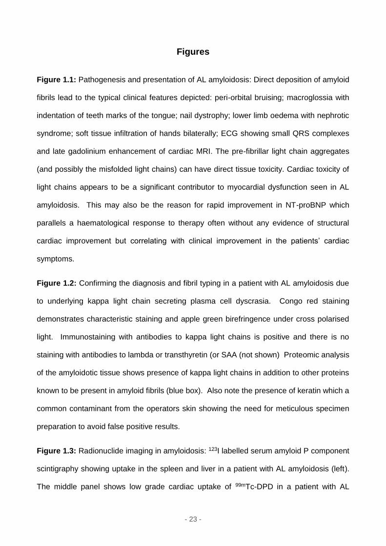
Figure 1.1: Pathogenesis and presentation of AL amyloidosis: Direct deposition of amyloid
fibrils lead to the typical clinical features depicted: peri-orbital bruising; macroglossia with
indentation of teeth marks of the tongue; nail dystrophy; lower limb oedema with nephrotic
syndrome; soft tissue infiltration of hands bilaterally; ECG showing small QRS complexes
and late gadolinium enhancement of cardiac MRI. The pre-fibrillar light chain aggregates
(and possibly the misfolded light chains) can have direct tissue toxicity. Cardiac toxicity of
light chains appears to be a significant contributor to myocardial dysfunction seen in AL
amyloidosis. This may also be the reason for rapid improvement in NT-proBNP which
parallels a haematological response to therapy often without any evidence of structural
cardiac improvement but correlating with clinical improvement in the patients’ cardiac
symptoms.
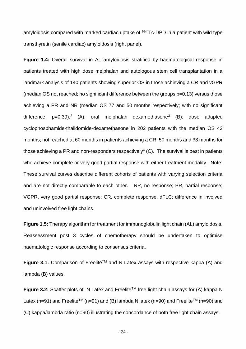
Figure 1.2: Confirming the diagnosis and fibril typing in a patient with AL amyloidosis due
to underlying kappa light chain secreting plasma cell dyscrasia. Congo red staining
demonstrates characteristic staining and apple green birefringence under cross polarised
light. Immunostaining with antibodies to kappa light chains is positive and there is no
staining with antibodies to lambda or transthyretin (or SAA (not shown) Proteomic analysis
of the amyloidotic tissue shows presence of kappa light chains in addition to other proteins
known to be present in amyloid fibrils (blue box). Also note the presence of keratin which a
common contaminant from the operators skin showing the need for meticulous specimen
preparation to avoid false positive results.
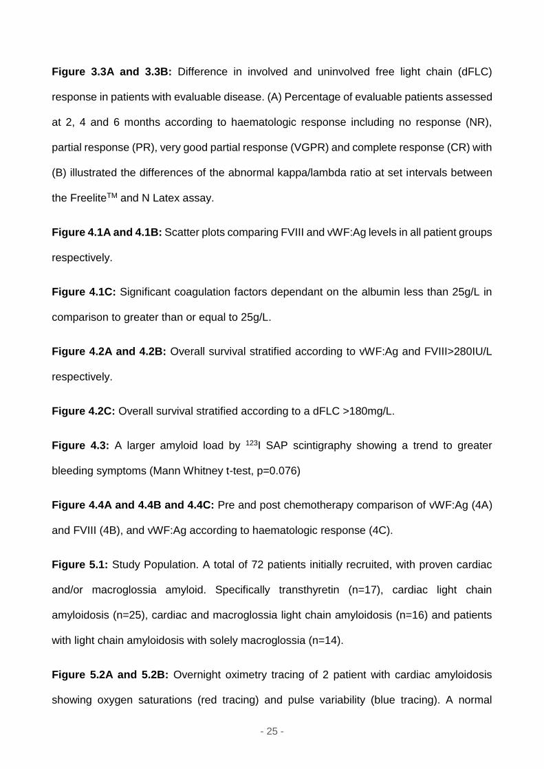
Figure 1.3: Radionuclide imaging in amyloidosis: 123I labelled serum amyloid P component
scintigraphy showing uptake in the spleen and liver in a patient with AL amyloidosis (left).
The middle panel shows low grade cardiac uptake of 99mTc-DPD in a patient with AL
- 24 -
amyloidosis compared with marked cardiac uptake of 99mTc-DPD in a patient with wild type
transthyretin (senile cardiac) amyloidosis (right panel).
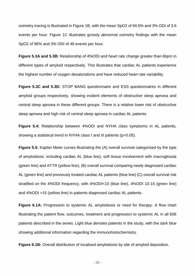
Figure 1.4: Overall survival in AL amyloidosis stratified by haematological response in
patients treated with high dose melphalan and autologous stem cell transplantation in a
landmark analysis of 140 patients showing superior OS in those achieving a CR and vGPR
(median OS not reached; no significant difference between the groups p=0.13) versus those
achieving a PR and NR (median OS 77 and 50 months respectively; with no significant
difference; p=0.39).2 (A); oral melphalan dexamethasone3 (B); dose adapted
cyclophosphamide-thalidomide-dexamethasone in 202 patients with the median OS 42
months; not reached at 60 months in patients achieving a CR; 50 months and 33 months for
those achieving a PR and non-responders respectively4 (C). The survival is best in patients
who achieve complete or very good partial response with either treatment modality. Note:
These survival curves describe different cohorts of patients with varying selection criteria
and are not directly comparable to each other. NR, no response; PR, partial response;
VGPR, very good partial response; CR, complete response, dFLC; difference in involved
and uninvolved free light chains.
Figure 1.5: Therapy algorithm for treatment for immunoglobulin light chain (AL) amyloidosis.
Reassessment post 3 cycles of chemotherapy should be undertaken to optimise
haematologic response according to consensus criteria.
Figure 3.1: Comparison of FreeliteTM and N Latex assays with respective kappa (A) and
lambda (B) values.
Figure 3.2: Scatter plots of N Latex and FreeliteTM free light chain assays for (A) kappa N
Latex (n=91) and FreeliteTM (n=91) and (B) lambda N latex (n=90) and FreeliteTM (n=90) and
(C) kappa/lambda ratio (n=90) illustrating the concordance of both free light chain assays.
- 25 -
Figure 3.3A and 3.3B: Difference in involved and uninvolved free light chain (dFLC)
response in patients with evaluable disease. (A) Percentage of evaluable patients assessed
at 2, 4 and 6 months according to haematologic response including no response (NR),
partial response (PR), very good partial response (VGPR) and complete response (CR) with
(B) illustrated the differences of the abnormal kappa/lambda ratio at set intervals between
the FreeliteTM and N Latex assay.
Figure 4.1A and 4.1B: Scatter plots comparing FVIII and vWF:Ag levels in all patient groups
respectively.
Figure 4.1C: Significant coagulation factors dependant on the albumin less than 25g/L in
comparison to greater than or equal to 25g/L.
Figure 4.2A and 4.2B: Overall survival stratified according to vWF:Ag and FVIII>280IU/L
respectively.
Figure 4.2C: Overall survival stratified according to a dFLC >180mg/L.
Figure 4.3: A larger amyloid load by 123I SAP scintigraphy showing a trend to greater
bleeding symptoms (Mann Whitney t-test, p=0.076)
Figure 4.4A and 4.4B and 4.4C: Pre and post chemotherapy comparison of vWF:Ag (4A)
and FVIII (4B), and vWF:Ag according to haematologic response (4C).
Figure 5.1: Study Population. A total of 72 patients initially recruited, with proven cardiac
and/or macroglossia amyloid. Specifically transthyretin (n=17), cardiac light chain
amyloidosis (n=25), cardiac and macroglossia light chain amyloidosis (n=16) and patients
with light chain amyloidosis with solely macroglossia (n=14).
Figure 5.2A and 5.2B: Overnight oximetry tracing of 2 patient with cardiac amyloidosis
showing oxygen saturations (red tracing) and pulse variability (blue tracing). A normal
- 26 -
oximetry tracing is illustrated in Figure 1B, with the mean SpO2 of 94.6% and 3% ODI of 3.6
events per hour. Figure 1C illustrates grossly abnormal oximetry findings with the mean
SpO2 of 96% and 3% ODI of 46 events per hour.
Figure 5.3A and 5.3B: Relationship of 4%ODI and heart rate change greater than 6bpm in
different types of amyloid respectively. This illustrates that cardiac AL patients experience
the highest number of oxygen desaturations and have reduced heart rate variability.
Figure 5.3C and 5.3D: STOP BANG questionnaire and ESS questionnaires in different
amyloid groups respectively, showing evident elements of obstructive sleep apnoea and
central sleep apnoea in these different groups. There is a relative lower risk of obstructive
sleep apnoea and high risk of central sleep apnoea in cardiac AL patients
Figure 5.4: Relationship between 4%ODI and NYHA class symptoms in AL patients,
showing a statistical trend in NYHA class I and III patients (p=0.05).
Figure 5.5: Kaplan Meier curves illustrating the (A) overall survival categorised by the type
of amyloidosis: including cardiac AL (blue line), soft tissue involvement with macroglossia
(green line) and ATTR (yellow line); (B) overall survival comparing newly diagnosed cardiac
AL (green line) and previously treated cardiac AL patients (blue line) (C) overall survival risk
stratified on the 4%ODI frequency, with 4%ODI<10 (blue line), 4%ODI 10-15 (green line)
and 4%ODI >15 (yellow line) in patients diagnosed cardiac AL patients.
Figure 6.1A: Progression to systemic AL amyloidosis or need for therapy. A flow chart
illustrating the patient flow, outcomes, treatment and progression to systemic AL in all 606
patients described in the series. Light blue denotes patients in the study, with the dark blue
showing additional information regarding the immunohistochemistry.
Figure 6.1B: Overall distribution of localised amyloidosis by site of amyloid deposition.
- 27 -
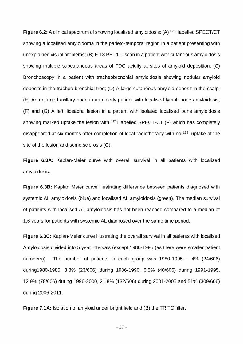
Figure 6.2: A clinical spectrum of showing localised amyloidosis: (A) 123I labelled SPECT/CT
showing a localised amyloidoma in the parieto-temporal region in a patient presenting with
unexplained visual problems; (B) F-18 PET/CT scan in a patient with cutaneous amyloidosis
showing multiple subcutaneous areas of FDG avidity at sites of amyloid deposition; (C)
Bronchoscopy in a patient with tracheobronchial amyloidosis showing nodular amyloid
deposits in the tracheo-bronchial tree; (D) A large cutaneous amyloid deposit in the scalp;
(E) An enlarged axillary node in an elderly patient with localised lymph node amyloidosis;
(F) and (G) A left iliosacral lesion in a patient with isolated localised bone amyloidosis
showing marked uptake the lesion with 123I labelled SPECT-CT (F) which has completely
disappeared at six months after completion of local radiotherapy with no 123I uptake at the
site of the lesion and some sclerosis (G).
Figure 6.3A: Kaplan-Meier curve with overall survival in all patients with localised
amyloidosis.
Figure 6.3B: Kaplan Meier curve illustrating difference between patients diagnosed with
systemic AL amyloidosis (blue) and localised AL amyloidosis (green). The median survival
of patients with localised AL amyloidosis has not been reached compared to a median of
1.6 years for patients with systemic AL diagnosed over the same time period.
Figure 6.3C: Kaplan-Meier curve illustrating the overall survival in all patients with localised
Amyloidosis divided into 5 year intervals (except 1980-1995 (as there were smaller patient
numbers)). The number of patients in each group was 1980-1995 – 4% (24/606)
during1980-1985, 3.8% (23/606) during 1986-1990, 6.5% (40/606) during 1991-1995,
12.9% (78/606) during 1996-2000, 21.8% (132/606) during 2001-2005 and 51% (309/606)
during 2006-2011.
Figure 7.1A: Isolation of amyloid under bright field and (B) the TRITC filter.
- 28 -
Figure 7.1C and 7.1D: Demarcation of the areas of amyloid to be dissected under TRITC
filter (C) and (D) under bright field.
Figure 7.1E: Tissue under bright field post dissecting the amyloid tissue.
Figure 7.1F: The dissected piece of tissue for proteomic analysis.
Figure 7.1G: Congo red staining of tissue
Figure 7.1H: Apple green birefringence of the tissue.
Figure 7.2A: Presenting symptoms in the laryngeal cohort.
Figure 7.2B: Presenting clinical symptoms in the tracheobronchial cohort.
Figure 7.3: Kaplan Meier curve comparing overall survival in patients diagnosed with
laryngeal amyloidosis with tracheobronchial amyloidosis, although not statistically significant
(p 0.66), the curves show a clear difference in the time course in each disease following
approximately 75 months.
Figure 7.4: Proteomic analysis of a micro-dissected amyloidotic area showing the presence
of the “amyloid signature proteins, along with light chains, 3 insulin growth factor (IGF)
binding protein complex and other proteins.
Figure 7.5: Protein sequence showing a repeating peptide sequence in patients with
localised laryngeal and tracheobronchial amyloidosis by proteomic analysis.
Figure 8.1A: Kaplan Meier estimated overall survival on an intention to treat basis. The
median overall survival has not been reached.
Figure 8.1B: Kaplan Meier estimates of progression free survival. The median progression
free survival was 44.5 months.
- 29 -
Figure 8.1C: Overall Survival stratified by the dFLC response achieved. Patients achieving
a partial dFLC response or greater (solid line) had a significantly better overall survival than
those who did not achieve a dFLC response (dashed line)
Figure 8.1D: Overall Survival stratified by the monoclonal protein response. There was no
significant impact of an isolated monoclonal protein response on survival (patients achieving
a partial haematological response or greater (solid line) and no response (dashed line)).
Figure 9.1A: The natural history of estimated glomerular filtration rate (eGFR) in hereditary
fibrinogen amyloidosis patients treated with CPHPC (solid lines) and CKD matched controls
(dashed lines).
Figure 9.1B: Natural history of creatinine in hereditary fibrinogen amyloidosis patients
treated with CPHPC (solid lines) and CKD matched controls (dashed lines).
Figure 9.1D: Renal survival in patients receiving CPHPC (solid line) was significantly better
(p<0.0001) than CKD matched controls (dashed line).
Fig 9.1E: Natural history of the creatinine pre CPHPC treatment in a select group of patients.
Figure 9.2: Recordings of VAS scores in 9 patients treated with CPHPC showing a
progressive decline in joint pain whilst patients were on treatment.
Figure 9.3A: Patient 1 with (β2M) amyloidosis illustrating a pictorial image of the recorded
VAS scores at baseline and following 12 months of treatment with CPHPC.
Figure 9.3B: Patient 2 with (β2M) amyloidosis illustrating a pictorial image of the recorded
VAS scores at baseline and following 12 months of treatment with CPHPC.
Figure 9.3C: Patient 3 with (β2M) amyloidosis illustrating a pictorial image of the recorded
VAS scores at baseline and following 12 months of treatment with CPHPC.
- 30 -
Figure 9.4A and 9.4B: illustrate the coronal images of a PET/CT with increased metabolic
activity in the thickened soft tissue surrounding both shoulders and hip joints at baseline (A)
and 12 months post treatment respectively (B).
Figure 9.5: Baseline QoL for AFib and DRA patients.
Figure 9.6A and 9.6B: QoL at 6 and 12 months in DRA patients.
Figure 9.7A, 9.7B and 9.7C: QoL at 6, 12 and 24 months in AFib patients with a good
eGFR.
Figure 9.8A, 9.8B and 9.8C: QoL at 6, 12 and 24 months in AFib patients with a poor eGFR.
- 31 -
Tables
Table 1.1: Classification of common types of systemic amyloidosis
Table 1.2: Conditions associated with underlying systemic AA amyloidosis
Table 1.3: Diagnostic and staging Investigations for systemic AL amyloidosis
Table 1.4: Consensus haematologic response in systemic light chain (AL) amyloidosis
Table 1.5: Chemotherapy regimens and ASCT studies in AL amyloidosis
Table 2.1: Genotyping for hereditary amyloidosis illustrating the primers used in this
technique
Table.2.2: Definition of the Eastern Co-operative Group Performance Status (ECOG)
Table 2.3: Definition of New York Heart Association Classification (NYHA)
Table 2.4: Definition of organ involvement and organ response in amyloidosis
Table 2.5: Consensus haematologic response in systemic light chain amyloidosis
Table 3.1: Patient characteristics
Table 3.2A: Kappa at presentation
Table 3.2B: Lambda at presentation
Table 3.2C: Kappa/lambda ratio at presentation
Table 3.3: Kappa/lambda ratios at presentation
Table 3.4: Concordance analysis for kappa (A), lambda (B) and kappa/lambda ratio (C) for
the two FLC assays
Table 3.5: Clinical sensitivity and specificity of the FreeliteTM assay and immunofixation
electrophoresis (IFE)
- 32 -
Table 3.6: Clinical sensitivity and specificity of the N Latex assay and immunofixation
electrophoresis (IFE)
Table 4.1: Patient characteristics
Table 4.2: Low anti-thrombin levels and nephrotic syndrome
Table 4.3: Variables associated with survival
Table 5.1: Patient characteristics
Table 5.2: Oximetry data in the different amyloid groups
Table 5.3: New cardiac AL
Table 6.1: Patient characteristics
Table 6.2: Patient characteristics according to amyloid type
Table 7.1: Patient characteristics in laryngeal group
Table 7.2: Patient characteristics in tracheobronchial group
Table 8.1: Patient characteristics
Table 8.2: Variables associated with survival
Table 9.1: Patient characteristics
Table 9.2: Comparison of clinical and laboratory features of AFib patients treated with
CPHPC and historical control
CHAPTER ONE
- 33 -
Chapter One: Introduction
This chapter is written in the context of my publication: Update on treatment of light
chain amyloidosis. Shameem Mahmood, Giovanni Palladini, Vaishali Sanchorawala,
Ashutosh Wechalekar. Haematologica. 2014, 99: 209-221, copyright permission obtained
from Haematologica office for use in my thesis.
Amyloidosis is a rare systemic disorder characterised by misfolding of aberrant precursor
proteins causing formation of unstable auto aggregates leading to amyloid fibril formation in
a predominant β-pleated sheet structure.5 These fibrils are deposited in different organs,
progressively affecting the organ’s architecture and function.6 The unstable protein may be
hereditary or acquired, with 25 different proteins to form amyloid fibrils,7 The most common
organs involved include the heart, kidneys, liver, gastrointestinal tract, autonomic and
peripheral nervous system. Amyloidosis varies in its disease phenotype with a clear
differentiation between a localised deposit of amyloid such as in localised amyloidosis and
systemic amyloidosis, the latter depending on the underlying fibril type, organ involved and
extent of amyloid deposition.
CHAPTER ONE
- 34 -
Amyloid proteins and fibrillogenesis
Early studies show that a vital part of the process of protein fibrillogenesis is partial unfolding
of the protein.8 Accelerated fibrillation in protein deposition diseases are associated with
certain mutations, and allow destabilisation of the native structure, resulting in an increased
quantity of steady-state concentration of partially folded conformers. Transthyretin in
solution has been shown to undergo dissociation to a monomer influenced by the
temperature, pH, ionic strength and protein concentration. Amyloid fibril formation related to
some TTR variants may be triggered by tetramer dissociation to a compact non-native
monomer with low conformational stability.9 The structure of the monomer may form a
partially unfolded monomeric structure and soluble aggregates. Variable domains have
been implicated in systemic AL amyloidosis, with initial hypothesis suggesting that
proteolysis occurred. Light chains are susceptible to aggregation due to a number of factors:
(1) certain somatic mutations are thought to de-stabilise the protein and hence favour the
formation of amyloid fibrils, (2) reduced thermodynamic stability and (3) the cellular
environment. Structural experiments have shown that most variable domains from AL
amyloidosis patients form crystals as monomers or dimers with the beta-pleated sheet.
Oxidative stress is also important, having been associated with amyloid fibril deposits and
cell death.10 Internalisation studies have shown that immunoglobulin light chains initially
internalise into the cardiomyocytes by migrating into the lysosomal compartments. Little is
known as to the underlying reason why certain organs are affected with these dynamic
amyloid deposits resulting in impairment of the affected organ,11 even with slight variations
within family members affected by the same hereditary variant protein. Previous
experiments have shown that development of systemic AA amyloidosis occurs when a
mouse is injected with protein from an AA amyloidosis mouse with an inflammatory
precipitant.12 This raises the hypothesis which proposes that once amyloid fibrils deposit in
CHAPTER ONE
- 35 -
a tissue, a particular template or backbone is formed. The ongoing deposition of further
precursor proteins allowing further amyloid deposits to be laid down within this template.
Regression of amyloid deposits is less well understood, with earlier studies showing that
this process is partly macrophage driven. Macrophage experiments in murine models have
shown the complete degradation of Aβ amyloid fibrils in vitro.13, 14 It has been postulated
that these macrophages infiltrating the amyloid deposits consequently result in the formation
of multinucleated giant cells, encircling the amyloid prior to engulfing the amyloid deposit.
Initial studies have explored this concept proposing that the clearance of amyloid may be
antibody mediated,15 with antibody-amyloid specific administration successful in reducing
the amyloid load in those diagnosed with Alzheimer’s disease16 or systemic AL.17 One
method of translating this concept visually is by use of I123SAP scintigraphy, whereby the
amyloid load may be assessed. It is variable as to which patients achieve amyloid regression
by this technique independent of the clonal or inflammatory suppression attained – with
clearly many factors which contribute to this phenomenon.
Epidemiology
Amyloidosis is a very rare condition, with the initial incidence based on deaths/post-mortem
information, initially quoted as 4.5/100000018 and an estimated 500 new cases seen at the
National Amyloidosis Centre each year. The subtle symptoms often masked as other
medical conditions are likely to underestimate this condition. Multiple myeloma patients may
have incidental amyloid deposits with no organ dysfunction in 38% of cases,19 with organ
dysfunction present in 3-7% of patients.20 The incidence of systemic AA amyloidosis
typically depends on the underlying inflammatory condition, with the prevalence increasing
from 18% to 30% at post-mortem.21 The incidence of hereditary amyloidosis varies greatly
between countries and remains little studied. Familial Amyloid Polyneuropathy varies
between countries with different mutation variants prevalent in different locations, with the
CHAPTER ONE
- 36 -
most common FAP variant in the UK and Ireland the T60A variant.22 Earlier studies have
estimated the V122I variant present in 3-4% of the Afro-Caribbean population, clinically
similar to the wild type variant ATTRwt.23, 24
Types of amyloidosis
Localised amyloidosis
Localised AL amyloidosis is characterised by amyloid deposits at a single site (commonly:
bladder, skin, larynx, lung) due to local production of light chains and no evidence of
systemic involvement. It has excellent prognosis with generally no need for systemic
therapy.25 We will elaborate more on this type of amyloidosis in chapter 7.
Systemic amyloidosis
This group comprises of systemic light chain (AL) amyloidosis, systemic amyloid A (AA)
amyloidosis, senile systemic amyloidosis (ATTRwt) and dialysis related amyloidosis (DRA).
Systemic light chain (AL) amyloidosis is the most common type; in which the amyloidogenic
protein is a monoclonal light chain secreted by a underlying clonal plasma cell (or rarely B
lymphoid) dyscrasia.6 Other hereditary amyloidoses are due to amyloidogenic mutations in
fibrinogen, Apolipoprotein A1 and A2, lysozyme and Gelsolin genes. AA amyloidosis occurs
due to deposition of serum amyloid A protein (an acute phase protein) in a spectrum of
disorders causing prolonged inflammation and treatment focuses upon reducing that
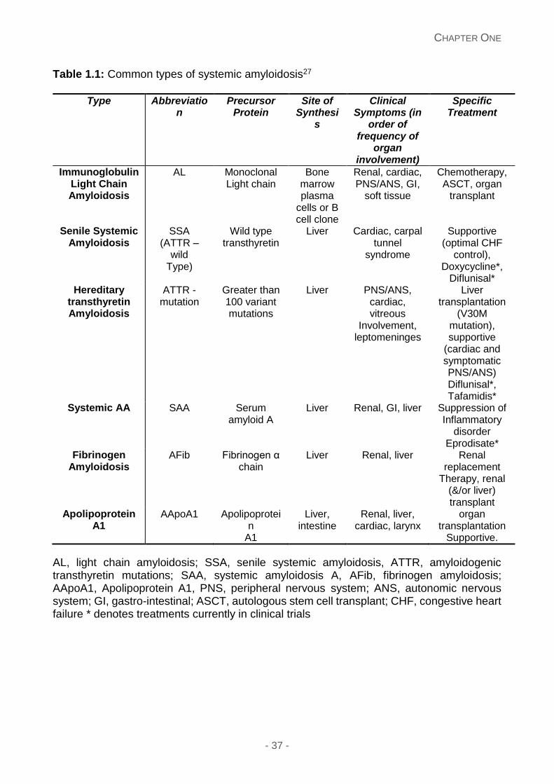
inflammatory drive. Table 1.1 illustrates the common types of systemic amyloidosis.26
CHAPTER ONE
- 37 -
Table 1.1: Common types of systemic amyloidosis27
Type Abbreviation
Precursor Protein
Site of Synthesi
s
Clinical Symptoms (in
order of frequency of
organ involvement)
Specific Treatment
Immunoglobulin Light Chain Amyloidosis
AL Monoclonal Light chain
Bone marrow plasma
cells or B cell clone
Renal, cardiac, PNS/ANS, GI,
soft tissue
Chemotherapy, ASCT, organ
transplant
Senile Systemic Amyloidosis
SSA (ATTR –
wild Type)
Wild type transthyretin
Liver Cardiac, carpal tunnel
syndrome
Supportive (optimal CHF
control), Doxycycline*,
Diflunisal* Hereditary
transthyretin Amyloidosis
ATTR - mutation
Greater than 100 variant mutations
Liver PNS/ANS, cardiac, vitreous
Involvement, leptomeninges
Liver transplantation
(V30M mutation), supportive
(cardiac and symptomatic PNS/ANS) Diflunisal*, Tafamidis*
Systemic AA SAA Serum amyloid A
Liver Renal, GI, liver Suppression of Inflammatory
disorder Eprodisate*
Fibrinogen Amyloidosis
AFib Fibrinogen α chain
Liver Renal, liver Renal replacement
Therapy, renal (&/or liver) transplant
Apolipoprotein A1
AApoA1 Apolipoprotein
A1
Liver, intestine
Renal, liver, cardiac, larynx
organ transplantation
Supportive.
AL, light chain amyloidosis; SSA, senile systemic amyloidosis, ATTR, amyloidogenic transthyretin mutations; SAA, systemic amyloidosis A, AFib, fibrinogen amyloidosis; AApoA1, Apolipoprotein A1, PNS, peripheral nervous system; ANS, autonomic nervous system; GI, gastro-intestinal; ASCT, autologous stem cell transplant; CHF, congestive heart failure * denotes treatments currently in clinical trials
CHAPTER ONE
- 38 -
Systemic AA amyloidosis
The amyloid fibrils in systemic AA amyloidosis arise from serum amyloid A protein in the
context of an underlying inflammatory condition. The most common inflammatory
arthropathies account for 50% in the Western world, whereas in the developing world the
majority of cases are secondary to infection. Hereditary periodic fever syndromes are
associated with increased inflammatory stimulus and hence the risk of systemic AA
amyloidosis. Table 1.2 describes the majority of inflammatory conditions.28 There are many
contributing factors which remain unexplained as to why AA amyloidosis and the production
of variable levels of SAA occur in this disease, with genetic polymorphisms possibly
contributing to this phenomenon.29
We recognise that systemic AA amyloidosis typically affects the kidneys in over 95% of
patients, clinically presenting with proteinuria in approximately 10% of patients in end stage
renal failure (ESRF) at diagnosis. Other organs involved can include the spleen (often seen
by 123I SAP scintigraphy, adrenal glands, liver and gastrointestinal involvement, with less
dysfunction of these organs.28 Approximately one third of patients will progress to ESRF
with the risk of renal decline very much dependent on the underlying inflammatory disorder
and treatment options, in recent years including newer biological agents. Renal
transplantation has been performed in selected patients with excellent outcomes.30

CHAPTER ONE
- 39 -
Table 1.2: Conditions associated with underlying systemic AA amyloidosis28
.
Inflammatory Arthritis Adult Still’s Disease Ankylosing Spondylitis Juvenile Idiopathic Arthritis Psoriatic Arthropathies Reiter’s Syndrome Rheumatoid Arthritis Gout Chronic Infections Bronchiectasis Chronic Cutaneous Ulcers Chronic Pyelonephritis Leprosy Osteomyelitis Q Fever Sub-acute Bacterial Endocarditis Tuberculosis Whipples Disease Immunodeficiency States Common Variable Immunodeficiency Cyclic Neutropenia Hyperimmunoglobulin M Syndrome Hypogammaglobulinaemia HIV/Aids Other Conditions Predisposing To Chronic Infections Cystic Fibrosis Epidermolysis Bullosa Injected Drug Abuse Jejuno-Ileal Bypass Kartagener’s Syndrome Paraplegia Sickle Cell Anaemia
Hereditary Periodic Fevers Cryopyrin associated periodic fever syndrome (CAPS) Familial Mediterranean fever (FMF) Mevalonate Kinase Deficiency (MKD or HIDS) TNF receptor associated periodic syndrome (TRAPS) Inflammatory Bowel Disease Crohn’s disease Ulcerative colitis Neoplasia Adenocarcinoma of the lung, gut, urogenital tract Basal cell carcinoma Carcinoid tumour Castleman’s disease Gastrointestinal stromal tumour Hairy cell leukaemia Hepatic adenoma Hodgkin’s disease Mesothelioma Renal cell carcinoma Sarcoma Systemic Vasculitis Behcet’s disease Giant cell arteritis Polyarteritis nodosa Polymyalgia rheumatic Systemic lupus erythematosis Takayasu's arteritis Additional Atrial myxoma Inflammatory abdominal aortic aneurysm Retroperitoneal fibrosis Sarcoidosis Sinus histiocytosis with massive lymphadenopathy
CHAPTER ONE
- 40 -
Dialysis related amyloidosis
This condition may arise secondary to long term dialysis with the underlying fibril β-
microglobulin (β2M). This molecule is typically filtered via the kidneys, specifically the
glomerulus and resorbed by the proximal tubular cells.31, 32 The majority of patients present
clinically following a period of 10 years on dialysis with symptoms of carpal tunnel syndrome,
spondyloarthropathies, arthralgia, subchondral bone cysts and fractures. The incidence of
DRA is lower with higher flux dialysis membranes with renal transplantation the only means
of reducing the former clinical symptoms.33
Wild type transthyretin amyloidosis, senile systemic amyloidosis
Amyloidosis caused by deposition of misfolded transthyretin (ATTR) is the next most
common, either hereditary (due to amyloidogenic ATTR mutations) or a disease of aging;
due to wild type ATTR deposition (senile systemic amyloidosis), with the fibril wild type
transthyretin.34 The latter is typically seen in older men, with the majority describing previous
Carpal Tunnel symptoms. Amyloid deposits with this fibril type can occur in the heart, and
found in 25% of autopsy findings in those older than 80 years of age,35 with deposits also
found in other tissues including the bladder, gastro-intestinal tract and soft tissue.36 Cardiac
manifestations of this disease are typically demonstrated by echocardiography and cardiac
MRI and tissue proof the ultimate test with wild type transthyretin gene sequencing. There
is limited data as to the natural progression of this disease, typically slower with the mainstay
of treatment centred on optimisation of heart failure treatment.
CHAPTER ONE
- 41 -
Hereditary Transthyretin amyloidosis
Hereditary Transthyretin amyloidosis accounts for the majority of hereditary amyloidosis with
100 mutations; with clinical presentation with progressive autonomic or peripheral
neuropathy. Cardiac involvement is present in the majority of patients, with other tissue
involvement including vitreous, gastrointestinal and less frequently the central nervous
system usually in the third decade; with variability due to the mutation involved.37-39
Transthyretin is produced in the liver in 95% of patients, with the rest produced in the choroid
plexus and retina.40 The most prevalent transthyretin mutation involves the substitution of
methionine for valine at position 30 (ATTRV30M). Clinical presentation is usually with an
ascending sensorimotor peripheral neuropathy, with cardiac involvement rare. The age of
onset occurs by the age of 30-40 years in the Portuguese, and presents approximately 20
years later in the Swedish one. Whilst the most common variant in the UK and Ireland is
T60A; clinical presentation including autonomic neuropathy and cardiac involvement by the
age of 50 years.22, 41 Another transthyretin variant present in the Afro-Caribbean population
(3-4%) is V122I variant with clinical cardiac disease after the age of 60 years.24 Evaluation
of family members at risk is also an issue, with genetic counselling important.
Orthotopic liver transplantation (OLT) was used initially in the 1990s with the premise that
the mutant TTR is produced in the liver.41, 42 Symptoms of peripheral neuropathy improved
when chosen earlier in the time course of the disease, with criteria such as age less than 60
years, limited polyneuropathy or autonomic neuropathy, no significant renal or cardiac
dysfunction important.43 However the mortality for this procedure is high.41, 44 There are
newer anti-amyloid therapies, targeting stabilisation of the soluble TTR in the blood and
inhibiting its production through silencing RNA and anti-sense oligonucleotide approaches.
CHAPTER ONE
- 42 -
Hereditary Aα-chain fibrinogen amyloidosis (AFib)
Hereditary fibrinogen amyloidosis accounts for the most common cause of renal amyloidosis
first described in 1993, with a variable penetrance and no family history described.45 There
are 9 reported variants with E526V the most common.46 Clinical presentation is with
proteinuria, hypertension and renal impairment at the age of 60 years and ESRF usually
within 5 years from diagnosis. The renal biopsy shows a characteristic abundant glomerular
amyloid infiltration with almost complete obliteration of the normal architecture but little or
no vascular or interstitial deposits.47
Combined renal and liver transplantation has been a treatment option for a selected cohort
of patients, given the amyloidogenic protein is produced exclusively by the liver. The long
term outcomes have been excellent but this procedure carries a high mortality risk.48 Renal
transplantation is another option with the median graft survival 7 years with recurrent
amyloid deposits typically the cause for further renal decline.47
Hereditary apolipoprotein A1 amyloidosis (AApoA1)
Apolipoprotein A1 is an HDL important in the role of cholesterol transport which is produced
in the liver (50%)49, 50 and intestines, with the liver and kidneys being major sites for Apo A1
catabolism. Thirteen variants have been reported, with the underlying pathogenesis
involving proteolytic cleavage at amino terminal 83-93 residues incorporated into the
amyloid fibrils.51 Each variant is associated with varying phenotypes and again differences
which occur within the family. The main organ involved is the kidney, and consequently
chronic renal failure, with neurological, cardiac, and hepatic dysfunction also reported. The
phenotype of the following six variants: Gly26Arg, Trp50Arg, Leu60Arg, Del70-72, Leu75Pro
and Leu64Pro involve renal involvement with hypertension and proteinuria with the clinical
presentation of hepatosplenomegaly.52 Other AApoAI variants (Leu90Pro, Arg173Pro,
Leu174Ser and Leu178His) have been reported with skin and cardiac amyloid deposits with
CHAPTER ONE
- 43 -
death usually occurring due to progressive cardiomyopathy within 10 years of diagnosis.53,
54 The exact underlying aetiology of this disease remains uncertain, with the hypothesis of
mutations destabilising the native structure and facilitating fibrillogenesis and consequent
proteolysis.55, 56Renal transplantation has offered one therapeutic option with renal graft
survival exceeding 10-15 years irrespective of recurrent amyloid in the transplanted organ.
57 Reduction in the variant ApoA1 by 50%, can be sufficient to facilitate extra-hepatic amyloid
regression in a certain cohort of patients, such as in liver transplantation.58
Apolipoprotein AII amyloidosis (AApoAII)
Apolipoprotein AII is an HDL apolipoprotein with Apoliprotein AII amyloidosis described
initially in 1973 by Weiss and Page,59 with 4 amyloidogenic variants reported to date, but
less well elucidated.60 Patients typically present with proteinuria and progressive renal
decline.
Hereditary Gelsolin amyloidosis (AGel)
The amyloid fibrils occur due to cleavage fragments of the variant Gelsolin initially described
by the Finnish ophthalmologist Jouko Meretoja in 1969.61 There are 2 main variants: G654A
described in Portugal, Japan and Iran, with G654T reported in countries including France,
Czech Republic and Denmark.62 Gelsolin is an actin-modulating protein facilitating the
migration into other cells, with the mutated form unable to bind to calcium and hence
susceptible to proteolysis and fibril formation.63-65 Patients typically present with corneal
lattice dystrophy during middle age with slowly progressive cranial neuropathies. The clinical
phenotype can vary greatly from a slight sensory neuropathy to severe ataxia, from mild
visual impairment to total blindness.66 Interestingly renal amyloid deposits are seen by 123I
SAP scintigraphy with no corresponding renal functional decline.
CHAPTER ONE
- 44 -
Lysozyme amyloidosis (ALys)
Lysozyme is a ubiquitous bacteriolytic enzyme typically found in high quantities in the liver,
articular surfaces, saliva and tears and expressed in granulocytes, monocytes and bone
marrow precursor cells. It is typically inherited in an autosomal dominant fashion. Pepys et
al first described Lysozyme amyloidosis,67 with seven amyloidogenic mutations described
including: Ile56Thr, Phe57Ile, Trp64Arg, Asp67His, Trp112Arg, Tyr54Asn and D67G with
patients presenting in their 3rd and 4th decade. Previous studies show that fibril formation by
human wild type lysozyme was accelerated by fibrils of the variant proteins, with wild type
lysozyme deposits significantly much lower in concentration compared to amyloidogenic
variants.67, 68 Patients clinically present with a slowly progressive decline in renal function
with organ involvement also involving liver, spleen, gastrointestinal tract and lymph node
involvement. Interestingly certain variants including Try64Arg and Asp67His and lung and
thyroid tissue involvement with the Ile56Thr variant describe Sicca syndrome due to salivary
gland amyloid deposits.69
Systemic light chain (AL) amyloidosis
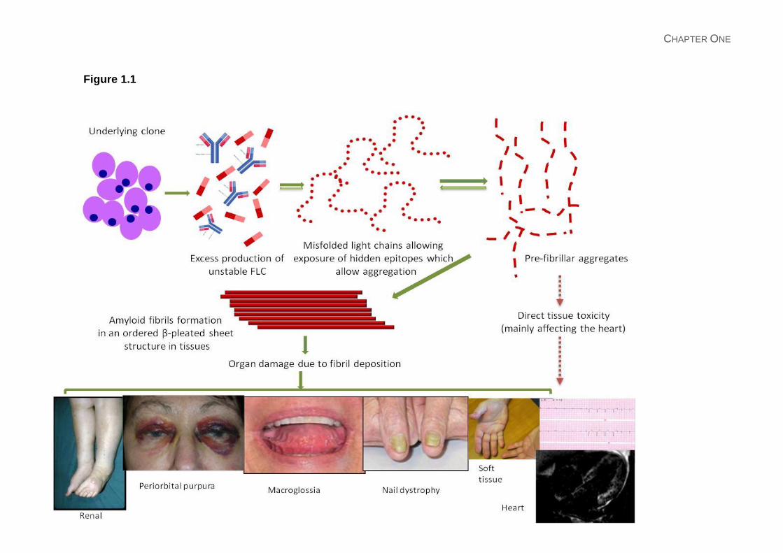
The presenting symptoms of AL amyloidosis have a wide spectrum: dyspnoea, lethargy,
weight loss, bleeding tendency, swelling of lower limbs, frothy urine, orthostatic hypotension
or peripheral neuropathy. Macroglossia and peri-orbital bruising are almost pathognomonic,
occurring only in a third of all cases (Figure 1.1). The diagnosis of AL amyloidosis is often
delayed as presenting features are subtle or mimic other more common conditions. Table
1.1 shows the most common other types of systemic amyloidosis.
CHAPTER ONE
- 45 -
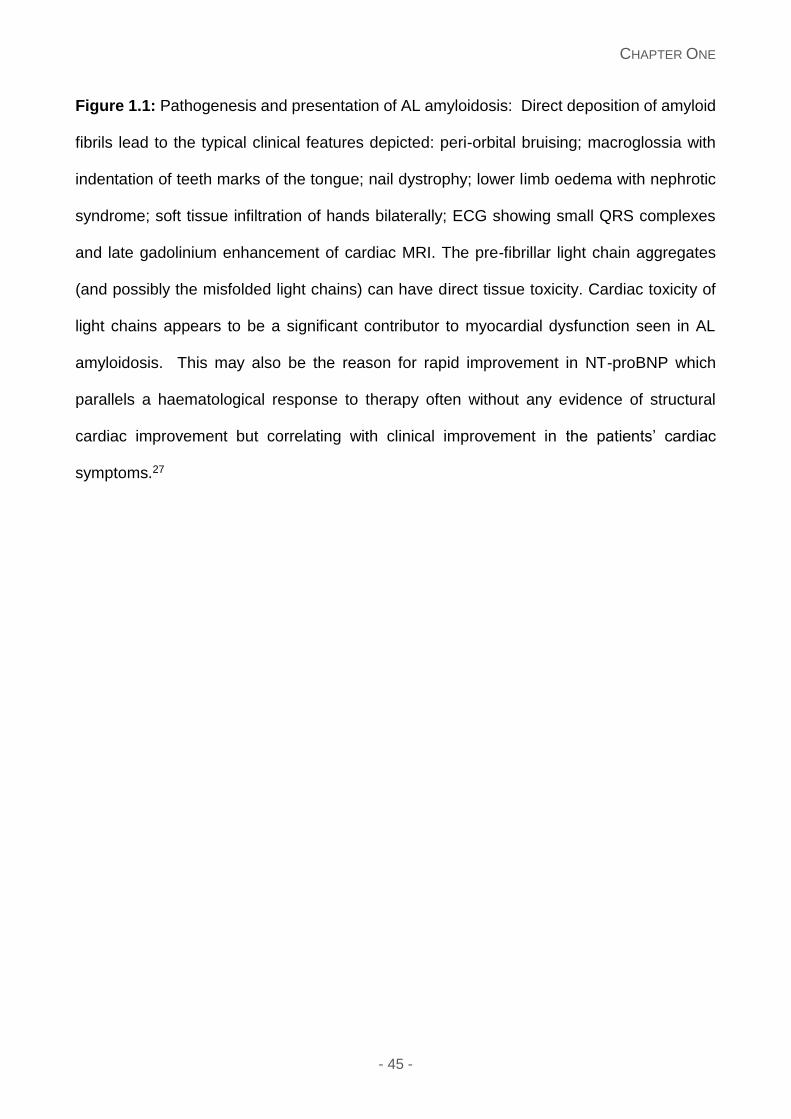
Figure 1.1: Pathogenesis and presentation of AL amyloidosis: Direct deposition of amyloid
fibrils lead to the typical clinical features depicted: peri-orbital bruising; macroglossia with
indentation of teeth marks of the tongue; nail dystrophy; lower limb oedema with nephrotic
syndrome; soft tissue infiltration of hands bilaterally; ECG showing small QRS complexes
and late gadolinium enhancement of cardiac MRI. The pre-fibrillar light chain aggregates
(and possibly the misfolded light chains) can have direct tissue toxicity. Cardiac toxicity of
light chains appears to be a significant contributor to myocardial dysfunction seen in AL
amyloidosis. This may also be the reason for rapid improvement in NT-proBNP which
parallels a haematological response to therapy often without any evidence of structural
cardiac improvement but correlating with clinical improvement in the patients’ cardiac
symptoms.27
CHAPTER ONE
- 46 -
Figure 1.1
CHAPTER ONE
- 47 -
Advanced organ dysfunction has often ensued prior to a clinical diagnosis of amyloidosis
although monoclonal gammopathy (MGUS)70 or myeloma usually predates a diagnosis of
amyloidosis. Fifteen percent of patients with myeloma have symptomatic AL amyloidosis
and up to 30% may have “incidental” deposits, which may become clinically significant with
improving long term outcomes in myeloma.71 Patients with MGUS and an abnormally
elevated free light chain (FLC) should be additionally monitored at each visit by
measurement of serum brain natriuretic peptide (BNP or its N-terminal fragment, NT-
proBNP) and urine for albuminuria – abnormal presence of either may herald development
of amyloidosis5 before advanced, symptomatic organ damage thus significantly reducing the
early deaths which are still observed.
Confirmation of diagnosis needs demonstration of amyloid deposition; pathognomonic apple
green birefringence by Congo red staining using crossed polarised light on histological
tissue sections of either the affected organ, bone marrow, rectum or abdominal fat aspirate
(latter being an easy bedside procedure available for all patients including those with
haemostatic impairment).72 Fibril typing is critical in deciding appropriate therapy and
performed by immunohistochemistry (widely available but specific only in 75-80% of cases
of AL),73 immuno-electron microscopy (highly specific but limited availability)74 or lately,
mass spectrometry of amyloid deposits obtained by laser capture (rapidly becoming an
invaluable adjunct)75 (Figure 1.2). Detecting the underlying clone requires serum and urine
electrophoresis and immunofixation, serum free light chain analysis, bone marrow
examination and imaging for presence of myeloma related bone disease.
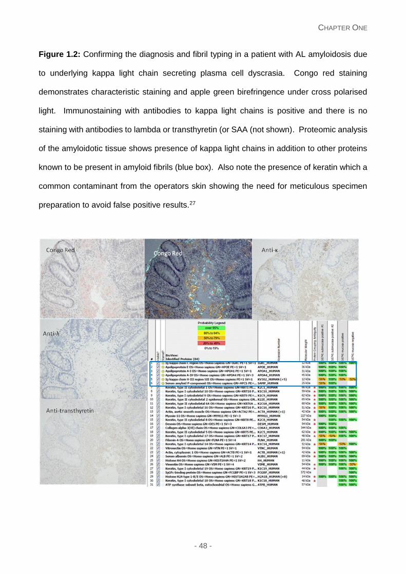
CHAPTER ONE
- 48 -
Figure 1.2: Confirming the diagnosis and fibril typing in a patient with AL amyloidosis due
to underlying kappa light chain secreting plasma cell dyscrasia. Congo red staining
demonstrates characteristic staining and apple green birefringence under cross polarised
light. Immunostaining with antibodies to kappa light chains is positive and there is no
staining with antibodies to lambda or transthyretin (or SAA (not shown). Proteomic analysis
of the amyloidotic tissue shows presence of kappa light chains in addition to other proteins
known to be present in amyloid fibrils (blue box). Also note the presence of keratin which a
common contaminant from the operators skin showing the need for meticulous specimen
preparation to avoid false positive results.27
CHAPTER ONE
- 49 -
Baseline assessment of organ function (Table 1.3) is important for prognosis and selection
of therapy. Formal testing for autonomic and peripheral neuropathy may be needed in
selected cases.
Table 1.3: Diagnostic and Staging Investigations for systemic AL amyloidosis27
Tissue diagnosis Abdominal fat aspirate Salivary gland or rectal biopsy Biopsy of involved organ
Amyloid typing Immunohistochemistry (immuno-electron microscopy if available) Mass spectrometry DNA analysis (if indicated)
Studies to detect an underlying plasma/B cell Clone
Serum and urine electrophoresis and immunofixation Serum Free light chain measurement Bone marrow aspirate / biopsy (plus FISH) Imaging studies for bone disease
Assessing of organ involvement and staging
Cardiac NT-proBNP (or BNP), cTnT (or hs-cTnT, or cTnI) Echocardiography (plus strain imaging) ECG (plus Holter ECG) Cardiac MRI (if indicated) 99mTc-DPD scan (if indicated) Renal 24 h urinary protein Serum creatinine (and eGFR) Liver Liver function tests Liver US / CT scan Nerves Nerve conduction studies (if indicated) Autonomic testing Whole body amyloid load 123I labelled SAP scintigraphy (if available)
FISH - fluorescent in situ hybridisation; NT-proBNP - N-terminal prohormone of brain natriuretic peptide; BNP – brain natriuretic peptide; cTnT or cTnI – troponin T or I; hs-cTnt – high sensitivity troponin T; ECG – electrocardiograph; 99mTc-DPD scan - 99mTc-dicarboxypropane diphosphonate scan; eGFR – estimated glomerular filtration rate; US – ultrasound; CT – computed tomography; MRI – magnetic resonance imaging; SAP – serum amyloid P.
CHAPTER ONE
- 50 -
123I labelled serum amyloid P component (SAP) scintigraphy (if available), is useful for
diagnosis, quantification and especially valuable in serial monitoring of amyloid deposits
(Figure 1.3).76 Cardiac magnetic resonance imaging (CMR) is more sensitive and specific
than echocardiography for diagnosis of cardiac amyloidosis, showing a characteristic
subendocardial late gadolinium enhancement. A cardiac biopsy maybe needed in a select
cohort of patients with isolated cardiac amyloidosis and MGUS to differentiate AL
amyloidosis from senile systemic amyloidosis/ATTR amyloidosis due to V122I mutation.
Bone scintigraphy tracers, 99mTc-dicarboxypropane diphosphonate (99mTc-DPD) and 99mTc-
pyrophosphate (99mTc-PYP), are avidly taken up in cardiac ATTR amyloid deposits but not
in AL77,78– an emerging non-invasive means of differentiating the two conditions (Figure 1.3).
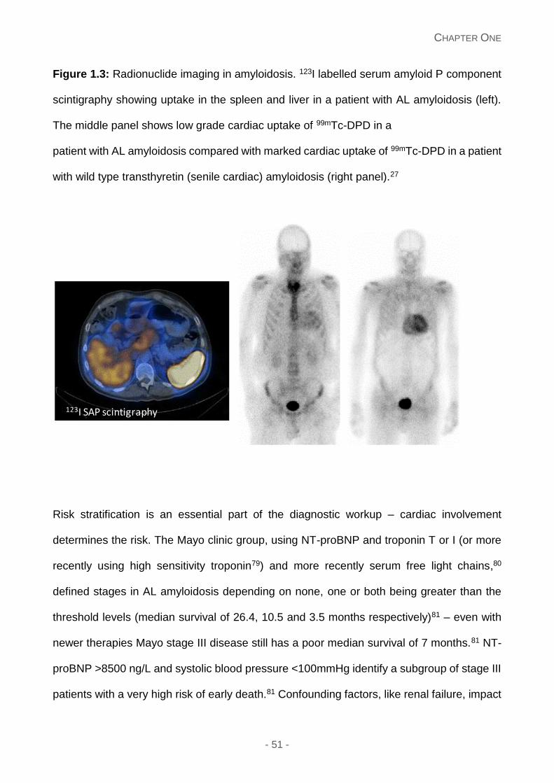
CHAPTER ONE
- 51 -
Figure 1.3: Radionuclide imaging in amyloidosis. 123I labelled serum amyloid P component
scintigraphy showing uptake in the spleen and liver in a patient with AL amyloidosis (left).
The middle panel shows low grade cardiac uptake of 99mTc-DPD in a
patient with AL amyloidosis compared with marked cardiac uptake of 99mTc-DPD in a patient
with wild type transthyretin (senile cardiac) amyloidosis (right panel).27
Risk stratification is an essential part of the diagnostic workup – cardiac involvement
determines the risk. The Mayo clinic group, using NT-proBNP and troponin T or I (or more
recently using high sensitivity troponin79) and more recently serum free light chains,80
defined stages in AL amyloidosis depending on none, one or both being greater than the
threshold levels (median survival of 26.4, 10.5 and 3.5 months respectively)81 – even with
newer therapies Mayo stage III disease still has a poor median survival of 7 months.81 NT-
proBNP >8500 ng/L and systolic blood pressure <100mmHg identify a subgroup of stage III
patients with a very high risk of early death.81 Confounding factors, like renal failure, impact
123I SAP scintigraphy
CHAPTER ONE
- 52 -
the concentration of NT-proBNP, and measurement of BNP may be preferred in this
setting.82
Treatment of AL amyloidosis
Goals of therapy
The aim of treatment in AL amyloidosis is eradicating the fibril precursor protein by
suppressing production of free light chains as rapidly as possible by targeting the underlying
clonal plasma/B cell dyscrasia, whilst minimising treatment related mortality (TRM) and
morbidity5 with supportive measures to preserve organ function. Since patients with AL
amyloidosis have a small clonal burden83 and lack the high risk cytogenetic features seen
in myeloma such as t(4:14) or del17p,84 shorter courses of dose adapted chemotherapy may
be adequate to achieve good haematologic responses.85
Response assessment has two components: haematologic and organ response – the latter
follows the former.86, 87 dFLC measurement is a key marker to assess clonal disease
response.87 Consensus criteria for haematologic response assessment have recently been
published (Table 1.4).88 A very good partial response (VGPR) (defined as dFLC less than
40mg/L) or better is associated with an OS of 80-90% at 3 years,88 and is currently
considered the minimum goal of therapy.
In addition to standard measures of organ function, cardiac biomarkers such as a reduction
in NT-proBNP of 30% and 300 ng/L from baseline following completion of therapy usefully
defines a cardiac organ response.87 Lack of such a decrease in NT-proBNP (in patients with
heart involvement) identifies a subgroup of patients achieving less than CR who need a
more profound haematologic response. Factors such as worsening renal failure or treatment
with immunomodulatory drugs may lead to elevated cardiac biomarkers, confounding
response assessment.89
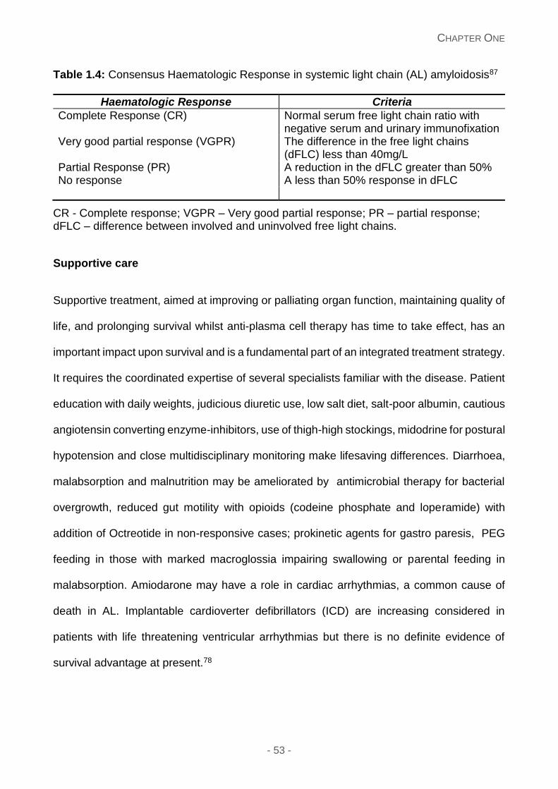
CHAPTER ONE
- 53 -
Table 1.4: Consensus Haematologic Response in systemic light chain (AL) amyloidosis87
Haematologic Response Criteria
Complete Response (CR)
Normal serum free light chain ratio with negative serum and urinary immunofixation
Very good partial response (VGPR) The difference in the free light chains (dFLC) less than 40mg/L
Partial Response (PR) A reduction in the dFLC greater than 50% No response A less than 50% response in dFLC
CR - Complete response; VGPR – Very good partial response; PR – partial response; dFLC – difference between involved and uninvolved free light chains.
Supportive care
Supportive treatment, aimed at improving or palliating organ function, maintaining quality of
life, and prolonging survival whilst anti-plasma cell therapy has time to take effect, has an
important impact upon survival and is a fundamental part of an integrated treatment strategy.
It requires the coordinated expertise of several specialists familiar with the disease. Patient
education with daily weights, judicious diuretic use, low salt diet, salt-poor albumin, cautious
angiotensin converting enzyme-inhibitors, use of thigh-high stockings, midodrine for postural
hypotension and close multidisciplinary monitoring make lifesaving differences. Diarrhoea,
malabsorption and malnutrition may be ameliorated by antimicrobial therapy for bacterial
overgrowth, reduced gut motility with opioids (codeine phosphate and loperamide) with
addition of Octreotide in non-responsive cases; prokinetic agents for gastro paresis, PEG
feeding in those with marked macroglossia impairing swallowing or parental feeding in
malabsorption. Amiodarone may have a role in cardiac arrhythmias, a common cause of
death in AL. Implantable cardioverter defibrillators (ICD) are increasing considered in
patients with life threatening ventricular arrhythmias but there is no definite evidence of
survival advantage at present.78
CHAPTER ONE
- 54 -
Autologous stem cell transplantation
High dose melphalan and autologous peripheral blood stem cell transplantation (ASCT) has
been routinely used as treatment for AL amyloidosis dating back to first reports in the mid
1990’s at Boston University90, 91 (Table 1.5). Contrary to the common experience with ASCT
in multiple myeloma, the toxicity of the procedure can be higher in AL e.g. a 15% incidence
of major complications during stem cell mobilisation and collection, and mortality 2-10%92 in
patients with cardiac or multi-organ involvement. Over the last decade, complications of
ASCT in AL patients have been well appreciated and addressed - appropriate patient
selection is the key to reduction in morbidity and mortality. A small French randomised
controlled trial failed to show a difference in survival with ASCT over conventional
chemotherapy with oral melphalan and dexamethasone, with clonal haematologic response
rates similar; 67% versus 68% respectively93 but major limitations included high TRM of 24%
in the ASCT arm and small sample size.94 Dose adapted melphalan strategy, with dose
reduction to 100, 140 and 200mg/m2 depending on renal, cardiac parameters and age,
increases a potentially suitable patient population, with lower doses potentially offering a
reduced toxicity, but also reduced haematologic responses.95
The largest transplant experience in AL amyloidosis comes from the Mayo clinic and Boston
University. At Boston University, assessment by a multi-disciplinary team and use of risk
stratification is employed to select patients for ASCT. Eligibility criteria include histological
proof of amyloidosis, clonal plasma cell dyscrasia, age greater than 18 years, performance
status (PS) 0-2 (Southwest Oncology Group or Zubrod), left ventricular ejection fraction
>40%, oxygen saturations >95% on air and supine BP > 90mmHg.5 Of 421 consecutive
patients treated with ASCT in this centre from July 1994 to December 2008, 55% received
200mg/m2 of high-dose melphalan and 45% received modified dose melphalan (100-
140mg/m2). On an intention-to-treat basis, 34% achieved a haematologic CR with an OS of
CHAPTER ONE
- 55 -
6.3 years. Of 340 evaluable patients, 43% achieved a CR and 78% achieved organ
responses. Comparison of those achieving a CR versus less than CR, the median event
free survivals (EFS) and OS were 8.3 and 13.2 years in the former and 2 and 5.9 years in
the latter group respectively. The overall TRM was 11.4%, with 5.6% in the latter 5 years,
suggesting further refinement to patient selection with added investigations and staging
criteria lowered the TRM.5
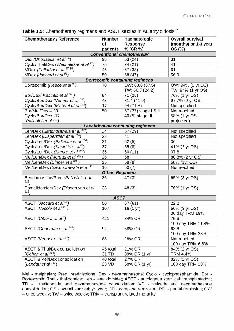
CHAPTER ONE
- 56 -
Table 1.5: Chemotherapy regimens and ASCT studies in AL amyloidosis27
Chemotherapy / Reference Number of patients
Haematologic Response % (CR %)
Overall survival (months) or 1-3 year OS (%)
Conventional chemotherapy
Dex (Dhodapkar et al 96) 93 53 (24) 31
Cyclo/Thal/Dex (Wechalekar et al 85) 75 74 (21) 41
MDex (Palladini et al 97, 98) 46 67 (33) 61
MDex (Jaccard et al 93) 50 68 (47) 56.9
Bortezomib containing regimens
Bortezomib (Reece et al 99) 70 OW: 68.8 (37.5) TW: 66.7 (24.2)
OW: 94% (1 yr OS) TW: 84% (1 yr OS)
Bor/Dex( Kastritis et al 100) 94 71 (25) 76% (1 yr OS)
Cyclo/Bor/Dex (Venner et al 101) 43 81.4 (41.9) 97.7% (2 yr OS)
Cyclo/Bor/Dex (Mikhael et al 102) 17 94 (71%) Not specified
Bor/Mel/Dex – 33 Cyclo/Bor/Dex -17 (Palladini et al 103)
50 67 (27) stage I & II 40 (5) stage III
Not reached 58% (1 yr OS projected)
Lenalidomide containing regimens
Len/Dex (Sanchorawala et al 104) 34 67 (29) Not specified
Len/Dex (Dispenzieri et al 105) 23 41 Not specified
Cyclo/Len/Dex (Palladini et al 106) 21 62 (5) 36
Cyclo/Len/Dex (Kastritis et al89) 37 55 (8) 41% (2 yr OS)
Cyclo/Len/Dex (Kumar et al 107) 35 60 (11) 37.8
Mel/Len/Dex (Moreau et al 108) 26 58 80.8% (2 yr OS)
Mel/Len/Dex (Dinner et al109) 25 58 (8) 58% (1yr OS)
Mel/Len/Dex (Sanchorawala et al 110 16 50 (7) Not reached
Other Regimens
Bendamustine/Pred (Palladini et al 111)
36 47 (3) 65% (3 yr OS)
Pomalidomide/Dex (Dispenzieri et al 112)
33 48 (3) 76% (1 yr OS)
ASCT
ASCT (Jaccard et al 93) 50 67 (61) 22.2
ASCT (Vesole et al 113) 107 16 (1 yr) 56% (3 yr OS) 30 day TRM 18%
ASCT (Cibeira et al 5) 421 34% CR 75.6 100 day TRM 11.4%
ASCT (Goodman et al 114) 92 58% CR 63.6 100 day TRM 23%
ASCT (Venner et al 115) 88 28% CR Not reached 100 day TRM 6.8%
ASCT & Thal/Dex consolidation (Cohen et al 116)
45 total 31 TD
21% CR 39% CR (1 yr)
84% (2 yr OS) TRM 4.4%
ASCT & Vel/Dex consolidation (Landau et al 117)
40 total 23 VD
27% CR 58% CR (1 yr)
82% (2 yr OS) 100 day TRM 10%
Mel - melphalan; Pred, prednisolone; Dex - dexamethasone; Cyclo - cyclophosphamide; Bor - Bortezomib; Thal - thalidomide; Len - lenalidomide;; ASCT - autologous stem cell transplantation; TD - thalidomide and dexamethasone consolidation; VD - velcade and dexamethasone consolidation; OS - overall survival; yr, year; CR - complete remission; PR - partial remission; OW – once weekly; TW – twice weekly; TRM – transplant related mortality
CHAPTER ONE
- 57 -
A landmark analysis at 1 year in 140 patients with evaluable FLC results, showed a superior
OS in those achieving a CR and vGPR versus those achieving a PR and NR (Figure 1.4A).2
The Mayo Clinic reported a series of 422 patients receiving an ASCT from March 1996 to
December 2009. Transplant eligibility included age < 70 years, PS 0-2, troponin T
<0.06ng/mL, creatinine clearance >30mls/min (unless on dialysis), New York Heart
Association class I-II and less than 2 organ involvement. The focus of this analysis was to
examine the TRM before and after January 2006, with TRM pre 2006 as 12% and 7% post-
2006.118 Troponin T >0.06ng/L and NT pro-BNP>5000pg/ml were associated with a high
TRM, whilst patients with both markers below the thresholds had a TRM of 1%.119 Refining
patient selection has allowed ASCT to be done safely, but eligibility of patients for ASCT
has reduced outside of centres with extensive transplant experience.
First major organ responses in AL were initially reported after ASCT in 2001 – 36% of
patients achieved a renal response at 12 months defined by the amyloidosis consensus
criteria5, 92, 95, 118 proving that clonal responses in AL translate into organ responses. The
depth of haematologic response strikingly correlated with renal responses. 71% of patients
in CR had renal responses compared to 11% with persistence of the plasma cell dyscrasia.
Over the last decade, improvements in quality of life, hepatic and cardiac responses have
been published. Similar to renal responses, clinical responses in other organ systems are
more evident with deeper haematologic responses. Organ responses can take up to 6-12
months or longer to occur. Haematologic CR occurs in just under half of all patients
undergoing ASCT; strategies to improve haematologic CR rates following ASCT are an
important focus. These include induction therapy prior to ASCT, novel conditioning and
consolidation therapy. A current phase II trial to evaluate the role of induction treatment prior
to ASCT using Bortezomib/Dex is ongoing at Boston. Among the first 22 patients treated,
by intention-to-treat analysis, haematologic responses occurred in 79% (53% CR and 26%
CHAPTER ONE
- 58 -
VGPR) of patients (for evaluable patients 67% CR).120 Tandem cycles of HDM have been
shown to improve the proportion of patients who ultimately achieve a haematologic CR,
leading to overall CR rate of 67%.121 A pilot study incorporating Bortezomib with HDM has
shown promising results with high haematologic response rates.120 Consolidation with
thalidomide and dexamethasone is too toxic for routine use116 but Bortezomib and
dexamethasone as consolidation give high responses and durable response rates including
CR in 58% at 12 months,117 with larger studies needed.
Combination chemotherapy
Alkylators and steroid based regimens
Alkylating agents have formed the backbone of treatment of AL for over 40 years with
melphalan and cyclophosphamide used in many current therapies. The first randomised
controlled trials proved the efficacy of melphalan and prednisone122 in this disease, but novel
treatment options make this regimen less attractive given few and slow haematologic
responses, poor survival of 18 months and rarer organ responses.122 Similarly, high dose
single agent dexamethasone96 and vincristine, Adriamycin and dexamethasone (VAD),123
although effective, have been superseded due to toxicity and ease of administration,
respectively, with recent regimens.
Melphalan-Dexamethasone (MDex) regimen, pioneered by the Italian amyloidosis group, is
well tolerated and associated with good haematologic and organ response rates of 67% and
33% respectively, a third of patients achieving a complete clonal response. The median PFS
and OS are 3.8 and 5.1 years respectively;98 results confirmed prospectively in subsequent
studies (Figure 1.4B).3, 93 This regimen is usually well tolerated, with 10-15% experiencing
severe adverse events, mainly fluid retention and cytopenias. Oral MDex is generally
considered as the standard of care for patients outside of clinical trials in a number of
countries. However, the role of MDex in patients with advanced cardiac disease is uncertain
CHAPTER ONE
- 59 -
where the median survival is 10.5 months124 and response rates (11% CR and 33% PR in
61 patients with cardiac AL) - another study reporting a median survival of 17 months and
28% mortality in the first three months in patients treated with this same regime.125 But high
early mortality makes this group difficult to treat regardless of the regimen. Intravenous
intermediate dose melphalan (25mg/m2) had a 12% TRM in a UK study with prolonged
remission in the responders126 but a recent prospective trial found this too toxic for routine
use.127
Reduced toxicity of low dose dexamethasone containing regimens in myeloma,128 make
these appealing in AL amyloidosis. Melphalan and once weekly dexamethasone was
associated with lower responses129 than standard MDex98 - raising doubts as to this
approach in AL where rapid and deep responses are desirable.
Bendamustine, an alkylator with a novel mechanism of action, with prednisone is useful in
relapsed/refractory disease and achieved haematologic responses in 47% (n=17) with a
survival advantage in responders in a 3 months landmark analysis (p=0.036). Three patients
achieved a VGPR or better in a heavily pre-treated patient group with a 3-year OS of 65%.111
Grade 3 or greater adverse events (seen in 33%), predominantly cytopenias, were
manageable. Bendamustine should be explored earlier in the disease course.
Immunomodulatory agents
The efficacy of immunomodulatory (IMiD’s) agents in plasma cell dyscrasias opened up
another treatment avenue for AL amyloidosis. Thalidomide, the first of these agents, has a
limited role in AL amyloidosis as monotherapy due to unacceptable toxicity at higher doses
and limited efficacy.130 The combination of thalidomide and dexamethasone (median
thalidomide dose 300mg) was effective and with clonal response rates of 48% (n=31), CR
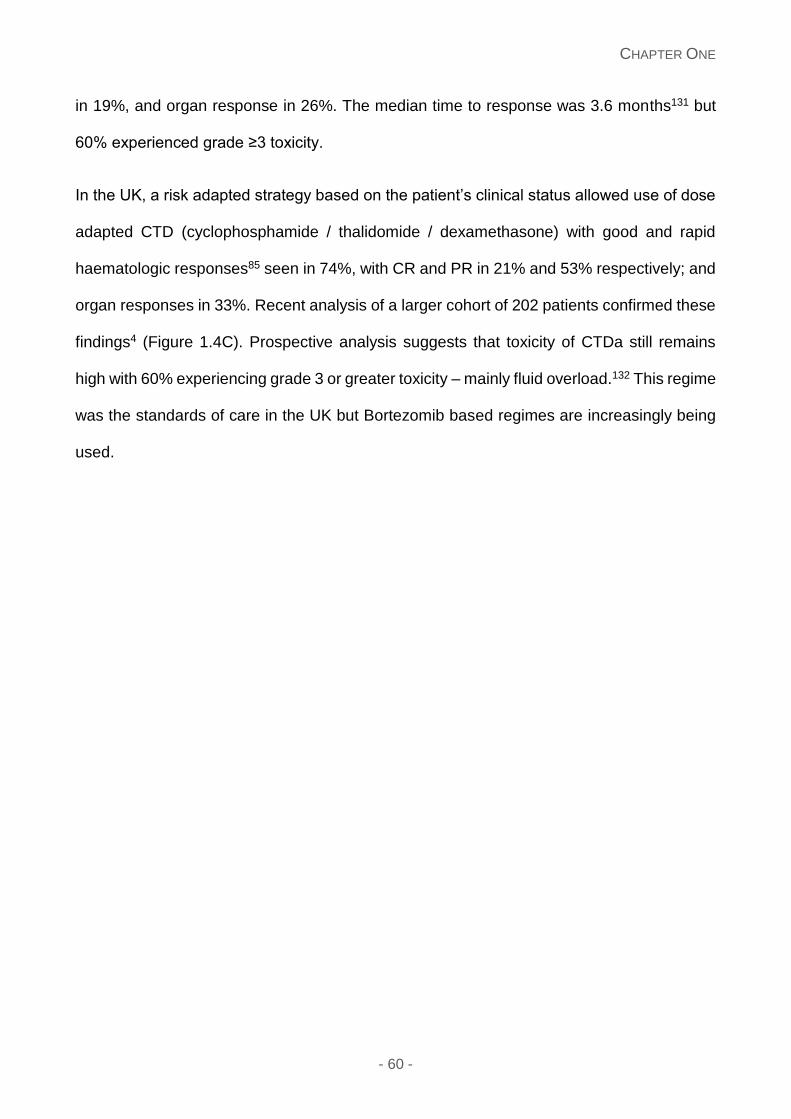
CHAPTER ONE
- 60 -
in 19%, and organ response in 26%. The median time to response was 3.6 months131 but
60% experienced grade ≥3 toxicity.
In the UK, a risk adapted strategy based on the patient’s clinical status allowed use of dose
adapted CTD (cyclophosphamide / thalidomide / dexamethasone) with good and rapid
haematologic responses85 seen in 74%, with CR and PR in 21% and 53% respectively; and
organ responses in 33%. Recent analysis of a larger cohort of 202 patients confirmed these
findings4 (Figure 1.4C). Prospective analysis suggests that toxicity of CTDa still remains
high with 60% experiencing grade 3 or greater toxicity – mainly fluid overload.132 This regime
was the standards of care in the UK but Bortezomib based regimes are increasingly being
used.
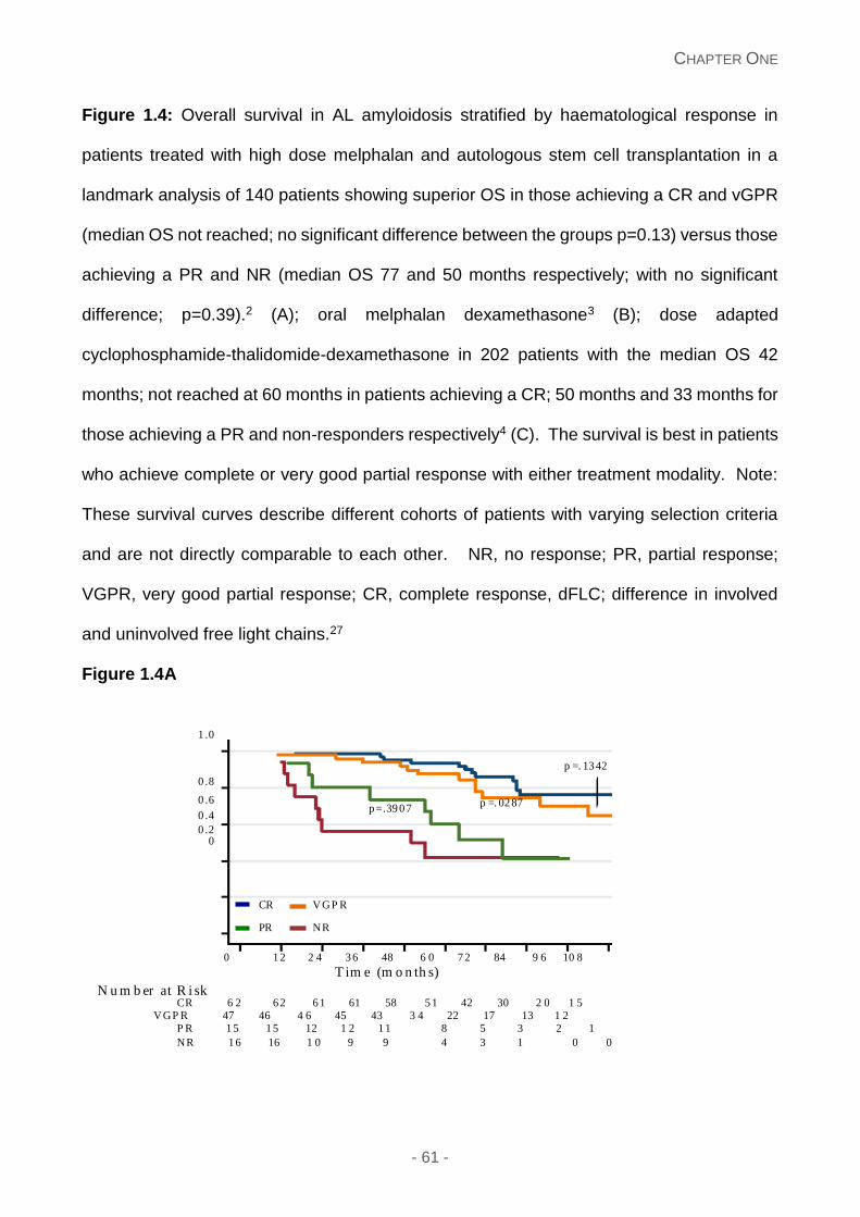
CHAPTER ONE
- 61 -
Figure 1.4: Overall survival in AL amyloidosis stratified by haematological response in
patients treated with high dose melphalan and autologous stem cell transplantation in a
landmark analysis of 140 patients showing superior OS in those achieving a CR and vGPR
(median OS not reached; no significant difference between the groups p=0.13) versus those
achieving a PR and NR (median OS 77 and 50 months respectively; with no significant
difference; p=0.39).2 (A); oral melphalan dexamethasone3 (B); dose adapted
cyclophosphamide-thalidomide-dexamethasone in 202 patients with the median OS 42
months; not reached at 60 months in patients achieving a CR; 50 months and 33 months for
those achieving a PR and non-responders respectively4 (C). The survival is best in patients
who achieve complete or very good partial response with either treatment modality. Note:
These survival curves describe different cohorts of patients with varying selection criteria
and are not directly comparable to each other. NR, no response; PR, partial response;
VGPR, very good partial response; CR, complete response, dFLC; difference in involved
and uninvolved free light chains.27
Figure 1.4A
0 1 2 2 4 3 6 48 6 0 7 2 84 9 6 10 8 T im e (m o n th s) N u m b er at R i sk CR 6 2 6 2 6 1 61 58 5 1 42 30 2 0 1 5 VGP R 47 46 4 6 45 43 3 4 22 17 13 1 2 P R 1 5 1 5 12 1 2 1 1 8 5 3 2 1 NR 1 6 16 1 0 9 9 4 3 1 0 0
1 .0
0 .8 0 .6 0 .4 0 .2
0
CR VGP R PR NR
p =. 13 42
p =. 02 87 p =.39 0 7
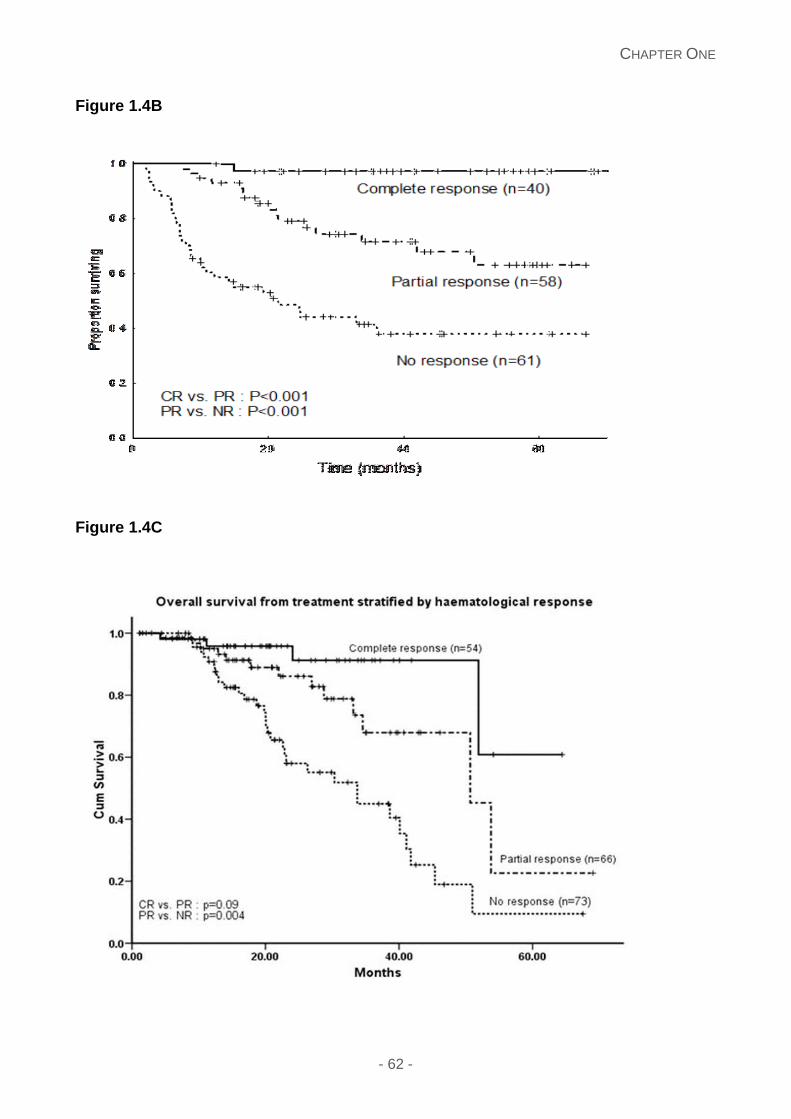
CHAPTER ONE
- 62 -
Figure 1.4B
Figure 1.4C
CHAPTER ONE
- 63 -
Lenalidomide is a second generation IMiD with greater anti-myeloma efficacy and
favourable toxicity profile. Two phase II studies reported that lenalidomide-dexamethasone
(Len/Dex) treatment showed good haematological responses in 67% (n=24), with CR and
PR in 29% and 38% respectively.104, 105 Standard lenalidomide doses of 25 mg were poorly
tolerated with much better tolerance with 15 mg daily. The most common side effects were
fatigue and myelosuppression accounting for 35%, and thromboembolism in 9% (n=3). Skin
rashes were experienced in 43%.133 Lenalidomide combined with dexamethasone has been
used in a small cohort of patients refractory to alkylators, Bortezomib and thalidomide, with
a 41% response rate and survival advantage in responders, indicating that IMiD’s can have
a role in salvage treatment,134 with no increased incidence of secondary malignancies.135
Complete response rates remain low with lenalidomide based regimes and a number of
phase II studies have focused on addition of an alkylator to improve the response rates.89,
106-109 Studies with additional cyclophosphamide or melphalan report overall haematological
response rates of 55-60% and CRs of approximately 20%. The toxicity of the combination
is high with nearly two thirds of patients reporting grade 3 or greater toxicity including
haematologic in 46%; other side effects including fatigue, oedema and gastro-intestinal
problems. The response rates in advanced stage patients are poor.89, 109 The exact place of
a lenalidomide-alkylator combination and advantages offered over Len/Dex remains to be
clearly defined.
Pomalidomide (a third generation IMiD effective in myeloma refractory to lenalidomide) with
dexamethasone achieved haematologic responses in 48% of patients with refractory AL112
with organ responses in 5 patients. The median OS and PFS were 28 months and 14 months
respectively. A third of all patients withdrew from the study due to adverse events.
Pomalidomide shows promise as salvage therapy in relapsed refractory patients and its
combination with proteasome inhibitors needs to be explored further.
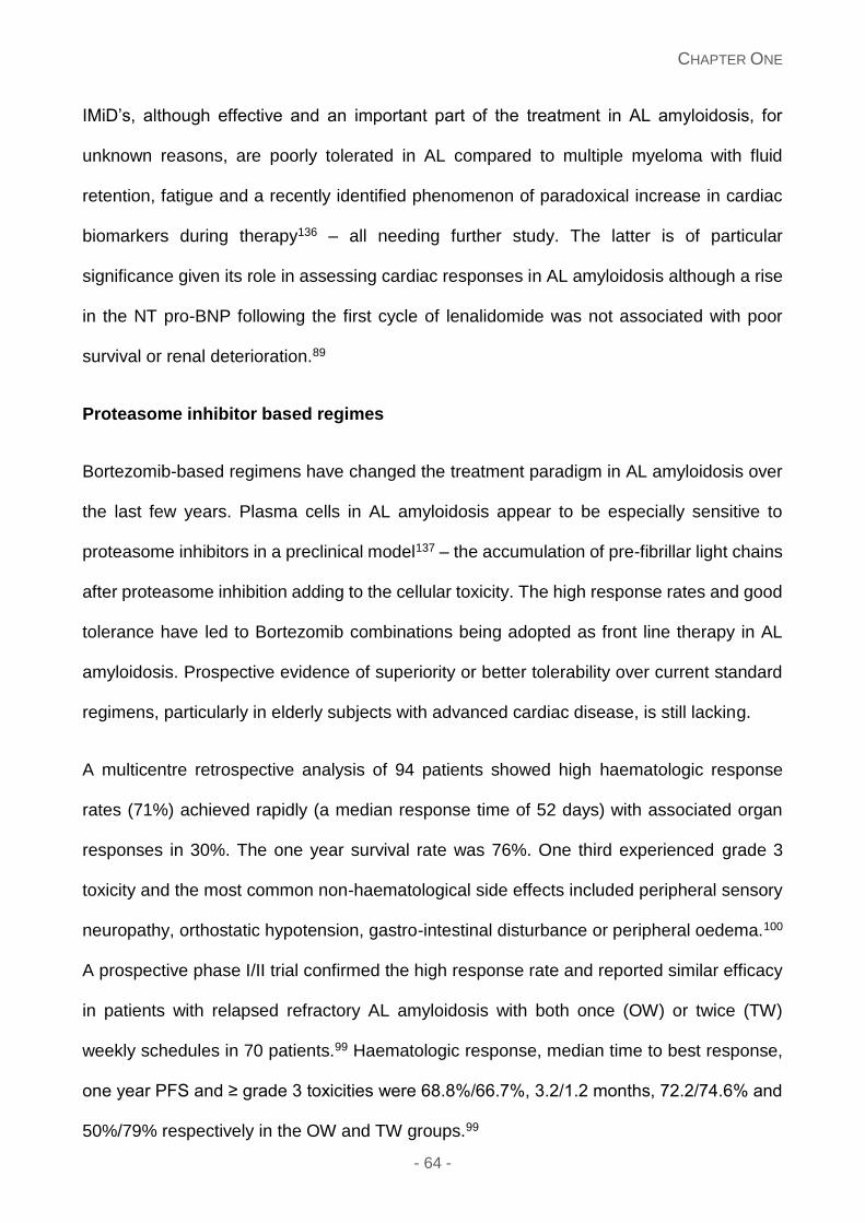
CHAPTER ONE
- 64 -
IMiD’s, although effective and an important part of the treatment in AL amyloidosis, for
unknown reasons, are poorly tolerated in AL compared to multiple myeloma with fluid
retention, fatigue and a recently identified phenomenon of paradoxical increase in cardiac
biomarkers during therapy136 – all needing further study. The latter is of particular
significance given its role in assessing cardiac responses in AL amyloidosis although a rise
in the NT pro-BNP following the first cycle of lenalidomide was not associated with poor
survival or renal deterioration.89
Proteasome inhibitor based regimes
Bortezomib-based regimens have changed the treatment paradigm in AL amyloidosis over
the last few years. Plasma cells in AL amyloidosis appear to be especially sensitive to
proteasome inhibitors in a preclinical model137 – the accumulation of pre-fibrillar light chains
after proteasome inhibition adding to the cellular toxicity. The high response rates and good
tolerance have led to Bortezomib combinations being adopted as front line therapy in AL
amyloidosis. Prospective evidence of superiority or better tolerability over current standard
regimens, particularly in elderly subjects with advanced cardiac disease, is still lacking.
A multicentre retrospective analysis of 94 patients showed high haematologic response
rates (71%) achieved rapidly (a median response time of 52 days) with associated organ
responses in 30%. The one year survival rate was 76%. One third experienced grade 3
toxicity and the most common non-haematological side effects included peripheral sensory
neuropathy, orthostatic hypotension, gastro-intestinal disturbance or peripheral oedema.100
A prospective phase I/II trial confirmed the high response rate and reported similar efficacy
in patients with relapsed refractory AL amyloidosis with both once (OW) or twice (TW)
weekly schedules in 70 patients.99 Haematologic response, median time to best response,
one year PFS and ≥ grade 3 toxicities were 68.8%/66.7%, 3.2/1.2 months, 72.2/74.6% and
50%/79% respectively in the OW and TW groups.99
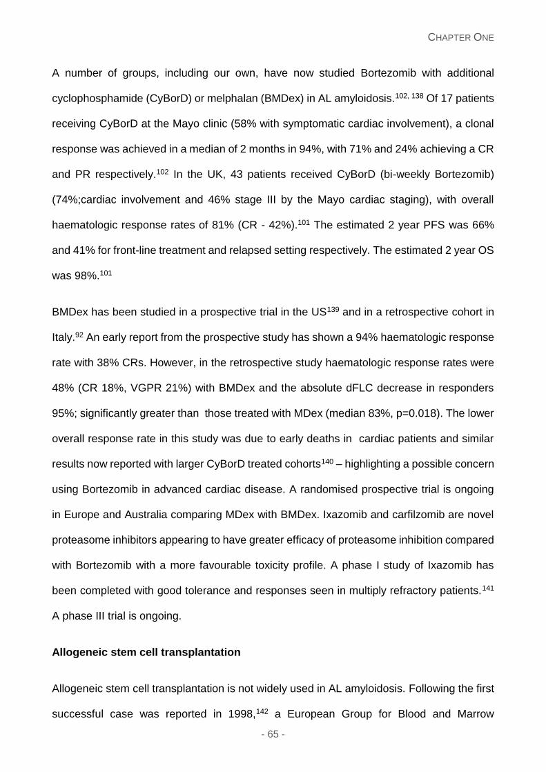
CHAPTER ONE
- 65 -
A number of groups, including our own, have now studied Bortezomib with additional
cyclophosphamide (CyBorD) or melphalan (BMDex) in AL amyloidosis.102, 138 Of 17 patients
receiving CyBorD at the Mayo clinic (58% with symptomatic cardiac involvement), a clonal
response was achieved in a median of 2 months in 94%, with 71% and 24% achieving a CR
and PR respectively.102 In the UK, 43 patients received CyBorD (bi-weekly Bortezomib)
(74%;cardiac involvement and 46% stage III by the Mayo cardiac staging), with overall
haematologic response rates of 81% (CR - 42%).101 The estimated 2 year PFS was 66%
and 41% for front-line treatment and relapsed setting respectively. The estimated 2 year OS
was 98%.101
BMDex has been studied in a prospective trial in the US139 and in a retrospective cohort in
Italy.92 An early report from the prospective study has shown a 94% haematologic response
rate with 38% CRs. However, in the retrospective study haematologic response rates were
48% (CR 18%, VGPR 21%) with BMDex and the absolute dFLC decrease in responders
95%; significantly greater than those treated with MDex (median 83%, p=0.018). The lower
overall response rate in this study was due to early deaths in cardiac patients and similar
results now reported with larger CyBorD treated cohorts140 – highlighting a possible concern
using Bortezomib in advanced cardiac disease. A randomised prospective trial is ongoing
in Europe and Australia comparing MDex with BMDex. Ixazomib and carfilzomib are novel
proteasome inhibitors appearing to have greater efficacy of proteasome inhibition compared
with Bortezomib with a more favourable toxicity profile. A phase I study of Ixazomib has
been completed with good tolerance and responses seen in multiply refractory patients.141
A phase III trial is ongoing.
Allogeneic stem cell transplantation
Allogeneic stem cell transplantation is not widely used in AL amyloidosis. Following the first
successful case was reported in 1998,142 a European Group for Blood and Marrow
CHAPTER ONE
- 66 -
Transplantation (EBMT) registry study in 2005, reported 19 patients with AL amyloidosis (7
patients full intensity conditioning; 8 reduced intensity conditioning (RIC)). The overall and
progression free survival was 60% and 53% respectively at one year, with TRM of 40%
(TRM 50% in patients receiving total body irradiation). Ten patients achieved haematologic
responses and 8 attaining organ responses.143 Anecdotal reports suggest RIC-SCT is
possible in AL.144 Currently, allogeneic stem cell transplantation may have a role in highly
selected young fit patients with relapsed disease but, ideally, should only be considered in
the context of a clinical trial.
An approach to treatment
The treatment strategy in a patient with AL amyloidosis is shaped by poignant background
factors including age, co-morbidities, performance status, contraindications to drugs and
patient wishes deciding the ultimate choice of the regimen, especially in the lack of clear
randomised evidence. Figure 1.5 provides a suggested treatment algorithm for patients
treated outside of clinical trials. The choice of upfront treatment is between autologous stem
cell transplantation (ASCT) and combination chemotherapy – one not necessarily precluding
the other. Patients with good performance status, limited organ involvement, good renal
function, cardiac ejection fraction >50%, CO diffusion capacity > 50%, cardiac troponin T
<0.06 ng/L and NT-proBNP <5000 ng/L appear to have a <5% TRM during ASCT119, 145, 146
and should be offered ASCT as a treatment choice. Borderline patients treated with a stem
cell sparing regime as improved organ function may allow ASCT at a later date. Bortezomib
has emerged as a key backbone in induction regimens – CyBorD or BMDex are becoming
the regimens of choice in most non-neuropathic patients. Cautious low dose regimens are
needed in advanced cardiac involvement (especially with NT-proBNP >8500ng/L). Patients
with neuropathy pose a dilemma, with melphalan/dexamethasone or
lenalidomide/dexamethasone as suitable upfront treatments. Response reassessment at

CHAPTER ONE
- 67 -
the 3 month point interval is key – patients with a poor response considered for either dose
increments or an additional drug to improve the response.
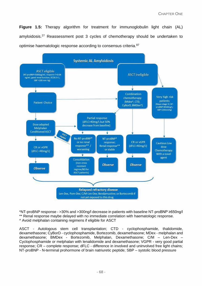
CHAPTER ONE
- 68 -
Figure 1.5: Therapy algorithm for treatment for immunoglobulin light chain (AL)
amyloidosis.27 Reassessment post 3 cycles of chemotherapy should be undertaken to
optimise haematologic response according to consensus criteria.87
*NT-proBNP response - >30% and >300ng/l decrease in patients with baseline NT-proBNP ≥650ng/l ** Renal response maybe delayed with no immediate correlation with haematologic response. ^ Avoid melphalan containing regimens if eligible for ASCT
ASCT - Autologous stem cell transplantation; CTD - cyclophosphamide, thalidomide, dexamethasone; CyBorD - cyclophosphamide, Bortezomib, dexamethasone; MDex - melphalan and dexamethasone; BMDex - Bortezomib, Melphalan, Dexamethasone; C/M – Len-Dex – Cyclophosphamide or melphalan with lenalidomide and dexamethasone; VGPR - very good partial response; CR – complete response; dFLC - difference in involved and uninvolved free light chains; NT-proBNP - N-terminal prohormone of brain natriuretic peptide; SBP – systolic blood pressure
CHAPTER ONE
- 69 -
Best organ responses occur in those achieving a haematologic CR/VGPR, but partial
responders may also achieve an organ response. Lenalidomide and Pomalidomide based
regimens or Bendamustine are useful in the relapsed/refractory setting. The value of
alkylator based regimes in patients relapsing after a novel agent based regime is uncertain.
IgM associated AL amyloidosis
4-7% of all patients with amyloidosis have an IgM secreting (mainly) lymphoplasmacytic
lymphoma (LPL) as the underlying cause of AL.147, 148 Treatment should be directed toward
the LPL clone. Single agent alkylators have limited efficacy. Regimes such as
melphalan/dexamethasone (n=14), purine analogues (n=17) confer good haematologic
responses of 64% and 73% respectively.147, 148 Recently,
Rituximab/Bortezomib/Dexamethasone shows promise, with overall haematologic
responses of 78% (n=7).149 Larger clinical trials are needed to evaluate this further.
Organ transplantation
Organ transplantation can be considered in patients attaining a VGPR or better with
irreversible end-stage organ function, or in a situation to facilitate aggressive chemotherapy;
not otherwise feasible due to organ dysfunction. Long term control of the underlying clonal
disorder is needed to prevent recurrence after organ transplantation or progression in other
organs.
Cardiac transplantation may be the only option to improve survival in younger patients with
advanced isolated heart involvement, accounting for 0.14% of heart transplants nationwide
according to the United Network for Organ Sharing (UNOS).150 A UK study reported 24
patients from 1984 to 2002 undergoing heart transplantation in 6 UK cardiac transplant
CHAPTER ONE
- 70 -
centres, 17 diagnosed with AL amyloidosis. The 1 and 5 year OS was 50% and 20%
respectively not receiving chemotherapy versus 71% and 36% who did.151 Another
multicentre study included 69 patients receiving heart transplants for amyloidosis (all types),
reported 1 and 5 year survival as 74.6% and 54% respectively.150 Control of the underlying
clone is important, and ASCT is generally considered most appropriate treatment. One US
series between 1994 and 2005 included 11 patients undergoing the two procedures serially;
9 patients surviving both with 3 eventually dying due to progressive amyloidosis; the earliest
55 months post ASCT. The 1 and 5 year survival following heart transplantation in this cohort
was 82% and 65% respectively;139,152 a marked improvement over the otherwise median
survival of advanced cardiac AL of 3-8 months. Scarcity of organs, risk of amyloid recurrence
and higher mortality of amyloid patients undergoing heart transplants still makes cardiac
transplantation a contentious issue.150
Renal transplantation in AL amyloidosis is considered to improve long term survival and
quality of life. The largest UK series reports 22 patients undergoing renal transplantation;
(19 cadaveric donors and 3 live donors), and 3 with extra-renal organ dysfunction. Nineteen
patients received chemotherapy or ASCT; 74% achieving a haematologic response (11 PR
and 3 CR) with no graft failures secondary to amyloid recurrence and 1 and 5 year OS 95%
and 67% respectively.153 The Mayo group reported 19 patients receiving renal
transplantation (1 cadaveric donor and 18 live donors), 12 having extra-renal involvement
from 1999 to 2008. All attained a haematologic response (18 CR and 1 PR); 8 receiving
renal transplantation followed by ASCT, 6 receiving ASCT followed by renal transplantation
and 5 receiving non-myeloablative chemotherapy followed by renal transplantation, with no
significant difference between these groups. The 1 and 5 year OS was 84% and 76%
respectively.153 Good long-term outcomes appear to be associated with renal
transplantation in AL in highly selected cases.
CHAPTER ONE
- 71 -
Outcomes with orthotopic liver transplantation (OLT) for advanced liver AL remain poor. A
UK study from 1984 to 2009 included 9 patients undergoing an OLT with the 1 and 5 year
survival from transplantation 33% and 22% respectively.153
In summary, AL Amyloidosis is rare. It is frequently diagnosed late due to the subtle signs
and symptoms of the disease. Increasing awareness of this disease poses major
challenges. Improved diagnostic techniques have enabled better detection of this disease.
Risk stratification using cardiac biomarkers has refined treatment selection and reduced
morbidity and TRM. Diagnosis and treatment planning in AL is complex and best considered
in specialist centres availing expertise of a multi-disciplinary team. Novel agent based
chemotherapy especially with Bortezomib has improved chemotherapy responses and is
being explored pre and post ASCT. Bendamustine, lenalidomide and Pomalidomide are
valuable therapies for relapsed refractory patients. Good haematologic responses (attaining
at least a VGPR being the goal of therapy87) translates into organ responses and improved
survival. While cardiac and haematologic responses can be simultaneous, responses of
other organs may evolve over months or years. Early mortality of patients with advanced
disease persists. Future directions involving therapies targeting the amyloid deposits - anti-
fibril antibodies are in early phase trials. Exploring the role of minimal residual disease, the
importance of eradicating this, gaining insight of the microenvironment is needed. Ultimately,
the most important factor is increasing the awareness to ensure early diagnosis to allow
therapy before extensive organ involvement.
CHAPTER ONE
- 72 -
Novel therapies for amyloidosis
A number of therapies are in development targeting various components of the
pathophysiology of amyloid fibrillogenesis. Glycosaminoglycans are a universal constituent
of all amyloid deposits and inhibition of the interaction between GAGs and amyloid fibrils
which seems a promising therapeutic approach AA type – which may also have merit in AL.
Eprodisate (Kiacta®, Bellus Health, Canada) is a negatively charged, sulfonated molecule
of low molecular weight is undergoing phase III trials.154 There is growing interest in
developing therapeutic antibodies to directly target amyloid deposits. Mu 11-1F4 is a
chimeric antibody reactive with many AL fibrils,155 localisation of which has been studied in
patients with PET imaging.156 Its administration as a therapeutic is planned. mAb2A4 is
another monoclonal antibody, binding AL/AA fibrils and human AL amyloid extracts,157
reported to cause amyloid regression in mouse models of AA and AL amyloidosis. This
antibody has been further developed as NEOD001 (Onclave Therapeutics Limited,
California), recently commencing phase I trials in systemic amyloidosis in the United States.
Pre-clinical development of small interfering RNAs (siRNAs) have also been explored as a
treatment in reducing the expression of the amyloid precursor protein, with in vitro studies
showing inhibition of synthesis of light chains in transfected cells, and in vivo reducing the
production and circulating free light chains.158
R-1-[6-[R-2-carboxy-pyrrolidin-1-yl]-6-oxo-hexanoyl]pyrrolidine-2-carboxylic acid (CPHPC)
is a drug which cross-links pairs of SAP molecules in vivo159 resulting in rapid clearance of
SAP from the liver and almost complete depletion of plasma SAP.160 Some SAP persists on
the amyloid deposits and lack of plasma SAP allows administration of an anti-SAP antibody
which can now target the amyloid deposits. Following promising results in transgenic mouse
model of AA amyloidosis, using anti-SAP immunoglobulin-G (IgG) antibodies and CPHPC,15

CHAPTER ONE
- 73 -
early phase clinical trials are underway to explore this further with initial patient recruitment
commencing this year.
CHAPTER ONE
- 74 -
Aims and scopes of this thesis
The introduction of this thesis provides a general overview of the different types of
amyloidosis with a comprehensive overview of systemic light chain amyloidosis and
treatment options available. This rare disease is often a delayed diagnosis, with the clinical
presentation masked by many other medical conditions. More recently, advances have been
paved with use of targeted treatment, monoclonal antibodies or accessibility of newer
chemotherapeutic agents. Prognostic stratification has emerged as an important tool in
treating amyloidosis patients.
The premise of this thesis focuses upon areas of interest. Firstly section one concentrates
on the investigations important in diagnosing, monitoring, and assessing the prognostic
utility and potential complications of patients diagnosed with systemic amyloidosis. Chapter
3 concentrates on the comparison of the utility of free light chain assays currently important
in diagnosing and monitoring these patients. The current serum FreeliteTM assay measures
immunoglobulin free light chains using polyclonal sheep antibodies directed against hidden
epitopes on the light chain molecule giving an accurate measurement of serum free kappa
and lambda and forms a standard part of the baseline and serial follow up assessments in
systemic AL amyloidosis161, 162, and is part of the revised Mayo staging system and standard
international consensus criteria for disease response assessment.80, 86, 87 A group in the
Netherlands working with Siemens, Germany, has developed this technology using a
mixture to two kappa and two lambda monoclonal antibodies to the hidden epitopes of the
constant region of the immunoglobulin light chain molecules.163 With increasing adoption
by laboratories of the novel assay, it is important to assess the utility of the new assay
compared to the current reference FreeliteTM assay. We report a comparison of serum free
light chains analysing the serum samples by both immunoassays at diagnosis and three
CHAPTER ONE
- 75 -
further time points in during the initial chemotherapy for patients with systemic AL
amyloidosis.
Chapter 4 outlines the coagulation abnormalities seen in systemic AL. Systemic light chain
(AL) amyloidosis is also known to be associated with a bleeding diathesis164, which may
range from small cutaneous bruising, pathgnomic “racoon eyes” or life threatening bleeds.
The crucial role of endothelial dysfunction in systemic AL may assist in understanding the
basis of the light chain toxicity in this disease. We designed a prospective study to
investigate the endothelial dysfunction in newly diagnosed systemic AL patients, using Von
Willebrand factor (vWF) antigen (Ag) and factor VIII to serve as a surrogate marker and
examine its relationship with light chain toxicity. Other coagulation factors were also
examined. This was performed examining the clinical bleeding manifestations using the
Royal Free Hospital v4 adapted bleeding questionnaire in conjunction with extensive
laboratory investigations such as factor assays, protein S, protein C, Anti-thrombin III,
vWF:Ag and VWF:Ricof and ADAMTS13 assays. Endothelial function was explored using
vWF antigen (Ag) and factor VIII to serve as a surrogate marker and examine its relationship
with light chain toxicity. The secondary aim was to assess the prognostic utility of these
investigations.
Chapter 5 focuses on a new area of interest which may pose a potential issue in patients
with systemic amyloidosis – sleep disordered breathing. Cardiac involvement and/or
macroglossia with soft tissue deposits, risk factors for central and obstructive sleep apnoea,
are common features of systemic AL amyloidosis. Little data exists on the occurrence of
sleep disordered breathing (SDB) or recurrent nocturnal hypoxia in amyloidosis. All were
screened for SDB using Epworth Sleepiness Score (ESS) and STOP BANG questionnaires.
We report here the results of a pilot study of overnight continuous pulse oximetry in patients
with systemic light chain (AL) amyloidosis, based on the hypothesis that recurrent nocturnal
CHAPTER ONE
- 76 -
hypoxaemia (likely due to sleep disordered breathing OSA and/or CSA) could occur in
patients with amyloidosis reporting a high incidence of recurrent nocturnal oxygen
desaturations and raise a question whether these desaturations may be the trigger for
sudden cardiac mortality.
The second section focuses on a rare type of amyloidosis - localised amyloidosis and a
subgroup – tracheobronchial amyloidosis. Chapter 6 elaborates on localised amyloidosis.
Localised deposits of amyloid can occur in various tissues in the body and are usually
presumed to be of AL type, with the consequent presence of a focal monoclonal B cell
dyscrasia within the affected tissue. As such, the clinical effects of the localised amyloid
deposits depend on their precise anatomical location, and can result in substantial morbidity.
Localised amyloidosis is much rarer than systemic types, and consequently remains very
poorly studied; with most knowledge arising from individual case reports or small series of
less than 20 patients. There are commonly reported sites include the urinary tract,
respiratory tract, larynx, skin and eyelids.165 Currently, data on long term outcomes and
progression to systemic disease is lacking with the need for further exploration in this field.
We have examined the clinical features and outcomes of a large series of 606 patients with
localised AL amyloidosis highlighting the striking differences from systemic AL amyloidosis
with respect to the lack of progression, benefit from debulking procedures, limited need for
cytotoxic chemotherapy therapy and excellent overall long term outcomes. Chapter 7
examines a subgroup of localised amyloidosis – laryngeal and tracheo-bronchial
amyloidosis. This group of patients have a more refractory control of their disease. We
examined the clinical features, treatment options and also sought to explore the laboratory
aspect of proteomic analysis in these biopsies to attempt to gain insight into any underlying
instigating factors. Although amyloid fibril formation and aggregation occurs when high
concentrations of the key partially folded intermediate is present, other factors including the
CHAPTER ONE
- 77 -
pH, temperature, amino acid sequence and concentration of the intermediate play a role.
The presence of ApoA1 and insulin-like growth factor binding protein complex in the LTBA
proteomic analysis suggests the potential involvement in the development of localised
amyloid in the laryngeal and tracheobronchial cases. Further studies are needed to
elucidate the function of these factors before a significant pathogenesis is attributed to the
latter.
The third section focuses on 2 types of treatments in systemic amyloidosis – a
chemotherapeutic option (lenalidomide/dexamethasone post novel agents) and ((R)-1-[6-
[(R)-2-carboxy-pyrrolidin-1-yl]-6-oxo-hexanoyl] pyrrolidine-2 carboxylic acid (CPHPC))
which has emerged as a potential therapeutic target to deplete SAP.
Chapter 8 relays our experience with a chemotherapeutic drug option –
lenalidomide/dexamethasone. We report a substantial cohort of patients treated with
lenalidomide in the relapsed/refractory setting, following the use of Bortezomib and/or
thalidomide with long term follow up assessing the impact of previous therapies on
haematologic responses.
Chapter 9 outlines a small cohort of patients treated with compassionate use of CPHPC.
Fibrinogen related amyloidosis has predominant renal involvement with end stage renal
failure within 5 years of presentation with proteinuria.47 Currently there are no definitive
therapeutic cures for this condition and we used CPHPC on a compassionate basis to try
and improve the renal survival duration in these patients, with the use of QoL questionnaires
to gain a better insight into the patients’ perspective. We also assessed its use in patients
with dialysis related amyloidosis.
CHAPTER TWO
- 78 -
Chapter Two: Materials and methods
Declaration
I have designed the studies, collected the data and performed analysis of the data
using 2 software programs – SPSS v20 (IBM SPSS) and Graph pad prism (version 5).
This thesis includes 3 prospective studies: the coagulation study, sleep disordered
breathing study and the CPHPC study. I recruited patients for the Coagulation study,
collected the data and performed the statistical analysis as a clinical research fellow
based at the National Amyloidosis Centre, University College Medical School (Royal
Free Campus). I also recruited patients for the sleep disordered breathing study, again
collected the data and performed the statistical analysis and sent the overnight pulse
oximetry downloads to Dr Milind Sovani (Respiratory physician) to analyse. I recruited
patients for the CPHPC study following discussion at our multidisciplinary meeting for
suitability for compassionate use of CPHPC in these patients. Ms Thirusha Lane, a
clinical lead nurse at the National Amyloidosis Centre used the QoL software (SF36)
in giving the output for the quality of life data. Various diagnostic methods were
undertaken by other individuals in the department and external sources as follows:
Measurement for biochemical serum and urinary investigations were performed by the
Royal Free Hospital laboratory services.
Frozen serum blood samples for the serum free light chain assay comparison were
collected by Wendy Taylor and Lois Cook, based at the National Amyloidosis Centre.
Coagulation assays were performed by the Haemophilia department at the Royal Free
Hospital and these investigations overseen by Ms Anne Riddle, Dr Keith Gomez and
Dr Pratima Chowdary.
CHAPTER TWO
- 79 -
Histological and immunohistochemical studies were performed by Janet Gilbertson
and Karen Boniface, both based at the National Amyloidosis Centre. Laser capture
techniques were undertaken by Janet Gilbertson and Nicola Botcher, both based at
the National Amyloidosis Centre. Proteomic analysis was undertaken using the
Scaffold program by Graham Taylor and Nigel Rendell, both based at the Wolfson
Centre Research Institute, National Amyloidosis Centre. Analysis of proteomic data
was performed by Dr Julian Gillmore and Janet Gilbertson.
The spinning and storing or serum samples for patients on compassionate use of
CPHPC were performed by the clinical nurses Thirusha Lane, Charlene Kearney and
Randolph Gaudia.
Genetic sequencing was performed by Dorota Rowczenio and Hadija Trojer, based at
the National Amyloidosis Centre.
Echocardiography was performed by Babaita Pawarova, Oliver Manalo, and Sevda
Ozer, based at the National Amyloidosis Centre.
123I-SAP scintigraphy was performed by David Hutt, Dorothea Gopaul and Stephanie
McKnight, based at the National Amyloidosis Centre.
Pulse oximetry output using the Minolta 300I pulse oximeter was read by Dr Milind
Sovani, a respiratory physician at Nottingham University hospital.
Patient selection
All of the patients recruited and studied in this thesis have been seen at the National
Amyloidosis Centre, London. Medical records were retrieved and an electronic
database has recorded details of all patients attending the centre, which I used. All
patients included in this thesis provided explicit informed consent. The database
CHAPTER TWO
- 80 -
recorded dates of deaths according to the Office of National Statistics, updates from
family members and details of the causes of deaths were specifically sought for by
contacting local general practioners or hospital consultants caring for the patients.
Only anonymised data was used.
Immunoassays
Serum free light chain assays
Kappa and lambda serum free immunoglobulin (FLC) concentrations were measured
using a latex-enhanced immunoassay (Binding Site, Birmingham, UK) on an Ehring
BNII auto analyser (Dade Behring Marburg, Germany). Antibodies were directed
against the FLC epitopes within the whole immunoglobulin molecules, with the
sensitivity quoted as<5mg/L for this assay. The reference range as provided by the
manufacturers for FreeliteTM: kappa 3.3-19.4mg/L, lambda 5.7-26.3mg/L and
kappa/lambda ratio 0.26-1.65. The N Latex (Siemens, Germany) immunoassay is a
turbid metric assay based on monoclonal antibodies. The reference range as provided
by the manufacturers for N Latex: kappa 6.7-22.4mg/L, lambda 8.3-27mg/L and
kappa/lambda ratio 0.31-1.56mg/L. In chapter 3, in total, 240 serum blood samples
were measured in duplicate using the FreeliteTM (Human kappa and lambda free kits,
Binding site Ltd, Birmingham, UK) and N Latex (Siemens Healthcare Diagnostics,
Germany) from stored frozen samples.
Histology
Congo red staining
Biopsies were logged into our database once received and the formalin fixed de-
paraffinised tissue sections 6-8µg thick were rehydrated and stained with
CHAPTER TWO
- 81 -
haematoxylin under running tap water. The Congo red method as described in
Puchtler el al166 was adopted with sections using this method. Increasing ethanol
concentrations to xylene were used to dehydrate the sections and eventually mounted
with DPX mounting medium. The stained slides once dry were viewed in bright field
under cross polarised light by microscopy.167 A known Congo red positive block
validated by laser micro dissection and proteomic analysis was used as a positive
control.
Immunohistochemistry
Immunohistochemical staining techniques were used to type the amyloid. Formalin
fixed de-paraffinised 2µm sections of amyloidotic tissue was used. These sections
were cleansed; washed with water and endogenous peroxidise activity was quenched
by incubation in aqueous (0.3%) hydrogen peroxide (H2O2) for approximately 30
minutes. Rinsing was performed using a phosphate-buffered saline (PBS) containing
0.05% Tween (Calbiochem) solution. Non-specific tissue binding was abolished by
incubation for a further 30 minutes in normal non-immune serum from the species
providing the secondary antibody (Vector Part of the ImmPRESS Kit). These sections
were incubated overnight with primary antisera at 4oC, and then rinsed with PBS
containing 0.05% Tween (Calbiochem) and labelled with secondary antibodies. Each
section was washed in PBS and bound enzyme-antibody bound complexes were
consequently visualised using a metal-enhanced DAB (Fisher Scientific solution).
Immunohistochemistry was possible with a panel of anti-human monospecific
antibodies reactive with: SAA (Eurodiagnostica, Huntington UK) AL kappa, lambda,
transthyretin and lysozyme (Dako Ltd, Denmark House Ely UK), Apolipoprotein AI
(Genzyme Diagnostics) and fibrinogen Aα chain (Calbiochem) were used where
CHAPTER TWO
- 82 -
appropriate. Congo red overlay was used in duplicate sections. Immunohistochemical
stained sections were counterstained in haematoxylin, ‘blued’ under running tap water
and stained with Congo-red.168
When performing TTR staining, pre-treatment was performed for enhanced antigen
retrieval using a 10 minute incubation with 1% sodium periodate. The slides were then
washed and incubated for a further 10 minutes with 0.1% sodium metabisulphate,
and washed again and incubated for 5 hours at room temperature with 6M Guanadine
in 0.9% sodium chloride.
Laser capture micro dissection and mass spectrometry
(Also described in chapter 7)
Laser capture micro dissection and mass spectrometry (LDMS) techniques were also
used to identify proteins from formalin-fixed, paraffin embedded tissues and described
in detail for chapter 7.169 10µm sections were cut from formalin fixed tissue on the
Director Expression Pathology 50001-024. Stained slides with Congo red (Putchler et
al) were left to dry after clearing with xylene.
Visualisation and locating the amyloid was done using bright field and Rhodamine filter
sets (TRITC) by the Zeis Palm Micro beam Laser capture microscope, with amyloid
typically yellow on a “red” background. Delineation of the areas of interest were drawn
around , appearing yellow under a Tritc florescent filter or under bright field and “cut”
out using laser capturing in to 0.5ml micro-centrifuge adhesive cap tubes (Zeiss) whilst
checking the cap to ensure that some tissue has been captured. A 10x concentration
stock solution of the following reagents were constituted: (1) TRIS: 0.1 M; use
1.2114g in 100ml, (0.6057g in 50 ml), (2) EDTA: 10 mM; use 0.37224g in 100ml,
(0.18612g in 50ml) and (3) Zwittergent (Z3/16): 0.02% w/v; use 0.02g in 100ml,
CHAPTER TWO
- 83 -
(0.01g in 50ml). A 1ml aliquot of each buffer is placed into 1.5 ml microfuge tubes and
stored in a separate snap seal bag, at -20°C. TEZ Buffer (Tris, EDTA, and
Zwittergent) and constituted by diluting each buffer in 1:10 in MQ. A glass vial of
100µg trypsin gold was reconstituted with 50mM Acetic acid and 5 µl aliquots were
stored in microfuge tubes at -80°C (>for 1 month 1mg/ml). To constitute working
strength trypsin, 5µl of stored frozen trypsin solution was added to 245µl of TEZ buffer
(1x)(4 fold concentration of trypsin) and made just prior to use. To constitute the
Dithiothreitol, 10 mg ml-1 solution in MilliQ water aliquot in 100ul volumes was used
and stored in a snap seal bag freezer.
35µl of TEZ buffer was added into each sample and then closed tightly, and the sample
vortexed. The sample was inverted and the left on the bench for 20 minutes. The
capped tubes were inverted, flicked to transfer the liquid to the cap and left to stand
for approximately 20 minutes at room temperature. The samples were centrifuged at
5000 rpm x 5 min (Eppendorf bench, 24 fixed positions) to recover the liquid into the
bottom of the vial.
The cap lock was fitted to the tubes, with care taken not to disturb the liquid at the
bottom of the vial and heated at 99°C x 90 minutes, mixing every 20-25 minutes.
Cooling next and centrifuged at 5000 rpm x 5 min to recover as much condensate as
possible from the tube lids. The Sonicater was cleaned with 70% alcohol (fume hood)
and refilled with fresh MQ water. The cap lock was fitted to tubes and sonicated for 60
minutes at room temperature. Samples were centrifuged at 5000 rpm x 5 min. Working
Trypsin Solution was made by taking 5µl of stored frozen trypsin solution and add to
245µl of TEZ buffer (1x) (4 fold concentration of trypsin). 1.5µl of working trypsin was
added to each sample, (total volume 36.5µl), and each sample vortexed. Again,
centrifuging at 5000 rpm x 5 min, with the cap locks fitted to the tubes and allowed to
CHAPTER TWO
- 84 -
digest overnight at 37ºC. Samples were centrifuge at 5000 rpm x 5 min. Addition of
5µl DTT to the Trypsin digested samples (reduced with Dithiothreitol (DTT) - (0.008M).
Centrifuged at 5000 rpm x 5 min. Cap locks were fitted and sample heated for 5
minutes at 98ºC (total volume 41.5 µl). The samples are cooled, then centrifuged at
5000 rpm x 5 min. The samples are taken to the equipment room to unfasten the caps
and place in freezer at -80.
On the freeze drier turn the drain valve and the air admittance valve clockwise. Ensure
the mode selector on the pump is at zero (0). Switch pump on at side and leave for 20
minutes. Dials should be set at 3mb and -40 temp. Select a small dissector and outlet
valve is turned fully anticlockwise and attach rubber pipe from manifold to outlet valve.
On manifold turn tail switch to on position (down) and check the dials move to the right
before falling again. This is removing the air from the rubber tube. Remove samples
from -80 and place in dissector ensuring lid is in place correctly. Quickly turn outlet
valve clockwise until the screw thread is just visible. Dials should be fully to the right.
Check that a vacuum has been created by gently lifting the dissector with the lid – the
bottom should be raised off the bench if not correct. Check that the dials are falling.
After 60 minutes check that the dials are at 3mb and -40 temp. Leave to freeze dry.
The person loading the MS will remove the samples however if this has not happened
within 24 hours. Remove the samples by reversing the steps and place in -80 with the
lids tightly shut.
The residues were reconstituted with 1% MeCN + 0.1% TFA (20µl/sample), vortexed
and centrifuged (10 000 rpm x 5 min). The supernatants transferred to Waters Total
Recovery vials for LC-MS/MS on the Velos. A QA sample (six protein mix tryptic
digest) should be run before and after the samples; all samples should be followed by
a blank run with a 1% MeCN + 0.1% TFA injection. The data was processed using
CHAPTER TWO
- 85 -
Proteome Discoverer for MASCOT searching of the NCBI and IPI databases enabling
mass spectrometry results to be produced.
Proteomic analysis was performed on the Velos platform and analysed following the
method of Rodriguez et al.75, 169 Following extraction into 10mM Tris/1mM
EDTA/0.002% Zwittergent buffer (99 °C, 1.5h) and sonication (1h), samples were
trypsinised (1.5µg w/v) overnight at 37°C and then reduced with Dithiothreitol (50µg)
at 99°C for 5 minutes. Digests were run on a nanoACQUITY™ UPLC system (Waters
Ltd., Elstree, and Hertfordshire, WD6 3SZ) coupled to an Orbitrap Velos Mass
Spectrometer (Thermo Electron, Bremen, Germany). MS data files were analysed
using Mascot software. (Matrix Science, London, UK).
Genetic sequencing
Patients with suspected hereditary amyloidosis or clinical features of laryngeal
involvement had genotyping performed. Patients at their baseline visit had whole blood
taken which was stored in an EDTA tube consequently frozen and stored, with
genomic DNA isolated as required.170 The polymerase chain reaction (PCR) adopting
the ‘ready to go” tubes (GE Healthcare) were used to amplify the coding regions for
the genes and appropriate exons including: transthyretin (exons 2, 3 and 4),
apolipoprotein AI (exons 3 and 4) and fibrinogen A α-chain (exon 5). HotStar Taq DNA
Polymerase kit (Qiagen) was used for the lysozyme gene (exon 2). The primers used
as part of the PCR process are outlined in Table 2.1.
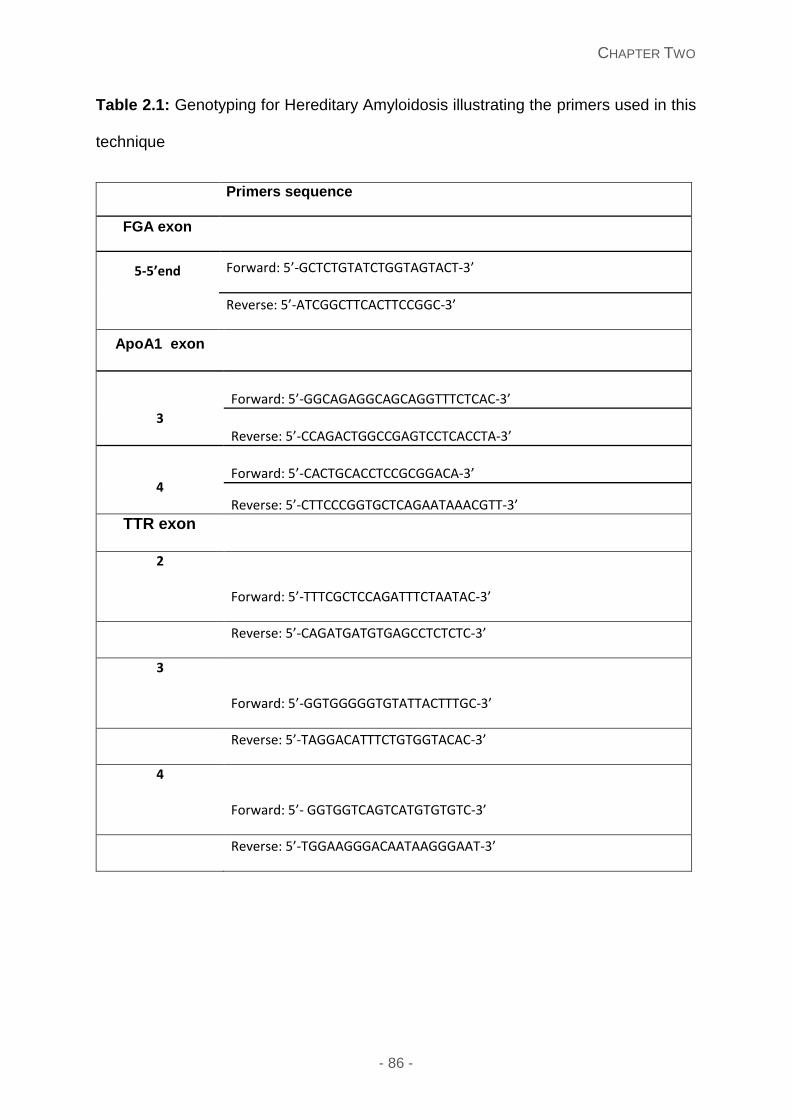
CHAPTER TWO
- 86 -
Table 2.1: Genotyping for Hereditary Amyloidosis illustrating the primers used in this
technique
Primers sequence
FGA exon
5-5’end
Forward: 5’-GCTCTGTATCTGGTAGTACT-3’
Reverse: 5’-ATCGGCTTCACTTCCGGC-3’
ApoA1 exon
3
Forward: 5’-GGCAGAGGCAGCAGGTTTCTCAC-3’
Reverse: 5’-CCAGACTGGCCGAGTCCTCACCTA-3’
4
Forward: 5’-CACTGCACCTCCGCGGACA-3’
Reverse: 5’-CTTCCCGGTGCTCAGAATAAACGTT-3’ TTR exon
2
Forward: 5’-TTTCGCTCCAGATTTCTAATAC-3’
Reverse: 5’-CAGATGATGTGAGCCTCTCTC-3’
3
Forward: 5’-GGTGGGGGTGTATTACTTTGC-3’
Reverse: 5’-TAGGACATTTCTGTGGTACAC-3’
4
Forward: 5’- GGTGGTCAGTCATGTGTGTC-3’
Reverse: 5’-TGGAAGGGACAATAAGGGAAT-3’
CHAPTER TWO
- 87 -
Overnight pulse oximetry
All patients recruited to the sleep apnoea study had overnight oximetry performed with
simplified instructions, appendix 2. This information was downloaded using the Minolta
300I software and then analysed by Dr Milind Sovani using this program.
Sleep questionnaires
Patients recruited to the sleep apnoea study completed an Epworth Sleepiness Score
(ESS) and STOP BANG questionnaires for obstructive sleep apnoea (OSA) to
correlate the clinical symptoms in conjunction with the overnight oximetry readings
(appendix 3 and 4 respectively).
SAP scintigraphy
This procedure was performed in all patients attending the National Amyloidosis
Centre at their first assessment, initially 1988. Each patient having this procedure
received approximately 200µg of SAP with 190MBq of 123I which is the equivalent of
3.8mSV of radiation. A dose of 60mg of potassium iodide was immediately given to
each patient prior to 123I SAP scintigraphy and a further 5 doses were administered
following this over the following 3 days to prevent thyroid uptake. Patients had anterior
(AP) or posterior (PA) imaging using an IGE-Starcam gamma-camera (IGE Medical
Systems, Slough, UK) at 6 or 24 hours following the injection.
A normal scan was defined upon having no abnormal localisation of the tracer. The
quantification of the amyloid load was based on: small – when uptake within one or
more organs was visible with the normal intensity in the blood pool; moderate – when
abnormal uptake was seen within organs and the blood pool was diminished; and
CHAPTER TWO
- 88 -
lastly large – when the blood pool signal was lost with adjustment of the grey scale to
include the target organs.
Progression of the amyloid was defined as increase of the tracer within the affected
organ(s) and/or decrease in the background blood pool. Regression of the amyloid
was defined as reduction of the tracer within the affected organ(s) and/or increase in
the background blood pool.
PET/CT imaging
This imaging uses a device combining the use of positron emission tomography (PET)
scanner and x-ray computed tomography (CT) scanner to acquire images sequentially
and combine to give a superposed image with the use of 18F-flurodeoxyglucose
(FDG) PET/CT. An intravenous bolus injection of a dose produced for 2 or 3-FDG is
made (doses range from 3.7-7.4 megabecaquerels per kilogram of body weight. The
functional aspect depicts the spatial distribution of metabolic or biochemical activity in
the body. Quantification of the size of the lesion and location are important. This
technique was used to assess patients with localised amyloidosis, specifically related
to lymph node, lung nodules or bone lesions.
Quality of life (QoL) questionnaire assessments
The quality of life was assessed at 6 monthly intervals for all patients by the validated
Quality Metric SF36v2® Health survey. This is designed to measure functional health
and well-being from the patient’s perspective. There are eight specified health
domains (physical functioning, role physical, bodily pain, general health, social
function, role emotional, mental health and vitality) which are assessed are scored
individually. The result is expressed in comparison to the American norms. The
CHAPTER TWO
- 89 -
average score for healthy controls in each measure is 50, with higher scores
representing a better QoL. A change of 10 points or greater in any domain between
administrations is considered clinically significant.
Measuring the quality of life (QoL) Until fairly recently the primary clinical endpoints such a hospitalisation or death were
the focus of most clinical trials or disease management programs. Previously patient-
reported outcomes (PRO) tended to be in relation to disease specific indicators, such
as joint pain in rheumatoid arthritis. However, this does not reflect the overall health-
related QoL. As people are living longer, healthcare must focus as much on quality as
length of life. Medicine regulatory bodies such as the United States (US) Food and
Drug Administration (FDA) and the United Kingdom (UK) Medicines and Healthcare
Products Regulatory Agency (MHRA) have become increasingly interested in patient-
reported health-related quality of life, and the pharmaceutical industry, in conjunction
with academic and clinical researchers, now expected to include validated QoL
measures as endpoints in clinical studies171
The Quality Metric SF-36v2® and other SF QoL questionnaires are widely recognised
as leading PRO measures; used and validated in patients with many different acute
and chronic conditions. The SF-36v2 has become invaluable to healthcare
professional as a useful utility in evaluating and monitoring disease and assessing the
success of therapy.172-175 The SF-36v2 provides a broad overview of a patient’s health
status and the effect of the disease/health status on his or her physical, social
emotional and mental functioning – i.e. disease burden.176 In addition to the utility of
the survey in measuring disease burden, the SF-36v2 may be administered to a single
patient at multiple time-points, offering a baseline comparator for long term monitoring
CHAPTER TWO
- 90 -
in chronic conditions and whilst having treatment. It is easy and quick to complete,
allowing easy incorporation into clinical practice177
The SF-36v2® is composed of 36 questions covering eight health domains or scales:
Physical functioning (PF): The PF domain is composed of 10 items covering distinct
aspects of physical functioning including a range of severe and minor physical
limitations, such as lifting and carrying groceries; climbing stairs; bending, kneeling,
and walking moderate distances. There is also one item dedicated to describing
limitations in self-care activities. Low scores indicate significant limitations in
performing physical activities, whilst high scores reflect little or no limitations in
performing physical activities.
Role-physical (RP): This domain includes four items covering a range of physical role
limitations, including limitations in carrying out work or other usual activities and
reduction in amount of time spent on work or other usual activities. Low scores indicate
interference of physical problems with work or typical usual activities.
Bodily pain (BP*): This domain includes two items: one designed to measure the
intensity of bodily pain and one measuring the extent of interference with normal work
activities as a result of bodily pain. Low scores indicate high levels of pain significantly
affecting normal activities, whilst high scores indicate no pain or impact on normal
activities.
General health (GH): The GH scale includes five items, including a rating of health
from poor to excellent, and four items addressing the patient’s opinion and
expectations of his/ her health. Low scores indicate a perception of poor general
health, which is expected to deteriorate. High scores indicate a perception of good
general health.
CHAPTER TWO
- 91 -
Vitality (VT): This scale includes a four-item measure of vitality (i.e. energy level and
fatigue). Low scores indicate feeling tired and worn out the majority of the time, whilst
high scores reflect feeling energetic most of the time.
Social functioning (SF). This domain includes 2 aspects; designed to assess health-
related physical or emotional problems on the quantity and quality of usual social
activities.
The low scores indicate extreme or frequent interference of health-related physical or
emotional problems with normal social activities.
Role-emotional (RE). This domain includes a three-item scale to assess the impact
of mental health-related problems with respect to limitations related to time spent on
work or other usual activities. Low scores reflect problems with work or other usual
activities as a result of emotional problems and high scores reflect no such limitations
due to emotional problems.
Mental health (MH). This five-item MH scale includes anxiety, depression, loss of
behavioural and/or emotional control, and psychological wellbeing. Low scores reflect
frequent feelings of nervousness and depression all or most of the time, whilst high
scores indicate feelings of peace, happiness, and calm.177
Each domain or scale is scored individually, with the result expressed in comparison
to American norms. Higher scores represent a better QoL and a change of 10 points
or more in a domain between administrations is considered clinically significant.
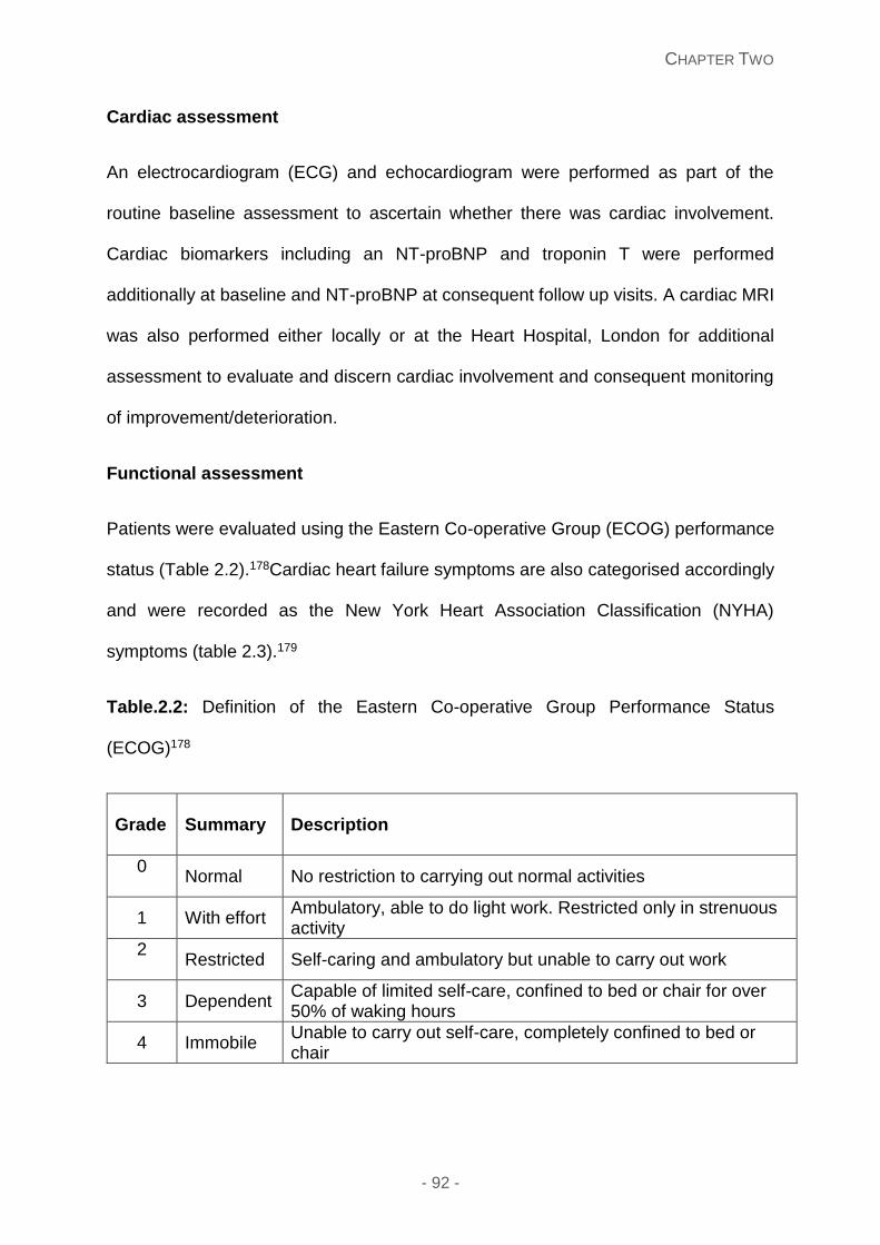
CHAPTER TWO
- 92 -
Cardiac assessment
An electrocardiogram (ECG) and echocardiogram were performed as part of the
routine baseline assessment to ascertain whether there was cardiac involvement.
Cardiac biomarkers including an NT-proBNP and troponin T were performed
additionally at baseline and NT-proBNP at consequent follow up visits. A cardiac MRI
was also performed either locally or at the Heart Hospital, London for additional
assessment to evaluate and discern cardiac involvement and consequent monitoring
of improvement/deterioration.
Functional assessment
Patients were evaluated using the Eastern Co-operative Group (ECOG) performance
status (Table 2.2).178Cardiac heart failure symptoms are also categorised accordingly
and were recorded as the New York Heart Association Classification (NYHA)
symptoms (table 2.3).179
Table.2.2: Definition of the Eastern Co-operative Group Performance Status
(ECOG)178
Grade
Summary Description
0
Normal No restriction to carrying out normal activities
1 With effort Ambulatory, able to do light work. Restricted only in strenuous activity
2
Restricted Self-caring and ambulatory but unable to carry out work
3 Dependent Capable of limited self-care, confined to bed or chair for over 50% of waking hours
4 Immobile Unable to carry out self-care, completely confined to bed or chair
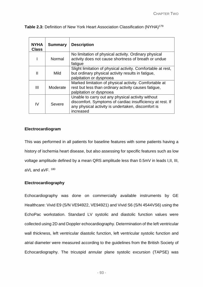
CHAPTER TWO
- 93 -
Table 2.3: Definition of New York Heart Association Classification (NYHA)179
NYHA Class
Summary Description
I Normal No limitation of physical activity. Ordinary physical activity does not cause shortness of breath or undue fatigue
II Mild Slight limitation of physical activity. Comfortable at rest, but ordinary physical activity results in fatigue, palpitation or dyspnoea
III Moderate Marked limitation of physical activity. Comfortable at rest but less than ordinary activity causes fatigue, palpitation or dyspnoea
IV Severe
Unable to carry out any physical activity without discomfort. Symptoms of cardiac insufficiency at rest. If any physical activity is undertaken, discomfort is increased
Electrocardiogram
This was performed in all patients for baseline features with some patients having a
history of ischemia heart disease, but also assessing for specific features such as low
voltage amplitude defined by a mean QRS amplitude less than 0.5mV in leads I,II, III,
aVL and aVF. 180
Electrocardiography
Echocardiography was done on commercially available instruments by GE
Healthcare: Vivid E9 (S/N VE94922, VE94921) and Vivid S6 (S/N 4544VS6) using the
EchoPac workstation. Standard LV systolic and diastolic function values were
collected using 2D and Doppler echocardiography. Determination of the left ventricular
wall thickness, left ventricular diastolic function, left ventricular systolic function and
atrial diameter were measured according to the guidelines from the British Society of
Echocardiography. The tricuspid annular plane systolic excursion (TAPSE) was
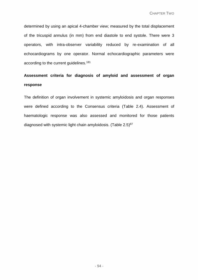
CHAPTER TWO
- 94 -
determined by using an apical 4-chamber view; measured by the total displacement
of the tricuspid annulus (in mm) from end diastole to end systole. There were 3
operators, with intra-observer variability reduced by re-examination of all
echocardiograms by one operator. Normal echocardiographic parameters were
according to the current guidelines.181
Assessment criteria for diagnosis of amyloid and assessment of organ
response
The definition of organ involvement in systemic amyloidosis and organ responses
were defined according to the Consensus criteria (Table 2.4). Assessment of
haematologic response was also assessed and monitored for those patients
diagnosed with systemic light chain amyloidosis. (Table 2.5)87
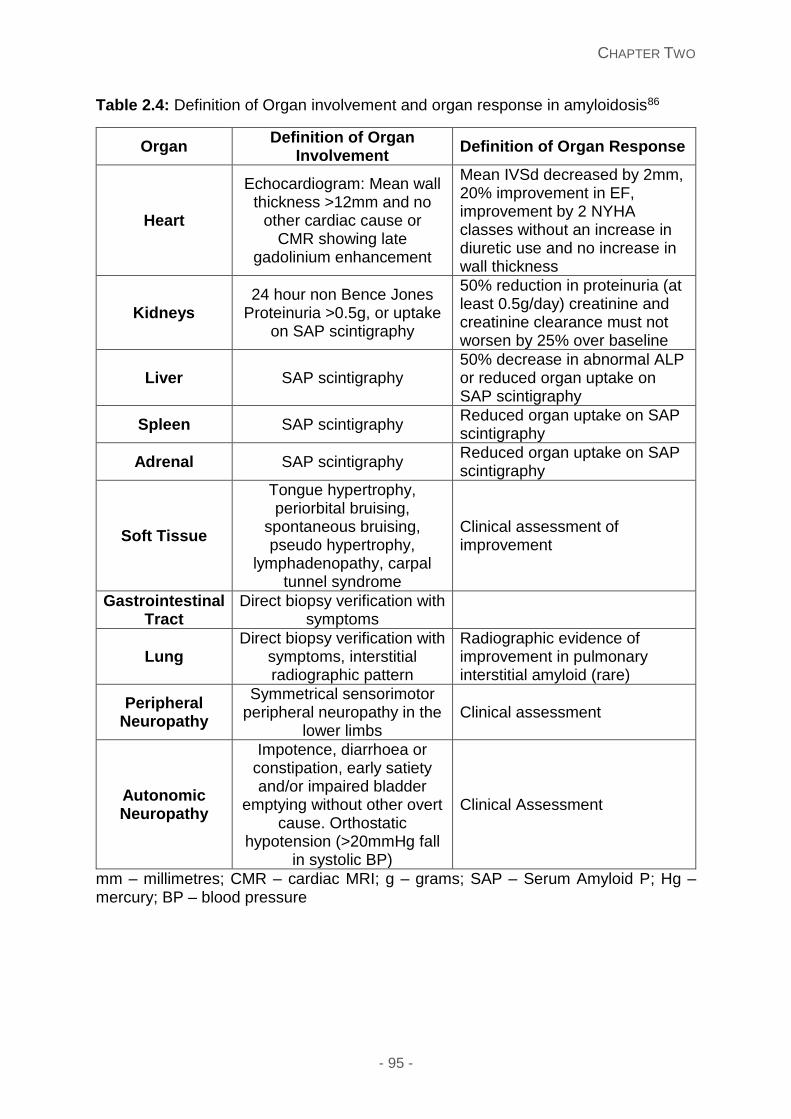
CHAPTER TWO
- 95 -
Table 2.4: Definition of Organ involvement and organ response in amyloidosis86
Organ Definition of Organ
Involvement Definition of Organ Response
Heart
Echocardiogram: Mean wall thickness >12mm and no
other cardiac cause or CMR showing late
gadolinium enhancement
Mean IVSd decreased by 2mm, 20% improvement in EF, improvement by 2 NYHA classes without an increase in diuretic use and no increase in wall thickness
Kidneys 24 hour non Bence Jones
Proteinuria >0.5g, or uptake on SAP scintigraphy
50% reduction in proteinuria (at least 0.5g/day) creatinine and creatinine clearance must not worsen by 25% over baseline
Liver SAP scintigraphy 50% decrease in abnormal ALP or reduced organ uptake on SAP scintigraphy
Spleen SAP scintigraphy Reduced organ uptake on SAP scintigraphy
Adrenal SAP scintigraphy Reduced organ uptake on SAP scintigraphy
Soft Tissue
Tongue hypertrophy, periorbital bruising,
spontaneous bruising, pseudo hypertrophy,
lymphadenopathy, carpal tunnel syndrome
Clinical assessment of improvement
Gastrointestinal Tract
Direct biopsy verification with symptoms
Lung Direct biopsy verification with
symptoms, interstitial radiographic pattern
Radiographic evidence of improvement in pulmonary interstitial amyloid (rare)
Peripheral Neuropathy
Symmetrical sensorimotor peripheral neuropathy in the
lower limbs Clinical assessment
Autonomic Neuropathy
Impotence, diarrhoea or constipation, early satiety and/or impaired bladder
emptying without other overt cause. Orthostatic
hypotension (>20mmHg fall in systolic BP)
Clinical Assessment
mm – millimetres; CMR – cardiac MRI; g – grams; SAP – Serum Amyloid P; Hg – mercury; BP – blood pressure
CHAPTER TWO
- 96 -
Table 2.5: Consensus Haematologic Response in systemic light chain amyloidosis87
Haematologic Response Criteria
Complete Response (CR)
Normal serum free light chain ratio with negative serum and urinary immunofixation
Very good partial response (VGPR) The difference in the free light chains (dFLC) less than 40mg/L
Partial Response (PR) A reduction in the dFLC greater than 50%
No response A less than 50% response in dFLC
mg/L – milligrams per litre
Statistical analysis
Statistical analysis was performed using SPSS v20 (IBM SPSS) software. Kaplan
Meier estimates were used to calculate the OS and PFS. P values <0.05 were
considered statistically significant. Graph Pad Prism (Version 5) was also used. The
statistical analysis is described in each methods/results section of each chapter.
- 97 -
Results Section One:
Diagnostic investigations and prognostic implications of
amyloidosis
CHAPTER THREE
- 98 -
Chapter Three: Comparison of free light chain assays: FreeliteTM
and N Latex in diagnosis, monitoring and predicting survival in
light chain amyloidosis
This chapter is written in context of my article: Comparison of free light chain
assays: FreeliteTM and N Latex in diagnosis, monitoring and predicting survival
in light chain amyloidosis. Shameem Mahmood, Nancy L Wassef, Simon J Salter,
Sajitha Sachchithanantham, T Lane, D Foard, Carol J Whelan, Helen J Lachmann,
Julian D Gilmore, Philip N Hawkins, Ashutosh D Wechalekar, American Journal of
Clinical Pathology, 2016 Jul;146 (1):78-85 (Original article) copyright permission
obtained from Oxford University Press, license no.:3917120648467 for use in my
thesis.
Introduction
Systemic AL amyloidosis is a rare protein deposition disease. The underlying
pathological process driving AL amyloidosis is an underlying clonal proliferation of
plasma cells producing an excess of unstable immunoglobulin light chains. There is
deposition of immunoglobulin light chains in tissues and organs leading to impairment
of the structure and function of the latter.6, 182 Tracking and monitoring AL amyloidosis
is a challenge as many patients have barely a detectable intact monoclonal
immunoglobulin. However, the discovery that serum free light chains could be
measured in 2000 led to a development that revolutionised this rare disease. The
serum FreeliteTM assay measures immunoglobulin free light chains using polyclonal
CHAPTER THREE
- 99 -
sheep antibodies directed against hidden epitopes on the light chain molecule giving
an accurate measurement of serum free kappa and lambda. Our group initially
reported the utility of this assay in 2002183 in this disease, with many studies following
since. This assay now forms a standard part of the baseline and serial follow up
assessments in systemic AL.161, 162 It is a part of the revised Mayo staging system and
standard international consensus criteria for the disease response assessment.80, 86,
87
Polyclonal antibodies to light chain epitopes in the FreeliteTM assay continues to be
prepared from specifically immunised sheep. However, in any naturally occurring
antibody mixture, standardisation remains complex and inter-assay variability is a
certainly a possibility. Efforts have been made to develop monoclonal antibodies to
the hidden light chain epitopes that would recognise and replicate the success of the
polyclonal assay and thus allow better inter-assay standardisation. A group in the
Netherlands working with Siemens, Germany, has developed this technology using a
mixture to two kappa and two lambda monoclonal antibodies to the hidden epitopes
of constant region of the immunoglobulin light chain molecules.163 The references
ranges for both the new N-Latex assay and the polyclonal FreeliteTM assay are similar.
With increasing adoption by laboratories of the novel assay, it is important to assess
the utility of the new assay compared to the current reference standard polyclonal free
light chain assay. We report a comparison of serum free light chains measured by
both immunoassays at diagnosis and three further time points in during the initial
chemotherapy for patients with systemic AL amyloidosis.
CHAPTER THREE
- 100 -
Methods
This study was conducted at the National Amyloidosis Centre, London, UK (NAC),
with recruitment of consecutive patients with systemic AL amyloidosis seen at the NAC
and under prospective follow up from January 2011 and April 2012 and undergoing
chemotherapy for AL amyloidosis. Patients were included if they had a baseline serum
sample at initial diagnosis and at least 3 of 4 blood samples available including from
baseline 2, 4 and 6 month follow up point intervals following treatment with
chemotherapy. Diagnosis of amyloidosis was confirmed in all cases with a tissue
biopsy showing the characteristic birefringence on Congo red staining. Typing of AL
amyloidosis was confirmed by immunohistochemical staining with appropriate
antibodies and exclusion of hereditary amyloidosis, where necessary, by genetic
sequencing of the genes implicated. All patients underwent systematic review at
presentation and detailed follow up assessments at 6 monthly intervals or dependant
on clinical indication. Assessment included clinical examination, detailed blood and
urine analysis (including assessment of serum and urine monoclonal immunoglobulin
and serum free light chains), serial 123I labelled serum amyloid P component (SAP)
scintigraphy to assess whole body amyloid load, electrocardiogram (ECG) and an
echocardiogram and routine observations including blood pressure, oxygen
saturations and pulse. Organ involvement was defined according to the standard
international amyloidosis consensus criteria.(18) Mayo disease stage was defined
according to the criteria published by Dispenzieri et al and Kumar et al80, 184
Serum samples were separated and stored at -80ºC. All retrieved samples were
tested in duplicate for measurement with FreeliteTM (The Binding site Ltd, Birmingham,
UK) and N-Latex (Siemens Healthcare Diagnostics, Germany) according to the
standard manufactures’ protocols on a BNTMII System nephelometer (Siemens,
CHAPTER THREE
- 101 -
Germany). Examination was undertaken to assess haematologic response on results
of the free light chain (FLC) assays in available samples at the following points
including 2 months; 90 FreeliteTM and 90 N Latex, 4 months; 91 FreeliteTM and 89 N
Latex, 6 months; 61 FreeliteTM and 62 N Latex. The discrepancies in the number of
available samples for analysis were secondary to the inadequate quantity of serum or
error readings in the analysed sample, hence excluded for analysis. In total, serum
free light chains were measured in a total of 240 serum samples in duplicate using
both the FreeliteTM and N Latex assays.
We performed a correlation analysis between the 2 FLC assays of results for kappa,
lambda and kappa/lambda ratio. Haematologic response was assessed according to
the international amyloidosis consensus criteria.86, 87 The same criteria were used to
assess a patient as a responder or not by both assays; although we recognise that the
criteria were developed with results obtained from the FreeliteTM assay. This study was
not powered or focused to develop new response criteria for the N-latex assay.
Statistical analysis was performed using SPSS software using a p value of less than
0.05 considered to be significant. We stated median values with minimum and
maximum ranges. We calculated the Pearson’s coefficient to evaluate the agreement
and concordance between the both FLC assays, with use of scatter plots to illustrate
this. Kaplan Meier curves were used to assess and describe the prognostic utility of
both assays.
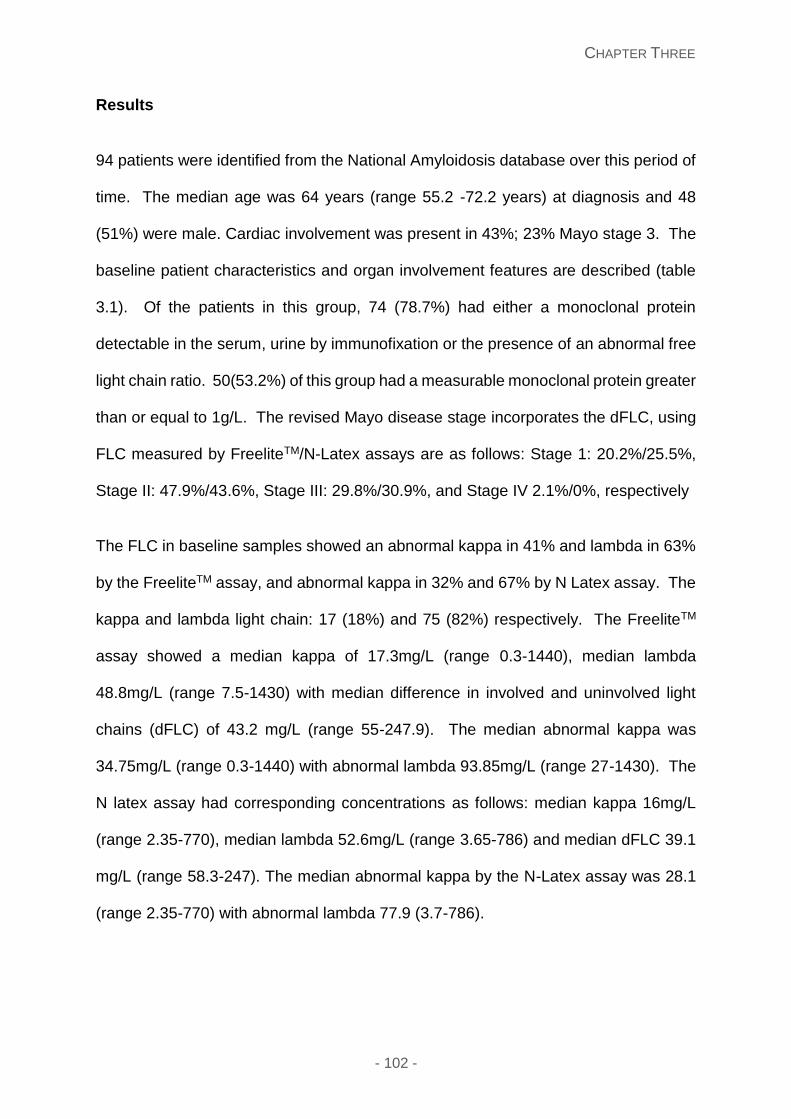
CHAPTER THREE
- 102 -
Results
94 patients were identified from the National Amyloidosis database over this period of
time. The median age was 64 years (range 55.2 -72.2 years) at diagnosis and 48
(51%) were male. Cardiac involvement was present in 43%; 23% Mayo stage 3. The
baseline patient characteristics and organ involvement features are described (table
3.1). Of the patients in this group, 74 (78.7%) had either a monoclonal protein
detectable in the serum, urine by immunofixation or the presence of an abnormal free
light chain ratio. 50(53.2%) of this group had a measurable monoclonal protein greater
than or equal to 1g/L. The revised Mayo disease stage incorporates the dFLC, using
FLC measured by FreeliteTM/N-Latex assays are as follows: Stage 1: 20.2%/25.5%,
Stage II: 47.9%/43.6%, Stage III: 29.8%/30.9%, and Stage IV 2.1%/0%, respectively
The FLC in baseline samples showed an abnormal kappa in 41% and lambda in 63%
by the FreeliteTM assay, and abnormal kappa in 32% and 67% by N Latex assay. The
kappa and lambda light chain: 17 (18%) and 75 (82%) respectively. The FreeliteTM
assay showed a median kappa of 17.3mg/L (range 0.3-1440), median lambda
48.8mg/L (range 7.5-1430) with median difference in involved and uninvolved light
chains (dFLC) of 43.2 mg/L (range 55-247.9). The median abnormal kappa was
34.75mg/L (range 0.3-1440) with abnormal lambda 93.85mg/L (range 27-1430). The
N latex assay had corresponding concentrations as follows: median kappa 16mg/L
(range 2.35-770), median lambda 52.6mg/L (range 3.65-786) and median dFLC 39.1
mg/L (range 58.3-247). The median abnormal kappa by the N-Latex assay was 28.1
(range 2.35-770) with abnormal lambda 77.9 (3.7-786).
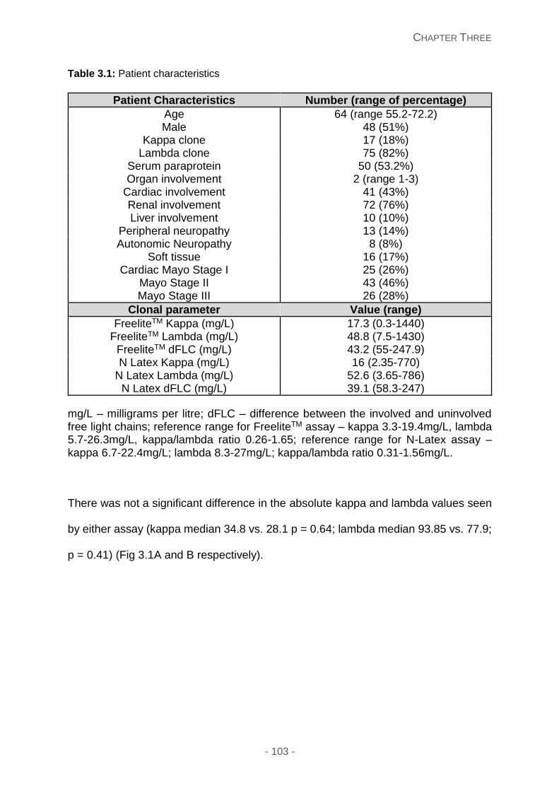
CHAPTER THREE
- 103 -
Table 3.1: Patient characteristics
Patient Characteristics Number (range of percentage)
Age 64 (range 55.2-72.2) Male 48 (51%)
Kappa clone 17 (18%) Lambda clone 75 (82%)
Serum paraprotein 50 (53.2%) Organ involvement 2 (range 1-3)
Cardiac involvement 41 (43%) Renal involvement 72 (76%) Liver involvement 10 (10%)
Peripheral neuropathy 13 (14%) Autonomic Neuropathy 8 (8%)
Soft tissue 16 (17%) Cardiac Mayo Stage I 25 (26%)
Mayo Stage II 43 (46%) Mayo Stage III 26 (28%)
Clonal parameter Value (range)
FreeliteTM Kappa (mg/L) 17.3 (0.3-1440) FreeliteTM Lambda (mg/L) 48.8 (7.5-1430)
FreeliteTM dFLC (mg/L) 43.2 (55-247.9) N Latex Kappa (mg/L) 16 (2.35-770)
N Latex Lambda (mg/L) 52.6 (3.65-786) N Latex dFLC (mg/L) 39.1 (58.3-247)
mg/L – milligrams per litre; dFLC – difference between the involved and uninvolved free light chains; reference range for FreeliteTM assay – kappa 3.3-19.4mg/L, lambda 5.7-26.3mg/L, kappa/lambda ratio 0.26-1.65; reference range for N-Latex assay – kappa 6.7-22.4mg/L; lambda 8.3-27mg/L; kappa/lambda ratio 0.31-1.56mg/L.
There was not a significant difference in the absolute kappa and lambda values seen
by either assay (kappa median 34.8 vs. 28.1 p = 0.64; lambda median 93.85 vs. 77.9;
p = 0.41) (Fig 3.1A and B respectively).
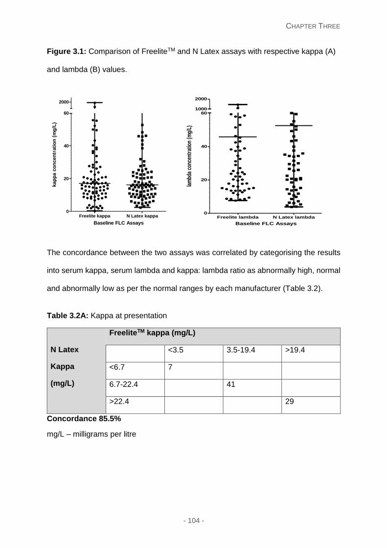
CHAPTER THREE
- 104 -
Figure 3.1: Comparison of FreeliteTM and N Latex assays with respective kappa (A)
and lambda (B) values.
The concordance between the two assays was correlated by categorising the results
into serum kappa, serum lambda and kappa: lambda ratio as abnormally high, normal
and abnormally low as per the normal ranges by each manufacturer (Table 3.2).
Table 3.2A: Kappa at presentation
N Latex
Kappa
(mg/L)
FreeliteTM kappa (mg/L)
<3.5 3.5-19.4 >19.4
<6.7 7
6.7-22.4 41
>22.4 29
Concordance 85.5%
mg/L – milligrams per litre
Freelite kappa N Latex kappa0
20
40
60
2000
Baseline FLC Assays
kap
pa c
on
cen
trati
on
(m
g/L
)
Freelite lambda N Latex lambda0
20
40
601000
2000
Baseline FLC Assays
lam
bda
conc
entr
atio
n (m
g/L)
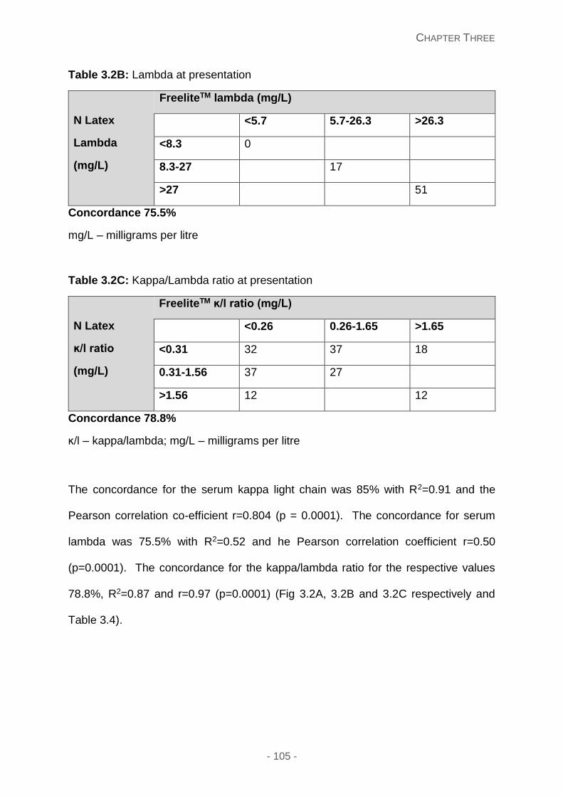
CHAPTER THREE
- 105 -
Table 3.2B: Lambda at presentation
N Latex
Lambda
(mg/L)
FreeliteTM lambda (mg/L)
<5.7 5.7-26.3 >26.3
<8.3 0
8.3-27 17
>27 51
Concordance 75.5%
mg/L – milligrams per litre
Table 3.2C: Kappa/Lambda ratio at presentation
N Latex
ĸ/Ɩ ratio
(mg/L)
FreeliteTM ĸ/Ɩ ratio (mg/L)
<0.26 0.26-1.65 >1.65
<0.31 32 37 18
0.31-1.56 37 27
>1.56 12 12
Concordance 78.8%
ĸ/Ɩ – kappa/lambda; mg/L – milligrams per litre
The concordance for the serum kappa light chain was 85% with R2=0.91 and the
Pearson correlation co-efficient r=0.804 (p = 0.0001). The concordance for serum
lambda was 75.5% with R2=0.52 and he Pearson correlation coefficient r=0.50
(p=0.0001). The concordance for the kappa/lambda ratio for the respective values
78.8%, R2=0.87 and r=0.97 (p=0.0001) (Fig 3.2A, 3.2B and 3.2C respectively and
Table 3.4).
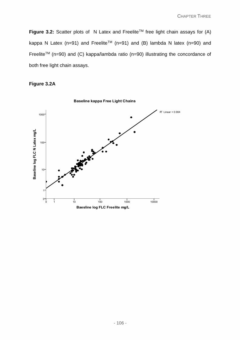
CHAPTER THREE
- 106 -
Figure 3.2: Scatter plots of N Latex and FreeliteTM free light chain assays for (A)
kappa N Latex (n=91) and FreeliteTM (n=91) and (B) lambda N latex (n=90) and
FreeliteTM (n=90) and (C) kappa/lambda ratio (n=90) illustrating the concordance of
both free light chain assays.
Figure 3.2A
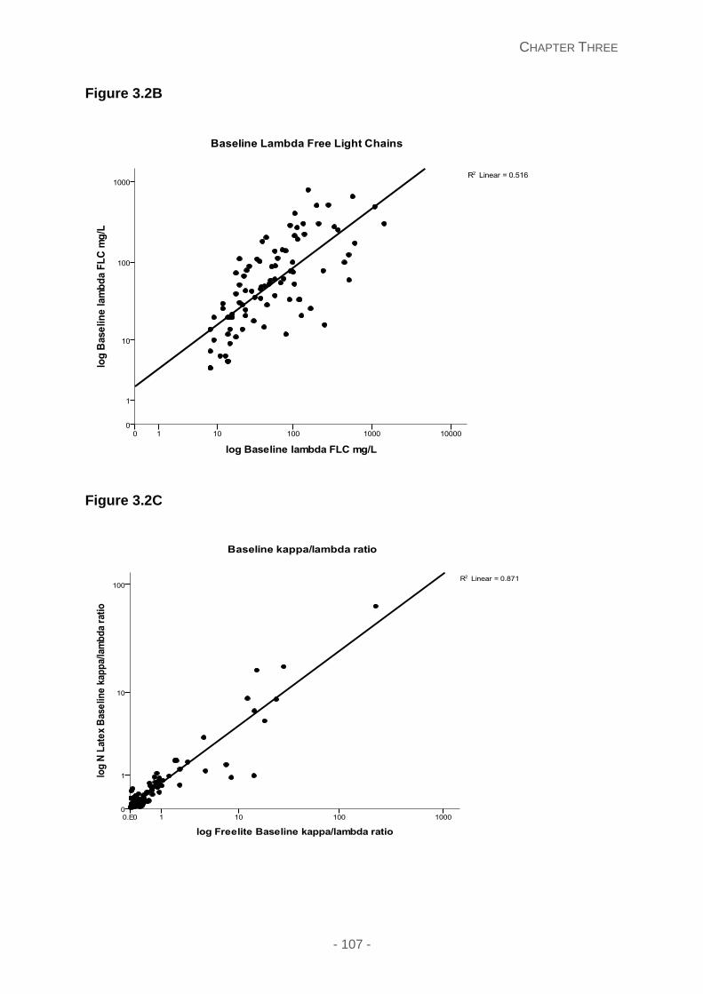
CHAPTER THREE
- 107 -
Figure 3.2B
Figure 3.2C
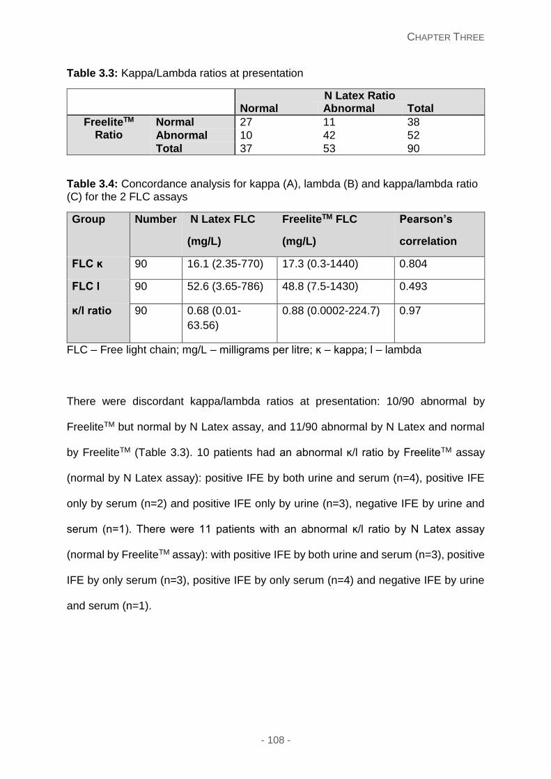
CHAPTER THREE
- 108 -
Table 3.3: Kappa/Lambda ratios at presentation
N Latex Ratio Normal Abnormal Total
FreeliteTM Ratio
Normal 27 11 38
Abnormal 10 42 52
Total 37 53 90
Table 3.4: Concordance analysis for kappa (A), lambda (B) and kappa/lambda ratio (C) for the 2 FLC assays
Group Number N Latex FLC
(mg/L)
FreeliteTM FLC
(mg/L)
Pearson’s
correlation
FLC ĸ 90 16.1 (2.35-770) 17.3 (0.3-1440) 0.804
FLC Ɩ 90 52.6 (3.65-786) 48.8 (7.5-1430) 0.493
ĸ/Ɩ ratio 90 0.68 (0.01-
63.56)
0.88 (0.0002-224.7) 0.97
FLC – Free light chain; mg/L – milligrams per litre; ĸ – kappa; Ɩ – lambda
There were discordant kappa/lambda ratios at presentation: 10/90 abnormal by
FreeliteTM but normal by N Latex assay, and 11/90 abnormal by N Latex and normal
by FreeliteTM (Table 3.3). 10 patients had an abnormal ĸ/Ɩ ratio by FreeliteTM assay
(normal by N Latex assay): positive IFE by both urine and serum (n=4), positive IFE
only by serum (n=2) and positive IFE only by urine (n=3), negative IFE by urine and
serum (n=1). There were 11 patients with an abnormal ĸ/Ɩ ratio by N Latex assay
(normal by FreeliteTM assay): with positive IFE by both urine and serum (n=3), positive
IFE by only serum (n=3), positive IFE by only serum (n=4) and negative IFE by urine
and serum (n=1).
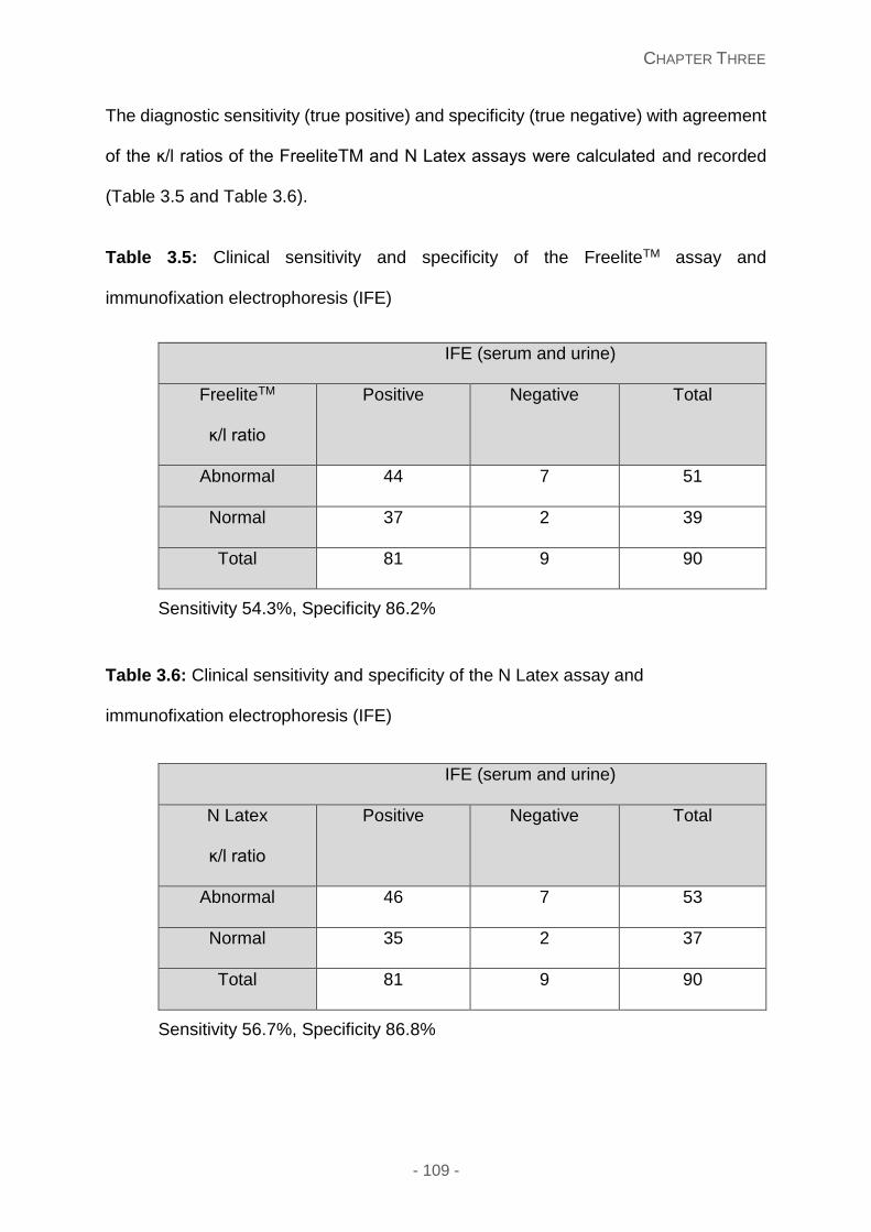
CHAPTER THREE
- 109 -
The diagnostic sensitivity (true positive) and specificity (true negative) with agreement
of the ĸ/Ɩ ratios of the FreeliteTM and N Latex assays were calculated and recorded
(Table 3.5 and Table 3.6).
Table 3.5: Clinical sensitivity and specificity of the FreeliteTM assay and
immunofixation electrophoresis (IFE)
IFE (serum and urine)
FreeliteTM
ĸ/Ɩ ratio
Positive Negative Total
Abnormal 44 7 51
Normal 37 2 39
Total 81 9 90
Sensitivity 54.3%, Specificity 86.2%
Table 3.6: Clinical sensitivity and specificity of the N Latex assay and
immunofixation electrophoresis (IFE)
IFE (serum and urine)
N Latex
ĸ/Ɩ ratio
Positive Negative Total
Abnormal 46 7 53
Normal 35 2 37
Total 81 9 90
Sensitivity 56.7%, Specificity 86.8%
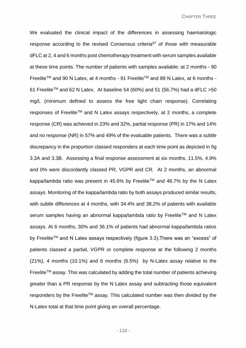
CHAPTER THREE
- 110 -
We evaluated the clinical impact of the differences in assessing haematologic
response according to the revised Consensus criteria87 of those with measurable
dFLC at 2, 4 and 6 months post chemotherapy treatment with serum samples available
at these time points. The number of patients with samples available: at 2 months - 90
FreeliteTM and 90 N Latex, at 4 months - 91 FreeliteTM and 89 N Latex, at 6 months -
61 FreeliteTM and 62 N Latex. At baseline 54 (60%) and 51 (56.7%) had a dFLC >50
mg/L (minimum defined to assess the free light chain response). Correlating
responses of FreeliteTM and N Latex assays respectively, at 2 months, a complete
response (CR) was achieved in 23% and 32%, partial response (PR) in 17% and 14%
and no response (NR) in 57% and 49% of the evaluable patients. There was a subtle
discrepancy in the proportion classed responders at each time point as depicted in fig
3.3A and 3.3B. Assessing a final response assessment at six months, 11.5%, 4.9%
and 0% were discordantly classed PR, VGPR and CR. At 2 months, an abnormal
kappa/lambda ratio was present in 45.6% by FreeliteTM and 46.7% by the N Latex
assays. Monitoring of the kappa/lambda ratio by both assays produced similar results,
with subtle differences at 4 months, with 34.4% and 38.2% of patients with available
serum samples having an abnormal kappa/lambda ratio by FreeliteTM and N Latex
assays. At 6 months, 30% and 36.1% of patients had abnormal kappa/lambda ratios
by FreeliteTM and N Latex assays respectively (figure 3.3).There was an “excess” of
patients classed a partial, VGPR or complete response at the following 2 months
(21%), 4 months (10.1%) and 6 months (6.5%) by N-Latex assay relative to the
FreeliteTM assay. This was calculated by adding the total number of patients achieving
greater than a PR response by the N Latex assay and subtracting those equivalent
responders by the FreeliteTM assay. This calculated number was then divided by the
N-Latex total at that time point giving an overall percentage.
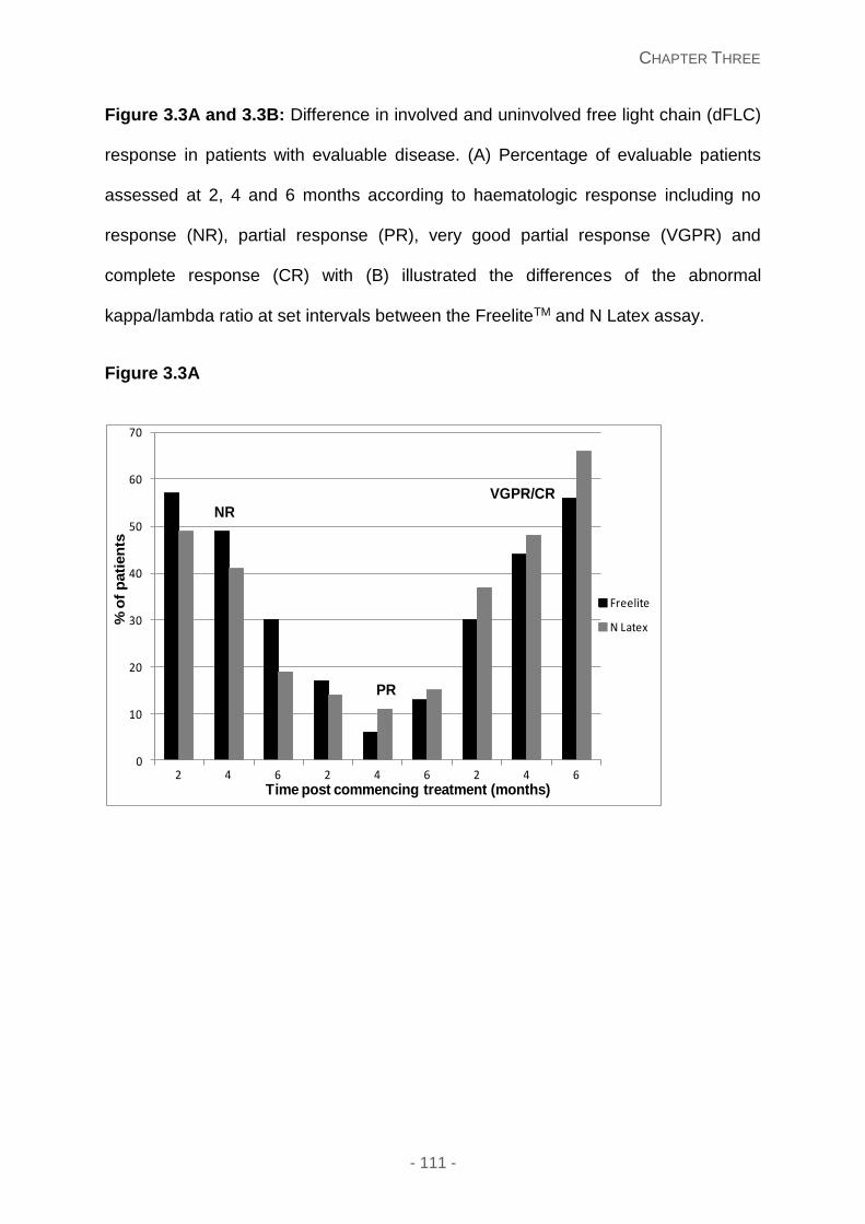
CHAPTER THREE
- 111 -
Figure 3.3A and 3.3B: Difference in involved and uninvolved free light chain (dFLC)
response in patients with evaluable disease. (A) Percentage of evaluable patients
assessed at 2, 4 and 6 months according to haematologic response including no
response (NR), partial response (PR), very good partial response (VGPR) and
complete response (CR) with (B) illustrated the differences of the abnormal
kappa/lambda ratio at set intervals between the FreeliteTM and N Latex assay.
Figure 3.3A
0
10
20
30
40
50
60
70
2 4 6 2 4 6 2 4 6
Freelite
N Latex
Time post commencing treatment (months)
NR
PR
VGPR/CR
% o
f p
ati
en
ts
CHAPTER THREE
- 112 -
Figure 3.3B
The estimated overall survival (OS) for the 94 patients was 24.1 months with a median
follow-up of 8.6 months. Given the differences in the proportion of patients classed as
achieving partial response by both assays at 4 months, we evaluated the overall
survival of patients achieving a partial response or greater at 4 months by both assays.
There was a significant improvement in the overall survival for patients classified as
achieving a partial response or better at 4 months by each assay. The median OS
was not reached in either assay. The survival benefit for those achieving a PR or
better by the FreeliteTM at 4 months was HR 7.84, 95% CI (0.94-65.57), log rank p =
0.03 and by the N-Latex assay HR 95.47, 95% CI (0.11-79601), log rank p = 0.004.
Freelite abnormal ratio
N latex abnormal ratio
Freelite normal ratioN Latex normal ratio
0
20
40
60
80
100
2 months 4 months 6 months
Freelite abnormal ratio
N latex abnormal ratio
Freelite normal ratio
N Latex normal ratio
Time post assessment (months)
% o
f patients
CHAPTER THREE
- 113 -
Discussion
We report here a comparative utility of using two different assays (one using polyclonal
anti-light chain antibodies and other using a combination of monoclonal anti-light chain
antibodies) for detection of and serial monitoring of serum free light chains in patients
with systemic AL amyloidosis undergoing chemotherapy. This study finds the two
assays are broadly comparable for detection of abnormal light chains in the serum
with a greater concordance for detection of kappa than for lambda immunoglobulin
free light chain. However, there were discrepancies present with the numbers
abnormal, the absolute light chain value and time point to reach thresholds for
haematological responses and also impact on survival.
The FreeliteTM assay was the only assay available for measurement of serum free light
chains until development of the N-Latex assay. This is a new and welcome
development to allow for a different method to study these complex molecules, but
standardisation is needed between the two assays. Both these assays have been
studied in myeloma as well as normal controls to assess imprecision, underestimation;
antigen excess or non-linear reactivity and overestimation; polymeric forms reacting
at multiple antigenic FLC sites of results.185-187 Our study did not focus on the
biochemical or technical aspects of either assay, but designed to assess the clinical
utility of each assay in a routine clinical setting in systemic where the serum free light
chains are typically abnormal, but low and where FLC measurements forms the
cornerstone of patient assessment and monitoring. Previous studies in patients with
a monoclonal gammopathy, have the kappa showing the best correlation between
both FLC assays, followed by kappa/lambda ratios then lambda, with the Pearson
coefficients 0.97, 0.83 and 0.91 respectively.188 Pretorius et al compared the utility of
CHAPTER THREE
- 114 -
both the FreeliteTM and N Latex assays in 116 samples showing large variations
between both assays, precluding the interchange of these results, with statistically
significant non-linearity occurring in approximately half of the monoclonal and
polyclonal samples (kappa and lambda) for both assays.189 The precision of the two
assays were further examined in two other studies, showing comparable levels190 and
reproducibility.191
The current study confirms that in systemic AL amyloidosis, there is generally excellent
concordance between both the assays in detecting an excess of serum free light
chains with better concordance of kappa than lambda as previously reported. The
overall concordance in our study showed kappa, lambda and kappa/lambda ratios as
85.5%, 75.5% and 78.8% respectively - similar to one study 70-80% 188 but lower than
in another study with the concordance quoted as approximately 90%.163 The
diagnostic sensitivity with respect to the serum and urine IFE results was lower than
expected by both assays, 54.3% and 56.7% by FreeliteTM and N Latex assays
respectively. Some patients had lower serum free light chain values at diagnosis, with
the presence of serum and/or urine IFE, clinical symptoms and diagnostic biopsy
needed to prove the diagnosis in these cases. The serum free light chains are often
lower than in Myeloma patients. A concerning result was that ~10% cases with each
assay detected an abnormal FLC ratio that was not detected by the other assay. The
underlying reason for this discordance is difficult to fully explain or ascertain, given the
details of antibodies used to detect the free light chains in both assays are
commercially confidential. Given the nature of the monoclonal disease underlying AL
amyloidosis, the mutational signature of the plasma cell DNA in each AL light chain is
unique. A likely explanation is the antibodies in each assay do not recognise the
epitopes on the respective light chains and hence there is a false negative signal. The
CHAPTER THREE
- 115 -
Mayo clinic group, has recently demonstrated that such samples also contain a
monoclonal light chain using mass spectrometry. Hence suggesting that currently
neither method is perfect for detection of all abnormal monoclonal free light chains but
as different patients “detected” or “missed” by either assay, there may be role for
repeating the FLC measurement by the other assay if an abnormality is not detected
in a patient with one assay. This may assist at least partly, overcome the problem in
AL amyloidosis of lack of evaluable light chains in a certain proportion of all patients.183
We compared the ĸ/Ɩ ratios and the IFE results showing that both assays had similar
sensitivities (54.3% and 56.7%) and specificities (86.2% and 86.8%) for FreeliteTM and
N Latex assays respectively.
This is the largest study to examine serial parallel sample assessment to assess the
serum free light chain responses by both assays simultaneously. Although there is
broad agreement in the response assessment as a percentage of total patients with
both assays, important differences are present in a proportion of patients who reach
the currently threshold used to define a partial or very good partial response. Eighteen
patients reached a PR and VGPR earlier by the N-Latex assay compared to the same
class of response by the FreeliteTM assay. This is concerning, but in a way not that
unexpected. The assays have very different antibodies which measure the free light
chain in the serum and hence the actual slopes of the standard curves are not exactly
identical. Hence, it is expected that the rate of change will be different. The response
criteria in AL amyloidosis have been derived using ROC analysis based on survival of
patients using various light chain thresholds attained by the FreeliteTM assay. Thus
the current thresholds are clinical and are not biochemical end points. Assessing the
results, it appears that the N-Latex assay classified patients as responders at an
earlier time point than the FreeliteTM assay, designing a study to examine a clinically
CHAPTER THREE
- 116 -
relevant threshold for defining a response with N-latex is important. Similarly
prognostic important thresholds of FLC in AL (dFLC >180 mg/L) may not necessary
be the same and will need re-defining for this new era. In this study, 3 patients were
misclassified as advanced stage III-IV by the revised classification using the different
assays.
It is encouraging that the classification of a patient as a haematological responder
(partial response or greater) translates into a survival benefit. However, earlier
classification of a greater proportion of patients as responders by the N-Latex assay
has risks, as treatment duration in amyloidosis is often shorter than in myeloma with
a lower threshold to stop treatment earlier in the responders particularly if there are
problems with tolerance to treatment. Unless thresholds are redefined, patients may
be classified as responders by the N-Latex assay may get undertreated and hence
carry a risk of shorter time to disease progression.
This current study needs to be interpreted cautiously in the context of certain
limitations. The numbers are still small and all patients did not have serial samples
available. Although the patients recruited in this study are serial AL patients seen at
the NAC, there is a selection bias as all patients were required to have serial samples
for six months, with only six month survivors are recruited thereby excluding a
substantial number of patients with stage III cardiac AL amyloidosis where there is still
30-40% mortality in the first six months.192 A study of truly unselected serial patients
is needed to assess the true correlation of the utility of both these assays. There was
higher than expected proportion of patients with non-evaluable light chains, possibly
due to selection of the survivors who are typically patients with early stage disease
and serum free light chain burden.
CHAPTER THREE
- 117 -
In summary, both FreeliteTM and N Latex assays are able to detect the abnormal FLC
in patients with systemic light chain amyloidosis. Although there is an excellent
correlation between these assays for detecting the abnormal light chain subtype the
discordance in the absolute values renders cross assay interpolation impossible.
Furthermore, each FLC assay appears to have limitations, in missing random yet
different patients suggesting a further need by both manufacturers to optimise the anti-
light chain antibodies underpinning the respective assays. Studies using clinical end
points are important to harmonise the response assessment criteria for each assay so
that even though the values may not be comparable, the response categorisation is
comparable. This is crucially important, given the emergence of the newer assay
becoming more widely adopted and possibly raising problems in the interpretation of
clinical and trial results. Designing future clinical trials in AL amyloidosis incorporating
FLC measurement by both assays would be ideal to aid in our understanding and
harmonisation of the field.
CHAPTER FOUR
- 118 -
Chapter Four: Bleeding diathesis, thrombotic tendencies and
endothelial dysfunction in amyloidosis patients
This chapter is written with reference to the publication: Utility of factor X
concentrate for the treatment of acquired factor X deficiency in systemic light-chain
amyloidosis. Shameem Mahmood, Julie Blundell, Anja Drebes, Philip N. Hawkins and
Ashutosh D. Wechalekar. Blood. 2014; 123(18):2899-900 (Original article).
Introduction
Systemic light chain (AL) amyloidosis is the most common systemic amyloidosis.
Typically the clonal proliferation of a plasma cell clone that consequently results in the
synthesis of an excess of light chains and toxic intermediates with specific structural
and mechanical effects resulting in systemic fibril toxicity.193, 194 These fibrils deposit
in different organs leading to progressive changes in the affected organ’s architecture
and function.6 Severity of this disease depends on the number of organs involved and
extent of amyloid deposits, with AL cardiomyopathy carrying a poor prognosis with
death arising as a consequence secondary to progressive heart failure, contractility
dysfunction, sudden death and associated arrhythmias or refractory untreatable
disease.195, 196 Some studies have explored the AL-light chain cardiotoxicity as an
important causal determinant in this disease.197, 198 Migrino et al demonstrated that AL
light chains cause microvascular dysfunction leading to apoptotic and necrotic insults
and related oxidative stress,199, 200 with other studies showing endothelial dysfunction
CHAPTER FOUR
- 119 -
in small and medium sized vessels, which ultimately may contribute to this
cardiotoxicity.201, 202
Endothelial dysfunction occurs with reduced bioavailability of vasodilators, especially
nitric oxide (NO) and consequent impairment of the endothelium and increased
endothelium contracting factors and pro-inflammatory state203 As such, it is associated
with coronary artery disease, hypertension, diabetes mellitus, chronic renal failure and
thought to represent a vital early trigger in the event of atherosclerosis204, 205 with a
complex interplay of low NO levels, increased oxidative stress and consequent
changes in the V-CAM, I-CAM and e-selectin adhesion molecules in this inflammatory
process205, 206 Of several methods used to assess the endothelium, plasma proteins
such as vWF have been examined extensively in cardiovascular disease and
atherosclerosis207-210 and used as a surrogate marker of endothelial dysfunction.
Numerous clinical and experimental reports imply that high vWF levels reflect
endothelium damage.211
Von Willebrand factor is a large glycoprotein synthesised by the vascular endothelial
cells through the luminal and abluminal membranes212 and megakaryocytes.
Secretion via the abluminal membrane results in some deposition within the vascular
subendothelium to bridge with circulating platelets212 and forms a non-covalent tightly
bound complex with factor VIII. Hence circulating levels of factor VIII are closely linked
to circulating levels of vWF, typically 1 molecule of FVIII to 50-100 vWF subunits.213
Von Willebrand factor has a dual role in haemostasis: promoting platelet adhesion to
thrombogenic surfaces and ensuring platelet-platelet cohesion during thrombus
formation, and acting as a carrier for FVIII; important in its production, stabilisation,
conformation and immunogenicity.214 Factor VIII serves a role in the intrinsic
coagulation pathway as a co-factor to accelerate the activation of factor X by factor
CHAPTER FOUR
- 120 -
IXa in the presence of calcium ions on a phospholipid surface.215 Thus the impact of
this intricate complex and its equilibrium may give us insight into the endothelial
dysfunction of this disease, but also possible thrombogenic and survival risk factors.
Systemic light chain (AL) amyloidosis is also known to be associated with a bleeding
diathesis,164 which may range from small cutaneous bruising, pathgnomic “racoon
eyes” to life threatening bleeds. There are a multitude of underlying factors which may
be responsible for the coagulation abnormalities in this disease including: factor
deficiencies and prothrombotic factors, vessel fragility, amyloid load, amyloid fibrils
and light chain burden216-218. Factor X deficiency has been extensively described in
the literature with an incidence of 6.3-14% in systemic AL patients164, 218. There is
limited data as to the clinical bleeding manifestations and the underlying coagulation
factors which may contribute to this environment.
This prospective study was designed to investigate the haemostasis and endothelial
dysfunction in newly diagnosed systemic light chain (AL) amyloidosis patients, with
further exploration of light chain toxicity in this disease. This was performed examining
the clinical bleeding manifestations using the Royal Free Hospital v4 adapted bleeding
questionnaire in conjunction with extensive laboratory investigations such as factor
assays, protein S, protein C, Anti-thrombin III, vWF:Ag and VWF:Ricof and
ADAMTS13 assays. Endothelial function was explored using vWF antigen (AG) and
factor VIII to serve as a surrogate marker and examine its relationship with light chain
toxicity. The secondary aim was to assess the prognostic utility of these investigations.
CHAPTER FOUR
- 121 -
Materials and methods
Patients
The study included 100 patients referred to the National Amyloidosis Centre with
probable newly diagnosed AL amyloidosis between May and December 2013.
Eligibility was open to all patients, with no exclusions according to age, performance
status, co-morbidities and naive to previous treatments No patient had a previous
history of an inherited factor deficiency or thrombotic trait. Eight patients were taking
warfarin and no patients were taking the newer anticoagulant agents.
Each patient had a detailed baseline assessment of organ function with investigations
including a full blood count, renal and liver function tests, serum protein
electrophoresis, serum free light chains, and urinary BJP, electrocardiogram and
echocardiography and 123I-labelled Serum Amyloid P (SAP) scintigraphy for visceral
amyloid deposition quantification. Each patient completed a Royal Free Hospital
bleeding questionnaire v4 (appendix 1). Additional blood samples were taken for
investigating bleeding and thrombotic tendencies in these patients. The diagnosis of
amyloidosis was confirmed on histology by Congo red staining and AL type confirmed
by specific immunostaining of amyloidotic tissue by antibodies to kappa or lambda light
chains and exclusion of hereditary amyloidosis by gene sequencing, and 99mTc-
dicarboxypropane diphosphonate (99mTc-DPD) scintigraphy for hereditary and non-
hereditary transthyretin based disease. Written consent for retrospective publication
of data was obtained from all patients in accordance with the Declaration of Helsinki.
Organ involvement was classified according to the updated international amyloidosis
consensus criteria.87
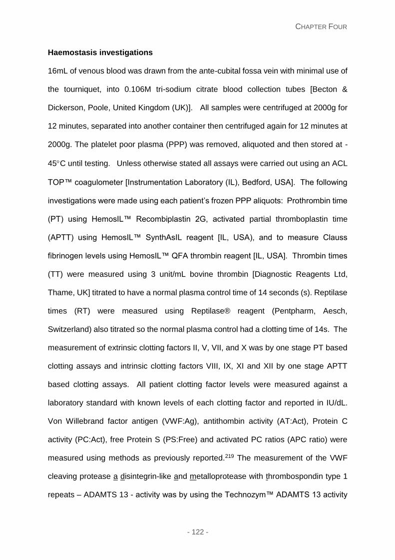
CHAPTER FOUR
- 122 -
Haemostasis investigations
16mL of venous blood was drawn from the ante-cubital fossa vein with minimal use of
the tourniquet, into 0.106M tri-sodium citrate blood collection tubes [Becton &
Dickerson, Poole, United Kingdom (UK)]. All samples were centrifuged at 2000g for
12 minutes, separated into another container then centrifuged again for 12 minutes at
2000g. The platelet poor plasma (PPP) was removed, aliquoted and then stored at -
45C until testing. Unless otherwise stated all assays were carried out using an ACL
TOP™ coagulometer [Instrumentation Laboratory (IL), Bedford, USA]. The following
investigations were made using each patient’s frozen PPP aliquots: Prothrombin time
(PT) using HemosIL™ Recombiplastin 2G, activated partial thromboplastin time
(APTT) using HemosIL™ SynthAsIL reagent [IL, USA), and to measure Clauss
fibrinogen levels using HemosIL™ QFA thrombin reagent [IL, USA]. Thrombin times
(TT) were measured using 3 unit/mL bovine thrombin [Diagnostic Reagents Ltd,
Thame, UK] titrated to have a normal plasma control time of 14 seconds (s). Reptilase
times (RT) were measured using Reptilase® reagent (Pentpharm, Aesch,
Switzerland) also titrated so the normal plasma control had a clotting time of 14s. The
measurement of extrinsic clotting factors II, V, VII, and X was by one stage PT based
clotting assays and intrinsic clotting factors VIII, IX, XI and XII by one stage APTT
based clotting assays. All patient clotting factor levels were measured against a
laboratory standard with known levels of each clotting factor and reported in IU/dL.
Von Willebrand factor antigen (VWF:Ag), antithombin activity (AT:Act), Protein C
activity (PC:Act), free Protein S (PS:Free) and activated PC ratios (APC ratio) were
measured using methods as previously reported.219 The measurement of the VWF
cleaving protease a disintegrin-like and metalloprotease with thrombospondin type 1
repeats – ADAMTS 13 - activity was by using the Technozym™ ADAMTS 13 activity
CHAPTER FOUR
- 123 -
assay [Technoclone GmbH, Vienna, Austria] according to the manufacturer’s
instructions.
Statistical analysis
The primary end point was to evaluate the prognostic utility of vWF Ag and FVIII in AL
patients. The median values and ranges are stated as minimum and maximum values.
Differences in group characteristics were compared (continuous variables) with use of
non-parametric t-test analysis with the Man Whitney test or the one way anova with
Post Turkey analysis for comparison of more than one group using Graph pad prism
5 software. Correlation analysis of coagulation factors was performed using
scatterplots and the Pearson correlation coefficient determined. ROC curve analysis
using SPSS v20 software was used to obtain an appropriate threshold for FVIII and
vWF:Ag levels for survival statistics. Overall survival (OS) was calculated from
diagnosis until death or last follow-up. Survival endpoints were examined to assess
the prognostic utility of the haemostasis investigations. Kaplan Meier estimates were
used using SPSS v20 (IBM SPSS) to calculate the OS and PFS. All p values are 2
sided, with significance values based on a p <0.05.
Results
Patient characteristics
Table 4.1 presents the baseline characteristics of all patient groups. There were 55
females and 45 males, with the median age 60.74 years (range 34-87.6). Patients
were then subdivided according to their diagnosis: 74 with systemic AL, 9 with no
evidence of amyloid but underlying multiple myeloma, 11 with other types of
amyloidosis; including transthyretin based disease (ATTR) and localised amyloid, and
6 with no evidence of amyloid. Examining patients with systemic AL: the median age
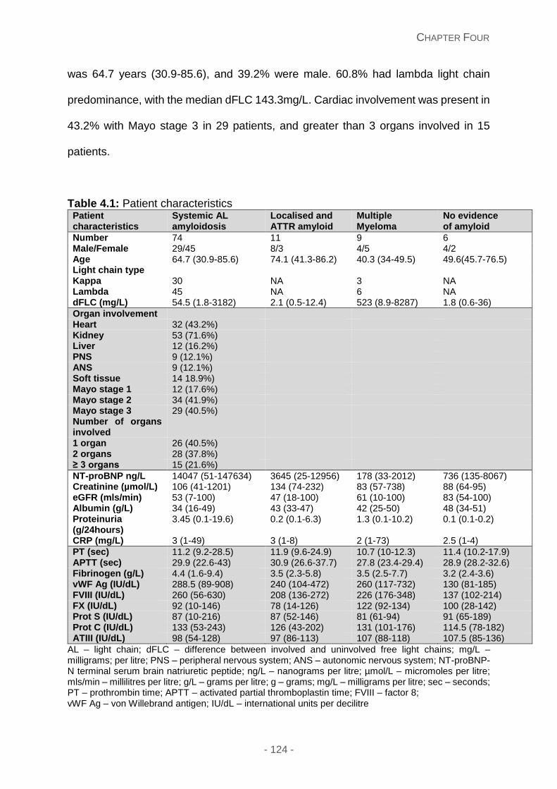
CHAPTER FOUR
- 124 -
was 64.7 years (30.9-85.6), and 39.2% were male. 60.8% had lambda light chain
predominance, with the median dFLC 143.3mg/L. Cardiac involvement was present in
43.2% with Mayo stage 3 in 29 patients, and greater than 3 organs involved in 15
patients.
Table 4.1: Patient characteristics Patient characteristics
Systemic AL amyloidosis
Localised and ATTR amyloid
Multiple Myeloma
No evidence of amyloid
Number 74 11 9 6 Male/Female 29/45 8/3 4/5 4/2 Age 64.7 (30.9-85.6) 74.1 (41.3-86.2) 40.3 (34-49.5) 49.6(45.7-76.5) Light chain type Kappa 30 NA 3 NA Lambda 45 NA 6 NA dFLC (mg/L) 54.5 (1.8-3182) 2.1 (0.5-12.4) 523 (8.9-8287) 1.8 (0.6-36)
Organ involvement Heart 32 (43.2%) Kidney 53 (71.6%) Liver 12 (16.2%) PNS 9 (12.1%) ANS 9 (12.1%) Soft tissue 14 18.9%) Mayo stage 1 12 (17.6%) Mayo stage 2 34 (41.9%) Mayo stage 3 29 (40.5%) Number of organs involved
1 organ 26 (40.5%) 2 organs 28 (37.8%) ≥ 3 organs 15 (21.6%)
NT-proBNP ng/L 14047 (51-147634) 3645 (25-12956) 178 (33-2012) 736 (135-8067) Creatinine (µmol/L) 106 (41-1201) 134 (74-232) 83 (57-738) 88 (64-95) eGFR (mls/min) 53 (7-100) 47 (18-100) 61 (10-100) 83 (54-100) Albumin (g/L) 34 (16-49) 43 (33-47) 42 (25-50) 48 (34-51) Proteinuria (g/24hours)
3.45 (0.1-19.6) 0.2 (0.1-6.3) 1.3 (0.1-10.2) 0.1 (0.1-0.2)
CRP (mg/L) 3 (1-49) 3 (1-8) 2 (1-73) 2.5 (1-4)
PT (sec) 11.2 (9.2-28.5) 11.9 (9.6-24.9) 10.7 (10-12.3) 11.4 (10.2-17.9) APTT (sec) 29.9 (22.6-43) 30.9 (26.6-37.7) 27.8 (23.4-29.4) 28.9 (28.2-32.6) Fibrinogen (g/L) 4.4 (1.6-9.4) 3.5 (2.3-5.8) 3.5 (2.5-7.7) 3.2 (2.4-3.6) vWF Ag (IU/dL) 288.5 (89-908) 240 (104-472) 260 (117-732) 130 (81-185) FVIII (IU/dL) 260 (56-630) 208 (136-272) 226 (176-348) 137 (102-214) FX (IU/dL) 92 (10-146) 78 (14-126) 122 (92-134) 100 (28-142) Prot S (IU/dL) 87 (10-216) 87 (52-146) 81 (61-94) 91 (65-189) Prot C (IU/dL) 133 (53-243) 126 (43-202) 131 (101-176) 114.5 (78-182) ATIII (IU/dL) 98 (54-128) 97 (86-113) 107 (88-118) 107.5 (85-136)
AL – light chain; dFLC – difference between involved and uninvolved free light chains; mg/L – milligrams; per litre; PNS – peripheral nervous system; ANS – autonomic nervous system; NT-proBNP- N terminal serum brain natriuretic peptide; ng/L – nanograms per litre; µmol/L – micromoles per litre; mls/min – millilitres per litre; g/L – grams per litre; g – grams; mg/L – milligrams per litre; sec – seconds; PT – prothrombin time; APTT – activated partial thromboplastin time; FVIII – factor 8; vWF Ag – von Willebrand antigen; IU/dL – international units per decilitre
CHAPTER FOUR
- 125 -
Clinical bleeding questionnaire
The clinical bleeding manifestations of all patients were assessed using the validated
bleeding questionnaire used in vWF type I disease. Amendments to this questionnaire
included denoting the duration of bleeding symptoms and the type of anticoagulation
if received. The bleeding questionnaire reported a bleeding score in 24/100 patients
(including 1 with multiple myeloma and one with localised gastrointestinal
amyloidosis), with a median bleeding score range 0-8 and median symptom duration
of 4 months (range 0.5-36). In comparison to other bleeding disorders, a bleeding
score greater than 4 was present in only 4 (5.4%) patients and bleeding score of 1
was present in 13 (17.6%) patients. The sites of bleeding included cutaneous (n=17),
oral (n=7), epistaxis (n=5), haemarthrosis (n=1), muscle haematoma (n=1) and
following surgery (n=2).
Coagulation abnormalities
Prolongation of the PT, APTT and TT occurred in 13 (17.6%), 13 (17.6%) and 35
(47.3%) patients respectively. Prolongation of both the PT and APTT occurred in 13
(17.6%) patients, of which 8 were on warfarin. Thirty one patients had a reptilase time
(RT) performed, and was prolonged in 25 (33.8%) patients. An elevated fibrinogen
was present in 42 (56.8%) patients.
Factor deficiencies
All patients had analysis of all factors including FII, FV, FVII, FVIII, FIX, FX, FXI, FXII
performed with a low FII in 5 patients, low FVII in 3 patients, low FVIII in 1 patient, low
FX in 8, low FIX in 3 patients and low FXII in 1 patient. The subnormal levels in FII (5),
FVII (2), FIX (3), and FX (6) corresponded with patients with concomitant warfarin use.
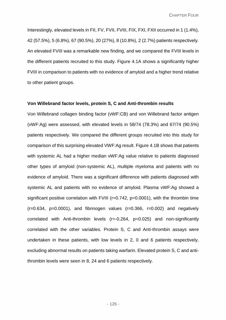
CHAPTER FOUR
- 126 -
Interestingly, elevated levels in FII, FV, FVII, FVIII, FIX, FXI, FXII occurred in 1 (1.4%),
42 (57.5%), 5 (6.8%), 67 (90.5%), 20 (27%), 8 (10.8%), 2 (2.7%) patients respectively.
An elevated FVIII was a remarkable new finding, and we compared the FVIII levels in
the different patients recruited to this study. Figure 4.1A shows a significantly higher
FVIII in comparison to patients with no evidence of amyloid and a higher trend relative
to other patient groups.
Von Willebrand factor levels, protein S, C and Anti-thrombin results
Von Willebrand collagen binding factor (vWF:CB) and von Willebrand factor antigen
(vWF:Ag) were assessed, with elevated levels in 58/74 (78.3%) and 67/74 (90.5%)
patients respectively. We compared the different groups recruited into this study for
comparison of this surprising elevated VWF:Ag result. Figure 4.1B shows that patients
with systemic AL had a higher median vWF:Ag value relative to patients diagnosed
other types of amyloid (non-systemic AL), multiple myeloma and patients with no
evidence of amyloid. There was a significant difference with patients diagnosed with
systemic AL and patients with no evidence of amyloid. Plasma vWF:Ag showed a
significant positive correlation with FVIII (r=0.742, p=0.0001), with the thrombin time
(r=0.634, p=0.0001), and fibrinogen values (r=0.366, r=0.002) and negatively
correlated with Anti-thrombin levels (r=-0.264, p=0.025) and non-significantly
correlated with the other variables. Protein S, C and Anti-thrombin assays were
undertaken in these patients, with low levels in 2, 0 and 6 patients respectively,
excluding abnormal results on patients taking warfarin. Elevated protein S, C and anti-
thrombin levels were seen in 8, 24 and 6 patients respectively.
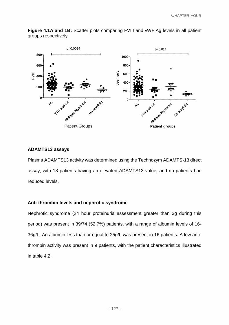
CHAPTER FOUR
- 127 -
Figure 4.1A and 1B: Scatter plots comparing FVIII and vWF:Ag levels in all patient groups respectively
ADAMTS13 assays
Plasma ADAMTS13 activity was determined using the Technozym ADAMTS-13 direct
assay, with 18 patients having an elevated ADAMTS13 value, and no patients had
reduced levels.
Anti-thrombin levels and nephrotic syndrome
Nephrotic syndrome (24 hour proteinuria assessment greater than 3g during this
period) was present in 39/74 (52.7%) patients, with a range of albumin levels of 16-
36g/L. An albumin less than or equal to 25g/L was present in 16 patients. A low anti-
thrombin activity was present in 9 patients, with the patient characteristics illustrated
in table 4.2.
p=0.0034
AL
TTR a
nd LA
Multi
ple M
yelo
ma
No a
myl
oid
0
200
400
600
800
Patient Groups
FV
III
AL
TTR a
nd LA
Multi
ple M
yelo
ma
No a
myl
oid
0
200
400
600
800
1000
p=0.014
Patient groups
VW
F:A
G
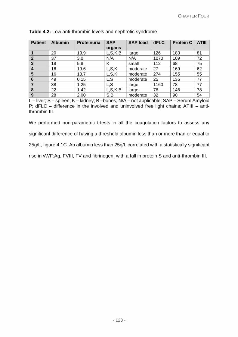
CHAPTER FOUR
- 128 -
Table 4.2: Low anti-thrombin levels and nephrotic syndrome
Patient Albumin Proteinuria SAP organs
SAP load dFLC Protein C ATIII
1 20 13.9 L,S,K,B large 126 183 81
2 37 3.0 N/A N/A 1070 109 72
3 18 5.8 K small 112 68 75
4 16 19.6 L,S,K moderate 27 169 62
5 16 13.7 L,S,K moderate 274 155 55
6 49 0.15 L,S moderate 25 136 77
7 38 1.25 L,S large 1160 78 77
8 22 1.42 L,S,K,B large 76 146 78
9 28 2.00 S,B moderate 32 90 54
L – liver; S – spleen; K – kidney; B –bones; N/A – not applicable; SAP – Serum Amyloid P; dFLC – difference in the involved and uninvolved free light chains; ATIII – anti-thrombin III. We performed non-parametric t-tests in all the coagulation factors to assess any
significant difference of having a threshold albumin less than or more than or equal to
25g/L, figure 4.1C. An albumin less than 25g/L correlated with a statistically significant
rise in vWF:Ag, FVIII, FV and fibrinogen, with a fall in protein S and anti-thrombin III.
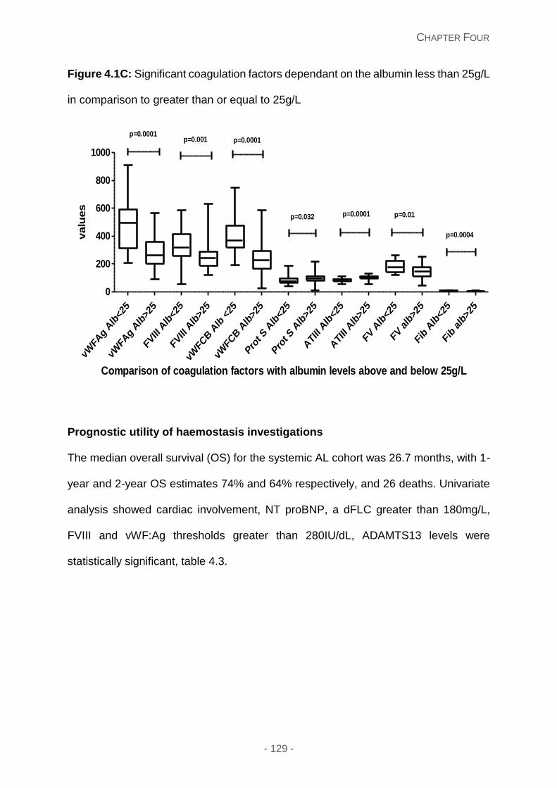
CHAPTER FOUR
- 129 -
Figure 4.1C: Significant coagulation factors dependant on the albumin less than 25g/L
in comparison to greater than or equal to 25g/L
Prognostic utility of haemostasis investigations
The median overall survival (OS) for the systemic AL cohort was 26.7 months, with 1-
year and 2-year OS estimates 74% and 64% respectively, and 26 deaths. Univariate
analysis showed cardiac involvement, NT proBNP, a dFLC greater than 180mg/L,
FVIII and vWF:Ag thresholds greater than 280IU/dL, ADAMTS13 levels were
statistically significant, table 4.3.
vWFA
g Alb
<25
vWFA
g Alb
>25
FVIII A
lb<2
5
FVIII A
lb>2
5
vWFC
B A
lb <
25
vWFC
B A
lb>2
5
Pro
t S A
lb<2
5
Pro
t S A
lb>2
5
ATI
II Alb
<25
ATI
II Alb
>25
FV A
lb<2
5
FV alb
>25
Fib
Alb
<25
Fib
alb>
25
0
200
400
600
800
1000
p=0.0001p=0.032
p=0.0001p=0.001p=0.0001
p=0.0004
p=0.01
Comparison of coagulation factors with albumin levels above and below 25g/L
valu
es
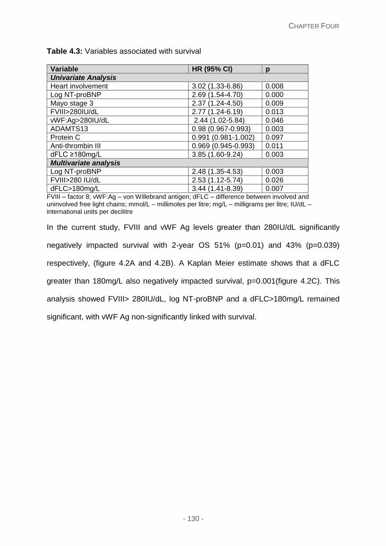
CHAPTER FOUR
- 130 -
Table 4.3: Variables associated with survival
Variable HR (95% CI) p
Univariate Analysis
Heart involvement 3.02 (1.33-6.86) 0.008
Log NT-proBNP 2.69 (1.54-4.70) 0.000
Mayo stage 3 2.37 (1.24-4.50) 0.009
FVIII>280IU/dL 2.77 (1.24-6.19) 0.013
vWF:Ag>280IU/dL 2.44 (1.02-5.84) 0.046
ADAMTS13 0.98 (0.967-0.993) 0.003
Protein C 0.991 (0.981-1.002) 0.097
Anti-thrombin III 0.969 (0.945-0.993) 0.011
dFLC ≥180mg/L 3.85 (1.60-9.24) 0.003
Multivariate analysis
Log NT-proBNP 2.48 (1.35-4.53) 0.003
FVIII>280 IU/dL 2.53 (1.12-5.74) 0.026
dFLC>180mg/L 3.44 (1.41-8.39) 0.007 FVIII – factor 8; vWF:Ag – von Willebrand antigen; dFLC – difference between involved and uninvolved free light chains; mmol/L – millimoles per litre; mg/L – milligrams per litre; IU/dL – international units per decilitre
In the current study, FVIII and vWF Ag levels greater than 280IU/dL significantly
negatively impacted survival with 2-year OS 51% (p=0.01) and 43% (p=0.039)
respectively, (figure 4.2A and 4.2B). A Kaplan Meier estimate shows that a dFLC
greater than 180mg/L also negatively impacted survival, p=0.001(figure 4.2C). This
analysis showed FVIII> 280IU/dL, log NT-proBNP and a dFLC>180mg/L remained
significant, with vWF Ag non-significantly linked with survival.
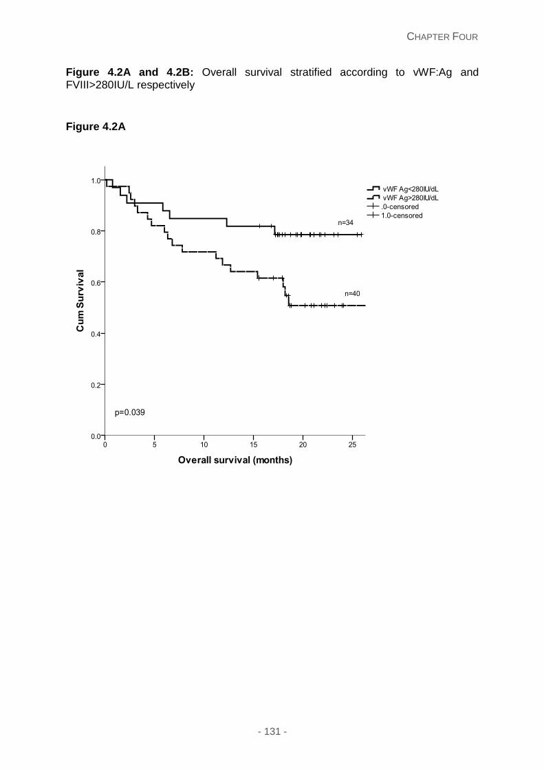
CHAPTER FOUR
- 131 -
Figure 4.2A and 4.2B: Overall survival stratified according to vWF:Ag and FVIII>280IU/L respectively
Figure 4.2A
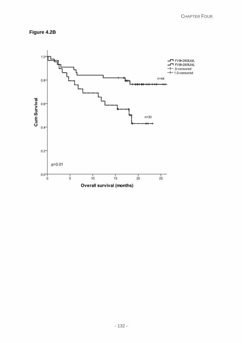
CHAPTER FOUR
- 132 -
Figure 4.2B
CHAPTER FOUR
- 133 -
Figure 4.2C: Overall survival stratified according to a dFLC >180mg/L
Bleeding symptoms and association with other variables
We examined the association of bleeding symptoms by amyloid load by 123I SAP
scintigraphy. Analysis by scatter plots showed that those patients with bleeding
symptoms had a non-significant higher median amyloid load by 123I SAP scintigraphy
in comparison to those not describing any bleeding symptoms, figure 4.3.
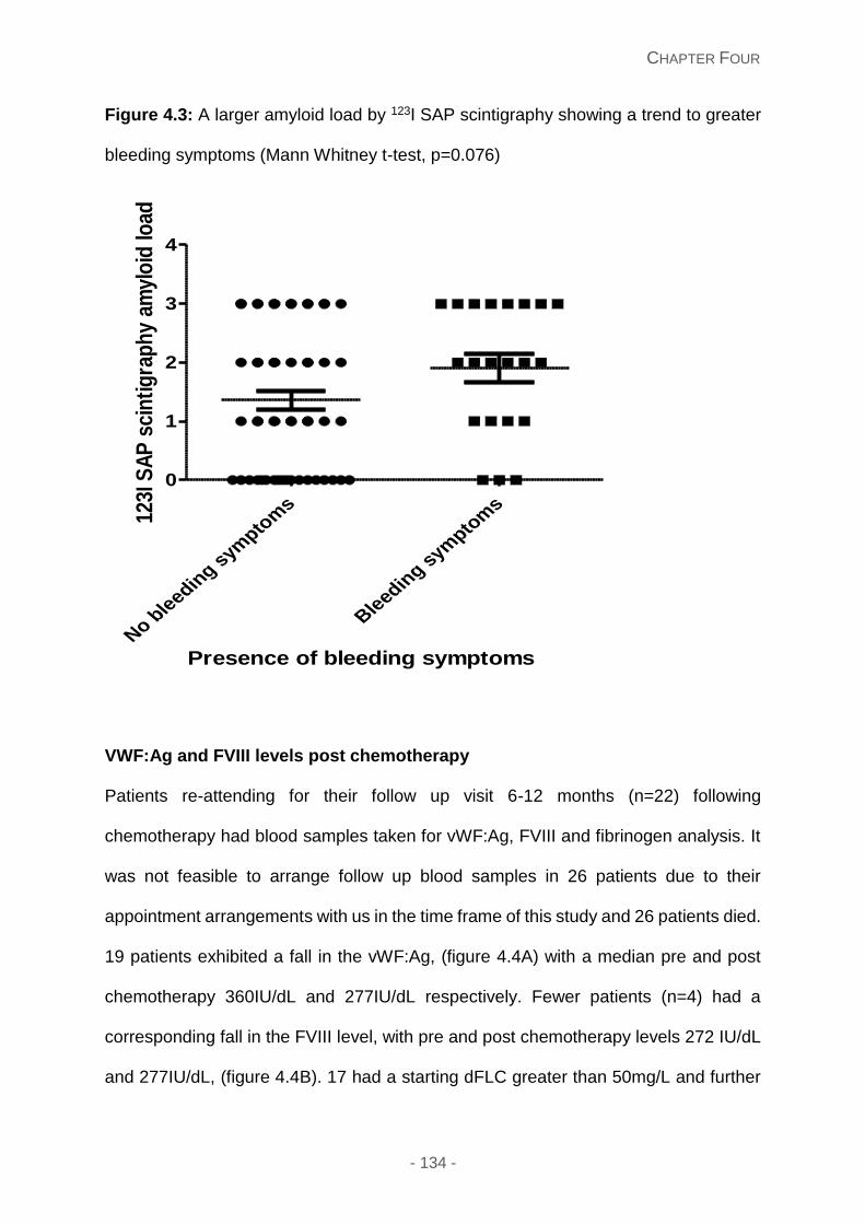
CHAPTER FOUR
- 134 -
Figure 4.3: A larger amyloid load by 123I SAP scintigraphy showing a trend to greater
bleeding symptoms (Mann Whitney t-test, p=0.076)
VWF:Ag and FVIII levels post chemotherapy
Patients re-attending for their follow up visit 6-12 months (n=22) following
chemotherapy had blood samples taken for vWF:Ag, FVIII and fibrinogen analysis. It
was not feasible to arrange follow up blood samples in 26 patients due to their
appointment arrangements with us in the time frame of this study and 26 patients died.
19 patients exhibited a fall in the vWF:Ag, (figure 4.4A) with a median pre and post
chemotherapy 360IU/dL and 277IU/dL respectively. Fewer patients (n=4) had a
corresponding fall in the FVIII level, with pre and post chemotherapy levels 272 IU/dL
and 277IU/dL, (figure 4.4B). 17 had a starting dFLC greater than 50mg/L and further
No ble
eding s
ympto
ms
Bleedin
g sym
ptom
s
0
1
2
3
4
Presence of bleeding symptoms
123I
SA
P s
cin
tig
rap
hy
amyl
oid
load
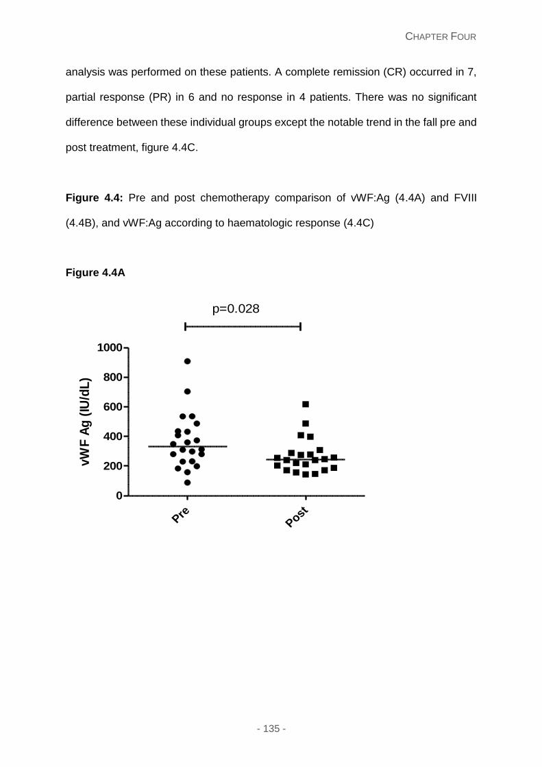
CHAPTER FOUR
- 135 -
analysis was performed on these patients. A complete remission (CR) occurred in 7,
partial response (PR) in 6 and no response in 4 patients. There was no significant
difference between these individual groups except the notable trend in the fall pre and
post treatment, figure 4.4C.
Figure 4.4: Pre and post chemotherapy comparison of vWF:Ag (4.4A) and FVIII
(4.4B), and vWF:Ag according to haematologic response (4.4C)
Figure 4.4A
PrePost
0
200
400
600
800
1000
p=0.028
vW
F A
g (
IU/d
L)
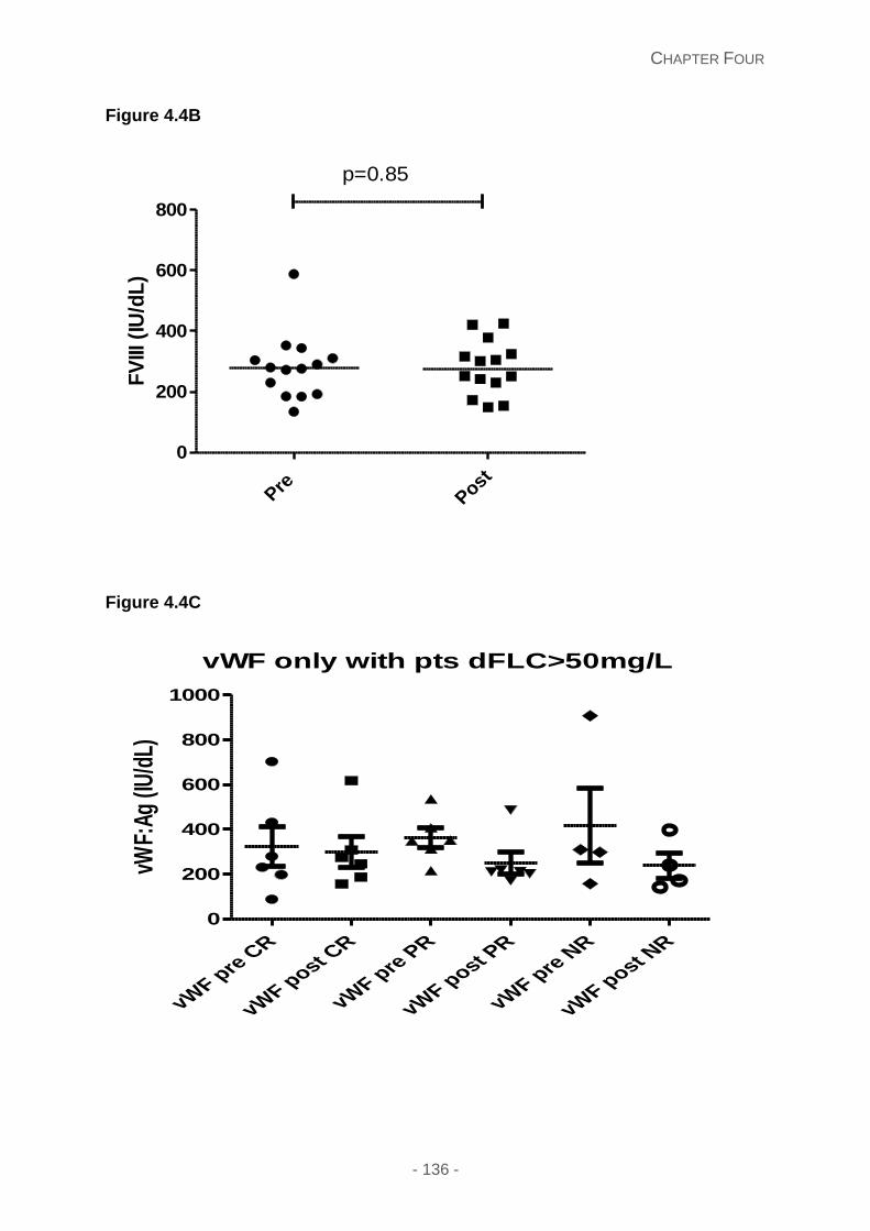
CHAPTER FOUR
- 136 -
Figure 4.4B
Figure 4.4C
Pre
Post
0
200
400
600
800
p=0.85
FV
III (
IU/d
L)
vWF only with pts dFLC>50mg/L
vWF p
re C
R
vWF p
ost CR
vWF p
re P
R
vWF p
ost PR
vWF p
re N
R
vWF p
ost NR
0
200
400
600
800
1000
vWF:
Ag (I
U/d
L)
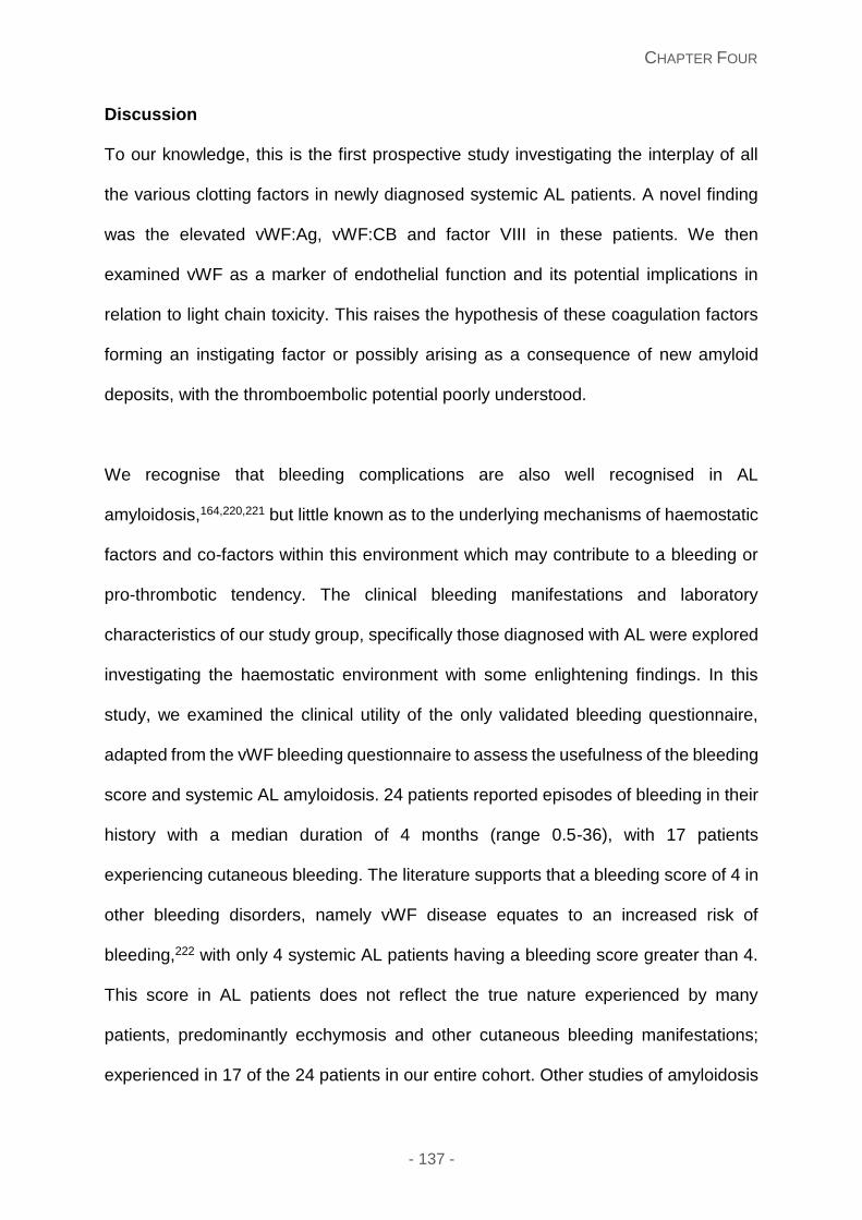
CHAPTER FOUR
- 137 -
Discussion
To our knowledge, this is the first prospective study investigating the interplay of all
the various clotting factors in newly diagnosed systemic AL patients. A novel finding
was the elevated vWF:Ag, vWF:CB and factor VIII in these patients. We then
examined vWF as a marker of endothelial function and its potential implications in
relation to light chain toxicity. This raises the hypothesis of these coagulation factors
forming an instigating factor or possibly arising as a consequence of new amyloid
deposits, with the thromboembolic potential poorly understood.
We recognise that bleeding complications are also well recognised in AL
amyloidosis,164,220,221 but little known as to the underlying mechanisms of haemostatic
factors and co-factors within this environment which may contribute to a bleeding or
pro-thrombotic tendency. The clinical bleeding manifestations and laboratory
characteristics of our study group, specifically those diagnosed with AL were explored
investigating the haemostatic environment with some enlightening findings. In this
study, we examined the clinical utility of the only validated bleeding questionnaire,
adapted from the vWF bleeding questionnaire to assess the usefulness of the bleeding
score and systemic AL amyloidosis. 24 patients reported episodes of bleeding in their
history with a median duration of 4 months (range 0.5-36), with 17 patients
experiencing cutaneous bleeding. The literature supports that a bleeding score of 4 in
other bleeding disorders, namely vWF disease equates to an increased risk of
bleeding,222 with only 4 systemic AL patients having a bleeding score greater than 4.
This score in AL patients does not reflect the true nature experienced by many
patients, predominantly ecchymosis and other cutaneous bleeding manifestations;
experienced in 17 of the 24 patients in our entire cohort. Other studies of amyloidosis
CHAPTER FOUR
- 138 -
patients have described haemostatic abnormalities with surprisingly fewer bleeding
complications, which may be explained by the laboratory investigations.223
The fibrinolytic pathway is often affected with prolongation of the PT, APTT, TT and
high levels of fibrinogen. In our study, prolongation of the PT, APTT and TT was a
predominant coagulation abnormality, concurring with other studies.164, 224 Previous
studies in Myeloma have shown high levels of immunoglobulins having a propensity
to increase the blood viscosity and interfering with the fibrin polymerisation225 Fibrin
formation is impaired, consequently interfering with the binding of plasmin and FXIII,
ultimately causing ineffectual clot retraction226 and abnormal clot clumps, with the
possible propensity to occlude smaller vessels. In our AL cohort, the TT was prolonged
in 35 patients, yet the majority of patients did not describe a bleeding tendency,
suggesting other haemostatic defects are needed to result in increased bleeding
manifestations such as vessel wall amyloid deposition and vessel fragility,227
enhanced fibrinolysis or down regulation or consumption of certain co-factors.
Within our study group, 2 of 74 (1%) of patients had a Factor X deficiency, compared
to previous studies of 7% 217 or 14%,164 with the variation clearly dependent upon
patient characteristics. Baseline FX levels are not predictive of bleeding risk.228
Previous studies show that rapid clearance of 131I-labelled FX from the circulation to
areas of amyloid deposition particularly within the spleen216, 229 suggests adsorption
on amyloid fibrils as a major cause FX deficiency in AL. Other suggested explanations
include synthetic dysfunction due to liver involvement or vitamin K deficiency and
discordance between FX activity and FX antigen.164 These 2 patients with factor X
deficiency exhibited prolongation of the PT with a normal APTT, with no bleeding
CHAPTER FOUR
- 139 -
complications. Further factor analysis was undertaken to assess any other associated
factor deficiencies encountered with this disease. Our results reflected a subnormal
FVIII in 1 patient and subnormal FXII in another patient, with prolongation of the APTT
in the former and no bleeding complications in either patient. The striking feature when
analysing the factor levels, was the extent of elevated values for all the factors,
predominantly for factor V, VIII and IX. Little data exists as to bleeding or thrombotic
complications related to FV, with only one patient having an activated protein C
resistance. Interestingly, FVIII results were elevated in 91%. High factor VIII levels are
thought to be an independent risk factor for venous thrombosis, with levels greater
than 150% associated with a five-fold increase risk of venous thrombosis.230 Heikal et
al explored elevated factor IX levels retrospectively in 81 patients, showing an
association with arterial and venous thrombosis231 The consequence of these elevated
factors remains undetermined but may provide a more prothrombotic environment.
The FVIII/vWF glycoprotein complex may serve as important platform from which
endothelial dysfunction can be assessed in this disease. Clearly intertwined in
equilibrium, vWF is a vital partner for FVIII’s function, stability, structure and
immunogenicity.214 Electrostatic interactions have been proposed as one driving force
for the FVIII-vWF complex,232 with Dimitrov et al showing binding of FVIII to vWF
demonstrated a high sensitivity to acidic pH and sensitivity to ionic strength.233 One
study showed that 50% high FVIII levels were associated with high vWF Ag levels,
implying that there are other factors which affect this interaction. Other studies have
shown that factors including BMI, diabetes mellitus, insulin, fibrinogen and triglycerides
are associated with elevated FVIII levels.234 Typically a rise in FVIII will lead to a
CHAPTER FOUR
- 140 -
concordant rise in vWF:Ag levels. Our study showed a concordant rise in both vWF
and FVIII levels.
In the present study, elevated plasma vWF was shown to be associated with patients
diagnosed with systemic AL amyloidosis, with 90.5% of patients having elevated vWF
activity, supporting previous studies examining vWF235 Kastritis et al, examined 81
patients with systemic AL with a serum vWF level of 181 IU/dL (range 20-557) with
vWF levels greater than 230 IU/dL were associated with a poor survival (median 4
months versus 47 months, p=0.001). In our study, the vWF activity was significantly
higher in AL patients in comparison with patients with no amyloid, p=0.014 in
comparison with other patient cohort groups. We found a significant survival
disadvantage with a factor VIII level and vWF antigen level greater than 280IU/dL in
comparison to the Greek group,235 although FVIII levels were not explored by the latter
group. The differences in the survival differences between the Greek group and our
study most likely reflect the patient cohorts recruited. In comparing all the patient
groups in our study, patients with systemic AL and Multiple myeloma (with the latter
not significantly different from controls) had higher median values of FVIII and vWF:Ag,
suggesting the underling plasma cell or light chains as the probable instigating factor.
Von Willebrand factor is a large multimeric glycoprotein produced in endothelial cells
and megakaryocytes playing an important role in haemostasis by promoting platelet
adhesion. A number of clinical and experimental studies have shown that elevated
vWF levels reflect endothelial damage and dysfunction.209 Previous studies have
proposed vWF as a marker of endothelial damage or dysfunction; which is secondary
to a pro-inflammatory and pro-coagulant state. The increased vWF:Ag and vWF:CB
CHAPTER FOUR
- 141 -
are likely to result in increased platelet plug formation. In systemic AL amyloidosis,
light chains misfold and aggregate as amyloid fibrils in vital organs. In vitro fibril studies
show that there are various factors which may play a part in the kinetics and
morphology of the fibrils: thermodynamic stability, low pH and oxidative stress.236-238
Earlier studies have shown that amyloid deposits occur earlier within the blood
vessels, with these patients presenting with early microcirculatory dysfunction.202 Thus
systemic AL patients with higher levels of vWF signify a unique marker for endothelial
dysfunction and may lead to a higher risk of thromboembolic disease and
cardiovascular disease.
Typically, large VWF multimers are produced in and released from vascular
endothelial cells, predominantly biologically active whilst interacting with platelets
under a high shear stress. The large multimers are cleaved and degraded into smaller
fragments by A Disintegrin And Metalloprotease with ThromboSpondin 1 repeats
(ADAMTS13); an enzyme produced by the liver. To investigate the significantly high
levels of vWF:Ag and vWF:CB levels, we examined the plasma ADAMTS13 activity in
these patients. The most remarkable observation of our study was the higher plasma
vWF Ag levels and normal and elevated levels of ADAMTS13 activity, with the
biological significance of this uncertain. Low levels of ADAMTS13 activity may be
secondary to high levels of substrate or sequestration of this protease to the vascular
endothelial cells. In AL patients, the vWF cleaving protease activity was normal and
elevated in 18 patients, likely to represent the increased levels of vWF:Ag and hence
increased need to ensure these multimers are cleaved.
CHAPTER FOUR
- 142 -
Proteins C and S are two vitamin K-dependent plasma proteins acting as natural
anticoagulants. Activated protein C acts as a proteolytic component of the complex,
whilst protein S is used as an activated protein C binding protein; both needed for
assembly of the anticoagulant complex on cell surfaces. This is primarily expressed
through the selective inactivation of Factors Va and VIIIa. Many patients deficient in
proteins C and S have been described with an associated thrombotic tendency. There
are various mechanisms and/or drugs which can lead to acquired deficiencies of these
proteins including oral anticoagulation, liver disease, DIC and in the case of protein S,
nephrotic syndrome, lupus erythematosus and certain hormones. Protein S plays an
important role as a cofactor in the inactivation of FVa and FVIIIa.
Protein C deficiency is known to be associated with thromboembolic phenomenon,
with little data as to increased protein C levels. In this study, 24 patients (32.4%) had
high levels of protein C with only 1 patient with activated protein C resistance. This
highlights the complex interplay of the various coagulation factors and bystanders that
contribute to proteinuria, not solely a low anti-thrombin level.
In our study nephrotic syndrome was present in 53% of the AL cohort, with
prolongation in the thrombin time and reptilase time in all these patients, suggesting a
close link between prolongation of the TT and RT and proteinuria, typically secondary
to the abnormal fibrin polymerisation that occurs in both cases.239, 240 Low anti-
thrombin levels in nephrotic syndrome have been described in many studies,241-243
likely secondary to urinary loss and increased consumption. Anti-thrombin acts as an
important inhibitor of serine protease clotting factors, with heparin and
Glycosaminoglycans (GAGS) accentuating its inhibitory effect; which bind to amyloid
CHAPTER FOUR
- 143 -
fibrils.244 Gamba et al described increased plasma values of thrombin-antithombin
(TAT) complexes and enhanced thrombin activation present, chronic consumption of
AT, accounting for differences in AT activity and AT:Ag levels. Thrombin generation
may be additionally enhanced by delayed fibrin formation.243 Of the nephrotic patients,
only 7 had reduced anti-thrombin values, with 5 patients having corresponding albumin
levels less than or equal to 25g/L. Interestingly the other 2 patients with albumin levels
of 28 and 38g/L had raised protein C levels and a low quantitative level of proteinuria.
There is very likely a change in the haemostatic factors including protein C which occur
prior to lower albumin levels and hence more extensive proteinuria.
Amyloid deposits in the kidneys typically occur within the glomerulus, and
consequently results in pathological damage, with proteinuria often seen which may
be nephrotic or sub-nephrotic. The renal tubules function to reabsorb the plasma
proteins (specifically in the proximal convoluted tubule) which pass through the
glomerular filtration barrier, which provides a charge and size selective barrier to
albumin. Podocytes are terminally differentiated glomerular epithelial cells, with
endothelium activation and loss of selectivity resulting in prolonged exposure of the
podocytes to these proteins.245 This overall damage leads to reduced vascular
endothelial growth factors (VEGF).246 Previous studies have explored the risk of
thromboembolic disease in nephrotic syndrome.247-249 In our study, comparison of
patients with an albumin less than 25g/L and those with an albumin greater than or
equal to 25g/L for the different coagulation factors showed definite differences. The
former group showed differences to the latter, with significantly higher vWF:Ag, FVIII,
FV, and fibrinogen levels and lower protein S and ATIII levels. This is the first study to
compare all coagulation factors to assess the possible aetiology of thrombotic
CHAPTER FOUR
- 144 -
phenomena or bleeding risks in patients diagnosed with systemic AL. Understanding
of the coagulation cascade has shown that elevated levels of FVIII and fibrinogen,
lower levels of protein S and anti-thrombin contribute to a prothrombotic tendency. The
latter findings were found in patients with an albumin less than 25g/L. Although these
factors work as part of a cog wheel together, this study highlights the potent risk of
prothrombotic tendency in patients with an albumin less than 25g/L, and hence the
awareness and need for prophylaxis or full anticoagulation consideration.
There is limited data as to managing patients given the paradox of bleeding and
thrombotic risks. A common finding in our study and previous studies164, 224, 250 was
the prolongation of TT and RT. Typically, hyperfibrinolysis may be related to lower
levels of α2-antiplasmin or increased urokinase type plasminogen activity. As such, ε-
aminocaproic acid are effective in controlling some bleeding complications.251 Until
recently, Factor X deficiency treatment deficiency therapeutic options included fresh
frozen plasma (FFP), prothrombin complex concentrates (PCCs), activated
prothrombin complex concentrates (aPCC) or recombinant factor VIIa (rFVIIa)252 –
each with significant risks in this fragile patient population. A larger series reported
44% of patients had complications when treated with rFVIIa preoperatively including
bleeding, thrombosis, or death.228. In addition to FFP, PCC, aPCC or rFVIIa,
tranexamic acid, plasma exchange253 and splenectomy217 were anecdotally reported
as treatments for FX deficiency in AL; the latter targeted at removing the splenic
amyloid fibril burden. As a result of a variable amount of FX in FFP, large volumes
are needed to achieve a haemostatic effect with the true clinical risk of fluid overload
in patients with cardiac involvement, especially those with advanced cardiac
involvement or elderly population. A serious concern arises regarding thrombotic risks
CHAPTER FOUR
- 145 -
of FVIIa or aPCC in patients with AL amyloidosis who are elderly with cardiovascular
risk factors and often nephrotic due to renal involvement (hence inherently
prothrombotic). BPL (Bio Products Laboratory Ltd, Elstree, UK) have also developed
a high purity plasma derived factor X concentrate and currently undergoing phase III
clinical trials used in patients with hereditary factor X deficiency. In one analysis, one
patient with inherited Factor X deficiency presenting with a shoulder haemarthrosis,
achieved good haemostatic response following FX dose of 25IU/kg daily254 with a
biological half-life 24 to 48 hours.
Our Centre also reported an initial report of using high purity factor X (HP-FX)
concentrate in two patients with systemic AL amyloidosis and acquired FX
deficiency.255 This involved 2 patients with biopsy proven AL amyloidosis, with
evidence of liver involvement and a large amyloid load by 123I serum amyloid P
component scintigraphy, one patient presenting with a forearm haematoma and the
second patient presenting with a ruptured spleen and knee haemarthrosis due to
acquired FX deficiency (both with FX levels 8 IU/dL). HP-FX concentrate was used to
treat each bleeding episode in each case given as a single dose of 40IU/kg (with the
FX activity assessed by one stage clotting prothrombin time (PT) based assays) and
haemostasis achieved in conjunction with supportive measures. There was an obvious
difference between the FX recovery times between patients with inherited FX
deficiency in comparison with our AL patients; the latter showed less predictable
kinetics, and a more rapid decline to baseline in 2-4 hours, consistent with the theory
of FX adsorption on amyloid fibrils. Each patient with AL amyloidosis has a different
amyloid load and a unique fibril sequence with a potentially different avidity for binding
FX – both likely to lead to large differences in the half life and dose needed as in the
second patient with a larger load and earlier decrease in FX levels. We tried to
CHAPTER FOUR
- 146 -
ascertain whether there was a correlation between bleeding symptoms and amyloid
load by 123I SAP scintigraphy, not showing a statistically significant difference.255 High
purity factor X concentrate is useful to treat bleeding due to acquired factor X
deficiency in systemic AL amyloidosis. Higher and/or more frequent dosing is likely to
be required to achieve adequate FX levels for haemostasis with frequent monitoring
of FX levels given the unpredictable kinetics; target FX thresholds similar to patients
with inherited FX deficiency: doses of 10-15 IU/dl for minor bleeding and >50 IU/dl for
major bleeding, trauma or surgery. Thus HP-FX has the advantage that the
haemostatic response can be monitored and treatment tailored to the patient individual
needs.255
Light chain toxicity may also serve as a further insult in this process of AL deposits.
Various experiments have attempted to explore the underlying mechanisms of light
chain toxicity, with no correlative factor for endothelial dysfunction. Previous studies
have proposed vWF as a marker of endothelial damage or dysfunction; which is
secondary to a pro-inflammatory and pro-coagulant state. The increased vWF:Ag and
vWF:CB are likely to result in increased platelet plug formation. Bauer et al described
the concept of vWF fibres laid down within the lumen of the cancer associated vessel
wall, with tumour derived VEGF inducing angiogenesis, thus affecting the
pathophysiology and activation of the endothelium.256 In systemic AL amyloidosis, light
chains misfold and aggregate as amyloid fibrils in vital organs. In vitro fibril studies
show that there are various factors which may play a part in the kinetics and
morphology of the fibrils: thermodynamic stability, low pH and oxidative stress.236-238
Earlier studies have shown that amyloid deposits occur earlier within the blood
vessels, with these patients presenting with early microcirculatory dysfunction. 202 As
such, it seems feasible that systemic AL patients have higher levels of vWF signifying
CHAPTER FOUR
- 147 -
a unique marker for endothelial dysfunction and may lead to a higher risk of
thromboembolic disease and cardiovascular disease. In comparing all the patient
groups in our study, patients with systemic AL and Multiple myeloma (with the latter
not significantly different from controls) had higher median values of FVIII and vWF:Ag,
suggesting the underling plasma cell or light chains as the probable instigating factor.
The underlying mechanism of light chain toxicity remains unknown, with some reports
showing that the prefibrillar light chain proteins, consequent endothelial dysfunction
and ischaemic vascular injury occurs prior to amyloid deposition. 200, 202 Exposure to
light chains and oxidative stress and microvascular dysfunction has been proposed in
the AL pathophysiology.199 Recently further exploration to examine the dysregulation
of an autophagy flux in the setting of proteotoxicity of the light chains has shown to be
important in those with cardiac AL. Guan et al found that inhibition of the autophagy
flux, specifically lysosomal dysfunction is important in the AL-light chain cardiotoxicity
and hence development of AL cardiomyopathy.257 In vitro experiments showed that
lysosomal dysfunction is an early point in the cascade of events that occur in cardiac
AL, specifically the light chain toxicity, followed by mitochondrial dysfunction, ROS
dysfunction and consequent cell death and dysfunction. This also highlights the
potential for a possible autophagy related targets, replicated in vivo halting cardiac AL
mortality using rapamycin and transient overexpression of transcription factor EB
(TFEB).257
This resultant rise in vWF and FVIII are likely to create a consequential pro-coagulant
and pro-inflammatory environment. Several studies have attempted to explore
whether FVIII or vWF are the causative stimulant in arterial or venous thrombogenesis.
CHAPTER FOUR
- 148 -
The ARIC study examined strong associations of FVIII and vWF as risk factors for
hypertension, diabetes mellitus, body mass index or triglycerides. Some of the latter
variables are associated with endothelial and vascular inflammation.211, 258 The
consequent high shear forces especially within narrowed vessels lead to increased
vWF secretion by the vascular endothelium, which stimulates platelet adhesion and
aggregation at the damaged arterial walls, with the high risk leading to thrombus
formation.259
Previous studies have shown that high levels of factor FVIII over time are present in
patients with thrombosis260, and not reflective of an acute phase reaction261 We
compared the C-reactive proteins (CRP) in each group to ascertain whether the
elevated vWF Ag and FVIII were secondary to an inflammatory or infective pathology;
with the former showing relatively low levels except for occasional patients with
infection (table 4.1). The latter finding negates underlying inflammation as a trigger for
the vWF rise.
In an attempt to explore the relationship between FVIII and vWF Ag with light chains,
we repeated blood samples for these coagulation factors post chemotherapy. This has
not been explored before, with the results showing that 86% (19/22 patients) with
these investigations undertaken exhibited a fall in the vWF:Ag level following
chemotherapy, suggesting a likely correlative relationship. Interestingly the FVIII levels
did not fall in the majority of patients. The type of haematological response did not
correlate with the level of fall in the vWF:Ag implying there is a much more complex
interplay of the light chains, the vascular endothelium and formation of amyloid
deposits. It is difficult to interpret this, with small numbers and no experimental or
CHAPTER FOUR
- 149 -
electron-microscopy to aid with understanding. One possible hypothesis would
suggest that treatment with chemotherapy in an attempt to reduce the light chains,
also serves to improve the function of the vascular endothelium, but may still provide
a thrombotic environment.
Our study highlights the prothrombotic potential in patients with newly diagnosed
systemic AL, irrespective of the presence of nephrotic syndrome or type of
chemotherapy regimen used. This raises the question as to what the optimum
treatment of thrombotic risk should involve. Ordinarily thromboprophylaxis would
suffice, but given the additional elevated risk factors of vWF:Ag, FVIII, fibrinogen and
lower anti-thrombin and protein S levels, further analysis is needed to explore whether
full anticoagulation is necessary.
In summary, this is the largest series prospectively examining all the underlying
coagulation factors, protein C and S, anti-thrombin in newly diagnosed AL amyloidosis.
Little is known in the setting of AL amyloidosis with the paradox of a bleeding diathesis
and thromboembolic disease. Haemostatic dysfunction likely results from different
interacting pathogenic pathways and coagulation factors. From our data, we showed
that prolongation of the thrombin time was a common finding and low levels of anti-
thrombin activity and proteinuria were associated with albumin levels less than 25g/L.
In addition these patients had elevated pro-thrombotic factors and reduced
anticoagulant factors which will contribute to the pro-thrombotic status of these
patients. An elevated FVIII and vWF:Ag level greater than 280IU/L carried a significant
survival disadvantage. Thromboprophylaxis or treatment anticoagulation is an
important consideration in all these patients not having bleeding complications.
CHAPTER FOUR
- 150 -
Our study points to underlying vascular endothelial damage that occurs in systemic
AL, and possibly contributing to the light chain toxicity environment. The correlative
fall in the vWF levels post chemotherapy may reflect the underlying vascular
endothelial changes which occur. Further studies to better understand the
pathophysiology of these findings are needed to explore the endothelium by electron
microscopy, and hence the utility of the prognostic findings as part of patient risk
stratification.
.
CHAPTER FIVE
- 151 -
Chapter Five: Frequent occurrence of recurrent nocturnal
desaturations in systemic AL amyloidosis
This chapter is written in the context of my publication: High prevalence of
recurrent nocturnal desaturations in systemic AL Amyloidosis: a cross-
sectional study. S Mahmood, M Sovani, P Smith, L George, C Quarta, S
Sachchithanantham, M Fontana, CJ Whelan, HJ Lachmann, JD Gillmore, PN
Hawkins, AD Wechalekar. Sleep Medicine. Accepted for publication and available
online December 21st 2016. (Original article). Copyright permission obtained from
Elsevier publishers, license no.: 4025341342092 for use in my thesis.
Introduction
Systemic AL amyloidosis is a rare disorder caused by deposition of misfolded
immunoglobulin light chains in organs or tissues. Outcomes of patients with
amyloidosis depend predominantly on the extent of cardiac disease, although
involvement of other organs and, particularly, soft tissues of the oropharynx contribute
to significant symptoms and morbidity. It is well recognised that early and unexpected
cardiac deaths account for 20-40% of all deaths within a few months of diagnosis in
AL amyloidosis. The exact terminal event and its triggers remain unclear.
Bradyarrhythmias262 or other arrhythmias may be the cause. Central sleep apnoea
(CSA), characterised by the faulty respiratory drive during sleep, is a well-recognised
complication of heart failure and can lead to recurrent episodes of nocturnal
hypoxemia worsening symptomatic heart failure,263 causing increased morbidity and
mortality. Obstructive sleep apnoea (OSA) is now a well-recognised cause of acute
and chronic adverse cardiovascular effects. In addition to cardiovascular effects, both
CHAPTER FIVE
- 152 -
CSA and OSA cause marked day time fatigue and/or sleepiness contributing to
morbidity.
Systemic AL amyloidosis is one of the few disorders which cause acquired progressive
heart failure and can also be associated with marked infiltration of the oropharyngeal
soft tissues – both potential risk factors for CSA or OSA, respectively, or CSA and
OSA together in patients with soft tissue and cardiac disease. The recurrent hypoxic
episodes could have a profound effect on the cardiac function in patients with
advanced amyloidosis.
We report here the results of a pilot study of overnight continuous pulse oximetry in
patients with systemic light chain (AL) amyloidosis, based on the hypothesis that
recurrent nocturnal hypoxaemia (likely due to sleep disordered breathing OSA and/or
CSA) could occur in patients with amyloidosis reporting a high incidence of recurrent
nocturnal oxygen desaturations and raise a question whether these desaturations may
be the trigger for sudden cardiac mortality.
Methods
Study population
This study included consecutive patients seen at the UK National Amyloidosis Centre
with amyloidosis between July 2013 and June 2014 who underwent overnight pulse
oximetry, using the Minolta 300I pulse oximeter. The main inclusion criteria were: the
presence of clinical macroglossia and/or cardiac amyloidosis. Written consent for
retrospective publication of data was obtained from all patients in accordance with the
Declaration of Helsinki. Amyloid deposition was confirmed on a tissue biopsy by the
presence demonstration of Congo red positivity under cross polarise light and fibril
CHAPTER FIVE
- 153 -
typing was done by immunohistochemistry or mass spectrometry. All patients had a
detailed assessment for organ involvement as per standard protocol at the National
Amyloidosis Centre. Blood tests included a full blood count, renal, liver and bone
profiles, cardiac biomarkers including N terminal fragment of the prohormone brain
natriuretic peptide (NT-proBNP) and troponin T. Other investigations included an
electrocardiograph (ECG), echocardiography and 123I serum amyloid P component
(SAP) scintigraphy. Organ involvement, haematologic and organ responses were
classified according to the updated international amyloidosis consensus criteria.87 All
patients underwent overnight pulse oximetry, with instructions given to each patient
(appendix 2). An episode of significant desaturation was defined as per the standard
definition as 4% or greater decrease in oxygen saturations from the average oxygen
saturations in the preceding 120 seconds lasting for more than 10 seconds. The
oxygen desaturation index (ODI), the hourly average number of desaturation episodes
over the whole night, was calculated. ODI is a standard measure used to score oxygen
desaturations, but may not always show evidence of hypopnoea.264 Heart rate (HR)
variability was defined as HR change of >6 beats per minute – tachycardia is a normal
physiological response to desaturation.181
All patients completed the Epworth Sleepiness Score (ESS) (appendix 3) and STOP
BANG (appendix 4) questionnaires for obstructive sleep apnoea (OSA). The
STOPBANG (Snoring, Tiredness during the daytime, Observed apnoea, high blood
Pressure, Body mass index, Age, Neck circumference and Gender) and ESS (Epworth
Sleepiness Scale) (included as supplementary data) are validated screening tools of
OSA with a high sensitivity265 but low specificity. Patients were classified as high risk
of OSA if the STOP-BANG score was greater than and equal to 3 and low risk if the
score was less than 3. The ESS (Epworth Sleepiness Scale) questionnaire is based
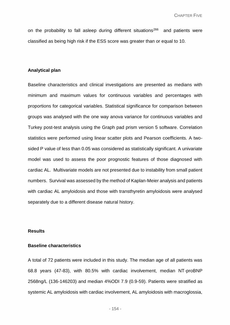
CHAPTER FIVE
- 154 -
on the probability to fall asleep during different situations266 and patients were
classified as being high risk if the ESS score was greater than or equal to 10.
Analytical plan
Baseline characteristics and clinical investigations are presented as medians with
minimum and maximum values for continuous variables and percentages with
proportions for categorical variables. Statistical significance for comparison between
groups was analysed with the one way anova variance for continuous variables and
Turkey post-test analysis using the Graph pad prism version 5 software. Correlation
statistics were performed using linear scatter plots and Pearson coefficients. A two-
sided P value of less than 0.05 was considered as statistically significant. A univariate
model was used to assess the poor prognostic features of those diagnosed with
cardiac AL. Multivariate models are not presented due to instability from small patient
numbers. Survival was assessed by the method of Kaplan-Meier analysis and patients
with cardiac AL amyloidosis and those with transthyretin amyloidosis were analysed
separately due to a different disease natural history.
Results
Baseline characteristics
A total of 72 patients were included in this study. The median age of all patients was
68.8 years (47-83), with 80.5% with cardiac involvement, median NT-proBNP
2568ng/L (136-146203) and median 4%ODI 7.9 (0.9-59). Patients were stratified as
systemic AL amyloidosis with cardiac involvement, AL amyloidosis with macroglossia,
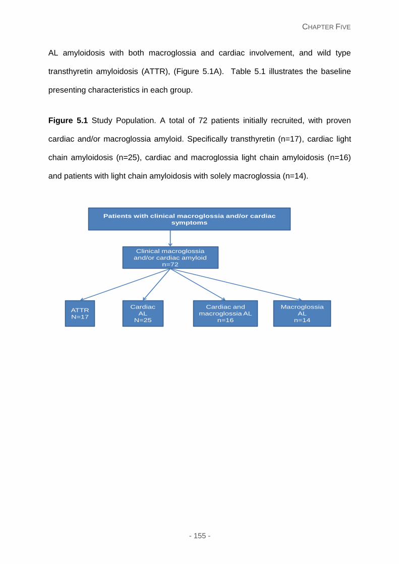
CHAPTER FIVE
- 155 -
AL amyloidosis with both macroglossia and cardiac involvement, and wild type
transthyretin amyloidosis (ATTR), (Figure 5.1A). Table 5.1 illustrates the baseline
presenting characteristics in each group.
Figure 5.1 Study Population. A total of 72 patients initially recruited, with proven
cardiac and/or macroglossia amyloid. Specifically transthyretin (n=17), cardiac light
chain amyloidosis (n=25), cardiac and macroglossia light chain amyloidosis (n=16)
and patients with light chain amyloidosis with solely macroglossia (n=14).
Patients with clinical macroglossia and/or cardiac
symptoms
Clinical macroglossia
and/or cardiac amyloid
n=72
ATTR
N=17
Cardiac
AL
N=25
Cardiac and
macroglossia AL
n=16
Macroglossia
AL
n=14
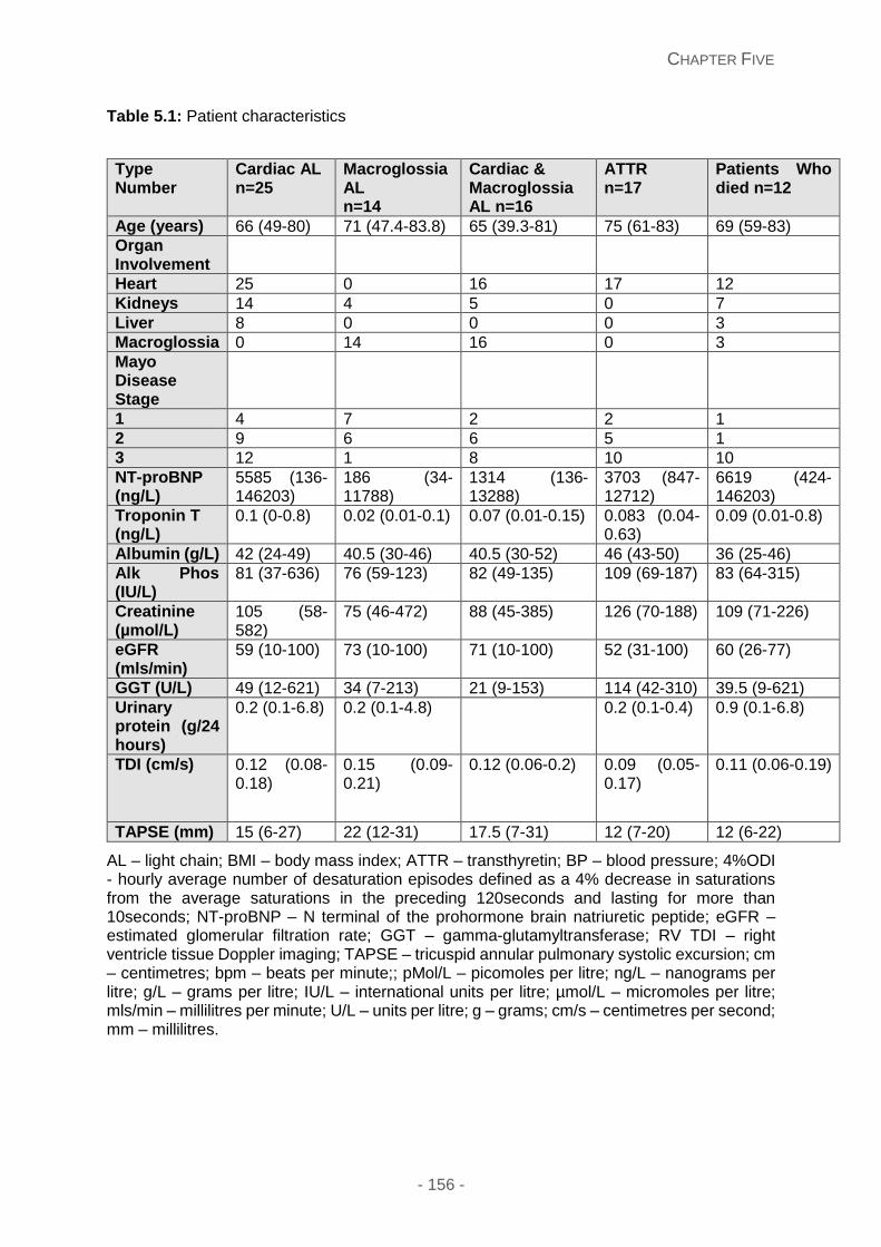
CHAPTER FIVE
- 156 -
Table 5.1: Patient characteristics
Type Number
Cardiac AL n=25
Macroglossia AL n=14
Cardiac & Macroglossia AL n=16
ATTR n=17
Patients Who died n=12
Age (years) 66 (49-80) 71 (47.4-83.8) 65 (39.3-81) 75 (61-83) 69 (59-83)
Organ Involvement
Heart 25 0 16 17 12
Kidneys 14 4 5 0 7
Liver 8 0 0 0 3
Macroglossia 0 14 16 0 3
Mayo Disease Stage
1 4 7 2 2 1
2 9 6 6 5 1
3 12 1 8 10 10
NT-proBNP (ng/L)
5585 (136-146203)
186 (34-11788)
1314 (136-13288)
3703 (847-12712)
6619 (424-146203)
Troponin T (ng/L)
0.1 (0-0.8) 0.02 (0.01-0.1) 0.07 (0.01-0.15) 0.083 (0.04-0.63)
0.09 (0.01-0.8)
Albumin (g/L) 42 (24-49) 40.5 (30-46) 40.5 (30-52) 46 (43-50) 36 (25-46)
Alk Phos (IU/L)
81 (37-636) 76 (59-123) 82 (49-135) 109 (69-187) 83 (64-315)
Creatinine (µmol/L)
105 (58-582)
75 (46-472) 88 (45-385) 126 (70-188) 109 (71-226)
eGFR (mls/min)
59 (10-100) 73 (10-100) 71 (10-100) 52 (31-100) 60 (26-77)
GGT (U/L) 49 (12-621) 34 (7-213) 21 (9-153) 114 (42-310) 39.5 (9-621)
Urinary protein (g/24 hours)
0.2 (0.1-6.8) 0.2 (0.1-4.8) 0.2 (0.1-0.4) 0.9 (0.1-6.8)
TDI (cm/s) 0.12 (0.08-0.18)
0.15 (0.09-0.21)
0.12 (0.06-0.2) 0.09 (0.05-0.17)
0.11 (0.06-0.19)
TAPSE (mm) 15 (6-27) 22 (12-31) 17.5 (7-31) 12 (7-20) 12 (6-22)
AL – light chain; BMI – body mass index; ATTR – transthyretin; BP – blood pressure; 4%ODI - hourly average number of desaturation episodes defined as a 4% decrease in saturations from the average saturations in the preceding 120seconds and lasting for more than 10seconds; NT-proBNP – N terminal of the prohormone brain natriuretic peptide; eGFR – estimated glomerular filtration rate; GGT – gamma-glutamyltransferase; RV TDI – right ventricle tissue Doppler imaging; TAPSE – tricuspid annular pulmonary systolic excursion; cm – centimetres; bpm – beats per minute;; pMol/L – picomoles per litre; ng/L – nanograms per litre; g/L – grams per litre; IU/L – international units per litre; µmol/L – micromoles per litre; mls/min – millilitres per minute; U/L – units per litre; g – grams; cm/s – centimetres per second; mm – millilitres.
CHAPTER FIVE
- 157 -
Overnight oximetry
Overnight oximetry tracings were recorded and collected in all 72 patients. Table 5.2
shows findings in systemic AL amyloidosis with cardiac involvement, AL amyloidosis
with macroglossia, AL amyloidosis with both macroglossia and cardiac involvement,
and wild type transthyretin amyloidosis (ATTR) and those patients who died. The
mean oxygen saturation of all patients in the study was 93% (±SD 1.99, 87-96). The
mean oxygen saturations were similar in all groups. The ATTR group had the lowest
number of abnormal oximetry tracings. Figures 5.2A and 5.2B illustrate a normal and
abnormal overnight oximetry tracing in 2 patients with cardiac amyloidosis
respectively.
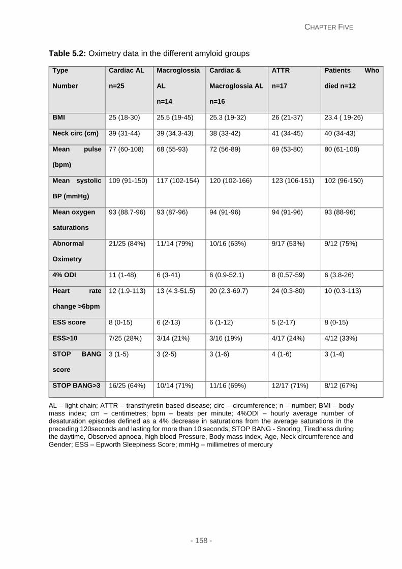
CHAPTER FIVE
- 158 -
Table 5.2: Oximetry data in the different amyloid groups
Type
Number
Cardiac AL
n=25
Macroglossia
AL
n=14
Cardiac &
Macroglossia AL
n=16
ATTR
n=17
Patients Who
died n=12
BMI 25 (18-30) 25.5 (19-45) 25.3 (19-32) 26 (21-37) 23.4 ( 19-26)
Neck circ (cm) 39 (31-44) 39 (34.3-43) 38 (33-42) 41 (34-45) 40 (34-43)
Mean pulse
(bpm)
77 (60-108) 68 (55-93) 72 (56-89) 69 (53-80) 80 (61-108)
Mean systolic
BP (mmHg)
109 (91-150) 117 (102-154) 120 (102-166) 123 (106-151) 102 (96-150)
Mean oxygen
saturations
93 (88.7-96) 93 (87-96) 94 (91-96) 94 (91-96) 93 (88-96)
Abnormal
Oximetry
21/25 (84%) 11/14 (79%) 10/16 (63%) 9/17 (53%) 9/12 (75%)
4% ODI 11 (1-48) 6 (3-41) 6 (0.9-52.1) 8 (0.57-59) 6 (3.8-26)
Heart rate
change >6bpm
12 (1.9-113) 13 (4.3-51.5) 20 (2.3-69.7) 24 (0.3-80) 10 (0.3-113)
ESS score 8 (0-15) 6 (2-13) 6 (1-12) 5 (2-17) 8 (0-15)
ESS>10 7/25 (28%) 3/14 (21%) 3/16 (19%) 4/17 (24%) 4/12 (33%)
STOP BANG
score
3 (1-5) 3 (2-5) 3 (1-6) 4 (1-6) 3 (1-4)
STOP BANG>3 16/25 (64%) 10/14 (71%) 11/16 (69%) 12/17 (71%) 8/12 (67%)
AL – light chain; ATTR – transthyretin based disease; circ – circumference; n – number; BMI – body mass index; cm – centimetres; bpm – beats per minute; 4%ODI – hourly average number of desaturation episodes defined as a 4% decrease in saturations from the average saturations in the preceding 120seconds and lasting for more than 10 seconds; STOP BANG - Snoring, Tiredness during the daytime, Observed apnoea, high blood Pressure, Body mass index, Age, Neck circumference and Gender; ESS – Epworth Sleepiness Score; mmHg – millimetres of mercury
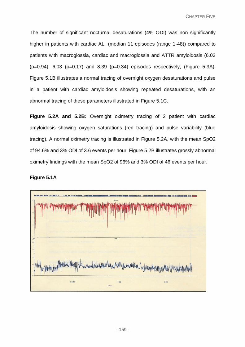
CHAPTER FIVE
- 159 -
The number of significant nocturnal desaturations (4% ODI) was non significantly
higher in patients with cardiac AL (median 11 episodes (range 1-48)) compared to
patients with macroglossia, cardiac and macroglossia and ATTR amyloidosis (6.02
(p=0.94), 6.03 (p=0.17) and 8.39 (p=0.34) episodes respectively, (Figure 5.3A).
Figure 5.1B illustrates a normal tracing of overnight oxygen desaturations and pulse
in a patient with cardiac amyloidosis showing repeated desaturations, with an
abnormal tracing of these parameters illustrated in Figure 5.1C.
Figure 5.2A and 5.2B: Overnight oximetry tracing of 2 patient with cardiac
amyloidosis showing oxygen saturations (red tracing) and pulse variability (blue
tracing). A normal oximetry tracing is illustrated in Figure 5.2A, with the mean SpO2
of 94.6% and 3% ODI of 3.6 events per hour. Figure 5.2B illustrates grossly abnormal
oximetry findings with the mean SpO2 of 96% and 3% ODI of 46 events per hour.
Figure 5.1A
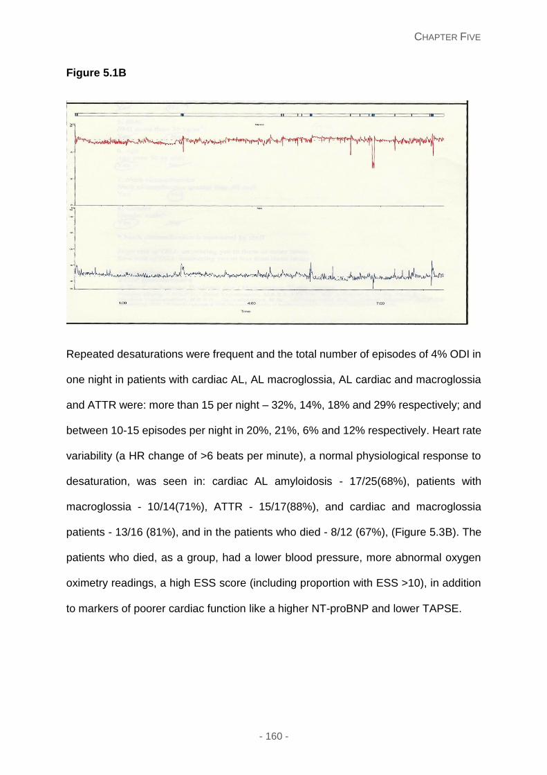
CHAPTER FIVE
- 160 -
Figure 5.1B
Repeated desaturations were frequent and the total number of episodes of 4% ODI in
one night in patients with cardiac AL, AL macroglossia, AL cardiac and macroglossia
and ATTR were: more than 15 per night – 32%, 14%, 18% and 29% respectively; and
between 10-15 episodes per night in 20%, 21%, 6% and 12% respectively. Heart rate
variability (a HR change of >6 beats per minute), a normal physiological response to
desaturation, was seen in: cardiac AL amyloidosis - 17/25(68%), patients with
macroglossia - 10/14(71%), ATTR - 15/17(88%), and cardiac and macroglossia
patients - 13/16 (81%), and in the patients who died - 8/12 (67%), (Figure 5.3B). The
patients who died, as a group, had a lower blood pressure, more abnormal oxygen
oximetry readings, a high ESS score (including proportion with ESS >10), in addition
to markers of poorer cardiac function like a higher NT-proBNP and lower TAPSE.
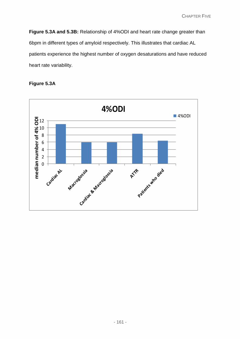
CHAPTER FIVE
- 161 -
Figure 5.3A and 5.3B: Relationship of 4%ODI and heart rate change greater than
6bpm in different types of amyloid respectively. This illustrates that cardiac AL
patients experience the highest number of oxygen desaturations and have reduced
heart rate variability.
Figure 5.3A
0
2
4
6
8
10
12
4%ODI4%ODI
me
dia
n n
um
be
r o
f 4
% O
DI
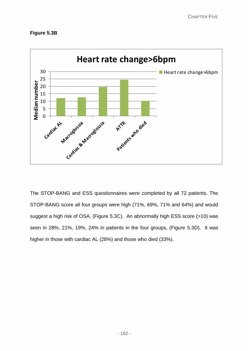
CHAPTER FIVE
- 162 -
Figure 5.3B
The STOP-BANG and ESS questionnaires were completed by all 72 patients. The
STOP-BANG score all four groups were high (71%, 69%, 71% and 64%) and would
suggest a high risk of OSA, (Figure 5.3C). An abnormally high ESS score (>10) was
seen in 28%, 21%, 19%, 24% in patients in the four groups, (Figure 5.3D). It was
higher in those with cardiac AL (28%) and those who died (33%).
0
5
10
15
20
25
30
Heart rate change>6bpmHeart rate change>6bpm
Me
dia
n n
um
be
r
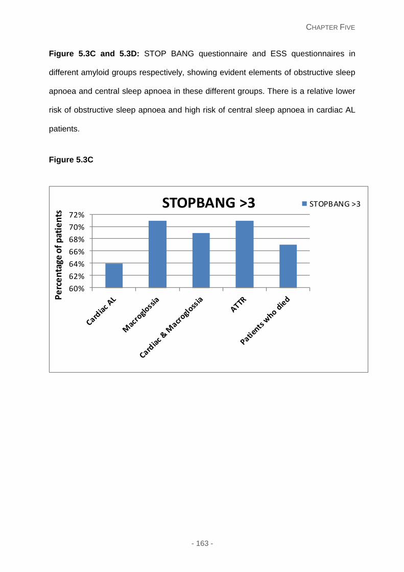
CHAPTER FIVE
- 163 -
Figure 5.3C and 5.3D: STOP BANG questionnaire and ESS questionnaires in
different amyloid groups respectively, showing evident elements of obstructive sleep
apnoea and central sleep apnoea in these different groups. There is a relative lower
risk of obstructive sleep apnoea and high risk of central sleep apnoea in cardiac AL
patients.
Figure 5.3C
60%
62%
64%
66%
68%
70%
72%STOPBANG >3 STOPBANG >3
Pe
rce
nta
ge o
f p
atie
nts
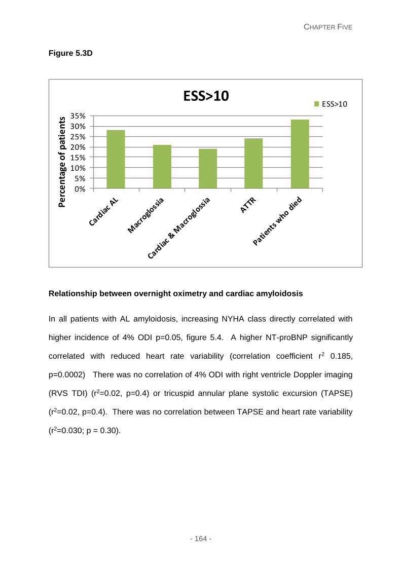
CHAPTER FIVE
- 164 -
Figure 5.3D
Relationship between overnight oximetry and cardiac amyloidosis
In all patients with AL amyloidosis, increasing NYHA class directly correlated with
higher incidence of 4% ODI p=0.05, figure 5.4. A higher NT-proBNP significantly
correlated with reduced heart rate variability (correlation coefficient r2 0.185,
p=0.0002) There was no correlation of 4% ODI with right ventricle Doppler imaging
(RVS TDI) (r2=0.02, p=0.4) or tricuspid annular plane systolic excursion (TAPSE)
(r2=0.02, p=0.4). There was no correlation between TAPSE and heart rate variability
(r2=0.030; p = 0.30).
0%5%
10%15%20%25%30%35%
ESS>10ESS>10
Pe
rce
nta
ge o
f p
atie
nts
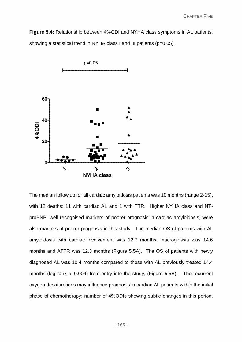
CHAPTER FIVE
- 165 -
Figure 5.4: Relationship between 4%ODI and NYHA class symptoms in AL patients,
showing a statistical trend in NYHA class I and III patients (p=0.05).
The median follow up for all cardiac amyloidosis patients was 10 months (range 2-15),
with 12 deaths: 11 with cardiac AL and 1 with TTR. Higher NYHA class and NT-
proBNP, well recognised markers of poorer prognosis in cardiac amyloidosis, were
also markers of poorer prognosis in this study. The median OS of patients with AL
amyloidosis with cardiac involvement was 12.7 months, macroglossia was 14.6
months and ATTR was 12.3 months (Figure 5.5A). The OS of patients with newly
diagnosed AL was 10.4 months compared to those with AL previously treated 14.4
months (log rank p=0.004) from entry into the study, (Figure 5.5B). The recurrent
oxygen desaturations may influence prognosis in cardiac AL patients within the initial
phase of chemotherapy; number of 4%ODIs showing subtle changes in this period,
1 2 3
0
20
40
60
p=0.05
NYHA class
4%
OD
I
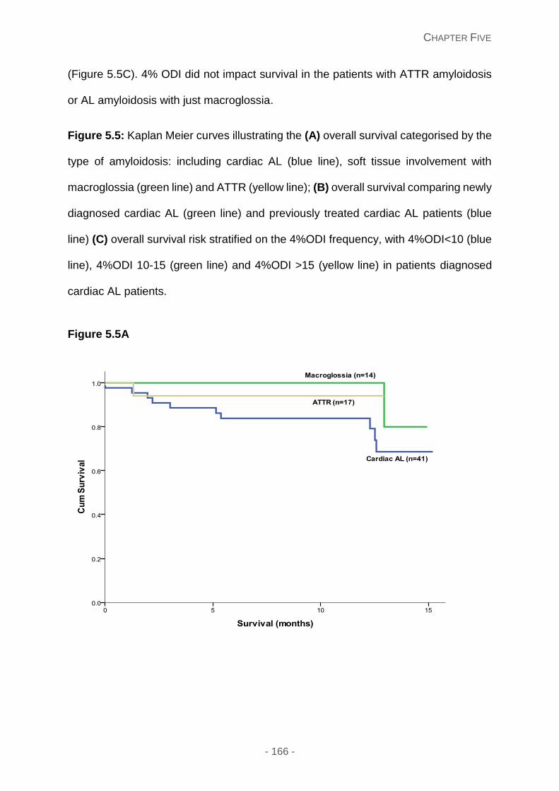
CHAPTER FIVE
- 166 -
(Figure 5.5C). 4% ODI did not impact survival in the patients with ATTR amyloidosis
or AL amyloidosis with just macroglossia.
Figure 5.5: Kaplan Meier curves illustrating the (A) overall survival categorised by the
type of amyloidosis: including cardiac AL (blue line), soft tissue involvement with
macroglossia (green line) and ATTR (yellow line); (B) overall survival comparing newly
diagnosed cardiac AL (green line) and previously treated cardiac AL patients (blue
line) (C) overall survival risk stratified on the 4%ODI frequency, with 4%ODI<10 (blue
line), 4%ODI 10-15 (green line) and 4%ODI >15 (yellow line) in patients diagnosed
cardiac AL patients.
Figure 5.5A
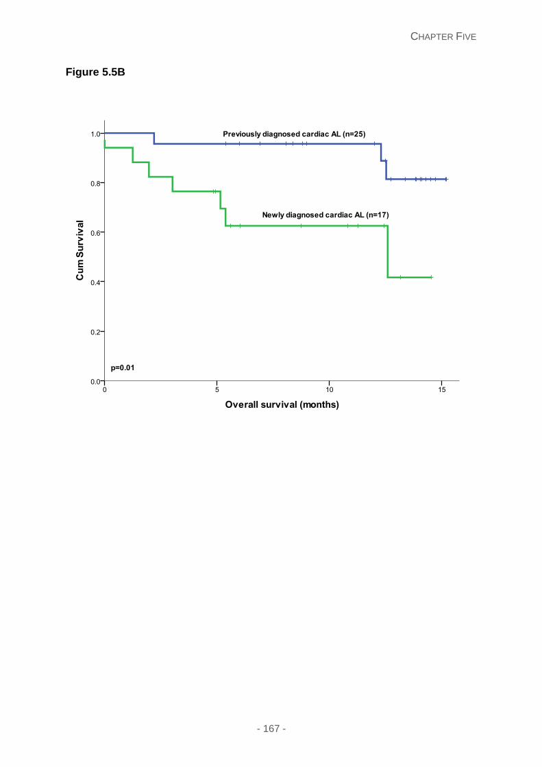
CHAPTER FIVE
- 167 -
Figure 5.5B
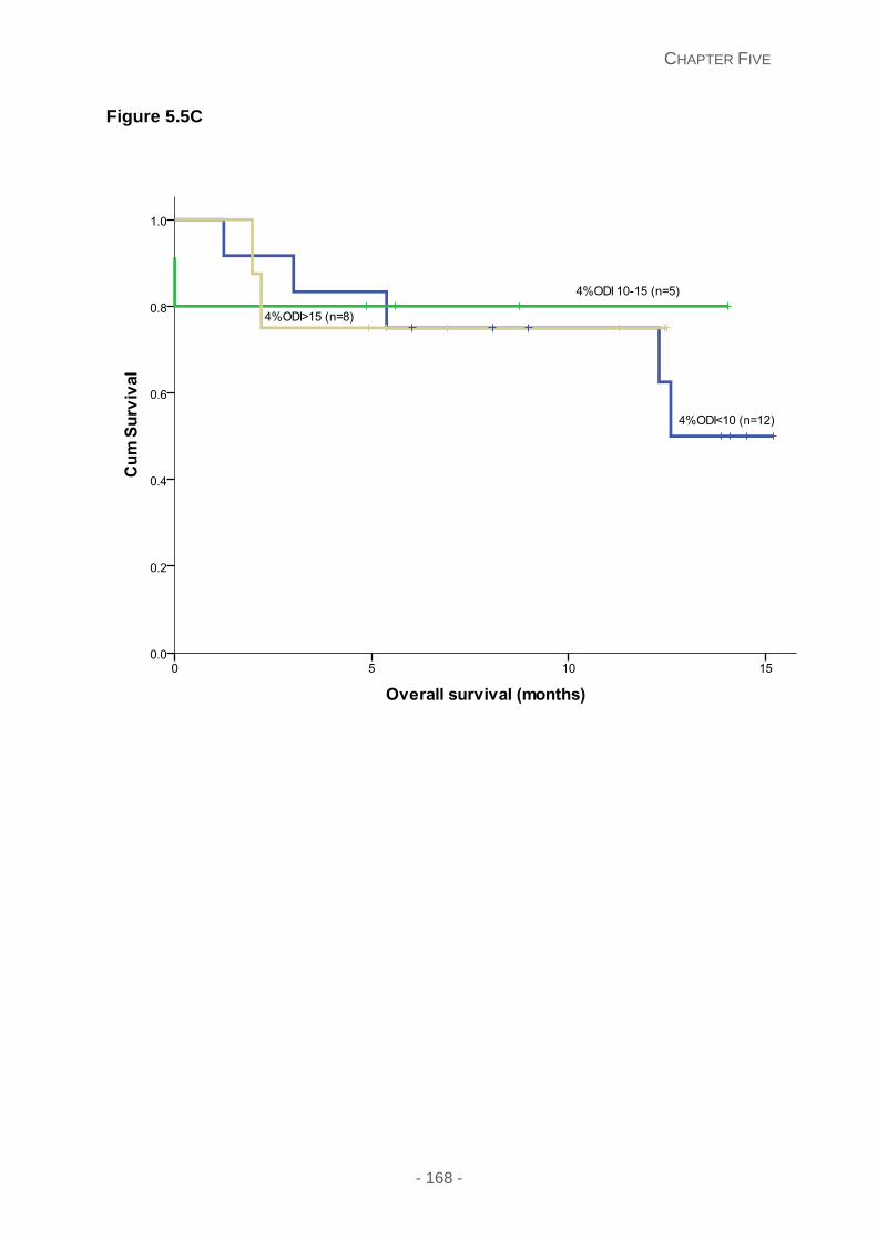
CHAPTER FIVE
- 168 -
Figure 5.5C
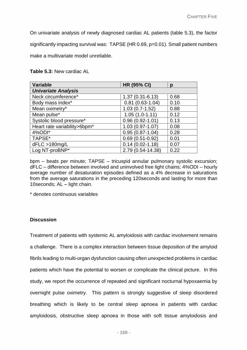
CHAPTER FIVE
- 169 -
On univariate analysis of newly diagnosed cardiac AL patients (table 5.3), the factor
significantly impacting survival was: TAPSE (HR 0.69, p=0.01). Small patient numbers
make a multivariate model unreliable.
Table 5.3: New cardiac AL
Variable HR (95% CI) p
Univariate Analysis
Neck circumference* 1.37 (0.31-6.13) 0.68
Body mass index* 0.81 (0.63-1.04) 0.10
Mean oximetry* 1.03 (0.7-1.52) 0.88
Mean pulse* 1.05 (1.0-1.11) 0.12
Systolic blood pressure* 0.96 (0.92-1.01) 0.13
Heart rate variability>6bpm* 1.03 (0.97-1.07) 0.08
4%ODI* 0.95 (0.87-1.04) 0.28
TAPSE* 0.69 (0.51-0.92) 0.01
dFLC >180mg/L 0.14 (0.02-1.18) 0.07
Log NT-proBNP* 2.79 (0.54-14.38) 0.22
bpm – beats per minute; TAPSE – tricuspid annular pulmonary systolic excursion; dFLC – difference between involved and uninvolved free light chains; 4%ODI – hourly average number of desaturation episodes defined as a 4% decrease in saturations from the average saturations in the preceding 120seconds and lasting for more than 10seconds; AL – light chain.
* denotes continuous variables
Discussion
Treatment of patients with systemic AL amyloidosis with cardiac involvement remains
a challenge. There is a complex interaction between tissue deposition of the amyloid
fibrils leading to multi-organ dysfunction causing often unexpected problems in cardiac
patients which have the potential to worsen or complicate the clinical picture. In this
study, we report the occurrence of repeated and significant nocturnal hypoxaemia by
overnight pulse oximetry. This pattern is strongly suggestive of sleep disordered
breathing which is likely to be central sleep apnoea in patients with cardiac
amyloidosis, obstructive sleep apnoea in those with soft tissue amyloidosis and
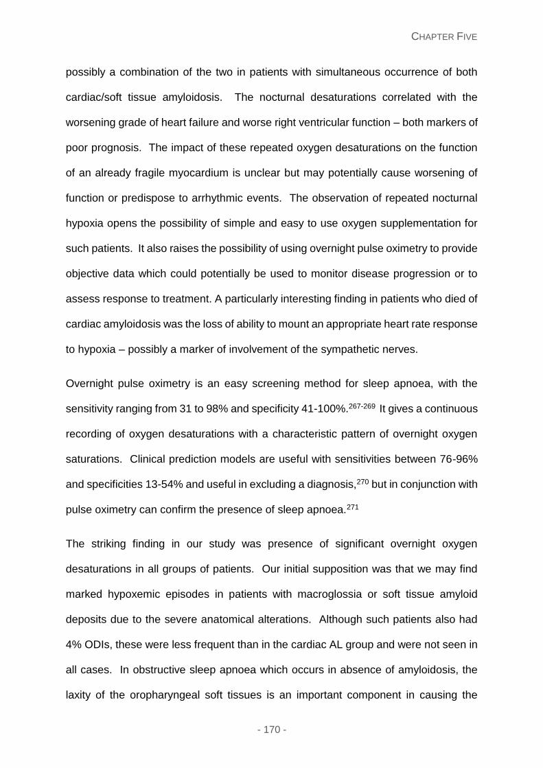
CHAPTER FIVE
- 170 -
possibly a combination of the two in patients with simultaneous occurrence of both
cardiac/soft tissue amyloidosis. The nocturnal desaturations correlated with the
worsening grade of heart failure and worse right ventricular function – both markers of
poor prognosis. The impact of these repeated oxygen desaturations on the function
of an already fragile myocardium is unclear but may potentially cause worsening of
function or predispose to arrhythmic events. The observation of repeated nocturnal
hypoxia opens the possibility of simple and easy to use oxygen supplementation for
such patients. It also raises the possibility of using overnight pulse oximetry to provide
objective data which could potentially be used to monitor disease progression or to
assess response to treatment. A particularly interesting finding in patients who died of
cardiac amyloidosis was the loss of ability to mount an appropriate heart rate response
to hypoxia – possibly a marker of involvement of the sympathetic nerves.
Overnight pulse oximetry is an easy screening method for sleep apnoea, with the
sensitivity ranging from 31 to 98% and specificity 41-100%.267-269 It gives a continuous
recording of oxygen desaturations with a characteristic pattern of overnight oxygen
saturations. Clinical prediction models are useful with sensitivities between 76-96%
and specificities 13-54% and useful in excluding a diagnosis,270 but in conjunction with
pulse oximetry can confirm the presence of sleep apnoea.271
The striking finding in our study was presence of significant overnight oxygen
desaturations in all groups of patients. Our initial supposition was that we may find
marked hypoxemic episodes in patients with macroglossia or soft tissue amyloid
deposits due to the severe anatomical alterations. Although such patients also had
4% ODIs, these were less frequent than in the cardiac AL group and were not seen in
all cases. In obstructive sleep apnoea which occurs in absence of amyloidosis, the
laxity of the oropharyngeal soft tissues is an important component in causing the
CHAPTER FIVE
- 171 -
collapse of the tissues during inspiration leading to obstruction of the upper airways.
The current findings of less than expected severe hypoxia in patients with significant
soft tissue amyloid deposits raises an interesting question whether increased stiffness
of the soft tissue, which is a hallmark of amyloid deposition, actually protects against
the “traditional” cause of OSA and true OSA in amyloidosis only occurs when there
enough amyloid deposition to alter the anatomy towards obstruction a supine position.
Central sleep apnoea (CSA) is a well-recognised complication of systolic heart failure
and leads to repeated episodes of nocturnal hypoxia. CSA is worse in patients with
an ejection fraction of <40% and has been reported to be associated with increase in
non-sustained ventricular tachycardia as well as reduction in heart rate variability.272
The occurrence of repeated severe nocturnal oxygen desaturations seen in patients
with cardiac amyloidosis strongly suggest CSA also occurs in AL amyloidosis – a
finding that will need formal confirmation by polysomnography. In our study, the
oxygen desaturations were greatest in those with cardiac AL and ATTR amyloid, again
with the median number of desaturations higher in those with cardiac AL. Moreover,
4% ODI correlated with NYHA class and NT-proBNP – suggesting a direct correlation
with worsening amyloid burden/heart failure with ODI. Although there was no direct
impact of the number of 4% ODIs on overall survival in patients with newly diagnosed
AL amyloidosis, these repeated insults with other factors are likely to influence these
patients. This is a strikingly different finding from that in systolic heart failure where the
cardiovascular impact of CSA is universal. In AL amyloidosis, newly diagnosed
patients have the most unstable heart disease with 30-40% patients dying of
cardiovascular complication within 6 months of the initial diagnosis. The impact of
nocturnal hypoxia correlating with increased deaths raises important questions – do
the repeated hypoxemic changes increase the vulnerability of an already fragile
CHAPTER FIVE
- 172 -
myocardium to arrhythmias? Ventricular arrhythmias have been proposed as a causal
mechanism for the peak of sudden cardiac death during sleeping hours in OSA
patients.273 The risk of cardiac arrhythmias with OSA appears to be related to disease
severity - majority of OSA patients presenting significant arrhythmias have moderate
or severe forms of the disease.274-276 The amyloidogenic light chains are directly toxic
to the myocardium in AL amyloidosis and additional hypoxemic insults may compound
this toxicity. Lastly, patients receive chemotherapy soon after diagnosis and
hypoxaemia may influence the toxicity of chemotherapy. Repeated nocturnal hypoxia
beyond the newly diagnosed setting may have different consequences. In CSA, the
hypoxaemia has consequences in addition to worse mortality, which include increase
in day time sleepiness, worsening fatigue and poorer quality of life.277, 278 Although
the latter two were not studied in our cohort, a very high proportion of patients with
cardiac AL amyloidosis and ATTR had abnormal ESS questionnaires – a measure of
day time sleepiness. Persistent and profound fatigue is a symptom in AL amyloidosis
which has never been adequately explained. It is always vaguely assumed to be
multifactorial from the wide range of organ involvement. The current findings suggest
that CSA/OSA may be a substantial contributor to this symptom. Recurrent hypoxemia
was seen in both AL and ATTR cardiac patients but were more marked in the AL
cohort – in keeping with the worse cardiac profile of AL patients compared to ATTR in
general.
An interesting finding in this study was the lack of or markedly reduced heart rate
variability in patients with cardiac AL amyloidosis. This lack of heart rate response to
hypoxia was greater in AL amyloidosis than those with ATTR cardiac amyloidosis.
Autonomic dysfunction is a known phenomenon in AL amyloidosis but the current
finding had no correlation with clinical autonomic neuropathy. Cardiac autonomic
CHAPTER FIVE
- 173 -
denervation is well recognised feature of systolic heart failure279, 280 and 123I-MIBG –
Metaiodobenzylguanidine (MIBG) scintigraphy is a useful tool for its identification.279,
281 MIBG scans have been reported to be abnormal in cardiac amyloidosis both AL
and ATTR type.281-284 The most marked lack of heart rate variability was seen in the
patients who died. Due to a restrictive left ventricle in cardiac AL, any needed
increases in cardiac output are largely dependent on appropriate increase in the heart
rate. The lack of physiologic tachycardia in response to hypoxia could make the
systemic consequences more profound. This finding suggests that further
assessment of cardiac autonomic denervation may be provide useful information in
AL amyloidosis and open a potential therapeutic avenue for consideration of device
therapy to counter a lack of the autonomic drive.
The markedly abnormal findings in this pilot study, if confirmed by polysomnography,
suggests possible avenues for intervention. For many years, there was a suggestion
that correction of CSA in systolic heart failure would improve outcomes. A randomised
study (SERV-HF) in systolic heart failure and moderate to severe heart disordered
breathing showed that assisted servo ventilation (ASV) controlled sleep disordered
breathing but it did not improve survival; in fact there was increased cardiac mortality
in the ASV group.285 The restrictive cardiomyopathy in AL is very different from the
patients in reported in that study and those findings cannot be extrapolated to cardiac
amyloidosis. In addition, newly diagnosed AL is a disease of very high mortality not
just chronic morbidity, in which SDB may impact. Just as drugs which treat systolic
heart failure effectively, like beta-blockers and ACE inhibitors, often have profoundly
negative consequences in AL - impact of intervention on sleep disordered breathing
in AL may have different consequences and will need a separate study of formal
polysomnography sleep study to confirm occurrence of SDB (CSA and/or OSA) as
CHAPTER FIVE
- 174 -
well as impacts of intervention with simple oxygen supplementation or indeed more
complex intervention for SDB.
We recognise the limitations of this pilot study – a small study cohort and the use of
overnight pulse oximetery only. The group of patients was mixed with newly
diagnosed AL, those on longer term follow up as well as ATTR amyloidosis limiting
ability to assess survival impact in subgroups. There was no polysomnography to
confirm the cause of the recurrent nocturnal hypoxia or clarify type of SDB.
In conclusion, recurrent nocturnal oxygen desaturations are very common in patients
with cardiac amyloidosis (both AL and ATTR type) as well as in patients with soft tissue
amyloid deposits affecting the oropharyngeal tract. A high proportion of patient score
‘high risk’ for sleep disordered breathing by questionnaires designed to screen for such
patients. Increased number and frequency of nocturnal desaturations may be
associated with poorer survival in patients with newly diagnosed cardiac AL
amyloidosis. Lack of heart rate variability (suggesting cardiac autonomic neuropathy)
is a frequent occurrence on cardiac AL and particularly in those patients who died –
findings which need further clarification. The role of hypoxia in precipitation of cardiac
arrhythmias or sudden death in AL needs to be clarified. Nocturnal hypoxia is a simple
target for intervention in cardiac AL amyloidosis and could potentially help to reduce
early mortality in AL which has remained an unmet medical need for over 25 years.
- 175 -
Results Section Two:
Localised amyloidosis and subtypes
CHAPTER SIX
- 176 -
Chapter Six: Natural history and outcomes in localised
immunoglobulin light chain (AL) amyloidosis: a long-term
observational study
This chapter is in context of my publication: Natural history and outcomes in
localised immunoglobulin light-chain amyloidosis: a long-term observational
study. Shameem Mahmood, Frank Bridoux, Christopher P Venner, Sajitha
Sachchithanantham, Janet A Gilbertson, Dorota Rowczenio, Thomas Wagner, Rabya
Sayed, Ketna Patel, Marianna Fontana, Carol J Whelan, Helen J Lachmann, Philip N
Hawkins, Julian D Gillmore, Ashutosh D Wechalekar. Lancet Haematology. 2015;
2(6):e241-50. (Original article) copyright permission obtained and given by Lancet
Haematology editors, 2015 for use in my thesis.
Introduction
Amyloidosis comprises a heterogeneous group of disorders produced by the
extracellular deposition of misfolded proteins in an insoluble fibrillar form.286 Systemic
light chain (AL) amyloidosis is the most common and serious type, where the amyloid
fibrils are derived from circulating monoclonal immunoglobulin light chains produced
by an underlying plasma cell or B-cell clone. Hereditary systemic amyloidosis is
associated with mutations in numerous variant proteins including transthyretin (ATTR
amyloidosis) and apolipoprotein AI (AApoA1 amyloidosis).287 In systemic types of
amyloidosis, the respective amyloid fibril precursor protein is present and circulating
CHAPTER SIX
- 177 -
in the bloodstream and results in relentless amyloid deposition throughout the body,
progressive vital organ dysfunction and early death.288
Localised deposits of amyloid can occur in various tissues in the body and are usually
presumed to be of AL type, with the consequent presence of a focal monoclonal B cell
dyscrasia within the affected tissue. As such, the clinical effects of the localised
amyloid deposits depend on their precise anatomical location, and can result in
substantial morbidity.
Localised amyloidosis is much rarer than systemic types, and consequently remains
very poorly studied; with most knowledge arising from individual case reports or small
series of less than 20 patients. There are commonly reported sites include the urinary
tract, respiratory tract, larynx, skin and eyelids.165 Currently, data on long term
outcomes and progression to systemic disease is lacking with the need for further
exploration in this field. We have examined the clinical features and outcomes of a
large series of patients with localised AL amyloidosis highlighting the striking
differences from systemic AL amyloidosis with respect to the lack of progression,
benefit from debulking procedures, limited need for cytotoxic chemotherapy therapy
and excellent overall long term outcomes.
Methods
Study design
All the patients with a specified disease setting of localised amyloidosis diagnosed,
assessed and followed up at the UK National Amyloidosis Centre (NAC) between
January 2, 1980 and December 15, 2011 were identified using the NAC database and
written medical records. There was no exclusion criteria dependant on age or
CHAPTER SIX
- 178 -
performance status, with all comorbidities permitted. All the patients were newly
diagnosed and treatment naïve. Localised amyloidosis was typed with biopsy proven
amyloid deposition confined to a single site or tissue (with the relevant histology of the
tissue examined), and no evidence of vital organ involvement, the latter defined as
cardiac, renal, liver involvement or peripheral or autonomic neuropathy. Soft tissue
involvement was defined as clinical evidence of a single deposit of amyloid in the
tongue or one site of deposition in other soft tissues. Standard tests of vital organ
function were assessed and absence of visceral uptake was documented by 123I serum
amyloid P component (SAP) scintigraphy.289 All patients had SAP scintigraphy
performed at their first visit except those diagnosed before 1988 (when the technique
was introduced into clinical practice), and had this following availability of the
procedure, which was invaluable in assessing for any visceral organ uptake. A
detailed protocolised assessment was followed by all patients at baseline and follow
up visits to assess disease evaluation including a full blood count, renal and liver
function tests, serum and urine protein electrophoresis and immunofixation,
electrocardiogram (ECG), echocardiography and 123I SAP scintigraphy. Serum free
light chains (FLC) were also measured prospectively on all patients diagnosed after
2002 on blood samples at presentation and consequent follow up visits; and also
retrospectively on stored serum samples, where available, for patients prior to 2002.
A bone marrow assessment was performed in patients with a proven plasma cell
dyscrasia for further investigation. Histological confirmation of amyloid deposition of
the affected tissue was conducted by showing Congo red staining and demonstration
of typical birefringence under cross polarised light. Fat biopsies were not performed in
addition to the affected tissue biopsy. The AL fibril type was determined by
immunohistochemical (IHC) staining using a panel of antibodies and defined as
CHAPTER SIX
- 179 -
staining with antibodies kappa and lambda immunoglobulin light chains and/or no
staining with antibodies to transthyretin, serum amyloid A protein (SAA) and ApoA1,
with AL the likely type. Hereditary amyloidosis was excluded by genetic sequencing in
patients in whom the clinical features could be in keeping with hereditary types –
particularly all patients with laryngeal symptoms underwent Apo A1 gene sequencing;
with laryngeal amyloid deposits a recognised feature of patients with Apo A1
mutations. Transthyretin gene sequencing was performed if any biopsies had
transthyretin staining to exclude ATTR. F18-flurodeoxyglucose positron emission
tomography (FDG-PET/CT) was performed in 18 patients following this technique
becoming an investigative tool at our centre.
Local disease progression was defined as progression or recurrence of the amyloid
deposit within the original localised site. Progression to systemic AL amyloidosis was
clarified as development of a new vital organ involvement or new evidence of visceral
amyloid deposits by 123I SAP scintigraphy, and stated as a number and percentage of
the total localised amyloidosis cohort. Statistical analysis in assessing survival
endpoints was performed using SPSS version 20. The overall survival (OS) was
determined from the date of diagnosis defined from the date at which a biopsy proof
of amyloidosis was obtained. The overall survival was estimated by the Kaplan Meier
method, calculated from the date of diagnosis until death or last follow-up, with the
causes of death known in those who died. All the patients consented to their details
being used anonymously and the study received approval from the Royal Free
Hospital ethics committee.
CHAPTER SIX
- 180 -
Results
Study population
The recruitment to this study included a total of 606 patients with localised amyloidosis
identified. Overall, this accounted for 12% (606/5050) of 5050 newly diagnosed
patients with all types of amyloidosis evaluated at our Centre during the study period.
97 patients with laryngeal symptoms had Apo A1 gene sequencing, and interestingly
transthyretin staining was present on 3 bladder biopsies, with transthyretin gene
sequencing performed to exclude ATTR. For the purposes of survival statistics, 11
patients were excluded as three patients had ATTR amyloid deposition on bladder
biopsy (with ATTR was not seen at any other localised site), 4 with lichen amyloidosis,
3 with insulin amyloidosis and 1 patient had AApoA1 amyloid deposition in a laryngeal
biopsy and shown to be heterozygous for ApoA1 Ala164Ser mutation by ApoA1 gene
sequencing. These patients were excluded from only survival analysis with few
numbers and little known as to underlying prognosis and complications of this group.
All remaining patients were included in analyses of survival and progression to
systemic disease illustrated in Figure 6.1A.
The survival outcomes are available for all patients with data obtained from the Office
of National Statistics in the UK.290 Baseline characteristics are illustrated in Table 6.1,
with the median age 59.5 years (range 48.8-87 yrs, IQR 50.2-74.5) and 51% (307/606)
were male. The median symptom duration was 7 months (range 0.5-360 months, IQR
4-24) prior to diagnosis. There were many sites of localised amyloidosis including:
bladder 95/606 patients (15.7%); laryngeal/tonsillar 92/606 (15.2%); cutaneous
84/606 (13.9%); pulmonary nodular 47/606 (7.8%); gastrointestinal 36/606 (5.9%);
oral 36/606 (5.9%); tracheobronchial 35/606 (5.8%); lymph node 31/606 (5.1%);
CHAPTER SIX
- 181 -
conjunctival 27/606 (4.5%); bone 24/606 (4%); eyelid 22/606 (3.6%); orbital 21/606
(3.5%); breast 13/606 (2.1%); ureteric 10/606 (1.7%); urethral 10/606 (1.7%); soft
tissue 10/606 (1.7%); prostate 9/606 (1.3%); cerebral 3/606 (0.5%) and aortic valve
1/606 (0.1%), (Figure 6.1B). Some patients had amyloid deposits at more than one
localised site including cutaneous (9 patients) and within the gastrointestinal tract (2
patients). The multiple deposits within the skin included - 5 patients with deposits in
both lower limbs, and 3 patients having 2 separate skin sites involved (right calf and
right elbow, face and skin over the breast, right hand and left elbow). Patients with
gastrointestinal involvement included (gastric involvement in 7 patients, small bowel
involvement in 10, 17 with large bowel involvement and 2 with small and large bowel
involvement).
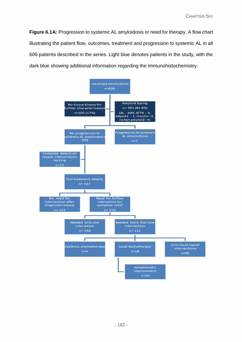
CHAPTER SIX
- 182 -
Figure 6.1A: Progression to systemic AL amyloidosis or need for therapy. A flow chart
illustrating the patient flow, outcomes, treatment and progression to systemic AL in all
606 patients described in the series. Light blue denotes patients in the study, with the
dark blue showing additional information regarding the immunohistochemistry.
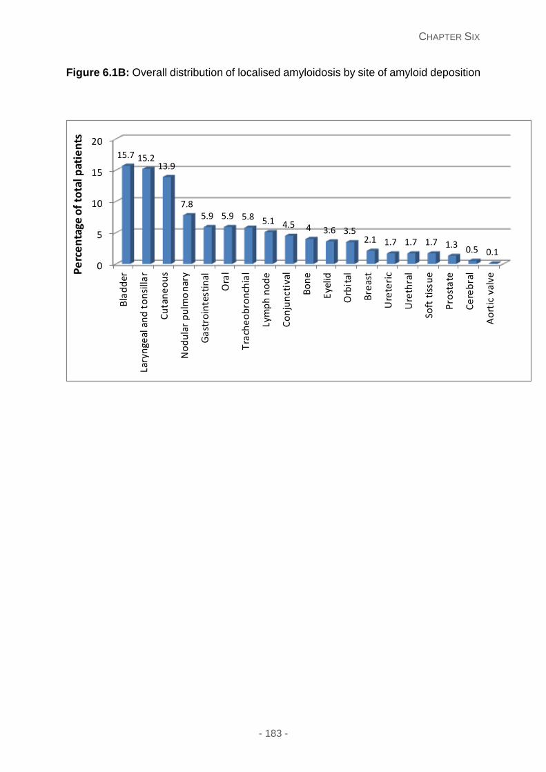
CHAPTER SIX
- 183 -
Figure 6.1B: Overall distribution of localised amyloidosis by site of amyloid deposition
0
5
10
15
20
Bla
dd
er
Lary
nge
al a
nd
to
nsi
llar
Cu
tan
eo
us
No
du
lar
pu
lmo
nar
y
Ga
stro
inte
stin
al
Ora
l
Tra
che
ob
ron
chia
l
Lym
ph
no
de
Co
nju
nct
ival
Bo
ne
Eye
lid
Orb
ital
Bre
ast
Ure
teri
c
Ure
thra
l
Soft
tis
sue
Pro
sta
te
Ce
reb
ral
Ao
rtic
val
ve
15.7 15.213.9
7.85.9 5.9 5.8 5.1 4.5 4 3.6 3.5
2.1 1.7 1.7 1.7 1.30.5 0.1
Pe
rce
nta
ge o
f to
tal p
atie
nts
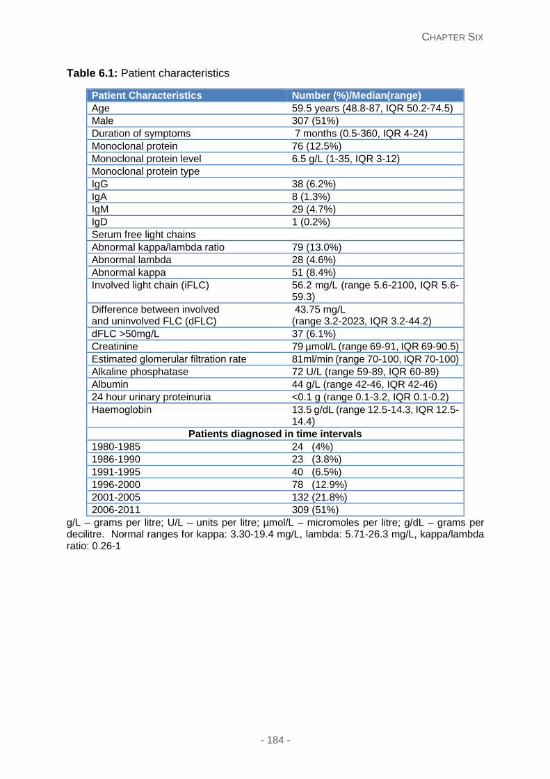
CHAPTER SIX
- 184 -
Table 6.1: Patient characteristics
Patient Characteristics Number (%)/Median(range)
Age 59.5 years (48.8-87, IQR 50.2-74.5)
Male 307 (51%)
Duration of symptoms 7 months (0.5-360, IQR 4-24)
Monoclonal protein 76 (12.5%)
Monoclonal protein level 6.5 g/L (1-35, IQR 3-12)
Monoclonal protein type
IgG 38 (6.2%)
IgA 8 (1.3%)
IgM 29 (4.7%)
IgD 1 (0.2%)
Serum free light chains
Abnormal kappa/lambda ratio 79 (13.0%)
Abnormal lambda 28 (4.6%)
Abnormal kappa 51 (8.4%)
Involved light chain (iFLC) 56.2 mg/L (range 5.6-2100, IQR 5.6-59.3)
Difference between involved and uninvolved FLC (dFLC)
43.75 mg/L (range 3.2-2023, IQR 3.2-44.2)
dFLC >50mg/L 37 (6.1%)
Creatinine 79 µmol/L (range 69-91, IQR 69-90.5)
Estimated glomerular filtration rate 81ml/min (range 70-100, IQR 70-100)
Alkaline phosphatase 72 U/L (range 59-89, IQR 60-89)
Albumin 44 g/L (range 42-46, IQR 42-46)
24 hour urinary proteinuria <0.1 g (range 0.1-3.2, IQR 0.1-0.2)
Haemoglobin 13.5 g/dL (range 12.5-14.3, IQR 12.5-14.4)
Patients diagnosed in time intervals
1980-1985 24 (4%)
1986-1990 23 (3.8%)
1991-1995 40 (6.5%)
1996-2000 78 (12.9%)
2001-2005 132 (21.8%)
2006-2011 309 (51%)
g/L – grams per litre; U/L – units per litre; µmol/L – micromoles per litre; g/dL – grams per decilitre. Normal ranges for kappa: 3.30-19.4 mg/L, lambda: 5.71-26.3 mg/L, kappa/lambda ratio: 0.26-1
CHAPTER SIX
- 185 -
The presenting symptoms were determined by the site of amyloid deposition and
Table 6.2 summarises the clinical features for each site of amyloid deposition, median
age and duration of symptoms and accompanying ranges stated as minimum and
maximum values. Figure 2 shows different types of localised amyloidosis. 67 of 606
patients (11.0%) were known to have an autoimmune disorder including 17/606 (2.8%)
with Sjögren’s syndrome, 18/606 (3.0%) with hypothyroidism, 8/606 (1.3%) having
rheumatoid arthritis and 6/606 (1.0%) diagnosed with systemic lupus erythematosus
(SLE). Taking into account the 17 patients with Sjögren’s syndrome, amyloid
deposition was present in the lungs in 5, breast in 3, conjunctiva in 2 - accounting for
10.6% (5/47), 23.1% (3/13), 7.4% (2/27) of all lung, breast and conjunctival
amyloidosis patients respectively- and four in the skin. The remainder had amyloid
deposition at a variety of other localised sites. An unrelated history of non-
haematological malignancies was present in 37 patients (6.1%); cervical and ovarian
cancer 3, carcinoma of the prostate 10, sigmoid cancer 6, transitional cell carcinoma
4, Essential Thrombocythaemia/Polycythaemia Rubra Vera 3, breast carcinoma 7,
renal cell carcinoma 2, brain tumour 1, and lung cancer 1. All these malignancies had
occurred at a site remote from the presenting site of localised amyloid deposition.
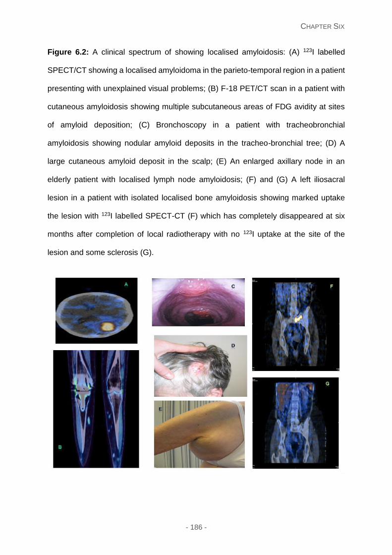
CHAPTER SIX
- 186 -
Figure 6.2: A clinical spectrum of showing localised amyloidosis: (A) 123I labelled
SPECT/CT showing a localised amyloidoma in the parieto-temporal region in a patient
presenting with unexplained visual problems; (B) F-18 PET/CT scan in a patient with
cutaneous amyloidosis showing multiple subcutaneous areas of FDG avidity at sites
of amyloid deposition; (C) Bronchoscopy in a patient with tracheobronchial
amyloidosis showing nodular amyloid deposits in the tracheo-bronchial tree; (D) A
large cutaneous amyloid deposit in the scalp; (E) An enlarged axillary node in an
elderly patient with localised lymph node amyloidosis; (F) and (G) A left iliosacral
lesion in a patient with isolated localised bone amyloidosis showing marked uptake
the lesion with 123I labelled SPECT-CT (F) which has completely disappeared at six
months after completion of local radiotherapy with no 123I uptake at the site of the
lesion and some sclerosis (G).
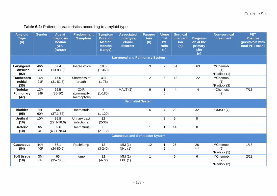
CHAPTER SIX
- 187 -
Table 6.2: Patient characteristics according to amyloid type
Amyloid Type (n)
Gender Age at diagnosis
Median yrs.
(range)
Predominant Symptom
Symptom Duration Median months (range)
Associated underlying
clonal disorder
Paraprotein (n)
Abnormal κ/λ
ratio (n)
Surgical Intervent
ion (n)
Progression at the primary
site (n)
Non-surgical treatment
PET Positive
(positive/n with total PET scan)
Laryngeal and Pulmonary System
Laryngeal+ Tonsillar
(92)
46M 46F
57.4 (13-83.3)
Hoarse voice 10.5 (1-360)
3 7 51 63 **Chemotx (1)
*Radiotx (1)
Tracheobronchial
(35)
14M 21F
47.6 (31-81.7)
Shortness of breath
4.3 (1-78)
2 5 18 22 **Chemotx (1)
*Radiotx (3)
Nodular Pulmonary
(47)
13M 34F
65.5 (36-80)
CXR abnormality
Haemoptysis
6 (1-180)
MALT (2) 9 10
4 4 *Chemotx (2)
7/18
Urothelial System
Bladder (95)
35F 60M
64 (37.1-87)
Haematuria 6 (1-120)
6 4 29 32 *DMSO (7)
Urethral (10)
10M 39.8 (27.1-78.6)
Urinary tract infections
12 (2-36)
2 5 5
Ureteric (10)
6M 4F
59.6 (43.1-78.4)
Haematuria 8 (2-112)
1 1 14 8
Cutaneous and Soft tissue System
Cutaneous (84)
44M 40F
56.1 (24-80.9)
Rash/lump 12 (3-240)
MM (1) NHL (1)
12 15
25 26 ***
**Chemotx (2)
*Radiotx (1)
1/18
Soft tissue (10)
3M 6F
65 (35-78.6)
lump 12 (4-72)
MM (1) LPL (1)
1 6 6 **Chemotx (2)
*Radiotx (2)
2/18
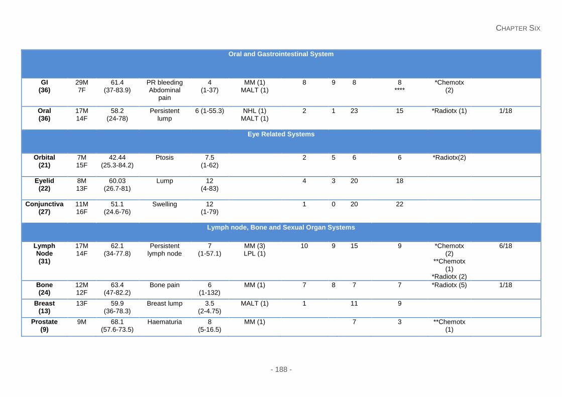
CHAPTER SIX
- 188 -
Oral and Gastrointestinal System
GI (36)
29M 7F
61.4 (37-83.9)
PR bleeding Abdominal
pain
4 (1-37)
MM (1) MALT (1)
8 9 8 8 ****
*Chemotx (2)
Oral (36)
17M 14F
58.2 (24-78)
Persistent lump
6 (1-55.3) NHL (1) MALT (1)
2 1 23 15 *Radiotx (1) 1/18
Eye Related Systems
Orbital (21)
7M 15F
42.44 (25.3-84.2)
Ptosis 7.5 (1-62)
2 5 6 6 *Radiotx(2)
Eyelid (22)
8M 13F
60.03 (26.7-81)
Lump 12 (4-83)
4 3 20 18
Conjunctiva(27)
11M 16F
51.1 (24.6-76)
Swelling 12 (1-79)
1 0 20 22
Lymph node, Bone and Sexual Organ Systems
Lymph Node (31)
17M 14F
62.1 (34-77.8)
Persistent lymph node
7 (1-57.1)
MM (3) LPL (1)
10 9 15 9 *Chemotx (2)
**Chemotx (1)
*Radiotx (2)
6/18
Bone (24)
12M 12F
63.4 (47-82.2)
Bone pain 6 (1-132)
MM (1) 7 8 7 7 *Radiotx (5)
1/18
Breast (13)
13F 59.9 (36-78.3)
Breast lump 3.5 (2-4.75)
MALT (1) 1 11 9
Prostate (9)
9M 68.1 (57.6-73.5)
Haematuria 8 (5-16.5)
MM (1) 7 3 **Chemotx (1)
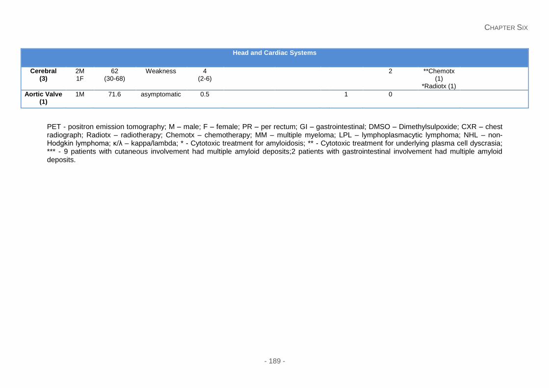
CHAPTER SIX
- 189 -
Head and Cardiac Systems
Cerebral (3)
2M 1F
62 (30-68)
Weakness 4 (2-6)
2 **Chemotx (1)
*Radiotx (1)
Aortic Valve (1)
1M 71.6 asymptomatic 0.5 1 0
PET - positron emission tomography; M – male; F – female; PR – per rectum; GI – gastrointestinal; DMSO – Dimethylsulpoxide; CXR – chest radiograph; Radiotx – radiotherapy; Chemotx – chemotherapy; MM – multiple myeloma; LPL – lymphoplasmacytic lymphoma; NHL – non-Hodgkin lymphoma; κ/λ – kappa/lambda; * - Cytotoxic treatment for amyloidosis; ** - Cytotoxic treatment for underlying plasma cell dyscrasia; *** - 9 patients with cutaneous involvement had multiple amyloid deposits;2 patients with gastrointestinal involvement had multiple amyloid deposits.
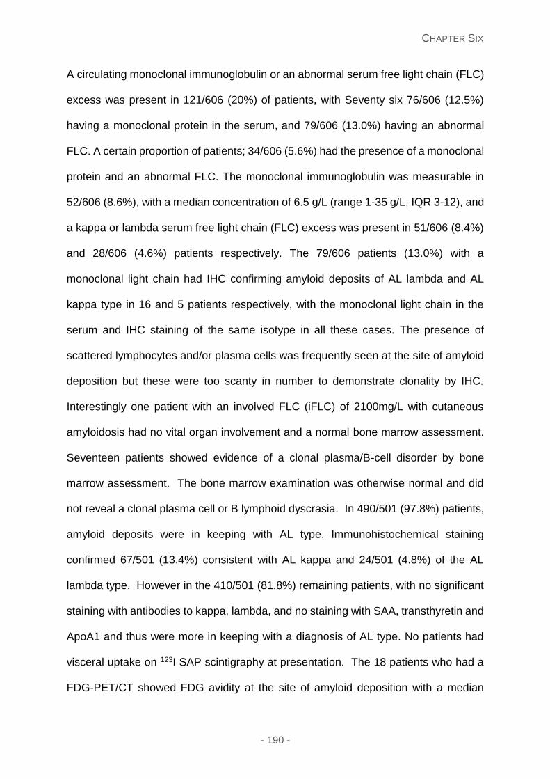
CHAPTER SIX
- 190 -
A circulating monoclonal immunoglobulin or an abnormal serum free light chain (FLC)
excess was present in 121/606 (20%) of patients, with Seventy six 76/606 (12.5%)
having a monoclonal protein in the serum, and 79/606 (13.0%) having an abnormal
FLC. A certain proportion of patients; 34/606 (5.6%) had the presence of a monoclonal
protein and an abnormal FLC. The monoclonal immunoglobulin was measurable in
52/606 (8.6%), with a median concentration of 6.5 g/L (range 1-35 g/L, IQR 3-12), and
a kappa or lambda serum free light chain (FLC) excess was present in 51/606 (8.4%)
and 28/606 (4.6%) patients respectively. The 79/606 patients (13.0%) with a
monoclonal light chain had IHC confirming amyloid deposits of AL lambda and AL
kappa type in 16 and 5 patients respectively, with the monoclonal light chain in the
serum and IHC staining of the same isotype in all these cases. The presence of
scattered lymphocytes and/or plasma cells was frequently seen at the site of amyloid
deposition but these were too scanty in number to demonstrate clonality by IHC.
Interestingly one patient with an involved FLC (iFLC) of 2100mg/L with cutaneous
amyloidosis had no vital organ involvement and a normal bone marrow assessment.
Seventeen patients showed evidence of a clonal plasma/B-cell disorder by bone
marrow assessment. The bone marrow examination was otherwise normal and did
not reveal a clonal plasma cell or B lymphoid dyscrasia. In 490/501 (97.8%) patients,
amyloid deposits were in keeping with AL type. Immunohistochemical staining
confirmed 67/501 (13.4%) consistent with AL kappa and 24/501 (4.8%) of the AL
lambda type. However in the 410/501 (81.8%) remaining patients, with no significant
staining with antibodies to kappa, lambda, and no staining with SAA, transthyretin and
ApoA1 and thus were more in keeping with a diagnosis of AL type. No patients had
visceral uptake on 123I SAP scintigraphy at presentation. The 18 patients who had a
FDG-PET/CT showed FDG avidity at the site of amyloid deposition with a median
CHAPTER SIX
- 191 -
standardised uptake values (SUV) of 2.7 (2.5-10) in 14 of the 18 patients with reported
SUV values (Appendix 5, Supplemental table 1).
Treatment
Complete details regarding interventions at the local disease site were available in
527/606 (87%) cases, and incomplete treatment details in 72/606 (11.9%), with
survival details also present in these patients. In total, 90/606 (14.9%) patients opted
for local follow up preferably, with the open option of re-attending the National
Amyloidosis Centre should they developed progressive symptoms or want to be seen
at the NAC. A certain proportion of patients, 257/527 (48.8%) required no
interventional treatment following the diagnostic biopsy or excision. 270/527 (51.2%)
proceeded to at least one interventional procedure for symptom relief and 112/527
(21.3%) patients required more than one repeat procedure. The symptom triggering
the need for intervention was often the predominant/presenting symptoms based on
site of deposits (table 6.2 illustrates these symptoms). The symptom of increasing size
of the lesions usually prompted the need for intervention in cutaneous, oral, soft tissue,
lymph node, nodular pulmonary and breast lesions. Pain was the trigger for in urethral
and bone amyloid deposits. Interventional procedures included surgical treatment in
44% and laser therapy in 7% (Table 6.2). The surgical removal (n=4) or radiotherapy
treatment (n=4) of the amyloid deposits in 8 patients led to disappearance of the
circulating clonal markers, (Appendix 5, Supplemental table 2).
Local radiotherapy has been used to treat patients more recently, and 18 patients were
treated with this modality in our cohort (table 2). Symptomatic improvement was
evident in 10/18 patients - 5 with bone lesions, 4 laryngeal/tracheobronchial and 1
patient with a localised amyloidoma in the femoral nerve/lumbar plexus. Three
CHAPTER SIX
- 192 -
patients received radiotherapy for tracheobronchial amyloidosis and one diagnosed
with laryngeal amyloidosis following failure of prior surgical resections and stent
insertions. With a median follow up of 11 months (range 2-14 months), all patients
noticed a significant improvement in their symptoms.
Chemotherapy was used in four patients: 2 having symptomatic pulmonary and 2 with
lymph node amyloid deposits with stabilisation of symptoms but no significant
improvement. There were a proportion of patients in whom corticosteroids alone were
used; 12/606 (2%) cases (in 7 patients with tracheobronchial amyloidosis). Topical
dimethylsulfoxide (DMSO) was used in 7 cases of bladder amyloidosis with 5 cases
noticing a symptomatic benefit. Eleven other patients were treated with chemotherapy
for progression of an underlying haematological disorder, not particularly for amyloid
progression; with no significant impact on the local amyloid deposits.
Progression and survival
The median follow up of the study cohort is 74.4 months (range 3.7-349.2 months,
IQR 37.2-132), and 51% (309/606) of patients were diagnosed after 2006 and 27.2%
(87/606) before 2000 (illustrated in table 6.1). Progression at the primary site was
present in 264/606 (43.5%) patients (table 6.2). Progression to systemic amyloidosis
was rare, occurring in only 7/606 cases (1.2%); (five had a circulating monoclonal
protein and one had a with detectable plasma cell clone in the marrow). The latter 7
patients included 5 patients with lymph node (LN) involvement, one had eyelid and
another bone involvement, progressing at a median on 51 months (20, 51, 60, 77, 84,
48 and 51 months in each case respectively) from the diagnosis point. The site of
progression to systemic amyloidosis included: three patients with soft tissue
progression, two developed renal amyloidosis (one with additional soft tissue) and one
CHAPTER SIX
- 193 -
each having new lymph node or bone lesions (Appendix 5, Supplemental table 3).
One patient developed asymptomatic uptake within the spleen by SAP scintigraphy.
During the follow up period, there were a total of 94 deaths, and only three deaths
directly attributed due to progression to systemic AL. The cause of death was
determined in 84 cases from the death certificate data from the Office for National
Statistics.290 In 10 patients, the patient’s general practice doctor was contacted to
ascertain the cause of death. The majority of patients had other co-morbidities
including chronic obstructive airways disease, sepsis, ischaemic heart disease,
autoimmune disease and cancer of other aetiology. The 5 and 10 year estimated
survival was 90.6% (95%CI; 87.7-92.9) and 80.3% (95%CI; 75.7-84.1) respectively
(illustrated in Figure 6.3A); with the median survival not reached for the entire cohort.
The median survival for patients aged 70 years or older was 12.1 years (95% CI; 10.5-
13.7). We compared our localised AL cohort to systemic AL patients diagnosed during
this same time frame, (Figure 6.3B) showing the estimated survival for systemic AL
amyloidosis over this time period was significantly shorter, 1.6 years (95%CI; 1.1-2.1)
and 5 and 10 year estimated survival 37.0% (95%CI;0.37-0.43) and 26% (95%CI;0.23-
0.28)% respectively. We examined the OS of patients diagnosed over different time
periods of this 30 year study when assessed as 5-yearly cohorts (except 1985-1995;
included as one group due to smaller patient numbers) (Figure 6.3C), showing no
significant difference and the numbers diagnosed showing an increasing trend. The 5
year survivals were as follows: 1980-1995 [(97%(95%CI;0.82-0.97) and
(87%(95%CI;0.75-0.94)], 1996-2000 [(88%(95%CI;0.79-0.93)and (77%(95%CI;0.67-
0.85)], 2001-2005 [(91%(95%CI;0.84-0.94) and (79%(95%CI;0.68-0.85)], 2006-2011
[(91%(95%CI;0.86-0.94) and (85%(95%CI;0.76-0.91)].
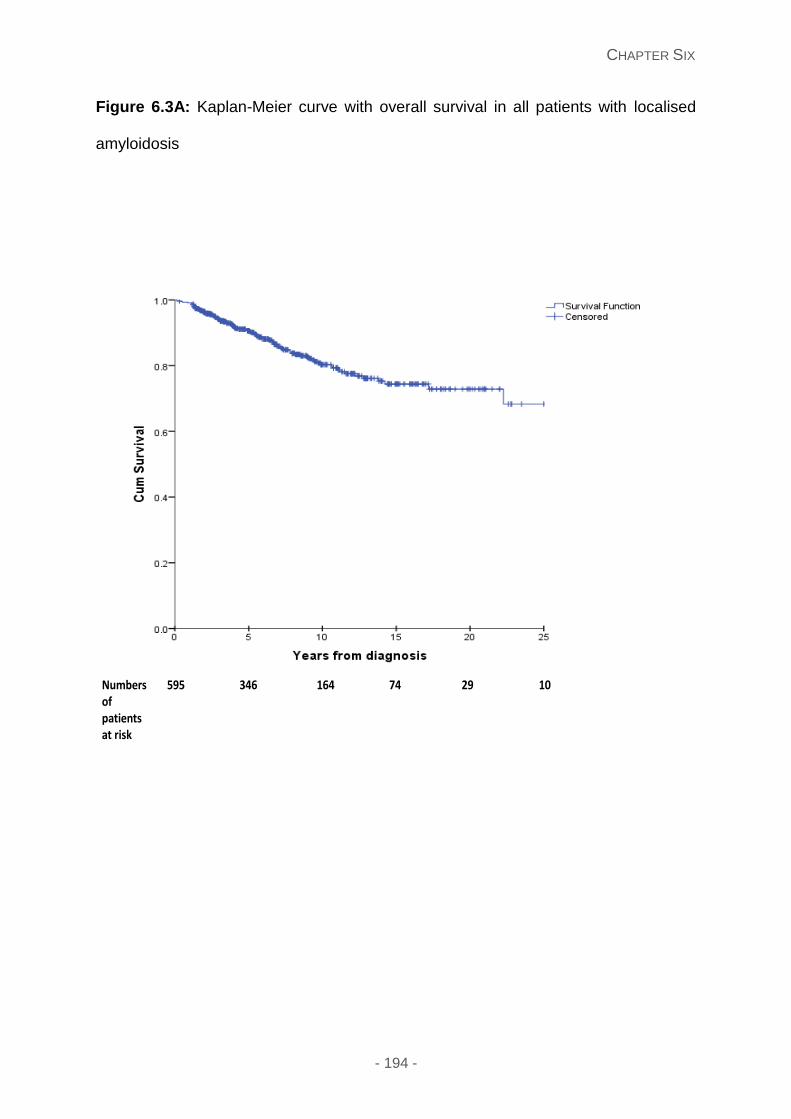
CHAPTER SIX
- 194 -
Figure 6.3A: Kaplan-Meier curve with overall survival in all patients with localised
amyloidosis
Numbers of patients at risk
595 346 164 74 29 10
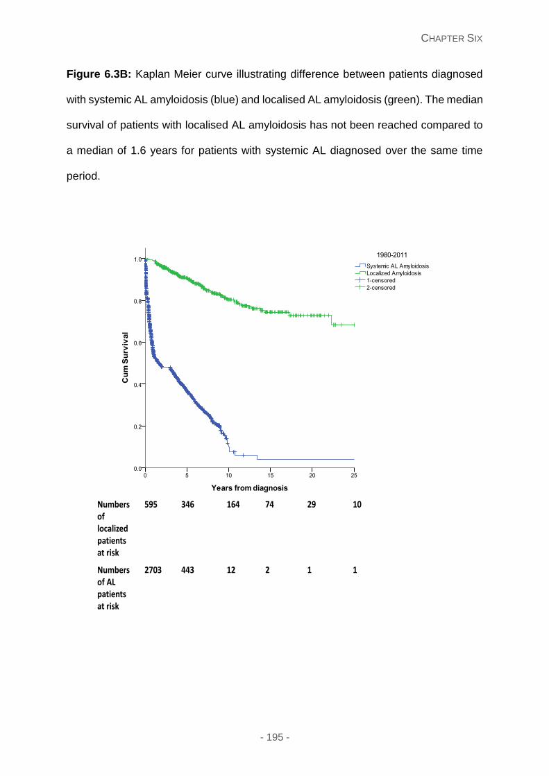
CHAPTER SIX
- 195 -
Figure 6.3B: Kaplan Meier curve illustrating difference between patients diagnosed
with systemic AL amyloidosis (blue) and localised AL amyloidosis (green). The median
survival of patients with localised AL amyloidosis has not been reached compared to
a median of 1.6 years for patients with systemic AL diagnosed over the same time
period.
Numbers of localized patients at risk
595 346 164 74 29 10
Numbers of ALpatientsat risk
2703 443 12 2 1 1
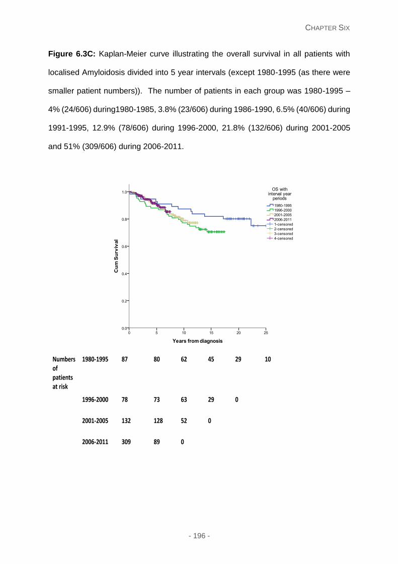
CHAPTER SIX
- 196 -
Figure 6.3C: Kaplan-Meier curve illustrating the overall survival in all patients with
localised Amyloidosis divided into 5 year intervals (except 1980-1995 (as there were
smaller patient numbers)). The number of patients in each group was 1980-1995 –
4% (24/606) during1980-1985, 3.8% (23/606) during 1986-1990, 6.5% (40/606) during
1991-1995, 12.9% (78/606) during 1996-2000, 21.8% (132/606) during 2001-2005
and 51% (309/606) during 2006-2011.
Numbers of patients at risk
1980-1995 87 80 62 45 29 10
1996-2000 78 73 63 29 0
2001-2005 132 128 52 0
2006-2011 309 89 0
CHAPTER SIX
- 197 -
Discussion
This study describes the largest cohort of patients with localised AL amyloidosis with
clinical features and natural disease course showing stark differences from systemic
AL amyloidosis. Localised AL amyloidosis is classified as having a relatively indolent
disease course that rarely evolves systemically. Most patients can be managed
adequately with local interventions, which is the primary treatment modality. In this
series, in striking contrast to systemic AL amyloidosis, localised amyloidosis does not
appear to impact survival and, with only 3 deaths directly attributed to progression to
systemic AL amyloidosis.
Defining localised amyloidosis is critically important in assessing isolated deposits and
the also in determining progression. We defined localised amyloidosis as a patient
with amyloid deposition at one single non-vital organ site with no evidence of
amyloidotic end organ damage by the diagnostic criteria of the international society of
amyloidosis of systemic organ involvement (in relation to no cardiac, renal, liver, nerve
or macroglossia). We had a unique modality to rule out visceral amyloid deposition
with a very high level of confidence, due to the availability of SAP scintigraphy (a very
sensitive and highly specific method for imaging visceral amyloid deposits) at our
centre. Hence, in absence of SAP scintigraphy, this diagnosis has to be approached
with greater caution especially with those with a detectable FLC excess or M-protein
and follow up would reveal the true nature of the diagnosis.
Systemic AL amyloidosis typically affects the heart, kidney, liver, peripheral and
autonomic nerves.86 In contrast, localised AL amyloidosis occurs mainly in mucosal
sites (affecting the airways, bladder, gastrointestinal tract, and conjunctiva), skin or
glandular sites (like breast) as in the current series and also previously reported.291-293
CHAPTER SIX
- 198 -
The presenting symptoms are site specific and may range from a localised lump (e.g.
under the skin or breast lump), bleeding due to mucosal erosion (symptoms of
haematuria or haemoptysis or melena) to obstructive symptoms when deposits are
located at a luminal site (bronchial, ureter or GI tract obstruction). Multiple sites of
involvement were present in certain patients with cutaneous and gastrointestinal
amyloidosis.
Localised amyloidosis has generally been reported to be of AL type. In the current
series, of a total of 606 patients with localised amyloidosis, we considered 98%
(490/501) patients to be in keeping with the AL type. However, we do recognise one
of the limitations of our study was typing amyloid deposits with a high proportion of
cases in the present series having staining with antibodies to both kappa and lambda
light chains (although lacking staining with antibodies to SAA, transthyretin and
ApoA1). In the era before access to laser capture followed by mass spectrometry (the
current gold standard), AL amyloidosis was a diagnosis of exclusion in cases where
specific staining with light chain antibodies could not be demonstrate by excluding the
presence of amyloid deposits of other types by appropriate staining. Due to the long
duration of this study, the vast majority of the patients were diagnosed using this
algorithm. However, it was notable that the staining with antibodies to kappa or
lambda was much lower than generally seen in patients with systemic AL amyloidosis,
and forward planning with the use of proteomic analysis will assist in this clarification.
A recent case series of nodular pulmonary amyloidosis from the Mayo group, using
laser capture micro dissection followed by tandem mass spectrometry (LC-MS) for
fibril typing, suggested that amyloid fibrils are a mixture of heavy (AH) and light chain
(AL) type;294 a potential explanation for difficulties in accurate staining by anti-light
chain antibodies. The retrospective nature of this study over a long period limited the
CHAPTER SIX
- 199 -
scope of LC-MS being undertaken for the current series and does remain a limitation
of this series.
Generally, over 95% of patients diagnosed with systemic AL amyloidosis have a
detectable monoclonal protein or abnormal FLC295 (which directly impacts on
prognosis296) and a detectable bone marrow plasma cell clone; in contrast with the
present series in which a normal bone marrow was seen in those with an abnormal
FLC or monoclonal protein (except 17 patients with an underlying plasma cell
dyscrasia/B-cell clone) and 80% with no evidence of a circulating M-protein or
abnormal FLC. In our study, 20% (121/606) had a monoclonal protein or abnormal
serum free light chain excess, with the presence of a monoclonal protein in 76/606
(12.5%) and abnormal serum free light chain excess in 79/606 (13.0%), and some
overlap of these groups. We recognise this figure may be higher than other centres,
and this difference predominantly due to referral bias and age of the referred
populations in different respective centres. Previous studies have shown that 5-10%
of the population have an incidental monoclonal protein in those older than 70 years
of age.297 Patients with lymph node involvement had a higher chance of having a
detectable monoclonal protein, with one study showing this group as having a high
risk of progressing to systemic AL.298 Of seven patients who progressed to systemic
AL, 5 had evidence of a detectable clonal marker in the serum. Patients with systemic
AL amyloidosis having abnormal light chains for many years prior to diagnosis,299
localised AL patients with an abnormal FLC still warrant long term follow up for
progression. Lack of clonality by bone marrow assessment in localised amyloidosis
supports previous reports of limited clonal proliferation at the site of amyloid deposition
as documented by highly sensitive methods like IgH-PCR300, 301 with only few patients
of the present series having an obvious local clonal cell infiltrate. The location of skin
CHAPTER SIX
- 200 -
and mucosal sites raise the possibility of chronic antigenic stimulation or autoimmunity
as drivers for local clonality302 particularly as Mucosa-associated lymphoid tissue
(MALT) lymphoma is a reported cause of lung and breast amyloidosis.303, 304 23% of
all cases of breast amyloidosis in this series were associated with Sjögren’s syndrome
(an association in 17 cases in total), also a previously reported association of localised
amyloidosis with MALT.305 The lack of detectable clonal cells remains an enigma in
most cases. Oligomeric light chains may be toxic to cells and tissues – with one theory
suggesting that plasma cells in localised AL are part of a “suicide” neoplasm and die
to due to light chain toxicity locally.306 Hence, this is an interesting hypothesis to
explain lack of cells as well as lack of systemic progression.
Serial monitoring of patients with localised amyloidosis is a difficult hurdle since the
standard serum or urine markers are frequently normal or not contributory with 123I
labelled SAP scintigraphy rarely abnormal. Surprisingly, all 18 patients who had a
FDG-PET/CT performed in the current series (Supplementary table 1) showed
presence of FDG avidity at the site of amyloid deposition, in keeping with previous
reports of high proportion of FDG-PET/CT positivity in localised amyloidosis.307 This
potentially useful finding is a metabolic conundrum given the lack of identifiable clonal
cell infiltration in the deposits. Inflammatory or giant cells within the amyloid deposits
have been suggested as a potential cause of the metabolic activity detected by FDG-
PET/CT.308 Further studies are needed to investigate the utility FDG PET/CT in both
diagnosis and monitoring of patients with Localised amyloidosis.
In contrast to systemic AL amyloidosis, approximately 50% of the patients in the
current series did not require any intervention following the diagnostic biopsy/excision.
Patients with obstructive/pressure symptoms or bleeding needed endoscopic or
CHAPTER SIX
- 201 -
surgical resection. Half of all patients needed one such procedure and a fifth of all
patients required repeated procedures for symptom control highlighting the need for
long term monitoring. Four patients in the current study needed chemotherapy for
localised amyloidosis. Radiotherapy (targeting the presumed clonal proliferation)
appears to be a useful, and possibly underutilised, treatment modality in localised
amyloidosis with all patients in the current series treated with radiotherapy showing
lack of progression and over half achieving good symptomatic improvement following
radiotherapy. Tracheo-bronchial deposits can be very troublesome and symptomatic
with marked morbidity and are a particular challenge amongst Localised AL. In this
group, radiotherapy has been reported to give symptomatic benefit (in 8/10 309 and 7/7
310 patients treated) and this was also evident in all three treated patients in the current
cohort. The dose and scheduling of radiotherapy require further systematic study.
Systemic AL amyloidosis typically remains a disease with a poor prognosis with a
median overall survival of about four years and, despite much progress, nearly a third
of all patients die in the first few months following diagnosis.192 We compared our
localised cohort with patients diagnosed with systemic AL during this same time frame,
with an explicitly better prognosis in the former. This large series reassuringly
examines the natural disease course of Localised amyloidosis, showing it has an
excellent prognosis with a median survival not reached in our cohort compared with
an overall surviall of 1.6 yrs for patients with systemic AL amyloidosis seen during the
same period. Most of the patients died of other causes and only three deaths
attributable directly to localised AL. The suvival of the patients compared with that
expected in the British population (medain survival of the over 70 yrs patients in the
current series 12.1 yrs which was similar to that for the British person at the age of 70
years) (date from Office of National Statistics UK).290 Another cruciaal point was that
CHAPTER SIX
- 202 -
only seven patients or 1.2% progressed to systemic AL amyloidosis. We specifically
ruled out visceral deposition by serum amyloid P (SAP) scintigraphy and cardiac
involvement by echocardiography plus normal cardiac biomarkers. There are two
groups of patients with localised AL which pose some problems – those with lymph
node involvement and those with a detectable circulating light chain/M-protein having
the same isotype as the amyloid type. It is contentious whether we can confidently
say that some patients with a localised lymph node amyloid deposit have localised
disease or “systemic” disease. Patients with no vital organ involvement and a localised
lymph node were categorised as localised amyloidosis, however this group is very
different from the other types of localised amyloidosis; with the increased risk of
“systemic disease” and spread through the lymphatic system, akin to other types of
lymphoma. In our series, 5/31 patients with lymph node involvement progressed to
systemic AL and 6/79 patients with a clonal light chain excess progressed (5 of these
having lymph node patients), with only one patient with no obvious light chain excess
progressed. We recognise that patients with isolated lymph node amyloidosis are
deemed at high risk of progression, and similarly those with an isotypic excess of FLC
also need careful monitoring. The very low chance of systemic progression in other
patients with localised amyloidosis implies that these patients should not be subjected
to frequent invasive monitoring.
There are a number of gaps in our knowledge of localised amyloidosis, with the
retrospective nature of this study. A longer median follow up is always important in
following and understanding a disease entity, recognising that 51% (309/606) of
patients were diagnosed after 2006. Accurate identification of the amyloid fibril type by
LC-MS is important with immunohistochemistry sometimes inadequate for fibril typing
in this setting. Novel approaches to clarify the nature of the underlying clonal
CHAPTER SIX
- 203 -
disorder(s) is required to guide therapy and unravel the prediliction for specific sites.
International colloborative studies are needed to prospectively evaluate the utility of
radiotherapy and other treatment modalities.
In summary, localised AL amyloidosis is a disease different from systemic AL
amyloidosis. Local surgical resection is adequate in most cases when treatment is
needed. Radiotherapy appears to have a useful role in patients not controlled by local
measures. Progression to systemic AL amyloidosis is extremely rare except in
patients with lymph node involvement. Patients with lymph node involvement and
those with a isotypic specific circulating free light chains warrant closer follow up for
development of systemic AL. The majority with localised AL have excellent long term
outcomes. Most die with the disease rather than as a consequence of it.

CHAPTER SEVEN
- 204 -
Chapter Seven: Laryngeal tracheobronchial amyloidosis: clinical
and proteomic analysis showing an association with Apo A1 and
insulin-like growth factor binding protein complex
Introduction
Laryngo-tracheobronchial amyloidosis is a rare type of localised amyloidosis,
characterised by insoluble fibrillar proteins deposited within the upper and lower airway
tract.311, 312 The underlying amino acid sequences and building block structure of this
and other types of amyloidoses share a common β-sheet conformation of the
polypeptide backbone,313 and thus forming a fibrillar, insoluble, proteolytic resistant
structure in all types of amyloid. Exploring the mechanism of the amyloid fibril
formation is vital in gaining insight into the mechanisms in the underlying
polymerisation of the soluble, monomeric peptide into the insoluble β-pleated sheet.
Earlier studies have shown that this conversion of a soluble peptide to an insoluble
fibril often includes the formation of a partially unfolded intermediate.314, 315 Some
studies propose a nucleation dependant polymerisation model to explain the fibril
formation.316 The underlying pathogenic factors are unknown, with the hypothesis
including a population of clonal plasma cells or B cells instigating this process in a
localised area. This entity is clearly different from systemic light chain (AL)
amyloidosis, in which the clonal plasma cells arise from the bone marrow and secrete
excessive quantities of monoclonal immunoglobulins which then circulate to target
organs.317
The aims of this study were to (a) report our experience of patients presenting with
laryngeal and tracheobronchial amyloidosis, examining the clinical symptoms,
CHAPTER SEVEN
- 205 -
treatment strategies and overall survival from a clinical perspective, (b) analyse the
laryngeal /tracheobronchial biopsies and undertaking proteomic analysis of these
patients and ascertain any potential underlying pathogenic instigators in this process
from a laboratory perspective.
Patients and methods
Patients
Patients with laryngeal and tracheobronchial amyloidosis between January 1980 and
December 2011 were identified from the database of the UK National Amyloidosis
Centre. Written consent was obtained from all patients in accordance with the
Declaration of Helsinki. Exclusion of systemic AL and vital organ involvement
(including cardiac, renal, liver, peripheral or autonomic neuropathy) was performed on
review of a detailed baseline assessment of organ function and no uptake by 123I
serum amyloid P component (SAP) scintigraphy, with patients before 1988 (availability
of 123I SAP scintigraphy) having this procedure later in their follow up course. Baseline
investigations included a full blood count, renal and liver function tests, serum protein
electrophoresis, serum free light chains (prospectively in samples after 2002 and
retrospectively on stored serum samples where available), and urinary BJP,
electrocardiogram and echocardiography and 123I SAP scintigraphy. Histological
analysis was performed on all biopsies with Congo red staining and apple green
birefringence and immunohistochemical staining for kappa, lambda, transthyretin or
serum amyloid A (SAA) antibodies undertaken in the majority. Genetic sequencing
was undertaken dependent upon clinical details and immunohistochemical staining,
such as Apolipoprotein (Apo) AI in all patients with available blood samples. During
this time frame, 63 patients with laryngeal involvement and 34 patients with
CHAPTER SEVEN
- 206 -
tracheobronchial amyloidosis with biopsy proven amyloid deposition were recruited.
The pathology slides were requested and reviewed at our centre and laser capture of
the slides undertaken to then perform proteomic analysis, with a total of 60 biopsies
retrieved. A total of 37 biopsies were not able to be analysed due to inaccessibility of
the original samples requested from the local hospitals/referring centres.
Patient questionnaire with quality of life assessment
A patient questionnaire was sent to all patients to ascertain the following data with 52
responses and 46 responses concerning quality of life. The questionnaire comprised
of 2 questions related to symptoms and duration and treatment options, and quality of
life with a grading score 0-10; score of 0 depicting a poor quality of life and 10
describing an excellent quality of life. The median overall score and ranges were
gained to reflect an understanding in the clinical impact of this disease from a patent’s
perspective.
Protocol for laser capture
Laser capture micro dissection and mass spectrometry (LDMS) is an invaluable tool
for identifying proteins from formalin-fixed, paraffin embedded tissues, with more in
depth procedure described in the methods chapter.169 Visualisation and locating the
amyloid was done using bright field (Figure 7.1A) and Rhodamine filter sets (TRITC)
on the Zeis Palm Microbeam Laser capture microscope, with amyloid typically yellow
on a “red” background. (Figure 7.1B) Areas of interest were drawn around, appearing
yellow under a Tritc florescent filter (Figure 7.1C) or under bright field (Figure 1D) and
“cut” out using laser capturing into 0.5ml micro-centrifuge adhesive cap tubes (Zeiss)
and checking the cap to ensure that some tissue had been captured. Figure 7.1D
illustrates the amyloid tissue post dissecting the tissue of interest and the figure 7.1E
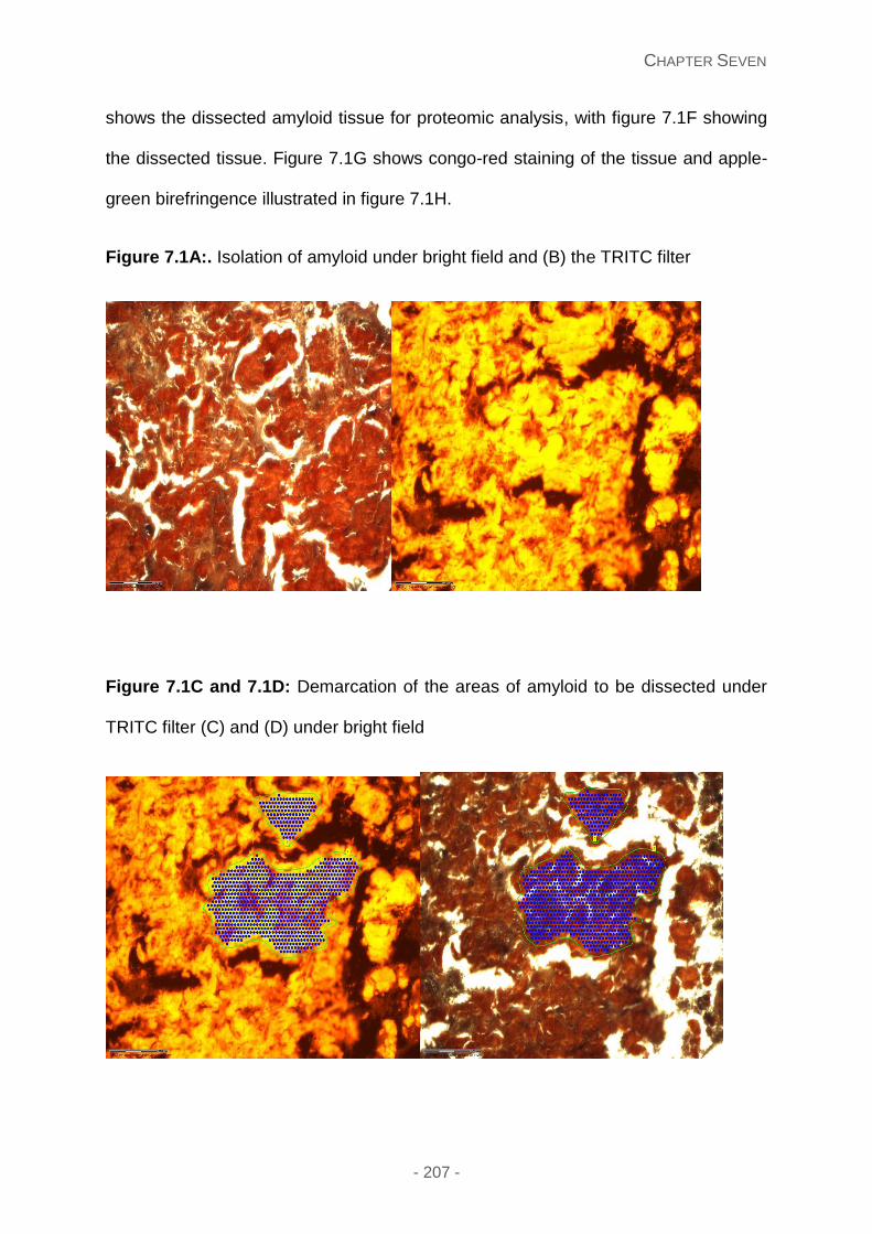
CHAPTER SEVEN
- 207 -
shows the dissected amyloid tissue for proteomic analysis, with figure 7.1F showing
the dissected tissue. Figure 7.1G shows congo-red staining of the tissue and apple-
green birefringence illustrated in figure 7.1H.
Figure 7.1A:. Isolation of amyloid under bright field and (B) the TRITC filter
Figure 7.1C and 7.1D: Demarcation of the areas of amyloid to be dissected under
TRITC filter (C) and (D) under bright field
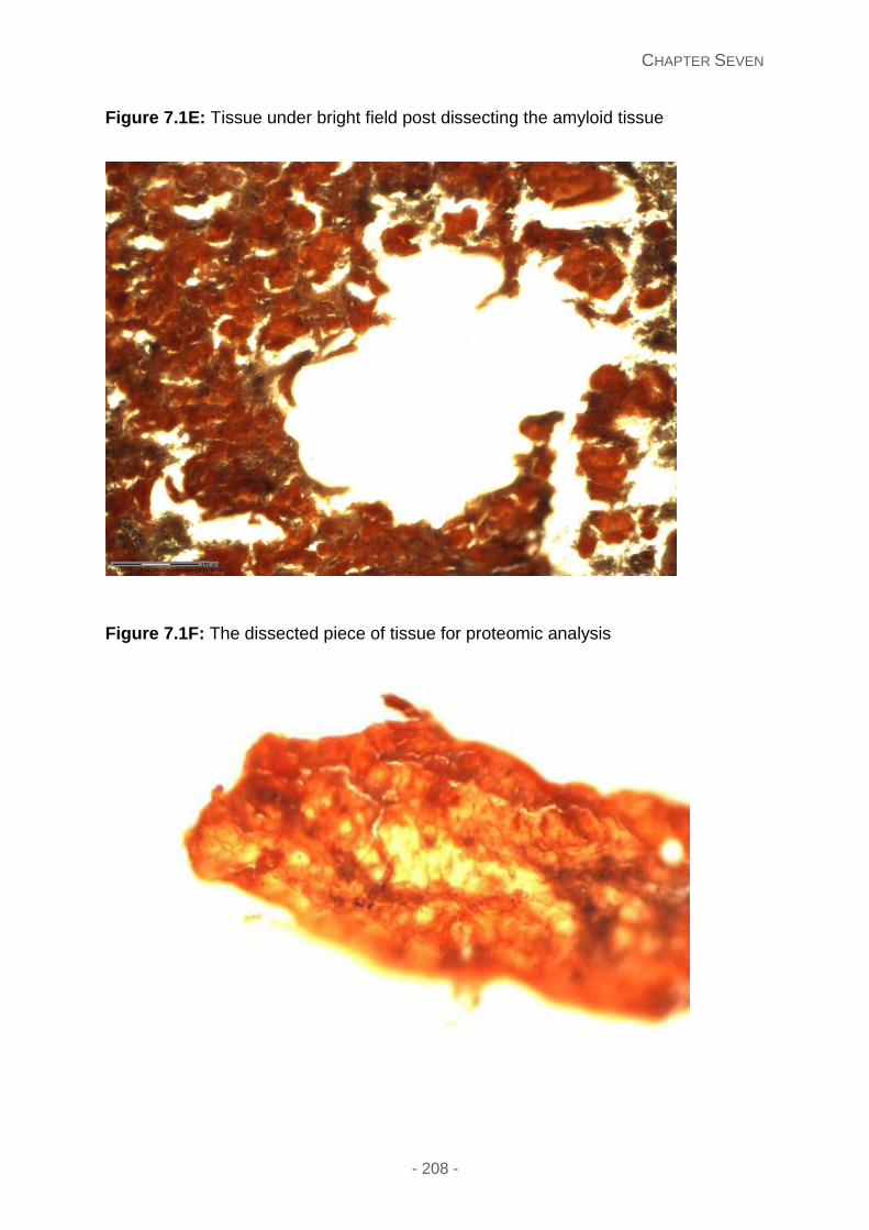
CHAPTER SEVEN
- 208 -
Figure 7.1E: Tissue under bright field post dissecting the amyloid tissue
Figure 7.1F: The dissected piece of tissue for proteomic analysis
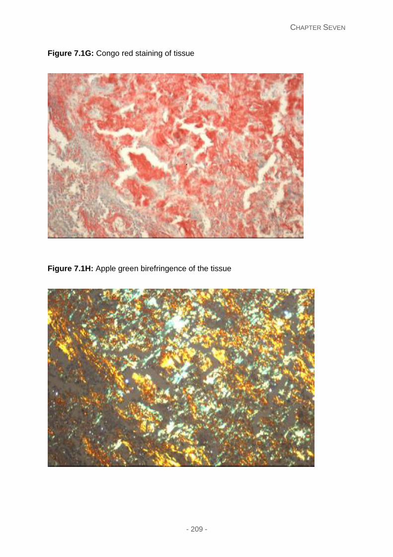
CHAPTER SEVEN
- 209 -
Figure 7.1G: Congo red staining of tissue
Figure 7.1H: Apple green birefringence of the tissue
CHAPTER SEVEN
- 210 -
The data was processed using Proteome Discoverer for MASCOT searching of the
NCBI and IPI databases enabling mass spectrometry results to be produced.
Statistical analysis and overall survival
Data was analysed and summarised using the number (percentage) and median
(minimum and maximum range). Quality of life assessments were based upon a score
0-10, with 0 and 10 equating to no quality of life and an excellent quality of life
respectively. Kaplan Meier curves were used to estimate the overall survival (OS);
calculated from the start of symptoms until death or last follow-up for the laryngeal and
tracheobronchial cohorts. Kaplan Meier estimates were used using SPSS v20 (IBM
SPSS) to calculate the OS and PFS. All p values are 2 sided, with significance values
based on a p <0.05.
Results
Patient population
The study was conducted between January 1980 and December 2011 and clinical
characteristics are summarised in table 7.1 and 7.2 with the symptoms within each
group illustrated in figure 2A and 2B. No patient developed systemic AL amyloidosis.
Genetic sequencing was performed in 59 patients, with only 1 patient diagnosed with
laryngeal involvement heterozygote for the Apolipoprotein A1 Ala160Ser mutation and
no evidence of other organ dysfunction.
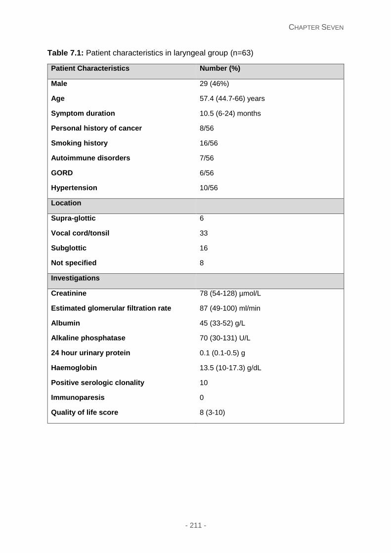
CHAPTER SEVEN
- 211 -
Table 7.1: Patient characteristics in laryngeal group (n=63)
Patient Characteristics Number (%)
Male 29 (46%)
Age 57.4 (44.7-66) years
Symptom duration 10.5 (6-24) months
Personal history of cancer 8/56
Smoking history 16/56
Autoimmune disorders 7/56
GORD 6/56
Hypertension 10/56
Location
Supra-glottic 6
Vocal cord/tonsil 33
Subglottic 16
Not specified 8
Investigations
Creatinine 78 (54-128) µmol/L
Estimated glomerular filtration rate 87 (49-100) ml/min
Albumin 45 (33-52) g/L
Alkaline phosphatase 70 (30-131) U/L
24 hour urinary protein 0.1 (0.1-0.5) g
Haemoglobin 13.5 (10-17.3) g/dL
Positive serologic clonality 10
Immunoparesis 0
Quality of life score 8 (3-10)
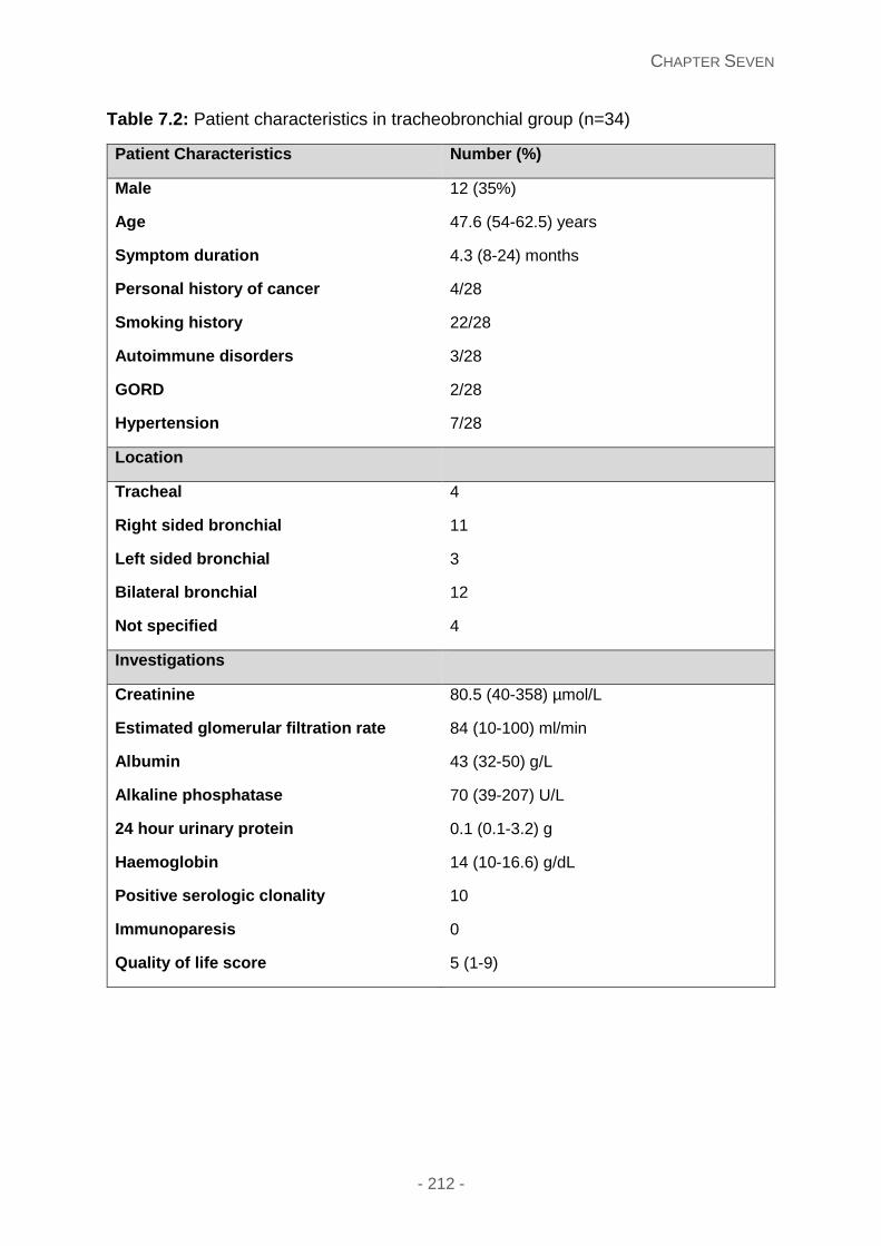
CHAPTER SEVEN
- 212 -
Table 7.2: Patient characteristics in tracheobronchial group (n=34)
Patient Characteristics Number (%)
Male 12 (35%)
Age 47.6 (54-62.5) years
Symptom duration 4.3 (8-24) months
Personal history of cancer 4/28
Smoking history 22/28
Autoimmune disorders 3/28
GORD 2/28
Hypertension 7/28
Location
Tracheal 4
Right sided bronchial 11
Left sided bronchial 3
Bilateral bronchial 12
Not specified 4
Investigations
Creatinine 80.5 (40-358) µmol/L
Estimated glomerular filtration rate 84 (10-100) ml/min
Albumin 43 (32-50) g/L
Alkaline phosphatase 70 (39-207) U/L
24 hour urinary protein 0.1 (0.1-3.2) g
Haemoglobin 14 (10-16.6) g/dL
Positive serologic clonality 10
Immunoparesis 0
Quality of life score 5 (1-9)
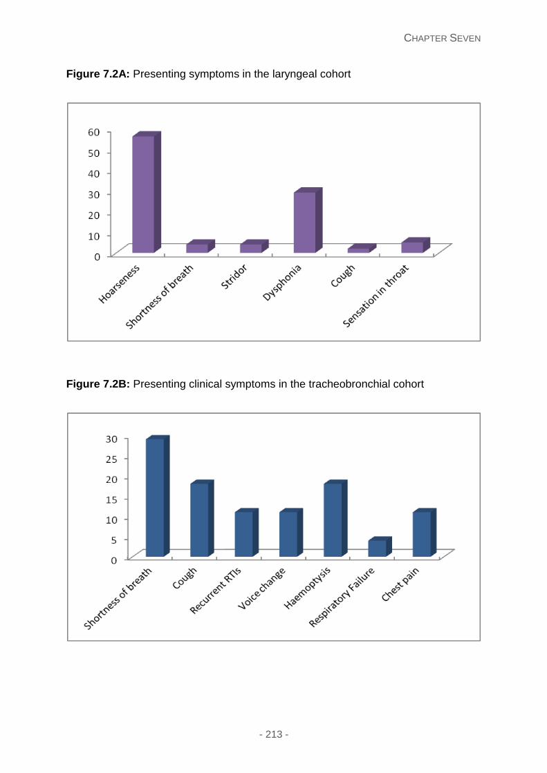
CHAPTER SEVEN
- 213 -
Figure 7.2A: Presenting symptoms in the laryngeal cohort
Figure 7.2B: Presenting clinical symptoms in the tracheobronchial cohort
CHAPTER SEVEN
- 214 -
Treatment strategies
Surgery
Surgical and laser therapeutic options were pursued in 39/63 (62%) and 18/34 (52.9%)
patients with laryngeal and tracheobronchial amyloidosis with 29% (n=18) and 24%
(n=8) requiring repeated procedures respectively.
Radiotherapy
A total of 4 patients with symptomatic airway amyloidosis involvement (1 laryngeal and
3 tracheobronchial) received radiotherapy with a median dose of 20 Gy in 10 fractions.
Following a median follow up of 18.7 months range (6-47), all patients had
experienced symptomatic local control.
Chemotherapy
One patient with laryngeal involvement and one patient with tracheobronchial
amyloidosis received chemotherapy; each with a lambda light chain excess and no
evidence of an excess of plasma cells by bone marrow assessment. Both patients
achieved a fall in the lambda light chains to less than 40mg/L with minimal
improvement in their symptoms. It is difficult to ascertain or comment on whether their
symptoms would have progressed with no treatment.
Quality of life assessment
A patient questionnaire was sent to all patients, with a response in 46 patients. The
median quality of life score in 33 laryngeal patients and 13 tracheobronchial patients
was 8 (range 3-10) and 5 (range 1-9) respectively. This is illustrated in table 7.1 and
7.2.
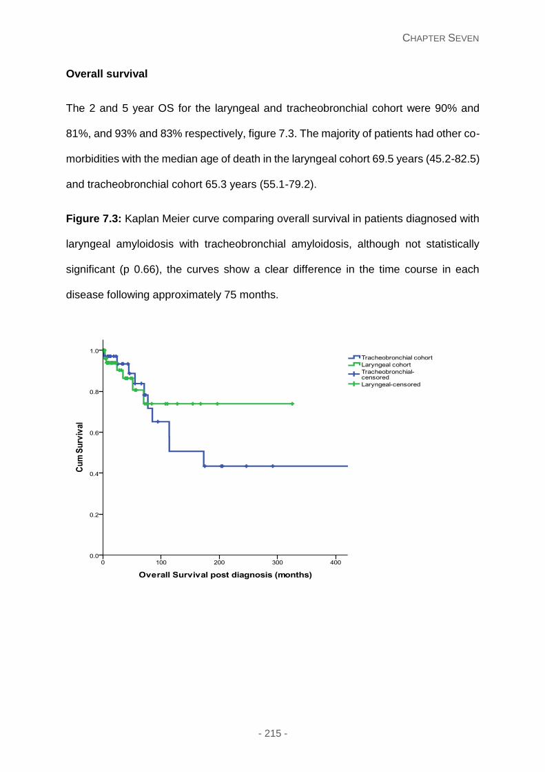
CHAPTER SEVEN
- 215 -
Overall survival
The 2 and 5 year OS for the laryngeal and tracheobronchial cohort were 90% and
81%, and 93% and 83% respectively, figure 7.3. The majority of patients had other co-
morbidities with the median age of death in the laryngeal cohort 69.5 years (45.2-82.5)
and tracheobronchial cohort 65.3 years (55.1-79.2).
Figure 7.3: Kaplan Meier curve comparing overall survival in patients diagnosed with
laryngeal amyloidosis with tracheobronchial amyloidosis, although not statistically
significant (p 0.66), the curves show a clear difference in the time course in each
disease following approximately 75 months.
CHAPTER SEVEN
- 216 -
Proteomic analysis
The proteomic analysis on 60 patients diagnosed with laryngeal and tracheobronchial
amyloidosis showed evidence of the 3 signature amyloid proteins – Serum amyloid P
(SAP), Apo A4 and Apo E. Interestingly, these patients all had evidence of insulin-like
growth factor binding protein complex and Apo A1. Comparison was made between
these samples and in those patients diagnosed with systemic immunoglobulin light
chain (AL) and transthyretin based disease, with patients with the systemic disease
showing no proteomic evidence of insulin-like growth factor binding protein complex
and a much lower ApoA1 peptide quantification than patients with laryngo-
tracheobronchial disease.
We analysed laryngeal and tracheobronchial normal tissue in 5 samples; acting as
normal controls. The results showed that insulin-like growth factor binding protein
complex and Apo A1 were not normal constituents of this tissue.
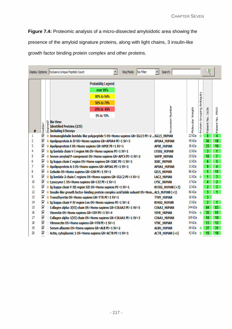
CHAPTER SEVEN
- 217 -
Figure 7.4: Proteomic analysis of a micro-dissected amyloidotic area showing the
presence of the amyloid signature proteins, along with light chains, 3 insulin-like
growth factor binding protein complex and other proteins.
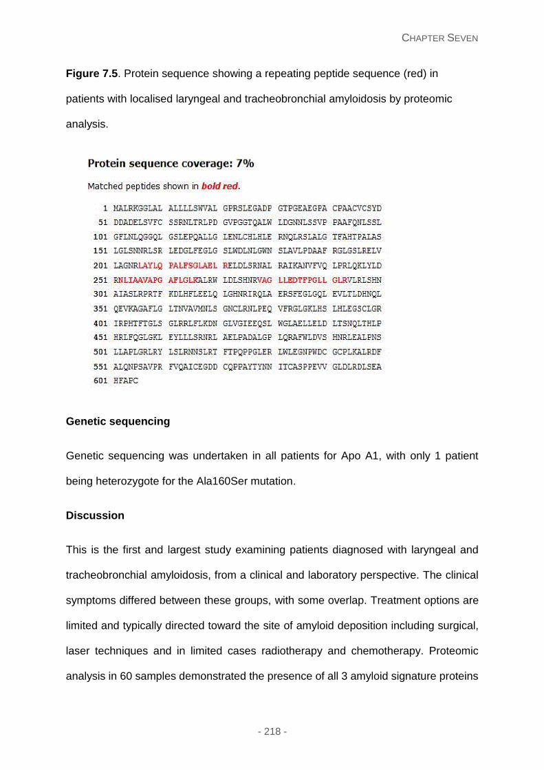
CHAPTER SEVEN
- 218 -
Figure 7.5. Protein sequence showing a repeating peptide sequence (red) in
patients with localised laryngeal and tracheobronchial amyloidosis by proteomic
analysis.
Genetic sequencing
Genetic sequencing was undertaken in all patients for Apo A1, with only 1 patient
being heterozygote for the Ala160Ser mutation.
Discussion
This is the first and largest study examining patients diagnosed with laryngeal and
tracheobronchial amyloidosis, from a clinical and laboratory perspective. The clinical
symptoms differed between these groups, with some overlap. Treatment options are
limited and typically directed toward the site of amyloid deposition including surgical,
laser techniques and in limited cases radiotherapy and chemotherapy. Proteomic
analysis in 60 samples demonstrated the presence of all 3 amyloid signature proteins
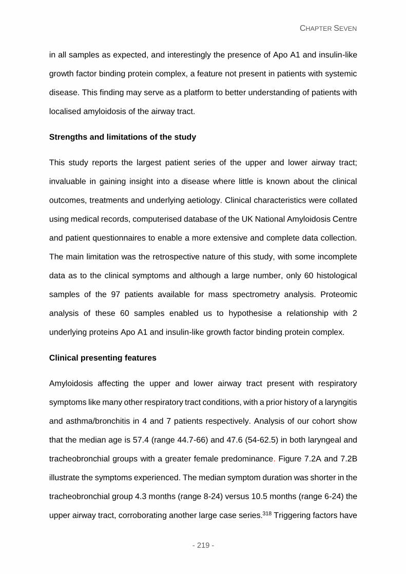
CHAPTER SEVEN
- 219 -
in all samples as expected, and interestingly the presence of Apo A1 and insulin-like
growth factor binding protein complex, a feature not present in patients with systemic
disease. This finding may serve as a platform to better understanding of patients with
localised amyloidosis of the airway tract.
Strengths and limitations of the study
This study reports the largest patient series of the upper and lower airway tract;
invaluable in gaining insight into a disease where little is known about the clinical
outcomes, treatments and underlying aetiology. Clinical characteristics were collated
using medical records, computerised database of the UK National Amyloidosis Centre
and patient questionnaires to enable a more extensive and complete data collection.
The main limitation was the retrospective nature of this study, with some incomplete
data as to the clinical symptoms and although a large number, only 60 histological
samples of the 97 patients available for mass spectrometry analysis. Proteomic
analysis of these 60 samples enabled us to hypothesise a relationship with 2
underlying proteins Apo A1 and insulin-like growth factor binding protein complex.
Clinical presenting features
Amyloidosis affecting the upper and lower airway tract present with respiratory
symptoms like many other respiratory tract conditions, with a prior history of a laryngitis
and asthma/bronchitis in 4 and 7 patients respectively. Analysis of our cohort show
that the median age is 57.4 (range 44.7-66) and 47.6 (54-62.5) in both laryngeal and
tracheobronchial groups with a greater female predominance. Figure 7.2A and 7.2B
illustrate the symptoms experienced. The median symptom duration was shorter in the
tracheobronchial group 4.3 months (range 8-24) versus 10.5 months (range 6-24) the
upper airway tract, corroborating another large case series.318 Triggering factors have
CHAPTER SEVEN
- 220 -
not been defined in these diseases 318, 319. In our series, smoking was present in 16/56
28.5% (laryngeal) and 22/28 78.6% (tracheobronchial). Within the laryngeal group, the
location of the lesion was situated in the supra-glottic region, vocal cords, subglottic
and not stated in 6, 33, 16 and 8 patients respectively. The predilection for bilateral
and right sided bronchial deposits may arise secondary to the anatomy of these lower
airway tracts, with the right bronchial tract wider, shorter and more vertical than the
left. Previous studies have reported the location of the lesion has having a major
bearing upon the risk to respiratory failure, specifically higher risk patients with
proximal and mid-bronchial lesions. 320
Treatment strategies
Excisional therapeutic options are the primary modality in managing these patients.321-
323 Here the focus is to remove the tissue containing amyloid deposits and associated
plasma/B cells. Systemic chemotherapy is not used frequently due to the low clonal
plasma/B cell burden, making the risks and toxicity of this treatment outweighing the
potential gain, despite the possible associated serum light chain excess or monoclonal
protein311
Radiotherapy has been described in some cases with LBTA309, 311, 318, 324 with the basis
that plasma cells are radiosensitive, with the majority of patients being treated with 20
Gy in 10 fractions.309 Similarly, in our series one patient with laryngeal involvement
and three patients with tracheobronchial amyloidosis received radiotherapy, following
failure with prior surgical resections and stent insertions to achieve local control. With
a median follow up of 18.7 (6-47) months, these patients have had notable
improvements in their quality of life, consistent with previously published literature.
CHAPTER SEVEN
- 221 -
Truong et al described outcomes in 10 patients post-radiotherapy with symptomatic
airway amyloidosis patients (3 laryngeal and 7 tracheobronchial). With a median follow
up of 6.7 years (1.5-10.3) 8 of 10 patients had symptomatic improvement.309
Overall survival
The OS of localised laryngeal and tracheobronchial amyloidosis is excellent with the
2 and 5 year survival 90% and 81%, and 93% and 83% respectively. Figure 3
illustrates the latter with comparison of both groups. Although not statistically
significant (p 0.66), the OS following 75 months shows a sharper decline in the
tracheobronchial group. These differences are most likely due to the underlying
pathogenic factors, possibly the extent of the localised deposits, smaller bronchial
anatomy and likeliness to occlude the airway tract.
Laser capture and proteomic analysis
Laryngeal and tracheobronchial amyloid deposition was characterised further with
laser capture and proteomic analysis. All 60 cases exhibited evidence of 3 amyloid
protein signature proteins serum amyloid P (SAP), Apo E and Apo AIV in conjunction
with Apo A1 and insulin-like growth factor binding protein complex. Proteomic analysis
was also performed in a total of 60 patients with systemic AL amyloidosis (renal,
cardiac and liver biopsies) or transthyretin based disease (cardiac, bone marrow) as
a control, with none of these samples showing evidence insulin-like growth factor
binding protein complex and a much lower peptide content of Apo A1.
Apo A1 has been described in hereditary and non-hereditary cases. In hereditary
ApoA1, deposition of amyloid occurs in different organs and tissue caused by the
germline mutation in the ApoA1 gene. Non-hereditary ApoA1 is characterised by the
CHAPTER SEVEN
- 222 -
non-variant protein and has been described in atherosclerotic intima325 and within the
respiratory tract; thought to be reduced in patients with chronic pulmonary disease
(COPD) resulting in airway limitation.326
Insulin growth factor (IGF) and insulin growth factor binding proteins (IGFBP)
proteases are involved in somatic growth, cell proliferation, cell transformation and
apoptosis.327-329 IGFBPs and IGF may form binary compounds and allowing selective
transport into tissues327 Insulin growth factor (IGF) is thought to be a key modulator
of lung fibroblast proliferation and is found at sites of inflammation330, 331 Further
studies have shown the regulation of IGF signalling via expression/down regulation of
ligands, receptors and/or regulatory binding proteins in previous malignancies.332 IGF-
1 genetic polymorphisms may represent the long term exposure of IGF-1 on the
cellular and circulatory sites.
The literature supports increasing evidence that amyloid fibril formation and
aggregation arises from a partially folded conformation of the aggregating protein, 333
with the initial lag in fibril formation thought to be secondary to the slow assembly of a
critical nucleus in a nucleation-polymerisation mechanism.334 Although aggregation
occurs when high concentrations of the key partially folded intermediate is present,
other factors including the pH, temperature, amino acid sequence and concentration
of the intermediate play a role. The presence of ApoA1 and insulin-like growth factor
binding protein complex in the LTBA proteomic analysis is a new finding and further
work is needed to elucidate the significance in localised amyloid in the laryngeal and
tracheobronchial cases. Further studies are needed to elucidate the function of these
factors before a significant pathogenesis is attributed with exploration in other types of
localised amyloidosis.
CHAPTER SEVEN
- 223 -
Future directions
Further studies are needed to ascertain whether Apo A1 and insulin-like growth factor
binding protein complex assist in instigating an underlying inflammatory process,
bystanders or involved in maintaining the self-limited localised nature of this disease.
In summary, we believe that LTBA is a type of localised amyloidosis with a more
troublesome clinical and hence problematic therapeutic quandary than other types of
localised amyloidosis.
- 224 -
Results Section Three:
Management therapeutic options for systemic amyloidosis
CHAPTER EIGHT
- 225 -
Chapter Eight: Lenalidomide and dexamethasone for systemic AL
amyloidosis following prior treatment with thalidomide or
Bortezomib regimens
This chapter is written in the context of the publication: Lenalidomide and
dexamethasone for systemic AL amyloidosis following prior treatment with
Thalidomide or Bortezomib regimens. Shameem Mahmood, Christopher P.
Venner, Sajitha Sachchithanantham, Thirusha Lane, Lisa Rannigan, Darren Foard,
Jenny H. Pinney, Simon D. J. Gibbs, Carol J. Whelan, Helen J. Lachmann, Julian D.
Gillmore, Philip N. Hawkins and Ashutosh D. Wechalekar. British Journal of
Haematology. 2014; 166(6):842-8. (Original article) copyright permission obtained
from John Wiley and Sons, license no.: 3784261189203 for use in my thesis.
Introduction
Amyloidosis is a disorder characterised by protein misfolding leading to formation and
deposition of amyloid fibrils in different tissues with change in structure and
consequent dysfunction of the affected organs. Systemic immunoglobulin light chain
(AL) amyloidosis is the most common type286 resulting from an underlying plasma cell
dyscrasia and treatment targeted toward suppressing this underlying clone.
Treatments are directed against the underlying clone and hence have been adapted
from those developed for treatment of myeloma. Immunomodulatory drugs (IMiDs) are
emerging as an important backbone for treatment of AL amyloidosis in both the
relapsed/refractory (Dispenzieri, et al 2007 Sanchorawala, et al 2010, Wechalekar, et
al 2007 Palladini, et al 2012) and upfront setting.85, 105, 108, 335 Small phase II studies
CHAPTER EIGHT
- 226 -
have shown good clonal responses using lenalidomide-dexamethasone in 67% 104, 105
and similar clonal response rates of 50%-62% with the addition of alkylating agents.
89, 106-108 Very few series have reported organ responses following lenalidomide
treatment. It is well recognised that organ responses are usually generally delayed in
AL amyloidosis with the exception of cardiac responses based on biomarkers, with the
latter difficult to interpret in patients in IMiD based therapies.136 Premature assessment
of an organ response may lead to not recognising the full clinical impact of any
treatment in this disease.
We report a substantial cohort of patients treated with lenalidomide in the
relapsed/refractory setting, following the use of Bortezomib and/or thalidomide with
long term follow up assessing the impact of previous therapies on haematologic
responses. This study reports an unexpectedly high rate of organ responses on long
term follow up in patients treated with Lenalidomide based treatment.
Patients and methods
This patient cohort comprised of all patients with systemic AL amyloidosis followed at
the UK National Amyloidosis Centre from July 2007 to August 2013 who received
treatment with lenalidomide for relapsed (n=62) or refractory (n=22) clonal disease,
with at least one prior line of therapy. Confirmation of amyloidosis was confirmed by
histology with Congo red staining and AL type confirmed by specific immunostaining
of amyloidotic tissue by antibodies to kappa or lambda light chains with exclusion of
hereditary amyloidosis by gene sequencing depending on the clinical details. All the
patients had evidence of either an abnormal serum free light chain (FLC) ratio or a
monoclonal immunoglobulin component by serum or urinary immunofixation by
electrophoresis. Written consent for retrospective publication of anonymous data was
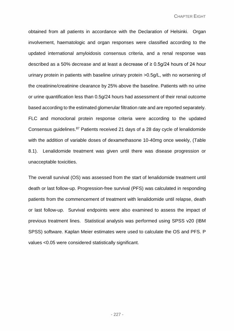
CHAPTER EIGHT
- 227 -
obtained from all patients in accordance with the Declaration of Helsinki. Organ
involvement, haematologic and organ responses were classified according to the
updated international amyloidosis consensus criteria, and a renal response was
described as a 50% decrease and at least a decrease of ≥ 0.5g/24 hours of 24 hour
urinary protein in patients with baseline urinary protein >0.5g/L, with no worsening of
the creatinine/creatinine clearance by 25% above the baseline. Patients with no urine
or urine quantification less than 0.5g/24 hours had assessment of their renal outcome
based according to the estimated glomerular filtration rate and are reported separately.
FLC and monoclonal protein response criteria were according to the updated
Consensus guidelines.87 Patients received 21 days of a 28 day cycle of lenalidomide
with the addition of variable doses of dexamethasone 10-40mg once weekly, (Table
8.1). Lenalidomide treatment was given until there was disease progression or
unacceptable toxicities.
The overall survival (OS) was assessed from the start of lenalidomide treatment until
death or last follow-up. Progression-free survival (PFS) was calculated in responding
patients from the commencement of treatment with lenalidomide until relapse, death
or last follow-up. Survival endpoints were also examined to assess the impact of
previous treatment lines. Statistical analysis was performed using SPSS v20 (IBM
SPSS) software. Kaplan Meier estimates were used to calculate the OS and PFS. P
values <0.05 were considered statistically significant.
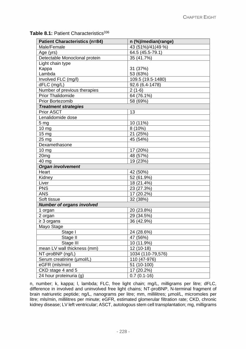
CHAPTER EIGHT
- 228 -
Table 8.1: Patient Characteristics336
Patient Characteristics (n=84) n (%)/median(range)
Male/Female 43 (51%)/41(49 %)
Age (yrs) 64.5 (45.5-79.1)
Detectable Monoclonal protein 35 (41.7%)
Light chain type Kappa Lambda
31 (37%) 53 (63%)
Involved FLC (mg/l) 109.5 (19.5-1480)
dFLC (mg/L) 92.6 (6.4-1478)
Number of previous therapies 2 (1-6)
Prior Thalidomide 64 (76.1%)
Prior Bortezomib 58 (69%)
Treatment strategies
Prior ASCT 13
Lenalidomide dose
5 mg 10 (11%)
10 mg 8 (10%)
15 mg 21 (25%)
25 mg 45 (54%)
Dexamethasone
10 mg 17 (20%)
20mg 48 (57%)
40 mg 19 (23%)
Organ involvement
Heart 42 (50%)
Kidney 52 (61.9%)
Liver 18 (21.4%)
PNS 23 (27.3%)
ANS 17 (20.2%)
Soft tissue 32 (38%)
Number of organs involved
1 organ 20 (23.8%)
2 organ 29 (34.5%)
≥ 3 organs 36 (42.9%)
Mayo Stage
Stage I 24 (28.6%)
Stage II 47 (56%)
Stage III 10 (11.9%)
mean LV wall thickness (mm) 12 (10-18)
NT-proBNP (ng/L) 1034 (110-79,576)
Serum creatinine (µmol/L) 110 (47-976)
eGFR (mls/min) 51 (10-100)
CKD stage 4 and 5 17 (20.2%)
24 hour proteinuria (g) 0.7 (0.1-16)
n, number; k, kappa; l, lambda; FLC, free light chain; mg/L, milligrams per litre; dFLC, difference in involved and uninvolved free light chains; NT-proBNP, N-terminal fragment of brain natriuretic peptide; ng/L, nanograms per litre; mm, millilitres; µmol/L, micromoles per litre; mls/min, millilitres per minute; eGFR, estimated glomerular filtration rate; CKD, chronic kidney disease; LV left ventricular; ASCT, autologous stem cell transplantation; mg, milligrams
CHAPTER EIGHT
- 229 -
Results
A total of 84 patients were identified from our database listing all registered patients.
The baseline clinical characteristics are illustrated in table 8.1. The median age was
64.5 years (range 45.5-79.1) and 52% were male. 37 (44%) had 3 or more organs
involved. 48 (56%) and 17 (20%) patients had a recorded pre-treatment estimated
glomerular filtration rate (eGFR) < 60 ml/min and < 30 ml/min respectively. The median
pre-treatment N-terminal fragment of brain natriuretic peptide (NT-proBNP) was 1034
ng/L with 11 patients having an NT-proBNP >8500ng/L, and a median threshold of
1881ng/L at 6 months. The median number of lines of treatment was 2 (range 1-6).
Seventy six percent and 68%, respectively received prior treatment with thalidomide
and Bortezomib based regimens. The median time from diagnosis to the
commencement of lenalidomide based treatment was 26.4 months (range 6-394). The
median duration of lenalidomide treatment was 6.5 months (1-52), with 31 (37%)
patients continuing following 12 cycles.
Haematologic response and survival
Lenalidomide regimens were used in the relapsed setting for 62 patients, with the 2
year OS and PFS 82% and 73%, respectively, versus 84% and 74% in the refractory
setting (n=22). On an intention-to-treat (ITT) basis, 51 (61%) achieved a
haematological response with a complete response (CR) in 17 (20%), very good
partial response (VGPR) in 6 (8%), partial response (PR) in 28 (33%) and no response
(NR) in 33 (39%) at 6 months (range 1-13) when the haematological response was
assessed. The median time to haematologic response was 3 months (range 1-19),
with haematological responses seen in 36 (56%) (VGPR or better in 14 (22%)) patients
CHAPTER EIGHT
- 230 -
treated with prior thalidomide and in 32 (55%) (VGPR or better in 13 (22%)) patients
treated with prior Bortezomib.
The median follow-up for this study was 21 months (range 1.2-80.6) and 21 deaths
occurred during this time. The estimated 1 and 2 year overall survival (OS) was 90%
and 84% respectively (Figure 8.1A) with the estimated 1 and 2 year PFS 82% and
73% respectively (Figure 8.1B). Previous exposure to thalidomide did not confer a
worse survival outcome and the estimated 2-year OS with and without thalidomide
treatment was 82% versus 83% ((HR 1.45 (0.48-4.38); p = 0.51), respectively. The
estimated 2-year PFS for those receiving prior thalidomide therapy was 80% in
comparison to 36%; those not receiving thalidomide ((HR 0.82 (0.36-1.85); p=0.623)).
Similarly, previous exposure to Bortezomib did not confer a worse survival (estimated
2-year OS 80% versus 85%, (HR 1.56 (0.6-4.06); p=0.36)) and the PFS ((estimated 2
year PFS 78% vs. 57%; HR 1.32 (0.60-2.91); p=0.492).
Patients who achieved a partial haematological response or better conferred an
estimated 2 year survival of 85% in comparison to 66% for those not responding (HR
0.45 (0.19-1.08); p 0.67). Achieving a light chain response but not necessarily a
paraprotein response was important in translating to a survival advantage with patients
achieving at least a partial dFLC response (PR or greater) having a median estimated
2 years OS of 79% in comparison to 65% for non-responders (HR 0.25 (0.08-0.72);
p=0.014)) (Fig. 8.1C). A paraprotein response in isolation, did not confer a similar
survival advantage (HR 0.71 (0.16-3.22); p=0.66)) (Fig. 8.1D). Multivariate analysis
showed that lack of a dFLC response and NT-proBNP >8500 ng/L were the sole
independent factors adversely impacting on survival (table 8.2).
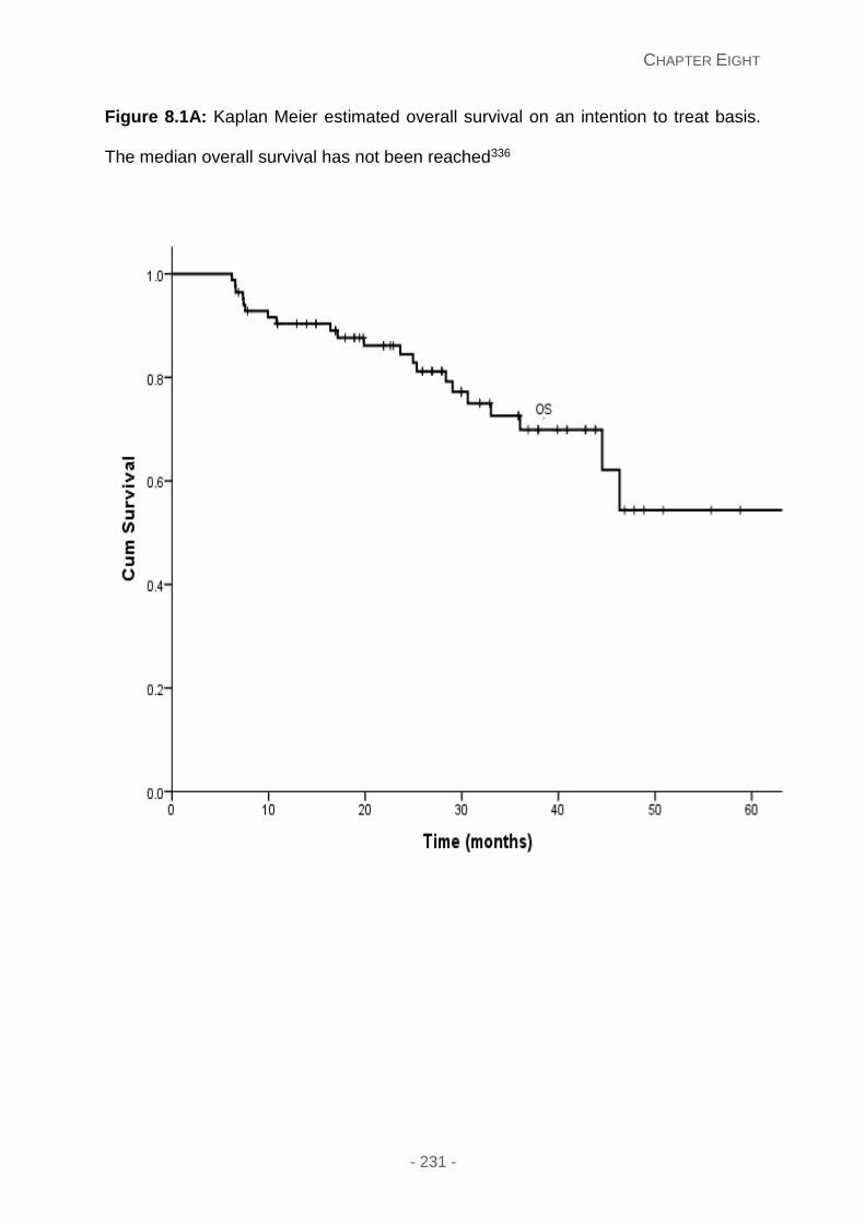
CHAPTER EIGHT
- 231 -
Figure 8.1A: Kaplan Meier estimated overall survival on an intention to treat basis.
The median overall survival has not been reached336
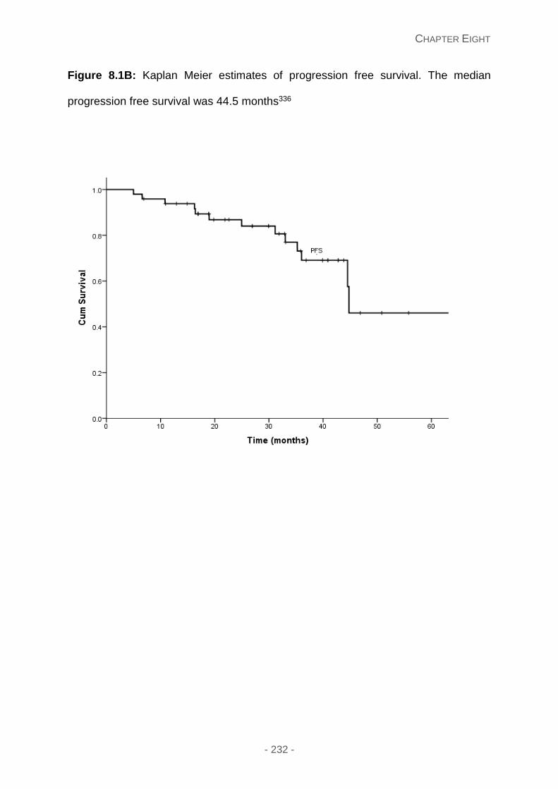
CHAPTER EIGHT
- 232 -
Figure 8.1B: Kaplan Meier estimates of progression free survival. The median
progression free survival was 44.5 months336
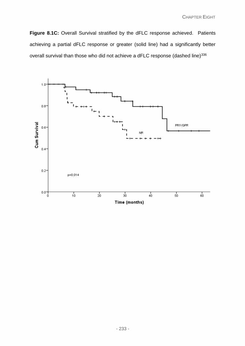
CHAPTER EIGHT
- 233 -
Figure 8.1C: Overall Survival stratified by the dFLC response achieved. Patients
achieving a partial dFLC response or greater (solid line) had a significantly better
overall survival than those who did not achieve a dFLC response (dashed line)336
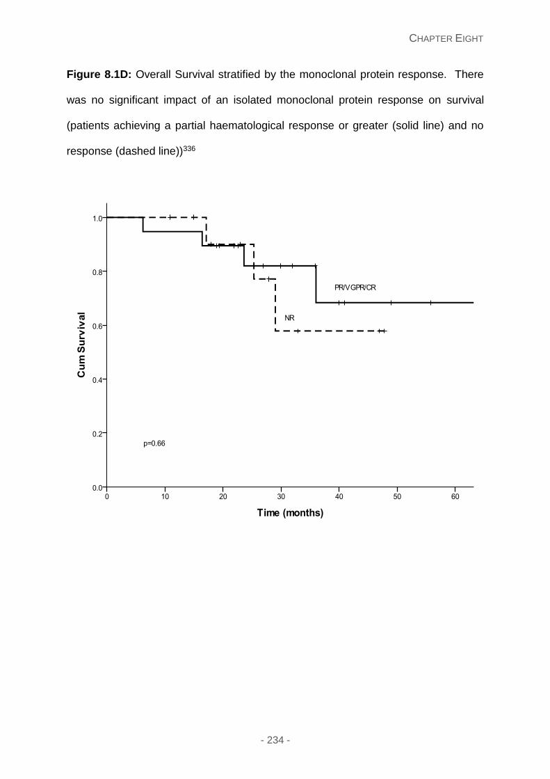
CHAPTER EIGHT
- 234 -
Figure 8.1D: Overall Survival stratified by the monoclonal protein response. There
was no significant impact of an isolated monoclonal protein response on survival
(patients achieving a partial haematological response or greater (solid line) and no
response (dashed line))336
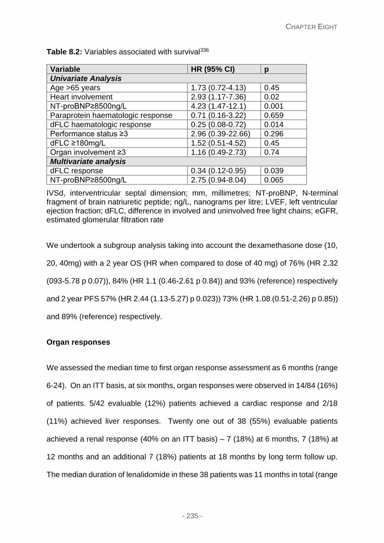
CHAPTER EIGHT
- 235 -
Table 8.2: Variables associated with survival336
Variable HR (95% CI) p
Univariate Analysis
Age >65 years 1.73 (0.72-4.13) 0.45
Heart involvement 2.93 (1.17-7.36) 0.02
NT-proBNP≥8500ng/L 4.23 (1.47-12.1) 0.001
Paraprotein haematologic response 0.71 (0.16-3.22) 0.659
dFLC haematologic response 0.25 (0.08-0.72) 0.014
Performance status ≥3 2.96 (0.39-22.66) 0.296
dFLC ≥180mg/L 1.52 (0.51-4.52) 0.45
Organ involvement ≥3 1.16 (0.49-2.73) 0.74
Multivariate analysis
dFLC response 0.34 (0.12-0.95) 0.039
NT-proBNP≥8500ng/L 2.75 (0.94-8.04) 0.065
IVSd, interventricular septal dimension; mm, millimetres; NT-proBNP, N-terminal fragment of brain natriuretic peptide; ng/L, nanograms per litre; LVEF, left ventricular ejection fraction; dFLC, difference in involved and uninvolved free light chains; eGFR, estimated glomerular filtration rate
We undertook a subgroup analysis taking into account the dexamethasone dose (10,
20, 40mg) with a 2 year OS (HR when compared to dose of 40 mg) of 76% (HR 2.32
(093-5.78 p 0.07)), 84% (HR 1.1 (0.46-2.61 p 0.84)) and 93% (reference) respectively
and 2 year PFS 57% (HR 2.44 (1.13-5.27) p 0.023)) 73% (HR 1.08 (0.51-2.26) p 0.85))
and 89% (reference) respectively.
Organ responses
We assessed the median time to first organ response assessment as 6 months (range
6-24). On an ITT basis, at six months, organ responses were observed in 14/84 (16%)
of patients. 5/42 evaluable (12%) patients achieved a cardiac response and 2/18
(11%) achieved liver responses. Twenty one out of 38 (55%) evaluable patients
achieved a renal response (40% on an ITT basis) – 7 (18%) at 6 months, 7 (18%) at
12 months and an additional 7 (18%) patients at 18 months by long term follow up.
The median duration of lenalidomide in these 38 patients was 11 months in total (range
CHAPTER EIGHT
- 236 -
6-52 months). An eGFR <15 ml/min was present in 5 patients and could not be
included in any renal response analysis. Two patients with proteinuria <0.5g had
stable eGFR values following treatment and 1 patient experienced a decline in eGFR
from 41ml/min to 15 ml/min. Fifty seven patients had evaluable visceral amyloid
deposition by 123I serum amyloid P component scintigraphy with regression evident in
17 (30%) patients at 6 months.
Toxicity
Twenty three patients experienced grade 3-4 toxicity (27%): neutropenia in 14
patients, sepsis in 4 patients, skin rash in 3 patients, thrombosis in 1 patient and
diarrhoea in 3 patients. 14 (16%) required dose reduction (from starting doses of
25mg) to 5-15mg primarily due to neutropenia. One patient developed repeated
thrombosis of a dialysis fistula which resulted in cessation of treatment but there were
no other thrombotic complications (including in 8 patients with a prior personal history
of thrombosis who received full intensity anticoagulation during their treatment). No
significant change in the eGFR was evident following treatment (baseline median 51
(range 10-100) mls/min to 47 (range 10-100) ml/min post therapy). No patients
developed secondary malignancies during this follow up period.
Discussion
There has been significant evolution of treatment of AL amyloidosis during the last
decade. Although oral melphalan-dexamethasone remains widely considered as a
standard of care, currently most patients receive a novel agent based regime, usually
as induction chemotherapy and again frequently at relapse. The optimal sequence for
using novel agent based therapies and the outcomes of relapsed patients have been
studied to a lesser degree. The current treatment of AL amyloidosis is directed against
CHAPTER EIGHT
- 237 -
the underlying clonal disorder; with the ultimate goal to achieve amyloid regression in
the affected organs. Modern treatments are increasingly successful in achieving the
former but the latter goal seems less tangible and also poorly studied and reported
less in studies of patients treated by chemotherapy based approaches. Ultimately it
is the organ response that will impact on patient survival and quality of life.
The present study reports the largest cohort of patients treated with lenalidomide-
dexamethasone (LD) currently, showing overall good haematological responses, in
systemic AL amyloidosis specifically in the relapsed/refractory setting. The overall
haematological response rate of 58% (CR -20%) achieved is reflective similarly to
earlier phase II studies of LD.104,105 The haematological response, particularly the
dFLC response translated into a significant survival advantage (HR 0.25 (0.08-0.72);
p=0.014)). The dFLC-responses were rapid and the median time to respond was 3
months – comparatively better than that reported in earlier LD studies 104, 105 and also
similar to those reported for LD-alkylator combinations, often in those treatment naïve
and previously treated 89, 106, 107 hence the benefit of an additional alkylator in treatment
of patients with systemic AL.
Data is limited data regarding the impact of prior Bortezomib or thalidomide based
treatments on lenalidomide responses. Myeloma studies analysis seems to suggest
a poorer response in previously thalidomide treated patients.337 Although
retrospective, this patient cohort had two thirds of all patients treated in a relatively
uniform manner with oral cyclophosphamide-thalidomide-dexamethasone 85 as the
standard first line therapy followed by Bortezomib at first clonal relapse and
lenalidomide dexamethasone at second clonal relapse - a treatment line consequence
as a result of treatment directed by funding availability clearly, in the UK, which is
CHAPTER EIGHT
- 238 -
regulated by the National Institute of Clinical Excellence (NICE). Italian colleagues in
a study showed that a median 2 year OS following lenalidomide dexamethasone with
Bortezomib pre-treatment was 47%.338 In the current study, treatment with
thalidomide or Bortezomib did not impact on either the haematological response,
overall or progression free survival (2 year OS was ~ 80% in those with and without
previous exposure to either chemotherapeutic agent). Our study showed that the 2
year OS was 76%, which was better than that reported in the Italian study – very likely
due to baseline disease characteristics which dominate survival outcomes in AL
amyloidosis. Typically cardiac involvement, specifically Mayo stage 3 carries a poorer
prognosis. The OS in this study is very different in compared to the reported outcomes
of newly diagnosed patient outcomes as reported for prospectively followed up
patients in our Alchemy study cohort339 predominantly due to a very high proportion
(29% early deaths in 714 patients (44% with Mayo stage 3)) in newly diagnosed
patients with AL amyloidosis. We performed a subgroup analysis determined by the
dexamethasone dose which showed a trend for an improved OS and PFS in those
having a higher dose but is difficult to interpret due to patient selection and the
retrospective nature of the cohort. This was also shown in the recent study illustrating
better outcomes in patients administered higher dose dexamethasone in the upfront
setting340 and may reflect rapid disease control in AL which may be different from
myeloma. Lack of impact of prior therapy on outcomes is encouraging and may be
partly explained by the treatment strategy. It is important to consider that apparent
poorer outcomes in patients who did not receive prior thalidomide may reflect patient
selection with significant neuropathic disease in which case tolerance may be poorer.
Due to significant risk of morbidity, it has become practice in the UK for patients to
discontinue chemotherapy following one or two cycles following best haematological
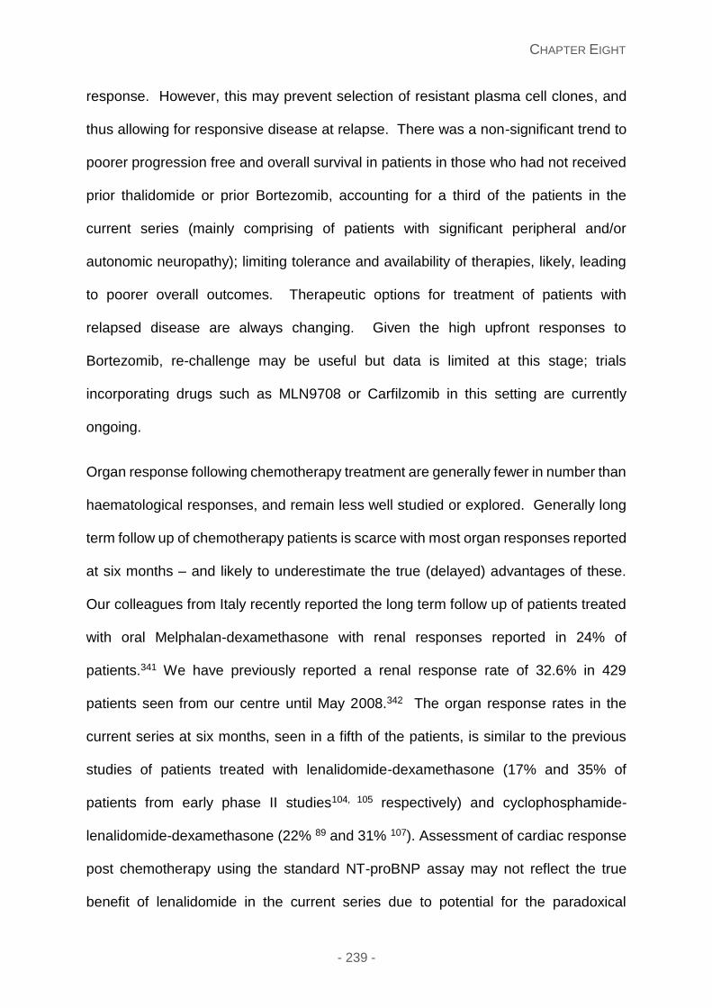
CHAPTER EIGHT
- 239 -
response. However, this may prevent selection of resistant plasma cell clones, and
thus allowing for responsive disease at relapse. There was a non-significant trend to
poorer progression free and overall survival in patients in those who had not received
prior thalidomide or prior Bortezomib, accounting for a third of the patients in the
current series (mainly comprising of patients with significant peripheral and/or
autonomic neuropathy); limiting tolerance and availability of therapies, likely, leading
to poorer overall outcomes. Therapeutic options for treatment of patients with
relapsed disease are always changing. Given the high upfront responses to
Bortezomib, re-challenge may be useful but data is limited at this stage; trials
incorporating drugs such as MLN9708 or Carfilzomib in this setting are currently
ongoing.
Organ response following chemotherapy treatment are generally fewer in number than
haematological responses, and remain less well studied or explored. Generally long
term follow up of chemotherapy patients is scarce with most organ responses reported
at six months – and likely to underestimate the true (delayed) advantages of these.
Our colleagues from Italy recently reported the long term follow up of patients treated
with oral Melphalan-dexamethasone with renal responses reported in 24% of
patients.341 We have previously reported a renal response rate of 32.6% in 429
patients seen from our centre until May 2008.342 The organ response rates in the
current series at six months, seen in a fifth of the patients, is similar to the previous
studies of patients treated with lenalidomide-dexamethasone (17% and 35% of
patients from early phase II studies104, 105 respectively) and cyclophosphamide-
lenalidomide-dexamethasone (22% 89 and 31% 107). Assessment of cardiac response
post chemotherapy using the standard NT-proBNP assay may not reflect the true
benefit of lenalidomide in the current series due to potential for the paradoxical
CHAPTER EIGHT
- 240 -
increase as previously reported,136 with a rise in the median NT-proBNP 1881ng/L at
6 months from baseline 1034ng/L in the current cohort. The striking result was the
fact that 55% of evaluable patients achieved a renal response by 18 months (with the
median duration on lenalidomide - 11 months). Interestingly, a select cohort of
patients treated with long term lenalidomide (median duration - 9 months) reported by
colleagues from Boston also showed an amyloidotic organ response in 44% of
evaluable patients.343 The renal responses in both these series appear to be
significantly higher than the previous reports of chemotherapy treated patients (25%
in long term follow up of MDex treated patients in comparison to 55% in the current
cohort) which appears to be similar to those treated with autologous stem cell
transplantation.344 The amyloidotic organ responses are important and in LD treated
patients are especially striking as the complete response rate is not particularly high
(approximately 20%). Lenalidomide is thought to increase the lymphoid immune
function by increasing the natural killer (NK) cell numbers and antibody-dependent
cell-mediated cytotoxicity and thus enhancing T-cell cytokine production.345
Lenalidomide also induces NK cells to stimulate production of granulocyte-
macrophage colony-stimulating factor, TNF-α, and various other immune recruiting
chemokines including RANTES, IL-8, MCP-1, and MIP-1α/β in response to antibody-
coated tumour cell lines, which contributes to a more effective cellular (including
macrophage) immune response.346 Previous studies from our group have shown a
natural slow clearance of amyloid from organs is affected by a macrophage type
mediated mechanism, which can be substantially enhanced with therapeutic
antibodies that target the deposits.15 Unexpectedly high percentage of renal
responses raises the possibility that lenalidomide may directly enhance the natural
regression of amyloid through its immunomodulatory effects. Supposing this was the
CHAPTER EIGHT
- 241 -
case, lenalidomide may play an adjuvant role in anti-amyloid immunotherapies which
are currently in development, as seen in its similar use in myeloma immunotherapy
with elotuzumab. There are a number of caveats which remain in bearing in mind
interpretations of these results: this is a retrospective series with limited patient
numbers, the possible selection of patients who are long term survivors and have the
resilience to tolerate long term therapy; with possible underestimation of organ
responses in other studies due to the early assessment time points.
Approximately a third of all patients in the current series developed grade 3 or greater
toxicity. Although worsening renal function347 or high incidence of skin toxicity has
previously been reported with lenalidomide; not seen in the current cohort. The major
grade 3 toxicity experienced was cytopenias which consequently led to dose
reductions. The grade 3 myelosuppression which developed improved following a
reduction of the lenalidomide dose, with a lenalidomide dose of 15 mg better tolerated
in our study, similar to previous reports,104, 106, 108 suggesting that this is an appropriate
starting dose. A few patients needed dose reduction after the first few cycles with the
stable dose achieved with good tolerance and considered for longer term therapy.
Although longer follow up is needed, there were no secondary malignancies in this
patient series. The toxicity profile reported in this series appears to better than that of
reported in prospective trials, very likely due to underestimation of toxicity due the
retrospective nature of this study and should be interpreted with caution accordingly.
Thus in summary, lenalidomide/dexamethasone is an effective regimen in treating
patients with systemic AL amyloidosis with relapsed and/or refractory clonal disease
with prior Bortezomib or thalidomide based regimens. Lower commencing doses
compared to that routinely used in treatment of patients with myeloma should be
CHAPTER EIGHT
- 242 -
considered to allow better tolerance. There was an encouragingly high rate of organ
responses recorded in this study in those patients receiving long term lenalidomide is
intriguing and needs further study in a larger patient cohort, with consideration of
evaluating this concept in experimental models to evaluate the potential of
lenalidomide in directly modulating the clearance of amyloid deposits.
CHAPTER NINE
- 243 -
Chapter Nine: Use of targeted therapy for reducing circulating SAP
in patients with hereditary fibrinogen amyloidosis and dialysis
related amyloidosis
Introduction
Systemic amyloidosis can be a fatal condition which is either acquired or hereditary,
leading to the accumulation of amyloid deposits within different tissues and organs,
resulting in dysfunction in the latter, with a high risk of mortality depending on the organ
involved.194 Hereditary fibrinogen amyloidosis was initially described in a Peruvian
kindred in 1993.45 Fibrinogen related amyloidosis has predominant renal involvement
with end stage renal failure within 5 years of presentation with proteinuria.47 There are
4 novel mutations reported with the main mutation E526V variant.45 Currently there
are no definitive therapeutic cures for this condition, with renal transplantation
performed in a selected cohort with a median graft survival of 6.7 years, with a
combined liver and renal transplantation considered only for very young, fit patients
but also associated with a high mortality.47
Beta-2-microglobulin (β2M) amyloidosis is a condition that arises in patients receiving
long term haemodialysis or continuous ambulatory peritoneal dialysis (CAPD).348, 349
Studies have shown that β2M is a major constituent of amyloid fibril formation, with
deposition within the synovial membranes and osteo-articular sites and hence
clinically destructive osteo-arthropathies, carpal tunnel syndrome, flexor tenosynovitis,
subchondral bone cysts, erosions and fractures.350
Serum amyloid P component (SAP) is a normal constituent plasma glycoprotein
previously shown to bind calcium-dependently to amyloid fibrils.351, 352 and an earlier
CHAPTER NINE
- 244 -
study using ((R)-1-[6-[(R)-2-carboxy-pyrrolidin-1-yl]-6-oxo-hexanoyl]pyrrolidine-2
carboxylic acid (CPHPC)) to target SAP as a treatment for systemic amyloidosis1 with
an excellent safety profile and variable SAP depletion in the various patient cohort.
We therefore investigated the clinical, safety, biochemical results with the quality of
life effects of CPHPC in patients with hereditary fibrinogen amyloidosis (n=10) and
dialysis related amyloidosis (n=3) in a pilot study.
Patients and methods
Patients
The primary cohort consisted of 10 patients diagnosed with hereditary fibrinogen A α-
chain amyloidosis (AFib) and 3 patients with dialysis related amyloid (Aβ2M) attending
the National Amyloidosis Centre initially recruited from August 2012, with patient
characteristics illustrated in table 9.1. We sought permission from the Royal Free
Hospital’s Drugs and Therapeutics Committee and all patients consented for
compassionate use of CPHPC, with written consent for retrospective publication of
data obtained from all patients in accordance with the Declaration of Helsinki.
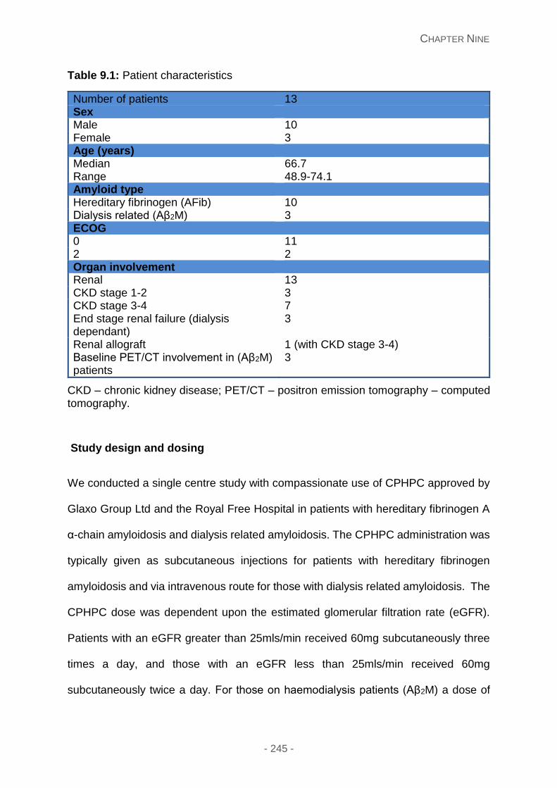
CHAPTER NINE
- 245 -
Table 9.1: Patient characteristics
Number of patients 13 Sex Male 10 Female 3 Age (years) Median 66.7 Range 48.9-74.1 Amyloid type Hereditary fibrinogen (AFib) 10 Dialysis related (Aβ2M) 3 ECOG 0 11 2 2 Organ involvement Renal 13 CKD stage 1-2 3 CKD stage 3-4 7 End stage renal failure (dialysis dependant)
3
Renal allograft 1 (with CKD stage 3-4) Baseline PET/CT involvement in (Aβ2M) patients
3
CKD – chronic kidney disease; PET/CT – positron emission tomography – computed tomography.
Study design and dosing
We conducted a single centre study with compassionate use of CPHPC approved by
Glaxo Group Ltd and the Royal Free Hospital in patients with hereditary fibrinogen A
α-chain amyloidosis and dialysis related amyloidosis. The CPHPC administration was
typically given as subcutaneous injections for patients with hereditary fibrinogen
amyloidosis and via intravenous route for those with dialysis related amyloidosis. The
CPHPC dose was dependent upon the estimated glomerular filtration rate (eGFR).
Patients with an eGFR greater than 25mls/min received 60mg subcutaneously three
times a day, and those with an eGFR less than 25mls/min received 60mg
subcutaneously twice a day. For those on haemodialysis patients (Aβ2M) a dose of
CHAPTER NINE
- 246 -
30mg CPHPC was given intravenously three times a week at the end of each dialysis
session. The clinical status of the patients were assessed at baseline and serial
monitoring performed with clinical assessment and investigations including
haematological, biochemical renal parameters and urinary quantification of
proteinuria, creatinine clearance, quality of life questionnaire 6 monthly, visual analog
scale (VAS) scores for any joint pain and whole body 123I-labelled SAP scintigraphy
(with a 4 week washout period to ensure no residual effect of the CPHPC effect). The
VAS score is a continuous scale comprised of a horizontal (10cm) line with the
extremes of pain 0; no pain and 10; maximum pain marked. It was self-completed by
each patient. Patients with dialysis related amyloid had pre-treatment 2-deoxy-2-
[fluorine-18] fluoro–D-glucose positron emission tomography- computed tomography
(18F FDG PET-CT) with reassessment of this imaging at 6 months post-treatment.
Quality of life (QoL) questionnaire assessments
The quality of life was assessed at 6 monthly intervals for all patients with the use of
the validated Quality Metric SF36v2® Health survey. This is designed to measure
functional health and well-being from the patient’s perspective. There are eight
specified health domains (physical functioning, role physical, bodily pain, general
health, social function, role emotional, mental health and vitality) which are assessed
are scored individually.177 The result is expressed in comparison to the American
norms. The average score for healthy controls in each measure is 50, with higher
scores representing a better QoL. A change of 10 points or greater in any domain
between administrations is considered clinically significant. This is explained in detail
in the methods chapter.
CHAPTER NINE
- 247 -
Results
Depletion of circulating SAP
The median (range) SAP concentration in our patient cohort was 25 mg/L (range 8-
38), with the normal range 8-55 mg/L.353 The first follow up appointment was arranged
for 6-8 weeks following the initial administration of CPHPC, showing the SAP
concentration had fallen to undetectable limits, <4mg/L at the second visit and
consequent visits, aside from visits which co-incided with an SAP scan; which the
CPHPC had been stopped 2 weeks previously. The quantification of the visceral
amyloid deposits by 123I labelled SAP scintigraphy performed yearly did not show any
significant difference in the amyloid load.
Clinical results
Among the 10 patients administered with CPHPC, 3 patients progressed to dialysis
(one patient post renal transplantation and renal function deteriorating secondary to a
further renal insult). The median follow up for the whole cohort was 28.9 months (range
9.1-38). The pre-treatment median eGFR was 31mls/minute (19-78) with pre-
treatment quantitative proteinuria 3g (range 1.1-18.6). Following treatment with
CPHPC 7 patients have remained dialysis independent. Comparison was made with
9 matched historical (eGFR matched) controls with reference made to the renal
parameters as end points, table 9.2. Monitoring of the natural course of the
biochemical parameters including the estimated glomerular filtration rate (eGFR) and
creatinine are illustrated in figure 9.1A and figure 9.1B respectively, showing a more
rapid decline in eGFR and corresponding rise in creatinine in the historical controls in
comparison to patients treated with CPHPC.
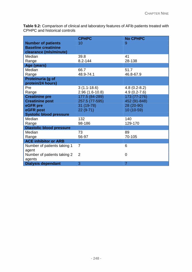
CHAPTER NINE
- 248 -
Table 9.2: Comparison of clinical and laboratory features of AFib patients treated with CPHPC and historical controls
CPHPC No CPHPC Number of patients 10 9 Baseline creatinine clearance (mls/minute)
Median 39.8 41 Range 8.2-144 28-138 Age (years) Median 66.7 51.7 Range 48.9-74.1 46.8-67.9 Proteinuria (g of protein/24 hours)
Pre 3 (1.1-18.6) 4.8 (0.2-8.2) Range 2.96 (1.6-10.8) 4.9 (0.2-7.6) Creatinine pre 177.5 (84-289) 173 (77-276) Creatinine post 257.5 (77-595) 452 (91-848) eGFR pre 31 (19-78) 28 (20-90) eGFR post 22 (9-71) 10 (10-59) Systolic blood pressure Median 132 140 Range 98-186 129-170 Diastolic blood pressure Median 73 89 Range 56-97 70-105 ACE inhibitor or ARB Number of patients taking 1 agent
7 6
Number of patients taking 2 agents
2 0
Dialysis dependant 3 7
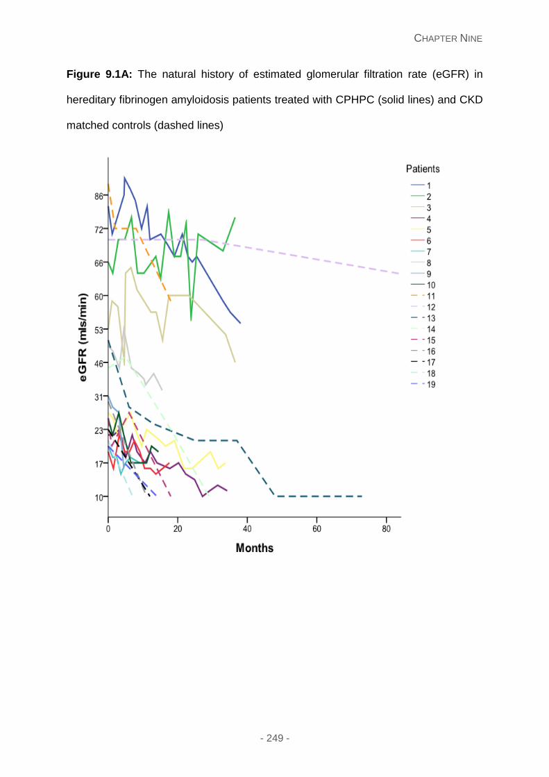
CHAPTER NINE
- 249 -
Figure 9.1A: The natural history of estimated glomerular filtration rate (eGFR) in
hereditary fibrinogen amyloidosis patients treated with CPHPC (solid lines) and CKD
matched controls (dashed lines)
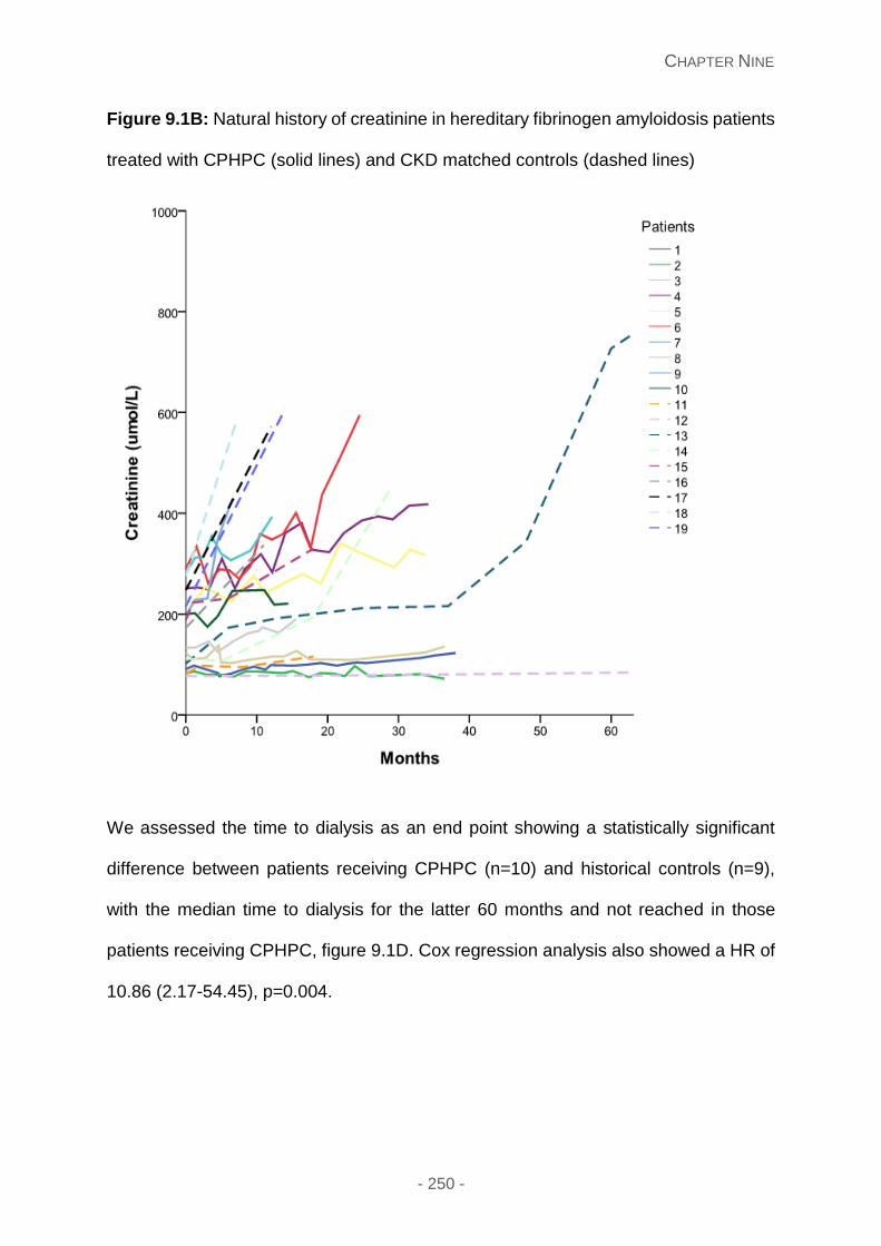
CHAPTER NINE
- 250 -
Figure 9.1B: Natural history of creatinine in hereditary fibrinogen amyloidosis patients
treated with CPHPC (solid lines) and CKD matched controls (dashed lines)
We assessed the time to dialysis as an end point showing a statistically significant
difference between patients receiving CPHPC (n=10) and historical controls (n=9),
with the median time to dialysis for the latter 60 months and not reached in those
patients receiving CPHPC, figure 9.1D. Cox regression analysis also showed a HR of
10.86 (2.17-54.45), p=0.004.
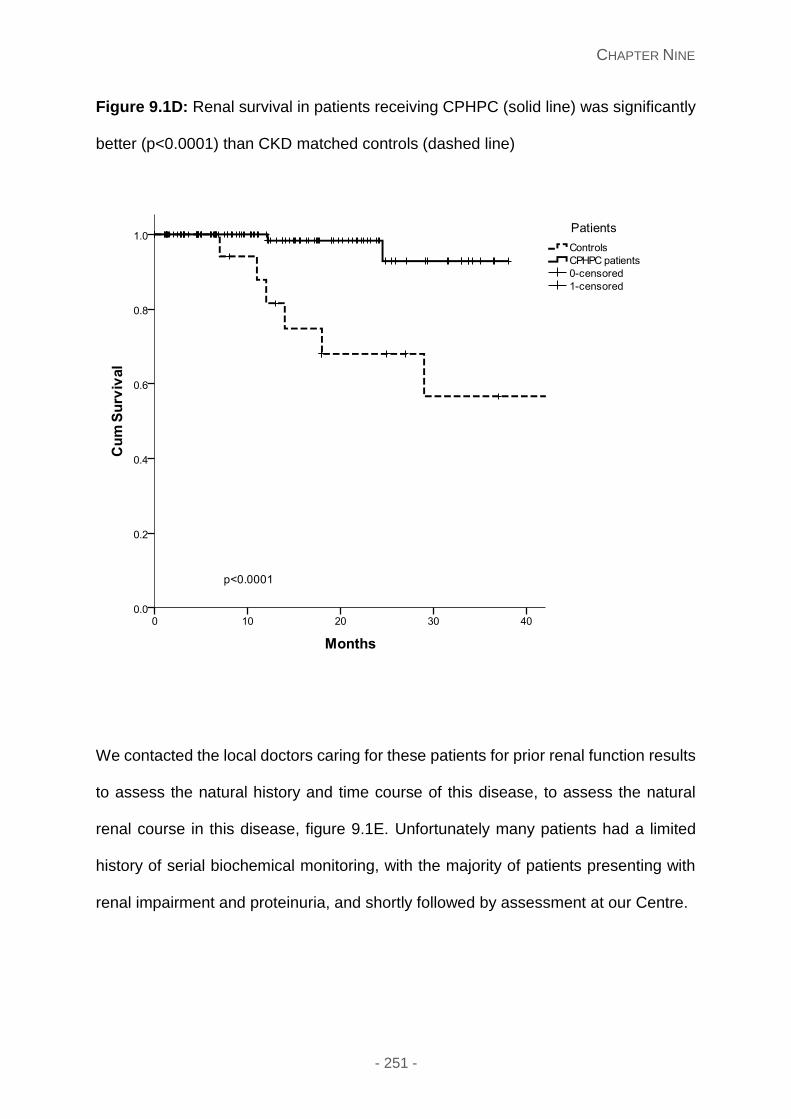
CHAPTER NINE
- 251 -
Figure 9.1D: Renal survival in patients receiving CPHPC (solid line) was significantly
better (p<0.0001) than CKD matched controls (dashed line)
We contacted the local doctors caring for these patients for prior renal function results
to assess the natural history and time course of this disease, to assess the natural
renal course in this disease, figure 9.1E. Unfortunately many patients had a limited
history of serial biochemical monitoring, with the majority of patients presenting with
renal impairment and proteinuria, and shortly followed by assessment at our Centre.
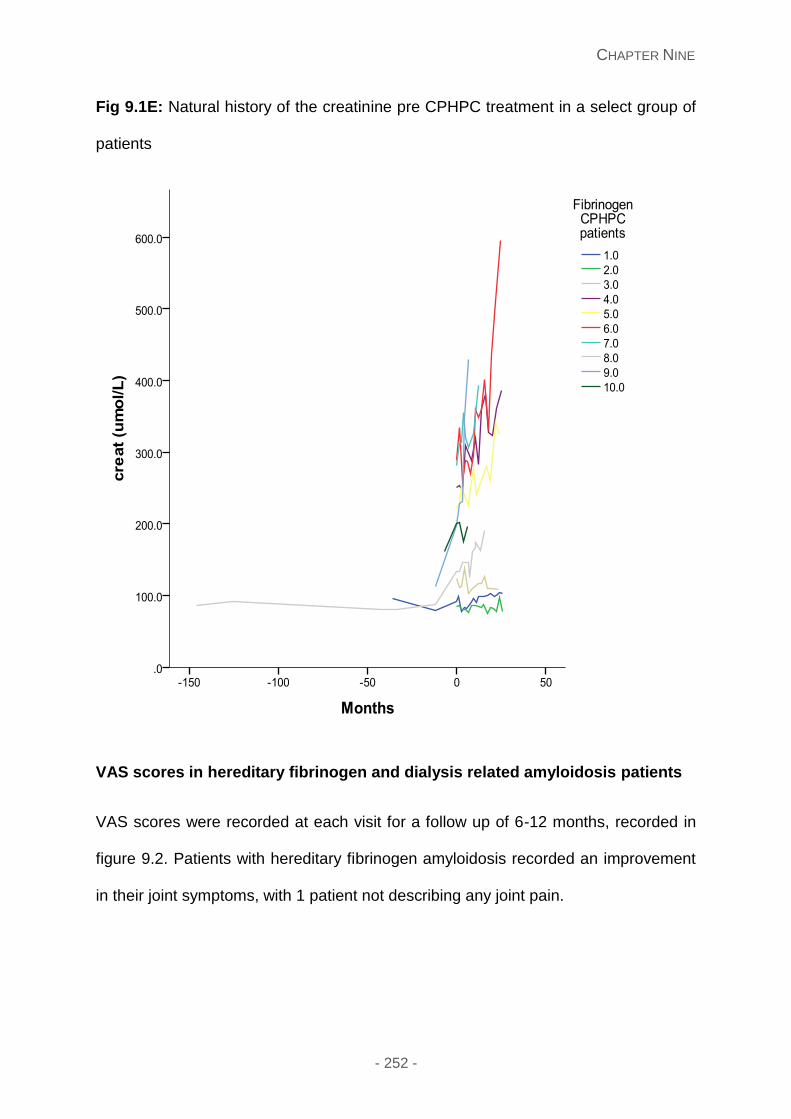
CHAPTER NINE
- 252 -
Fig 9.1E: Natural history of the creatinine pre CPHPC treatment in a select group of
patients
VAS scores in hereditary fibrinogen and dialysis related amyloidosis patients
VAS scores were recorded at each visit for a follow up of 6-12 months, recorded in
figure 9.2. Patients with hereditary fibrinogen amyloidosis recorded an improvement
in their joint symptoms, with 1 patient not describing any joint pain.
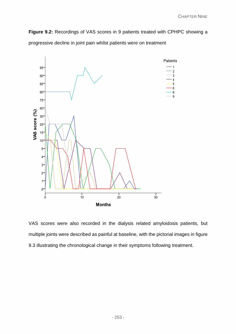
CHAPTER NINE
- 253 -
Figure 9.2: Recordings of VAS scores in 9 patients treated with CPHPC showing a
progressive decline in joint pain whilst patients were on treatment
VAS scores were also recorded in the dialysis related amyloidosis patients, but
multiple joints were described as painful at baseline, with the pictorial images in figure
9.3 illustrating the chronological change in their symptoms following treatment.
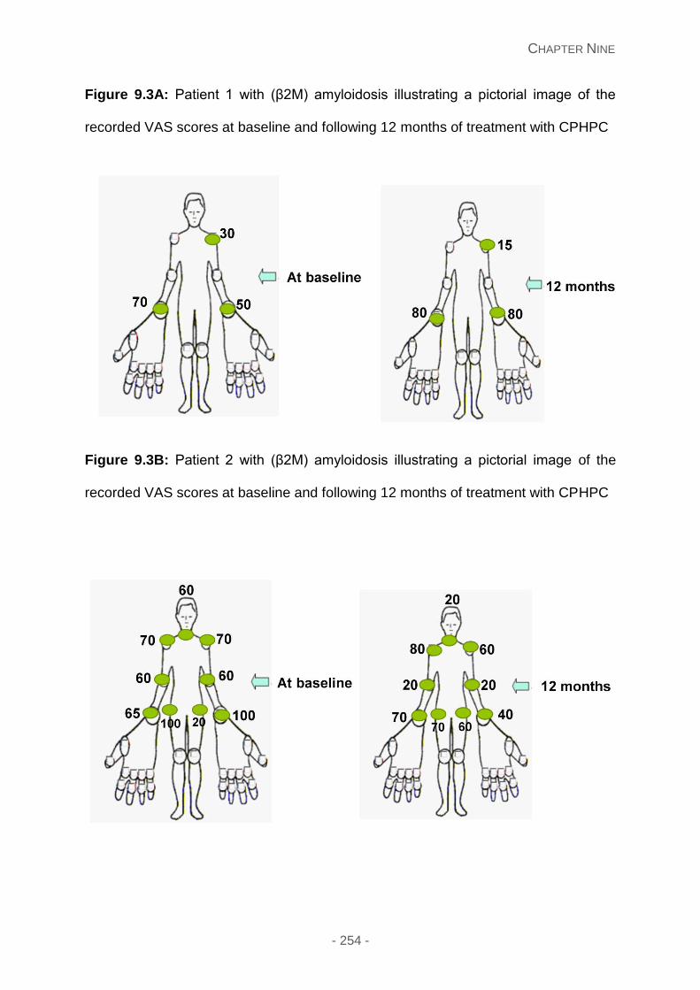
CHAPTER NINE
- 254 -
Figure 9.3A: Patient 1 with (β2M) amyloidosis illustrating a pictorial image of the
recorded VAS scores at baseline and following 12 months of treatment with CPHPC
Figure 9.3B: Patient 2 with (β2M) amyloidosis illustrating a pictorial image of the
recorded VAS scores at baseline and following 12 months of treatment with CPHPC
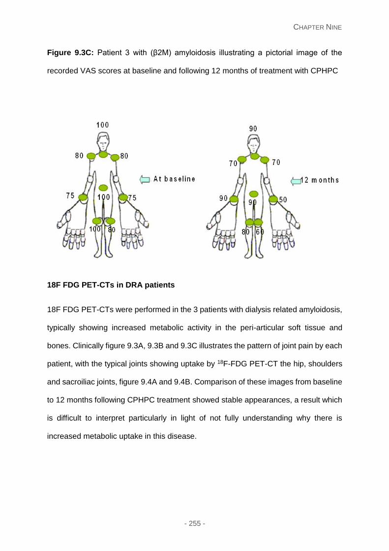
CHAPTER NINE
- 255 -
Figure 9.3C: Patient 3 with (β2M) amyloidosis illustrating a pictorial image of the
recorded VAS scores at baseline and following 12 months of treatment with CPHPC
18F FDG PET-CTs in DRA patients
18F FDG PET-CTs were performed in the 3 patients with dialysis related amyloidosis,
typically showing increased metabolic activity in the peri-articular soft tissue and
bones. Clinically figure 9.3A, 9.3B and 9.3C illustrates the pattern of joint pain by each
patient, with the typical joints showing uptake by 18F-FDG PET-CT the hip, shoulders
and sacroiliac joints, figure 9.4A and 9.4B. Comparison of these images from baseline
to 12 months following CPHPC treatment showed stable appearances, a result which
is difficult to interpret particularly in light of not fully understanding why there is
increased metabolic uptake in this disease.

CHAPTER NINE
- 256 -
Figure 9.4A and 9.4B: illustrate the coronal images of a PET/CT with increased
metabolic activity in the thickened soft tissue surrounding both shoulders and hip joints
at baseline (A) and 12 months post treatment respectively (B)
Figure 9.4A Figure 9.4B
CHAPTER NINE
- 257 -
Safety and adverse side effects
No serious adverse incidents occurred. All hereditary fibrinogen patients administered
the CPHPC subcutaneously either twice or three times per day, governed by the
underlying eGFR. Initially patients reported momentary local stinging at the injection
site. The formulation was diluted slightly at 12 months post commencement of
treatment resulting in less stinging. No patients withdrew from the study. During the
winter months, a few patients reported crystallisation of the CPHPC which improved
following warming the vial by placing it in a container containing warm water. All 3 DRA
patients received CPHPC following each session of haemodialysis with no
complications.
Quality of life
The SFv36 data was available in all patients recruited, with results subcategorised into
AFib patients; good eGFR (eGFR>30mls/min) and low eGFR (eGFR<30mls/min), and
DRA patients showing a declining trend of results from the former to latter groups,
pictorially in figure 9.5.
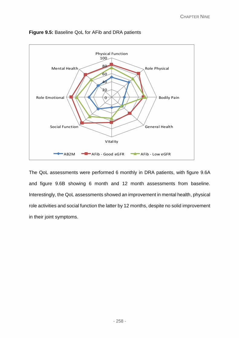
CHAPTER NINE
- 258 -
Figure 9.5: Baseline QoL for AFib and DRA patients
The QoL assessments were performed 6 monthly in DRA patients, with figure 9.6A
and figure 9.6B showing 6 month and 12 month assessments from baseline.
Interestingly, the QoL assessments showed an improvement in mental health, physical
role activities and social function the latter by 12 months, despite no solid improvement
in their joint symptoms.
0
20
40
60
80
100Physical Function
Role Physical
Bodily Pain
General Health
Vitality
Social Function
Role Emotional
Mental Health
AB2M AFib - Good eGFR AFib - Low eGFR
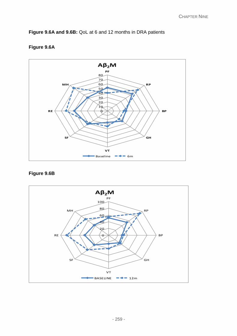
CHAPTER NINE
- 259 -
Figure 9.6A and 9.6B: QoL at 6 and 12 months in DRA patients
Figure 9.6A
Figure 9.6B
0
10
20
30
40
50
60
70
80PF
RP
BP
GH
VT
SF
RE
MH
Aβ2M
Baseline 6m
0
20
40
60
80
100PF
RP
BP
GH
VT
SF
RE
MH
Aβ2M
BASELINE 12m
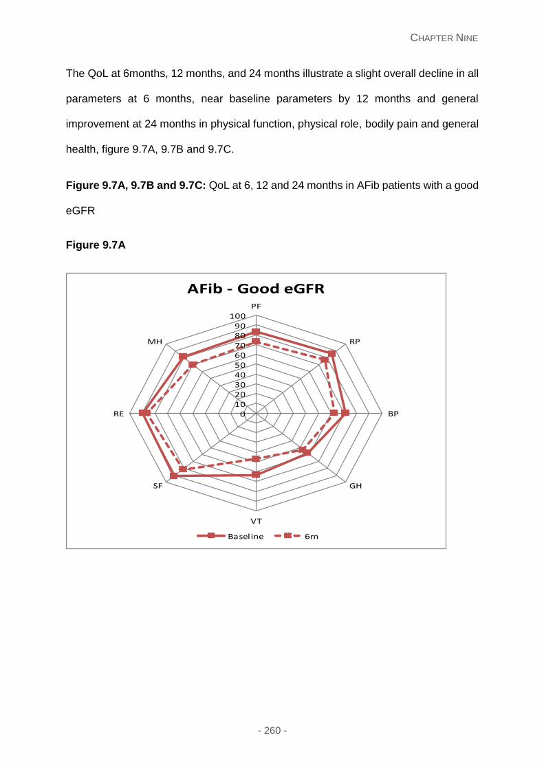
CHAPTER NINE
- 260 -
The QoL at 6months, 12 months, and 24 months illustrate a slight overall decline in all
parameters at 6 months, near baseline parameters by 12 months and general
improvement at 24 months in physical function, physical role, bodily pain and general
health, figure 9.7A, 9.7B and 9.7C.
Figure 9.7A, 9.7B and 9.7C: QoL at 6, 12 and 24 months in AFib patients with a good
eGFR
Figure 9.7A
0102030405060708090
100PF
RP
BP
GH
VT
SF
RE
MH
AFib - Good eGFR
Baseline 6m
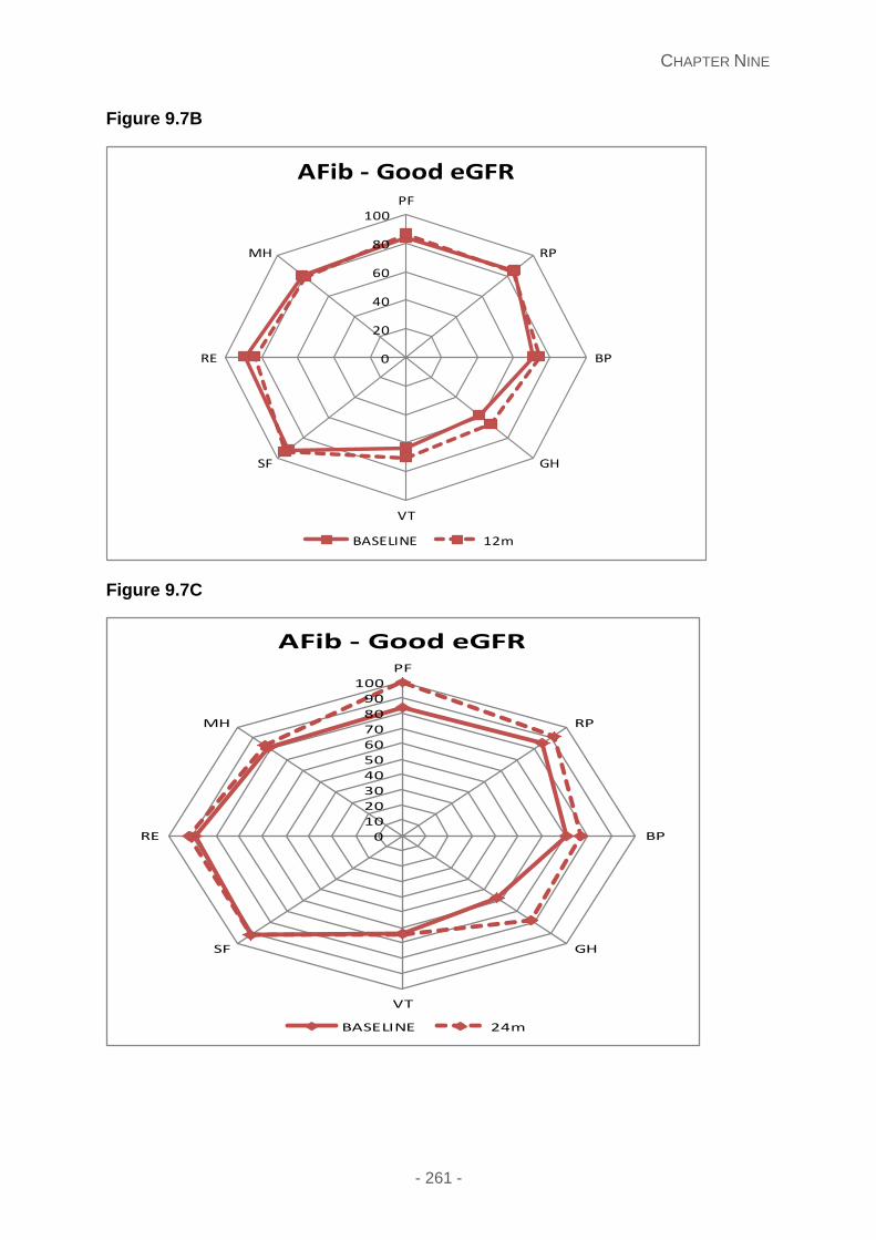
CHAPTER NINE
- 261 -
Figure 9.7B
Figure 9.7C
0
20
40
60
80
100PF
RP
BP
GH
VT
SF
RE
MH
AFib - Good eGFR
BASELINE 12m
0
10
20
30
40
50
60
70
80
90
100PF
RP
BP
GH
VT
SF
RE
MH
AFib - Good eGFR
BASELINE 24m
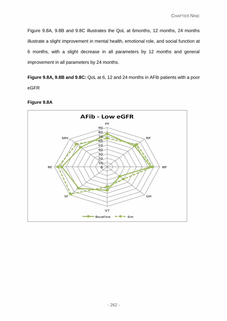
CHAPTER NINE
- 262 -
Figure 9.8A, 9.8B and 9.8C illustrates the QoL at 6months, 12 months, 24 months
illustrate a slight improvement in mental health, emotional role, and social function at
6 months, with a slight decrease in all parameters by 12 months and general
improvement in all parameters by 24 months.
Figure 9.8A, 9.8B and 9.8C: QoL at 6, 12 and 24 months in AFib patients with a poor
eGFR
Figure 9.8A
0
10
20
30
40
50
60
70
80
90PF
RP
BP
GH
VT
SF
RE
MH
AFib - Low eGFR
Baseline 6m
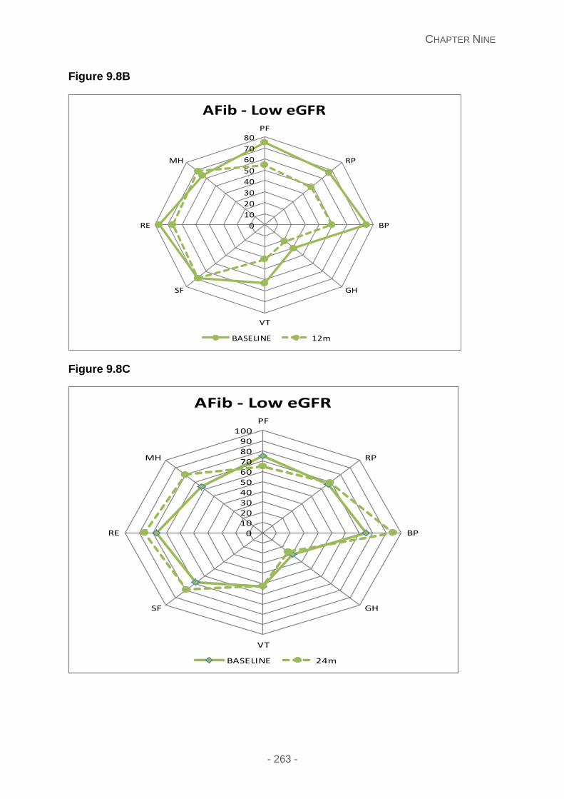
CHAPTER NINE
- 263 -
Figure 9.8B
Figure 9.8C
0
10
20
30
40
50
60
70
80PF
RP
BP
GH
VT
SF
RE
MH
AFib - Low eGFR
BASELINE 12m
0102030405060708090
100PF
RP
BP
GH
VT
SF
RE
MH
AFib - Low eGFR
BASELINE 24m
CHAPTER NINE
- 264 -
Discussion
In systemic amyloidosis, amyloid deposits may occur in many tissues and hence result
in impairment of the mechanical, structural and functional capacity of the tissue
affected. Fibrinogen amyloidosis typically affects the renal function and beta-2-
microglobulin amyloidosis is recognised to affect the osteo-articular surfaces, bone
and Carpal Tunnel Syndrome, with limited therapeutic options for both.160 Thus using
this principle of reducing the serum amyloid P, and possible removal of the amyloid
deposits, restoration of the organ function was the ultimate goal. Earlier studies have
explored the use of CPHPC to deplete circulating CPHPC, and we wanted to analyse
its use in these 2 groups of systemic amyloidosis.
The predominant finding was that the regular administration of CPHPC resulted in
sustained depletion of circulating serum amyloid P, and hence the most probable
reason for a slower decline in the renal function in patients with fibrinogen related
amyloidosis, in comparison to controls. This finding was variable with the various
factors influencing the latter including the estimated glomerular filtration rate, degree
of quantitative proteinuria and other incipient renal insults. The resulting improved
renal survival was clearly apparent with the renal decline in controls described as
13mls/min (6-21) per year, and previously described 25.8 (7.1-49.2) mls/min per
year.160 In our study, the median renal decline of CPHPC treated patients was
4.5mls/min per year (range 2.3-26.6), with 1 patient having an improvement of
3.9mls/min per year. We also demonstrated a significantly improved renal survival in
patients receiving CPHPC (p<0.0001) in comparison to CKD matched historical
controls.
CHAPTER NINE
- 265 -
As in the earlier study by Gillmore et al, the current data supports the slowing of the
natural decline in renal function seen in patients with fibrinogen amyloidosis, with early
use of this drug likely to result in later need for dialysis. Studies with CPHPC and IgG
antibody anti-SAP show promising results; with rapid clearance of the amyloid from
the liver and other tissues.354
During the study we recorded patients joint symptoms with a VAS score. Although
these patients did not have confirmed osteoporosis or a diagnosis of joint disease,
(with non-specific joint pain in a particular joint), they acknowledged that their pain
score improved over the course of time, and can only describe this as a possible
consequence secondary to CPHPC use with formal studies needed for its use in
patients with a diagnosis of osteoporosis. There were minimal complications with an
excellent safety profile.
Given the profound joint symptoms that patients with beta-2-microglobulin amyloidosis
have, a suitable hypothesis arises that CPHPC may deplete the circulating SAP and
hence improvement in their joint symptoms. The latter finding was seen in patients on
haemodialysis who then had a renal transplant, resulting in a marked reduction in their
joint symptoms.355 In our study, all three patients did not report a noticeable difference
in their joint symptoms; as evidence by no significant change in their VAS scores.
Earlier studies have described multiple myeloma patients with arthropathy,356 with MRI
imaging showing a homogenous low-to-intermediate signal intensity by both T1 and
T2 weighted images. Peri-articular amyloid may demonstrate mild enhancement
following gadolinium administration.357, 358 A case report of a patient with multiple
myeloma and amyloid deposition within the peri-articular joints was also described359
hence we thought to explore the utility of this investigation in this cohort of patients
CHAPTER NINE
- 266 -
and also in assessing their response following CPHPC treatment. 18F FDG PET-CT is
a useful non-invasive imaging modality which demonstrates the amyloid arthropathy
when other imaging such as x-rays do not delineate these changes. Our study also
confirmed this finding and their potential use in diagnosing and assessing patients with
dialysis related amyloidosis, or indeed in patients with systemic amyloidosis.
The QoL serves as an important assessment in patients concerns and satisfaction
with different facets affecting their lifestyle. In our study, we used this to assess
whether this played an impact in their treatment. Given there were different groups,
we subcategorised the groups into DRA, good and poor eGFR (the latter dependent
upon the eGFR ≥30mls/min or <30mls/min respectively). The QoL showed a subtle
improvement in different areas including mental health, physical role activities and
social function but documented no overall improvement in their joint symptoms. All
patients with AFib noticed an improving trend by 24 months, with a slight decline in
parameters prior to this. Interestingly the decline in their parameters was earlier in the
good eGFR group at 6 months compared to the poor eGFR group at 12 months. This
may be partly due to the concern for the overall outcome of this treatment and the
effectiveness over time.
In summary, our pilot study shows the slower renal decline and hence effects of
amyloid accumulation following commencement of CPHPC administration and clinical
efficacy in patients with hereditary fibrinogen amyloidosis. Our study showed a
significant improvement in the natural progression of renal decline with improved renal
survival in the latter group with an excellent safety profile. There was some anecdotal
improvement in joint symptoms, which will need further exploration in prospective

CHAPTER NINE
- 267 -
controlled studies for osteoporosis. The efficacy of this drug remains undetermined in
patients with beta-2-microglobulin amyloidosis, with minimal clinical benefit seen.
.
CHAPTER TEN
- 268 -
Chapter Ten: General conclusions
The studies undertaken in this thesis illustrate important unique findings in the
diagnostic investigations, prognosis and management of systemic amyloidosis, and
inform a greater understanding of localised amyloidosis and subtypes.
Serum free light chain measurements have a crucial role in diagnosing and monitoring
patients with systemic AL amyloidosis, now forming a standard part of the baseline
and serial follow up clinical assessments.161, 162 Inclusion of free light chain
concentration has lately been incorporated into the revised Mayo staging system and
the international consensus criteria for assessment of disease response to treatment
assessment.80,86,87 Our study is the largest study to date exploring the two
commercially available free light chain assays and comparing the results in the clinical
setting. The serum FreeliteTM assay measures immunoglobulin free light chains using
polyclonal sheep antibodies directed against hidden epitopes on the light chain
molecule producing an estimate of serum free kappa and lambda light chains. The N
Latex assay uses a mixture to two kappa and two lambda monoclonal antibodies to
the hidden epitopes of constant region of the immunoglobulin light chain molecules.
We showed that there is an excellent correlation between the different assays for
detecting the abnormal light chain subtype, but with the discordance in the absolute
values of the FLC; crucially, this renders cross assay interpolation impossible. This is
critically important as the newer N Latex assay becomes more widely adopted, with
considerable potential for causing misinterpretation of patients’ responses to
chemotherapy.
CHAPTER TEN
- 269 -
Coagulation abnormalities are often present in newly diagnosed systemic AL patients.
There is a well described associated bleeding diathesis,164 which may range from
small cutaneous bruising, pathgnomic “racoon eyes” or life threatening bleeds, with
little known as to the underlying coagulation factor abnormalities aside from factor X
deficiency and dysfibrinogenaemia. We described the novel finding of elevated factor
VIII and vWF antigen levels in these patients, with normal ADAMTS13 results; thus
excluding autoantibodies. This finding was not evident in normal controls. An albumin
threshold less than 25g/L corresponded with a statistically significant rise in vWF:Ag,
FVIII, FV and fibrinogen, with a fall in protein S and anti-thrombin III; the latter factors
previously shown in prothrombotic states. Thus this finding emphasises the increasing
importance of consideration of thromboprophylaxis or full anticoagulation in these
patients. We also explored light chain toxicity using vWF Ag as a surrogate marker,
with a FVIII and vWF Ag >280IU/L negatively impacting survival. Our study
hypothesises that underlying vascular endothelial damage occurs in systemic AL, and
possibly contributes to the light chain toxicity environment. The correlative fall in the
vWF levels post chemotherapy may reflect the underlying vascular endothelial
changes which occur. Further studies to better understand the pathophysiology of
these findings are needed to explore the endothelium by electron microscopy, and
hence the utility of the prognostic findings as part of patient risk stratification.
Fatigue is most universal in systemic amyloidosis and sleep disordered breathing
patterns were shown to be frequent and a potential contributor to fatigue symptoms.
Recurrent nocturnal oxygen desaturations are very common in patients with cardiac
amyloidosis (both AL and ATTR type), and also in patients with soft tissue amyloid
deposits affecting the oropharyngeal tract. Sleep disordered breathing questionnaires
such as STOPBANG and ESS questionnaires report a high proportion of patients
CHAPTER TEN
- 270 -
scoring ‘high risk’ for sleep disordered breathing. Increased number and frequency of
nocturnal desaturations may be associated with poorer survival in patients with cardiac
AL amyloidosis. Lack of heart rate variability (suggesting cardiac autonomic
neuropathy) is a frequent occurrence in cardiac AL and also noted in those patients
who died – findings which need further clarification. The role of hypoxia in precipitation
of cardiac arrhythmias or sudden death in AL needs to be clarified. Nocturnal hypoxia
is a simple target for intervention in cardiac AL amyloidosis and could potentially help
to reduce early mortality in AL which has remained an unmet medical need for over
25 years.
Localised amyloidosis is a very rare type of amyloidosis, with commonly reported sites
including the urinary tract, respiratory tract, larynx, skin and eyelids.165 This has been
poorly studied and our study describes the largest cohort of patients with localised AL
amyloidosis with clinical features and natural disease course showing stark differences
from systemic AL amyloidosis. Patients with obstructive/pressure symptoms or
bleeding needed endoscopic or surgical resection. Our results show that half of all
patients needed one such procedure and a fifth of all patients required repeated
procedures for symptom control highlighting the need for long term monitoring. Four
patients in the current study needed chemotherapy for localised amyloidosis.
Radiotherapy (targeting the presumed clonal proliferation) appears to be a useful, and
possibly underutilised, treatment modality in localised amyloidosis with all patients in
the current series treated with radiotherapy showing lack of progression and over half
achieving good symptomatic improvement following radiotherapy. Surprisingly, all 18
patients who had a FDG-PET/CT study performed in the current series showed
presence of FDG avidity at the site of amyloid deposition, in keeping with previous
reports of high proportion of FDG-PET/CT positivity in localised amyloidosis.307
CHAPTER TEN
- 271 -
Progression to systemic AL amyloidosis is extremely rare except in patients with
lymph node involvement. Patients with lymph node involvement and those with a
isotypic specific circulating free light chains warrant closer follow up for development
of systemic AL. The majority with localised AL have excellent long term outcomes.
Most die with the disease rather than as a consequence of it.
Laryngo-tracheobronchial amyloidosis is a rare type of localised amyloidosis,
characterised by insoluble fibrillar proteins deposited within the upper and lower airway
tract.311, 312 and we describe the largest series to date. In our study, we describe the
laryngeal and tracheobronchial amyloid deposition was characterised further with
laser capture and proteomic analysis. All 60 cases exhibited evidence of 3 amyloid
protein signature proteins serum amyloid P (SAP), Apo E and Apo AIV in conjunction
with Apo A1 and insulin-like growth factor binding protein complex. Proteomic analysis
was also performed in a total of 60 patients with systemic AL amyloidosis (renal,
cardiac and liver biopsies) or transthyretin based disease (cardiac, bone marrow) as
a control, with none of these samples showing evidence insulin-like growth factor
binding protein complex and a much lower peptide content of Apo A1. Excisional
therapeutic options are the primary modality in managing these patients.321-323
Radiotherapy has been described in some cases with LBTA309, 311, 318, 324 with the basis
that plasma cells are radiosensitive, with the majority of patients being treated with 20
Gy in 10 fractions.309 Similarly, in our series one patient with laryngeal involvement
and three patients with tracheobronchial amyloidosis received radiotherapy, following
failure with prior surgical resections and stent insertions to achieve local control. With
a median follow up of 18.7 (6-47) months, these patients have had notable
improvements in their quality of life, consistent with previously published literature.

CHAPTER TEN
- 272 -
Lenalidomide based treatment following prior novel agent based therapy has been
little studied, which we endeavoured to do so in 84 systemic AL amyloidosis patients
with relapsed/refractory clonal disease following prior treatment with thalidomide
(76%) and/or Bortezomib (68%). On an intention to treat (ITT) basis, overall
haematologic response rate was 61%, including 20% complete responses. The
median overall survival (OS) has not been reached; 2 year OS and progression free
survival (PFS) was 84% and 73% respectively. Achieving a free light chain (FLC)
response was an independent good prognostic factor for OS in multivariate analysis.
16% achieved an organ response at 6-months, with a marked improvement in organ
responses in patients on long term therapy (median duration 11 months), 55%
achieving renal responses by 18 months. Lower commencing doses compared to that
routinely used in treatment of patients with myeloma should be considered to allow
better tolerance. Lenalidomide/dexamethasone therapy achieves good
haematological responses in patients with AL amyloidosis with relapsed/refractory
clonal disease. The rate of renal responses among patients who received prolonged
treatment was unexpectedly high, raising the possibility that immunomodulatory
effects of lenalidomide therapy might enhance the otherwise slow natural regression
of amyloid deposits.
((R)-1-[6-[(R)-2-carboxy-pyrrolidin-1-yl]-6-oxo-hexanoyl]pyrrolidine-2 carboxylic acid
(CPHPC)) has emerged as a potential therapeutic target to deplete SAP as a
treatment for systemic amyloidosis.1 We explored this treatment in patients with
hereditary fibrinogen amyloidosis (AFib) and in patients with dialysis related
amyloidosis. Our study showed a significant improvement in the natural progression
of renal decline with improved renal survival in the latter group with an excellent safety
profile. The resulting improved renal survival was clearly apparent with the renal
CHAPTER TEN
- 273 -
decline in controls described as 13mls/min (6-21) per year, and previously described
25.8 (7.1-49.2) mls/min per year.160 In our study, the median renal decline of CPHPC
treated patients was 4.5mls/min per year (range 2.3-26.6), with 1 patient having an
improvement of 3.9mls/min per year. We also demonstrated a significantly improved
renal survival in patients receiving CPHPC (p<0.0001) in comparison to CKD matched
historical controls. There was some anecdotal improvement in joint symptoms, which
will need further exploration in prospective controlled studies for osteoporosis. The
QoL questionnaires were a useful adjunct in assessing patient’s treatment, showing a
general improvement in several parameters from baseline. The efficacy of this drug
remains undetermined in patients with beta-2-microglobulin amyloidosis, with minimal
clinical benefit seen.
Thus this thesis highlights the important techniques of FLC assays, coagulation
assays, sleep disordered breathing important in the setting of diagnostic, monitoring
and prognostic tests in systemic AL. Our knowledge of localised disease and a
subtype – laryngeal and tracheobronchial amyloidosis are enriched following this
study, showing this disease a separate entity to systemic AL, with the potential
repeating peptide sequence in these patients a possible insight to this disease.
Management is dependent on the type of systemic amyloidosis, with chemotherapy
for systemic AL and treatments targeted against depletion of SAP for hereditary
fibrinogen amyloidosis.
- 274 -
Future studies planned
Results from chapter 4 describe endothelial dysfunction that occurs in systemic AL.
Von Willebrand factor has been used in other diseases as a surrogate marker of
endothelial dysfunction and preliminary results show likely light chain toxicity
implications. There are ongoing plans to initiate a new study examining this
relationship further by electron microscopy.
The results from chapter 5 describe sleep disordered breathing and oxygen
desaturations in systemic amyloidosis. This is a large study outlining the latter issues
in patients with systemic amyloidosis, with the hypothesis that these oxygen
desaturations may increase morbidity and mortality in this group. Initial plans are
underway to study this prospectively including the use of supplemental oxygen in
newly diagnosed systemic AL amyloidosis patients with Mayo stage 3 cardiac
biomarkers.
Chapter 6 outlines the largest detailed characteristics and natural history and survival
features in localised amyloidosis. There are initial plans to form a collaborative group
for this disease, enabling the patient details, treatment and outcome measures to be
recorded prospectively.
The results of chapter 7 describe a large series of patients diagnosed with laryngeal
and tracheo-bronchial amyloidosis, elaborating on the clinical and proteomic analysis
of these patients. The interesting finding of increased quantification of Apo A1 and
presence of IGBF in these patients and not found in those diagnosed with systemic
AL or transthyretin based disease, has initiated plans to study this further in another
proteomic based study.
- 275 -
Chapter 8 outlines the use of lenalidomide based treatment in systemic AL. This was
a large series of 84 patients, but retrospective in nature, and we are planning on
confirming these results in a larger patient series treated with long term lenalidomide.
We aim to also use a matched series of patients treated with other chemotherapy
regimens. There was an encouragingly but surprisingly high rate of organ responses
recorded in this study for those patients receiving long term lenalidomide. This is
intriguing and needs further study in a larger patient cohort, with plans for
consideration of evaluating this concept in experimental models to evaluate the
potential of lenalidomide in directly modulating the clearance of amyloid deposits.
- 276 -
References
1. Gillmore JD, Tennent GA, Hutchinson WL, et al. Sustained pharmacological depletion
of serum amyloid P component in patients with systemic amyloidosis. Br J Haematol
2010; 148(5): 760-7.
2. Girnius S, Seldin DC, Cibeira MT, Sanchorawala V. New Hematologic Response Criteria
Predict Survival in Patients With Immunoglobulin Light Chain Amyloidosis Treated With
High-Dose Melphalan and Autologous Stem-Cell Transplantation. Journal of Clinical
Oncology; 31(21): 2749-50.
3. Palladini G, Russo P, Foli A, et al. Treatment with Oral Melphalan and Dexamethasone
of An Extended Patient Population with AL Amyloidosis. Blood 2009; 114: 3889.
4. Gibbs SDJ, Sattianayagam PT, Lachmann HJ, et al. Risk-Adapted Cyclophosphamide,
Thalidomide and Dexamethasone (CTD) for the Treatment of Systemic AL Amyloidosis:
Long Term Outcomes among 202 Patients. Blood 2008; 112: 611.
5. Cibeira MT SV, Seldin DS, Quillen K, Berk JL, Dember LM, et al. Outcome of AL
amyloidosis after high-dose melphalan and autologous stem cell transplantation: long-
term results in a series of 421 patients. Blood 2011; 118(16): 4346-52.
6. Kyle RA GM. Primary systemic amyloidosis: clinical and laboratory features in 474
cases. Semin Hematol 1995; 32(1): 45-59.
7. Westermark P, Araki S, Benson MD, et al. Nomenclature of amyloid fibril proteins.
Amyloid: Int J Exp Clin Invest 1999; 6: 63-6.
8. Dobson CM, Karplus M. The fundamentals of protein folding: bringing together theory
and experiment. Curr Opin Struct Biol 1999; 9: 92-101.
9. Quintas A, Vaz DC, Cardoso I, Saraiva MJ, Brito RM. Tetramer dissociation and
monomer partial unfolding precedes protofibril formation in amyloidogenic transthyretin
variants. J Biol Chem 2001; 276: 27207-13.
10. G. M, P W. The systemic amyloidoses: clearer understanding of the molecular
mechanisms offers hope for more effective therapies. J Intern Med 2004; 255(2): 158-
78.
11. Pepys MB, Hawkins PN. Amyloidosis. In: Austen KF, Frank MM, Atkinson JP, Cantor H,
eds. Samter's Immunologic Diseases. Sixth ed. Philadelphia: Lippincott Williams &
Wilkins; 2001: 401-12.
12. Lundmark K, Westermark GT, Nystrom S, Murphy CL, Solomon A, Westermark P.
Transmissibility of systemic amyloidosis by a prion-like mechanism. Proc Natl Acad Sci
USA 2002; 99: 6979-84.
- 277 -
13. Van Rooijen N, Sanders A. Liposome mediated depletion of macrophages: mechanism
of action, preparation of liposomes and applications. J Immunol Methods 1994; 174: 83-
93.
14. Majumdar A, Chung H, Dolios G, et al. Degradation of fibrillar forms of Alzheimer's
amyloid beta-peptide by macrophages. Neurobiol Aging 2008; 29(5): 707-15.
15. Bodin K, Ellmerich S, Kahan MC, et al. Antibodies to human serum amyloid P
component eliminate visceral amyloid deposits. Nature 2010; 468(7320): 93-7.
16. Dodel RC, Du Y, Depboylu C, et al. Intravenous immunoglobulins containing antibodies
against beta-amyloid for the treatment of Alzheimer's disease. J Neurol Neurosurg
Psychiatry 2004; 75(10): 1472-4.
17. Hrncic R, Wall J, Wolfenbarger DA, et al. Antibody-mediated resolution of light chain-
associated amyloid deposits. Am J Pathol 2000; 157: 1239-46.
18. Simms RW, Prout MN, Cohen AS. The epidemiology of AL and AA amyloidosis.
Baillieres Clin Rheumatol 1994; 8: 627-34.
19. Desikan KR, Dhodapkar MV, Hough A, et al. Incidence and impact of light chain
associated (AL) amyloidosis on the prognosis of patients with multiple myeloma treated
with autologous transplantation. Leuk Lymphoma 1997; 27(3-4): 315-9.
20. Kyle RA. Multiple myeloma: review of 869 cases. Mayo Clin Proc 1975; 50(1): 29-40.
21. Koivuniemi R, Paimela L, Suomalainen R, Tornroth T, Leirisalo-Repo M. Amyloidosis is
frequently undetected in patients with rheumatoid arthritis. Amyloid 2008; 15(4): 262-8.
22. Reilly MM, Staunton H, Harding AE. Familial amyloid polyneuropathy (TTR ala 60) in
north west Ireland: a clinical, genetic, and epidemiological study. J Neurol Neurosurg
Psychiatry 1995; 59: 45-9.
23. Jacobson DR, Ittmann M, Buxbaum JN, Wieczorek R, Gorevic PD. Transthyretin Ile 122
and cardiac amyloidosis in African-Americans. 2 case reports. Tex Heart Inst J 1997;
24: 45-52.
24. Jacobson DR, Pastore RD, Yaghoubian R, et al. Variant-sequence transthyretin
(isoleucine 122) in late-onset cardiac amyloidosis in black Americans. N Engl J Med
1997; 336: 466-73.
25. Sipe JD BM, Buxbaum JN, Ikeda SI, Merlini G, Saraiva MJ, Westermark P. Amyloid fibril
protein nomenclature: 2012 recommendations from the Nomenclature Committee of the
International Society of Amyloidosis. Amyloid 2012; 19(4): 167-70.
26. Merlini G SD, Gertz, MA. Amyloidosis: pathogenesis and new therapeutic options. J Clin
Oncol 2011; 29(14): 1924-33.
27. Mahmood S, Palladini G, Sanchorawala V, Wechalekar A. Update on treatment of light
chain amyloidosis. Haematologica 2014; 99(2): 209-21.
- 278 -
28. Lachmann HJ, Goodman HJB, Gilbertson JA, et al. Natural history and outcome in
systemic AA amyloidosis. N Engl J Med 2007; 356: 2361-71.
29. Ajiro J, Narita I, Sato F, et al. SAA1 gene polymorphisms and the risk of AA amyloidosis
in Japanese patients with rheumatoid arthritis. Mod Rheumatol 2006; 16: 294-9.
30. Pinney JH, Lachmann HJ, Sattianayagam PT, et al. Renal transplantation in systemic
amyloidosis-importance of amyloid fibril type and precursor protein abundance. Am J
Transplant 2013; 13(2): 433-41.
31. Dember LM, Jaber BL. Dialysis-related amyloidosis: late finding or hidden epidemic?
Semin Dial 2006; 19: 105-9.
32. Bernier GM, Conrad ME. Catabolsm of human beta-2-microglobulin by the rat kidney.
AmJ Physiol 1969; 217: 1359-62.
33. Campistol JM, Pnz E, Munoz-Gomez J, et al. Renal transplantation for dialysis
amyloidosis. Transplant Proc 1992; 24: 118-9.
34. Westermark P, Sletten K, Johansson B, Cornwell GG. Fibril in senile systemic
amyloidosis is derived from normal transthyretin. Proc Natl Acad Sci USA 1990; 87:
2843-5.
35. Cornwell GG, 3rd, Murdoch WL, Kyle RA, Westermark P, Pitkanen P. Frequency and
distribution of senile cardiovascular amyloid. A clinicopathologic correlation. Am J Med
1983; 75: 618-23.
36. Westermark P, Bergstrom J, Solomon A, Murphy C, Sletten K. Transthyretin-derived
senile systemic amyloidosis: clinicopathologic and structural considerations. Amyloid
2003; 10 Suppl 1: 48-54.
37. Lobato L. Portuguese-type amyloidosis (transthyretin amyloidosis, ATTR V30M). J
Nephrol 2003; 16: 438-42.
38. Said G. Familial amyloid polyneuropathy: mechanisms leading to nerve degeneration.
Amyloid 2003; 10 Suppl 1: 7-12.
39. Sousa MM, Saraiva MJ. Neurodegeneration in familial amyloid polyneuropathy: from
pathology to molecular signaling. Prog Neurobiol 2003; 71: 385-400.
40. Benson MD, Uemichi T. Transthyretin amyloidosis. Amyloid: Int J Exp Clin Invest 1996;
3: 44-56.
41. Stangou AJ, Hawkins PN. Liver transplantation in transthyretin-related familial amyloid
polyneuropathy. Curr Opin Neurol 2004; 17: 615-20.
42. Holmgren G, Steen L, Ekstedt J, et al. Biochemical effect of liver transplantation in two
Swedish patients with familial amyloidotic polyneuropathy (FAP-met30). Clin Genet 1991;
40: 242-6.
- 279 -
43. Adams D, Samuel D, Goulon-Goeau C, et al. The course and prognostic factors of
familial amyloid polyneuropathy after liver transplantation. Brain 2000; 123 ( Pt 7): 1495-
504.
44. Yazaki M, Tokuda T, Nakamura A, et al. Cardiac amyloid in patients with familial amyloid
polyneuropathy consists of abundant wild-type transthyretin. Biochem Biophys Res
Commun 2000; 274: 702-6.
45. Benson MD, Liepnieks J, Uemichi T, Wheeler G, Correa R. Hereditary renal amyloidosis
associated with a mutant fibrinogen a-chain. Nature Genetics 1993; 3: 252-5.
46. Uemichi T, Liepnieks JJ, Benson MD. Hereditary renal amyloidosis with a novel variant
fibrinogen. J Clin Invest 1994; 93: 731-6.
47. Gillmore JD, Lachmann HJ, Rowczenio D, et al. Diagnosis, pathogenesis, treatment,
and prognosis of hereditary fibrinogen A alpha-chain amyloidosis. J Am Soc Nephrol
2009; 20: 444-51.
48. Stangou AJ, Banner NR, Hendry BM, et al. Hereditary fibrinogen A alpha-chain
amyloidosis: phenotypic characterization of a systemic disease and the role of liver
transplantation. Blood 2010; 115: 2998-3007.
49. Gordon DJ, Rifkind BM. High-density lipoprotein--the clinical implications of recent
studies. N Engl J Med 1989; 321: 1311-6.
50. Castro G, Nihoul LP, Dengremont C, et al. Cholesterol efflux, lecithin-cholesterol
acyltransferase activity, and pre-beta particle formation by serum from human
apolipoprotein A-I and apolipoprotein A-I/apolipoprotein A-II transgenic mice consistent
with the latter being less effective for reverse cholesterol transport. Biochemistry 1997;
36: 2243-9.
51. Benson MD. The hereditary amyloidoses. Best Pract Res Clin Rheumatol 2003; 17: 909-
27.
52. Obici L, Palladini G, Giorgetti S, et al. Liver biopsy discloses a new apolipoprotein A-I
hereditary amyloidosis in several unrelated Italian families. Gastroenterology 2004; 126:
1416-22.
53. Obici L, Bellotti V, Mangione P, et al. The new apolipoprotein A-I variant Leu174 ® Ser
causes hereditary cardiac amyloidosis, and the amyloid fibrils are constituted by the 93-
residue N-terminal polypeptide. Am J Pathol 1999; 155: 695-702.
54. Hamidi Asl L, Liepnieks JJ, Hamidi Asl K, et al. Hereditary amyloid cardiomyopathy
caused by a variant apolipoprotein A1. Am J Pathol 1999; 154: 221-7.
55. Booth DR, Tan SY, Booth SE, et al. A new apolipoprotein AI variant, Trp50Arg, causes
hereditary amyloidosis. QJ Med 1995; 88: 695-702.
56. Soutar AK, Hawkins PN, Vigushin DM, et al. Apolipoprotein AI mutation Arg-60 causes
autosomal dominant amyloidosis. Proc Natl Acad Sci USA 1992; 89: 7389-93.
- 280 -
57. Gillmore JD, Stangou AJ, Tennent GA, et al. Clinical and biochemical outcome of
hepatorenal transplantation for hereditary systemic amyloidosis associated with
apolipoprotein AI Gly26Arg. Transplantation 2001; 71: 986-92.
58. Gillmore JD, Stangou AJ, Lachmann HJ, et al. Organ transplantation in hereditary
apolipoprotein AI amyloidosis. Am J Transplant 2006; 6: 2342-7.
59. Weiss SW, Page DL. Amyloid nephropathy of Ostertag with special reference to renal
glomerular giant cells. Am J Pathol 1973; 72: 447-55.
60. Higuchi K, Kitagawa K, Naiki H, Hanada K, Hosokawa M, Takeda T. Polymorphism of
apolipoprotein A-II (apoA-II) among inbred strains of mice. Relationship between the
molecular type of apoA-II and mouse senile amyloidosis. Biochem J 1991; 279: 427-33.
61. Meretoja J. Familial systemic paramyloidosis with lattice dystrophy of the cornea,
progressive cranial neuropathy, skin changes and various internal symptoms. A
previously unrecognized heritable syndrome. Ann Clin Res 1969; 1: 314-24.
62. de la Chapelle A, Kere J, Sack GH, Jr., Tolvanen R, Maury CPJ. Familial amyloidosis,
Finnish type: G654 - a mutation of the gelsolin gene in Finnish families and an unrelated
American family. Genomics 1992; 13: 898-901.
63. Kazmirski SL, Isaacson RL, An C, et al. Loss of a metal-binding site in gelsolin leads to
familial amyloidosis-Finnish type. Nat Struct Biol 2002; 9: 112-6.
64. Burtnick LD, Urosev D, Irobi E, Narayan K, Robinson RC. Structure of the N-terminal
half of gelsolin bound to actin: roles in severing, apoptosis and FAF. Embo J 2004; 23:
2713-22.
65. Chen CD, Huff ME, Matteson J, et al. Furin initiates gelsolin familial amyloidosis in the
Golgi through a defect in Ca(2+) stabilization. Embo J 2001; 20: 6277-87.
66. Kiuru-Enari S, Haltia M. Chapter 39 - Hereditary gelsolin amyloidosis. In: Gérard S,
Christian K, eds. Handbook of Clinical Neurology: Elsevier; 2013: 659-81.
67. Pepys MB, Hawkins PN, Booth DR, et al. Human lysozyme gene mutations cause
hereditary systemic amyloidosis. Nature 1993; 362: 553-7.
68. Morozova-Roche LA, Zurdo J, Spencer A, et al. Amyloid fibril formation and seeding by
wild-type human lysozyme and its disease-related mutational variants. Journal of
structural biology 2000; 130: 339-51.
69. Benson MD. Ostertag revisited: the inherited systemic amyloidoses without neuropathy.
Amyloid 2005; 12: 75-87.
70. Weiss BM HJ, Cordaro D, Roschewski MJ, Abbott KC, Olson SW. Monoclonal
gammopathy of undetermined significance (MGUS) precedes the diagnosis of AL
amyloidosis by up to 14 years. Blood 2011; 118: 1827.
- 281 -
71. Rajkumar SV DA, Kyle RA. Monoclonal gammopathy of undetermined significance,
Waldenstrom macroglobulinemia, AL amyloidosis, and related plasma cell disorders:
diagnosis and treatment. Mayo Clin Proc 2006; 81(5): 693-703.
72. Gertz MA M, G. Definition of organ involvement and response to treatment in AL
amyloidosis: an updated consensus opinion. Amyloid-Journal of Protein Folding
Disorders 2010; 17: 48-9.
73. Schönland SO, Hegenbart U, Bochtler T, et al. Immunohistochemistry in the
classification of systemic forms of amyloidosis: a systematic investigation of 117
patients. Blood 2012; 119(2): 488-93.
74. Arbustini E, Verga L, Concardi M, Palladini G, Obici L, Merlini G. Electron and immuno-
electron microscopy of abdominal fat identifies and characterizes amyloid fibrils in
suspected cardiac amyloidosis. Amyloid 2002; 9(2): 108-14.
75. Vrana JA, Gamez J.D, Madden BJ, Theis JD, Bergen HR 3rd, Dogan A. Classification
of amyloidosis by laser microdissection and mass spectrometry-based proteomic
analysis in clinical biopsy specimens. Blood 2009; 114(24): 4957-9.
76. Hawkins PN, Lavender JP, Pepys MB. Evaluation of systemic amyloidosis by
scintigraphy with 123I-labelled serum amyloid P component. N Engl J Med 1990; 323(8):
508-13.
77. Rapezzi C, Quarta CC, Guidalotti PL, et al. Usefulness and limitations of 99mTc-3,3-
diphosphono-1,2-propanodicarboxylic acid scintigraphy in the aetiological diagnosis of
amyloidotic cardiomyopathy. Eur J Nucl Med Mol Imaging 2011; 38(3): 470-8.
78. Bokhari S, Castaño A, Pozniakoff T, Deslisle S, Latif F, Maurer MS. 99mTc-
Pyrophosphate Scintigraphy for Differentiating Light-Chain Cardiac Amyloidosis From
the Transthyretin-Related Familial and Senile Cardiac Amyloidoses. Circulation:
Cardiovascular Imaging; 6(2): 195-201.
79. Palladini G, Barassi A, Klersy C, et al. The combination of high-sensitivity cardiac
troponin T (hs-cTnT) at presentation and changes in N-terminal natriuretic peptide type
B (NT-proBNP) after chemotherapy best predicts survival in AL amyloidosis. Blood
2010; 116(18): 3426-30.
80. Kumar S DA, Lacy MQ, Hayman SR, Buadi FK, Colby C, Laumann K, Zeldenrust SR,
Leung N, Dingli D, Greipp PR, Lust JA, Russell SJ, Kyle RA, Rajkumar SV, Gertz MA.
Revised prognostic staging system for light chain amyloidosis incorporating cardiac
biomarkers and serum free light chain measurements. J Clin Oncol 2012; 30(9): 989-95.
81. Wechalekar A SS, Kastritis E, Gillmore JD, Dimopoulos MA, Lane, et al. European
Collaborative Study of Treatment Outcomes in 346 Patients with Systemic AL
Amyloidosis with Mayo Stage III Disease. J Clin Oncol 2013; 30(9): 989-95.
- 282 -
82. Palladini G FA, Milani P, Russo P, Albertini R, Lavatelli F, et al. Best use of cardiac
biomarkers in patients with AL amyloidosis and renal failure. America Journal of
Hematology 2012; 87(5): 465-71.
83. Gertz MA, Greipp PR, Kyle RA. Classification of amyloidosis by the detection of clonal
excess of plasma cells in the bone marrow. J Lab Clin Med 1991; 118(1): 33-9.
84. Bryce AH, Ketterling RP, Gertz MA, et al. Translocation t(11;14) and survival of patients
with light chain (AL) amyloidosis. Haematologica 2009; 94(3): 380-6.
85. Wechalekar AD, Goodman HJB, Lachmann HJ, Offer M, Hawkins PN, Gillmore JD.
Safety and efficacy of risk-adapted cyclophosphamide, thalidomide, and
dexamethasone in systemic AL amyloidosis. Blood 2007; 109(2): 457-64.
86. Gertz MA, Comenzo R, Falk RH, et al. Definition of organ involvement and treatment
response in immunoglobulin light chain amyloidosis (AL): A consensus opinion from the
10th International Symposium on Amyloid and Amyloidosis. Am J Hematol 2005; 79(4):
319-28.
87. Comenzo RL, Reece D, Palladini G, et al. Consensus guidelines for the conduct and
reporting of clinical trials in systemic light-chain (AL) amyloidosis. Leukemia 2012;
26(11): 2317-25.
88. Palladini G, Dispenzieri A, Gertz MA, et al. New criteria for response to treatment in
immunoglobulin light chain amyloidosis based on free light chain measurement and
cardiac biomarkers: impact on survival outcomes. J Clin Oncol 2012; 30(36): 4541-9.
89. Kastritis E, Terpos E, Roussou M, et al. A phase 1/2 study of lenalidomide with low-dose
oral cyclophosphamide and low-dose dexamethasone (RdC) in AL amyloidosis. Blood
2012; 119(23): 5384-90.
90. Sanchorawala V, Skinner M, Quillen K, Finn KT, Doros G, Seldin DC. Long-term
outcome of patients with AL amyloidosis treated with high-dose melphalan and stem-
cell transplantation. Blood 2007; 110(10): 3561-3.
91. Dispenzieri A, Lacy MQ, Kyle RA, et al. Eligibility for hematopoietic stem-cell
transplantation for primary systemic amyloidosis is a favorable prognostic factor for
survival. J Clin Oncol 2001; 19(14): 3350-6.
92. Milani P PG, Foli A, Lavatelli F, Nuvolone M, Merlini G. Melphalan and dexamethasone
vs Bortezomib, melphalan and dexamethasone in AL amyloidosis: a matched
comparison. XIVth Myeloma Workshop 2013.
- 283 -
93. Jaccard A MP, Leblond V, Leleu X, Benboubker L, Hermine O, Recher C, Asli B, Lioure
B, Royer B, Jardin F, Bridoux F, Grosbois B, Jaubert J, Piette JC, Ronco P, Quet F,
Cogne M, Fermand, J.P for the Myélome Autogreffe (MAG) and Intergroupe
Francophone du Myélome (IFM) Intergroup. High-dose melphalan versus melphalan
plus dexamethasone for AL amyloidosis. N Engl J Med 2007; 357(11): 1083-93.
94. Seldin DC AJ, Sanchorawala V, Malek K, Wright DG, Quillen, K, Finn KT, Berk JL,
Dember LM, Falk RH, Skinner M. Improvement in quality of life of patients with AL
amyloidosis treated with high-dose melphalan and autologous stem cell transplantation.
Blood 2004; 104(6): 1888-93.
95. Gertz MA LM, Dispenzieri A, Ansell SM, Elliott MA, Gastineau DA, Inwards DJ, Micallef
IN, Porrata LF, Tefferi A, Litzow MR. Risk-adjusted manipulation of melphalan dose
before stem cell transplantation in patients with amyloidosis is associated with a lower
response rate. Bone Marrow Transplant 2004; 34(12): 1025-31.
96. Dhodapkar MV, Hussein MA, Rasmussen E, et al. Clinical efficacy of high-dose
dexamethasone with maintenance dexamethasone/alpha interferon in patients with
primary systemic amyloidosis: results of United States Intergroup Trial Southwest
Oncology Group (SWOG) S9628. Blood 2004; 104(12): 3520-6.
97. Palladini G, Perfetti V, Obici L, et al. Association of melphalan and high-dose
dexamethasone is effective and well tolerated in patients with AL (primary) amyloidosis
who are ineligible for stem cell transplantation. Blood 2004; 103(8): 2936-8.
98. Palladini G, Russo P, Nuvolone M, et al. Treatment with oral melphalan plus
dexamethasone produces long-term remissions in AL amyloidosis. Blood 2007; 110(2):
787-8.
99. Reece DE, Hegenbart U, Sanchorawala V, et al. Efficacy and safety of once-weekly and
twice-weekly bortezomib in patients with relapsed systemic AL amyloidosis: results of a
phase 1/2 study. Blood 2011; 118(4): 865-73.
100. Kastritis E, Wechalekar AD, Dimopoulos MA, et al. Bortezomib with or without
dexamethasone in primary systemic (light chain) amyloidosis. J Clin Oncol 2010; 28(6):
1031-7.
101. Venner CP, Lane T, Foard D, et al. Cyclophosphamide, bortezomib and dexamethasone
therapy in AL amyloidosis is associated with high clonal response rates and prolonged
progression free survival. Blood 2012; 119(11): 4387-90.
102. Mikhael JR, Schuster SR, Jimenez-Zepeda VH, et al. Cyclophosphamide-bortezomib-
dexamethasone (CyBorD) produces rapid and complete hematologic response in
patients with AL amyloidosis. Blood 2012; 119(19): 4391-4.
- 284 -
103. Palladini G, Milani P, Foli A, et al. Treatment of AL Amyloidosis with Bortezomib
Combined with Alkylating Agents: Results From a Prospective Series of Unselected
Patients. Blood 2011; 118(21): 1702.
104. Sanchorawala V, Wright DG, Rosenzweig M, et al. Lenalidomide and dexamethasone
in the treatment of AL amyloidosis: results of a phase 2 trial. Blood 2007; 109(2): 492-6.
105. Dispenzieri A, Lacy MQ, Zeldenrust SR, et al. The activity of lenalidomide with or without
dexamethasone in patients with primary systemic amyloidosis. Blood 2007; 109(2): 465-
70.
106. Palladini G, Russo P, Milani P, et al. A phase II trial of cyclophosphamide, lenalidomide
and dexamethasone in previously treated patients with AL amyloidosis. Haematologica
2013; 98(3): 433-6.
107. Kumar SK, Hayman SR, Buadi FK, et al. Lenalidomide, cyclophosphamide, and
dexamethasone (CRd) for light-chain amyloidosis: long-term results from a phase 2 trial.
Blood 2012; 119(21): 4860-7.
108. Moreau P, Jaccard A, Benboubker L, et al. Lenalidomide in combination with melphalan
and dexamethasone in patients with newly diagnosed AL amyloidosis: a multicenter
phase 1/2 dose-escalation study. Blood 2010; 116(23): 4777-82.
109. Dinner S, Witteles W, Afghahi A, et al. Lenalidomide, melphalan and dexamethasone in
an immunoglobulin light chain amyloidosis patient population with high rates of
advanced cardiac involvement. Haematologica 2013.
110. Sanchorawala V, Patel JM, Sloan JM, Shelton AC, Zeldis JB, Seldin DC. Melphalan,
lenalidomide and dexamethasone for the treatment of immunoglobulin light chain
amyloidosis: results of a phase II trial. Haematologica 2013; 98(5): 789-92.
111. Palladini G SS, Milani P, Kimmich C, Foli A, Bochtler T, Merlini G, Hegenbart U.
Treatment of AL Amyloidosis with Bendamustine. Blood 2012; (21): 120.
112. Dispenzieri A, Buadi F, Laumann K, et al. Activity of pomalidomide in patients with
immunoglobulin light-chain amyloidosis. Blood 2012; 119(23): 5397-404.
113. Vesole DH, Pérez WS, Akasheh M, Boudreau C, Reece DE, Bredeson CN. High-Dose
Therapy and Autologous Hematopoietic Stem Cell Transplantation for Patients With
Primary Systemic Amyloidosis: A Center for International Blood and Marrow Transplant
Research Study. Mayo Clinic Proceedings 2006; 81(7): 880-8.
114. Goodman HJ, Gillmore JD, Lachmann HJ, Wechalekar AD, Bradwell AR, Hawkins PN.
Outcome of autologous stem cell transplantation for AL amyloidosis in the UK. Br J
Haematol 2006; 134(4): 417-25.
- 285 -
115. Venner CP LT, Foard D, Rannigan L, Gibbs S, Pinney JH, Whelan CJ, Lachmann HJ,
Gillmore JD, Roussel M, Hawkins PN, Wechalekar AD. Stringent patient selection
improves outcomes in patients with AL Amyloidosis undergoing Autologous Stem Cell
Transplantation. XIIIth International Symposium on Amyloidosis 2012: 211.
116. Cohen AD ZP, Chou, J, Teruya-Feldstein J, Reich L, Hassoun H, Levine B, Filippa DA,
Riedel E, Kewalramani T, Stubblefield MD, Fleisher M, Nimer S,, RL. C. Risk-adapted
autologous stem cell transplantation with adjuvant dexamethasone +/- thalidomide for
systemic light-chain amyloidosis: results of a phase II trial. Br J Haematol 2007; 139(2):
224-33.
117. Landau H, Hassoun H, Rosenzweig MA, et al. Bortezomib and dexamethasone
consolidation following risk-adapted melphalan and stem cell transplantation for patients
with newly diagnosed light-chain amyloidosis. Leukemia 2013; 27(4): 823-8.
118. Gertz MA LM, Dispenzieri A, Kumar SK, Buadi FK, Dingli D, Leung, N, Hogan WJ,
Hayman SR. Trends in day 100 and 2-year survival after auto-SCT for AL amyloidosis:
outcomes before and after 2006. Bone Marrow Transplant 2011; 46(7): 970-5.
119. Gertz MA LM, Dispenzieri A, Kumar SK, Dingli D, Leung N, Hogan WJ, Buadi FK,
Hayman SR. Refinement in patient selection to reduce treatment-related mortality from
autologous stem cell transplantation in amyloidosis. Bone Marrow Transplant 2013;
48(4): 557-61.
120. Sanchorawala V QK, Sloan JM, Anrea NT, Seldin DC. Bortezomib and high dose
melphalan conditioning for stem cell transplantation for AL amyloidosis: a pilot study.
Haematologica 2011; 96(12): 1890-92.
121. Sanchorawala V, Wright DG, Quillen K, et al. Tandem cycles of high-dose melphalan
and autologous stem cell transplantation increases the response rate in AL amyloidosis.
Bone Marrow Transplant 2007; 40(6): 557-62.
122. Kyle RA, Gertz MA, Greipp PR, Witzig TE, Lust JA, Lacy MQ, et al. A trial of three
regimens for primary amyloidosis: colchicine alone, melphalan and prednisone, and
melphalan, prednisone, and colchicine. N Engl J Med 1997; 336(17): 1202-7.
123. Goodman HJB, Wechalekar AD, Lachmann HJ, Bradwell AR, Hawkins PN. Survival and
responses in 229 patients with AL amyloidosis treated with VAD or VAD-like
chemotherapy. Hematologica 2005; 90 (Suppl. 1): 201
124. Lebovic D, Hoffman J, Levine BM, et al. Predictors of survival in patients with systemic
light-chain amyloidosis and cardiac involvement initially ineligible for stem cell
transplantation and treated with oral melphalan and dexamethasone. Br J Haematol
2008; 143(3): 369-73.
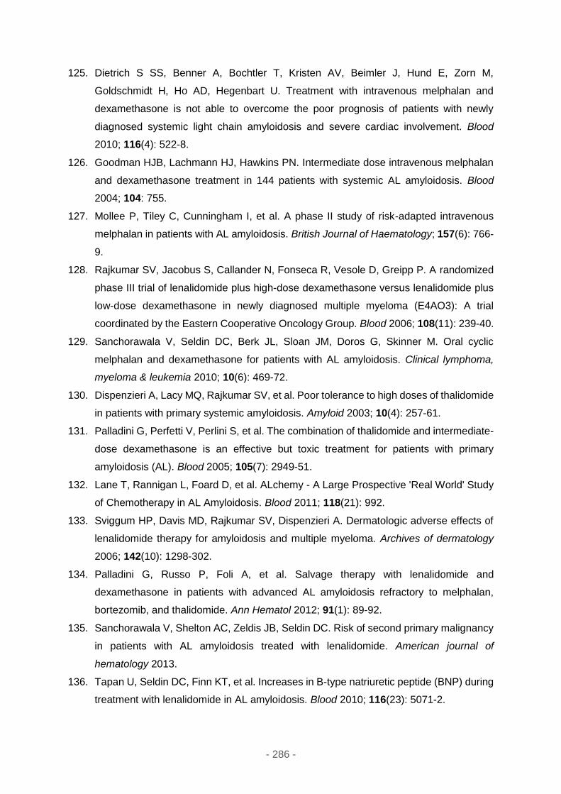
- 286 -
125. Dietrich S SS, Benner A, Bochtler T, Kristen AV, Beimler J, Hund E, Zorn M,
Goldschmidt H, Ho AD, Hegenbart U. Treatment with intravenous melphalan and
dexamethasone is not able to overcome the poor prognosis of patients with newly
diagnosed systemic light chain amyloidosis and severe cardiac involvement. Blood
2010; 116(4): 522-8.
126. Goodman HJB, Lachmann HJ, Hawkins PN. Intermediate dose intravenous melphalan
and dexamethasone treatment in 144 patients with systemic AL amyloidosis. Blood
2004; 104: 755.
127. Mollee P, Tiley C, Cunningham I, et al. A phase II study of risk-adapted intravenous
melphalan in patients with AL amyloidosis. British Journal of Haematology; 157(6): 766-
9.
128. Rajkumar SV, Jacobus S, Callander N, Fonseca R, Vesole D, Greipp P. A randomized
phase III trial of lenalidomide plus high-dose dexamethasone versus lenalidomide plus
low-dose dexamethasone in newly diagnosed multiple myeloma (E4AO3): A trial
coordinated by the Eastern Cooperative Oncology Group. Blood 2006; 108(11): 239-40.
129. Sanchorawala V, Seldin DC, Berk JL, Sloan JM, Doros G, Skinner M. Oral cyclic
melphalan and dexamethasone for patients with AL amyloidosis. Clinical lymphoma,
myeloma & leukemia 2010; 10(6): 469-72.
130. Dispenzieri A, Lacy MQ, Rajkumar SV, et al. Poor tolerance to high doses of thalidomide
in patients with primary systemic amyloidosis. Amyloid 2003; 10(4): 257-61.
131. Palladini G, Perfetti V, Perlini S, et al. The combination of thalidomide and intermediate-
dose dexamethasone is an effective but toxic treatment for patients with primary
amyloidosis (AL). Blood 2005; 105(7): 2949-51.
132. Lane T, Rannigan L, Foard D, et al. ALchemy - A Large Prospective 'Real World' Study
of Chemotherapy in AL Amyloidosis. Blood 2011; 118(21): 992.
133. Sviggum HP, Davis MD, Rajkumar SV, Dispenzieri A. Dermatologic adverse effects of
lenalidomide therapy for amyloidosis and multiple myeloma. Archives of dermatology
2006; 142(10): 1298-302.
134. Palladini G, Russo P, Foli A, et al. Salvage therapy with lenalidomide and
dexamethasone in patients with advanced AL amyloidosis refractory to melphalan,
bortezomib, and thalidomide. Ann Hematol 2012; 91(1): 89-92.
135. Sanchorawala V, Shelton AC, Zeldis JB, Seldin DC. Risk of second primary malignancy
in patients with AL amyloidosis treated with lenalidomide. American journal of
hematology 2013.
136. Tapan U, Seldin DC, Finn KT, et al. Increases in B-type natriuretic peptide (BNP) during
treatment with lenalidomide in AL amyloidosis. Blood 2010; 116(23): 5071-2.
- 287 -
137. Oliva L CF PG, Orfanelli U, Pengo N, Cascio P, Rognoni P, Sitia R, Merlini G, Cenci S.
Investigating vulnerability to proteosome inhibition in primary light chain amyloidosis.
XIIIth International Symposium on Amyloidosis 2012: 31.
138. Mateos MV, Richardson PG, Schlag R, et al. Bortezomib plus melphalan and prednisone
compared with melphalan and prednisone in previously untreated multiple myeloma:
updated follow-up and impact of subsequent therapy in the phase III VISTA trial. J Clin
Oncol 2010; 28(13): 2259-66.
139. Zonder J SV, Snyder RM, Matous J, Terebelo H, Janakiraman N, Mapara MY, Lalo S,
Tageja N, Webb C, Monsma D, Sellers C, Abrams J and Gasparetto C. Melphalan
and Dexamethasone Plus Bortezomib Induces Hematologic and Organ Responses in
AL-Amyloidosis with Tolerable Neurotoxicity. Blood 2009; 114: 746.
140. Venner CP LT, Foard D, Rannigan L, Mahmood S, Gibbs SD, et al. A Matched
Comparison of Cyclophosphamide, Bortezomib and Dexamethasone (CVD) Versus
Cyclophosphamide, Thalidomide and Dexamethasone (CTD) in the Treatment of Mayo
Cardiac Stage III Patients with AL Amyloidosis. Blood 2012; 120(21): 2966.
141. Sanchorawala V CR, Zonder JA, Kukreti V, Cohen AD, Dispenzieri A, Schoenland SO,
Jaccard A, Berg D, Yuan Z, Di Bacco A, Gupta N, Hui AM, Palladini G, Merlini G.
MLN9708, an investigational oral proteasome inhibitor (PI) in relapsed or refractory light-
chain (AL) Amyloidosis. 2013.
142. Gillmore JD, Davies J, Iqbal A, Madhoo S, Russell NH, Hawkins PN. Allogeneic bone
marrow transplantation for systemic AL amyloidosis. Brit J Haematol 1998; 100(1): 226-
8.
143. Schonland SO, Lokhorst H, Buzyn A, et al. Allogeneic and syngeneic hematopoietic cell
transplantation in patients with amyloid light-chain amyloidosis: a report from the
European Group for Blood and Marrow Transplantation. Blood 2006; 107(6): 2578-84.
144. Imamura T, Ogata M, Kohno K, et al. Successful reduced intensity allogeneic stem cell
transplantation for systemic AL amyloidosis. Am J Hematol 2006; 81(4): 281-3.
145. Palladini G, Merlini G. Transplantation vs. conventional-dose therapy for amyloidosis.
Curr Opin Oncol 2011; 23(2): 214-20.
146. Dispenzieri A, Kyle RA, Lacy MQ, et al. Superior survival in primary systemic
amyloidosis patients undergoing peripheral blood stem cell transplantation: a case-
control study. Blood 2004; 103(10): 3960-3.
147. Terrier B, Jaccard A, Harousseau J-L, et al. The Clinical Spectrum of IgM-Related
Amyloidosis: A French Nationwide Retrospective Study of 72 Patients. Medicine 2008;
87(2): 99-109 10.1097/MD.0b13e31816c43b6.
- 288 -
148. Wechalekar AD, Lachmann HJ, Goodman HJ, Bradwell A, Hawkins PN, Gillmore JD.
AL amyloidosis associated with IgM paraproteinemia: clinical profile and treatment
outcome. Blood 2008; 112(10): 4009-16.
149. Palladini G, Foil A, Russo P, et al. Treatment of IgM-Associated AL Amyloidosis With
the Combination of Rituximab, Bortezomib, and Dexamethasone. Clinical Lymphoma
Myeloma & Leukemia 2011; 11(1): 143-5.
150. Kpodonu J, Massad MG, Caines A, Geha AS. Outcome of heart transplantation in
patients with amyloid cardiomyopathy. J Heart Lung Transplant 2005; 24(11): 1763-5.
151. Dubrey SW, Burke MM, Hawkins PN, Banner NR. Cardiac transplantation for amyloid
heart disease: the United Kingdom experience. J Heart Lung Transplant 2004; 23(10):
1142-53.
152. Lacy MQ, Dispenzieri A, Hayman SR, et al. Autologous stem cell transplant after heart
transplant for light chain (Al) amyloid cardiomyopathy. J Heart Lung Transplant 2008;
27(8): 823-9.
153. Sattianayagam PT, Gibbs SDJ, Pinney JH, et al. Solid organ transplantation in AL
amyloidosis. Am J Transplant 2010; 10(9): 2124-31.
154. Kisilevsky R, Szarek WA. Novel glycosaminoglycan precursors as anti-amyloid agents
part II. J Mol Neurosci 2002; 19(1-2): 45-50.
155. O'Nuallain B, Hrncic R, Wall JS, Weiss DT, Solomon A. Diagnostic and therapeutic
potential of amyloid-reactive IgG antibodies contained in human sera. J Immunol 2006;
176(11): 7071-8.
156. Wall JS, Kennel SJ, Stuckey AC, et al. Radioimmunodetection of amyloid deposits in
patients with AL amyloidosis. Blood 2010; 116(13): 2241-4.
157. Wall JS, Kennel SJ, Williams A, et al. AL amyloid imaging and therapy with a monoclonal
antibody to a cryptic epitope on amyloid fibrils. PloS one 2012; 7(12): e52686.
158. Hovey BM, Ward JE, Soo Hoo P, O'Hara CJ, Connors LH, Seldin DC. Preclinical
development of siRNA therapeutics for AL amyloidosis. Gene therapy; 18(12): 1150-6.
159. Pepys MB, Herbert J, Hutchinson WL, et al. Targeted pharmacological depletion of
serum amyloid P component for treatment of human amyloidosis. Nature 2002;
417(6886): 254-9.
160. Gillmore JD, Tennent GA, Hutchinson WL, et al. Sustained pharmacological depletion
of serum amyloid P component in patients with systemic amyloidosis. Br J Haematol
2010: [Epub ahead of print].
161. Dispenzieri A, Kyle R, Merlini G, et al. International Myeloma Working Group guidelines
for serum-free light chain analysis in multiple myeloma and related disorders. Leukemia
2009; 23(2): 215-24.
- 289 -
162. Kumar S, Dispenzieri A, Katzmann JA, et al. Serum immunoglobulin free light chain
measurement in AL amyloidosis: prognostic value and correlations with clinical features.
Blood 2010; 116((24)): 5126-9.
163. te Velthuis H KI, Stam P, van den Broek M, Bos HK, Hol S, Teunissen E, Fischedick KS,
Althaus H, Schmidt B, Wagner C, Melsert R. N Latex FLC - new monoclonal high
performance assays for the determination of free light chain kappa and lambda. Clin
Chem Lab Med 2011; 49: 1323-32.
164. Mumford AD, O'Donnell J, Gillmore JD, Manning RA, Hawkins PN, Laffan M. Bleeding
symptoms and coagulation abnormalities in 337 patients with AL amyloidosis. Br J
Haematol 2000; 110: 454-60.
165. Westermark P, Benson MD, Buxbaum JN, Cohen AS, Frangione B, Ikeda S ea. A primer
of amyloid nomenclature. Amyloid 2007; 14(3): 179-83.
166. Puchtler H, Sweat F, Levine M. On the binding of Congo red by amyloid. J Histochem
Cytochem 1962; 10: 355-64.
167. Puchtler H, Waldrop FS, Meloan SN. A review of light, polarization and fluorescence
microscopic methods for amyloid. Appl Pathol 1985; 3(1-2): 5-17.
168. Tennent GA, Cafferty KD, Pepys MB, Hawkins PN. Congo red overlay
immunohistochemistry aids classification of amyloid deposits. In: Kyle RA, Gertz MA,
eds. Amyloid and Amyloidosis 1998. Pearl River, New York: Parthenon Publishing;
1999: 160-2.
169. Rodriguez FJ, Gamez JD, Vrana JA, et al. Immunoglobulin derived depositions in the
nervous system: novel mass spectrometry application for protein characterization in
formalin-fixed tissues. Lab Invest 2008; 88: 1024-37.
170. Talmud P, Tybjaerg-Hansen A, Bhatnagar D, et al. Rapid screening for specific
mutations in patients with a clinical diagnosis of familial hypercholesterolaemia.
Atherosclerosis 1991; 89: 137-41.
171. Health USDo, Human Services FDACfDE, Research, et al. Guidance for industry:
patient-reported outcome measures: use in medical product development to support
labeling claims: draft guidance. Health and Quality of Life Outcomes 2006; 4: 79-.
172. Russell AS, Conner-Spady B, Mintz A, Maksymowych WP. The responsiveness of
generic health status measures as assessed in patients with rheumatoid arthritis
receiving infliximab. The Journal of Rheumatology 2003; 30(5): 941-7.
173. Strand V, Tugwell P, Bombardier C, et al. Function and health-related quality of life:
Results from a randomized controlled trial of leflunomide versus methotrexate or
placebo in patients with active rheumatoid arthritis. Arthritis & Rheumatism 1999; 42(9):
1870-8.
- 290 -
174. Russell AS, Conner-Spady B, Mintz A, Maksymowych WP. The responsiveness of
generic health status measures as assessed in patients with rheumatoid arthritis
receiving infliximab. J Rheumatol 2003; 30(5): 941-7.
175. Strand V, Tugwell P, Bombardier C, et al. Function and health-related quality of life:
results from a randomized controlled trial of leflunomide versus methotrexate or placebo
in patients with active rheumatoid arthritis. Leflunomide Rheumatoid Arthritis
Investigators Group. Arthritis Rheum 1999; 42(9): 1870-8.
176. Hudson M, Thombs BD, Steele R, et al. Quality of Life in Patients with Systemic
Sclerosis Compared to the General Population and Patients with Other Chronic
Conditions. The Journal of Rheumatology 2009; 36(4): 768-72.
177. Maurish ME. USer's manual for the SF-36v2 Health Survey.: Lincoln RI: Quality Metric
Incorporated; 2011.
178. Oken MM, Creech RH, Tormey DC, et al. Toxicity and response criteria of the Eastern
Cooperative Oncology Group. Am J Clin Oncol 1982; 5: 649-55.
179. Little. Nomenclature and criterion for diagnosis of diseases of the heart and great
vessels. The Criteria Committee New York Heart Association 9ed. Boston, MA.: Little,
Brown & Co; 1994: 253-6.
180. Carroll JD, Gaasch WH, McAdam KP. Amyloid cardiomyopathy: characterization by a
distinctive voltage/mass relation. Am J Cardiol 1982; 49: 9-13.
181. Rudski LG, Lai WW, Afilalo J, et al. Guidelines for the Echocardiographic Assessment
of the Right Heart in Adults: A Report from the American Society of Echocardiography.
Journal of the American Society of Echocardiography 2010; 23(7): 685-713.
182. Gertz MA, Lacy MQ, Dispenzieri A, Hayman SR. Amyloidosis. Best Pract Res Clin
Haematol 2005; 18: 709-27.
183. Lachmann HJ, Gallimore R, Gillmore JD, et al. Outcome in systemic AL amyloidosis in
relation to changes in concentration of circulating free immunoglobulin light chains
following chemotherapy. Br J Haematol 2003; 122: 78-84.
184. Dispenzieri A, Gertz M, Kyle R, et al. Serum cardiac troponins and N-terminal pro-brain
natriuretic peptide: a staging system for primary systemic amyloidosis. J Clin Oncol
2004; 22(18): 3751-7.
185. Katzmann JA CR, Abraham RS, Bryant S, Lymp JF, Bradwell AR, Kyle RA. Serum
reference intervals and diagnostic ranges for free kappa and free lambda
immunoglobulin light chains: relative sensitivity for detection of monoclonal light chains.
Clin Chem 2002; 48: 1437-44.
186. Briand PY DO, Caillon H, Grosbois B, Le Treut A , Guenet L. Analytical performance of
the serum free light chain assay. Clin Chem Lab Med 2010; 48: 73-9.
- 291 -
187. Tate J BS, Sykes S, Mollee P. Quantitative serum free light chain assay - analytical
issues. Clin Biochem Rev 2009; 30: 131-40.
188. Kim H-S, Kim HS, Shin K-S, et al. Clinical Comparisons of Two Free Light Chain Assays
to Immunofixation Electrophoresis for Detecting Monoclonal Gammopathy. BioMed
Research International 2014; 2014: 7.
189. Pretorius CJ KS, Tate J, Wilgen U, Ungerer JP. Evaluation of the N Latex FLC free light
chain assay on the Siemens BN analyser: precision, agreement, linearity and variation
between reagent lots. Annals of clinical biochemistry 2012; 49: 450-5.
190. Sharrod-Cole H, Matters D, Showell P, Harding S. Serum free light chain assessments:
Comparison of precision and linearity. Biochemica Clinica 2013; 37: 390a.
191. Lock R, Saleem R, Roberts E, et al. A multicentre study comparing two methods for
serum free light chain analysis. Annals of clinical biochemistry 2013; 50(3): 255-61.
192. Wechalekar A, Schonland SO, Kastritis E, Gillmore JD, Dimopoulos MA, Lane Tea.
European Collaborative Study of Treatment Outcomes in 346 Patients with Systemic AL
Amyloidosis with Mayo Stage III Disease. J Clin Oncol 2013; 30(9): 989-95.
193. Sikkink LA, Ramirez-Alvarado M. Cytotoxicity of amyloidogenic immunoglobulin light
chains in cell culture. Cell Death and Dis 2010; 1: e98.
194. Pepys MB. Amyloidosis. Annu Rev Med 2006; 57: 223-41.
195. Falk RH. Diagnosis and Management of the Cardiac Amyloidoses. Circulation 2005;
112(13): 2047-60.
196. Dubrey SW, Cha K, Anderson J, et al. The clinical features of immunoglobulin light-chain
(AL) amyloidosis with heart involvement; 1998.
197. Mishra S, Guan J, Plovie E, et al. Human amyloidogenic light chain proteins result in
cardiac dysfunction, cell death, and early mortality in zebrafish; 2013.
198. Franco DA, Truran S, Burciu C, et al. Protective role of clusterin in preserving endothelial
function in AL amyloidosis. Atherosclerosis 2012; 225(1): 220-3.
199. Migrino RQ, Hari P, Gutterman DD, et al. Systemic and microvascular oxidative stress
induced by light chain amyloidosis. International Journal of Cardiology 2010; 145(1): 67-
8.
200. Migrino RQ, Truran S, Gutterman DD, et al. Human microvascular dysfunction and
apoptotic injury induced by AL amyloidosis light chain proteins; 2011.
201. Modesto KM, Dispenzieri A, Gertz M, et al. Vascular abnormalities in primary
amyloidosis; 2007.
202. Berghoff M, Kathpal M, Khan F, Skinner M, Falk R, Freeman R. Endothelial dysfunction
precedes C-fiber abnormalities in primary (AL) amyloidosis. Annals of Neurology 2003;
53(6): 725-30.
- 292 -
203. Endemann DH, Schiffrin EL. Endothelial Dysfunction. Journal of the American Society
of Nephrology 2004; 15(8): 1983-92.
204. Celermajer DS, Sorensen KE, Gooch VM, et al. Non-invasive detection of endothelial
dysfunction in children and adults at risk of atherosclerosis. The Lancet 1992;
340(8828): 1111-5.
205. Khan BV, Harrison DG, Olbrych MT, Alexander RW, Medford RM. Nitric oxide regulates
vascular cell adhesion molecule 1 gene expression and redox-sensitive transcriptional
events in human vascular endothelial cells. Proceedings of the National Academy of
Sciences 1996; 93(17): 9114-9.
206. Szmitko PE, Wang C-H, Weisel RD, de Almeida JR, Anderson TJ, Verma S. New
Markers of Inflammation and Endothelial Cell Activation: Part I. Circulation 2003;
108(16): 1917-23.
207. Lee KW, Lip GYH, Tayebjee M, Foster W, Blann AD. Circulating endothelial cells, von
Willebrand factor, interleukin-6, and prognosis in patients with acute coronary
syndromes; 2005.
208. Conway DSG, Pearce LA, Chin BSP, Hart RG, Lip GYH. Prognostic Value of Plasma
von Willebrand Factor and Soluble P-Selectin as Indices of Endothelial Damage and
Platelet Activation in 994 Patients With Nonvalvular Atrial Fibrillation. Circulation 2003;
107(25): 3141-5.
209. Constans J, Conri C. Circulating markers of endothelial function in cardiovascular
disease. Clinica Chimica Acta 2006; 368(1–2): 33-47.
210. Brill A, Fuchs TA, Chauhan AK, et al. von Willebrand factor–mediated platelet adhesion
is critical for deep vein thrombosis in mouse models; 2011.
211. Lip GYH, Blann A. von Willebrand factor: a marker of endothelial dysfunction in vascular
disorders?; 1997.
212. Wagner DD. Cell Biology of von Willebrand Factor. Annual Review of Cell Biology 1990;
6(1): 217-42.
213. Kaufman RJ, Dorner AJ, Fass DN. von Willebrand Factor Elevates Plasma Factor VIII
Without Induction of Factor VIII Messenger RNA in the Liver; 1999.
214. Federici A. The factor VIII/von Willebrand factor complex: basic and clinical issues;
2003.
215. Hamer RJ, Koedam JA, Beeser-Visser NH, Sixma JJ. The effect of thrombin on the
complex between factor VIII and von Willebrand factor. European Journal of
Biochemistry 1987; 167(2): 253-9.
216. Furie B, Voo L, McAdam KP, Furie BC. Mechanism of factor X deficiency in systemic
amyloidosis. N Engl J Med 1981; 304(14): 827-30.
- 293 -
217. Greipp PR, Kyle RA, Bowie EJ. Factor-X deficiency in amyloidosis: a critical review. Am
J Hematol 1981; 11(4): 443-50.
218. Choufani EB, Sanchorawala V, Ernst T, et al. Acquired factor X deficiency in patients
with amyloid light-chain amyloidosis: incidence, bleeding manifestations, and response
to high-dose chemotherapy. Blood 2001; 97: 1885-7.
219. Gatt A, Riddell A, Calvaruso V, Tuddenham EG, Makris M, Burroughs AK. Enhanced
thrombin generation in patients with cirrhosis-induced coagulopathy. J Thromb Haemost
2010; 9: 1994-2000.
220. Lackner H. Hemostatic abnormalities associated with dysproteinemias. Semin Hematol
1973; 10: 125-33.
221. Yood RA, Skinner M, Rubinow A, Talarico L, Cohen AS. Bleeding manifestations in 100
patients with amyloidosis. JAMA 1983; 249: 1322-4.
222. Tosetto A, Rodeghiero F, Castaman G, et al. Quantitative analysis of bleeding
symptoms in type 1 von Willebrand disease: results from a multicenter European study
(MCMDM-1 VWD). J Thromb Haemost 2006; 4(4): 766-73.
223. Marcatti M, Mauri S, Tresoldi M, et al. Unusual bleeding manifestations in a case of
primary amyloidosis with factor X deficiency but elevations of in vivo markers of thrombin
formation and activity. Thromb Res 1995; 80: 333-7.
224. Gastineau DA, Gertz MA, Daniels TM, Kyle RA, Bowie EJ. Inhibitor of the thrombin time
in systemic amyloidosis: a common coagulation abnormality. Blood 1991; 77: 2637-40.
225. O'Kane MJ, Wisdom GB, Desai ZR, Archbold GP. Inhibition of fibrin monomer
polymerisation by myeloma immunoglobulin. J Clin Pathol 1994; 47(3): 266-8.
226. Carr MEJ, Zekert SL. Abnormal clot retraction, altered fibrin structure, and normal
platelet function in multiple myeloma. Am J Physiol 1994; 266: 1195-201.
227. Hoshino Y, Hatake K, Muroi K, et al. Bleeding tendency caused by the deposit of amyloid
substance in the perivascular region. Intern Med 1993; 32(11): 879-81.
228. Thompson CA, Kyle R, Gertz M, Heit J, Pruthi R, Pardanani A. Systemic AL amyloidosis
with acquired factor X deficiency: A study of perioperative bleeding risk and treatment
outcomes in 60 patients. Am J Hematol 2010; 85(3): 171-3.
229. Zangari M, Elice F, Fink L, Tricot G. Hemostatic dysfunction in paraproteinemias and
amyloidosis. Semin Thromb Hemost 2007; 33(4): 339-49.
230. Koster T, Blann AD, Briet E, Vandenbroucke JP, Rosendaal FR. Role of clotting factor
VIII in effect of von Willebrand factor on occurrence of deep-vein-thrombosis. Lancet
1995; 346(8943): 152-5.
231. Heikal NM, Murphy KK, Crist RA, Wilson AR, Rodgers GM, Smock KJ. Elevated factor
IX is associated with an increased odds ratio for both arterial and venous thrombotic
events. Am J Clin Pathol 2013; 140(5): 680-5.
- 294 -
232. Lenting PJ, van Mourik JA, Mertens K. The Life Cycle of Coagulation Factor VIII in View
of Its Structure and Function; 1998.
233. Dimitrov JD, Christophe O, Kang J, et al. Thermodynamic Analysis of the Interaction of
Factor VIII with von Willebrand Factor. Biochemistry 2012; 51(20): 4108-16.
234. Conlon M, Folsom A, Finch A, et al. Associations of factor VIII and von Willebrand factor
with age, race, sex, and risk factors for atherosclerosis. The Atherosclerosis Risk in
Communities (ARIC) Study. Thromb Haemost 1993; 70(3): 380-5.
235. Kastritis E, Papassotiriou I, Terpos E, et al. Elevated Serum Levels Of Von Willebrand
Factor (vWF) Predict For Early Death and Shorter Survival In Patients With Primary
Systemic Light Chain (AL) Amyloidosis Independently Of Cardiac Biomarkers. Blood
2013: 3194.
236. Wall J, Schell M, Murphy C, Hrncic R, Stevens FJ, Solomon A. Thermodynamic
Instability of Human λ6 Light Chains: Correlation with Fibrillogenicity. Biochemistry
1999; 38(42): 14101-8.
237. Zhu M, Han S, Zhou F, Carter SA, Fink AL. Annular Oligomeric Amyloid Intermediates
Observed by in Situ Atomic Force Microscopy. Journal of Biological Chemistry 2004;
279(23): 24452-9.
238. Mohiuddin I, Chai H, Lin PH, Lumsden AB, Yao Q, Chen C. Nitrotyrosine and
Chlorotyrosine: Clinical Significance and Biological Functions in the Vascular System.
Journal of Surgical Research 2006; 133(2): 143-9.
239. Abshire TC, Fink LK, Christian J, Hathaway WE. The prolonged thrombin time of
nephrotic syndrome. Journal of Pediatric Hematology and Oncology 1995; 17: 156-62.
240. Mysliwiec M, Ralston A, Ackrill P, Poller L. A study of impaired fibrin polymerisation in
patients with nephrotic syndrome. Folia Haematol Int Mag Klin Morphol Blutforsch 1990;
117(1): 73-8.
241. Chen TY, Huang CC, Tsao CJ. Hemostatic molecular markers in nephrotic syndrome.
Am J Hematol 1993; 44(4): 276-9.
242. Kanfer A. Coagulation factors in nephrotic syndrome. Am J Nephrol 1990; 10(Suppl 1):
63-8.
243. Gamba G, Montani N, Anesi E, et al. Abnormalities in thrombin-antithrombin pathway in
AL amyloidosis. Amyloid: Int J Exp Clin Invest 1999; 6: 273-7.
- 295 -
244. Husby G, Stenstad T, Magnus JH, Sletten K, Nordvag BY, Marhaug G. Interaction
between circulating amyloid fibril protein precursors and extracellular tissue matrix
components in the pathogenesis of systemic amyloidosis. Clin Immunol Immunopathol
1994; 70: 2-9.
245. Durvasula RV, Petermann AT, Hiromura K, et al. Activation of a local tissue angiotensin
system in podocytes by mechanical strain1. Kidney Int 2004; 65(1): 30-9.
246. Eremina V, Baelde HJ, Quaggin SE. Role of the VEGF-A Signaling Pathway in the
Glomerulus: Evidence for Crosstalk between Components of the Glomerular Filtration
Barrier. Nephron Physiology 2007; 106(2): p32-p7.
247. Singhal R, Brimble KS. Thromboembolic complications in the nephrotic syndrome:
Pathophysiology and clinical management. Thrombosis Research 2006; 118(3): 397-
407.
248. Orth SR, Ritz E. The nephrotic syndrome. New England Journal of Medicine 1998;
338(17): 1202-11.
249. Yang Y, Lv J, Zhou F, et al. Risk Factors of Pulmonary Thrombosis/Embolism in
Nephrotic Syndrome. The American Journal of the Medical Sciences 2014; 348(5): 394-
8.
250. Gamba G, Montani N, Anesi E, et al. Clotting alterations in primary systemic amyloidosis.
Haematologica 2000; 85: 289-92.
251. Sane DC, Pizzo SV, Greenberg CS. Elevated urokinase-type plasminogen activator
level and bleeding in amyloidosis: a case report and literature review. Am J Hematol
1989; 31(1): 53-7.
252. Boggio L, Green D. Recombinant human factor VIIa in the management of amyloid-
associated factor X deficiency. Br J Haematol 2001; 112: 1074-5.
253. Beardell FV, Varma M, Martinez J. Normalization of plasma factor X levels in
amyloidosis after plasma exchange. Am J Hematol 1997; 54(1): 68-71.
254. Alvarez MT, Fernandez I, Luddington R, Norton M, Dash C. First in human clinical
experience of a high purity factor X concentrate. Haemophilia 2010; 16(6): 973-.
255. Mahmood S, Blundell J, Drebes A, Hawkins PN, Wechalekar AD. Utility of factor X
concentrate for the treatment of acquired factor X deficiency in systemic light-chain
amyloidosis. Blood 2014; 123(18): 2899-900.
256. Borsig L. VWF fibers induce thrombosis during cancer; 2015.
257. Guan J, Mishra S, Qiu Y, et al. Lysosomal dysfunction and impaired autophagy underlie
the pathogenesis of amyloidogenic light chain-mediated cardiotoxicity. EMBO Molecular
Medicine 2014; 6(11): 1493-507.
258. Blann A. von Willebrand factor and the endothelium in vascular disease. Br J Biomed
Sci 1993; 50: 125-34.
- 296 -
259. Ruggeri Z. Von Willebrand factor. J Clin Invest 1997; 99: 559-64.
260. O'Donnell J, Mumford AD, Manning RA, Laffan M. Elevation of FVIII : C in Venous
Thromboembolism Is Persistent and Independent of the Acute Phase Response.
Thrombosis and Haemostasis 2000; 83(1): 10-3.
261. Kraaijenhagen RA, in 't Anker PS, Koopman MMW, et al. High Plasma Concentration of
Factor VIIIc Is a Major Risk Factor for Venous Thromboembolism. Thrombosis and
Haemostasis 2000; 83(1): 5-9.
262. Sayed RH, Rogers D, Khan F, et al. A study of implanted cardiac rhythm recorders in
advanced cardiac AL amyloidosis; 2015.
263. Eckert D, Jordan A, Merchia P, Malhotra A. Central Sleep Apnea. Chest 2007; 131(2):
595-607.
264. Iber C, Ancoli-Israel S, Quan SF, Medicine ftAAoS. Westchester, IL: American Academy
of Sleep Medicine. The AASM Manual for the Scoring of Sleep and Associated Events:
rules, terminology and technical speci- fications 2007; (1st Edition).
265. Chung F, Yegneswaran B, Liao P, et al. STOP questionnaire: a tool to screen patients
for obstructive sleep apnea. Anesthesiology 2008; 108: 812-21.
266. MW J. A new method for measuring daytime sleepiness: the Epworth sleepiness scale.
Sleep 1991; 14(6): 540-45.
267. Series F, Marc I, Cormier Y, La Forge J. Utility of Nocturnal Home Oximetry for Case
Finding in Patients with Suspected Sleep Apnea Hypopnea Syndrome. Annals of
Internal Medicine 1993; 119(6): 449-53.
268. Netzer N, Eliasson AH, Netzer C, Kristo DA. Overnight pulse oximetry for sleep-
disordered breathing in adults*: A review. Chest 2001; 120(2): 625-33.
269. Whitelaw WA, Brant RF, Flemons WW. Clinical Usefulness of Home Oximetry
Compared with Polysomnography for Assessment of Sleep Apnea. American Journal of
Respiratory and Critical Care Medicine 2005; 171(2): 188-93.
270. Wasson JH, Sox HC, Neff RK, Goldman L. Clinical Prediction Rules. New England
Journal of Medicine 1985; 313(13): 793-9.
271. Rofail LM, Wong KKH, Unger G, Marks GB, Grunstein RR. Comparison between a
Single-Channel Nasal Airflow Device and Oximetry for the Diagnosis of Obstructive
Sleep Apnea. Sleep 2010; 33(8): 1106-14.
272. Lanfranchi PA, Somers VK, Braghiroli A, Corra U, Eleuteri E, Giannuzzi P. Central sleep
apnea in left ventricular dysfunction: prevalence and implications for arrhythmic risk.
Circulation 2003; 107(5): 727-32.
273. Gami AS, Howard DE, Olson EJ, Somers VK. Day–Night Pattern of Sudden Death in
Obstructive Sleep Apnea. New England Journal of Medicine 2005; 352(12): 1206-14.
- 297 -
274. Becker HF, Koehler U, Stammnitz A, Peter JH. Heart block in patients with sleep
apnoea. Thorax 1998; 53: S29-S32.
275. Mehra R, Benjamin EJ, Shahar E, et al. Association of nocturnal arrhythmias with sleep-
disordered breathing: The Sleep Heart Health Study. Am J Respir Crit Care Med 2006;
173: 910-6.
276. Simantirakis EN, Schiza SI, Marketou ME, et al. Severe bradyarrhythmias in patients
with sleep apnoea: The effect of continous positive airway pressure treatment: A long-
term evaluation using an insertable loop recorder. Eur Heart J 2004; 25: 1070-6.
277. Costanzo MR, Khayat R, Ponikowski P, et al. Mechanisms and Clinical Consequences
of Untreated Central Sleep Apnea in Heart Failure. Journal of the American College of
Cardiology 2015; 65(1): 72-84.
278. Colten HR AB. Sleep Disorders and Sleep Deprivation: An Unmet Public Health
Problem. 2006.
279. J. Boogers M, E. Veltman C, J. Bax J. Cardiac Autonomic Nervous System in Heart
Failure: Imaging Technique and Clinical Implications. Current Cardiology Reviews 2011;
7(1): 35-42.
280. Florea VG, Cohn JN. The Autonomic Nervous System and Heart Failure. Circulation
Research 2014; 114(11): 1815-26.
281. Bokhari S, Shahzad R, Castaño A, Maurer M. Nuclear imaging modalities for cardiac
amyloidosis. J Nucl Cardiol 2014; 21(1): 175-84.
282. Nakata T, Shimamoto K, Yonekura S, et al. Cardiac Sympathetic Denervation in
Transthyretin-Related Familial Amyloidotic Polyneuropathy: Detection with Iodine-123-
MIBG. Journal of Nuclear Medicine 1995; 36(6): 1040-2.
283. Tanaka M, Hongo M, Kinoshita O, et al. Iodine-123 Metaiodobenzylguanidine
Scintigraphic Assessment of Myocardial Sympathetic Innervation in Patients With
Familial Amyloid Polyneuropathy. Journal of the American College of Cardiology 1997;
29(1): 168-74.
284. Hongo M, Urushibata K, Kai R, et al. Iodine-123 metaiodobenzylguanidine scintigraphic
analysis of myocardial sympathetic innervation in patients with AL (primary) amyloidosis.
American Heart Journal 2002; 144(1): 122-9.
285. Cowie MR, Woehrle H, Wegscheider K, et al. Adaptive Servo-Ventilation for Central
Sleep Apnea in Systolic Heart Failure. New England Journal of Medicine 2015; 373(12):
1095-105.
286. Merlini G, Bellotti V. Molecular mechanisms of amyloidosis. N Engl J Med 2003; 349:
583-96.
287. Sipe JD, Benson MD, Buxbaum JN, et al. Nomenclature 2014 : Amyloid fibril proteins
and clinical classification of the amyloidosis. Amyloid 2014; 21(4): 221-4.
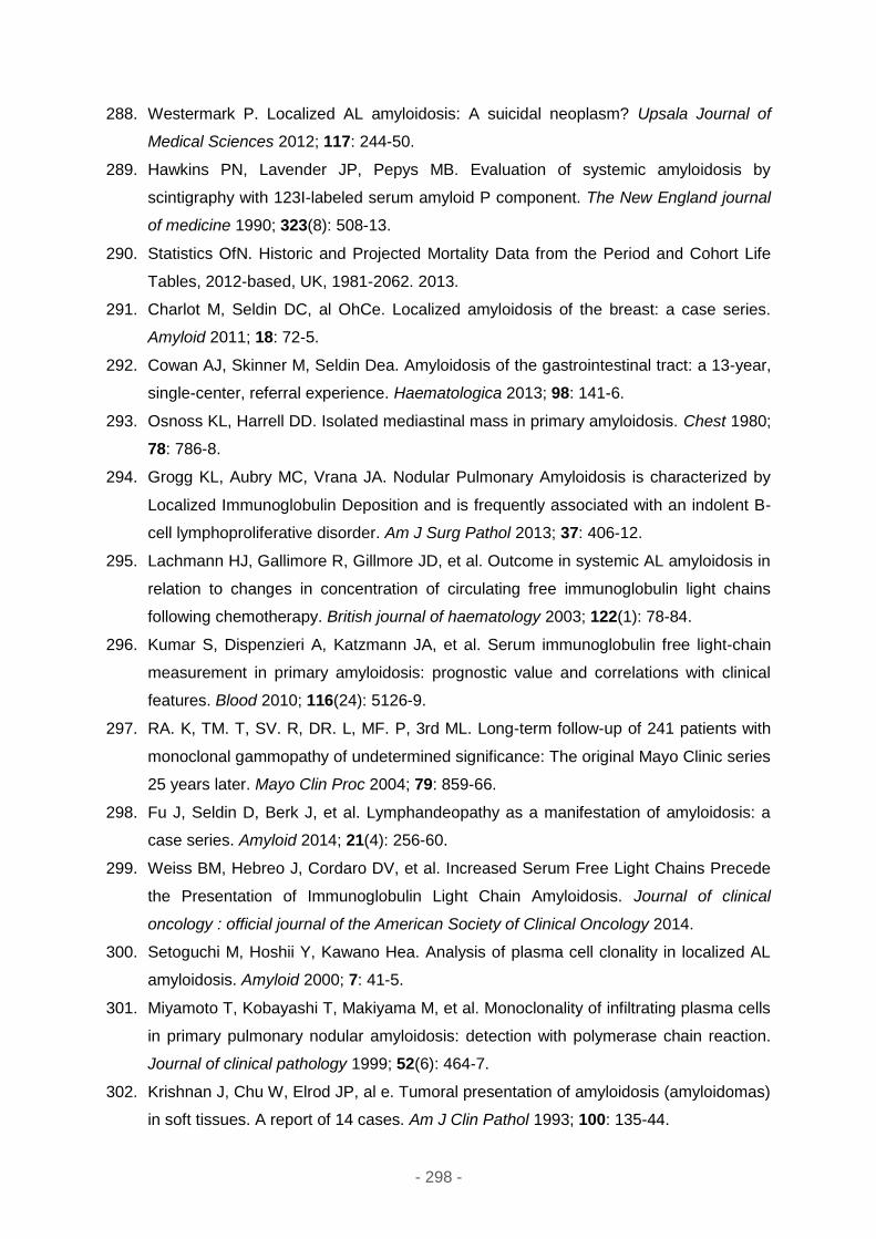
- 298 -
288. Westermark P. Localized AL amyloidosis: A suicidal neoplasm? Upsala Journal of
Medical Sciences 2012; 117: 244-50.
289. Hawkins PN, Lavender JP, Pepys MB. Evaluation of systemic amyloidosis by
scintigraphy with 123I-labeled serum amyloid P component. The New England journal
of medicine 1990; 323(8): 508-13.
290. Statistics OfN. Historic and Projected Mortality Data from the Period and Cohort Life
Tables, 2012-based, UK, 1981-2062. 2013.
291. Charlot M, Seldin DC, al OhCe. Localized amyloidosis of the breast: a case series.
Amyloid 2011; 18: 72-5.
292. Cowan AJ, Skinner M, Seldin Dea. Amyloidosis of the gastrointestinal tract: a 13-year,
single-center, referral experience. Haematologica 2013; 98: 141-6.
293. Osnoss KL, Harrell DD. Isolated mediastinal mass in primary amyloidosis. Chest 1980;
78: 786-8.
294. Grogg KL, Aubry MC, Vrana JA. Nodular Pulmonary Amyloidosis is characterized by
Localized Immunoglobulin Deposition and is frequently associated with an indolent B-
cell lymphoproliferative disorder. Am J Surg Pathol 2013; 37: 406-12.
295. Lachmann HJ, Gallimore R, Gillmore JD, et al. Outcome in systemic AL amyloidosis in
relation to changes in concentration of circulating free immunoglobulin light chains
following chemotherapy. British journal of haematology 2003; 122(1): 78-84.
296. Kumar S, Dispenzieri A, Katzmann JA, et al. Serum immunoglobulin free light-chain
measurement in primary amyloidosis: prognostic value and correlations with clinical
features. Blood 2010; 116(24): 5126-9.
297. RA. K, TM. T, SV. R, DR. L, MF. P, 3rd ML. Long-term follow-up of 241 patients with
monoclonal gammopathy of undetermined significance: The original Mayo Clinic series
25 years later. Mayo Clin Proc 2004; 79: 859-66.
298. Fu J, Seldin D, Berk J, et al. Lymphandeopathy as a manifestation of amyloidosis: a
case series. Amyloid 2014; 21(4): 256-60.
299. Weiss BM, Hebreo J, Cordaro DV, et al. Increased Serum Free Light Chains Precede
the Presentation of Immunoglobulin Light Chain Amyloidosis. Journal of clinical
oncology : official journal of the American Society of Clinical Oncology 2014.
300. Setoguchi M, Hoshii Y, Kawano Hea. Analysis of plasma cell clonality in localized AL
amyloidosis. Amyloid 2000; 7: 41-5.
301. Miyamoto T, Kobayashi T, Makiyama M, et al. Monoclonality of infiltrating plasma cells
in primary pulmonary nodular amyloidosis: detection with polymerase chain reaction.
Journal of clinical pathology 1999; 52(6): 464-7.
302. Krishnan J, Chu W, Elrod JP, al e. Tumoral presentation of amyloidosis (amyloidomas)
in soft tissues. A report of 14 cases. Am J Clin Pathol 1993; 100: 135-44.
- 299 -
303. Lim JK, Lacy MQ, Kurtin PJ, Kyle RA, Gertz MA. Pulmonary marginal zone lymphoma
of MALT type as a cause of localised pulmonary amyloidosis. Journal of clinical
pathology 2001; 54(8): 642-6.
304. Kambouchner M, Godmer P, Guillevin L, Raphael M, Droz D, Martin A. Low grade
marginal zone B cell lymphoma of the breast associated with localised amyloidosis and
corpora amylacea in a woman with long standing primary Sjogren's syndrome. Journal
of clinical pathology 2003; 56(1): 74-7.
305. Meijer JM, Schonland SO, Palladini G, et al. Sjögren's syndrome and localized nodular
cutaneous amyloidosis: coincidence or a distinct clinical entity? Arthritis Rheum 2008;
58(7): 1992-9.
306. Westermark P. Localized AL amyloidosis: a suicidal neoplasm? Upsala journal of
medical sciences 2012; 117(2): 244-50.
307. Mekinian A, Jaccard A, Soussan M, et al. 18F-FDG PET/CT in patients with amyloid
light-chain amyloidosis: case-series and literature review. Amyloid : the international
journal of experimental and clinical investigation : the official journal of the International
Society of Amyloidosis 2012; 19(2): 94-8.
308. Yoshida A, Borkar S, Singh Bea. Incidental detection of concurrent extramedullary
plasmacytoma and amyloidoma of the nasopharynx on [18F]fluorodeoxyglucose
positron emission tomography/computed tomography. J Clin Oncol 2008; 26: 5817-9.
309. Truong MT, Kachnic LA, Grillone GA, et al. Long-Term Results of Conformal
Radiotherapy for Progressive Airway Amyloidosis. Int J Radiat Oncol Biol Phys 2012;
83(2): 734-9.
310. Neben-Wittich M, Foote R, Kalra S. External Beam Radiation Therapy for
Tracheobronchial Amyloidosis. Chest 2007; 132: 262-7.
311. Capizzi S, Betancourt E, Prakash U. Tracheobronchial amyloidosis. Mayo Clin Proc
2000; 75: 1148-52.
312. Gillmore JD, Hawkins PN. Amyloidosis and the respiratory tract. Thorax 1999; 54: 444-
51.
313. Eanes ED, Glenner GG. X-ray diffraction studies on amyloid filaments. J Histochem
Cytochem 1968; 16: 673-7.
314. Jahn TR, Parker MJ, Homans SW, Radford SE. Amyloid formation under physiological
conditions proceeds via a native-like folding intermediate. Nat Struct Mol Biol 2006;
13(3): 195-201.
315. Eakin CM, Berman AJ, Miranker AD. A native to amyloidogenic transition regulated by
a backbone trigger. Nat Struct Mol Biol 2006; 13: 202-8.
316. Come J, Fraser P, Lanbury PJ. A Kinetic Model for Amyloid Formation in the Prion
Diseases, Importance of Seeding. PNAS USA 1993; 90: 5959-63.
- 300 -
317. Falk RH, Comenzo RL, Skinner M. The systemic amyloidoses. N Engl J Med 1997; 337:
898-909.
318. O'Regan A, Fenlon HM, Beamis JF, Jr., Steele MP, Skinner M, Berk JL.
Tracheobronchial amyloidosis. The Boston University experience from 1984 to 1999.
Medicine (Baltimore) 2000; 79: 69-79.
319. Cordier JF, Loire R, Brune J. Amyloidosis of the lower respiratory tract. Clinical and
pathologic features in a series of 21 patients. Chest 1986; 90: 827-31.
320. Berk JL, O'Regan A, Skinner M. Pulmonary and tracheobronchial amyloidosis. Semin
Respir Crit Care Med 2002; 23: 155-65.
321. Rubinow A, Celli BR, Cohen AS, Rigden BG, Brody JS. Localized amyloidosis of the
lower respiratory tract. Am Rev Respir Dis 1978; 118: 603-11.
322. Breuer R, Simpson GT, Rubinow A, Skinner M, Cohen AS. Tracheobronchial
amyloidosis: treatment by carbon dioxide laser photoresection. Thorax 1985; 40: 870-1.
323. Yap J, Wang Y, SC. P. A case of primary diffuse tracheobronchial amyloidosis treated
with laser therapy. Sing Med J 1992; 33: 198-200.
324. Kurrus JA, Hayes JK, Hoidal JR, Menendez MM, Elstad MR. Radiation therapy for
tracheobronchial amyloidosis. Chest 1998; 114: 1489-92.
325. Rocken C, Tautenhahn J, Buhling F, et al. Prevalence and Pathology of Amyloid in
Atherosclerotic Arteries. Arteriosclerosis, Thrombosis, and Vascular Biology 2006;
26(3): 676-7.
326. Nicholas BL, Skipp P, Barton S, et al. Identification of Lipocalin and Apolipoprotein A1
as Biomarkers of Chronic Obstructive Pulmonary Disease. American Journal of
Respiratory and Critical Care Medicine; 181(10): 1049-60.
327. Sandhu MS, Dunger DB, Giovannucci EL. Insulin, Insulin-Like Growth Factor-I (IGF-I),
IGF Binding Proteins, Their Biologic Interactions, and Colorectal Cancer. Journal of the
National Cancer Institute 2002; 94(13): 972-80.
328. LeRoith D, Baserga R, Helman L, Roberts Jr CT. Insulin-like Growth Factors and
Cancer. Annals of Internal Medicine 1995; 122(1): 54-9.
329. Baserga R. The Insulin-like Growth Factor I Receptor: A Key to Tumor Growth? Cancer
Research 1995; 55(2): 249-52.
330. Price WA, Moats-Staats BM, Stiles AD. Pro- and Anti-inflammatory Cytokines Regulate
Insulin-like Growth Factor Binding Protein Production by Fetal Rat Lung Fibroblasts.
American Journal of Respiratory Cell and Molecular Biology 2002; 26(3): 283-9.
331. Yamashita N, Tashimo H, Ishida H, et al. Role of insulin-like growth factor-I in allergen-
induced airway inflammation and remodeling. Cellular Immunology 2005; 235(2): 85-91.
332. Korampalli TS, Stafford ND. Expression of angiogenic growth factors in laryngeal
carcinoma. Mol Clin Oncol 2013; 6: 1013-8.
- 301 -
333. Booth DR, Sunde M, Bellotti V, et al. Instability, unfolding and aggregation of human
lysozyme variants underlying amyloid fibrillogenesis. Nature 1997; 385: 787-93.
334. Khurana R, Gillespie JR, Talapatra A, et al. Partially Folded Intermediates as Critical
Precusors of Light Chain Amyloid Fibrils and Amorphous Aggregates. Biochemistry
2001; 40: 3525-35.
335. Sanchorawala V, Finn KT, Fennessey S, et al. Durable hematologic complete responses
can be achieved with lenalidomide in AL amyloidosis. Blood 2010; 116(11): 1990-1.
336. Mahmood S, Venner CP, Sachchithanantham S, et al. Lenalidomide and
dexamethasone for Systemic AL Amyloidosis following prior treatment with thalidomide
or bortezomib regimens. British Journal of Haematology: n/a-n/a.
337. Wang M, Dimopoulos M, Chen C, al e. Lenalidomide plus dexamethasone is more
effective than dexamethasone alone in patients with relapsed or refractory multiple
myeloma regardles of prior thalidomide exposure. Blood 2008; 112: 4445-51.
338. Palladini G, Russo P, Foli A, et al. Salvage therapy with lenalidomide and
dexamethasone in patients with advanced AL amyloidosis refractory to melphalan,
bortezomib, and thalidomide. Ann Hematol 2012; 91(1): 89-92.
339. Wechalekar A, Foard D, Rannigan L, et al. Characteristics and outcomes of 714 patients
with systemic AL amyloidosis - analysis of a prospective study (Alchemy study). XIVth
International Symposium on Amyloidosis 2014: 49.
340. Palladini G, Milani P, Foli A, et al. Oral melphalan and dexamethasone grants extended
survival with minimal toxicity in AL amyloidosis: long-term results of a risk-adapted
approach. Haematologica 2014; 99(4): 743-50.
341. Palladini G, Milani P, Foli A, et al. Oral melphalan and dexamethasone grants extended
survival with minimal toxicity in AL amyloidosis: long-term results of a risk-adapted
approach. Haematologica.
342. Pinney JH, Lachmann HJ, Bansi L, et al. Outcome in Renal AL Amyloidosis following
Chemotherapy. Journal of Clinical Oncology 2011; 29(6): 674-81.
343. Sanchorawala V SA, Zeldis JB, Seldin DC. Risk of second primary malignancy in
patients with AL amyloidosis treated with lenalidomide. Am J Hematol 2013; 88(8): 719.
344. Cibeira M, Sanchorawala V, Seldin D, al e. Outcome of AL amyloidosis after high-dose
melphalan and autologous stem cell transplantation: long term results in a series of 421
patients. Blood 2011; 118(16): 4346-52.
345. Lonial S, Vij R, Harousseau J-L, et al. Elotuzumab in Combination With Lenalidomide
and Low-Dose Dexamethasone in Relapsed or Refractory Multiple Myeloma. Journal of
Clinical Oncology.
- 302 -
346. Wu L, Parton A, Lu L, Adams M, Schafer P, Bartlett JB. Lenalidomide enhances
antibody-dependent cellular cytotoxicity of solid tumor cells in vitro: influence of host
immune and tumor markers. Cancer Immunology, Immunotherapy; 60(1): 61-73.
347. Specter R, Sanchorawala V, Seldin DC, et al. Kidney dysfunction during lenalidomide
treatment for AL amyloidosis. Nephrology Dialysis Transplantation; 26(3): 881-6.
348. Tan SY, Baillod R, Brown E, et al. Clinical, radiological and serum amyloid P component
scintigraphic features of beta2-microglobulin amyloidosis associated with continuous
ambulatory peritoneal dialysis. Nephrol Dial Transplant 1999; 14: 1467-71.
349. Drueke TB. Beta2-microglobulin and amyloidosis. Nephrol Dial Transplant 2000; 15
Suppl 1: 17-24.
350. Bely M, Kapp P, Szabo TS, Lakatos T, Apathy A. Electron microscopic characteristics
of beta2-microglobulin amyloid deposits in long-term haemodialysis. Ultrastruct Pathol
2005; 29(6): 483-91.
351. Pepys MB, Dyck RF, de Beer FC, Skinner M, Cohen AS. Binding of serum amyloid P
component (SAP) by amyloid fibrils. Clin Exp Immunol 1979; 38: 284-93.
352. Pepys MB, Booth DR, Hutchinson WL, Gallimore JR, Collins PM, Hohenester E. Amyloid
P component. A critical review. Amyloid: Int J Exp Clin Invest 1997; 4: 274-95.
353. Nelson SR, Tennent GA, Sethi D, et al. Serum amyloid P component in chronic renal
failure and dialysis. Clin Chim Acta 1991; 200: 191-200.
354. Richards DB, Cookson LM, Berges AC, et al. Therapeutic Clearance of Amyloid by
Antibodies to Serum Amyloid P Component. New England Journal of Medicine 2015;
373(12): 1106-14.
355. Tan SY, Irish A, Winearls CG, et al. Long term effect of renal transplantation on dialysis-
related amyloid deposits and symptomatology. Kidney Int 1996; 50: 282-9.
356. Fautrel B, Fermand J-P, Sibilia J, Nochy D, Rousselin B, Ravaud P. Amyloid arthropathy
in the course of multiple myeloma. The Journal of Rheumatology 2002; 29(7): 1473-81.
357. Katoh N, Tazawa K-i, Ishii W, Matsuda M, Ikeda S-i. Systemic AL Amyloidosis Mimicking
Rheumatoid Arthritis. Internal Medicine 2008; 47(12): 1133-8.
358. Miyata M, Sato N, Watanabe H, et al. Magnetic Resonance Imaging Findings in Primary
Amyloidosis-associatedArthropathy. Internal Medicine 2000; 39(4): 313-9.
359. Kecler-Pietrzyk A, Kok HK, Lyburn ID, Torreggiani WC. Dialysis Related Amyloid
Arthropathy on (18)FDG PET-CT. The Ulster Medical Journal 2014; 83(2): 117-8.

- 303 -
Appendices
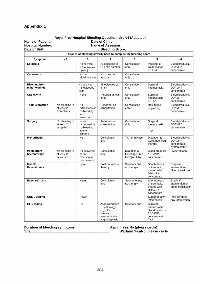
- 304 -
Appendix 1
Royal Free Hospital Bleeding Questionnaire v4 (Adapted) Name of Patient: Date of Clinic: Hospital Number: Name of Assessor: Date of Birth: Bleeding Score:
Grades of bleeding severity used to compute the bleeding score
Symptom - 1 0 1 2 3 4
Epistaxis - No or trivial
(<5 episodes / year )
>5 episodes or >10 min duration
Consultation only
Packing or cauterisation or TXA
Blood products / DDAVP / concentrate
Cutaneous - No or trivial (<1 cm)
>1cm and no trauma
Consultation only
Bleeding from minor wounds
- No or trivial (<5 episodes / year )
>5 episodes or > 5 min
Consultation only
Surgical haemostasis
Blood products / DDAVP / concentrate
Oral cavity - None Referred at least once
Consultation only
Surgical haemostasis or TXA
Blood products / DDAVP / concentrate
Tooth extraction No bleeding in at least 2 extractions
No extractions or no bleeding in 1 extraction
Reported, no consultation
Consultation only
Resuturing
or packing
Blood products / DDAVP / concentrate
Surgery No bleeding in at least 2 surgeries
None performed or no bleeding in one Surgery
Reported, no consultation
Consultation only
Surgical haemostasis or
TXA
Blood products / DDAVP / concentrate
Menorrhagia - No Consultation only
TXA or pill use Dilatation & Curettage, iron therapy
Blood products / DDAVP / concentrate / hysterectomy
Postpartum haemorrhage
No bleeding in at least 2 deliveries
No deliveries or no bleeding in one delivery
Consultation only
Dilatation & Curettage, iron therapy, TXA
Blood products / DDAVP / concentrate
Hysterectomy
Muscle Haematomas
- Never Post-trauma no therapy
Spontaneous, no therapy
Spontaneous or traumatic, treated with DDAVP / concentrate
Surgical intervention or blood transfusion
Haemarthrosis - Never Consultation only
Spontaneous, no therapy
Spontaneous or traumatic, treated with DDAVP / concentrate
Surgical intervention or blood transfusion
CNS Bleeding - Never - - Subdural, any intervention
Intra cerebral, any intervention
GI Bleeding - No Associated with GI pathology e.g. ulcer, varices, haemorrhoids, angiodysplasia
Spontaneous Surgical haemostasis Blood products / DDAVP / concentrate/ TXA
Duration of bleeding symptoms __________________ Aspirin Yes/No (please circle) Site__________________________________________ Warfarin Yes/No (please circle
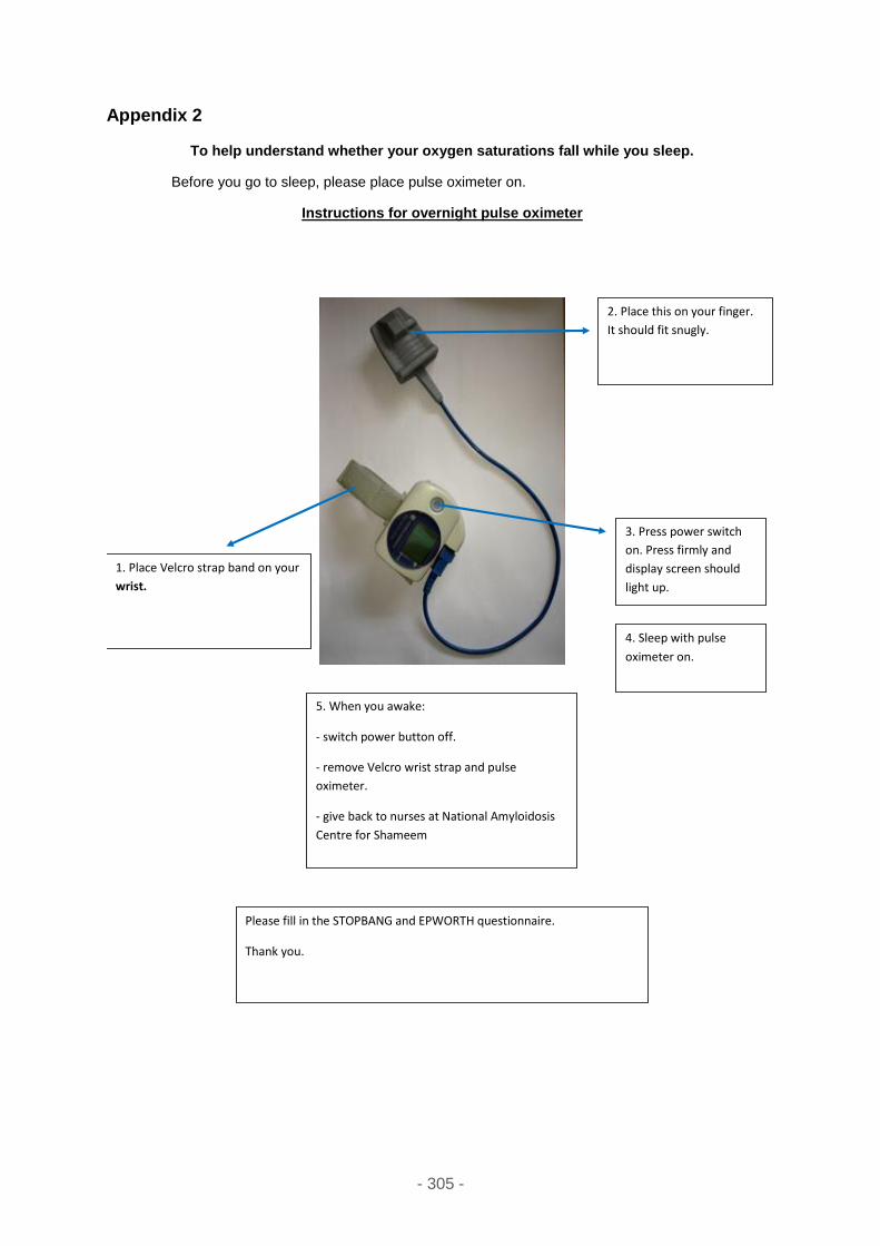
- 305 -
Appendix 2
To help understand whether your oxygen saturations fall while you sleep.
Before you go to sleep, please place pulse oximeter on.
Instructions for overnight pulse oximeter
1. Place Velcro strap band on your
wrist.
2. Place this on your finger.
It should fit snugly.
5. When you awake:
- switch power button off.
- remove Velcro wrist strap and pulse
oximeter.
- give back to nurses at National Amyloidosis
Centre for Shameem
3. Press power switch
on. Press firmly and
display screen should
light up.
4. Sleep with pulse
oximeter on.
Please fill in the STOPBANG and EPWORTH questionnaire.
Thank you.

- 306 -
Appendix 3
EPWORTH SLEEPINESS SCORE How to work out your score How likely are you to doze off or fall asleep during the following situations, in contrast to just feeling tired? For each of the situations listed below, give yourself a score of 0 to 3 where: 0 = would never doze 1 = slight chance 2 = moderate chance 3 = high chance Work out your total score by adding up your individual scores for situations 1 to 8 (If you have not been in the following situations recently, think about how you would have been affected).
Situation
Score (0 – 3)
Sitting and reading
Watching television
Sitting inactive in a public place e.g. theatre, meeting
As a passenger in a car for an hour without a break
Laying down for a rest in the afternoon
Sitting and talking to someone
Sitting quietly after lunch (when you have had no alcohol)
In a car, whilst stopped in traffic
Total

- 307 -
Appendix 4
STOP BANG QUESTIONAIRRE
Height inches/cm Weight lb/kg
Age Male/Female BMI
Collar size of shirt: S, M, L or XL Neck Circumference * cm
1. Snoring.
Do you snore loudly (louder than talking or loud enough to be heard through closed doors)?
Yes No
2. Tired.
Do you often feel tired, fatigued or sleepy during daytime?
Yes No
3. Observed.
Has anyone observed you stop breathing during your sleep?
Yes No
4. Blood pressure.
Do you have or are you being treated for high blood pressure?
Yes No
5. BMI.
BMI more than 35 kg/m2?
Yes No
6. Age
Age greater than 50?
Yes No
7. Neck circumference.
Neck circumference greater than 40cm?
Yes No
8. Gender.
Gender male?
Yes No
* Neck circumference measured by staff
High risk of OSA: answering yes to 3 or more items.
Low risk of OSA: answering yes to less than 3 items
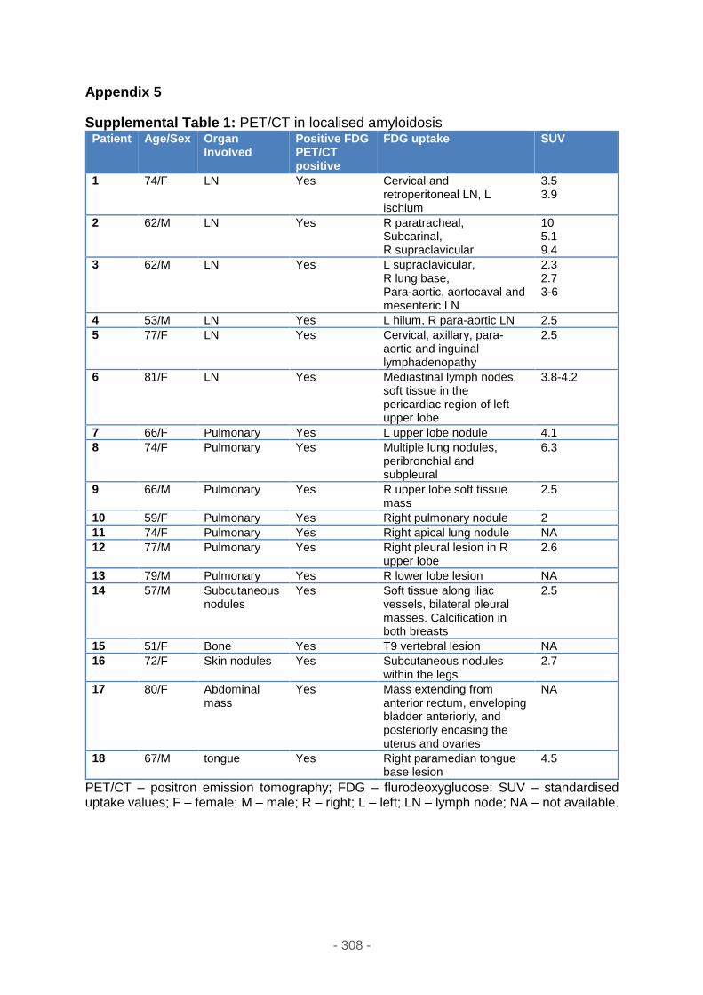
- 308 -
Appendix 5
Supplemental Table 1: PET/CT in localised amyloidosis Patient Age/Sex Organ
Involved Positive FDG PET/CT positive
FDG uptake SUV
1 74/F LN Yes Cervical and retroperitoneal LN, L ischium
3.5 3.9
2 62/M LN Yes R paratracheal, Subcarinal, R supraclavicular
10 5.1 9.4
3 62/M LN Yes L supraclavicular, R lung base, Para-aortic, aortocaval and mesenteric LN
2.3 2.7 3-6
4 53/M LN Yes L hilum, R para-aortic LN 2.5
5 77/F LN Yes Cervical, axillary, para-aortic and inguinal lymphadenopathy
2.5
6 81/F LN Yes Mediastinal lymph nodes, soft tissue in the pericardiac region of left upper lobe
3.8-4.2
7 66/F Pulmonary Yes L upper lobe nodule 4.1
8 74/F Pulmonary Yes Multiple lung nodules, peribronchial and subpleural
6.3
9 66/M Pulmonary Yes R upper lobe soft tissue mass
2.5
10 59/F Pulmonary Yes Right pulmonary nodule 2
11 74/F Pulmonary Yes Right apical lung nodule NA
12 77/M Pulmonary Yes Right pleural lesion in R upper lobe
2.6
13 79/M Pulmonary Yes R lower lobe lesion NA
14 57/M Subcutaneous nodules
Yes Soft tissue along iliac vessels, bilateral pleural masses. Calcification in both breasts
2.5
15 51/F Bone Yes T9 vertebral lesion NA
16 72/F Skin nodules Yes Subcutaneous nodules within the legs
2.7
17 80/F Abdominal mass
Yes Mass extending from anterior rectum, enveloping bladder anteriorly, and posteriorly encasing the uterus and ovaries
NA
18 67/M tongue Yes Right paramedian tongue base lesion
4.5
PET/CT – positron emission tomography; FDG – flurodeoxyglucose; SUV – standardised uptake values; F – female; M – male; R – right; L – left; LN – lymph node; NA – not available.
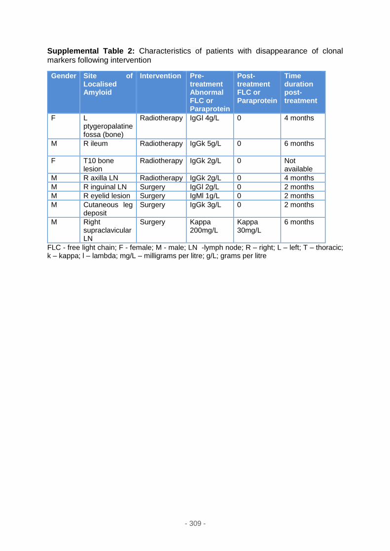
- 309 -
Supplemental Table 2: Characteristics of patients with disappearance of clonal markers following intervention
Gender Site of Localised Amyloid
Intervention Pre-treatment Abnormal FLC or Paraprotein
Post-treatment FLC or Paraprotein
Time duration post-treatment
F L ptygeropalatine fossa (bone)
Radiotherapy IgGl 4g/L 0 4 months
M R ileum Radiotherapy IgGk 5g/L 0
6 months
F T10 bone lesion
Radiotherapy IgGk 2g/L 0 Not available
M R axilla LN Radiotherapy IgGk 2g/L 0 4 months
M R inguinal LN Surgery IgGl 2g/L 0 2 months
M R eyelid lesion Surgery IgMl 1g/L 0 2 months
M Cutaneous leg deposit
Surgery IgGk 3g/L 0 2 months
M Right supraclavicular LN
Surgery Kappa 200mg/L
Kappa 30mg/L
6 months
FLC - free light chain; F - female; M - male; LN -lymph node; R – right; L – left; T – thoracic; k – kappa; l – lambda; mg/L – milligrams per litre; g/L; grams per litre
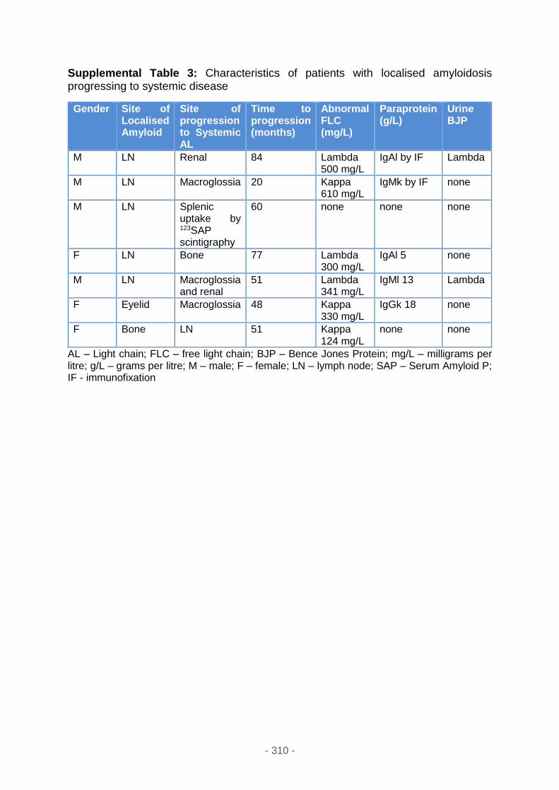
- 310 -
Supplemental Table 3: Characteristics of patients with localised amyloidosis progressing to systemic disease
Gender Site of Localised Amyloid
Site of progression to Systemic AL
Time to progression (months)
Abnormal FLC (mg/L)
Paraprotein (g/L)
Urine BJP
M LN Renal 84 Lambda 500 mg/L
IgAl by IF Lambda
M LN Macroglossia 20 Kappa 610 mg/L
IgMk by IF none
M LN Splenic uptake by 123SAP scintigraphy
60 none none none
F LN Bone 77 Lambda 300 mg/L
IgAl 5 none
M LN Macroglossia and renal
51 Lambda 341 mg/L
IgMl 13 Lambda
F Eyelid Macroglossia 48 Kappa 330 mg/L
IgGk 18 none
F Bone LN 51 Kappa 124 mg/L
none none
AL – Light chain; FLC – free light chain; BJP – Bence Jones Protein; mg/L – milligrams per litre; g/L – grams per litre; M – male; F – female; LN – lymph node; SAP – Serum Amyloid P; IF - immunofixation