amyloidosis and porphyria.ppt
TRANSCRIPT
DAPM RV DENTAL COLLEGE DEPARTMENT OF ORAL AND MAXILLOFACIAL PATHOLOGY
REFERENCES
Shafer’s textbook of oral pathology-
Shafer , Hine , Levy. Oral and Maxillofacial Pathology- Neville, Damm, Allen, Bouquot.
Textbook of Biochemistry- DM Vasudevan and Sreekumari
REFERENCES
Pathologic Basis of Disease – Robbins and Cotran
Essential Pathology for Dental Student- Harsh Mohan.
Pathologic outlines.com Wikipedia.org Emedicine .medscape.com
AMYLOIDOSIS
DEFINITION :
Term used for a group of diseases characterized by extracellular deposition of a fibrillar protein called amyloid .
First described by ROKTINASKY in 1842
( Pathologic Basis Of Disease- Robbins And Cotran)
AMYLOIDOSIS
Named by Virchow Amylon = starch
Oid = like
Synonym= beta fibrillosis (Pathologic Basis Of Disease- Robbins And Cotran)
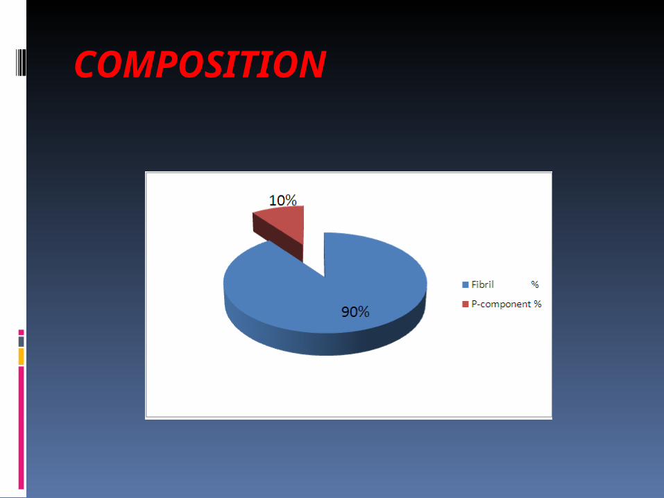
COMPOSITION
COMPOSITION
FIBRIL PROTEIN
oBy electron microscopy , the fibrils are non branching filaments, delicate.olying singly or 1-4 laterally aligned .oEach fibril is composed of protofibrils.oBy X ray crystallography, the fibrils have a cross beta-pleated sheet configuration.( Essential Pathology for Dental Students- Harsh Mohan)
COMPOSITION
P COMPONENT
oBy electron microscopy, it has a pentagonal profile .oHas a internal diameter = 4nm External diameter = 9nmoChemical analysis, it is a glycoprotein resembling the serum alpha one glycoprotein. ( Essential Pathology for Dental Students- Harsh Mohan)
CHEMICAL NATURE OF AMYLOID Chemically analysis of the fibril protein revealed two types of amyloids :
AL (amyloid light chain ) protein.
AA ( amyloid associated ) protein.
AL( Amyloid light chain ) PROTEIN Consists of poly peptides. Produced by immunoglobulin secreting protein.
Molecular weight = 7500 to 23,000
Either lambda or kappa light chains form the AL protein.
Lambda chains are two times more common.
(Pathologic Basis of Disease- Robbins and cotran )
AA (Amyloid associated) Protein Consists of polypeptides Derived from Serum Amyloid Protein.
SAA circulates in association with HDL.
Synthesized in the liver. Molecular weight= 8500
(Pathologic Basis of Disease- Robbins and cotran )
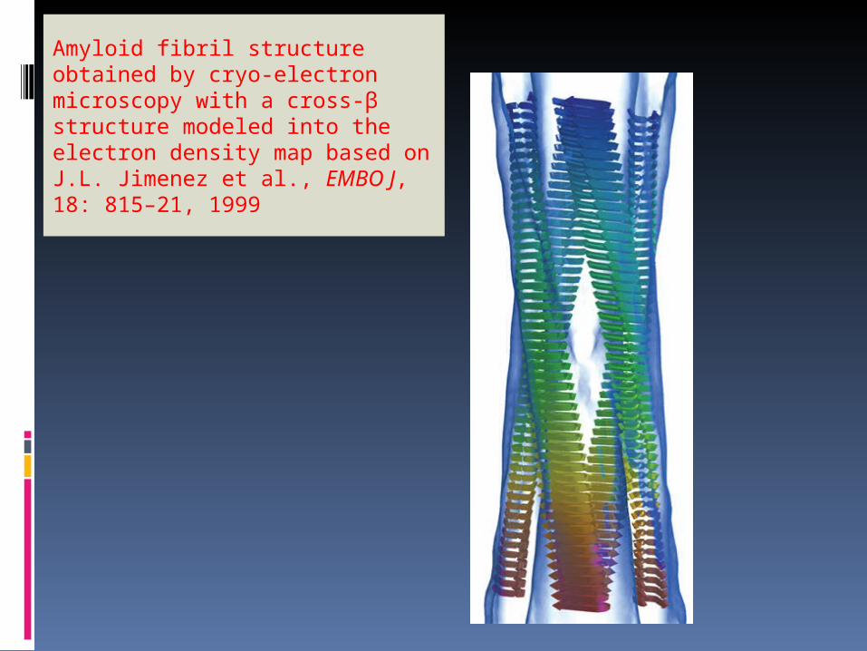
Amyloid fibril structure obtained by cryo-electron microscopy with a cross-β structure modeled into the electron density map based on J.L. Jimenez et al., EMBO J, 18: 815–21, 1999
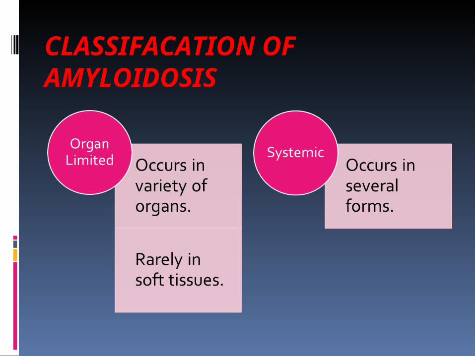
CLASSIFACATION OF AMYLOIDOSIS
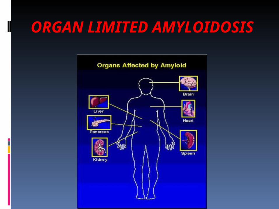
ORGAN LIMITED AMYLOIDOSIS
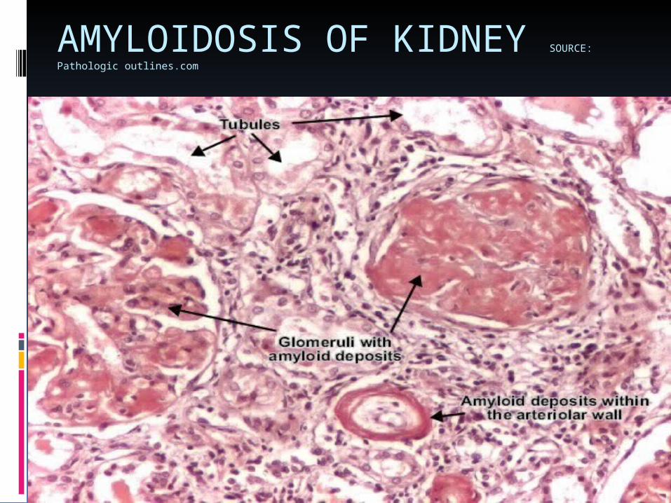
AMYLOIDOSIS OF KIDNEY SOURCE:
Pathologic outlines.com
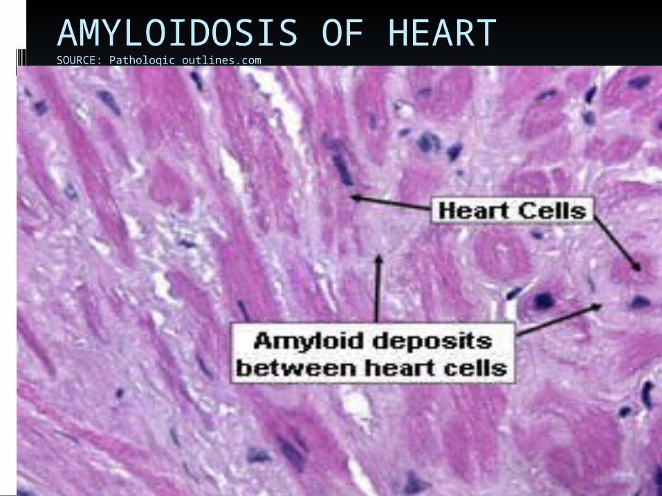
AMYLOIDOSIS OF HEART SOURCE: Pathologic outlines.com
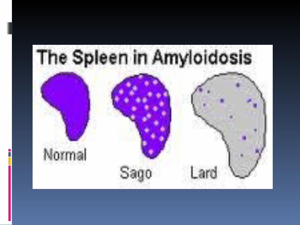
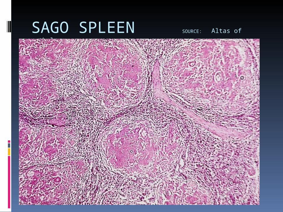
SAGO SPLEEN SOURCE: Altas of
pathology
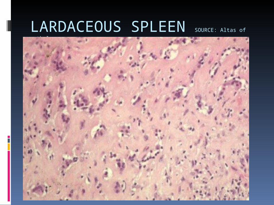
LARDACEOUS SPLEEN SOURCE: Altas of
pathology
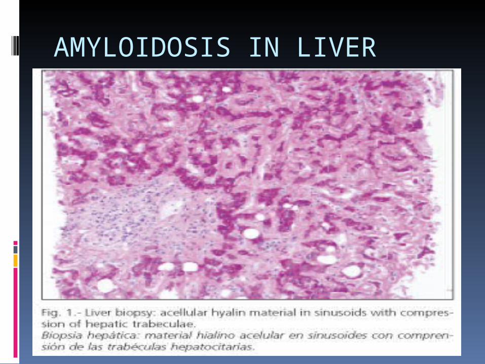
AMYLOIDOSIS IN LIVER
SYSTEMIC AMYLOIDOSIS
SYSTEMIC AMYLOIDOSIS
PRIMARY and MYELOMA ASSOCIATEDCLINICAL FEATURES
Age : older adults, above 65 years.Slight male predilection.SIGNS AND SYMPTOMSFatigue, weight loss, paresthesia, hoarseness, edema and orthostatic hypotension.(Textbook of oral and maxillofacial Pathology- Neville, Second Edi.)
PRIMARY and MYELOMA ASSOCIATEDSIGNS AND SYMPTOMSEventually, carpal tunnel syndrome, muco-cutaneous lesions, hepatomegaly and macroglossia.Skin lesions are smooth, firm and waxy .Lesions are associated with petechaiae and ecchymoses.(Textbook of oral and maxillofacial Pathology- Neville, Second Edi.)
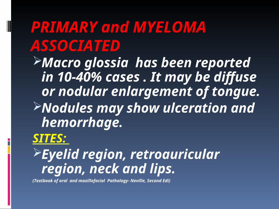
PRIMARY and MYELOMA ASSOCIATEDMacro glossia has been reported in 10-40% cases . It may be diffuse or nodular enlargement of tongue.
Nodules may show ulceration and hemorrhage.
SITES: Eyelid region, retroauricular region, neck and lips.
(Textbook of oral and maxillofacial Pathology- Neville, Second Edi)
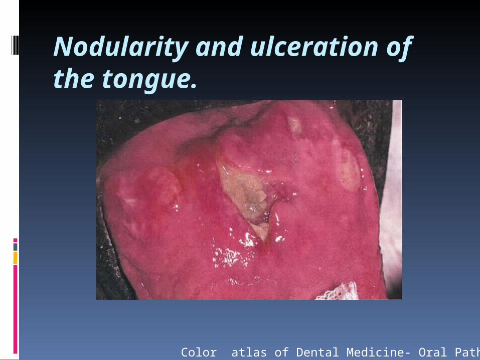
Nodularity and ulceration of the tongue.
Color atlas of Dental Medicine- Oral Pathology
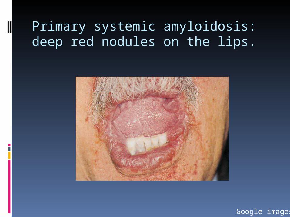
Primary systemic amyloidosis: deep red nodules on the lips.
Google images
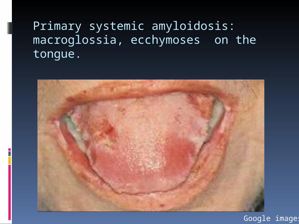
Primary systemic amyloidosis: macroglossia, ecchymoses on thetongue.
Google images

Patient exhibits a firm, waxy, smooth lesion near the eye.
Google images
PRIMARY and MYELOMA ASSOCIATED
Infrequently patients may complain of dry eyes and dry mouth which are secondary to amyloid infiltration and destruction of salivary and lacrimal glands.
(Textbook of oral and maxillofacial Pathology- Neville, Second Edi.)
SECONDARY AMYLOIDOSIS Develops as a result of long standing chronic inflammatory process like : Osteomyelitis
Tuberculosis SarcoidosisSITES: Kidney, Liver, spleen, adrenals.
(Textbook of oral and maxillofacial Pathology- Neville, Second Edi.)
HEMODIALYSIS ASSOCIATED AMYLOIDOSIS Seen in patients who have under
gone long term renal dialysis. Due to accumulation of beta-2 microglobulin, an amyloid protein.
Deposits in the bones and joints . Tongue involvement is common.
(Textbook of oral and maxillofacial Pathology- Neville, Second Edi.)
HEREDOFAMILIAL AMYLOIDOSIS Uncommon disease. Inherited autosomal dominant trait.
These conditions appear as polyneuropathies, cardiomyopathy, cardiac arrhythmias, CCF and renal failure.
(Textbook of oral and maxillofacial Pathology- Neville, Second Edi.)
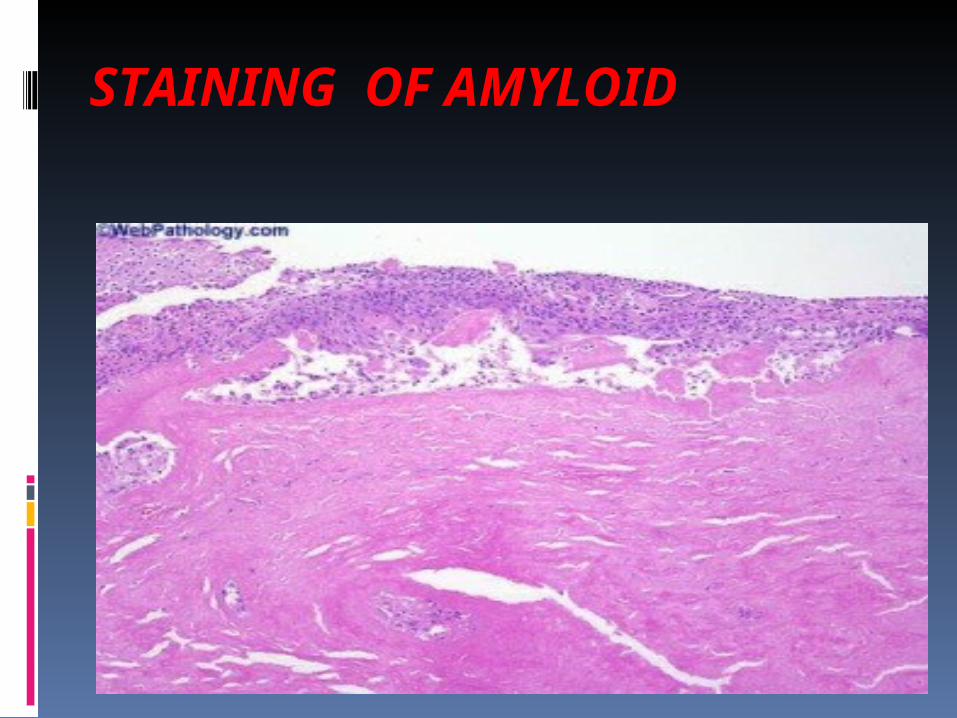
STAINING OF AMYLOID
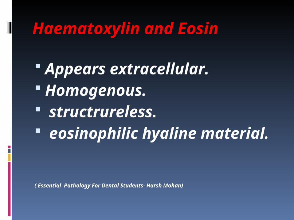
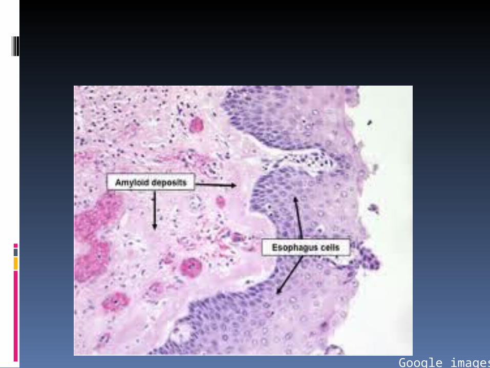
Haematoxylin and Eosin
Appears extracellular. Homogenous. structrureless. eosinophilic hyaline material.
( Essential Pathology For Dental Students- Harsh Mohan)
Google images
IODINE STAIN
Oldest method. Lugol’s iodine is used. It imparts mahogany- brown color to the amyloid containing area.
Addition of H2SO4 turns it violet.
( Essential Pathology For Dental Students- Harsh Mohan)
Google images
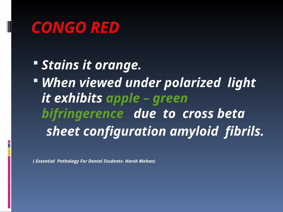
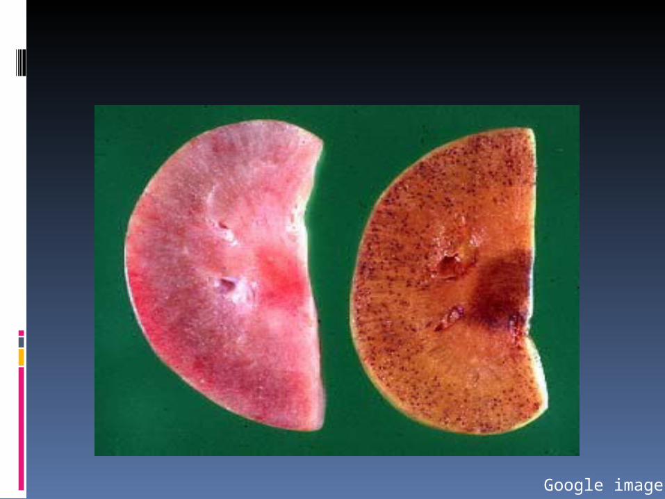
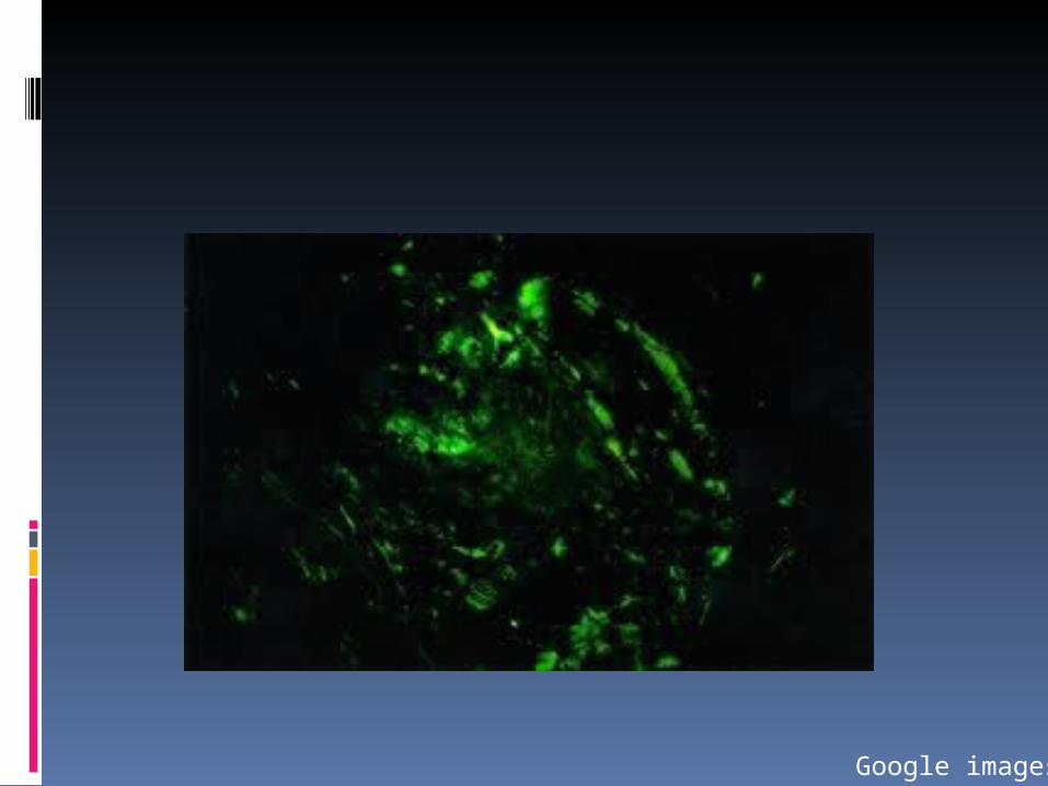
CONGO RED
Stains it orange. When viewed under polarized light it exhibits apple – green bifringerence due to cross beta
sheet configuration amyloid fibrils.
( Essential Pathology For Dental Students- Harsh Mohan)
Google images
CONGO RED
Also used to differentiate between AA and AL amyloid.
AA amyloid fails to stain if previously treated with KMnO4.
Most reliable method.
( Essential Pathology For Dental Students- Harsh Mohan)
METACHROMATIC STAIN
Amyloid has a property of metachromasia.
Stains employed are :a.Methyl Violetb.Crystal violetC. Gentian violet. Imparts rose pink color.
( Essential Pathology For Dental Students- Harsh Mohan)
FLUORESCENT STAINS
Stains like thioflavin S and T bind to amyloid
fluoresce yellow under U V light.
( Essential Pathology For Dental Students- Harsh Mohan)
DIAGNOSIS
BIOPSY EXAMINATION. IN VIVO CONGO RED TEST. IMMNOELECTROPHORESIS. BONE MARROW ASPIRATION.
TREATMENT AND PROGNOSIS No effective therapy. Surgical debulking of tongue. Treatment for primary amyloidosis with Prednisone and melphalan.
Most patients die of cardiac failure , arrhythmia or renal disease within few months or years of diagnosis.
(Textbook of oral and maxillofacial Pathology- Neville, Second Edi.)
INTRODUCTION
This disease occurs due to inborn defect of porphyrin metabolism.
Porphyrias are a group of rare disorders passed down through families, in which an important part of hemoglobin, called heme, is not made properly
CLASSIFICATION
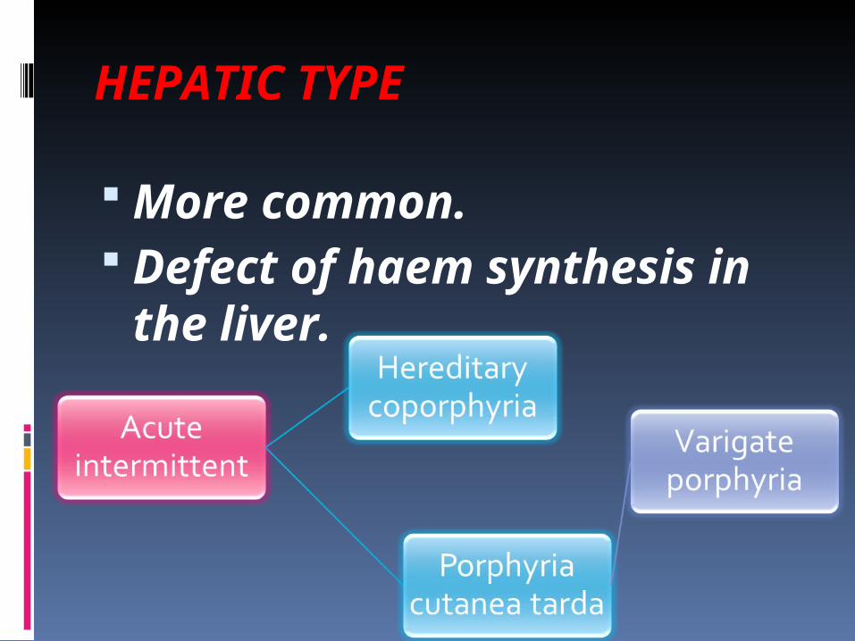
HEPATIC TYPE
More common. Defect of haem synthesis in the liver.
ACUTE INTERMITTENT PORPHYRIA Deficiency of UROPORPHYINOGEN I SYNTHETASE
Abdominal pain. Vomiting Cardiovascular abnormalities
HEREDITARY COPORPHYRIA Deficiency of enzyme coproporphyrinogen III oxidase.
Autosomal dominant. Causes purple urine, photosensitivity, and attacks of abdominal pain.
Very rare.
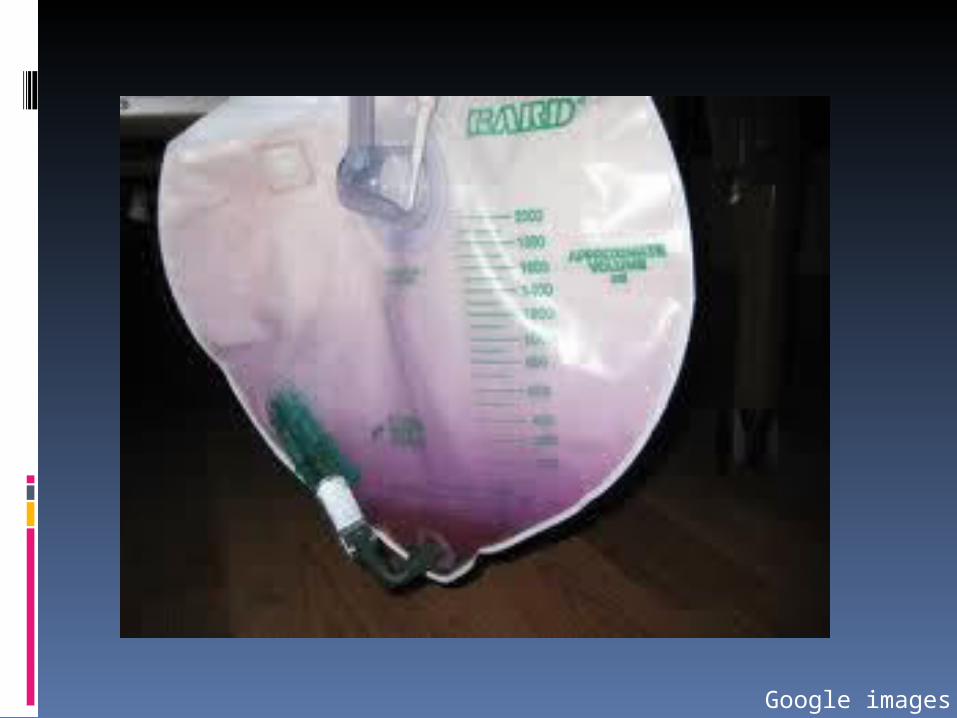
Google images
VARIGATE PORPHYRIA
South African porphyria.
Abdominal pain. vomiting. Diarrhea.
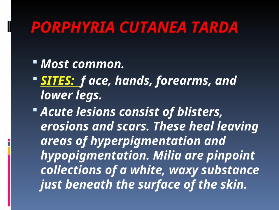
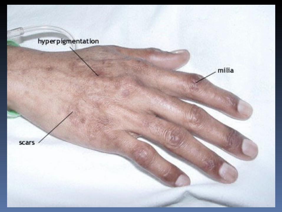
PORPHYRIA CUTANEA TARDA Most common. SITES: f ace, hands, forearms, and lower legs.
Acute lesions consist of blisters, erosions and scars. These heal leaving areas of hyperpigmentation and hypopigmentation. Milia are pinpoint collections of a white, waxy substance just beneath the surface of the skin.
ERYTHROPOIETIC PORPHYRIA
Common childhood porphyria.
Defective synthesis of heme in erythrocytes.
The enzyme deficiency occurs in the red blood cells
ERYTHROPOIETIC PROTOPORPHYRIA
mild form of porphyria. Severe photosensitivity. Exposure to even indoor light sources can cause the rash.
Prolonged exposure lead s to edema and blistering.
CONGENITAL ERYTHROPIETIC PORPHYRIA Inborn-error of haem-
porphyrin synthesis. Autosomal recessive . Also known as GüNTHER’S DISEASE
Caused by deficiency of the enzyme uroporphyrinogen synthetase.
(Oral and maxillofacial pathology- Neville)
CLINICAL FEATURES
No sex predilection. The first sign is red coloration of urine by uroporphyrin.
Photosensitivity. Hypertrichosis
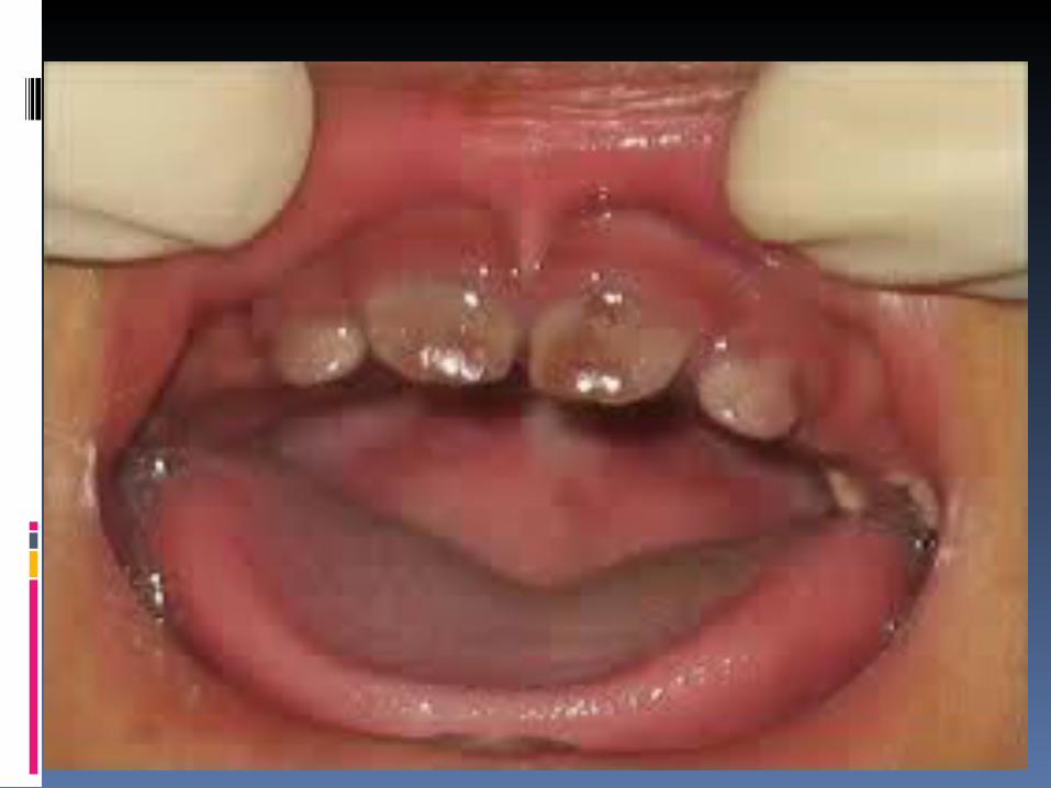
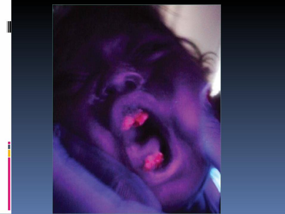
ORAL MANIFESTATIONS
Both deciduous and permanent dentition show pigmentation, usually brown. More intense in deciduous teeth.
Under Wood’s UV ray, teeth show a striking red fluorescence.
Ground sections show porphyrin pigments deposited in enamel, dentin and cementum.
TREATMENT
No treatment is available.
Discolored teeth may be cosmetically restored with porcelain –veneered crown.

THANK YOU