amersham™ ecl™ prime western blotting detection reagent · amersham ecl prime western blotting...
TRANSCRIPT
Amersham™ ECL™ Prime Western BlottingDetection Reagent
Product booklet
Code: RPN2232 RPN2236
3
Page Finder1. Legal 4
2. Description 5
2.1 Introduction 5 2.2 Design and features 6 2.3 Membrane Compatibility 6
3. Important user information 7 3.1 Intended use 7 3.2 Safety notices 7 3.3 Quality control 7
4. Handling 8
4.1 Safety precautions 8 4.2 Storage 8 4.3 Expiry 8
5. Required components 9 5.1 Kit components 9 5.2 Solutions 9 5.3 Membrane 9 5.4 Blocking reagents 9
5.5 Immunodetection reagents 10
6. Western blotting optimization 10
6.1 Introduction 11
6.2 Molecular weight markers 11
6.3 Membranes 12
6.4 Transfer 12
6.5 Blocking 12
6.6 Western blotting handbook 13
7. Protocol 14 7.1 Protocol overview 14
328-9829-42 AF 01-2014
7.2 Electrophoresis and transfer 15 7.3 Blocking 15 7.4 Antibody probing 15 7.5 Detection 18 7.6 Image analysis 19
8. Additional information 21 8.1 Stripping and reprobing membranes 21 8.2 Determination of optimum antibody concentration 22
9. Troubleshooting guide 25
10. Related products 28
10.1 Blotting equipment 28
10.2 Amersham ECL HRP-linked secondary antibodies 29
10.3 Detection reagents 30
428-9829-42 AF 01-2014
5
1. Legal GE, imagination at work and GE monogram are trademarks of General Electric Company.
Amersham, ECL, ECL DualVue, ECL Plex, Hybond, Hyperfilm, ImageQuant and Rainbow are trademarks of GE Healthcare companies.
Amersham ECL Prime is manufactured and sold under license from Cyanagen Srl and is subject of US Canadian and EU patent application number US7803573; EP1962095; US7855287; EP1950207 US2012009603(A1); CA2742025; EP2405016, together with other equivalent granted patents and patent applications in other countries.
Tween is a trademark of ICI Americas Inc.
© 2010-2014 General Electric Company – All rights reserved. Previously published Sept 2010
All goods and services are sold subject to the terms and conditions of sale of the company within GE Healthcare which supplies them. A copy of these terms and conditions is available on request.Contact your local GE Healthcare representative for the most current information.
http://www.gelifesciences.com/ecl
GE Healthcare UK Limited. Amersham Place, Little Chalfont, Buckinghamshire, HP7 9NA UK
528-9829-42 AF 01-2014
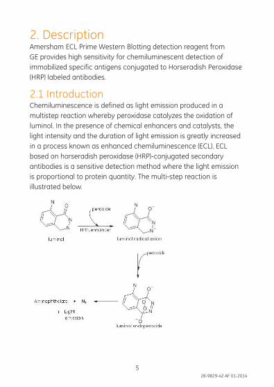
2. Description Amersham ECL Prime Western Blotting detection reagent from GE provides high sensitivity for chemiluminescent detection of immobilized specific antigens conjugated to Horseradish Peroxidase (HRP) labeled antibodies.
2.1 Introduction Chemiluminescence is defined as light emission produced in a multistep reaction whereby peroxidase catalyzes the oxidation of luminol. In the presence of chemical enhancers and catalysts, the light intensity and the duration of light emission is greatly increased in a process known as enhanced chemiluminescence (ECL). ECL based on horseradish peroxidase (HRP)-conjugated secondary antibodies is a sensitive detection method where the light emission is proportional to protein quantity. The multi-step reaction is illustrated below.
628-9829-42 AF 01-2014
7
2.2 Design and features Amersham ECL Prime detection reagent is designed to provide long signal duration (24 hours) and high sensitivity (picogram levels). The high intensity light output means that highly diluted antibodies can be used, and makes detection optimal using a CCD camera (e.g., ImageQuant™ LAS systems from GE). The light output can also be detected using autoradiography film (e.g., Amersham Hyperfilm™ product range).
2.3 Membrane compatibilityAmersham ECL Prime detection reagent is optimized for use with Amersham Hybond™ PVDF membrane where the performance compared to standard chemiluminescent substrates is most enhanced, but is also compatible with Amersham Protran nitrocellulose membrane.
728-9829-42 AF 01-2014
Amersham ECL Prime detection reagent is intended for chemiluminescent detection in Western blotting. Amersham ECL Prime detection reagent is intended for research use only, and shall not be used in any clinical procedures, or for diagnostic purposes.
3.2 Safety noticesThis user documentation contains CAUTIONS concerning the safe use of Amersham ECL Prime detection reagent. See definitions below.
Cautions
CAUTION CAUTION indicates a hazardous situation which, if not avoided, could result in minor or moderate injury. It is important not to proceed until all stated conditions are met and clearly understood.
3.3 Quality controlAmersham ECL Prime detection reagent is manufactured in compliance with our ISO 9001 certified quality management system, and is in conformity with the acceptance criteria set up for the product.
3. Important user information3.1 Intended use
828-9829-42 AF 01-2014
9
4. Handling
4.1 Safety precautions It is recommended to read the Safety Data Sheet (SDS) before using Amersham ECL Prime detection reagent.
CAUTION Hazardous substances. When using hazardous chemicals, take all suitable protective measures, such as wearing protective glasses and gloves resistant to the substances used. Follow local and/or national regulations for safe operation.
4.2 Storage On receipt, all components should be stored in a refrigerator at 2°C to 8°C. Amersham ECL Prime detection reagent is sensitive to prolonged exposure to light. Always store the individual reagents in the light-tight containers in which they are provided.
4.3 Expiry The components are stable for at least 3 months when stored under the recommended conditions. See expiry date on package.
928-9829-42 AF 01-2014
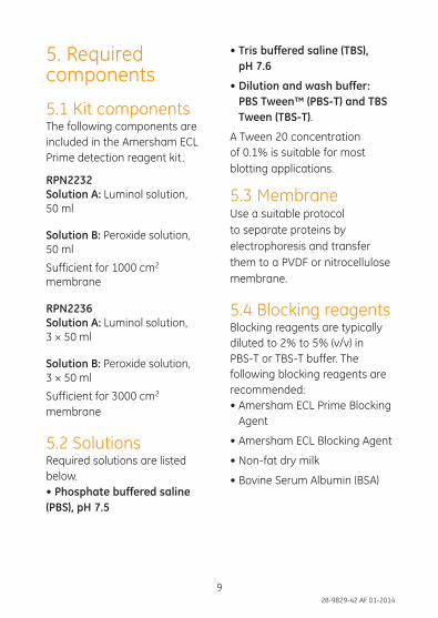
5. Required components
5.1 Kit components The following components are included in the Amersham ECL Prime detection reagent kit.
RPN2232Solution A: Luminol solution, 50 ml
Solution B: Peroxide solution, 50 ml
Sufficient for 1000 cm2 membrane
RPN2236Solution A: Luminol solution, 3 × 50 ml
Solution B: Peroxide solution, 3 × 50 ml
Sufficient for 3000 cm2 membrane
5.2 Solutions Required solutions are listed below. • Phosphate buffered saline (PBS), pH 7.5
• Tris buffered saline (TBS), pH 7.6
• Dilution and wash buffer: PBS Tween™ (PBS-T) and TBS Tween (TBS-T).
A Tween 20 concentration of 0.1% is suitable for most blotting applications.
5.3 MembraneUse a suitable protocol to separate proteins by electrophoresis and transfer them to a PVDF or nitrocellulose membrane.
5.4 Blocking reagents Blocking reagents are typically diluted to 2% to 5% (v/v) in PBS-T or TBS-T buffer. The following blocking reagents are recommended:• Amersham ECL Prime Blocking
Agent
• Amersham ECL Blocking Agent
• Non-fat dry milk
• Bovine Serum Albumin (BSA)
1028-9829-42 AF 01-2014
11
5.5 Immunodetection reagents• Primary antibody specific to the target protein(s)
• HRP conjugated secondary antibody specific to the primary antibody. See Amersham ECL HRP-linked secondary antibodies, on page 29.
Dilute the antibodies in PBS-T or TBS-T according to the recommendations in Dilution ranges, on page 16.
1128-9829-42 AF 01-2014
6. Western blotting optimization
6.1 IntroductionTo achieve an optimal Western blotting result with high signal tonoise ratio and best possible sensitivity and linearity, it is important to optimize the method and to select compatible products.
Consider the following:
• Sample quality and loading amount – It is important that the sample is of good quality and that detectable levels of target protein is present.
• Membrane and blocking – Select membranes and blocking agents compatible with sample and antibodies.
• Primary and secondary antibodies – Always select specific antibodies of high quality and optimize the antibody dilution.
• Detection and imaging – Select detection reagent according to your application
need. A CCD imager offers high sensitivity and broad dynamic range and provide better quantification than X-ray film.
This chapter describes products recommended for use with Amersham ECL Prime detection reagent.
6.2 Molecular weight markers Molecular weight markers are used to determine protein size. In addition, pre-stained markers allow confirmation of protein transfer and orientation (as the colored bands transfer to the membrane).
• Amersham Rainbow™ Markers are pre-stained multicolored markers for monitoring progress of protein electrophoresis, confirming transfer efficiency and determination of molecular weight of blotted proteins.
• Amersham ECL DualVue™ Markers are markers optimized for use with Amersham ECL, Amersham ECL Prime and Amersham ECL Select and contains a
1228-9829-42 AF 01-2014
13
combination of pre-stained and tagged proteins markers. These markers enable monitoring of electrophoresis, confirming transfer efficiency and determination of molecular weight of blotted proteins without staining on gel and membrane, as well as in chemiluminescence detection.
6.3 Membranes • Amersham Hybond are
PVDF membranes with high protein binding capacity and mechanical strength, which makes them ideal for Western blotting applications where stripping and re-probing are needed. The membrane is optimal for use with Amersham ECL Prime and Amersham ECL Select detection reagents.
• Amersham Protran are nitrocellulose membranes compatible with all chemiluminescent Western blotting substrates. The main advantage is the normally low background.
6.4 Transfer • Wet transfer is the most
commonly used transfer method. It provides efficient transfer of small to large proteins.
• Semi-dry transfer is faster than wet transfer and consumes less buffer. Semi-dry transfer works well for most proteins but transfer may be less efficient for large proteins. It might have reduced sensitivity for very low abundance proteins.
6.5 Blocking After protein transfer the membrane need to be incubated in a blocking solution to prevent non-specific binding of antibodies, which can cause background and non-specific protein bands on the blot. The blocking agent should be optimized for best results, no single blocking agent is optimal for all proteins and antibodies. GE Healthcare recommend the following blocking agents compatible with Amersham
1328-9829-42 AF 01-2014
ECL, Amersham ECL Prime and Amersham ECL Select:
• Amersham ECL Prime Blocking Agent
• Amersham ECL Blocking Agent
• BSA Blocking Agent
• Non-fat dry milk
6.6 Western blotting handbook More technical help, tips, and best practices can be found in the handbook Western Blotting Principles and Methods from GE Healthcare (code no. 28-9998-97).
1428-9829-42 AF 01-2014
15
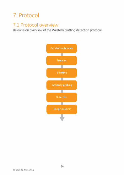
7. Protocol
7.1 Protocol overviewBelow is an overview of the Western blotting detection protocol.
1528-9829-42 AF 01-2014
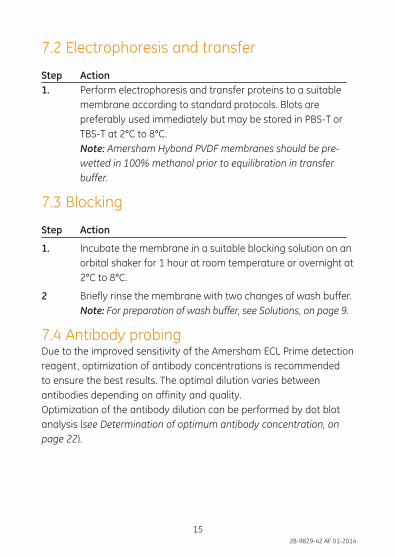
7.2 Electrophoresis and transfer
Step Action 1. Perform electrophoresis and transfer proteins to a suitable
membrane according to standard protocols. Blots are preferably used immediately but may be stored in PBS-T or TBS-T at 2°C to 8°C.
Note: Amersham Hybond PVDF membranes should be pre-wetted in 100% methanol prior to equilibration in transfer buffer.
7.3 Blocking
Step Action
1. Incubate the membrane in a suitable blocking solution on an orbital shaker for 1 hour at room temperature or overnight at 2°C to 8°C.
2 Briefly rinse the membrane with two changes of wash buffer. Note: For preparation of wash buffer, see Solutions, on page 9.
7.4 Antibody probingDue to the improved sensitivity of the Amersham ECL Prime detection reagent, optimization of antibody concentrations is recommended to ensure the best results. The optimal dilution varies between antibodies depending on affinity and quality.Optimization of the antibody dilution can be performed by dot blot analysis (see Determination of optimum antibody concentration, onpage 22).
1628-9829-42 AF 01-2014
17
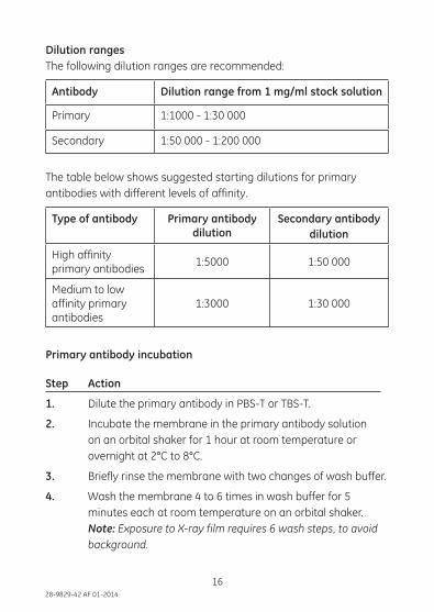
Dilution rangesThe following dilution ranges are recommended:
Antibody Dilution range from 1 mg/ml stock solution
Primary 1:1000 - 1:30 000
Secondary 1:50 000 - 1:200 000
The table below shows suggested starting dilutions for primary antibodies with different levels of affinity.
Type of antibody Primary antibodydilution
Secondary antibodydilution
High affinity primary antibodies
1:5000 1:50 000
Medium to low affinity primary antibodies
1:3000 1:30 000
Primary antibody incubation
Step Action
1. Dilute the primary antibody in PBS-T or TBS-T.
2. Incubate the membrane in the primary antibody solution on an orbital shaker for 1 hour at room temperature or overnight at 2°C to 8°C.
3. Briefly rinse the membrane with two changes of wash buffer.
4. Wash the membrane 4 to 6 times in wash buffer for 5 minutes each at room temperature on an orbital shaker. Note: Exposure to X-ray film requires 6 wash steps, to avoid background.
1728-9829-42 AF 01-2014
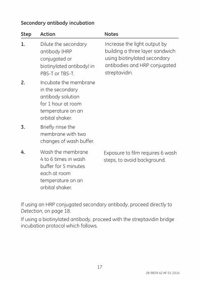
If using an HRP conjugated secondary antibody, proceed directly toDetection, on page 18.
If using a biotinylated antibody, proceed with the streptavidin bridgeincubation protocol which follows.
Secondary antibody incubation
Step Action Notes
1. Dilute the secondary antibody (HRP conjugated or biotinylated antibody) in PBS-T or TBS-T.
2. Incubate the membrane in the secondary antibody solution for 1 hour at room temperature on an orbital shaker.
3. Briefly rinse the membrane with two changes of wash buffer.
4. Wash the membrane 4 to 6 times in wash buffer for 5 minutes each at room temperature on an orbital shaker.
Increase the light output by building a three layer sandwich using biotinylated secondary antibodies and HRP conjugated streptavidin.
Exposure to film requires 6 wash steps, to avoid background.
1828-9829-42 AF 01-2014
19
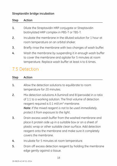
Streptavidin bridge incubation
Step Action
1. Dilute the Streptavidin HRP conjugate or Streptavidin biotinylated HRP complex in PBS-T or TBS-T.
2. Incubate the membrane in the diluted solution for 1 hour at room temperature on an orbital shaker.
3. Briefly rinse the membrane with two changes of wash buffer.
4. Wash the membrane by suspending it in enough wash buffer to cover the membrane and agitate for 5 minutes at room temperature. Replace wash buffer at least 4 to 6 times.
7.5 Detection
Step Action
1. Allow the detection solutions to equilibrate to room temperature for 20 minutes.
2. Mix detection solutions A (luminol) and B (peroxide) in a ratio of 1:1 to a working solution. The final volume of detection reagent required is 0.1 ml/cm2 membrane. Note: If the mixed reagent is not to be used immediately, protect it from exposure to the light.
3. Drain excess wash buffer from the washed membrane and place it protein side up in a suitable box or on a sheet of plastic wrap or other suitable clean surface. Add detection reagent onto the membrane and make sure it completely covers the membrane.
4. Incubate for 5 minutes at room temperature.
5. Drain off excess detection reagent by holding the membrane edge gently against a tissue.
1928-9829-42 AF 01-2014
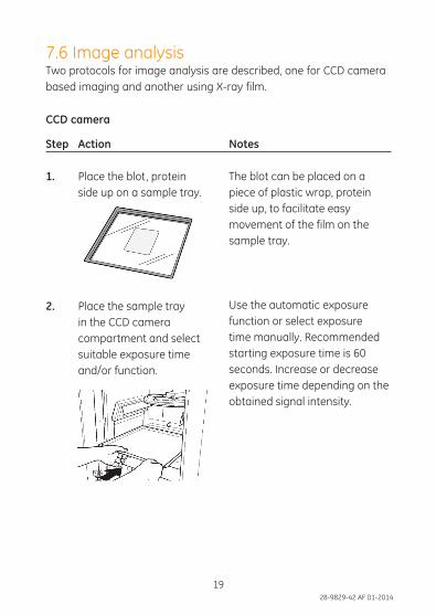
7.6 Image analysisTwo protocols for image analysis are described, one for CCD camera based imaging and another using X-ray film.
CCD camera
Step Action Notes
1. Place the blot, protein side up on a sample tray.
2. Place the sample tray in the CCD camera compartment and select suitable exposure time and/or function.
The blot can be placed on a piece of plastic wrap, protein side up, to facilitate easy movement of the film on the sample tray.
Use the automatic exposure function or select exposure time manually. Recommended starting exposure time is 60 seconds. Increase or decrease exposure time depending on the obtained signal intensity.
2028-9829-42 AF 01-2014
21
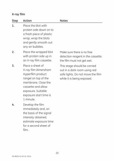
1. Place the blot with protein side down on to a fresh piece of plastic wrap, wrap the blots and gently smooth out any air bubbles.
2. Place the wrapped blot with protein side up in an X-ray film cassette.
3. Place a sheet of X-ray film (Amersham Hyperfilm product range) on top of the membrane. Close the cassette and allow exposure. Suitable exposure start time is 1 minute.
4. Develop the film immediately and, on the basis of the signal intensity obtained, estimate exposure time for a second sheet of film.
Make sure there is no free detection reagent in the cassette; the film must not get wet.
This stage should be carried out in a dark room using red safe lights. Do not move the film while it is being exposed.
X-ray film
Step Action Notes
2128-9829-42 AF 01-2014
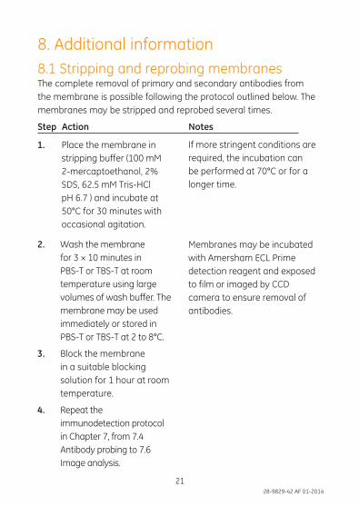
8. Additional information8.1 Stripping and reprobing membranesThe complete removal of primary and secondary antibodies fromthe membrane is possible following the protocol outlined below. Themembranes may be stripped and reprobed several times.
Step Action Notes
1. Place the membrane in stripping buffer (100 mM 2-mercaptoethanol, 2% SDS, 62.5 mM Tris-HCl pH 6.7 ) and incubate at 50°C for 30 minutes with occasional agitation.
2. Wash the membrane for 3 × 10 minutes in PBS-T or TBS-T at room temperature using large volumes of wash buffer. The membrane may be used immediately or stored in PBS-T or TBS-T at 2 to 8°C.
3. Block the membrane in a suitable blocking solution for 1 hour at room temperature.
4. Repeat the immunodetection protocol in Chapter 7, from 7.4 Antibody probing to 7.6 Image analysis.
If more stringent conditions are required, the incubation can be performed at 70°C or for a longer time.
Membranes may be incubated with Amersham ECL Prime detection reagent and exposed to film or imaged by CCD camera to ensure removal of antibodies.
2228-9829-42 AF 01-2014
23
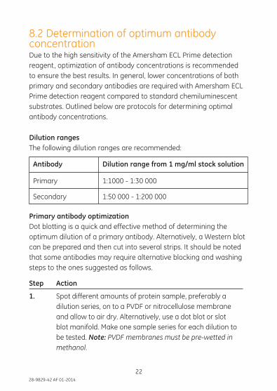
8.2 Determination of optimum antibody concentrationDue to the high sensitivity of the Amersham ECL Prime detection reagent, optimization of antibody concentrations is recommended to ensure the best results. In general, lower concentrations of both primary and secondary antibodies are required with Amersham ECL Prime detection reagent compared to standard chemiluminescent substrates. Outlined below are protocols for determining optimal antibody concentrations.
Dilution rangesThe following dilution ranges are recommended:
Antibody Dilution range from 1 mg/ml stock solution
Primary 1:1000 - 1:30 000
Secondary 1:50 000 - 1:200 000
Primary antibody optimizationDot blotting is a quick and effective method of determining the optimum dilution of a primary antibody. Alternatively, a Western blot can be prepared and then cut into several strips. It should be noted that some antibodies may require alternative blocking and washing steps to the ones suggested as follows.
Step Action
1. Spot different amounts of protein sample, preferably a dilution series, on to a PVDF or nitrocellulose membrane and allow to air dry. Alternatively, use a dot blot or slot blot manifold. Make one sample series for each dilution to be tested. Note: PVDF membranes must be pre-wetted in methanol.
2328-9829-42 AF 01-2014
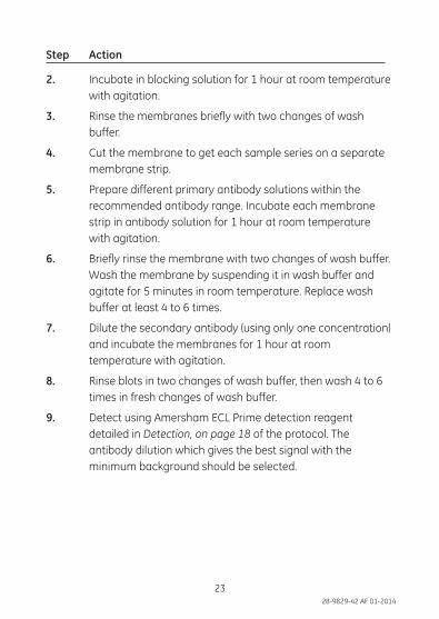
Step Action
2. Incubate in blocking solution for 1 hour at room temperature with agitation.
3. Rinse the membranes briefly with two changes of wash buffer.
4. Cut the membrane to get each sample series on a separate membrane strip.
5. Prepare different primary antibody solutions within the recommended antibody range. Incubate each membrane strip in antibody solution for 1 hour at room temperature with agitation.
6. Briefly rinse the membrane with two changes of wash buffer. Wash the membrane by suspending it in wash buffer and agitate for 5 minutes in room temperature. Replace wash buffer at least 4 to 6 times.
7. Dilute the secondary antibody (using only one concentration) and incubate the membranes for 1 hour at room temperature with agitation.
8. Rinse blots in two changes of wash buffer, then wash 4 to 6 times in fresh changes of wash buffer.
9. Detect using Amersham ECL Prime detection reagent detailed in Detection, on page 18 of the protocol. The antibody dilution which gives the best signal with the minimum background should be selected.
2428-9829-42 AF 01-2014
25
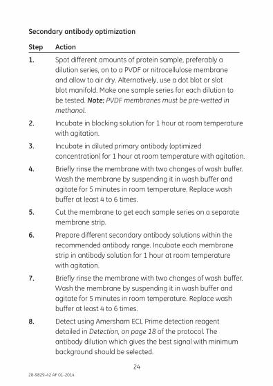
Secondary antibody optimization
Step Action
1. Spot different amounts of protein sample, preferably a dilution series, on to a PVDF or nitrocellulose membrane and allow to air dry. Alternatively, use a dot blot or slot blot manifold. Make one sample series for each dilution to be tested. Note: PVDF membranes must be pre-wetted in methanol.
2. Incubate in blocking solution for 1 hour at room temperature with agitation.
3. Incubate in diluted primary antibody (optimized concentration) for 1 hour at room temperature with agitation.
4. Briefly rinse the membrane with two changes of wash buffer. Wash the membrane by suspending it in wash buffer and agitate for 5 minutes in room temperature. Replace wash buffer at least 4 to 6 times.
5. Cut the membrane to get each sample series on a separate membrane strip.
6. Prepare different secondary antibody solutions within the recommended antibody range. Incubate each membrane strip in antibody solution for 1 hour at room temperature with agitation.
7. Briefly rinse the membrane with two changes of wash buffer. Wash the membrane by suspending it in wash buffer and agitate for 5 minutes in room temperature. Replace wash buffer at least 4 to 6 times.
8. Detect using Amersham ECL Prime detection reagent detailed in Detection, on page 18 of the protocol. The antibody dilution which gives the best signal with minimum background should be selected.
2528-9829-42 AF 01-2014
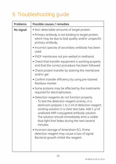
9. Troubleshooting guide
Problems Possible causes / remedies
No signal • Non-detectable amounts of target protein.
• Primary antibody is not binding to target protein, which may be due to bad quality and/or unspecific primary antibody.
• Incorrect species of secondary antibody has been used.
• PVDF membrane not pre-wetted in methanol.
• Check that transfer equipment is working properly and that the correct procedure has been followed.
• Check protein transfer by staining the membrane and/or gel.
• Confirm transfer efficiency by using pre-stained Rainbow marker.
• Some proteins may be affected by the treatments required for electrophoresis.
• Detection reagents do not function properly.- To test the detection reagent activity, in a
darkroom prepare 1 to 2 ml of detection reagent working solution in a clear test tube. Add 1 μl of undiluted HRP-conjugated antibody solution. The solution should immediately emit a visible blue light that fades during the next several minutes.
• Incorrect storage of Amersham ECL Prime detection reagent may cause a loss of signal. Bacterial growth inhibit the reagent.
2628-9829-42 AF 01-2014
27
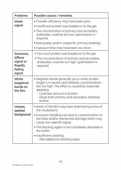
Problems Possible causes / remedies
Weaksignal
• Transfer efficiency may have been poor.
• Insufficient protein was loaded on to the gel.
• The concentration of primary and secondary antibodies could be too low; optimization is required.
• Bad quality and/or unspecific primary antibody.
• Exposure time may have been too short.
Excessive,diffuse signal or Rapidly fading signal
• Too much protein was loaded on to the gel.
• The concentrations of primary and secondary antibodies could be too high; optimization is required.
White(negative)bands onthe film
• Negative bands generally occur when protein target is in excess and antibody concentrations are too high. The effect is caused by substrate depletion.- Load less amount of protein.- Dilute both primary and secondary antibody
further.
Uneven,spotted background
• Areas of the blot may have dried during some of the incubations.
• Incorrect handling can lead to contamination on the blots and/or membrane damage which may cause non-specific signal.
• The blocking agent is not completely dissolved in the buffer.
• Insufficient washing.- Add additional washing steps.
2728-9829-42 AF 01-2014
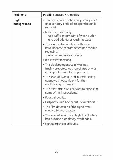
Problems Possible causes / remedies
Highbackgrounds
• Too high concentrations of primary and/or secondary antibodies; optimization is required.
• Insufficient washing.- Use sufficient amount of wash buffer
and add additional washing steps.
• Transfer and incubation buffers may have become contaminated and require replacing.- Always use fresh solutions
• Insufficient blocking.
• The blocking agent used was not freshly prepared, was too diluted or was incompatible with the application.
• The level of Tween used in the blocking agent was not sufficient for the application performed.
• The membrane was allowed to dry during some of the incubations.
• Poor gel quality.
• Unspecific and bad quality of antibodies.
• The film detection of the signal was allowed to over expose.
• The level of signal is so high that the film has become completely overloaded.
• Non compatible products.
2828-9829-42 AF 01-2014
29
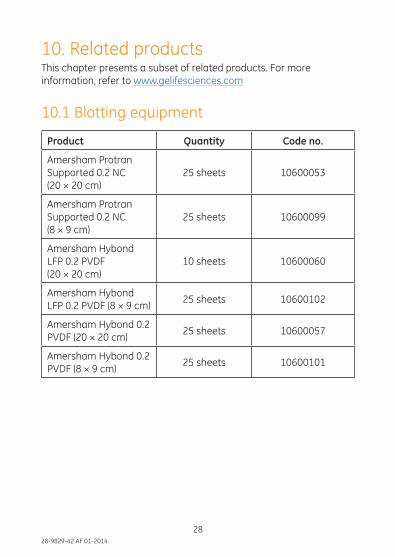
10. Related productsThis chapter presents a subset of related products. For more information, refer to www.gelifesciences.com
10.1 Blotting equipment
Product Quantity Code no.
Amersham Protran Supported 0.2 NC (20 × 20 cm)
25 sheets 10600053
Amersham Protran Supported 0.2 NC (8 × 9 cm)
25 sheets 10600099
Amersham Hybond LFP 0.2 PVDF (20 × 20 cm)
10 sheets 10600060
Amersham Hybond LFP 0.2 PVDF (8 × 9 cm)
25 sheets 10600102
Amersham Hybond 0.2 PVDF (20 × 20 cm)
25 sheets 10600057
Amersham Hybond 0.2 PVDF (8 × 9 cm)
25 sheets 10600101
2928-9829-42 AF 01-2014
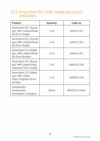
10.2 Amersham ECL HRP-linked secondary antibodies
Product Quantity Code no.
Amersham ECL Mouse IgG, HRP-Linked Whole Ab (from sheep)
1 ml NA931-1ML
Amersham ECL Human IgG, HRP-Linked Whole Ab (from sheep)
1 ml NA933-1ML
Amersham ECL Rabbit IgG, HRP-Linked Whole Ab (from donkey)
1 ml NA934-1ML
Amersham ECL Mouse IgG, HRP-Linked F(ab)2 fragment (from sheep)
1 ml NA9310-1ML
Amersham ECL Rabbit IgG, HRP-Linked F(ab)2 fragment (from donkey)
1 ml NA9340-1ML
Streptavidin-Horseradish Peroxidase Conjugate
100 μl RPN1231-100UL
3028-9829-42 AF 01-2014
31
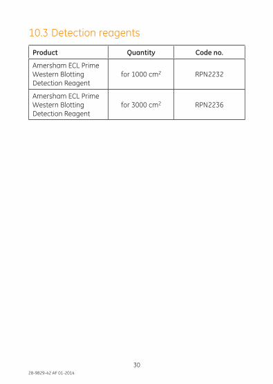
10.3 Detection reagents
Product Quantity Code no.
Amersham ECL Prime Western Blotting Detection Reagent
for 1000 cm2 RPN2232
Amersham ECL Prime Western Blotting Detection Reagent
for 3000 cm2 RPN2236

3128-9829-42 AF 01-2014
Page intentionally left blank

imagination at work
28-9829-42 AF 01-2014
For your local office contact information, visit www.gelifesciences.com/contact
GE Healthcare UK Limited Amersham Place Little Chalfont, Buckinghamshire, HP7 9NA, UK
http://www.gelifesciences.com
GE Healthcare offices:
GE Healthcare Bio-Sciences AB
Björkgatan 30, 751 84
Uppsala
Sweden
GE Healthcare Europe GmbH
Munzinger Strasse 5 D-79111
Freiburg
Germany
GE Healthcare Bio-Sciences Corp.
800 Centennial Avenue
P.O. Box 1327
Piscataway,
NJ 08855-1327
USA
GE Healthcare Japan Corporation
Sanken Bldg. 3-25-1
Hyakunincho Shinjuku-ku
Tokyo 169-0073
Japan