ameloblastin induces tumor suppressive phenotype and ... · ameloblastin induces tumor suppressive...
TRANSCRIPT
1
Ameloblastin induces tumor suppressive phenotype and enhances
chemosensitivity to doxorubicin via Src-Stat3 inactivation in
osteosarcoma
Toshinori Ando1†, Yasusei Kudo1,6†, Shinji Iizuka1, Takaaki Tsunematsu1,6, Hanako
Umehara1, Madhu Shrestha1, Toshihiro Matsuo7, Tadahiko Kubo2, Shouji Shimose5, Koji
Arihiro3, Ikuko Ogawa4, Mitsuo Ochi2, Takashi Takata1*
1Department of Oral and Maxillofacial Pathobiology, 2Department of Orthopaedic Surgery,
Institute of Biomedical and Health Sciences, Hiroshima University, Hiroshima, Japan,
3Anatomical Pathology, 4Center of Oral Clinical Examination, Hiroshima University
Hospital, Hiroshima, Japan, 5Division of Orthopaedic Surgery, National Hospital
Organization Kure Medical Center, Kure, Japan, 6Department of Oral Molecular Pathology,
Institute of Health Biosciences, The University of Tokushima Graduate School, Tokushima,
Japan, 7Department of Orthopedic Surgery, Aichi Medical University, Nagakute, Aichi,
Japan.
†These authors contributed equally to this work.
*To whom correspondence should be addressed. Takashi Takata D.D.S., Ph.D., Department
of Oral and Maxillofacial Pathobiology, Basic Life Science, Institute of Biomedical and

2
Health Sciences, Hiroshima University, 1-2-3 Kasumi, Minami-ku, Hiroshima, Japan
734-8553. Tel: +81-82-257-5634, E-mail: [email protected]
3
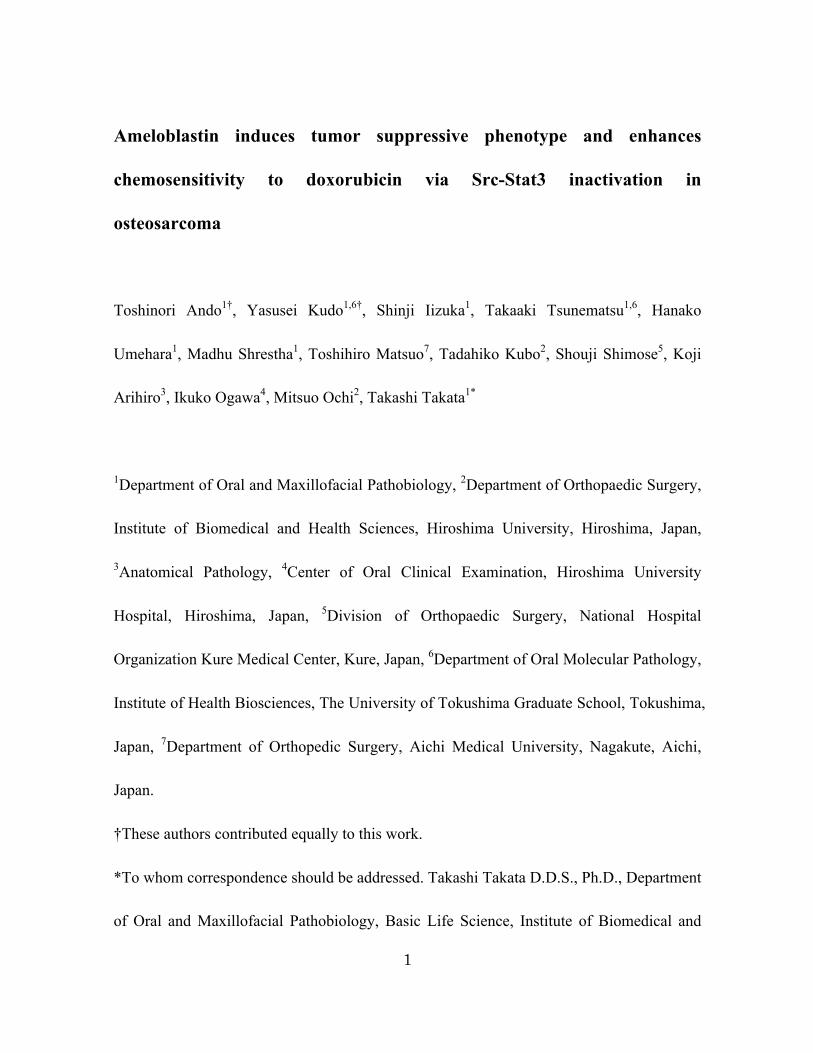
Figure S1. AMBN induces apoptosis and sensitivity to doxorubicin in osteosarcoma cells,
related to Figure 1. (A) Representative results of cell cycle distributions analyzed by PI
staining and FACS in control and AMBN-stable 143B-Luc cells are shown. (B) The
expression of FLAG-AMBN and cleaved caspase-3 in control and AMBN-stable 143B-Luc
cells was examined. (C) After the treatment with DMSO and doxorubicin (0.5 µg/mL) for
24 h in control and AMBN-stable 143B-Luc cells, cleaved caspase-3 expression was
examined.
Supplemental Figure S1�
0 200 400 600 800 1000FL2-Area
Data.005
M1
M2
M3M4
0 200 400 600 800 1000FL2-Area
Data.001
M1
M2
M3M4
Sub G1: 4.08 G1: 60.92 S: 11.53 G2/M: 18.52�
Sub G1: 11.25 G1: 55.65 S: 12.97 G2/M: 16.40�
Control� AMBN-stable�
A B
FLAG�
Cleaved caspase-3�
β-actin�
143B-Luc�
Cleaved caspase-3 (short exposure)�
β-actin�
DMSO� DOX�
143B-Luc�
Cleaved caspase-3 (long exposure)�
C
4
Supplemental Figure S2�
DMSO� DOX�
143B-Luc (Cumate day3)�
C
A
B
D
■Control ■AMBN-inducible�
U2-OS�
Days (Cumate)�
Num
ber
of c
ells
(×10
3 )�
0
5
10
15
20
25
30
35
0 1 2 3
β-actin�
Cleaved caspase-3�
DMSO� DOX�
U2-OS (Cumate day3)�
FLAG�
β-actin�
pY705-Stat3�
t-Stat3�
U2-OS (Cumate day3)�
143B-Luc (Cumate day3)�
FLAG�
pY416-Src�
t-Src�
Cleaved caspase-3�
β-actin�
143B-Luc (Cumate day3)�
Control� AMBN-inducible
0 30 60 0 30 60 After cell attachment on the dish (min)�
E
143B-Luc�
Num
ber
of c
ells
(×10
3 )�
Days (Cumate)�
0
5
10
15
20
25
30
35
0 1 2 3
■Control ■AMBN-inducible�
***�***�
Cleaved caspase-3�
β-actin�
FLAG�
AMBN-inducible�Control�
Cumate 0 1 2 3 0 1 2 3 days�
143B-Luc�

5
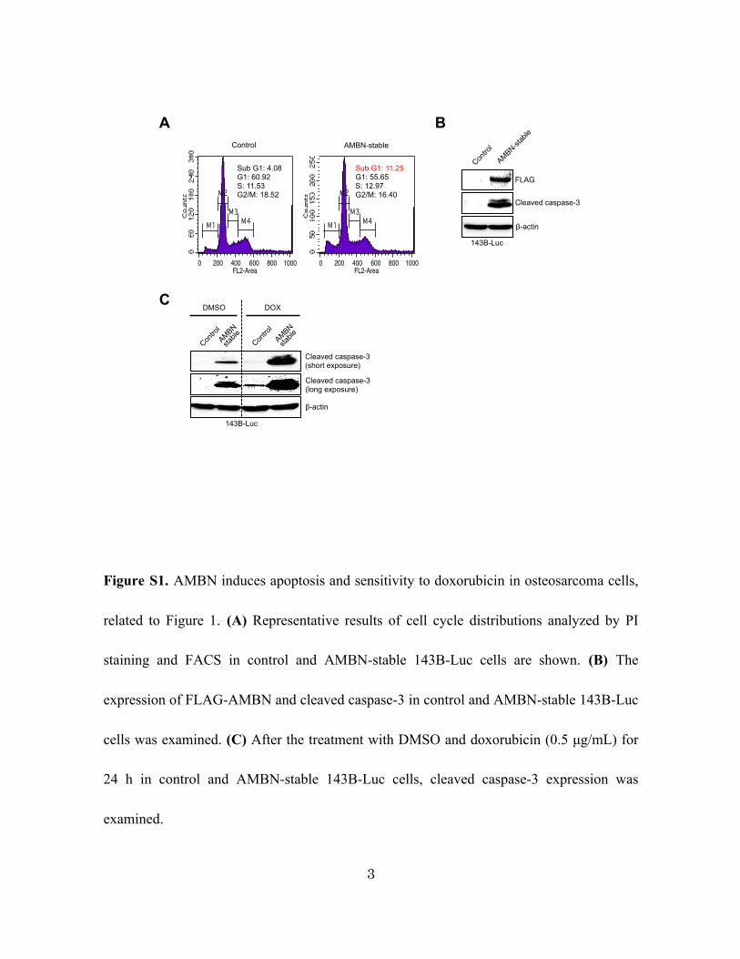
Figure S2. AMBN induces apoptosis and sensitivity to doxorubicin through the
inactivation of Src-Stat3 pathway in osteosarcoma cells, related to Figure 1. (A) Control
and AMBN-inducible 143B-Luc cells were cultured for 3 days with Cumate solution
(300µg/mL). The cells were collected each day and the expression of FLAG-AMBN and
cleaved caspase-3 was evaluated. (B) Control and AMBN-inducible 143B-Luc and U2-OS
cells were cultured with Cumate solution, and cell growth was counted on days 0, 1, 2, and
3 (N=3). (C) Control and AMBN-inducible 143B-Luc and U2-OS cells were cultured with
Cumate for 3 days, and these cells were treated with DMSO and doxorubicin (0.5 µg/mL)
at last 24 h. The expression of FLAG-AMBN and cleaved caspase-3 was examined. (D)
The expression of FLAG-AMBN, pY705-Stat3, total-Stat3 and cleaved caspase-3 was
examined. (E) The expression of FLAG-AMBN, pY416-Src, total-Src and cleaved
caspase-3 after attachment on the culture dish (0, 30, 60 minutes) was examined.
Mean±SEM (B); ***, P<0.001.

6
Figure S3. CD63 is expressed among human osteosarcoma cell lines and is needed for
Stat3 inactivation induced by AMBN, related to Figure 1. (A) The expression of CD63 at
the protein level in NOS-1, SaOS-2, U2-OS and 143-B Luc cells was examined. (B)
shScramble and shCD63 were transfected into AMBN-inducible 143B-Luc cells. The
expression of FLAG-AMBN, CD63, pY705-Stat3 and total-Src at the protein level in was
examined.
β-actin�
t-Stat3�
pY705-Stat3�
CD63�
FLAG�
AMBN-inducible
143B-Luc (Cumate day3)�
CD63 (long exposure)�
CD63 (short exposure)�
β-actin�
A B
Supplemental Figure S3�
7
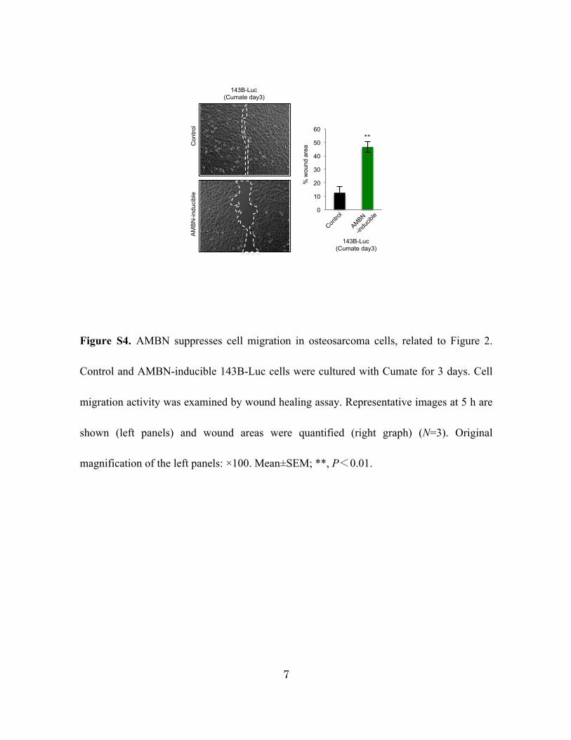
Figure S4. AMBN suppresses cell migration in osteosarcoma cells, related to Figure 2.
Control and AMBN-inducible 143B-Luc cells were cultured with Cumate for 3 days. Cell
migration activity was examined by wound healing assay. Representative images at 5 h are
shown (left panels) and wound areas were quantified (right graph) (N=3). Original
magnification of the left panels: ×100. Mean±SEM; **, P<0.01.
0
10
20
30
40
50
60
143B-Luc (Cumate day3)
% w
ound
are
a
**
3.30
143B-Luc (Cumate day3)
Con
trol
AM
BN
-indu
cibl
e
Supplemental Figure S4
8
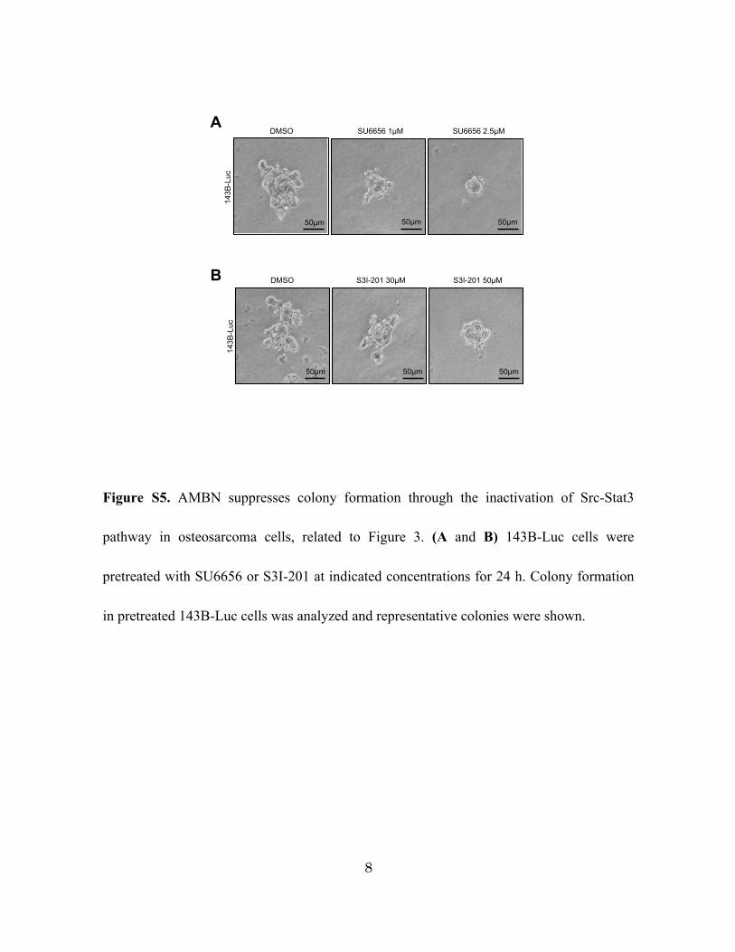
Figure S5. AMBN suppresses colony formation through the inactivation of Src-Stat3
pathway in osteosarcoma cells, related to Figure 3. (A and B) 143B-Luc cells were
pretreated with SU6656 or S3I-201 at indicated concentrations for 24 h. Colony formation
in pretreated 143B-Luc cells was analyzed and representative colonies were shown.
Supplemental Figure S5�
143B
-Luc�
DMSO� SU6656 1µM� SU6656 2.5µM�
50µm� 50µm� 50µm�14
3B-L
uc�
DMSO� S3I-201 30µM� S3I-201 50µM�
50µm� 50µm� 50µm�
B�
A�
9
Supplemental Figure S6�
shScramble SU6656 0µM
shAMBN2 SU6656 0µM
shAMBN2 SU6656 1µM
shAMBN2 SU6656 2.5µM
NOS-1
NOS-1
shScramble S3I201 0µM
shAMBN2 S3I201 0µM
shAMBN2 S3I201 50µM
shAMBN2 S3I201 100µM
A� B�
C� D�
NOS-1
shScramble SU6656 0µM
shAMBN2 SU6656 0µM
shAMBN2 SU6656 1µM
shAMBN2 SU6656 2.5µM
50µm�
50µm�
50µm�
50µm�
NOS-1
shAMBN2 S3I201 200µM
shAMBN2 S3I201 100µM
shAMBN2 S3I201 0µM
shScramble S3I201 0µM
50µm�
50µm�
50µm�
50µm�
10
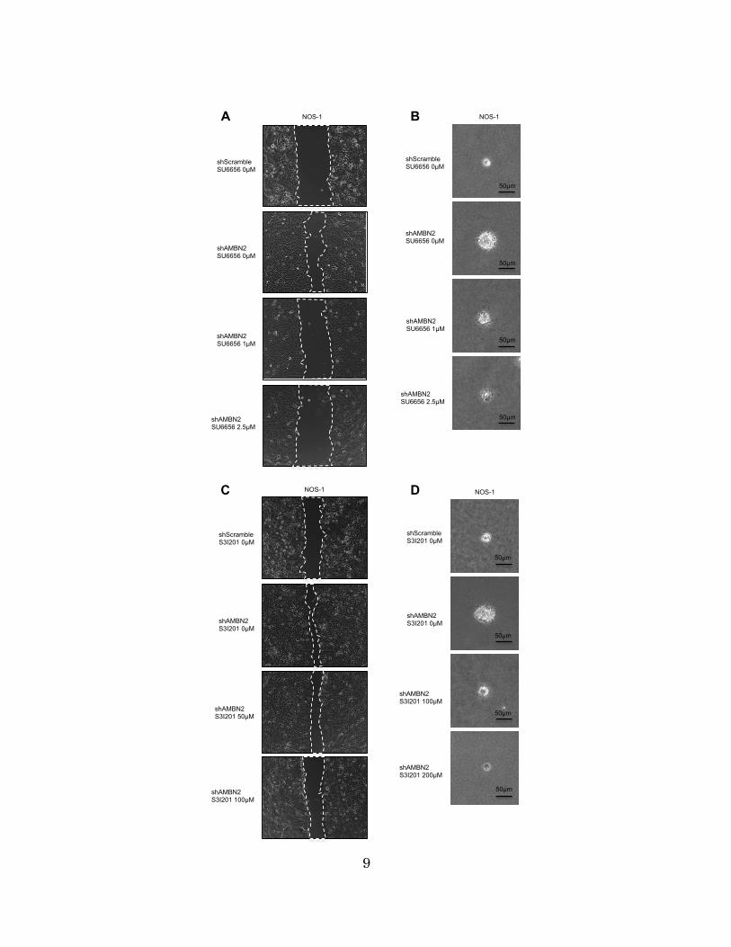
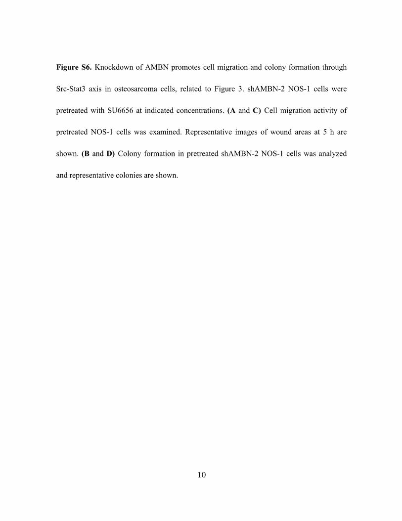
Figure S6. Knockdown of AMBN promotes cell migration and colony formation through
Src-Stat3 axis in osteosarcoma cells, related to Figure 3. shAMBN-2 NOS-1 cells were
pretreated with SU6656 at indicated concentrations. (A and C) Cell migration activity of
pretreated NOS-1 cells was examined. Representative images of wound areas at 5 h are
shown. (B and D) Colony formation in pretreated shAMBN-2 NOS-1 cells was analyzed
and representative colonies are shown.
11
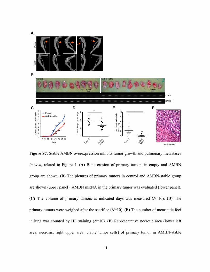
Figure S7. Stable AMBN overexpression inhibits tumor growth and pulmonary metastases
in vivo, related to Figure 4. (A) Bone erosion of primary tumors in empty and AMBN
group are shown. (B) The pictures of primary tumors in control and AMBN-stable group
are shown (upper panel). AMBN mRNA in the primary tumor was evaluated (lower panel).
(C) The volume of primary tumors at indicated days was measured (N=10). (D) The
primary tumors were weighed after the sacrifice (N=10). (E) The number of metastatic foci
in lung was counted by HE staining (N=10). (F) Representative necrotic area (lower left
area: necrosis, right upper area: viable tumor cells) of primary tumor in AMBN-stable
Supplemental Figure S7�
10mm�
Control� AMBN-stable�
10mm�
AMBN�
GAPDH�
B�
E
1�
2�
3�
Tum
or w
eigh
t (×
103 m
g)� **�
0�
C D
0 1 2 3 4 5 6 7 8
7 9 11 13 15 17 19 21 23 days�
**�
Tum
or v
olum
e (×
103 m
m3 )�
AMBN-stable Control�
cont, v
ecl
AMBN
, vec
l
0
2
4
6
8
10
Num
ber o
f met
asta
tic
foci
in lu
ng�
*�
0�
2�
4�
6�
8�
10�
Em
pty�
AM
BN
A
F
AMBN-stable�

12
group is shown. Original magnification: ×200. Mean±SEM (C-E); **, P<0.01; *, P<
0.05.
13
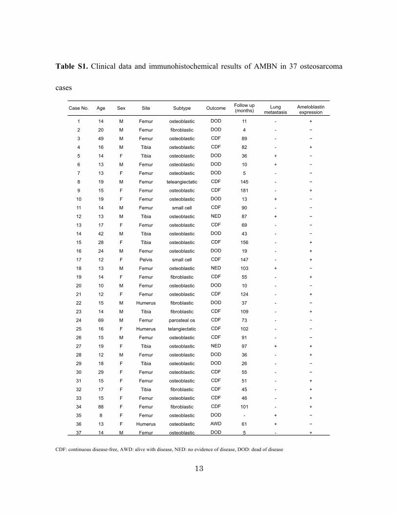
Table S1. Clinical data and immunohistochemical results of AMBN in 37 osteosarcoma
cases
CDF: continuous disease-free, AWD: alive with disease, NED: no evidence of disease, DOD: dead of disease
Case No. Age Sex Site Subtype Outcome Follow up (months)
Lung metastasis
Ameloblastin expression
1 14 M Femur osteoblastic DOD�� 11 - +
2 20 M Femur fibroblastic DOD 4 - −
3 49 M Femur osteoblastic CDF 89 - −
4 16 M Tibia osteoblastic CDF 82 - +
5 14 F Tibia osteoblastic DOD 36 + −
6 13 M Femur osteoblastic DOD 10 + −
7 13 F Femur osteoblastic DOD 5 - −
8 19 M Femur teleangiectatic CDF 145 - −
9 15 F Femur osteoblastic CDF 181 - +
10 19 F Femur osteoblastic DOD 13 + −
11 14 M Femur small cell CDF 90 - −
12 13 M Tibia osteoblastic NED 87 + −
13 17 F Femur osteoblastic CDF 69 - −
14 42 M Tibia osteoblastic DOD 43 - −
15 28 F Tibia osteoblastic CDF 156 - +
16 24 M Femur osteoblastic DOD 19 - +
17 12 F Pelvis small cell CDF 147 - +
18 13 M Femur osteoblastic NED 103 + −
19 14 F Femur fibroblastic CDF 55 - +
20 10 M Femur osteoblastic DOD 10 - −
21 12 F Femur osteoblastic CDF 124 - +
22 15 M Humerus fibroblastic DOD 37 - −
23 14 M Tibia fibroblastic CDF 109 - +
24 69 M Femur parosteal os CDF 73 - −
25 16 F Humerus telangiectatic CDF 102 - −
26 15 M Femur osteoblastic CDF 91 - −
27 19 F Tibia osteoblastic NED 97 + +
28 12 M Femur osteoblastic DOD 36 - +
29 18 F Tibia osteoblastic DOD 26 - −
30 29 F Femur osteoblastic CDF 55 - −
31 15 F Femur osteoblastic CDF 51 - +
32 17 F Tibia fibroblastic CDF 45 - +
33 15 F Femur osteoblastic CDF 46 - +
34 88 F Femur fibroblastic CDF 101 - +
35 8 F Femur osteoblastic DOD - + −
36 13 F Humerus osteoblastic AWD 61 + −
37 14 M Femur osteoblastic DOD 5 - +
Supplemental Table S1�
Clinical data and immunohistochemical results of AMBN in 37 osteosarcoma cases
CDF: continuous disease-free AWD: alive with disease NED: no evidence of disease DOD: dead of disease�